lit review endo
TRANSCRIPT
Abigail Martin 07360932 4BY2
1
Genes involved in embryo- endometrium interactions in mares during Maternal
Recognition of Pregnancy (MRP)
Abstract
Introduction
There are many important reasons for studying equine reproduction and improving its
efficiency for its own sake [1]. 16% of thoroughbreds experience pregnancy failure, with
60% happening within the first three weeks of gestation [2]. Economically, the equine
industry is substantial in many countries and poor reproductive results are very costly.
Although equine reproductive studies are still in their early stages, it is known that genes
expressed in the endometrium (the lining of the uterus) of mares play a necessary role in the
establishment and maintenance of maternal pregnancy. The actions of the conceptus (the
spherical shaped sac containing the embryo proper, its associated membranes and fluid) in the
uterus must be considered as it is also important in MRP [3].
Using biochemical pathways, the conceptus signals its presence causing arrest of the
oestrous cyclic process and maintains primary Corpus Luteum (CL) function. The events
controlling these processes are known as “maternal recognition of pregnancy” (MRP) [4]. For
all ruminants it is therefore understood that for MRP to transpire, communication between the
conceptus and the endometrium is necessary, thus creating a synchronous uterine
environment for embryonic development. Very little is known about the molecular crosstalk
between the embryo and endometrium in equines because their MRP mechanisms are
different to other ruminants [5].
Progesterone is a principal hormone involved in maintaining pregnancy in all
mammals. In the presence of an embryo in the uterus, the life span of the CL (corpus luteum)
is prolonged and endometrial release of prostaglandin F2α (PGF2α) into the bloodstream is
blocked, preventing luteolysis (regression of the CL) [3]. MRP depends on species- specific
signals produced by the embryo and recognised by the uterine lining and vice versa.
The aim of this review is to first, give an overview of the first 3 weeks of pregnancy
in the mare and its embryonic development. Secondly, to highlight key prostaglandins and
genes that have a strong influence on MRP involved in the conceptus- endometrium
interactions, especially those leading up to implantation in equids and in ruminants. Finally,
to discuss the values of studying equine embryology.
Abigail Martin 07360932 4BY2
2
Early stage development of the equine embryo
After fertilisation, the zygote cleaves via first mitosis to give two cells called
blastomeres surrounded by the zona pellucida. Cleavages are divisions of cells in the early
embryo. The cleavage ends with the formation of the blastula. These begin during the
transport of the embryo through the oviduct into the uterus but, this process is a species-
specific stage during development. Mares are unusual to other animals with regard to passage
through the oviduct, whereby only developing embryos can pass through unlike unfertilised
oocytes which cannot [3,6].
The equine blastocyst enters the oviduct around day 6.5 after ovulation has occurred.
The blastocyst contains an inner cell mass and a blastocyst cavity surrounded by a monolayer
of trophectoderm. By day 7 the zona pellucida has shed and the embryo is surrounded by a
tough acellular mucin-like glycoprotein capsule [7]. The capsule acts to protect, and prevents
the trophoblast from elongating, unlike other domestic animals such as cattle and pigs, and
gives it its spherical structure. It facilitates rapid growth between days 11 and 16, nutrition
and migration of the conceptus in the uterine lumen via peristaltic myometrial forces [5].
During the migration from one uterine horn to the other, the conceptus has surface
contact with the endometrium. The conceptus signals its presence in the uterus via anti-
luteolytic signals thus, the embryo prevents luteolysis by first, placing itself near to the
endometrium then attaching to it [3], between day 17 and 21 [8]. This event is known as
fixation, where the conceptus containing the embryo proper becomes attached to the
endometrial surface. Fixation is the termination of the extended mobility stage in the uterus,
positioning and fixing the embryo at the base of one of the uterine horns. This termination is
because of an increase in uterine tone and embryo growth, predominantly in the right horn of
mares [8]. In rodents and primates the term implantation is used here but, in these animals the
embryo penetrates the epithelium and advances into the endometrial connective tissue where
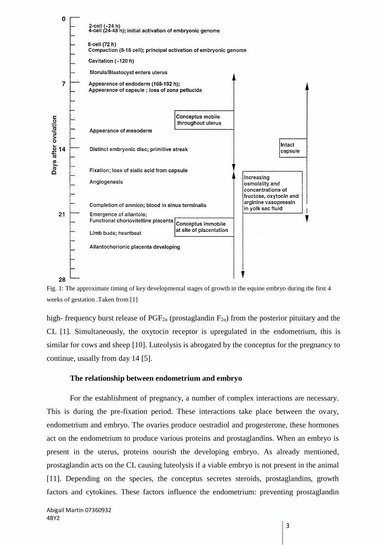
it is implanted [3]. Fig.1 outlines the stages of development of an equine embryo during MRP
from day 0 to day 28.
In mares luteolysis can occur as early as days 11 and 13 [5,9]. Luteolysis can be
defined as the functional and/ or structural regression of the CL [10]. It is a crucial process in
the ovarian cycle whereby, post-ovulation, luteolysis is initiated by an Oxytocin-dependant
Abigail Martin 07360932 4BY2
3
Fig. 1: The approximate timing of key developmental stages of growth in the equine embryo during the first 4
weeks of gestation .Taken from [1]
high- frequency burst release of PGF2α (prostaglandin F2α) from the posterior pituitary and the
CL [1]. Simultaneously, the oxytocin receptor is upregulated in the endometrium, this is
similar for cows and sheep [10]. Luteolysis is abrogated by the conceptus for the pregnancy to
continue, usually from day 14 [5].
The relationship between endometrium and embryo
For the establishment of pregnancy, a number of complex interactions are necessary.
This is during the pre-fixation period. These interactions take place between the ovary,
endometrium and embryo. The ovaries produce oestradiol and progesterone, these hormones
act on the endometrium to produce various proteins and prostaglandins. When an embryo is
present in the uterus, proteins nourish the developing embryo. As already mentioned,
prostaglandin acts on the CL causing luteolysis if a viable embryo is not present in the animal
[11]. Depending on the species, the conceptus secretes steroids, prostaglandins, growth
factors and cytokines. These factors influence the endometrium: preventing prostaglandin
Abigail Martin 07360932 4BY2
4
secretion and stimulating protein secretion, or by directly acting on the ovary to stimulate
progesterone secretion [12].
The pig, horse and guinea pig are oxytocin dependant species. During MPR the CL
produces progesterone and oxytocin stimulating endometrial synthesis of prostaglandin thus,
causing luteolysis. Interferon- tau (IFN-τ) is the MRP factor in ruminants such as sheep,
cows, goats [13, 14]. IFN-τ genes are not present in horses and equine conceptuses do not
produce interferon like proteins during early pregnancy but, it is an important factor in other
ruminants and therefore must be acknowledged [15]. IFN-τ silences expression of the
estrogen receptor (ESR1) preventing expression of endometrial oxytocin receptors. If an
embryo is present, oxytocin cannot induce PGF2α synthesis preventing luteolysis. It is
released from the endometrial glands and stimulates histotrophe production, i.e. secretions
that nourish the embryo in the uterine cavity. Pigs have a different mechanism to halt
luteolysis. Oxytocin is produced in the CL and promotes synthesis of PGF2α like ruminants.
The trophectoderm of a pig trophoblast produces oestradiol, thus preventing luteolysis. PGF2α
is secreted into the uterine cavity where it is degraded by rate limiting enzymes instead of the
maternal bloodstream as in equids and ruminants [3].
Conceptus associated proteins involved in endometrium communication
The components of systems for the exchanging of materials between the conceptus
and the mare are often found in the cellular yolk sac wall called the capsule. The proteins
β2M, uterocalin, uteroglobulin, ganglioslide activator protein (GM2AP) and phospholipase
A2 (PLA2) are mostly expressed in large amounts approaching the time before fixation.
A major luteolysis- associated protein of the conceptus capsule is β2 Microglobulin
(β2M). It is the most abundant protein within the capsule. β2M undergoes proteolysis in the
capsule and its sequence is shortened around the time of fixation [16]. The removal of amino
acids is due to the conversion of the intact form of β2M to its cleaving form to the
endometrium around day 17, when the embryo becomes fixed to the uterine lumen. The
origin of the β2M has not been determined but Quinn et al. state that it is likely to stem from
the endometrium or yolk sac wall tissue. Its function is the light chain complex of various
MHC class 1 complexes. It is believed to also have an earlier function in the capsule before
degradation occurs [17].
Uterocalin/ P19 is a characterised lipocalin which act as a small hydrophilic carrier
protein. It is secreted by the endometrial glands in late diestrus and in early pregnancy in
equines. The cationic protein binds to capsule and its main function is to transport and deliver
Abigail Martin 07360932 4BY2
5
lipophillic substances such as nutrients into the yolk sac [16]. It is only present in the yolk sac
fluid before fixation. Uterocalin has only been demonstrated in equines so far. Similar to β2M
it lessens as the capsule degrades, therefore uterocalin and β2M only appear to be essential
during early embryonic growth [17].
The uteroglobin gene is expressed in the equine uterus. First identified in the uterine
of rabbits, it is a member of the large secretoglobulin super family of proteins. Like
Uterocalin it binds small lipophillic molecules and also, is present in ample amounts in the
uterine lavage fluids but, only in the yolk sac before fixation [16, 17]. Uteroglobulin is found
in various mammals including some without encapsulate conceptuses.
The ganglioslide activator protein (GM2AP) is expressed in large amounts by the
equine capsule up until around day 18.5 of pregnancy. This protein is involved in the
transport of glycolipids. It has only been found to be expressed by the capsule so far [18].
Phospholipase A2 (PLA2) are a complex group of enzymes that cleave glycerol based
phospholipids and a fatty acid, usually arachidonic acid, a cyclooxygenase substrate from the
eicosanoid family (a group of signalling molecules constructed by the oxidation of twenty-
carbon essential fatty acids) [16, 17, 19]. The PLA2 enzymes play a valuable role in
regulating ovarian function, pregnancy and delivery, they are also important in inflammation
and haemostasis. PLA2 synthesis in the equine uterus was found to bind to the capsule and
increase its concentration when PGF2α was administered to block fixation [16, 17, 19]. It is
also believable that synthesis of PLA2 assists in the degradation and removal of the
conceptus capsule.
Abigail Martin 07360932 4BY2
6
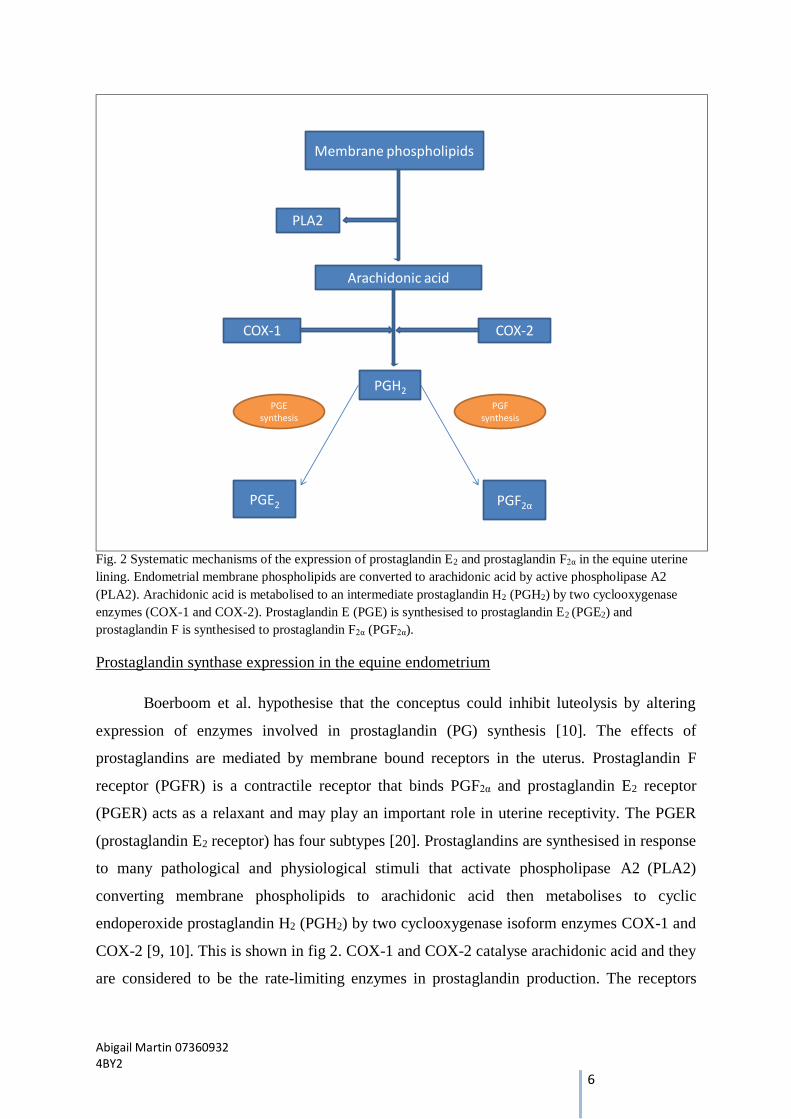
Membrane phospholipids
PLA2
Arachidonic acid
COX-1 COX-2
PGH2
PGE2 PGF2α
PGE synthesis
PGF synthesis
Fig. 2 Systematic mechanisms of the expression of prostaglandin E2 and prostaglandin F2α in the equine uterine
lining. Endometrial membrane phospholipids are converted to arachidonic acid by active phospholipase A2
(PLA2). Arachidonic acid is metabolised to an intermediate prostaglandin H2 (PGH2) by two cyclooxygenase
enzymes (COX-1 and COX-2). Prostaglandin E (PGE) is synthesised to prostaglandin E2 (PGE2) and
prostaglandin F is synthesised to prostaglandin F2α (PGF2α).
Prostaglandin synthase expression in the equine endometrium
Boerboom et al. hypothesise that the conceptus could inhibit luteolysis by altering
expression of enzymes involved in prostaglandin (PG) synthesis [10]. The effects of
prostaglandins are mediated by membrane bound receptors in the uterus. Prostaglandin F
receptor (PGFR) is a contractile receptor that binds PGF2α and prostaglandin E2 receptor
(PGER) acts as a relaxant and may play an important role in uterine receptivity. The PGER
(prostaglandin E2 receptor) has four subtypes [20]. Prostaglandins are synthesised in response
to many pathological and physiological stimuli that activate phospholipase A2 (PLA2)
converting membrane phospholipids to arachidonic acid then metabolises to cyclic
endoperoxide prostaglandin H2 (PGH2) by two cyclooxygenase isoform enzymes COX-1 and
COX-2 [9, 10]. This is shown in fig 2. COX-1 and COX-2 catalyse arachidonic acid and they
are considered to be the rate-limiting enzymes in prostaglandin production. The receptors
Abigail Martin 07360932 4BY2
7
PGER and PGFR along with the cyclooxygenase enzymes play an important role in
degrading prostaglandin during MRP [20].
COX-1 expression was shown to remain low in the uterus during the oestrus cycle and
become upregulated during MRP. It is understood that its expression may be influenced by
oestradiol [20]. COX-2 is a key modifier of PG metabolism during early pregnancy in mares.
It can be stimulated in different cell types by different factors, i.e. Follicle stimulating
hormone and luteinising hormone in ovarian follicles or by cytokines influencing epithelial
cells [20]. PGH2 is the intermediate prostaglandin formed from the prostaglandin synthesis
reaction, it is then converted to prostaglandin E2 (PGE2) and PGF2α by prostaglandin E
synthesis (PGEs) and prostaglandin F synthesis (PGFs) [15]. Balances between these two
biosynthesis are considered important in ruminants for successful pregnancy to take place. It
is not fully understood whether the relative expression of enzymes PGE2 and PGF2α changes
throughout the oestrous cycle and pregnancy in equid endometrium [21]. Ealy et al. state that
in equids, the primary target for conceptus secretions is COX-2, which leads to believe that
endometrial COX-2 is blocked by the presence of the conceptus but, the amount of PGEs and
PGFs is not affected [10, 13].
In relation to differences in the biological roles in the reproductive tracts interactions
in equid verses ruminant, Boerboom el al’s findings were that there was a lack of endometrial
expression of PGE2 in equines and also, that it does not have an effect on the lifespan of the
CL or in luteolysis. In bovine endometrium PGE2 regulation plays an important role in
parallel with COX-2 in the establishment of pregnancy. PGE2 is produced in large quantities
by the conceptus, Boerboom et al. hypothesise that it may have the responsibility of acting as
a supplement for the endometrium rather than serving to stimulate myometrial contractions
therefore, moving the conceptus around the uterus distributing antiluteotic signal molecules
which is a process common in ruminants. By targeting the expression of the COX-2 enzyme
the equine conceptus can regulate endometrial prostaglandin synthesis [10, 15].
In cows and sheep endometrial COX-1 expression was almost undetectable and COX-
2 was found to increase and be temporarily expressed around the time of luteolysis (i.e., day
15) [10]. COX-2 expression levels in these ruminants is prolonged by pregnancy, of which
does not affect the amount of PGF2α released by the uterus of the ewe but affects the
pulsatility of its secretion [10].
Abigail Martin 07360932 4BY2
8
There are fundamental differences in the molecular mechanisms underlying MRP in
mares, cows and ewes. Oxytocin stimulation of PGF2α secretion is reduced when pregnant in
these animals but in mares the receptor number is higher at time of luteolysis resulting in
prevention of luteolysis being more dependent on inhibiting the up-regulation of COX-2
rather than oxytocin receptor [22].
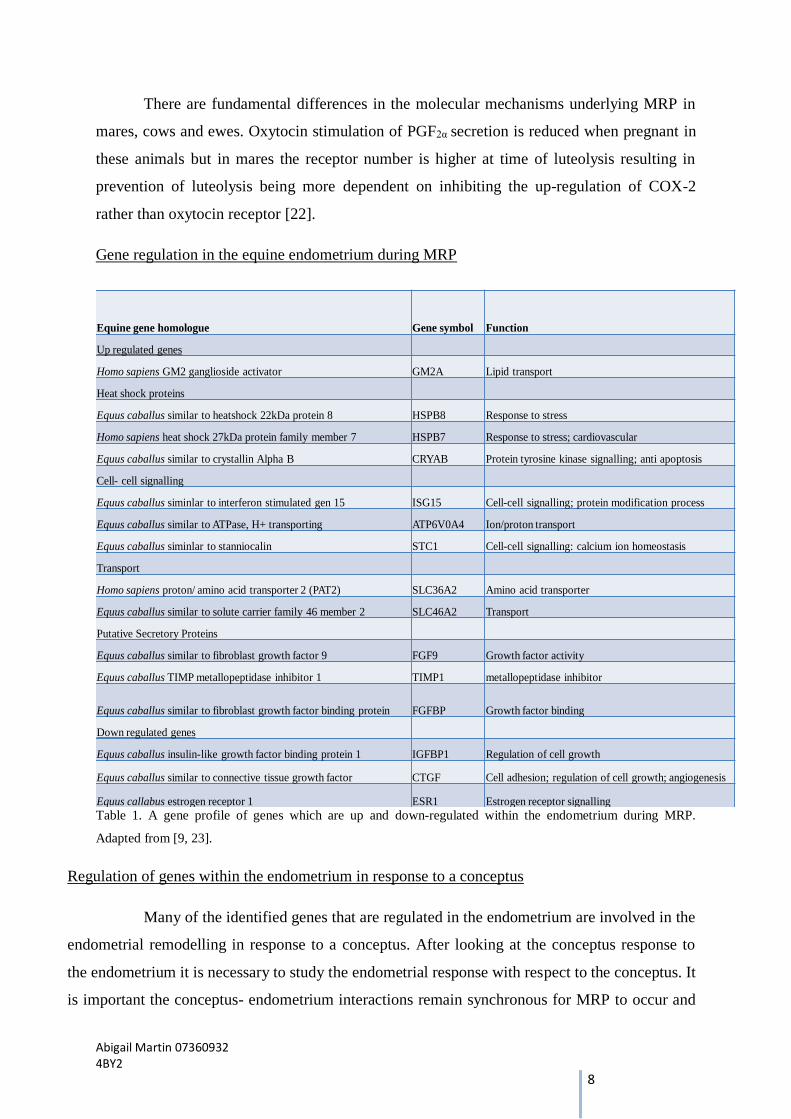
Gene regulation in the equine endometrium during MRP
Equine gene homologue Gene symbol Function
Up regulated genes
Homo sapiens GM2 ganglioside activator GM2A Lipid transport
Heat shock proteins
Equus caballus similar to heatshock 22kDa protein 8 HSPB8 Response to stress
Homo sapiens heat shock 27kDa protein family member 7 HSPB7 Response to stress; cardiovascular
Equus caballus similar to crystallin Alpha B CRYAB Protein tyrosine kinase signalling; anti apoptosis
Cell- cell signalling
Equus caballus siminlar to interferon stimulated gen 15 ISG15 Cell-cell signalling; protein modification process
Equus caballus similar to ATPase, H+ transporting ATP6V0A4 Ion/proton transport
Equus caballus siminlar to stanniocalin STC1 Cell-cell signalling: calcium ion homeostasis
Transport
Homo sapiens proton/ amino acid transporter 2 (PAT2) SLC36A2 Amino acid transporter
Equus caballus similar to solute carrier family 46 member 2 SLC46A2 Transport
Putative Secretory Proteins
Equus caballus similar to fibroblast growth factor 9 FGF9 Growth factor activity
Equus caballus TIMP metallopeptidase inhibitor 1 TIMP1 metallopeptidase inhibitor
Equus caballus similar to fibroblast growth factor binding protein FGFBP Growth factor binding
Down regulated genes
Equus caballus insulin-like growth factor binding protein 1 IGFBP1 Regulation of cell growth
Equus caballus similar to connective tissue growth factor CTGF Cell adhesion; regulation of cell growth; angiogenesis
Equus callabus estrogen receptor 1 ESR1 Estrogen receptor signalling
Table 1. A gene profile of genes which are up and down-regulated within the endometrium during MRP.
Adapted from [9, 23].
Regulation of genes within the endometrium in response to a conceptus
Many of the identified genes that are regulated in the endometrium are involved in the
endometrial remodelling in response to a conceptus. After looking at the conceptus response to
the endometrium it is necessary to study the endometrial response with respect to the conceptus. It
is important the conceptus- endometrium interactions remain synchronous for MRP to occur and
Abigail Martin 07360932 4BY2
9
for embryo growth and development to be successful. Table 1 shows a summary of commonly
upregulated and down regulated genes that are expressed in the endometrium during early
pregnancy in response to the conceptus.
Merkl et al. determined differential expression of genes in the endometrium on day 8
and 12 of pregnant and cyclic mares, taking endometrial biopsies on the relevant days and using
qPCR and microarrays to show total mRNA expression in response to a conceptus. By day 12,
they noticed a significant difference in the expressions recorded. They found the higher mRNA
levels were due to a response to the conceptus that prevented the down regulation of the genes
they analysed, except for FGF9 gene, a fibroblast growth factor, which was additionally
upregulated. Merkl et al. stated that the regulation of steroid hormones may be responsible for
these interactions [9].
Heat shock proteins are proteins whose expression is upregulated when introduced to
a stressful environment. The three main proteins regulated by oestrogens and involved in the
equine endometrium are heat shock proteins B7 and B8 (HSPB7 and HSPB8) and Crystalin, alpha
B (CRYAB). The endometrium is sensitive and receptive towards the conceptus. Klein et al.
hypothesised that these proteins prepare the endometrium for fixation in the conceptus [23].
Cell-cell signalling mediates the transfer of information from one cell to another [24].
This process and its upregulation of genes are essential for the successful establishment of
pregnancy. Stanniocalcin 1 (STC1) is a pregnant-specific uterine gene which is upregulated in the
endometrium across species. In the endometrium of equids STC1 expression has not been
reported but, its expression has been recorded in sheep, pigs, humans and mice. In sheep, STC1
mRNA and protein are required for regulating growth and differentiation of the embryo and its
placenta, and act as a marker for implantation in pigs. In humans and mice STC1 expression is
highest during embryo implantation in the endometrium. It therefore plays a species- independent
role in early pregnancy [9, 23].
During MRP, amino acid transport is necessary for embryo development. Transport
mediator protein, solute carrier 36 member 2 (SLC36A2) is a proton/ amino acid symporter. This
simply means that two molecules can be transported by this protein in the same direction through
the conceptus capsule using a common carrier mechanism. Klein et al. assume that its
upregulation is due to the demands of rapidly growing conceptus for nutrients [23].
Abigail Martin 07360932 4BY2
10
Secretory proteins make up a considerable amount of gene material upregulated in the
uterine cavity. When secretory protein production is poor, pregnancies often show detrimental
effects [25]. Commonly upregulated secretory proteins were luteinizing hormone (LH), tissue
inhibitor of metalloproteinase 1 (TIMP1) and insulin growth factors (IGFs). LHB has been
observed in women and is believed to prepare the uterus for implantation. It interrupts cellular
apoptosis, encourages angiogenesis and employs a particular immune responses [11, 25]. This is
understood to be similar in equids in preparing the endometrium for fixation. TIMPs regulate the
extracellular matrix production and tissue reconstruction during implantation and fixation thus, it
is a contributing factor to endometrial reconstruction in various species and equine pregnancies
[23]. As described earlier, FGF9 a fibroblast growth factor is also potentially involved in
endometrial reconstruction [9, 23]. IGFBP1 and IGF1 are both found to be upregulated and
expressed in Klein et al. and Merkl et al. studies on expressions in the endometrium. It is
suggested IGFBP1 and IGF1 have a function as transporters of IGFs between the endometrium
and uterine lumen of the horse [9, 23].
A gene of interest is estrogen receptor 1 (ESR1), involved in the initiation of
luteolysis in cyclic mares. Pre-fixation period ESR1 is down regulated. This is due to the intrusion
of the conceptus [23]
The value of equines in reproductive studies
When it comes to looking at maternal recognition of pregnancy, equines exhibit many
differences in comparison to other domestic ruminants (goats, sheep, pigs, cattle). But there
are also similarities. For example in cattle 70%- 80% of total embryonic loss occurs between
days 8-16 after insemination, a similar time-frame like equines in embryonic loss. Equines
have many advantages in using them as models over other ruminants for the study of
pregnancy in the first month, and for the total embryonic loss that occurs during this time.
There is no other domestic species of which a conceptus can be collected intact i.e within the
first four weeks of pregnancy while undergoing advanced organogenesis. It can be obtained
from the uterus repeatedly, atraumatically and easily with no maternal tissues attached [1].
Although methods and techniques for this research have developed greatly over the past
decade, there are still many unanswered questions in embryonic development and its
interactions within the uterus. Fortunately, this acts as an incentive to undertake this research
and combat the problem of early embryonic loss and extend our knowledge of endometrium-
Abigail Martin 07360932 4BY2
11
embryo cross-talk. Betteridge states that “research will quite certainly also pay dividends in
unanticipated ways in the broader field of reproductive biology” [1].
A disadvantage is that the embryo cannot be accessed easily until around day 6.5
after the blastocyst has been released from the oviduct. At present, surgery or slaughter is the
necessary procedure to collect a blastocyst from the oviduct. Once the blastocyst enters the
uterus it can be accessed easily using harmful techniques. Surgery or slaughter is most
certainly not efficient. It is possible to successfully culture cleavage stage embryos in vitro.
There is not a system that can meet the requirements of a growing blastocyst and provide it
with the correct environment to allow it to produce a capsule as it does in the uterus [26].
Therefore, there is a need to improve methods to produce embryos in vitro.
From an immunological point of view, the embryo- maternal interactions has
highlighted events that struggle to be detected in other species [25].
Conclusion
It is clear that embryo- endometrial interactions are not detected by a single gene, but
by a series of individual genes and pathways regulating specific phases of endometrial
function, conceptus development, and the influences the conceptus has on endometrial
function during maternal recognition of pregnancy. In only very recent years has there been
much understanding of the embryo- maternal cross talk. Transcriptional profiling of the equid
endometrium during MRP has only been published this year, of which are the first few of
their kind to be reported. Common features have been found in equine and human
pregnancies [11, 25, 27]. Equines have proven to be a positive addition in the studies of
early reproductive research. Many questions remain in this area of research, such as further
investigations into communication between the embryo and endometrium via gene
expression, the physical relationship between the capsule and the yolk sac and its signalling
efforts towards the endometrium and the conceptus synchronisation in the uterine cavity. By
focusing on particular genes that are expressed during early pregnancy it will be possible to
improve methods for diagnosis and treatment of mares that are prone to losing the conceptus
during the first three weeks of pregnancy. There is the need for rigour in the study of
embryonic development and its products presumed to play an important role in
communication between embryo and mother in any species.
Abigail Martin 07360932 4BY2
12
References
1. Betteridge KJ: Comparative aspects of equine embryonic development. Animal Reproduction Science 2000, 60:691-702.
2. Morris LH AW: Reproductive efficiency of intensively managed thoroughbred mares in Newmarket. Equine Vet J 2002:51-60.
3. Hyttel P SF, Vejsted M: Essentials of domestic animal embryology: Saunders Elsevier; 2009. 4. Fuller W. Bazer, Ott TL, Spencer TE: Maternal recognition of pregnancy:Comparative aspects - A
review. Placenta 1998, 19 Supplement 2:375-386. 5. Allen WR, Wilsher S: A Review of implantation and early placentation in the mare. Placenta
2009, 30:1005-1015. 6. Betteridge KJ: Equine embryology: An inventory of unanswered questions. Theriogenology 2007,
68:S9-S21. 7. Oriol JG B: Mucin-like glycoproteins in the equine embryonic capsule. Mol Repro Dev 1993,
34:255-265. 8. Sharma S, Davies Morel MCG, Dhaliwal GS, Dadarwal D: The pattern of embryonic fixation and its
relationship to pregnancy loss in thoroughbred mares. Reproduction in Domestic Animals 2010, 45:361-367.
9. Merkl M, Ulbrich S, Otzdorff C, Herbach N, Wanke R, Wolf E, Handler J, Bauersachs S: Microarray analysis of equine endometrium at Days 8 and 12 of pregnancy. Biol Reprod 2010.
10. Boerboom D, Brown KA, Vaillancourt D: Expression of key progtaglandin synthesis in equine endometrium during late diestrus and early pregnancy. Biol Reprod 2003:391-399.
11. Bazer FW, Spencer TE, Johnson GA, Burghardt RC, Wu G: Comparative aspects of implantation. Reproduction 2009:195–209.
12. Goff AK: Embryonic signals and survival. Reproduction in Domestic Animals 2002, 37:133-139. 13. Spencer TE, Gray A, Johnson GA, Taylor KM, Gertle A, Gootwine l, Ott TL, Bazer FW: Effects of
recombinant ovine interferon tau placental lactogen, and growth hormone on the ovine uterus. Biology of reproduction 1999, 61:1409-1418.
14. Poser NL: The control of prostaglandin production by the endometrium in relation to luteolysis and menstruation. Prostaglandins Leukotrienes and Essential Fatty Acids 1995:147-195.
15. Ealy AD, Ehroh ML, Sharp DC: Progstalandin H synthase type 2 is differentially expressed in endometrium bases on pregnancy status in pony mares and responds to oxytocin and conceptus secretions in explant culture. Anim Reprod Sci 2010.
16. Hayes MA, Quinn BA, Keirstead ND, Katavolos P, Waelchli RO, Betteridge KJ: Proteins associated with the early intrauterine equine conceptus. Reproduction in Domestic Animals 2008, 43:232-237.
17. Quinn BA, Hayes MA, Waelchli R, Kennedy MW, Betteridge KJ: Changes in major proteins in the embryonic capsule during immobilization (fixation) of the conceptus in the third week of pregnancy in the mare. Reproduction 2007, 134:161-170.
18. Quinn BA, Caswell DE, Lillie BN, Waelchli RO, Betteridge KJ, Hayes MA: The GM2-activator protein is a major protein expressed by the encapsulated equine trophoblast. Animal Reproduction Science 2006, 94:391-394.
19. Ababneh M, Ababneh H, Shidaifat F: Expression of cytosolic phospholipase A2 in equine endometrium during the oestrous cycle and early pregnancy. Reprod Domest Anim 2010.
20. Atli M, Kurar E, Kayis S, Aslan S, Semacan A, Celik S, Guzeloglu A: Evaluation of genes involved in prostaglandin action in equine endometrium during estrous cycle and early pregnancy. Anim Reprod Sci 2010.
21. Guzeloglu A, Atli MO, Kurar E, Semacan A, Aslan S, Kayis SA, Celik S: Evaluation of genes involved in prostaglandin action in equine endometrium during estrous cycle and early pregnancy. Animl Reprod Sci 2010, 122:124-132.

Abigail Martin 07360932 4BY2
13
22. Allen WR: Luteal deficiency and embryo mortality in the mare. Reproduction in Domestic Animals 2001, 36:121-131.
23. Klein C, Scoggin K, Ealy A, Troedsson M: Transcriptional profiling of equine endometrium during the time of maternal recognition of pregnancy. Biol Reprod 2010, 83:102-113.
24. Nelson DL, Cox MM: Lehninger Principles of biochemistry Fourth Edition: New York : W.H. Freeman; 2005.
25. Leela E. Noronha, Antczak DF: Maternal immune responses to trophoblast: The contribution of the horse to pregnancy immunology. Reprod Immunol 2010:231-244.
26. Betteridge KJ:Farm animal embryo technologies: Achievements and perspectives. Theriogenology 2006, 65:905-913.
27. Teklenburg G, Macklon NS: In Vitro Models for the Study of early human embryo-endometrium interactions. Reproductive Sciences 2009, 16:811-818.