liposomes: advancements and innovation in the
TRANSCRIPT

This is a peer-reviewed, accepted author manuscript of the following research article, Shah, S., Dhawan, V., Holm, R., Nagarsenker, M. S., & Perrie, Y. (2020). Liposomes: advancements and innovation in the manufacturing process. Advanced Drug Delivery Reviews, 154-155, 102-122. https://doi.org/10.1016/j.addr.2020.07.002
Liposomes: Advancements and innovation in the manufacturing process 1
2
Sanket Shaha,b, Vivek Dhawanb,c, René Holma, d, Mangal S. Nagarsenkerc,e, Yvonne Perrief,* 3
4
5
6
*Author for correspondence 7
Yvonne Perrie 8
Strathclyde Institute of Pharmacy and Biomedical Sciences, 9
161 Cathedral St, University of Strathclyde, Glasgow, Scotland. G4 0RE. 10
Email: [email protected] 11
a Drug product development, Janssen R&D, Johnson & Johnson, Turnhoutseweg 30, 2340 Beerse, Belgium b These authors contributed equally c Department of Pharmaceutics, Bombay College of Pharmacy, Kalina, Santacruz, Mumbai, India - 400098. d Department of Science and Environment, Roskilde University, 4000 Roskilde, Denmark. e Department of Pharmaceutics, VES College of Pharmacy, Chembur, Mumbai, India 400074 f Strathclyde Institute of Pharmacy and Biomedical Sciences, 161 Cathedral St, The University of Strathclyde, Glasgow, Scotland. G4 0RE

2
Highlights 1
• Liposomes and their related constructs offer unique advantages in terms of drug and 2
vaccine delivery. 3
• However, current processes used for the manufacture of liposomes present a range of 4
challenges, driving up cost, and limiting production. 5
• New production methods can address these issues and support the cost-effective 6
manufacture of current liposomal systems and facilitate the development of new 7
liposomal products. 8

3
Abstract 1
Liposomes are well recognised as effective drug delivery systems, with a range of products 2
approved, including follow on generic products. Current manufacturing processes used to 3
produce liposomes are generally complex multi-batch processes. Furthermore, liposome 4
preparation processes adopted in the laboratory setting do not offer easy translation to large 5
scale production, which may delay the development and adoption of new liposomal systems. 6
To promote advancement and innovation in liposome manufacturing processes this review 7
considers the range of manufacturing processes available for liposomes, from laboratory scale 8
and scale up, through to large-scale manufacture and evaluates their advantages and 9
limitations. The regulatory considerations associated with the manufacture of liposomes is 10
also discussed. New innovations that support leaner scalable technologies for liposome 11
fabrication are outlined including self-assembling liposome systems and microfluidic 12
production. The critical process attributes that impact on the liposome product attributes are 13
outlined to support potential wider adoption of these innovations. 14

4
Graphical abstract 1
2

5
Keywords 1
Liposomes; Lipid nanoparticulate; Microfluidics; Leciplex; Phospholipids; cationic liposomes; 2
nanomedicine; monoclonal antibody; oligonucleotide; targeted delivery 3
4

6
Abbreviations 1
Chol: Cholesterol 2
cryoTEM: cryo transmission electron microscopy 3
Da: Daltons 4
DLin-MC3-DMA: (6Z,9Z,28Z,31Z)-heptatriaconta-6,9,28,31-tetren-19-yl-4-5
(dimethylamino)butanoate 6
DMG-PEG2000: (R)-2,3-bis(tetradecyloxy)propyl 1-(methoxypoly(ethylene 7
glycol)20000)propyl carbamate 8
DOPE: 1,2-dioleoyl-sn-glycero-3-phosphoethanolamine 9
DOTAP: 1,2-dioleoyl-3-trimethylammonium-propane (chloride salt) 10
DPPC: 1,2-Dipalmitoyl-3-sn-phosphatidylcholine 11
DSPC: 1,2-distearoyl-sn-glycero-3-phosphocholine 12
EMA: European medicines agency 13
FDA: Food and drug administration 14
FRR: flow rate ratio 15
FVR: flow velocity ratio 16
GMP: Good manufacturing practice 17
GUVs: Giant unilamellar vesicles 18
HGL: High gravity level 19
ICH: International Council for Harmonisation 20
IPA: iso-propyl alcohol 21
IPQC: In-process quality control 22
LFH: Lipid film hydration 23
LNPs: Lipid nanoparticles 24
7
Log P: Partition co-efficient 1
LUVs: Large Unilamellar vesicles 2
MC: microchannel 3
MLVs: Multilamellar vesicles 4
MVVs: Multivesicular vesicles 5
OLVs: Oligo lamellar vesicles 6
PBS: Phosphate buffer saline 7
PC: Phosphatidyl choline 8
PDI: Polydispersity index 9
PTFE: polytetrafluoroethylene 10
PVA: polyvinyl alcohol 11
RNAi: Ribose nucleic acid interference 12
siRNA: small interfering ribose nucleic acid 13
SUVs: Small unilamellar vesicles 14
TBA: tert-butyl alcohol 15
TFF: tangential flow filtration 16
ULVs: unilamellar vesicles 17
w/o/w emulsion: Water-in-oil-in-water emulsion 18
w/o: water-in-oil 19
20

8
Contents 1
Highlights ................................................................................................................................................ 2 2
Abstract ................................................................................................................................................... 3 3
Graphical abstract ................................................................................................................................... 4 4
Keywords ................................................................................................................................................. 5 5
Abbreviations .......................................................................................................................................... 6 6
1. Introduction and overview of application of liposomes ............................................................... 9 7
2. Manufacturing of liposomes ........................................................................................................ 13 8
2.1. Laboratory scale manufacturing ........................................................................................... 13 9
2.1.1. Solvent evaporation ...................................................................................................... 15 10
2.1.2. Solvent dispersion ......................................................................................................... 16 11
2.1.3. Reverse phase evaporation ........................................................................................... 16 12
2.1.4. Size manipulation .......................................................................................................... 17 13
2.1.5. Final liposomal drug product ........................................................................................ 17 14
2.2. Industrial manufacturing and scale up considerations ......................................................... 19 15
2.3. Potential for innovation in large scale liposome manufacturing .......................................... 23 16
3. Regulatory overview of liposomes ................................................................................................ 25 17
4. Advances in scalable technologies for liposome fabrication ........................................................ 30 18
4.1. Self-assembled vesicular drug delivery system .................................................................... 31 19
4.1.1. Heating methods ........................................................................................................... 31 20
4.1.2. Nanoprecipitation and Ionic interaction ....................................................................... 32 21
4.1.3. Solvent exchange .......................................................................................................... 33 22
4.1.4. High Shear ..................................................................................................................... 34 23
4.1.5. Emulsification and solvent evaporation ....................................................................... 35 24
4.1.6. Packed bed-based reactors ........................................................................................... 36 25
4.1.7. Gel assisted self-assembly ............................................................................................ 38 26
4.1.8. Spray drying and fluid bed drying ................................................................................. 39 27
4.1.10. Freeze drying. ................................................................................................................ 40 28
4.1.11. Supercritical fluid techniques........................................................................................ 41 29
4.2. Microfluidics .......................................................................................................................... 45 30
4.2.1. Microfluidic cartridge design ........................................................................................ 46 31
4.2.2. Microfluidic material and production parameter considerations ................................ 49 32
5. Conclusion .................................................................................................................................... 56 33
6. References .................................................................................................................................... 58 34
35
36

9
1. Introduction and overview of application of liposomes 1
Lipid-based nanomedicines are used to 1) protect drugs from degradation in vivo, 2) control 2
drug release, 3) modify biodistribution, 4) target drug delivery to the site of disease and 5) 3
enhance solubility and bioavailability. Lipid based delivery systems are also effective as 4
vaccine adjuvants through their ability to protect and deliver antigens (peptide, protein and 5
nucleic acid systems) to the antigen presenting cells and stimulating protective immune 6
responses. Suitable engineering of nanomedicines in terms of their composition, particle size, 7
and surface charge can aid in achieving spatial and temporal delivery of drugs. This applies to 8
the delivery of traditional small molecules and to lipid-based nanoparticles used to deliver 9
nucleic acid-based drugs; patisiran (Onpattro®; Alnylam), approved by the FDA and the EMA 10
in 2018, is the first siRNA-based drug approval. Patisiran is indicated for polyneuropathy of 11
hereditary transthyretin-mediated amyloidosis. Onpattro® contains 2.0 mg/mL of patisiran (a 12
double stranded siRNA which is the active substance) incorporated into lipid nanoparticles. 13
The nanoparticles are 60 – 100 nm in size with a near neutral surface change at biological pH. 14
The nanoparticles are formed during production as a result of the lipids associating with the 15
siRNA. The nanoparticles are built from four lipids. DSPC is used with cholesterol to support 16
the formation and stability of the lipid nanoparticles. An ionisable lipid (DLin-MC3-DMA) is 17
incorporated which electrostatically interacts with the siRNA and promotes high drug loading 18
and a pegylated lipid DMG-PEG2000 is incorporated to improve the stability of the formed 19
LNPs [1]. Of these lipid-based nanomedicines, liposomes are generally the most well-20
established systems. 21
Liposomes are lipid based spherical shaped vesicular systems, in which a lipophilic bilayer is 22
sandwiched between two hydrophilic layers. The versatility and advantages of liposomes as 23
a drug delivery system for small molecules, peptides, gene, and monoclonal antibodies is well 24
studied and acknowledged in the peer-reviewed scientific literature [2-6]. Liposomes fall into 25
the general category of nanomedicines and play a key role in many diverse areas of health 26
and have found an application in the treatment of patients suffering from cardiovascular 27
disease, neurodegenerative disease, diabetes, cancer and inflammation. Parenteral delivery 28
offers the advantage of bypassing first pass metabolism, poor gastrointestinal permeability 29
and gastrointestinal side effects (a problem common to oral delivery of drugs) and parenteral 30
10
administration provides an opportunity for targeted delivery of drugs resulting in higher 1
bioavailability and reduced off target side effects. 2
A myriad of reports exist in the literature on application of liposomes to deliver drugs 3
[7-10] and genes [11-14] parenterally. Phospholipids being biodegradable and biocompatible 4
and bearing resemblance to the lipids present in cellular membranes, are widely explored for 5
their drug delivery potential associated with their assemblies to organise structure (Fig. 1). 6
Liposomes on account of being formed due to self-assembly possess a thermodynamic 7
advantage. They have been used for cancer treatment to improve tumour targeting and 8
reduce off-target toxicity (e.g. Doxil®) and to treat patients with severe infections or 9
immunocompromised conditions (e.g. AmBisome®). Whilst access to these advanced 10
treatments can be limited (mainly due to their cost), in 2015, the global liposomal doxorubicin 11
market alone was valued at USD 814.6 million [15]. Furthermore, there are many other 12
marketed oncology nanomedicines using lipid-based nanotechnology (e.g. DaunoXome®, 13
Myocet®, DepoCyt®, Marqibo® and Onivyde®) and more recently Onpattro®, the first FDA-14
approved RNAi therapeutic. Indeed, the nanomedicines market is recognised as a high risk, 15
high return market and has enjoyed unprecedented growth over the last five years. 16

11
1
Fig. 1. Schematic showing the different phases of a phospholipid molecule. The phospholipid molecules in an 2 aqueous environment, under right conditions, forms a bilayered lamella spherical in shape called liposomes. 3 Depending on the types and forms, liposomes can be called a LUVs, SUVs, and MLVs. Reprinted from Trends in 4 Biotechnology, 16/7, Dan D Lasic, Novel applications of liposomes, 307-321, Copyright (2020), with permission 5 from Elsevier [16]. 6
The global nanomedicine market was valued at USD 135 billion in 2015 and it is 7
anticipated that this will reach USD 350.8 billion by 2025 [17]. However, using traditional 8
manufacturing methods, the scale-up production of nanomedicines presents a significant 9
challenge to their clinical development and the cost of commercial manufacture is a 10
recognised barrier to their translation from bench to beside. Despite their widespread 11
research, it is well recognised that the current processes used for manufacturing of liposomes 12
suffers from many severe problems, including: i) multi-step batch processes; ii) the need for 13
particle size reduction (often involving specialized tools and equipment such as extrusion and 14
high-pressure homogenization) and iii) limited batch sizes. This drive cost upwards, limits 15
production and hinders development of liposomes. This was exemplified by the global 16
shortage of Doxil® due to closure of a sterile injectables production site due to manufacturing 17

12
challenges as the principle reason. Global shortages of this anti-cancer treatment lasted for 1
more than two years [18]. Hence, it is important to identify ways of making the liposomal 2
manufacture process leaner and identifying ways to make this drug delivery option more 3
attractive from an industrial point of view. 4
13
2. Manufacturing of liposomes 1
2.1. Laboratory scale manufacturing 2
Numerous reviews and research articles have been published elsewhere on the 3
composition, preparation, and characterization of liposomes and readers are requested to 4
refer them for more in-depth understanding [19-30]. Almost all the techniques involve 5
dissolution of phospholipids in an organic solvent followed by removal of the organic solvent, 6
later in the process. This prior dissolution followed by removal of organic solvent is important 7
for the formation of liposomes. The building blocks of liposomes are phospholipids and/or 8
cholesterol. The critical micelle concentration of most commonly used phospholipids is in the 9
nanomolar range and the concentration of phospholipids used for liposomes manufacturing 10
is much above the critical micelle concentration. This along with the three-dimensional 11
cylinder like shape of each phospholipid (Fig. 2) leads to formation of liposomes along with 12
lipid aggregates when phospholipids, as such, are exposed to an aqueous environment. In 13
order to make uniform liposomal dispersions, it is important to make thin lipid sheets before 14
exposing it to an aqueous phase or introduce the organic phospholipid solution in a controlled 15
manner in an aqueous environment for the formation of liposomes. This is why all the 16
reported techniques of liposome manufacturing i.e. solvent evaporation, solvent 17
dispersion/antisolvent addition, or detergent removal focus on first disaggregating the 18
phospholipids into individual phospholipid molecules followed by exposure to aqueous 19
environment to enable formation of different types of liposomes viz. MLVs, SUVs, GUVs, OLVs, 20
MVVs (Table 1) [31, 32]. The detergent removal technique is not discussed in this , but 21
interested reader can be referred to other literature sources [33, 34]. A special mention of 22
the reverse phase evaporation method is discussed as it is the preferred laboratory technique 23
for obtaining high entrapment efficiency of hydrophilic drugs [35]. 24
25

14
1
Fig. 2. Different molecular shapes of a surfactant/phospholipid like molecules. Most of the commonly used 2 phospholipids have a cylinder like shape and form a bilayered lamellae when exposed to aqueous medium. By 3 addition of cone shape or inverted cone shape species, the properties of the bilayered lamellae can be altered to 4 make it more rigid/leaky. Reprinted from Publication Trends in Biotechnology, 16/7, Dan D Lasic, Novel 5 applications of liposomes, 307-321, Copyright (2020), with permission from Elsevier [16]. 6
Table 1. Classification of different liposome formations. 7
Types of liposome formations
Commonly used abbreviations
Particle size Number of lamellae
Small unilamellar vesicles SUVs 20-100 nm 1
Large unilamellar vesicles LUVs >100 nm 1
Giant unilamellar vesicles GUVs >1000 nm 1
Multilamellar vesicles MLVs >500 nma >5
Oligolamellar vesicles OLVs 100-1000 nm 2-5
Multivesicular liposomes MVVs >1000 nm 1
(Vesicle inside a vesicle)
8
a This is a typical particle size, however, MLVs with a particle size of 100 nm have been reported [36] J.A. Kulkarni, M.M. Darjuan, J.E. Mercer, S. Chen, R. van der Meel, J.L. Thewalt, Y.Y.C. Tam, P.R. Cullis, On the Formation and Morphology of Lipid Nanoparticles Containing Ionizable Cationic Lipids and siRNA, ACS Nano, 12 (2018) 4787-4795.

15
2.1.1. Solvent evaporation 1
In this technique, also known as lipid film hydration, phospholipids are dissolved in an organic 2
solvent (more often an equimolar mixture of chloroform and methanol, others could be ether, 3
ethanol, or dichloromethane) [37, 38]. The drug, if lipophilic, is also added to the organic 4
solvent to form a one-phase solution. The organic solvent is subsequently removed slowly 5
under vacuum to form thin sheets of lipid films in which the drug is uniformly dispersed. The 6
thin sheets of lipids are hydrated with an aqueous buffer phase above the glass transition 7
phase of the lipid. If the drug is hydrophilic, it should be dissolved in the aqueous buffer 8
solution. The resulting dispersion gives MLVs with particle size in the micrometer range. This 9
technique is more suitable for lipophilic drugs as a high entrapment efficiency (>90%) can be 10
obtained. For hydrophilic drugs, depending on the physicochemical properties, the 11
entrapment efficiency would routinely around 10-30 % by this passive process. Low 12
entrapment efficiency values have been reported for cytarabine, streptomycin sulphate, 13
chloramphenicol, oxytetracycline, and sulfamerazine [39, 40]. The entrapment efficiency can 14
be increased further for hydrophilic drugs by use of active loading technique. Active loading 15
technique involves transmembrane gradient (like pH or ionic) of unionized species to effect 16
higher entrapment of the drug. A classic example of the active loading technique being used 17
is in manufacture of doxorubicin liposomes. Liposomes are manufactured either by solvent 18
evaporation, solvent dispersion (Section 2.1.2) or reverse phase evaporation technique 19
(Section 2.1.3) using ammonium sulphate solution as the aqueous phase. The MLVs are 20
further manipulated for size (Section 2.1.4) using suitable technique followed by removal of 21
un-entrapped ammonium sulphate using dialysis/diafiltration. The pH of external phase is 22
adjusted (using dialysis/diafiltration) to 7.4 to create a transmembrane pH gradient and a 23
solution of doxorubicin hydrochloride is added that results in entrapment of high amounts of 24
doxorubicin in the aqueous core as precipitates of doxorubicin sulphate [41-43]. Hydrophilic 25
drugs that have been developed into liposomes with high entrapment efficiency using the 26
active loading technique are bupivacaine [44], kanamycin [45], ciprofloxacin [46], chloroquine 27
diphosphate [47], primaquine [48], topotecan [49, 50], and vincristine [51]. For a more 28
detailed theoretical basis readers can refer an excellent book chapter by Boris Čeh [52] and 29
for a more comprehensive preparation techniques for liposomes a review by Has and Sunthar 30
is recommended [53]. 31

16
2.1.2. Solvent dispersion 1
In this technique the phospholipids are dissolved in an organic solvent that is often miscible 2
with water, ethanol being the preferred solvent [20, 54, 55]. A lipophilic drug would be 3
dissolved in the ethanolic solution together with the phospholipids (other water miscible 4
solvents could be used if the lipophilic drug is not soluble in ethanol). The ethanolic 5
phospholipid/drug solution is added to an aqueous buffer solution, which leads to dilution of 6
the ethanol into the water and thereby spontaneous formation of MLVs. The particle size 7
MLVs is in the micrometer range. This technique is most suited for lipophilic drugs, which can 8
yield high entrapment efficiency. For hydrophilic drugs the entrapment efficiency is normally 9
in the sub 20%. However, as described in Section 2.1.1, the entrapment efficiency can be 10
increased significantly reaching >90%. 11
12
2.1.3. Reverse phase evaporation 13
This technique is the most preferred techniques for loading a hydrophilic drug in liposomes. 14
For hydrophilic drugs, the internal aqueous core is the only region where the drug can be 15
loaded. Hence, a technique, that can entrap a large amount of aqueous core during formation 16
of liposome will yield a high entrapment efficiency and hence a high drug load. In the reverse 17
phase evaporation method, a w/o emulsion is prepared by dissolving the hydrophilic drug in 18
water and dissolving the phospholipid in water-immiscible solvent (usually chloroform). The 19
organic solvent is then slowly removed, under vacuum, and a gel phase is formed. Further 20
evaporation of the organic solvent yields liposomal dispersion with high entrapment of the 21
aqueous core in the internal core of liposomes. This technique can yield up to 30-50% 22
entrapment of a hydrophilic drug passively [56, 57] and can be increased to >90% using the 23
active loading technique described in Section 2.1.1. This technique is discussed extensively 24
by Szoka and Papahadjopoulos [35]. This method and is suited for making small volume 25
parenterals. However, its use on an industrial scale is limited due to the complex 26
manufacturing process. 27
28
29

17
2.1.4. Size manipulation 1
Both, solvent evaporation and solvent dispersion, produce MLVs which are in the micrometer 2
range. For drug delivery application, it is important to further reduce the particle size of these 3
liposomes in the submicron range, more specifically in the 50-200 nm, as the particle size of 4
liposomes has a huge impact on the pharmacokinetic and pharmacodynamic profile in vivo 5
and hence can have an impact on the therapeutic efficacy of the final formulation [58-60]. On 6
the laboratory scale, there are numerous techniques available that can reduce the particle 7
size of liposomes viz. sonication [8], freeze thaw [61], homogenization [62, 63], and extrusion 8
[64, 65]. All the techniques have their merits and demerits. Sonication is a rather fast 9
technique of reducing the particle size with a high amount of energy dissipated in a small 10
volume. During sonication there is generation of heat, which may lead to degradation of 11
phospholipids and heat labile drugs. Freeze thaw can also be used to convert the MLVs into 12
smaller vesicles as SUVs or LUVs, however, in many cases the it can only reduce the particle 13
size to a certain extent with a rather high particle size distribution, i.e. polydispersity index. 14
Homogenization can also be used for particle size reduction and is a batch process. Liposomes 15
are soft matters that can be reduced in particle size by application of a high-pressure 16
homogenizer. The liposomes obtained by high pressure homogenizer have a higher 17
polydispersity index (~0.2) compared to liposomes prepared using extrusion [66, 67] and at 18
high lipid loads (>100 mg/mL) the size reduction efficiency may also be reduced [63]. 19
Extrusion of liposomes through a polycarbonate membrane can produce liposomes of a 20
defined pore size and acceptable PDI (≤0.1). The extrusion process is a laborious and time-21
consuming process; however, it is the most acceptable and reproducible process for making 22
liposomes with known defined characteristics. 23
24
2.1.5. Final liposomal drug product 25
The final dosage form, for parenteral administration, of the liposomal drug product can vary 26
depending on the change in physical and chemical properties of liposomes over its storage. It 27
is important to bring the conversation around stability, earlier in the development, as it is 28
leaner to develop all the target product profile early during the development, rather than 29
doing it in late phase development. The factors that one should consider while determining 30
18
the final dosage form for liposomes apart from physical and chemical stability is the preferred 1
storage for the commercial product. It is a given that, room temperature storage is preferred 2
over a cold storage for logistical and economic reasons. More often than not the liposomal 3
dispersion stability at room temperature is a challenge and hence most of the marketed 4
products that are presented as a liquid dosage form are required to be stored between 2-8 5
°C. The particle size, drug loading, and chemical stability are the three important liposomal 6
critical to quality attributes that can get affected during storage of liposomal dispersion as a 7
liquid at room temperature. Another way, to improve the chemical and/or physical stability, 8
at room temperature, is by converting the liposomal dispersion into a dried powder/freeze 9
dried cake for reconstitution. Because of the reduced interaction of the drug product 10
components in the solid state as compared to the liquid state, there is a general trend towards 11
a better chemical and/or physical stability. However, it might not be always possible to have 12
dry liposomal powder for reconstitution due to various reasons. One reason being that often 13
the liposomes do not have the same particle size before and after reconstitution and for these 14
reasons freeze drying/spray drying of liposomes into powders/cake might not be always 15
possible. The second reason being that if the drug is not lipophilic, then it would be inside the 16
liposomal aqueous core and the process of spray drying/freeze drying can change the 17
entrapment efficiency of hydrophilic drugs and the amount of liposome entrapped drug will 18
change after reconstitution. This phenomenon also holds true for drugs which have been 19
loaded in the interior aqueous core of liposomes using active loading technique as in case of 20
doxorubicin loaded liposomes. Hence, in such scenarios, a liposomal dispersion as a liquid 21
that is stored between 2-8 °C is the preferred dosage form. Even, with a better chemical and 22
physical stability profile of a freeze dried/spray dried liposomal product at room temperature, 23
it might still be commercialized as a liquid dosage form to be stored at 2-8 °C. The reason 24
being, that, converting a liposomal dispersion from a liquid dosage form to a solid freeze-25
dried cake or spray dried powder requires an additional step (along with other in process 26
quality control tests) in the liposome manufacturing under aseptic conditions. Hence, this 27
decision is more driven by the product needs, target product profile, target climatic zones of 28
the marketed product, and the organizational preference of having a cold chain storage or a 29
freeze drying/spray drying capability. 30
31

19
2.2. Industrial manufacturing and scale up considerations 1
Given that the majority of liposomal formulations are designed to improve drug delivery and 2
reduce off-target toxicity associated with the incorporated cytotoxic drug, the manufacturing 3
process employed must control liposomes’ critical quality attributes. These includes particle 4
size (generally < 100 nm), high drug loading and retention (which can be achieved by including 5
high transition temperature lipids and cholesterol) and a near neutral surface charge and/or 6
PEGylation [68]. 7
Despite several methods available for producing liposomes at laboratory scale, there 8
are only a few methods that are used for commercial manufacture that can deliver liposomes 9
with the required critical quality attributes. Of all the methods previously described, ethanol 10
injection followed by extrusion is the most commonly used method of manufacture of large-11
scale parenteral liposomes. The reason is the reproducibility of liposome particle size and 12
polydispersity index compared to other small-scale manufacturing techniques and the 13
preference of using ethanol (Solvent diffusion) over chloroform (Solvent evaporation) [69]. 14
The particle size and the associated polydispersity index has an influence on the 15
biodistribution and pharmacokinetics of liposomes and hence an impact on the efficacy of 16
liposomes [23]. Hence, a strict control on the particle size is needed, and this is why the 17
extrusion process is critical. 18
Large-scale manufacturing of liposomes is a long and laborious process and the 19
number of unit operations and associated tests are quite exhaustive (Fig. 3). A typical process 20
would involve; buffer preparation, filtration, phospholipid solution preparation, filtration, 21
lipid hydration, extrusion, diafiltration, dilution, sterile filtration, and finally filling. The 22
associated in-process controls for every step increases the complexity of the overall process. 23
A typical quality control would involve pH control at critical steps, filter integrity test, particle 24
size and zeta potential measurements, phospholipid content, bioburden testing bulk drug 25
product assay/pH/related substances, and visual inspection at critical steps. This is a basic 26
large-scale liposome manufacturing process that considers no other additional complexity 27
like active loading as in case of doxorubicin liposomes [70] or freeze drying at the end of 28
manufacturing [71, 72]. Every such step will add to this already complex manufacturing 29
process. If one looks at a typical large-scale manufacturing process of making liposomes (Fig. 30
3) using the ethanol injection method followed by extrusion process, for a model lipophilic or 31
20
hydrophilic drug, it involves approximately 9-unit operations. And further, the complexity is 1
increased as every unit operation requires an in-process quality control which makes it a long 2
and labour-intensive process. 3
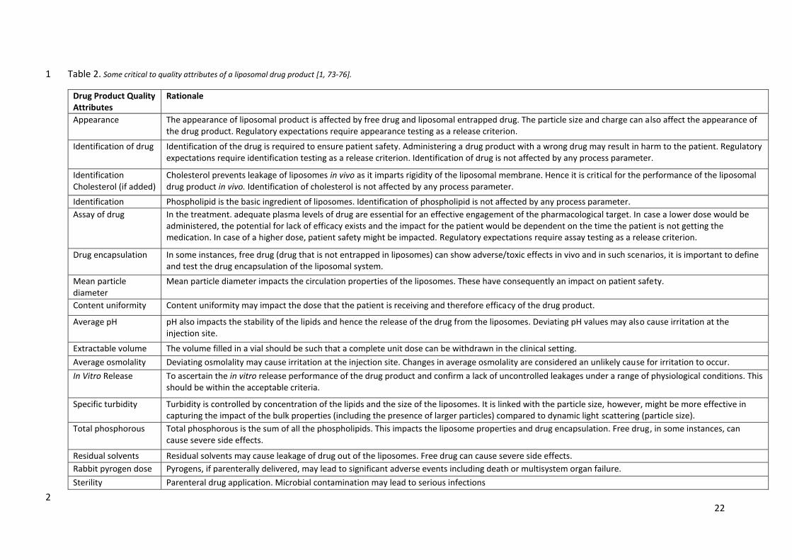
Apart from the complex large-scale manufacturing of liposomes, there are numerous 4
critical to quality attributes of a liposomal drug product that requires careful consideration as 5
it can affect the end drug product during manufacturing, storage, or its clinical performance 6
(Table 2). 7
8
9

21
# Components Process steps
(Lipophilic drug) Process steps
(Hydrophilic drug) In-process controls
Innovation impacta
1 Water for injection
Buffer salts Buffer preparation Buffer pH
2 Water for injection
Buffer salts Drug
Drug solution preparation Solution pH
3 Appropriate filter
Filtration to remove
particulates
Filtration to remove particulates Filter integrity test
4 Phospholipid Cholesterol
Ethanol Lipid dissolution
5
Phospholipid Cholesterol
Ethanol Drug
Lipid and drug
dissolution
6 Appropriate filter
Filtration to remove
particulates
Filtration to remove particulates Filter integrity test
7 Mixing Lipid hydration Lipid hydration MF
8 Polycarbonate
membrane Extrusion of MLVs Extrusion of MLVs
Liposome particle size
Zeta potential Total phosphorous
content
MF/ SA
9 Membrane
Diafiltration (To remove
ethanol)
Diafiltration (To remove ethanol and un-entrapped
drug)
Filter integrity test Dispersion pH
Total phosphorous content
SA
10 Buffer solution Post extrusion
dilution Post diafiltration
dilution
Bioburden Bulk drug product quality (pH, assay)
11 Membrane Sterile filtration Sterile filtration
Filter integrity Bulk drug product
Bioburden Assay
12 Vials Filling Filling Visual inspection
Total steps 9 9 12-15
Fig. 3. A typical large-scale liposomal manufacturing process.1
a MF is microfluidics technology and SA is self-assembled vesicular drug delivery systems. Innovation in liposome manufacturing is expected to impact these traditional liposomal manufacturing steps leading to, hopefully, a lean way of liposome production.
22
Table 2. Some critical to quality attributes of a liposomal drug product [1, 73-76]. 1
Drug Product Quality Attributes
Rationale
Appearance The appearance of liposomal product is affected by free drug and liposomal entrapped drug. The particle size and charge can also affect the appearance of the drug product. Regulatory expectations require appearance testing as a release criterion.
Identification of drug Identification of the drug is required to ensure patient safety. Administering a drug product with a wrong drug may result in harm to the patient. Regulatory expectations require identification testing as a release criterion. Identification of drug is not affected by any process parameter.
Identification Cholesterol (if added)
Cholesterol prevents leakage of liposomes in vivo as it imparts rigidity of the liposomal membrane. Hence it is critical for the performance of the liposomal drug product in vivo. Identification of cholesterol is not affected by any process parameter.
Identification phospholipid
Phospholipid is the basic ingredient of liposomes. Identification of phospholipid is not affected by any process parameter.
Assay of drug In the treatment. adequate plasma levels of drug are essential for an effective engagement of the pharmacological target. In case a lower dose would be administered, the potential for lack of efficacy exists and the impact for the patient would be dependent on the time the patient is not getting the medication. In case of a higher dose, patient safety might be impacted. Regulatory expectations require assay testing as a release criterion.
Drug encapsulation In some instances, free drug (drug that is not entrapped in liposomes) can show adverse/toxic effects in vivo and in such scenarios, it is important to define and test the drug encapsulation of the liposomal system.
Mean particle diameter
Mean particle diameter impacts the circulation properties of the liposomes. These have consequently an impact on patient safety.
Content uniformity Content uniformity may impact the dose that the patient is receiving and therefore efficacy of the drug product.
Average pH pH also impacts the stability of the lipids and hence the release of the drug from the liposomes. Deviating pH values may also cause irritation at the injection site.
Extractable volume The volume filled in a vial should be such that a complete unit dose can be withdrawn in the clinical setting.
Average osmolality Deviating osmolality may cause irritation at the injection site. Changes in average osmolality are considered an unlikely cause for irritation to occur.
In Vitro Release To ascertain the in vitro release performance of the drug product and confirm a lack of uncontrolled leakages under a range of physiological conditions. This should be within the acceptable criteria.
Specific turbidity Turbidity is controlled by concentration of the lipids and the size of the liposomes. It is linked with the particle size, however, might be more effective in capturing the impact of the bulk properties (including the presence of larger particles) compared to dynamic light scattering (particle size).
Total phosphorous Total phosphorous is the sum of all the phospholipids. This impacts the liposome properties and drug encapsulation. Free drug, in some instances, can cause severe side effects.
Residual solvents Residual solvents may cause leakage of drug out of the liposomes. Free drug can cause severe side effects.
Rabbit pyrogen dose
Pyrogens, if parenterally delivered, may lead to significant adverse events including death or multisystem organ failure.
Sterility Parenteral drug application. Microbial contamination may lead to serious infections
2
23
2.3. Potential for innovation in large scale liposome manufacturing 1
Liposomes were first reported in 1960s, and the first liposomal approved product in the 2
market was approved in 1990. Since, then in US and Europe approximately a range of 3
products been approved which use vesicular system as the basis for drug delivery. It seems 4
that, despite the numerous advantages, academic research, and innumerable peer-reviewed 5
publications on liposomes, the total number of liposome/vesicular based products in the 6
highly regulated pharmaceutical space are scarce. Despite the many advantages of liposomes 7
as a versatile drug delivery system, one of the major roadblocks for their commercialization 8
is the difficulty to have a simpler method of making liposomes at laboratory scale as well as 9
commercial scale (as discussed in Section 2.2). The current preferred method of liposome 10
manufacture is the ethanol injection method followed by extrusion of the preformed vesicles. 11
The total number of unit operations using the present method of liposome manufacturing is 12
a barrier to a robust formulation and process development. The complexity is immense and 13
hence scale up and/or technology transfer becomes a challenge. Over the years, thousands 14
of research publications have reported different methods of making liposomes which 15
includes, but are not limited to, lipid film hydration, ethanol injection and detergent removal, 16
and a technique that can reduce the overall complexity of liposomes is something that has 17
kept the formulation scientists busy. A lean way of making liposomes will make this drug 18
delivery technology a more attractive prospect in development of new chemical entities as 19
the advantages it offers can make a druggable candidate more druggable and potent, and 20
eventually benefit the patients with reduced total drug load and associated side effects. 21
As shown in Fig. 3, there are numerous steps that are executed during liposome 22
manufacturing. From, an industrial viewpoint, lipid hydration; membrane extrusion; and 23
diafiltration steps are very energy and time intensive as it requires a lot of expertise and allied 24
in process controls. Any technology that can address this problem, either by elimination or 25
modification of the extrusion process can make this a more lean and robust process. The 26
nanoprecipitation/antisolvent technique (of which LeciPlex® is an example), in which the 27
phospholipids and a stabilizer dissolved in a bio-compatible solvent spontaneously forms sub-28
micron vesicular system when exposed to aqueous environment. These are self-assembled 29
technologies that give a specific particle size and PDI for a given lipid and stabilizer 30
composition. This technology if optimized further could eliminate the extrusion and/or the 31
24
diafiltration step as the need for solvent removal is not needed and avoids altogether the use 1
of organic solvents. A different technology that can make liposomes by controlled 2
precipitation (as in microfluidics) with desired particle size. Microfluidics can eliminate the 3
need for lipid hydration and extrusion as the vesicles are formed and hydrated in the 4
microfluidic chamber itself eliminating the need for a separate extrusion step. 5
6

25
3. Regulatory overview of liposomes 1
The academical research in liposomes has been very extensive and continues to represent 2
an exciting field of science. Crommelin et al. [77] recently made an overview of commercial 3
liposomal drug product marketed in US in the EMA region and reported that 19 products 4
based upon liposome was approved for pharmaceutical purposes, of which 2 have been 5
discontinued, i.e. there is currently 17 marketed products in US and EMA region. Liposomes 6
is used mostly within oncology, but also for fungal infections, pain management, and as 7
carrier systems in vaccine products. Safety is one of the main drivers for the use of liposomes 8
in these cases [77]. 9
Bangham et al. first described liposomes in 1961 [78], while the first pharmaceutical 10
product with a liposome-based formulation was approved in 1990, i.e. 30 years later. Doxil, 11
being the first stealth liposome, was approved in 1995, and of the 4 approved stealth 12
liposomes three contains doxorubicin, i.e. conventional liposomes seem to be the preferred 13
formulation strategy for liposomes. Six of the 19 products are freeze dried, hence most is 14
presented as an aqueous dispersion, while stability do not seem to be a general issue. Vyxeos 15
was the first and only liposomal drug product to contain two active compounds, an approach 16
that could be very interesting also for other compounds with synergistic effects. 17
Of the 19 products that are or have been approved by FDA and/or EMA four contained 18
doxorubicin and two bupivacaine and 3 was three had vaccines, see Table 3, while in total 12 19
small molecules (~70%) are included in a liposome drug product. Doxorubicin liposome 20
formulations is the only generic version approved by FDA/EMA. Worldwide there is a higher 21
amount of generic liposomal products containing doxorubicin or amphotericin B outside the 22
FDA/EMA regions, see Table 4. In total 10 and 5 generic liposomal product have been 23
registered for doxorubicin and amphotericin B, respectively. Generic versions of liposomes is 24
still a debated topic in the scientific literature [79, 80], despite the many approvals. 25
The molecules included in the liposomes had a mean molecular weight on 559 ± 271 g/mol 26
spanning from 243 to 1278 g/mol, so overall aligned with the molecular range for orally 27
administered compounds, though with a shift towards the higher molecular weight range 28
often seen for orally administered compounds [81]. The average log P of the compounds is 29
1.3 spaning from -3.2 to 5.5, the average melting point is 215 °C ranging from 108 to 255 °C, 30
i.e. compounds formulated in liposomes spans very widely on these two parameters. Hence, 31

26
the versatility of liposomes for drug molecules with varied physicochemical properties is also 1
validated by commercially approved products. 2

27
Table 3. List of commercial liposomal drug products approved in US and/or EMA region. Table modified based upon data from [77]. 1
Product name
Active ingredient
Indication yeara Lipids in
formulationb Liposome
type PSD
Finished product
MW (g/mol)c
Log Pc MP
(°C) c AS
(mg/L) c
AmBisome Amphotericin Fungal infections 1990 HSPC:DSPG, chol
2: 0.8 : 1 M Conventional
< 100 nm
Freeze dried 924.08 0.8 170 750
Doxil/ Caelyx
Doxorubicin
Breast neoplasms; multiple myeloma; ovarian neoplasms;
Kaposi's sarcoma
1995 HSPC:chol:DSPE-PEG
56:39:5 M Stealth 100 nm
Aqueous dispersion
543.52 1.3 229-231 Soluble
DaunoXome Daunorubicin Cancer advanced HIV-
associated Kaposi's sarcoma
1996 DSPC: chol
2 : 1 M Conventional
40–80 nm
Aqueous dispersion
527.52 1.8 208-209 39.2
DepoCyt Cytarabine Neoplastic meningitis 1999d DOPC:DPPG Conventional 20 µm Aqueous
dispersion 243.22 -2.8 186-188
Freely soluble
Epaxal Inactivated hepatitis A
virus Hepatitis A 1999
DOPC:DOPE 75:25M
Conventional 150 nm Aqueous
dispersion na na na na
Myocet Doxorubicin Breast neoplasms 2000 EPC:chol 55:45 M
Conventional 80-90
nm Freeze dried 543.52 1.3 229-231 Soluble
Visudyne Verteporfin Sub foveal choroidal neovascularization
2000 EPG:DMPC
3:5 M Conventional
18-104 nm
Freeze dried 718.79 2.1 No data 0.013
DepoDur Morphine Pain relief 2004e DOPC:DPPG Conventional 17-23
µm Aqueous
dispersion 285.34 0.9 255 149
a Year of first approval b Abbreviations used in table: chol: cholesterol; EPC: egg phosphatidylcholine; EPG: egg phosphatidylglycerol; DEPC: 1,2-dierucoylphosphatidylcholine; DOPC: dioleylphosphatidylcholine; DOPE: dioleoly-sn-glycero-phophoethanolamine; DOPS: dioleoylphosphatidylserine; DPPC: dipalmitoyl phosphatidylcholine; DPPG: dipalmitoylphosphatidylcholine; DMPC: dimyristoylphosphatidylcholine; DSPC: distearoylphophatidylcoline; DSPE-PEG: distearoylphosphatidylcholine polyethylene glycol; DSPG; distearoylphosphatidylglycerol; HSPC: hydrogenated soy bean phosphatidylcholine; SPH: sphingomyelin; PSD: Particle size distribution; MW: Molecular weight; MP: Melting point; AS: Aqueous solubility. c Data from https://www.drugbank.ca/drugs/ d Product discontinued 2017 e Product discontinued 2014

28
Marqibo Vincristine
Philadelphia chromosome-negative
acute lymphoblastic leukemia
2009 SPH:chol
6:4 M Conventional 100 nm Freeze dried 824.97 2.8 220 3 g/L
Mepact Mifamurtide Osteosarcoma 2009 DOPC:DOPS
3:7 M Conventional 1-5 µm Freeze dried 1277.52 5.5 No data 0.0013
Exparel Bupivacaine Anesthetic 2011 DEPC:DPPG:chol:tric
aprylin Conventional
24-31 µm
Aqueous dispersion
288.43 3.1 107-108 2400
Lipodox Doxorubicin Breast neoplasms 2013 HSPC:chol:DSPE-PEG
56:39:5 M Stealth 100 nm
Aqueous dispersion
543.52 1.3 229-231 Soluble
Onivyde Irinotecan Metastatic Pancreatic
Cancer SHARE
2015 DSPC:chol:DSPE-PEG
3:2:0.015 Stealth 110 nm
Aqueous dispersion
586.68 3.2 222-223 Soluble
Mosquirix RTS, S antigen-based vaccine
Vaccination to help against malaria caused by the parasite Plasmodium
falciparum
2015 DOPC:chol Conventional 50-100
nm Aqueous
dispersion na na na na
Doxorubicin Doxorubicin Breast neoplasms 2017 HSPC:chol:DSPE-PEG Stealth 100 nm Aqueous
dispersion 543.52 1.3 229-231 Soluble
Nocita Bupivacaine Anesthetic 2017 DEPC:DPPG:chol:tric
aprylin Conventional
25-31 µm
Aqueous dispersion
288.43 3.1 107-108 2400
Vyxeos Daunorubicin
Cytarabine Acute myeloid leukemia 2017
DSPC:DSPG:chol 7:2:1
Conventional 107 nm Freeze dried 527.52 243.22
1.8 -2.8
208-209 186-188
39.2 Freely
soluble
Shingrix Glycoprotein E based vaccine
vaccine for prevention of herpes zoster
2017 DOPC:chol Conventional 50-100
nm Aqueous
dispersion na na na na
Arikayce Amikacin Mycobacterium avium co
mplex lung disease 2018 DPPC:chol Conventional 300 nm
Aqueous dispersion
585.60 -3.2 214 50000
1 2

29
Table 4. Generic doxorubicin and amphotericin B liposomal products and their manufacturer. 1
Drug Original product
Generic version Manufacturer
Doxorubicin hydrochloride
Doxil®
Adropeg 20® Axiommax Oncology Pvt. Ltd
DOXOrubicin® Dr. Reddy’s
Doxulip® United Biotech
i-dox® Getwell
Lipodox® Sun Pharma
Lippod™ Celon Labs
Natdox-LP® Natco Pharma Ltd.
Pegadria 50® Intas pharmaceutical Ltd.
Rubilong™ Zuventus Healthcare Ltd.
SinaDoxosome® Exir Nano Sina Co
Amphotericin B
AmBisome®
Abhope® Abbott
Ambilip® United Biotech
Amflight™ Celon Labs
Amphonex® Bharat serums and vaccines Ltd.
Phosome 10® Cipla
2
30
4. Advances in scalable technologies for liposome fabrication 1
As discussed in section 2.2 and section 2.3, the current manufacturing of liposomes on a 2
large-scale is a challenge as it involves a multi-step multi-test process and that the innovation 3
in this space is much needed. Before, a technology gets explored at the large/commercial 4
scale, it is important to see what has been done in the academic or basic research space to 5
understand the potential solutions and potential pitfalls before larger scale considerations is 6
initiated. A search of self-assembled technology for liposomes and microfluidics for liposomes 7
on PubMed was conducted to understand the trend in the interest of the academia and basic 8
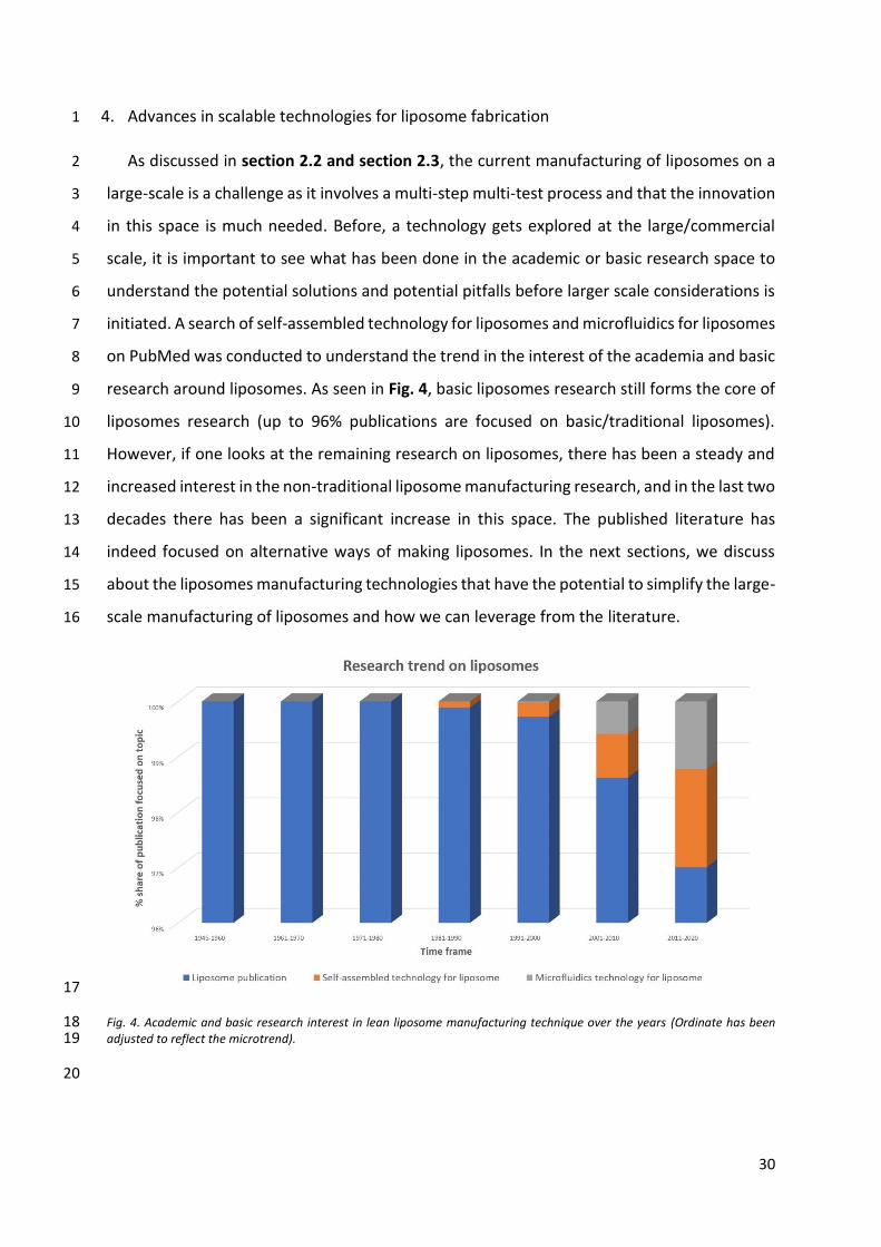
research around liposomes. As seen in Fig. 4, basic liposomes research still forms the core of 9
liposomes research (up to 96% publications are focused on basic/traditional liposomes). 10
However, if one looks at the remaining research on liposomes, there has been a steady and 11
increased interest in the non-traditional liposome manufacturing research, and in the last two 12
decades there has been a significant increase in this space. The published literature has 13
indeed focused on alternative ways of making liposomes. In the next sections, we discuss 14
about the liposomes manufacturing technologies that have the potential to simplify the large-15
scale manufacturing of liposomes and how we can leverage from the literature. 16
17
Fig. 4. Academic and basic research interest in lean liposome manufacturing technique over the years (Ordinate has been 18 adjusted to reflect the microtrend). 19
20
31
4.1. Self-assembled vesicular drug delivery system 1
There have been several advancements in the manufacturing techniques of liposomes since 2
their advent in therapeutics. Conventional methods of liposome fabrication involve hydration 3
of a thin film of phospholipids (Section 2.1.1. and Table 2) with buffer/aqueous phase to 4
render self-assembly to form vesicular structures [78]. It is necessary that the hydration 5
occurs above the phase transition temperature above which lipids exist in fluid state for self-6
assembly to take place. Other parameters include concentration of lipids, nature of lipids, 7
volume, type and ionic strength of buffer, temperature of hydration and curing, and agitation 8
time. This section deals with some recently reported novel approaches to render self-9
assembly yielding vesicular structures. 10
11
4.1.1. Heating methods 12
Nkanga and Krause et al. have recently reported liposomes encapsulating cyclodextrin 13
complexes of isoniazid conjugated pthalocyanin prepared by solvent free, easy to scale up 14
heating method [82]. The method involves the use of ethylene glycol, propylene glycol, or 15
glycerol as hydrating adjuvant. Phospholipid and the cyclodextrin-drug complex were 16
hydrated for 60 mins at room temperature with water followed by addition of adjuvant and 17
further stirring for an hour at 70℃ during which phospholipids self-assembled to form 18
liposomes encapsulating the complex. Authors reported 58-70% entrapment efficiency of the 19
cyclodextrin-drug complex in liposomes with a particle size of 150 – 650 nm. Entrapment 20
efficiency was observed to be independent of the hydrating adjuvant used. Authors, on the 21
contrary observed a greater entrapment (71%) when liposomes were prepared without the 22
hydrating adjuvant. Liposomes prepared by heating method without hydrating adjuvant 23
exhibited higher entrapment efficiency and also greater size as compared to liposomes 24
fabricated by film hydration method. Surfactant vesicles encapsulating alpha tocopherol have 25
also been recently reported by Basiri et al. employing a modification of the heating method 26
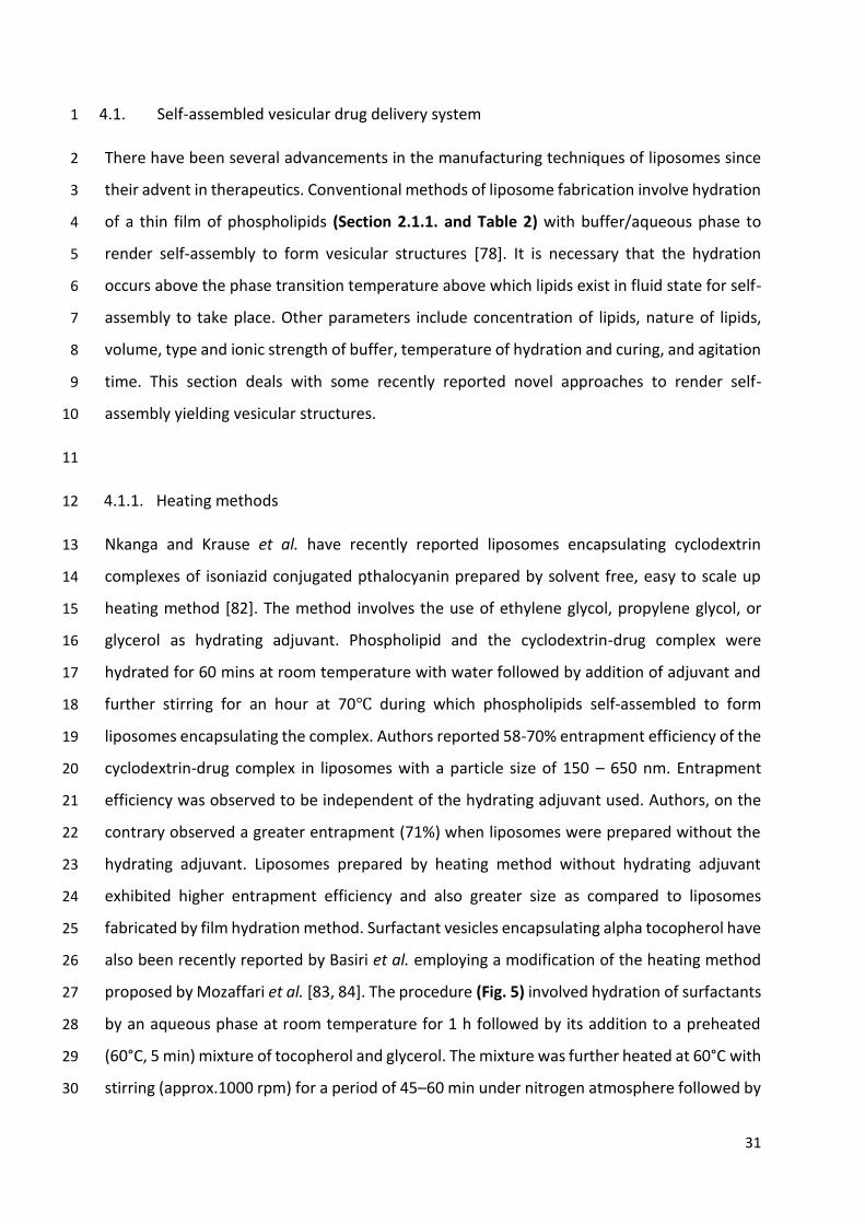
proposed by Mozaffari et al. [83, 84]. The procedure (Fig. 5) involved hydration of surfactants 27
by an aqueous phase at room temperature for 1 h followed by its addition to a preheated 28
(60°C, 5 min) mixture of tocopherol and glycerol. The mixture was further heated at 60°C with 29
stirring (approx.1000 rpm) for a period of 45–60 min under nitrogen atmosphere followed by 30

32
sonication as a size reduction step [83]. Authors prepared niosomes with different ratios of 1
Span 60, Tween 60, cholesterol, and dicetyl phosphate. They observed an increase in size with 2
decreasing hydrophilicity of lipid mixture. Around 80% tocopherol could be encapsulated in 3
the niosomes using higher ratios of Span 60: Tween 60 that increased the hydrophobicity of 4
the system. Incorporation of cholesterol to impart rigidity and dicetyl phosphate as charge 5
imparting agent was also thought to contribute to high entrapment. The method utilizes 6
principles of green chemistry, is amenable to scale up, and can be applied to encapsulate both 7
hydrophilic and lipophilic drugs. 8
9
Fig. 5. Schematic representation of heating process to fabricate liposomes. 10
11
4.1.2. Nanoprecipitation and Ionic interaction 12
Recently, Nagarsenker et al. fabricated a vesicular system termed LeciPlex® (Fig. 6) on the 13
basis of charged interaction leading to enhanced thermodynamic stability using a single step 14
fabrication procedure amenable to scale up. The procedure, a form of solvent dispersion 15
method/anti-solvent method, (Fig. 7) involved dissolution of phospholipids in a 16
biocompatible solvent like transcutol HP, that also included a charge imparting agent. The 17
biocompatible solvent phase was heated above the phase transition temperature of the lipids 18
followed by hydration with an aqueous medium at the same temperature. The resultant 19
dispersion contained self-assembled vesicular structures (Fig. 7) in the nano range with 20
excellent shelf stability [85]. The system has successfully been explored for encapsulating 21
various drugs like azelaic acid [86], carvedilol [87], idebenone [86], nelfinavir mesylate [88], 22
quercetin [89, 90], silibinin [91], and spironolactone [92]. Authors have also explored various 23
biocompatible solvents such as transcutol, ethanol, glycofurol [93]. Further, depending on the 24
hydrophilicity of the cationic/charge imparting agent, the drug was solubilized either in 25
aqueous phase or organic phase. Authors have reported the system to be very versatile since 26
it has been explored to encapsulate small molecule drugs with molecular weight ranging from 27

33
300 Da to 600 Da and with log P value from -2 to 4. Authors have also reported studies with 1
encapsulation of genetic materials like DNA [85]. 2
3
4
Fig. 6. Schematic representation of LeciPlex® system depicting its possible applications in drug delivery systems. 5
6
7
Fig. 7. LeciPlex® fabrication using a single step scalable procedure. 8
9
4.1.3. Solvent exchange 10
Buboltz et al. have devised a novel apparatus to fabricate liposomes based on rapid solvent 11
exchange [94]. The apparatus was made up of a tube containing buffer mounted on a vortexer 12
which on actuation formed the buffer into a cylindrical shell. A solution of lipids in an organic 13
solvent was injected under vacuum into an aqueous buffer so that vaporization of the solvent 14
began, along with some evaporative cooling. The vortexing buffer served as a heat reservoir, 15
transferring heat to the droplets to allow vaporization to proceed to completion [94]. Rieder 16
et al. have further optimized the protocol for DPPC MLVs by equipping the apparatus with 17
additional controls in regulating temperature, as well as pumping speed, and vortex velocity 18
[95]. Authors conclude that the mechanical forces during vortex mixing and evacuation 19
speeds have greatest effect on formation of liposomes. Rapid evaporation with high vortex 20
speeds resulted in formation of ULVs rather than MLVs. Authors inferred that rapid 21

34
evaporation resulted in rupture of lipid membranes, thereby, leading to formation of ULVs. 1
Furthermore, they also observed reducing the vortex speed from 2500 rpm to 600 rpm 2
resulted in formation of concave meniscus at the bottom instead of thin films on the surface. 3
This resulted in foaming which further reduced the evaporation rate leading to formation of 4
MLVs. The overall time for sample preparation was reported to be 4 mins. Authors also 5
observed that reducing the amount of lipids and the ratio of organic solvent to water resulted 6
in formation of ULVs over MLVs. These factors were more significant contributors when 7
samples were prepared in buffers than water. Authors have explained the formation of ULVs 8
over MLVs on basis of microscopic instabilities present in aqueous phase during liposome 9
formation. These turbulences were thought to provide nucleation points for formation of lipid 10
vesicles where self-assembly takes place. Greater microturbulences resulted in more 11
nucleation points and hence amount of lipid present per unit point reduced resulting in 12
formation of ULVs over MLVs. 13
14
4.1.4. High Shear 15
Recently, Anderson et al. have patented a protocol to prepare liposomes using high shear 16
method [96, 97]. Briefly, the process involved dispersing dried powder of lipids in a suitable 17
buffer. Using an equipment that provides high shear, the dispersion was heated to a 18
temperature above the phase transition temperature. Initially a low shear was applied to 19
avoid foaming. After the phase transition temperature was reached, mixture was stirred at 20
high shear until a desired size distribution was reached which was followed by cooling to room 21
temperature. Authors reported an average size of liposomes to be 163.2 ± 0.493 nm with a 22
PDI of 0.258. The method has been patented for fabrication of immunogenic liposomes 23
containing vaccine adjuvants [96, 97]. Shen et al. have also studied the effect of high shear to 24
a surfactant solution containing MLVs to produce ULVs. They observed the surfactant system 25
without shearing to contain ULVs and MLVs with a size range of 300 – 500 nm. 26
Homogenization of the system at 200 bar resulted in conversion of MLVs to ULVs of the size 27
range of 50 – 75 nm. Authors, however, observed an increase in size of this ULVs over 9 days 28
of storage. Cryo-TEM images revealed the ULVs to be potato shaped rather than spherical in 29
nature. Authors attributed this observation to emulsification at high pressure which caused 30
ULVs to be in a unstable state leading to increase in size after storage in a bid to reach low 31
energy state [98]. 32

35
Wang et al. have reported glass beads to produce shear to reduce size of liposomes in 1
nanoscale as opposed to conventional methods, where glass beads were only used to 2
increase the surface area for film formation. The method comprised of dissolving lipids in 3
chloroform followed by solvent evaporation by rotary evaporation to yield a thin film to which 4
aqueous solution of drug was added along with glass beads followed by prolonged shaking to 5
yield vesicles [99, 100]. Wang et.al developed liposomes in nano range (60 to 550 nm) by 6
employing glass beads of different sizes to yield shear. The size of liposomes was observed to 7
increase with the increase in diameter of glass beads used during preparation. The smaller 8
glass beads (2 mm) possessed less density and therefore did not provide adequate shear 9
forces resulting in small fraction of large vesicles (800 nm) along with small sized vesicles (100 10
nm). Increasing the time of shear to 24 h, however resulted in further lowering of size (67 nm) 11
with good PDI. Further, use of large glass beads (5 mm) lead to formation of liposomes of 12
greater size with good PDI. With increased time of shear, a reduction in size of liposomes to 13
100 nm was observed. Authors reported 3 mm and 4 mm beads to produce liposomes of 100 14
nm size with 1 h of shearing, however PDI values remained high. The authors also observed 2 15
mm and 5 mm glass beads to yield best entrapment efficiencies of amphotericin B (Up to 92%) 16
[99]. 17
18
4.1.5. Emulsification and solvent evaporation 19
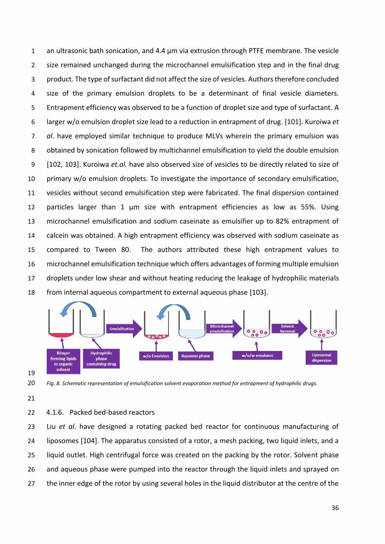
Suzuki et al. have recently reported a multiple emulsification-solvent evaporation method to 20
prepare liposomes that can yield higher entrapment efficiency for hydrophilic moieties [101]. 21
The process (Fig. 8) comprised of primary emulsification to formulate a water in-oil (w/o) 22
emulsion that contained the drug and a volatile organic solvent containing a mixture of 23
bilayer-forming lipids. This was followed by a secondary emulsification step to obtain w/o/w 24
emulsions effected by means of microchannel emulsification technique. The primary 25
emulsion was forced through channels in an aqueous phase to form the w/o/w emulsion 26
followed by evaporation of solvent to yield self-assembled lipid vesicles entrapping the 27
hydrophilic drug moieties. Authors reported the method to possess a wide control over 28
particle size range of vesicles with 0.2 µm to several micron size particles being obtained. The 29
size of the liposomes obtained was observed to be dependent upon the globule sizes of 30
primary emulsion and the technique used to effect emulsification. The mean diameters of 31
water droplets in the primary w/o emulsions was 0.2 µm with probe sonication, 1.2 µm with 32
36
an ultrasonic bath sonication, and 4.4 µm via extrusion through PTFE membrane. The vesicle 1
size remained unchanged during the microchannel emulsification step and in the final drug 2
product. The type of surfactant did not affect the size of vesicles. Authors therefore concluded 3
size of the primary emulsion droplets to be a determinant of final vesicle diameters. 4
Entrapment efficiency was observed to be a function of droplet size and type of surfactant. A 5
larger w/o emulsion droplet size lead to a reduction in entrapment of drug. [101]. Kuroiwa et 6
al. have employed similar technique to produce MLVs wherein the primary emulsion was 7
obtained by sonication followed by multichannel emulsification to yield the double emulsion 8
[102, 103]. Kuroiwa et.al. have also observed size of vesicles to be directly related to size of 9
primary w/o emulsion droplets. To investigate the importance of secondary emulsification, 10
vesicles without second emulsification step were fabricated. The final dispersion contained 11
particles larger than 1 µm size with entrapment efficiencies as low as 55%. Using 12
microchannel emulsification and sodium caseinate as emulsifier up to 82% entrapment of 13
calcein was obtained. A high entrapment efficiency was observed with sodium caseinate as 14
compared to Tween 80. The authors attributed these high entrapment values to 15
microchannel emulsification technique which offers advantages of forming multiple emulsion 16
droplets under low shear and without heating reducing the leakage of hydrophilic materials 17
from internal aqueous compartment to external aqueous phase [103]. 18
19
Fig. 8. Schematic representation of emulsification solvent evaporation method for entrapment of hydrophilic drugs. 20
21
4.1.6. Packed bed-based reactors 22
Liu et al. have designed a rotating packed bed reactor for continuous manufacturing of 23
liposomes [104]. The apparatus consisted of a rotor, a mesh packing, two liquid inlets, and a 24
liquid outlet. High centrifugal force was created on the packing by the rotor. Solvent phase 25
and aqueous phase were pumped into the reactor through the liquid inlets and sprayed on 26
the inner edge of the rotor by using several holes in the liquid distributor at the centre of the 27

37
reactor. Liposomes were manufactured by injecting methanolic solution containing lipids and 1
aqueous phase into separate inlets of the reactor at increasing temperatures maintained by 2
circulating water in the jacket of the tanks. Liposome suspension was collected from the 3
reactor outlet and then dialyzed against PBS (pH at 7.4) to remove residual organic solvent. 4
Authors observed flow rate ratio (FRR) of solvent phase to aqueous phase to be one of major 5
factors determining particle size. The flow rate of organic solvent was maintained at 20 6
mL/min and aqueous phase flow rate was increased from 20 mL/min to 300 mL/min. An 7
increase in the FRR intensified the two-phase velocity difference, thereby, enhancing mass 8
transfer and nucleation leading to formation of small size liposomes. The high gravity level 9
(HGL), a parameter, calculated by the authors as a measure of centrifugal force generated by 10
the rotor was another critical process parameter affecting liposome size. Increasing HGL also 11
resulted in enhanced micromixing and greater mass transfer as the fluids were split into thin 12
streams and tinier droplets. The temperature did not seem to affect the size of the liposomes. 13
In contrast, a slight increase in the particle size was observed from 208 nm to 232 nm when 14
the temperature was increased from 20 ℃ to 50 ℃. A further increase in temperature to 60 15
℃ did not affect the particle size. The authors attributed this particle size increase to fusion 16
of liposomes at higher temperature. An increase in entrapment efficiency of sorafenib was 17
observed on increasing the temperature from 20 ℃ to 40 ℃ after which a decrease in 18
entrapment efficiency was observed when temperature reached 60 ℃. Authors attributed 19
the reduction in entrapment efficiency to hydrolysis of phosphatidylcholine at higher 20
temperature thereby disrupting lipid bilayers and causing leakage of drug. Authors also report 21
a similar trend of FRR on entrapment efficiency. A lower FRR resulted in reduced contact 22
between the two phases with drug being retained in organic phase due to higher solubility 23
leading to low entrapment efficiency. An increase of FRR above an optimum value resulted in 24
decreased contact between lipids and drug resulting in low entrapment efficiency. Using 25
optimal conditions, authors could obtain liposomes with mean particle size of 200 nm and 26
entrapment efficiency of 89%. The authors also report an output of 33.6 kg/day of drug loaded 27
liposomes under optimum conditions thereby suggesting this technique to demonstrate high 28
potential for liposome production in large scale [104]. 29
30
31
32

38
4.1.7. Gel assisted self-assembly 1
Weinberger et al. have devised a novel method of liposome formation in solid state assisted 2
using PVA [105]. Authors prepared a 5% (w/w) PVA solution and coated microscope coverslips 3
by spreading 100–300 µL of PVA solution onto it followed by oven drying. Lipids solubilized in 4
chloroform were spread on the dried PVA film and placed under vacuum for 30 min for 5
evaporation of the solvent. Buffer was placed in a Vitrex chamber formed on cover slip and 6
GUV formation was tracked using phase contrast microscopy. Authors have utilized the 7
fabrication method to encapsulate proteins in liposomes [105]. 8
9
4.1.8. Spray drying and fluid bed drying 10
Conventional spray drying has been used by many researchers to prepare liposomal 11
dispersions. Maniyar et al. prepared liposomes using a onestep spray drying process as 12
previously reported in the literature [106]. Briefly, lipids were dissolved in methanol: 13
chloroform (1:1) solvent system to which drug was added and finally lactose was added as a 14
carrier to yield the dispersion for spray drying. The dispersion was then subjected to spray-15
drying with the inlet and outlet temperatures set to 80 ℃ and 50 ℃ respectively and feed 16
rate of 5 mL/min. The spray flow rate was set to 1.5 kg/cm2. Authors reported the liposomes 17
to be of 270 nm size with a PDI of 0.239 and an entrapment efficiency of 56.38% [106]. 18
Gala et al. have reported a novel approach utilizing methods that are industrially 19
feasible such as fluid bed coating, high pressure homogenization, and freeze-drying [107]. 20
Briefly, authors prepared pro-liposomes by spraying the ethanolic solution of 21
phosphatidylcholine onto sucrose particles in a fluid bed coater. Authors performed the 22
coating and drying at temperature as low as 30 ℃ to avoid lipid melting and thereby particle 23
agglomeration and to ensure proper spreading of wet phospholipid on the sucrose carrier 24
particles. The process was completed in two hours and yields was reported as high as 20% 25
w/w from the original weight of sucrose. The pro-liposomes were further hydrated for two 26
hours at 60 °C and freeze dried to yield the final product. Sucrose is thought to be 27
advantageous in its dual role which it plays as a carrier in the formulation of proliposomes 28
and as a cryoprotectant during freeze-drying. Pro-liposomes and liposomes generated after 29
hydration were nanosize which were further size reduced to the range of 70 – 125 nm using 30
high pressure homogenization and freeze dried. Authors report that the freeze-drying of the 31
nano-liposomes retained the size below 155 nm post reconstitution. An increase in 32

39
entrapment efficiency of beclomethasone dipropionate was also observed, probably, due to 1
an increased interaction between drug and lipids on removal of water. Presence of residual 2
ethanol was stated to cause interdigitations in liposome bilayers resulting in very poor 3
entrapment efficiencies and hence authors also suggest the drying time to be at least 2 hours 4
to evaporate all the ethanol [107]. 5
Nirale and Nagarsenker have also explored the possibility of preparation of liposomes 6
by spray drying a methanolic solution containing phospholipids and lactose dissolved in it. 7
Spray dried powder on hydration with saline yielded giant vesicles of size ranging from 800 8
nm to 6 µm while hydration with saline yielded liposomes of mean size of 3 µm [108]. 9
10
4.1.9. Solvent diffusion-based methods 11
Many researchers have attempted to advance the traditional solvent based method to render 12
it a single step process to yield the final product. In this regard, Costa et al. have reported a 13
modification of the conventional ethanol injection method [109]. The equipment consisted of 14
three pressurized tanks containing lipid solutions in ethanol, which were pumped under a 15
controlled rate to a static mixer, which ensured proper mixing of all lipids prior to it reaching 16
the injection port where the organic and aqueous streams converged. Authors reported flow 17
rate of 5 to 40 mL/min for organic phase and 60 to 400 mL/min for the aqueous phase. The 18
entire process was automated using computer algorithms where the user has to define final 19
lipid concentration and molar ratios of lipids. Liposomes that were formed were unilamellar, 20
monodispersed and possessed a size of ~25 nm to >465 nm depending on the lipid type and 21
flow rate [109]. Pulseless flow rates, Reynolds number of mixed ethanol/aqueous flow stream 22
and FVR were three parameters that determined the formation of jet and governed the PDI 23
of liposomes. Low FVR and low Reynolds number resulted in a stratified stream and limited 24
mixing leading to formation of polydisperse liposomes. High Reynolds number along with low 25
FVR lead to formation of weak jet thereby yielding polydisperse liposomes. Maintaining a high 26
FVR results in monodisperse liposomes, with size being governed by Reynolds number. 27
Further, the size was also observed to be more dependent on flow rate of aqueous phase 28
rather than lipid concentration. 29
Another novel inline method that integrates all processes involved in liposome 30
preparation has been developed by Araki et al. [110]. The equipment consists of the in-line 31
40
thermal mixing device with modified counterflow dialysis to yield in-line closed liposome 1
production system. The process comprised of dissolving lipids with aid of heat and drugs in 2
isopropanol followed by dilution with maltose and a sodium phosphate solution. This 3
dispersion was delivered to the in-line thermal mixing device. The solution during heating was 4
passed through 0.22 µm filter thereby achieving sterilization followed by cooling which 5
resulted in self-assembly of lipids to form liposomes. The heating and cooling temperatures 6
were set at 80 °C and 20 °C respectively. The dispersion was then subjected to a counterflow 7
dialysis against buffer solution to remove the organic solvent and concentrate the dispersion. 8
The liposomes were further freeze dried using polysorbate 80 as the cryo-protectant. Authors 9
obtained a monodisperse liposomal vesicles of 100 nm size using this process without an 10
additional homogenization step. Decreasing the amount of organic solvent was shown to 11
reduce the size of the liposomes due to increased hydrophobic interactions between 12
phospholipid molecules. Authors state that solubility of the lipids determines the amount of 13
organic solvent to be used which bears an influence on the size of the liposomes. Authors 14
reported the process to be scalable with a scale-up production that can be set up with a simple 15
parallel processing. Authors further reported that the process bears an aseptic production 16
capability which is amenable to complete automation without additional human intervention 17
[110]. 18
19
4.1.10. Freeze drying. 20
Recently, Liu et al. have prepared liposome using a lyophilization monophase solution 21
technique [111]. The technique as reported involves dissolving the lipids, drug, and 22
lyoprotectants in a TBA/water system followed by freeze-drying to yield the pro-liposomes. 23
On hydration, these give the liposomal dispersion. This method is a one-step process and is 24
amenable for large-scale liposome preparation. Using this technique, Liu et al. prepared 25
liposomes encapsulating glycyrrhetinic acid. Briefly, authors dissolved drug and lipids in TBA 26
at 45 °C, and lyoprotectant was dissolved in 45 °C water. When mixed at appropriate ratios, 27
these two solutions produced a third clear isotropic monophase solution. This solution was 28
sterilized by filtration and lyophilized. Prefreezing was performed for 12 h at − 40 °C followed 29
by primary drying at temperature of − 50 °C for 24 h with a chamber pressure of 1–20 Pa. 30
Authors report the entrapment efficiency and particle size of the reconstituted liposome to 31
be 72.82% and 198 nm respectively. The dispersion was stable for 6 months at 25 °C. Authors 32
41
observed that entrapment efficiency was affected by amount of phosphatidylcholine, amount 1
of cholesterol, and volume percentage of TBA; while particle size was observed to be more 2
dependent on volume percentage of TBA. Further, sublimation rate during lyophilisation was 3
observed to increase with increasing volume percentage of TBA [111]. 4
5
4.1.11. Supercritical fluid techniques 6
Recently, researchers have also employed supercritical fluids to assist in liposome preparation 7
[112]. The process is called as Supercritical assisted Liposome formation (SuperLip) [113]. 8
Lipids were dissolved in ethanol which were then mixed with pure carbon dioxide in a 9
saturator to obtain an expanded fluid. The saturator was filled with baffles, maintained under 10
high pressure and thermally heated by thin bands to produce the supercritical fluid. The 11
mixture was fed to a high-pressure formation vessel to which an aqueous solution in an 12
atomized form containing the drug was also introduced. The working temperature of the 13
saturator and the formation vessel was set to 40 °C and pressure to 100 bar. The liposome 14
suspension was recovered from the bottom of the vessel; a decompression step was used to 15
separate carbon dioxide and ethanol using a stainless-steel separator which was maintained 16
at 30 °C and 10 bar. The technique has been explored to encapsulate hydrophilic drugs like 17
theophylline where up to 98% entrapment efficiency has been reported. Multilamellar 18
liposomes of ~200 nm size with >90% entrapment efficiencies for lipophilic drugs have been 19
reported by this technique. The encapsulation efficiency was observed to depend on flow rate 20
of the aqueous solution with reduced flow rates resulting in higher values of entrapment. 21
Higher flow rates of aqueous phase caused greater velocity of droplets after atomization, and 22
thereby, impacted the disruption of the droplets on the receiving container at the bottom. 23
This resulted in lower entrapment efficiencies of theophylline. Lower flow rates in contrast 24
produced bigger droplets resulting in dispersion with smaller mean size but larger size 25
distribution. Trucillo et al. have also investigated this technique to prepare liposomes with 26
three antioxidants, Farnesol, Linalool, and limonene [114]. Lipid soluble antioxidants were 27
loaded in a stabilized emulsion which was atomized and introduced in the formation vessel 28
containing the ternary mixture viz. CO2/ethanol/Lipids to obtain the formation of liposomes. 29
Farnesol, linalool, and limonene were the anti-oxidants investigated for encapsulation. Two 30
approaches were used for encapsulating these molecules in liposomes. In the first approach, 31
the lipids along with anti-oxidants were dissolved in ethanol while in second approach, lipid 32

42
soluble antioxidants were dissolved in isopropyl myristate and loaded in a stabilized emulsion. 1
The aqueous phase/emulsion phase was atomized and introduced in the formation vessel 2
containing the ternary mixture viz. CO2/ethanol/Lipids to obtain the formation of liposomes. 3
Authors attribute various parameters such as atomization in a high-pressure environment, 4
turbulent shear forces, and viscosity of the medium to affect liposome formation and the size. 5
Authors report liposomes to be smooth, homogenous with a size range of 300 – 600 nm and 6
entrapment efficiency of >90% prepared using this technique [114]. Table 5 list array of/ 7
number of all the techniques that involve self-assembly of liposomes. 8

43
Table 5. Comparison of different methods based on self-assembly with respect to manufacturing and scalability. 1
Method Process Parameters to be controlled Size Range Drug type Liposome
type Scalability Comments
Conventional Thin film hydration
• Hydration time
• Hydration temperature
• Sonication time.
>1 µm Lipophilic GUVs Presently
used commercially
• Multi step process.
• Homogenization/Extrusion is required post fabrication for size reduction
• Sterilization operation post fabrication/aseptic processing is required.
• Can be “tens of” to “hundreds of” litres on large scale
Conventional Ethanol injection
• Rate of injection
• Volume of solvent/aqueous phase
• Agitation rate
>100 nm [54] Lipophilic & hydrophilic
[20] SUVs, MLVs
Presently
used
commercially
• Pilot plant scale design using this method has been reported with liposomes [54].
• Removal of ethanol using rotary evaporator is an added step.
• Up to 3L batch has been prepared [54].
• Can be “tens of” to “hundreds of” litres on large scale
Heating methods • Temperature
• Time of hydration and self-assembly >1 µm Lipophilic GUVs +++
• Multiple step process.
• Size reduction is needed post fabrication to attain desired size.
Nanoprecipitation
• Volume of aqueous and organic phase
• Agitation rate and time
• Rate of addition
• Temperature of operation
200 to 500 nm [85]
Lipophilic GUVs, OLVs +++++
• Simple fabrication procedure.
• Use of biocompatible solvent is an advantage
• Size reduction operation may be required subsequently
• Sterilization operation post fabrication/aseptic processing is required.
Solvent exchange method
• Temperature and pressure during operation
• Pumping and vortexing speed
• Volume of solvent
>1 µm Lipophilic MLVs +
• Rapid process
• Polydispersity of product is high.
• Size reduction techniques are needed post fabrication to attain desired size.
High Shear method
• Temperature
• Time of heating
• Shear force (e.g. speed of rotor)
~200 nm (good PDI)
[98]
Charged drugs
SUVs +++++ • Simple easy to scale up technique
• Avoids use of organic solvents.
Emulsification evaporation
• Homogenization technique to prepare primary emulsion
• Microchannel emulsification equipment dimensions
>1 µm Lipophilic & hydrophilic
MLVs, GUVs [71]
+++
• Multi step process.
• Leakage of hydrophilic drug during fabrication may result in less entrapment.
• Size reduction/sterilization operation is required post fabrication.

44
• Flow rates of primary emulsion and aqueous phase to yield secondary emulsion
Packed Bed Reactors using high gravity
technology
• Flow rate of aqueous and organic phases
• Temperature
• Type of packing
• Rotational speed of the reactor
~100 nm (good PDI)
[115] Lipophilic SUVs +++
• Reduced number of unit operations
• Provides liposomes of desired size
• Removal of residual solvent is necessary before further processing.
Gel assisted methods • Surface of gel formed
• Temperature set during process >1 µm [105]
gene/peptide/monoclonal antibodies?
GUVs + • Easy process yields rapid formation of GUVs
without need for special equipment
• Scale up may be challenging
Spray drying/Fluid bed coating
• Inlet and outlet temperature
• Spray flow rate
100 nm to >1 µm [106-108]
Lipophilic MLVs +++ • Process yields pro-liposomes;
• Yields are however low.
Freeze drying
• Pre-freezing time and temperature
• Primary drying conditions
• Secondary drying conditions
~200 nm (good PDI)
[111] Lipophilic SUVs +++
• Pre-filtration of solution followed by lyophilisation can result in sterile final product with reduced operations.
Modified solvent-based methods
• Feed rates of organic and aqueous phase
• Temperature during operation.
50 to 500 nm [109, 110]
Lipophilic SUVs +++++
• Process has a smaller number of unit operations to yield desired liposomal size
• Ease of sterilization is advantage.
• Removal of residual solvents is necessary from final product.
• Up to 4L batches have been attempted [110].
Supercritical fluid assisted fabrication
• Temperature and Pressure of formation cell and separator chambers
• Feed rates of carbon dioxide and aqueous solutions
• Dimensions of nozzle to produce atomization.
100 to 200 nm (good PDI) [114]
Lipophilic & hydrophilic
MLVs +++
• Process can be used to encapsulate hydrophilic and lipophilic drugs
• Process has a smaller number of unit operations but is complex as compared to other techniques
• Sterilization may be achieved by pre-filtration of solutions.
1

45
4.2. Microfluidics 1
The application of microfluidics for the manufacture of liposomes has gained considerable 2
academic interest over recent years, as shown in Fig. 4. Microfluidics offers the ability to 3
consistently produce optimised, uniform nanoparticles [116]. The production can be scale-4
independent [117], allowing translation of formulations from laboratory to GMP [118], 5
addressing many of the limitations of traditional bulk production methods. In liposome 6
production, microfluidics can replace the lipid hydration and extrusion steps during liposome 7
production and replace it with a single-step process for liposome production where particle 8
size is process controlled (Fig. 3). The ability to achieve this is related to the process offering 9
the miniaturisation of the fluidic mixing environment; by using intersecting microchannels, 10
nanolitre volumes of fluids are mixed in a highly controlled format [119, 120]. A key feature 11
of many of the microfluidics systems is the generation of laminar flow, which is challenging to 12
produce in macroscale systems. Using laminar flow, nanoprecipitation can be easily 13
controlled. Single phase mixing systems are the most commonly adopted due to process 14
simplicity. These systems control the mixing of two or more miscible solvents (commonly an 15
aqueous phase mixed with a water miscible alcohol such as methanol, ethanol or 16
isopropanol). During the mixing, the change in polarity promotes nanoprecipitation and the 17
formation of liposomes [121] (Fig. 9). This process can also be referred to an ‘anti-solvent 18
approach’ [122]. A hypothesis for self-assembly of liposomes resulting from this process was 19
proposed by Zook and Vreeland [123]. Within their model, as the alcohol and aqueous phase 20
mix, the polarity of the mixture increases. With this increase in polarity, the lipids become 21
progressively less soluble and the lipid monomers self-associate into planar bilayer discs. As 22
these discs increase in size, this increases the surface area of the hydrophobic chains around 23
the edge of the discs that is in contact with the polar solvent. To reduce this hydrophobic 24
surface area, the discs bend and form spherical liposomes [123]. By incorporating water 25
soluble drugs in the aqueous phase and/or lipid soluble drugs in the solvent phase the drug 26
can be simultaneously incorporated into the liposomes. Using this method, the ability to 27
entrap small molecules [124, 125], nucleic acids, [126] and proteins [17] within liposomes 28
have been demonstrated. Further, it has been demonstrated that the method is a scale-29
independent production of liposomal adjuvants [15, 127, 128]. 30
31

46
4.2.1. Microfluidic cartridge design 1
Fluid flow can occur in two different ways: laminar flow or turbulent flow. The type of flow 2
produced depends on the velocity and viscosity of the fluid and mixing in macroscopic flow is 3
generally turbulent [129]. However, microflows are more commonly laminar, and mixing 4
under standard conditions involves molecular diffusion processes only, which is inefficient 5
[129-131]. Therefore, to address this and enhance mixing efficiency, micromixers have been 6
optimised with regard to channel geometry and architecture such that effective mixing can 7
be achieved within short mixing channels and with high throughput [132]. Indeed, a key factor 8
in the formation of liposomes using microfluidics is the residence time within the mixing 9
chamber as well as the geometry of the microfluidic mixer as this controls the rate of 10
nanoprecipitation and vesicle formation. 11
A wide range of micromixers have been developed and tested for liposome 12
production, including toroidal mixers, staggered herringbone mixers, T-mixers and 13
hydrodynamic flow focusing (See Fig. 9 and Table 6). For example, hydrodynamic flow 14
focusing can be achieved in 2D or 3D. With the 2D device, fluid is injected concurrently 15
through three inlets. A central stream of a water-miscible solvent containing lipids is focused 16
horizontally by aqueous fluid streams introduced perpendicular to the central stream [133, 17
134]. With this system, lipid aggregation and liposome formation can occur at the wall of the 18
microfluidic channel [134]. This can present issues in particle size control and micromixer 19
fouling/blocking [133, 135]. To address this, 3D hydrodynamic flow focus mixing has been 20
developed. Within this system, the alcohol solvent stream is introduced by a capillary 21
surrounded by the aqueous stream. This avoids aggregation of the nanoparticle components 22
at the walls of the mixer [134, 136]. Hood et al., demonstrated the use of hydrodynamic flow 23
focusing to produce liposomes and within their microfluidic cartridge they also included 24
microdialysis for buffer exchange and establishment of a pH gradient which supported drug 25
loading of doxorubicin to give complete on-chip production [137]. 26

47
1
Fig. 9. Schematic representations of example micromixer cartridge designs that can be used. A) a toroidal mixer with planar 2 geometry employing centrifugal forces to encourage uniform mixing, b) the staggered herringbone micromixer with 3 embossed chevrons, c) a basic T-mixer with two inlets where fluids are forced into a T junction and d) hydrodynamic flow 4 focusing with three inlets where a central stream of solvent is focused by aqueous fluid streams either in 2D or 3D. 5
6

48
Table 6. Examples of microfluidic mixers used to produce liposomes. Table modified based upon data from [117]. 1
Microfluidic architecture
Formulationa Entrapped material
Loading Particle size
Reference
Toroidal mixer DSPC:Chol
DSPC:Chol:PS DOPE:DOTAP
OVA OVA
PolyA
26 - 36 % 15 – 25 %
95 – 100 %
50-60 100-120
40-60 [117]
Staggered herringbone
mixer
POPC:cholesterol Doxorubicin 60 to 100 %
depending on formulation
20 – 30 [138]
DLinkE2-DMA:DSPC:Chol:PEG-DMA
si-RNA 95 – 100 %
30 - 55 [139]
DSPC:Chol Metformin
and Glipizide 20 – 25 % 38 – 44 %
50 - 60 [124]
DMPC:Chol / DSPC:Chol Atenolol and
quinine 100%
50 – 80 % 200 – 360 [140]
Tween85:Chol:DDA siRNA Not
determined 70 – 230 [141]
ATX:DSPC:Chol:DMG-PEG:PEG2000
si-RNA Not
determined 40 – 50 [142]
T-mixer Triolein:POPC:DSPE-PEG2000 Iron oxide 0.43 w/w 35 - 140 [143]
Hydrodynamic flow focussing
DMPC:Chol:DPPE-PEG2000 Doxorubicin Up to 72%
80 - 190 [137]
2
To increase the efficiency of mixing, the contact area between the aqueous and 3
solvent phases can be increased through appropriate microchannel configuration [132]. 4
Passive mixers, such as the staggered herringbone mixer, have been developed to achieve 5
this and have been used to manufacture a range of liposome formulations (Table 6). The 6
staggered herringbone design has an in-groove pattern in the microchannel with an 7
asymmetric herringbone shape [144, 145]. As a result of this structure, the fluid streams are 8
passed over a series of protruding herringbone structures causing chaotic flow. This creates 9
transverse vortices that are repeatedly changed because of the asymmetric geometry [146]. 10
This micromixer construct can be used to produce size-controlled liposomes with their size 11
being tightly defined by process parameters [138]. However, the complex structure of this 12
micromixer present practical limitations in terms of fabrication costs, and throughput speeds 13
[117]. To address this, an alternative design based on a toroidal mixer design has been 14
developed. This micromixer promotes laminar flow at high fluid speeds by using circular 15
a Abbreviations. DSPC: 1,2-distearoyl-sn-glycero-3-phosphocholine; Chol: Cholesterol; PS: phosphatidylserine; DOPE: 1,2-dioleyl-sn-glycerol-3-phosphoethanolamine; DOTAP: 1,2-dioleoyl-3-trimethylammonium-propane; POPC: 1-palmitoyl-2-oleoyl-sn-glycero-3-phosphocholine; DLinkE2-DMA and ATX: proprietary lipids; PEG-DMA: N-[(methoxy poly(ethylene glycol)2000)carbamyl]-1,2-dimyristyloxlpropyl-3-amine; DMPC: 1,2-dimyristoyl-sn-glycero-3-phosphocholine; DMG-PEG2000: 1,2-Dimyristoyl-sn-glycerol, methoxypolyethylene glycol-2000; DSPE-PEG: 1,2-distearoyl-sn-glycero-3-phosphoethanolamine-N-[methoxy-(polyethylene glycol)-2000]; DPPE-PEG2000: 1,2-Dimyristoyl-sn-glycerol methoxypolyethylene glycol -200.

49
structures within the flow path. This induces chaotic advection through increasing the 1
number of vortices and centrifugal forces created between the columns within the cartridge, 2
allowing for improved mixing and higher throughput [131]. Using this new toroidal 3
micromixer design, the production of liposomes from laboratory-scale to GMP scale was 4
demonstrated [117]. 5
In addition to reduced complexity and time for manufacture, the use of microfluidics 6
offers further advantages. For example, size-tuned production of liposomes (from 40 to 500 7
nm depending on the formulation and process parameters e.g. [17, 138, 148]) with low 8
polydispersity (often below <0.1 PDI) can be achieved through the control of process 9
parameters. The use of microfluidics has also been shown to promotes higher loading within 10
liposomes compared to other commonly used methods; for example, a forty-fold increase in 11
iron oxide loading was achieved using a T mixer compared to other methods [143]. A five to 12
ten-fold increase in protein loading was also achieved when microfluidics was adopted as the 13
manufacturing process compared to liposomes produced by lipid hydration and sonication or 14
extrusion [17]. This was achieved with small homogenous (50 – 60 nm; <0.2 PDI) liposome 15
formulations that could be scaled from bench scale to GMP production rates [117]. An 16
additional advantage of this microfluidic production method is that it can be conducted at 17
ambient temperture negating the need to work above lipid transition temperatures, as is 18
normally needed for lipid hydration based methods [17]. In the case of complexation of 19
nucleic acids within lipid nanoparticles, whilst loading is generally 100% irrespective of the 20
production method used, microfluidics offers controlled complexation and size tuned 21
production (e.g. [139]). With liposomes where high drug loading is achieved using active 22
loading (e.g. doxorubicin loading), microfluidics offer the opportunity to support liposomal 23
drug formulations in a reduced time, with minimal reagent waste [137]. 24
25
4.2.2. Microfluidic material and production parameter considerations 26
During the production process, the focus is on optimising the key driving forces of vesicle 27
assembly including the component solubilities, concentrations and process parameters. As 28
with the other manufacturing methods discussed, the selected process parameters can 29
impact on the end product attributes which includes size, size distribution, lipid composition, 30
and drug loading/release characteristics (Table 7). 31
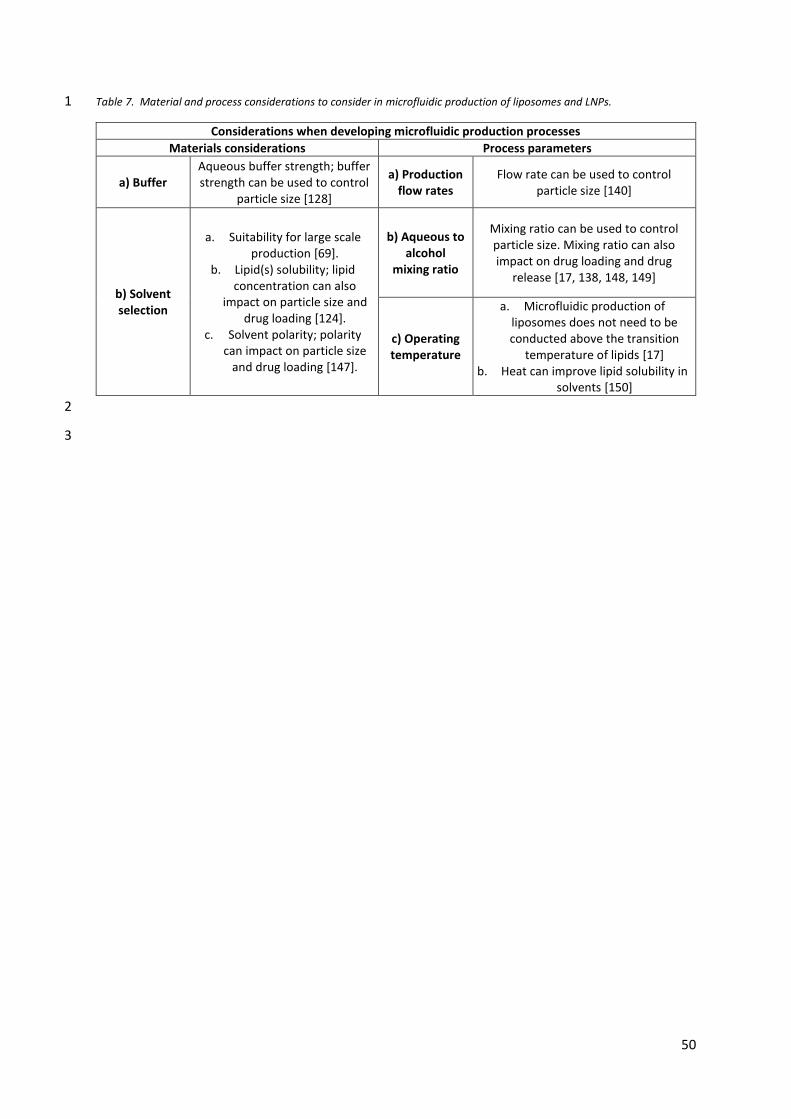
50
Table 7. Material and process considerations to consider in microfluidic production of liposomes and LNPs. 1
Considerations when developing microfluidic production processes
Materials considerations Process parameters
a) Buffer Aqueous buffer strength; buffer strength can be used to control
particle size [128]
a) Production flow rates
Flow rate can be used to control particle size [140]
b) Solvent selection
a. Suitability for large scale production [69].
b. Lipid(s) solubility; lipid concentration can also
impact on particle size and drug loading [124].
c. Solvent polarity; polarity can impact on particle size
and drug loading [147].
b) Aqueous to alcohol
mixing ratio
Mixing ratio can be used to control particle size. Mixing ratio can also impact on drug loading and drug
release [17, 138, 148, 149]
c) Operating temperature
a. Microfluidic production of liposomes does not need to be conducted above the transition
temperature of lipids [17] b. Heat can improve lipid solubility in
solvents [150]
2
3

51
4.2.2.1. Aqueous buffer selection 1
When considering the initial fluid attributes, the choice of aqueous phase, alcohol phase and 2
lipid concentration all impact on the liposome product attributes. For example, recent work 3
[128] has shown that by controlling the aqueous buffer concentration, the particle size of 4
cationic and anionic liposomes could be controlled. Through controlling the buffer 5
concentration of Tris in the aqueous phase, highly monodisperse, cationic liposomes at 6
selected size between 40 nm and 500 nm were produced. In vivo biodistribution studies in 7
mice also showed that by using this method to creating small (<50 nm) liposomes, the 8
clearance rates of these liposomes from the injection site was increased and increased 9
accumulation to the draining lymphatics promoted. 10
11
4.2.2.2. Alcohol solvent selection 12
The alcohol selection in the production process is a key factor to consider as the suitability for 13
scale-up manufacturing and the solubility of the selected lipids in the solvent must be 14
considered. Working with solvents that have low toxicity potential and defined as class 3 in 15
the ICH Q3C (R6) [69] (e.g. ethanol and IPA) is preferable followed by those in class 2 (e.g. 16
methanol). However, the choice of solvent can have an impact upon liposome attributes 17
[147]. Results show that in general, reducing the polarity of the solvent (e.g. by replacing 18
methanol with isopropanol) increased the liposome particle size and reduced drug loading. 19
However, the choice of solvent did not impact on liposome short-term stability or drug release 20
characteristics. By using solvent combinations such as methanol/isopropanol mixtures to 21
modify solvent polarity, the resultant liposome particle size was also similarly modified. 22
However, not all liposome formulations were sensitive to the impact of solvent choice; 23
liposomes containing charged lipids and formulations containing increased concentrations of 24
cholesterol or pegylated-lipids were less sensitive to solvent choice [147]. In the proposed 25
model of liposome self-assembly during microfluidics [123], vesicle size is determined by two 26
factors: the growth rate of planar bilayer discs and 2) the rate the discs close into spherical 27
vesicles. As the alcohol and buffer mix, polarity of the mixture increases and lipid discs form. 28
Thus, depending on the polarity of the alcohol adopted, the overall polarity of the mixture 29
running through the micromixer can be reduced. Thus, if the solvent is switched from 30
methanol to isopropanol, this may result in larger lipid discs forming (Fig. 9) and subsequently 31
larger liposomes form. In terms of lipid concentration, some studies suggest that low initial 32
52
lipid concentrations tend to promote larger and more heterogeneous liposome suspensions 1
and generally, at higher initial lipid concentrations there is no impact (e.g. [17, 127]). 2
However, the relationship between lipid concentration and particle size is not clear and it 3
maybe formulation and microfluidic cartridge design dependent. For example, in studies 4
using single hydrodynamic focusing, increasing lipid concentrations increased particle size 5
[151, 152]. This trend was also shown with the production of LNPs using a chaotic mixer [153]. 6
Within this study [153], that authors note the size of lipid discs formed (and the subsequent 7
size of vesicles) is controlled by the rate of mixing, which controls the ethanol concentration 8
and mixture polarity. With a more rapid reduction in ethanol concentration, smaller vesicles 9
formed [153]. The authors also propose that at high lipid concentrations, larger lipid discs 10
form at the saline-ethanol interface promoting larger LNP formation [153]. 11
12
4.2.2.3. Process parameters – aqueous to alcohol mixing ratio 13
Like the impact of solvent selection, the rate of mixing of the aqueous and alcohol during the 14
production of liposomes impacts on liposome attributes. Generally, low mixing ratios tend to 15
form larger vesicles; for example studies by Kastner et al. using liposomal formulation 16
DOPE:DOTAP and PC:Chol on a staggered herringbone micromixer chip demonstrated a 17
reduction in average liposome size (from 200 nm to 50 nm) as the alcohol content in the 18
mixture reduced from 50% to 17% [125, 126]. Jahn et al. also showed that as the flow rate 19
ratio increased (and the alcohol content in the mixture reduced), the resulting particle size 20
decreased [121]. Zizzari et al. also demonstrated this size-control effect for the liposomal 21
formulation HSPC:Chol:DSPE-PEG-2000 produced over a range of flow rate ratios [154]. There 22
are two possible rationales why the polarity gradient produced can be used to control 23
liposome particle size. Jahn et al. [155] propose that during microfluidic mixing, the initially 24
formed liposomes may take-up alcohol within the bilayer. This can promote in some partial 25
disassembly of the liposomes. As the solvent phases continue to mix, then the alcohol 26
concentration in the liposomes will decrease, resulting in re-assembly. When the flow rate 27
ratio is such that the overall alcohol concentration is low, this may limit the assembly / re-28
assembly cycle and reduce liposome size [155]. Alternatively, Zizzari et al. note that at higher 29
flow rate ratios, a smaller solvent stream results and as the lipid discs formed at the liquid 30
interface, they may bend and forming liposomes more rapidly in the presence of decreasing 31
solvent concentration. The length of time these lipid discs can grow will directly impact upon 32

53
the final resulting particle size, with shorter times leading to smaller liposomes. Thus at high 1
alcohol content flow rate ratios (e.g. 1:1) the longer the time available for lipid discs to expand 2
and for larger vesicles to form [156]. Interesting the mixing flow rates was also shown to 3
impact on drug loading and drug release properties, with lower alcohol concentrations in the 4
flow mix producing smaller liposomes, with lower protein loading and higher release rates 5
[17]. 6
7
4.2.2.4. Process parameters – flow rate 8
The operating flow rate adopted in microfluidics is an important consideration as it will dictate 9
production speeds. The flow rate though the micromixer can have an impact on liposome 10
size; however, this tends to be micromixer and formulation dependent. For example, Sedighi 11
et al. [157], use design of experiments to rapidly screen and optimise various liposomes 12
formulations. They tested the impact of both flow rate ratio and total flow rate. From their 13
studies using a staggered herringbone mixer, they reported that the flow rate ratio had a 14
significant impact on particle size and size distribution (as covered in section 4.2.1.3), whilst 15
liposome characteristics remained constant at flow rates above 8 mL/min. We have also 16
recently shown the ability to produce liposomes with mapped characteristics (size, 17
polydispersity and drug loading) with flow rates from 12 to 200 mL/min using a toroidal mixer 18
[117]. However, with cationic liposomes prepared from dimethyldioctadecylammonium 19
bromide (DDAB) and trehalose 6,6′-dibehenate (TDB), their vesicle size was shown to be 20
controlled by both the flow rate ratio and total flow rate [127]. With these systems, increasing 21
the flow rate and flow rate ratio was shown to reduce sizes from around 1000 nm (1:1 flow 22
rate ratio and 5 mL/min flow rate) down to 160 nm (5:1 flow rate ratio, 20 mL/min flow rate). 23
However, due to their cationic nature the different liposome sizes showed similar protein 24
loading and clearance from the injection site after intramuscular injection [127]. 25
26
4.2.2.5. Process parameters – operating temperature 27
In many liposome production methods, liposome manufacture is undertaken at temperatures 28
above the transition temperature of the lipid bilayer [158]; for example, DSPC liposomes are 29
commonly prepared above 55oC. This can present issues for thermos-liable drugs, particularly 30
proteins. However, when microfluidics is employed, liposomes can be produced at ambient 31
temperature irrespective of their lipid composition [17, 117]; studies have shown that using 32

54
microfluidic production, liposomes produced at room temperatures or at temperatures above 1
the main lipid transition temperature are similar in size with DSPC:Cholesterol liposomes of 2
the same size being produced at operating temperatures from 20 to 60oC. This demonstrates 3
there is no requirement to work above the lipid transition temperature during the microfluidic 4
manufacturing process [17]. This may be a result of alcohol being present within the liposomal 5
membranes during the initial production stage. When interacting with membranes, ethanol 6
and other short chain alcohols can locate in the headgroup region where the hydrogen bonds 7
between the alcohols and the phosphate and carbonyl groups of the lipids can form [159, 8
160]. This changes the packing in the lipid membrane and can increase membrane fluidity 9
[161]. Fatty alcohols have also been used as an alternative to cholesterol in the formulation 10
of liposomes and can reduce the transition temperature of liposome bilayers in a similar 11
manner to cholesterol [162]. However, it can be useful to use elevated temperatures to 12
improve the solubility of some lipids in solvents during the processes irrespective of their Tc 13
(e.g. [127]). 14
15
4.2.2.6. Scale-independent production, continuous manufacturing using microfluidics and 16
down-stream processing considerations. 17
Microfluidics offers the advantage of being scale-independent. This allows rapid optimisation 18
of liposomes characteristics to be undertaken using a design-of-experiment approach [156], 19
followed by translation of the formulation and production parameters from laboratory 20
production through to continuous manufacturing (which can be described as a processing 21
concept whereby product constantly flows out). Continuous manufacturing has a long history 22
in the non-pharmaceutical industries and has been adopted for active pharmaceutical 23
ingredients and solid oral dosage forms such as ORKAMBITM, PREZISTA®, VERZENIOTM and 24
SYMDEKO®. The advantage of continuous manufacturing includes, lower capital expenditure, 25
smaller factory footprint and lower Cost of Goods. For lower volume production, semi-26
continuous can also be adopted. Continuous manufacturing has evolved from bulk drug, to 27
solid dosage forms through to more complex biologicals. Therefore, the transition of this to 28
complex formulations is the next step. The development of down-stream processing can also 29
support this and selected relevant processing techniques and principles can be applied to the 30
production of liposomes. Liposome manufacturing has some inherent aspects that make 31
scalable manufacture and continuous production appropriate. Liposomes have some 32
55
elements of solid oral products (drug manufacturing/sourcing/supply chain) and some from 1
biopharma (mixing vessels, tangential flow filtration, filtration etc) and some unique elements 2
(particle size, size distribution and drug loading) [163]. For example, the application of TFF 3
can be applied to liposomal manufacture. TFF offers two key processing steps for liposomes 4
1) purification (removal of solvent and/or free drug) and 2) concentration adjustment. TFF 5
processes can be run in a recirculating loop or as a single pass format, where multiple TFF 6
cassettes are run in series. This can offer lower system hold-up volumes. After purification of 7
the system, sterilisation can be achieved by incorporating in-line sterile filtration as part of 8
the process train. 9
As part of a continuous or semi-continuous production process, process analytical 10
technology should be included to analyse and control the manufacturing process and to 11
monitor the liposome critical quality attributes. Microfluidics offers opportunities in terms of 12
design flexibility, process control and parameter predictability. It also offers ample 13
opportunities for modular production setup, process feedback and process control [164]. This 14
requires rapid in-line or at-line methods to monitor product attributes such as particle size 15
analysis and drug loading. However, currently there is no process analytical methods available 16
for microbial contamination, so sterility assurance would need to be assured through the 17
design and validation of the system. For batch production, this can be achieved by testing a 18
single bulk sample prior to filling and capping. If continuous filling is used, then a 19
representative bulk is not applicable. Therefore, further developments are needed in these 20
areas to allow fully continuous production to be adopted. 21
22

56
5. Conclusion 1
Liposomes, and lipid-based nanomedicines play a key role in healthcare. Their ability 2
to protect, deliver and target drugs provides enhanced efficacy and reduced toxicity, which 3
have been explored for both classical small molecules as well as for RNA delivery. However, 4
as a complex drug product their manufacture presents challenges with multi-batch processes 5
commonly used. This limits their wider application and challenge their general applicability as 6
it complicates the development of a robust, scalable and affordable process, that may 7
challenge the value proposition of the product in some diseases and regions. With the 8
development of new manufacturing processes, which offer leaner manufacturing and scale-9
independent manufacture, the application of liposomes and other lipid-based nanomedicines 10
can be more readily translated from the pre-clinical research through to production and 11
clinical use. To support this, rapid on-line and at-line analytical tools are required that can 12
support the characterisation and quality assurance of the drug product. 13
57
Acknowledgements 1
This research did not receive any specific grant from funding agencies in the public, 2
commercial, or not-for-profit sectors. 3
4
CRediT 5
All authors (SS, VD, RH, MSN, YP) contributed to the conceptualisation, visualization, writing 6
– original draft, writing – reviewing and editing of this manuscript. 7

58
6. References 1
[1] E.m. agency, Assessment report: Onpattro, in, European medicines agency, 30 Churchill 2
Place, Canary Wharf, London E14 5EU, United Kingdom, 2018, pp. 188. 3
[2] R. Dutta, R.I. Mahato, Recent advances in hepatocellular carcinoma therapy, 4
Pharmacology and Therapeutics, 173 (2017) 106-117. 5
[3] B. Yu, X. Zhao, J.L. Lee, R.J. Lee, Targeted delivery systems for oligonucleotide therapeutics, 6
AAPS Journal, 11 (2009) 195-203. 7
[4] H.B. Newton, Advances in strategies to improve drug delivery to brain tumors, Expert 8
Review of Neurotherapeutics, 6 (2006) 1495-1509. 9
[5] J.O. Eloy, R. Petrilli, L.N.F. Trevizan, M. Chorilli, Immunoliposomes: A review on 10
functionalization strategies and targets for drug delivery, Colloids and surfaces. B, 11
Biointerfaces, 159 (2017) 454-467. 12
[6] V. Agrahari, V. Agrahari, A.K. Mitra, Nanocarrier fabrication and macromolecule drug 13
delivery: challenges and opportunities, Therapeutic delivery, 7 (2016) 257-278. 14
[7] A.S. Jain, P.N. Goel, S.M. Shah, V.V. Dhawan, Y. Nikam, R.P. Gude, M.S. Nagarsenker, 15
Tamoxifen guided liposomes for targeting encapsulated anticancer agent to estrogen 16
receptor positive breast cancer cells: In vitro and in vivo evaluation, Biomedicine & 17
Pharmacotherapy, 68 (2014) 429-438. 18
[8] S.M. Shah, P.N. Goel, A.S. Jain, P.O. Pathak, S.G. Padhye, S. Govindarajan, S.S. Ghosh, P.R. 19
Chaudhari, R.P. Gude, V. Gopal, M.S. Nagarsenker, Liposomes for targeting hepatocellular 20
carcinoma: Use of conjugated arabinogalactan as targeting ligand, International Journal of 21
Pharmaceutics, 477 (2014) 128-139. 22
[9] S.M. Shah, P.O. Pathak, A.S. Jain, C.R. Barhate, M.S. Nagarsenker, Synthesis, 23
characterization, and in vitro evaluation of palmitoylated arabinogalactan with potential for 24
liver targeting, Carbohydrate Research, 367 (2013) 41-47. 25
[10] M.S. Nagarsenker, A.S. Jain, S.M. Shah, Functionalized Lipid Particulates in Targeted Drug 26
Delivery, in: P.V. Devarajan, S. Jain (Eds.) Targeted Drug Delivery : Concepts and Design, 27
Springer International Publishing, Cham, 2015, pp. 411-431. 28
[11] N.S. Templeton, D.D. Lasic, New directions in liposome gene delivery, Molecular 29
Biotechnology, 11 (1999) 175-180. 30
[12] N.S. Templeton, D.D. Lasic, P.M. Frederik, H.H. Strey, D.D. Roberts, G.N. Pavlakis, 31
Improved DNA: liposome complexes for increased systemic delivery and gene expression, 32
Nature Biotechnology, 15 (1997) 647-652. 33
[13] Y. Liu, D. Liggitt, W. Zhong, G. Tu, K. Gaensler, R. Debs, Cationic liposome-mediated 34
intravenous gene delivery, The Journal of biological chemistry, 270 (1995) 24864-24870. 35
[14] S.-L. Huang, Liposomes in ultrasonic drug and gene delivery, Advanced drug delivery 36
reviews, 60 (2008) 1167-1176. 37
[15] S. Khadke, C.B. Roces, A. Cameron, A. Devitt, Y. Perrie, Formulation and manufacturing 38
of lymphatic targeting liposomes using microfluidics, Journal of Controlled Release, 307 39
(2019) 211-220. 40
[16] D.D. Lasic, Novel applications of liposomes, Trends in Biotechnology, 16 (1998) 307-321. 41
[17] N. Forbes, M.T. Hussain, M.L. Briuglia, D.P. Edwards, J.H.t. Horst, N. Szita, Y. Perrie, Rapid 42
and scale-independent microfluidic manufacture of liposomes entrapping protein 43
incorporating in-line purification and at-line size monitoring, International Journal of 44
Pharmaceutics, 556 (2019) 68-81. 45

59
[18] A. Wilkinson, E. Lattmann, C.B. Roces, G.K. Pedersen, D. Christensen, Y. Perrie, Lipid 1
conjugation of TLR7 agonist Resiquimod ensures co-delivery with the liposomal Cationic 2
Adjuvant Formulation 01 (CAF01) but does not enhance immunopotentiation compared to 3
non-conjugated Resiquimod+CAF01, Journal of Controlled Release, 291 (2018) 1-10. 4
[19] K.S. Ahmed, S.A. Hussein, A.H. Ali, S.A. Korma, Q. Lipeng, C. Jinghua, Liposome: 5
composition, characterisation, preparation, and recent innovation in clinical applications, 6
Journal of Drug Targeting, 27 (2019) 742-761. 7
[20] C. Jaafar-Maalej, R. Diab, V. Andrieu, A. Elaissari, H. Fessi, Ethanol injection method for 8
hydrophilic and lipophilic drug-loaded liposome preparation, Journal of Liposome Research, 9
20 (2010) 228-243. 10
[21] A.S. Domazou, P. Luigi Luisi, SIZE DISTRIBUTION OF SPONTANEOUSLY FORMED 11
LIPOSOMES BY THE ALCOHOL INJECTION METHOD, Journal of Liposome Research, 12 (2002) 12
205-220. 13
[22] H. Daraee, A. Etemadi, M. Kouhi, S. Alimirzalu, A. Akbarzadeh, Application of liposomes 14
in medicine and drug delivery, Artificial Cells, Nanomedicine, and Biotechnology, 44 (2016) 15
381-391. 16
[23] M. Anderson, A. Omri, The Effect of Different Lipid Components on the In Vitro Stability 17
and Release Kinetics of Liposome Formulations, Drug Delivery, 11 (2004) 33-39. 18
[24] C. Zylberberg, S. Matosevic, Pharmaceutical liposomal drug delivery: a review of new 19
delivery systems and a look at the regulatory landscape, Drug Delivery, 23 (2016) 3319-3329. 20
[25] Y.-D. Dong, E. Tchung, C. Nowell, S. Kaga, N. Leong, D. Mehta, L.M. Kaminskas, B.J. Boyd, 21
Microfluidic preparation of drug-loaded PEGylated liposomes, and the impact of liposome size 22
on tumour retention and penetration, Journal of Liposome Research, 29 (2019) 1-9. 23
[26] S. Garg, G. Heuck, S. Ip, E. Ramsay, Microfluidics: a transformational tool for 24
nanomedicine development and production, Journal of Drug Targeting, 24 (2016) 821-835. 25
[27] J. Gubernator, Active methods of drug loading into liposomes: recent strategies for stable 26
drug entrapment and increased in vivo activity, Expert Opinion on Drug Delivery, 8 (2011) 27
565-580. 28
[28] K. Kogure, R. Moriguchi, K. Sasaki, M. Ueno, S. Futaki, H. Harashima, Development of a 29
non-viral multifunctional envelope-type nano device by a novel lipid film hydration method, 30
Journal of Controlled Release, 98 (2004) 317-323. 31
[29] K. Yang, J.T. Delaney, U.S. Schubert, A. Fahr, Fast high-throughput screening of 32
temoporfin-loaded liposomal formulations prepared by ethanol injection method, Journal of 33
Liposome Research, 22 (2012) 31-41. 34
[30] K. Yachi, H. Harashima, H. Kikuchi, R. Sudo, H. Yamauchi, K. Ebihara, H. Matsuo, K. Funato, 35
H. Kiwada, BIOPHARMACEUTICAL EVALUATION OF THE LIPOSOMES PREPARED BY 36
REHYDRATION OF FREEZE-DRIED EMPTY LIPOSOMES (FDELs) WITH AN AQUEOUS SOLUTION 37
OF A DRUG, Biopharmaceutics & Drug Disposition, 17 (1996) 589-605. 38
[31] R.N. Assil Kk Fau - Weinreb, R.N. Weinreb, Multivesicular liposomes. Sustained release of 39
the antimetabolite cytarabine in the eye. 40
[32] M. Salim, H. Minamikawa, A. Sugimura, R. Hashim, Amphiphilic designer nano-carriers 41
for controlled release: from drug delivery to diagnostics, MedChemComm, 5 (2014) 1602-42
1618. 43
[33] W. Jiskoot, T. Teerlink, E.C. Beuvery, D.J.A. Crommelin, Preparation of liposomes via 44
detergent removal from mixed micelles by dilution, Pharmaceutisch Weekblad, 8 (1986) 259-45
265. 46

60
[34] J. Lehtinen, Z. Hyvönen, A. Subrizi, H. Bunjes, A. Urtti, Glycosaminoglycan-resistant and 1
pH-sensitive lipid-coated DNA complexes produced by detergent removal method, Journal of 2
Controlled Release, 131 (2008) 145-149. 3
[35] F. Szoka, Jr., D. Papahadjopoulos, Procedure for preparation of liposomes with large 4
internal aqueous space and high capture by reverse-phase evaporation, Proc Natl Acad Sci U 5
S A, 75 (1978) 4194-4198. 6
[36] J.A. Kulkarni, M.M. Darjuan, J.E. Mercer, S. Chen, R. van der Meel, J.L. Thewalt, Y.Y.C. 7
Tam, P.R. Cullis, On the Formation and Morphology of Lipid Nanoparticles Containing 8
Ionizable Cationic Lipids and siRNA, ACS Nano, 12 (2018) 4787-4795. 9
[37] H. Zhang, Thin-Film Hydration Followed by Extrusion Method for Liposome Preparation, 10
in: G.G.M. D'Souza (Ed.) Liposomes: Methods and Protocols, Springer New York, New York, 11
NY, 2017, pp. 17-22. 12
[38] J.L.V.W.M. Brandl, Preparation of liposomes, in: V.P.T.a.V. Weissig (Ed.) Liposomes: A 13
practical approach, Oxford university press, New York, 2007, pp. 396. 14
[39] L.D. Mayer, M.B. Bally, M.J. Hope, P.R. Cullis, Techniques for encapsulating bioactive 15
agents into liposomes, Chem Phys Lipids, 40 (1986) 333-345. 16
[40] S.M. Gruner, R.P. Lenk, A.S. Janoff, N.J. Ostro, Novel multilayered lipid vesicles: 17
comparison of physical characteristics of multilamellar liposomes and stable plurilamellar 18
vesicles, Biochemistry, 24 (1985) 2833-2842. 19
[41] S. Clerc, Y. Barenholz, Loading of amphipathic weak acids into liposomes in response to 20
transmembrane calcium acetate gradients, Biochimica et biophysica acta, 1240 (1995) 257-21
265. 22
[42] G. Haran, R. Cohen, L.K. Bar, Y. Barenholz, Transmembrane ammonium sulfate gradients 23
in liposomes produce efficient and stable entrapment of amphipathic weak bases, Biochimica 24
et biophysica acta, 1151 (1993) 201-215. 25
[43] D. Zucker, D. Marcus, Y. Barenholz, A. Goldblum, Liposome drugs' loading efficiency: A 26
working model based on loading conditions and drug's physicochemical properties, Journal 27
of Controlled Release, 139 (2009) 73-80. 28
[44] G.J. Grant, Y. Barenholz, E.M. Bolotin, M. Bansinath, H. Turndorf, B. Piskoun, E.M. 29
Davidson, A novel liposomal bupivacaine formulation to produce ultralong-acting analgesia, 30
Anesthesiology, 101 (2004) 133-137. 31
[45] O.R. Justo, A.M. Moraes, Kanamycin incorporation in lipid vesicles prepared by ethanol 32
injection designed for tuberculosis treatment, The Journal of pharmacy and pharmacology, 33
57 (2005) 23-30. 34
[46] J.P. Wong, H. Yang, K.L. Blasetti, G. Schnell, J. Conley, L.N. Schofield, Liposome delivery of 35
ciprofloxacin against intracellular Francisella tularensis infection, Journal of Controlled 36
Release, 92 (2003) 265-273. 37
[47] L. Qiu, N. Jing, Y. Jin, Preparation and in vitro evaluation of liposomal chloroquine 38
diphosphate loaded by a transmembrane pH-gradient method, International Journal of 39
Pharmaceutics, 361 (2008) 56-63. 40
[48] G. Stensrud, S.A. Sande, S. Kristensen, G. Smistad, Formulation and characterisation of 41
primaquine loaded liposomes prepared by a pH gradient using experimental design, 42
International Journal of Pharmaceutics, 198 (2000) 213-228. 43
[49] S.A. Abraham, K. Edwards, G. Karlsson, N. Hudon, L.D. Mayer, M.B. Bally, An evaluation 44
of transmembrane ion gradient-mediated encapsulation of topotecan within liposomes, 45
Journal of Controlled Release, 96 (2004) 449-461. 46

61
[50] D. Zucker, A.V. Andriyanov, A. Steiner, U. Raviv, Y. Barenholz, Characterization of 1
PEGylated nanoliposomes co-remotely loaded with topotecan and vincristine: relating 2
structure and pharmacokinetics to therapeutic efficacy, Journal of Controlled Release, 160 3
(2012) 281-289. 4
[51] Y. Tokudome, N. Oku, K. Doi, Y. Namba, S. Okada, Antitumor activity of vincristine 5
encapsulated in glucuronide-modified long-circulating liposomes in mice bearing Meth A 6
sarcoma, Biochimica et Biophysica Acta (BBA) - Biomembranes, 1279 (1996) 70-74. 7
[52] B. Čeh, Theory of Loading of Agents into Liposomes, in: Y. Barenholz (Ed.) Handbook of 8
Nonmedical Applications of Liposomes, CRC Press, Boca Raton, 1996, pp. 28. 9
[53] C. Has, P. Sunthar, A comprehensive review on recent preparation techniques of 10
liposomes, Journal of Liposome Research, (2019) 1-30. 11
[54] C. Charcosset, A. Juban, J.-P. Valour, S. Urbaniak, H. Fessi, Preparation of liposomes at 12
large scale using the ethanol injection method: Effect of scale-up and injection devices, 13
Chemical Engineering Research and Design, 94 (2015) 508-515. 14
[55] M. Pons, M. Foradada, J. Estelrich, Liposomes obtained by the ethanol injection method, 15
International Journal of Pharmaceutics, 95 (1993) 51-56. 16
[56] C. Pidgeon, S. McNeely, T. Schmidt, J.E. Johnson, Multilayered vesicles prepared by 17
reverse-phase evaporation: liposome structure and optimum solute entrapment, 18
Biochemistry, 26 (1987) 17-29. 19
[57] G. Chen, D. Li, Y. Jin, W. Zhang, L. Teng, C. Bunt, J. Wen, Deformable liposomes by reverse-20
phase evaporation method for an enhanced skin delivery of (+)-catechin, Drug Development 21
and Industrial Pharmacy, 40 (2014) 260-265. 22
[58] U. Isele, K. Schieweck, P. van Hoogevest, H.-G. Capraro, R. Kessler, Pharmacokinetics and 23
body distribution of liposomal zinc phthalocyanine in tumor-bearing mice: Influence of 24
aggregation state, particle size, and composition, Journal of Pharmaceutical Sciences, 84 25
(1995) 166-173. 26
[59] R. Janknegt, S. de Marie, I.A.J.M. Bakker-Woudenberg, D.J.A. Crommelin, Liposomal and 27
Lipid Formulations of Amphotericin B, Clinical Pharmacokinetics, 23 (1992) 279-291. 28
[60] A. Gabizon, H. Shmeeda, Y. Barenholz, Pharmacokinetics of Pegylated Liposomal 29
Doxorubicin, Clinical Pharmacokinetics, 42 (2003) 419-436. 30
[61] J.D. Castile, K.M.G. Taylor, Factors affecting the size distribution of liposomes produced 31
by freeze–thaw extrusion, International Journal of Pharmaceutics, 188 (1999) 87-95. 32
[62] R. Barnadas-Rodrıguez, M. Sabés, Factors involved in the production of liposomes with a 33
high-pressure homogenizer, International Journal of Pharmaceutics, 213 (2001) 175-186. 34
[63] D. Bachmann, M. Brandl, G. Gregoriadis, Preparation of liposomes using a Mini-Lab 8.30 35
H high-pressure homogenizer, International Journal of Pharmaceutics, 91 (1993) 69-74. 36
[64] F. Olson, C.A. Hunt, F.C. Szoka, W.J. Vail, D. Papahadjopoulos, Preparation of liposomes 37
of defined size distribution by extrusion through polycarbonate membranes, Biochimica et 38
Biophysica Acta (BBA) - Biomembranes, 557 (1979) 9-23. 39
[65] F. Szoka, F. Olson, T. Heath, W. Vail, E. Mayhew, D. Papahadjopoulos, Preparation of 40
unilamellar liposomes of intermediate size (0.1–0.2 μm) by a combination of reverse phase 41
evaporation and extrusion through polycarbonate membranes, Biochimica et Biophysica Acta 42
(BBA) - Biomembranes, 601 (1980) 559-571. 43
[66] S.K. Chung, G.H. Shin, M.K. Jung, I.C. Hwang, H.J. Park, Factors influencing the 44
physicochemical characteristics of cationic polymer-coated liposomes prepared by high-45
pressure homogenization, Colloids and Surfaces A: Physicochemical and Engineering Aspects, 46
454 (2014) 8-15. 47

62
[67] M. Brandl, D. Bachmann, M. Drechsler, K.H. Bauer, Liposome Preparation by a New High 1
Pressure Homogenizer Gaulin Micron Lab 40, Drug Development and Industrial Pharmacy, 16 2
(1990) 2167-2191. 3
[68] T.M. Allen, P.R. Cullis, Liposomal drug delivery systems: from concept to clinical 4
applications, Advanced drug delivery reviews, 65 (2013) 36-48. 5
[69] E.m. agency, ICH guideline Q3C (R6) on impurities: guideline for residual solvents, step 5, 6
in, Spark building, Orlyplein 24, 1043 DP Amsterdam, The Netherlands, 2019, pp. 39. 7
[70] S.A. Abraham, D.N. Waterhouse, L.D. Mayer, P.R. Cullis, T.D. Madden, M.B. Bally, The 8
Liposomal Formulation of Doxorubicin, in: Methods in Enzymology, Academic Press, 2005, 9
pp. 71-97. 10
[71] G.M. Jensen, The care and feeding of a commercial liposomal product: liposomal 11
amphotericin B (AmBisome®), Journal of Liposome Research, 27 (2017) 173-179. 12
[72] B. Rivnay, J. Wakim, K. Avery, P. Petrochenko, J.H. Myung, D. Kozak, S. Yoon, N. Landrau, 13
A. Nivorozhkin, Critical process parameters in manufacturing of liposomal formulations of 14
amphotericin B, International Journal of Pharmaceutics, 565 (2019) 447-457. 15
[73] E.m. agency, Reflection paper on the data requirements for intravenous liposomal 16
products developed with reference to an innovator liposomal product, in, 2013, pp. 13. 17
[74] FDA, Draft guidance on doxorubicin hydrochloride, in, 2018, pp. 5. 18
[75] FDA, Liposome Drug Products: Chemistry, Manufacturing, and Controls; Human 19
Pharmacokinetics and Bioavailability; and Labeling Documentation, Guidance for Industry, in, 20
2018, pp. 15. 21
[76] J. MHLW, Guideline for the Development of Liposome Drug Products, (2016) 30. 22
[77] D.J.A. Crommelin, P. van Hoogevest, G. Storm, The role of liposomes in clinical 23
nanomedicine development. What now? Now what?, Journal of Controlled Release, 318 24
(2020) 256-263. 25
[78] A.D. Bangham, M.M. Standish, J.C. Watkins, Diffusion of univalent ions across the 26
lamellae of swollen phospholipids, Journal of Molecular Biology, 13 (1965) 238-IN227. 27
[79] L.F. Hsu, J.D. Huang, A statistical analysis to assess the most critical bioequivalence 28
parameters for generic liposomal products, International journal of clinical pharmacology and 29
therapeutics, 52 (2014) 1071-1082. 30
[80] R.N. Mamidi, S. Weng, S. Stellar, C. Wang, N. Yu, T. Huang, A.P. Tonelli, M.F. Kelley, A. 31
Angiuoli, M.C. Fung, Pharmacokinetics, efficacy and toxicity of different pegylated liposomal 32
doxorubicin formulations in preclinical models: is a conventional bioequivalence approach 33
sufficient to ensure therapeutic equivalence of pegylated liposomal doxorubicin products?, 34
Cancer chemotherapy and pharmacology, 66 (2010) 1173-1184. 35
[81] F. Ditzinger, D.J. Price, A.R. Ilie, N.J. Kohl, S. Jankovic, G. Tsakiridou, S. Aleandri, L. Kalantzi, 36
R. Holm, A. Nair, C. Saal, B. Griffin, M. Kuentz, Lipophilicity and hydrophobicity considerations 37
in bio-enabling oral formulations approaches - a PEARRL review, The Journal of pharmacy and 38
pharmacology, 71 (2019) 464-482. 39
[82] C.I. Nkanga, R.W.M. Krause, Encapsulation of Isoniazid-conjugated Phthalocyanine-In-40
Cyclodextrin-In-Liposomes Using Heating Method, Scientific Reports, 9 (2019) 11485. 41
[83] L. Basiri, G. Rajabzadeh, A. Bostan, α-Tocopherol-loaded niosome prepared by heating 42
method and its release behavior, Food Chemistry, 221 (2017) 620-628. 43
[84] M.R. Mozafari, Nanoliposomes: Preparation and Analysis, in: V. Weissig (Ed.) Liposomes: 44
Methods and Protocols, Volume 1: Pharmaceutical Nanocarriers, Humana Press, Totowa, NJ, 45
2010, pp. 29-50. 46

63
[85] A.A. Date, D. Srivastava, M.S. Nagarsenker, R. Mulherkar, L. Panicker, V. Aswal, P.A. 1
Hassan, F. Steiniger, J. Thamm, A. Fahr, Lecithin-based novel cationic nanocarriers (LeciPlex) 2
I: fabrication, characterization and evaluation, Nanomedicine (Lond), 6 (2011) 1309-1325. 3
[86] S.M. Shah, M. Ashtikar, A.S. Jain, D.T. Makhija, Y. Nikam, R.P. Gude, F. Steiniger, A.A. 4
Jagtap, M.S. Nagarsenker, A. Fahr, LeciPlex, invasomes, and liposomes: A skin penetration 5
study, International Journal of Pharmaceutics, 490 (2015) 391-403. 6
[87] D.H. Hassan, R. Abdelmonem, M.M. Abdellatif, Formulation and Characterization of 7
Carvedilol Leciplex for Glaucoma Treatment: In-Vitro, Ex-Vivo and In-Vivo Study, 8
Pharmaceutics, 10 (2018) 197. 9
[88] T. Belubbi, S. Shevade, V. Dhawan, V. Sridhar, A. Majumdar, R. Nunes, F. Araújo, B. 10
Sarmento, K. Nagarsenker, F. Steiniger, A. Fahr, A. Magarkar, A. Bunker, M. Nagarsenker, Lipid 11
Architectonics for Superior Oral Bioavailability of Nelfinavir Mesylate: Comparative in vitro 12
and in vivo Assessment, AAPS PharmSciTech, 19 (2018) 3584-3598. 13
[89] A.A. Date, M.S. Nagarsenker, S. Patere, V. Dhawan, R.P. Gude, P.A. Hassan, V. Aswal, F. 14
Steiniger, J. Thamm, A. Fahr, Lecithin-Based Novel Cationic Nanocarriers (Leciplex) II: 15
Improving Therapeutic Efficacy of Quercetin on Oral Administration, Molecular 16
Pharmaceutics, 8 (2011) 716-726. 17
[90] V.V. Dhawan, G.V. Joshi, A.S. Jain, Y.P. Nikam, R.P. Gude, R. Mulherkar, M.S. Nagarsenker, 18
Apoptosis induction and anti-cancer activity of LeciPlex formulations, Cellular Oncology, 37 19
(2014) 339-351. 20
[91] V. Dhawan, B. Sutariya, A. Lokras, J. Thamm, M. Saraf, U. Warawdekar, A. Fahr, M. 21
Nagarsenker, Lipid nanoconstructs for superior hepatoprotection: In vitro assessments as 22
predictive tool for in vivo translation, International Journal of Pharmaceutics, 579 (2020) 23
119176. 24
[92] A. Salama, M. Badran, M. Elmowafy, M.G. Soliman, Spironolactone-Loaded LeciPlexes as 25
Potential Topical Delivery Systems for Female Acne: In Vitro Appraisal and Ex Vivo Skin 26
Permeability Studies, Pharmaceutics, 12 (2019). 27
[93] S.M. Shah, A.S. Jain, R. Kaushik, M.S. Nagarsenker, M.J. Nerurkar, Preclinical 28
Formulations: Insight, Strategies, and Practical Considerations, AAPS PharmSciTech, 15 (2014) 29
1307-1323. 30
[94] J.T. Buboltz, G.W. Feigenson, A novel strategy for the preparation of liposomes: rapid 31
solvent exchange, Biochimica et Biophysica Acta (BBA) - Biomembranes, 1417 (1999) 232-245. 32
[95] A.A. Rieder, D. Koller, K. Lohner, G. Pabst, Optimizing rapid solvent exchange preparation 33
of multilamellar vesicles, Chemistry and Physics of Lipids, 186 (2015) 39-44. 34
[96] L.V. Andreasen, G. Wood, D. Christensen, METHODS FOR PRODUCING LIPOSOMES, in: 35
USPTO (Ed.), Statens Serum Institut, Copenhagen S USA, 2014, US 2014/0112979 A1, pp. 27. 36
[97] L.V. Andreasen, G. Wood, D. Christensen, METHODS FOR PRODUCING LIPOSOMES, in: 37
USPTO (Ed.), USA, 2019, US2019/0201340A1, pp. 27. 38
[98] Y. Shen, H. Hoffmann, Formation of Unique Unilamellar Vesicles from Multilamellar 39
Vesicles under High-Pressure Shear Flow, The Journal of Physical Chemistry B, 122 (2018) 40
8706-8711. 41
[99] A. Wang, A. Ahmad, S. Ullah, L. Cheng, L. Ke, Q. Yuan, A Cheap and Convenient Method 42
of Liposome Preparation Using Glass Beads as a Source of Shear Force, AAPS PharmSciTech, 43
18 (2017) 3227-3235. 44
[100] R. Tanasescu, U. Mettal, A. Colom, A. Roux, A. Zumbuehl, Facile and Rapid Formation of 45
Giant Vesicles from Glass Beads, in: Polymers (Basel), 2018. 46

64
[101] A. Suzuki, T. Kuroiwa, K. Horikoshi, A. Kanazawa, S. Ichikawa, Freeze-dryable lipid 1
vesicles with size tunability and high encapsulation efficiency prepared by the multiple 2
emulsification-solvent evaporation method, Colloids and Surfaces B: Biointerfaces, 159 (2017) 3
412-418. 4
[102] T. Kuroiwa, K. Horikoshi, A. Suzuki, M.A. Neves, I. Kobayashi, K. Uemura, M. Nakajima, 5
A. Kanazawa, S. Ichikawa, Efficient Encapsulation of a Water-Soluble Molecule into Lipid 6
Vesicles Using W/O/W Multiple Emulsions via Solvent Evaporation, Journal of the American 7
Oil Chemists' Society, 93 (2016) 421-430. 8
[103] E.C. Ossai, T. Kuroiwa, K. Horikoshi, Y. Otsuka, J. Terasawa, A. Kanazawa, S. Sato, S. 9
Ichikawa, Lipid Vesicle Preparation Using W/O/W Multiple Emulsions Via Solvent Evaporation: 10
The Effect of Emulsifiers on the Entrapment Yield of Hydrophilic Materials, Journal of the 11
American Oil Chemists' Society, 96 (2019) 1405-1416. 12
[104] Y. Liu, K. Wu, J. Wang, Y. Le, L. Zhang, Continuous production of antioxidant liposome 13
for synergistic cancer treatment using high-gravity rotating packed bed, Chemical Engineering 14
Journal, 334 (2018) 1766-1774. 15
[105] A. Weinberger, F.-C. Tsai, Gijsje H. Koenderink, Thais F. Schmidt, R. Itri, W. Meier, T. 16
Schmatko, A. Schröder, C. Marques, Gel-Assisted Formation of Giant Unilamellar Vesicles, 17
Biophysical Journal, 105 (2013) 154-164. 18
[106] M.G. Maniyar, C.R. Kokare, Formulation and evaluation of spray dried liposomes of 19
lopinavir for topical application, Journal of Pharmaceutical Investigation, 49 (2019) 259-270. 20
[107] R.P. Gala, I. Khan, A.M.A. Elhissi, M.A. Alhnan, A comprehensive production method of 21
self-cryoprotected nano-liposome powders, International journal of pharmaceutics, 486 22
(2015) 153-158. 23
[108] N.M. Nirale N, Design and optimization of particulate systems for pulmonary delivery, 24
in: Department of Pharmaceutics, Bombay college of pharmacy, University of Mumbai, 25
Mumbai, 2013, pp. 256. 26
[109] A.P. Costa, X. Xu, M.A. Khan, D.J. Burgess, Liposome Formation Using a Coaxial Turbulent 27
Jet in Co-Flow, Pharmaceutical Research, 33 (2016) 404-416. 28
[110] R. Araki, T. Matsuzaki, A. Nakamura, D. Nakatani, S. Sanada, H.Y. Fu, K. Okuda, M. 29
Yamato, S. Tsuchida, Y. Sakata, T. Minamino, Development of a novel one-step production 30
system for injectable liposomes under GMP, Pharmaceutical Development and Technology, 31
23 (2018) 602-607. 32
[111] T. Liu, W. Zhu, C. Han, X. Sui, C. Liu, X. Ma, Y. Dong, Preparation of Glycyrrhetinic Acid 33
Liposomes Using Lyophilization Monophase Solution Method: Preformulation, Optimization, 34
and In Vitro Evaluation, Nanoscale Research Letters, 13 (2018) 324. 35
[112] P. Trucillo, R. Campardelli, E. Reverchon, Supercritical CO2 assisted liposomes 36
formation: Optimization of the lipidic layer for an efficient hydrophilic drug loading, Journal 37
of CO2 Utilization, 18 (2017) 181-188. 38
[113] L. Lesoin, C. Crampon, O. Boutin, E. Badens, Development of a continuous dense gas 39
process for the production of liposomes, The Journal of Supercritical Fluids, 60 (2011) 51-62. 40
[114] P. Trucillo, R. Campardelli, E. Reverchon, Antioxidant loaded emulsions entrapped in 41
liposomes produced using a supercritical assisted technique, The Journal of Supercritical 42
Fluids, 154 (2019) 104626. 43
[115] S.K. Sundar, M.S. Tirumkudulu, Synthesis of Sub-100-nm Liposomes via Hydration in a 44
Packed Bed of Colloidal Particles, Industrial & Engineering Chemistry Research, 53 (2014) 198-45
205. 46

65
[116] L. Capretto, D. Carugo, S. Mazzitelli, C. Nastruzzi, X. Zhang, Microfluidic and lab-on-a-1
chip preparation routes for organic nanoparticles and vesicular systems for nanomedicine 2
applications, Advanced drug delivery reviews, 65 (2013) 1496-1532. 3
[117] C. Webb, N. Forbes, C.B. Roces, G. Anderluzzi, G. Lou, S. Abraham, L. Ingalls, L. Ingalls, 4
K. Marshall, T.J. Leaver, J.A. Watts, J.W. Aylott, Y. Perrie, Using microfluidics for scalable 5
manufacturing of nanomedicines from bench to GMP: A case study using protein-loaded 6
liposomes, International Journal of Pharmaceutics, (2020) 119266. 7
[118] P.M. Valencia, O.C. Farokhzad, R. Karnik, R. Langer, Microfluidic technologies for 8
accelerating the clinical translation of nanoparticles, Nature Nanotechnology, 7 (2012) 623-9
629. 10
[119] I.U. Khan, C.A. Serra, N. Anton, T.F. Vandamme, Production of nanoparticle drug delivery 11
systems with microfluidics tools, Expert Opinion on Drug Delivery, 12 (2015) 547-562. 12
[120] G.M. Whitesides, The origins and the future of microfluidics, Nature, 442 (2006) 368-13
373. 14
[121] A. Jahn, W.N. Vreeland, D.L. DeVoe, L.E. Locascio, M. Gaitan, Microfluidic directed 15
formation of liposomes of controlled size, Langmuir : the ACS journal of surfaces and colloids, 16
23 (2007) 6289-6293. 17
[122] S. Streck, L. Hong, B.J. Boyd, A. McDowell, Microfluidics for the Production of 18
Nanomedicines: Considerations for Polymer and Lipid-based Systems, Pharmaceutical 19
nanotechnology, 7 (2019) 423-443. 20
[123] J.M. Zook, W.N. Vreeland, Effects of temperature, acyl chain length, and flow-rate ratio 21
on liposome formation and size in a microfluidic hydrodynamic focusing device, Soft Matter, 22
6 (2010) 1352-1360. 23
[124] S. Joshi, M.T. Hussain, C.B. Roces, G. Anderluzzi, E. Kastner, S. Salmaso, D.J. Kirby, Y. 24
Perrie, Microfluidics based manufacture of liposomes simultaneously entrapping hydrophilic 25
and lipophilic drugs, Int J Pharm, 514 (2016) 160-168. 26
[125] E. Kastner, V. Verma, D. Lowry, Y. Perrie, Microfluidic-controlled manufacture of 27
liposomes for the solubilisation of a poorly water soluble drug, Int J Pharm, 485 (2015) 122-28
130. 29
[126] E. Kastner, R. Kaur, D. Lowry, B. Moghaddam, A. Wilkinson, Y. Perrie, High-throughput 30
manufacturing of size-tuned liposomes by a new microfluidics method using enhanced 31
statistical tools for characterization, International Journal of Pharmaceutics, 477 (2014) 361-32
368. 33
[127] C.B. Roces, S. Khadke, D. Christensen, Y. Perrie, Scale-Independent Microfluidic 34
Production of Cationic Liposomal Adjuvants and Development of Enhanced Lymphatic 35
Targeting Strategies, Molecular Pharmaceutics, 16 (2019) 4372-4386. 36
[128] G. Lou, G. Anderluzzi, S. Woods, C.W. Roberts, Y. Perrie, A novel microfluidic-based 37
approach to formulate size-tuneable large unilamellar cationic liposomes: Formulation, 38
cellular uptake and biodistribution investigations, European journal of pharmaceutics and 39
biopharmaceutics : official journal of Arbeitsgemeinschaft fur Pharmazeutische 40
Verfahrenstechnik e.V, 143 (2019) 51-60. 41
[129] V. Rudyak, A. Minakov, Modeling and Optimization of Y-Type Micromixers, 42
Micromachines, 5 (2014) 886-912. 43
[130] A.E. Kamholz, P. Yager, Theoretical Analysis of Molecular Diffusion in Pressure-Driven 44
Laminar Flow in Microfluidic Channels, Biophysical Journal, 80 (2001) 155-160. 45
[131] C.-Y. Lee, W.-T. Wang, C.-C. Liu, L.-M. Fu, Passive mixers in microfluidic systems: A 46
review, Chemical Engineering Journal, 288 (2016) 146-160. 47

66
[132] C.-Y. Lee, C.-L. Chang, Y.-N. Wang, L.-M. Fu, Microfluidic mixing: a review, Int J Mol Sci, 1
12 (2011) 3263-3287. 2
[133] X. Kang, C. Luo, Q. Wei, C. Xiong, Q. Chen, Y. Chen, Q. Ouyang, Mass production of highly 3
monodisperse polymeric nanoparticles by parallel flow focusing system, Microfluidics and 4
Nanofluidics, 15 (2013) 337-345. 5
[134] S. Streck, L. Hong, B. J. Boyd, A. McDowell, Microfluidics for the Production of 6
Nanomedicines: Considerations for Polymer and Lipid-based Systems, Pharmaceutical 7
nanotechnology, 7 (2019) 423-443. 8
[135] R. Othman, G.T. Vladisavljević, H.C. Hemaka Bandulasena, Z.K. Nagy, Production of 9
polymeric nanoparticles by micromixing in a co-flow microfluidic glass capillary device, 10
Chemical Engineering Journal, 280 (2015) 316-329. 11
[136] J.-M. Lim, N. Bertrand, P.M. Valencia, M. Rhee, R. Langer, S. Jon, O.C. Farokhzad, R. 12
Karnik, Parallel microfluidic synthesis of size-tunable polymeric nanoparticles using 3D flow 13
focusing towards in vivo study, Nanomedicine: Nanotechnology, Biology and Medicine, 10 14
(2014) 401-409. 15
[137] R.R. Hood, W.N. Vreeland, D.L. DeVoe, Microfluidic remote loading for rapid single-step 16
liposomal drug preparation, Lab on a chip, 14 (2014) 3359-3367. 17
[138] I.V. Zhigaltsev, N. Belliveau, I. Hafez, A.K. Leung, J. Huft, C. Hansen, P.R. Cullis, Bottom-18
up design and synthesis of limit size lipid nanoparticle systems with aqueous and triglyceride 19
cores using millisecond microfluidic mixing, Langmuir : the ACS journal of surfaces and 20
colloids, 28 (2012) 3633-3640. 21
[139] N.M. Belliveau, J. Huft, P.J. Lin, S. Chen, A.K. Leung, T.J. Leaver, A.W. Wild, J.B. Lee, R.J. 22
Taylor, Y.K. Tam, C.L. Hansen, P.R. Cullis, Microfluidic Synthesis of Highly Potent Limit-size 23
Lipid Nanoparticles for In Vivo Delivery of siRNA, Molecular therapy. Nucleic acids, 1 (2012) 24
e37. 25
[140] M. Guimaraes Sa Correia, M.L. Briuglia, F. Niosi, D.A. Lamprou, Microfluidic 26
manufacturing of phospholipid nanoparticles: Stability, encapsulation efficacy, and drug 27
release, Int J Pharm, 516 (2017) 91-99. 28
[141] M.A. Obeid, C. Dufes, S. Somani, A.B. Mullen, R.J. Tate, V.A. Ferro, Proof of concept 29
studies for siRNA delivery by nonionic surfactant vesicles: in vitro and in vivo evaluation of 30
protein knockdown, J Liposome Res, 29 (2019) 229-238. 31
[142] T. Yanagi, K. Tachikawa, R. Wilkie-Grantham, A. Hishiki, K. Nagai, E. Toyonaga, P. 32
Chivukula, S. Matsuzawa, Lipid Nanoparticle-mediated siRNA Transfer Against 33
PCTAIRE1/PCTK1/Cdk16 Inhibits In Vivo Cancer Growth, Molecular therapy. Nucleic acids, 5 34
(2016) e327. 35
[143] J.A. Kulkarni, Y.Y.C. Tam, S. Chen, Y.K. Tam, J. Zaifman, P.R. Cullis, S. Biswas, Rapid 36
synthesis of lipid nanoparticles containing hydrophobic inorganic nanoparticles, Nanoscale, 9 37
(2017) 13600-13609. 38
[144] Y. Du, Z. Zhang, C. Yim, M. Lin, X. Cao, A simplified design of the staggered herringbone 39
micromixer for practical applications, Biomicrofluidics, 4 (2010) 024105. 40
[145] A.D. Stroock, S.K.W. Dertinger, A. Ajdari, I. Mezić, H.A. Stone, G.M. Whitesides, Chaotic 41
Mixer for Microchannels, Science, 295 (2002) 647-651. 42
[146] E.L. Tóth, E.G. Holczer, K. Iván, P. Fürjes, Optimized Simulation and Validation of Particle 43
Advection in Asymmetric Staggered Herringbone Type Micromixers, Micromachines, 6 (2015) 44
136-150. 45
67
[147] C. Webb, S. Khadke, S.T. Schmidt, C.B. Roces, N. Forbes, G. Berrie, Y. Perrie, The Impact 1
of Solvent Selection: Strategies to Guide the Manufacturing of Liposomes Using Microfluidics, 2
Pharmaceutics, 11 (2019). 3
[148] C. van Ballegooie, A. Man, I. Andreu, B.D. Gates, D. Yapp, Using a Microfluidics System 4
to Reproducibly Synthesize Protein Nanoparticles: Factors Contributing to Size, Homogeneity, 5
and Stability, Processes, 7 (2019) 290. 6
[149] W.S. Lin, N. Malmstadt, Liposome production and concurrent loading of drug simulants 7
by microfluidic hydrodynamic focusing, European biophysics journal : EBJ, 48 (2019) 549-558. 8
[150] C.B. Roces, S. Khadke, D. Christensen, Y. Perrie, Scale-Independent Microfluidic 9
Production of Cationic Liposomal Adjuvants and Development of Enhanced Lymphatic 10
Targeting Strategies, Mol Pharm, 16 (2019) 4372-4386. 11
[151] P. Pradhan, J. Guan, D. Lu, P.G. Wang, L.J. Lee, R.J. Lee, A facile microfluidic method for 12
production of liposomes, Anticancer Research, 28 (2008) 943-947. 13
[152] T.A. Balbino, N.T. Aoki, A.A.M. Gasperini, C.L.P. Oliveira, A.R. Azzoni, L.P. Cavalcanti, L.G. 14
de la Torre, Continuous flow production of cationic liposomes at high lipid concentration in 15
microfluidic devices for gene delivery applications, Chemical Engineering Journal, 226 (2013) 16
423-433. 17
[153] M Maeki, Y Fujishima, Y Sato, T Yasui, N Kaji, A Ishida, H Tani, Y Baba, H Harashima, M 18
Tokeshi, Understanding the formation mechanism of lipid nanoparticles in microfluidic 19
devices with chaotic micromixers, PLoS ONE 12(11) (2017):e0187962.https://doi.org/ 20
10.1371/journal.pone.0187962 21
[154] A. Zizzari, M. Bianco, L. Carbone, E. Perrone, F. Amato, G. Maruccio, F. Rendina, V. Arima, 22
Continuous-Flow Production of Injectable Liposomes via a Microfluidic Approach, Materials 23
(Basel, Switzerland), 10 (2017). 24
[155] A. Jahn, S.M. Stavis, J.S. Hong, W.N. Vreeland, D.L. DeVoe, M. Gaitan, Microfluidic 25
Mixing and the Formation of Nanoscale Lipid Vesicles, ACS Nano, 4 (2010) 2077-2087. 26
[156] A. Zizzari, M. Bianco, L. Carbone, E. Perrone, F. Amato, G. Maruccio, F. Rendina, V. Arima, 27
Continuous-Flow Production of Injectable Liposomes via a Microfluidic Approach, Materials, 28
10 (2017) 1411. 29
[157] M. Sedighi, S. Sieber, F. Rahimi, M.A. Shahbazi, A.H. Rezayan, J. Huwyler, D. Witzigmann, 30
Rapid optimization of liposome characteristics using a combined microfluidics and design-of-31
experiment approach, Drug delivery and translational research, 9 (2019) 404-413. 32
[158] F. Szoka, Jr., D. Papahadjopoulos, Comparative properties and methods of preparation 33
of lipid vesicles (liposomes), Annual review of biophysics and bioengineering, 9 (1980) 467-34
508. 35
[159] S.E. Feller, C.A. Brown, D.T. Nizza, K. Gawrisch, Nuclear Overhauser enhancement 36
spectroscopy cross-relaxation rates and ethanol distribution across membranes, Biophysical 37
journal, 82 (2002) 1396-1404. 38
[160] H. Rottenberg, Probing the interactions of alcohols with biological membranes with the 39
fluorescent probe Prodan, Biochemistry, 31 (1992) 9473-9481. 40
[161] F.W. Stetter, T. Hugel, The nanomechanical properties of lipid membranes are 41
significantly influenced by the presence of ethanol, Biophys J, 104 (2013) 1049-1055. 42
[162] M.H. Ali, D.J. Kirby, A.R. Mohammed, Y. Perrie, Solubilisation of drugs within liposomal 43
bilayers: alternatives to cholesterol as a membrane stabilising agent, Journal of Pharmacy and 44
Pharmacology, 62 (2010) 1646-1655. 45
[163] R.D. Worsham, V. Thomas, S.S. Farid, Potential of Continuous Manufacturing for 46
Liposomal Drug Products, Biotechnology journal, 14 (2019) e1700740. 47

68
[164] S. Colombo, M. Beck-Broichsitter, J.P. Bøtker, M. Malmsten, J. Rantanen, A. Bohr, 1
Transforming nanomedicine manufacturing toward Quality by Design and microfluidics, 2
Advanced drug delivery reviews, 128 (2018) 115-131. 3
4
5