liposomes and stealth liposomes
DESCRIPTION
ppt on liposome and stealth liposomeTRANSCRIPT
LIPOSOMES MD ASIF IQBAL M.PHARM (PHARMACEUTICS) JAMIA HAMDARD NEW DELHI-62
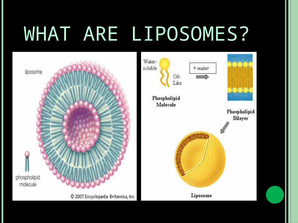
WHAT ARE LIPOSOMES? Liposomes are concentric bilayered vescicles in which an aqueous volume is entirely enclosed by a membranous lipid bilayer mainly composed of natural or synthetic phospholipids.
Liposomes can be composed of naturally-derived phospholipids with mixed lipid chains (like egg phosphatidylethanolamine), or of pure surfactant components like DOPE (dioleoylphosphatidylethanolamine). Liposomes, usually but not by definition, contain a core of aqueous solution; lipid spheres that contain no aqueous material are called micelles.
WHAT ARE LIPOSOMES?
COMPARISON OF LIPOSOMES AND NIOSOMES
Niosomes are non-ionic surfactant based liposomes. They are mostly formed by cholesterol incorporation as an excipient. Other excipients can also be used.
They are structurally similar to liposomes in having a bilayer, however, the materials used to prepare niosomes makes them more stable and thus niosomes offer many more advantages over liposomes.
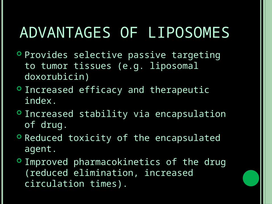
ADVANTAGES OF LIPOSOMES Provides selective passive targeting to tumor tissues (e.g. liposomal doxorubicin)
Increased efficacy and therapeutic index.
Increased stability via encapsulation of drug.
Reduced toxicity of the encapsulated agent.
Improved pharmacokinetics of the drug (reduced elimination, increased circulation times).
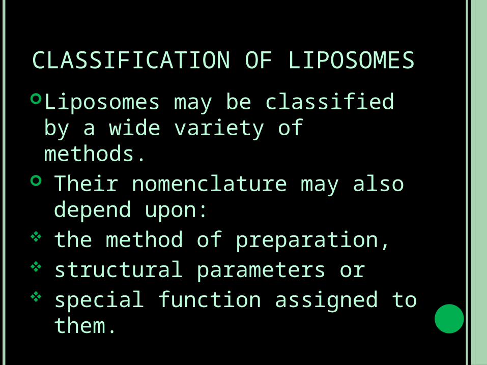
CLASSIFICATION OF LIPOSOMES
Liposomes may be classified by a wide variety of methods.
Their nomenclature may also depend upon:
the method of preparation, structural parameters or special function assigned to them.
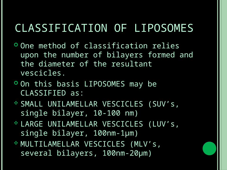
CLASSIFICATION OF LIPOSOMES One method of classification relies upon the number of bilayers formed and the diameter of the resultant vescicles.
On this basis LIPOSOMES may be CLASSIFIED as:
SMALL UNILAMELLAR VESCICLES (SUV’s, single bilayer, 10-100 nm)
LARGE UNILAMELLAR VESCICLES (LUV’s, single bilayer, 100nm-1µm)
MULTILAMELLAR VESCICLES (MLV’s, several bilayers, 100nm-20µm)
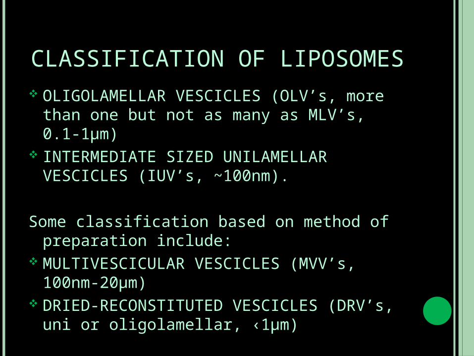
CLASSIFICATION OF LIPOSOMES OLIGOLAMELLAR VESCICLES (OLV’s, more than one but not as many as MLV’s, 0.1-1µm)
INTERMEDIATE SIZED UNILAMELLAR VESCICLES (IUV’s, ~100nm).
Some classification based on method of preparation include:
MULTIVESCICULAR VESCICLES (MVV’s, 100nm-20µm)
DRIED-RECONSTITUTED VESCICLES (DRV’s, uni or oligolamellar, ‹1µm)
CLASSIFICATION OF LIPOSOMESREVERSE PHASE EVAPORATION VESCICLES (REV’s, unilamellar, ~0.5µm)
MICRO-EMULSIFICATION LIPOSOMES (MEL, multilayered, 0.1-0.2µm)
LARGE UNILAMELLAR VESCICLES PREPARED BY EXTRUSION (VET, single/bilayered, 100nm-1µm)
STABLE PLURILAMELLAR VESCICLES (SPLV’s, multilayered, 100nm-2µm)
MECHANISM OF LIPOSOME FORMATION
WHAT ARE PHOSPHOLIPIDS AND THEIR PHYSICO CHEMICAL PROPERTIES?
Phospholipids are amphipathic molecules as they have a hydrophobic tail and an hydrophilic or polar head.
The hydrophilic and hydrophobic domains/ segments within the molecular geometry of amphiphilic lipids orient and self organise in ordered supramolecular structure when confronted with solvents.
In aqueous medium the molecules in self-assembled structures are oriented in such a way that the polar portion of the molecule remains in contact with the polar environment and at the same time shields the non-polar part.
MECHANISM OF LIPOSOME FORMATION
In aqueous mixtures these molecules are able to form various phases, some of them are stable and others remain in the metastable state.
At high concentrations of these polar lipids, liquid crystalline phases are formed that upon dilution with excess water can be stabilised into relatively stable colloidal particles.
The macroscopic structures often include lamellar, hexagonal or cubic phases dispersed as colloidal nanoconstructs referred as LIPOSOMES, or HEXASOMES or CUBOSOMES, respectively.
MECHANISM OF LIPOSOME FORMATION
MECHANISM OF LIPOSOME FORMATION
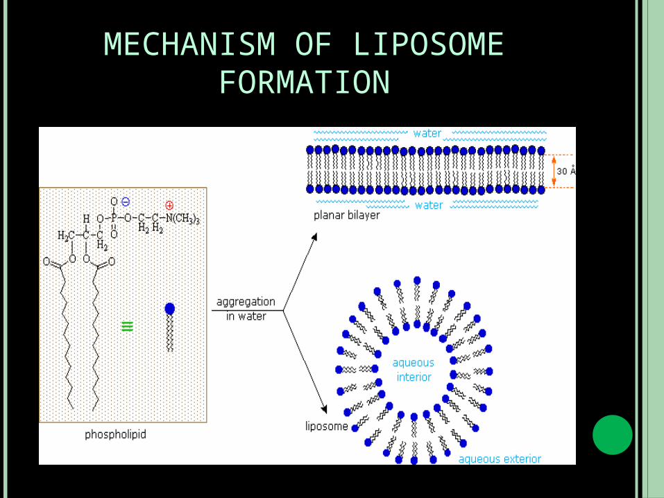
The amphipathic nature of phospholipids and their analogues renders them the ability to form closed concentric bilayers in the presence of water.
Liposomes are formed when thin lipid films or lipid cakes (of amphiphilic nature) are hydrated and stacks of liquid crystalline bilayers become fluid and swell.
The hydrated lipid sheets detach during agitation and self close to form large, multi cellular vescicles which prevent the interaction of water with the hydrocarbon core of the bilayer at the edges.

METHODS OF LIPOSOME PREPARATION AND DRUG
LOADING
Liposomes are manufactured in majority using procedures in which the water soluble (hydrophilic) materials are entrapped using aqueous solution of these materials as hydrating fluid or by the addition of drug/drug solution at some stage during the manufacture.
The lipid soluble (lipophilic) materials are solubilized in the organic solution of the constitutive lipid(s) and then evaporated to a dry drug containing lipid film followed by its hydration.
METHODS OF LIPOSOME PREPARATION AND DRUG
LOADING Methods of liposome preparation and drug loading are as follows:
PASSIVE LOADING TECHNIQUES: Mechanical dispersion methods of passive loading.
Solvent dispersion methods for passive loading.
Detergent depletion methods of passive loading.
REMOTE LOADING
PASSIVE LOADING TECHNIQUES
Passive loading technique involves three different groups of methods working on different principles:
MECHNICAL DISPERSIONSOLVENT DISPERSIONDETERGENT SOLUBILIZATION
MECHANICAL DISPERSION METHODS OF PASSIVE LOADING
The methods covered under this category begin with a lipid solution in organic solvent and end up with lipid dispersion in water.
The various components are typically combined by co-dissolving the lipids in an organic solvent.
The organic solvent is then removed and the solid lipid mixture is hydrated using an aqueous buffer.
The lipids spontaneously swell and hydrate to form liposomes.
MECHANICAL DISPERSION METHODS OF PASSIVE LOADING
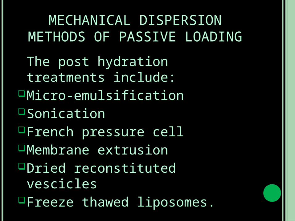
The post hydration treatments include:
Micro-emulsificationSonicationFrench pressure cellMembrane extrusionDried reconstituted vescicles
Freeze thawed liposomes.
SOLVENT DISPERSION METHODS FOR PASSIVE LOADING
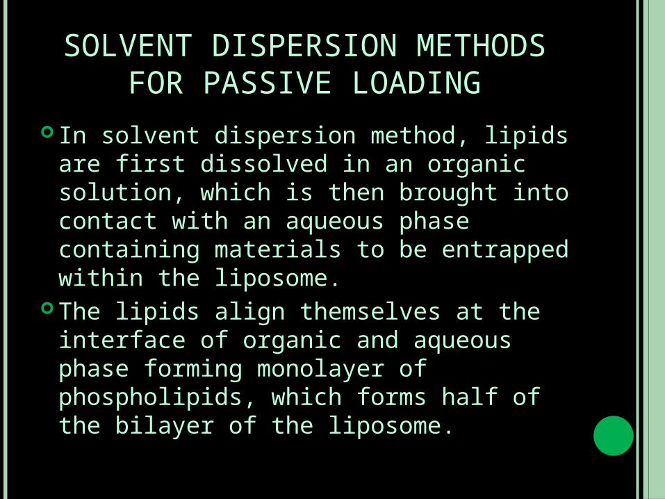
In solvent dispersion method, lipids are first dissolved in an organic solution, which is then brought into contact with an aqueous phase containing materials to be entrapped within the liposome.
The lipids align themselves at the interface of organic and aqueous phase forming monolayer of phospholipids, which forms half of the bilayer of the liposome.
SOLVENT DISPERSION METHODS FOR PASSIVE LOADING
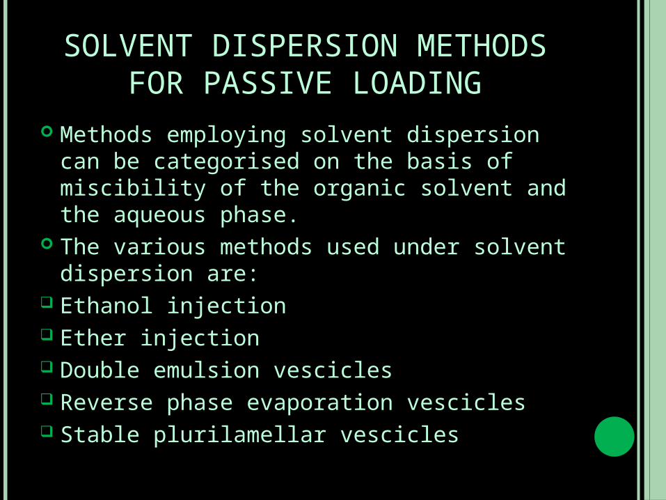
Methods employing solvent dispersion can be categorised on the basis of miscibility of the organic solvent and the aqueous phase.
The various methods used under solvent dispersion are:
Ethanol injection Ether injection Double emulsion vescicles Reverse phase evaporation vescicles Stable plurilamellar vescicles
DETERGENT DEPLETION METHODS OF PASSIVE LOADING
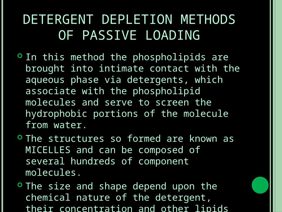
In this method the phospholipids are brought into intimate contact with the aqueous phase via detergents, which associate with the phospholipid molecules and serve to screen the hydrophobic portions of the molecule from water.
The structures so formed are known as MICELLES and can be composed of several hundreds of component molecules.
The size and shape depend upon the chemical nature of the detergent, their concentration and other lipids involved.
REMOTE (ACTIVE) LOADING Active loading methods has the following advantages over the passive encapsulation methods:
A high encapsulation efficiency and capacity.
A reduced leakage of the encapsulated compounds.
Avoidance of biological active compounds during preparation steps in the dispersion thus reducing safety hazards.
REMOTE (ACTIVE) LOADING The technique in general applies the concept of improved loading of drugs due to various transmembrane gradients, such as electrical, ionic or specific salt gradients.
This technique brings about improved loading into preformed liposomes using pH gradients and potential difference across liposomal membranes.
A concentration difference in proton concentration across the membrane of liposomes can drive the loading of amphipathic molecules.
CHARACTERIZATION OF LIPOSOMES
Characterization of liposomes is done to ensure their in vitro and in vivo performances.
The different parameters that are characterised are:
Vescicle shape and lamellarity Vescicle size and size distribution Surface charge Encapsulation efficiency and trapped volume
Chemical characterization of liposomes.
APPLICATIONS OF LIPOSOMES Liposomes have been used for the following therapeutic and pharmaceutical applications:
Liposomes as drug/protein delivery vehicles.
Controlled and sustained drug release in situ.
Altered pharmacokinetics and biodistribution.
Enzyme replacement therapy and lysosomal storage disorders.
Enhanced drug solubilization.
APPLICATION OF LIPOSOMES Liposomes in anti microbial, antifungal and antiviral therapy.
Liposomal drugs Liposome biological response modifiers Liposomes in tumor therapy. Carriers of small cytotoxic molecules Vehicles for macromolecules as cytokines or genes.
Liposomes in gene therapy. Gene and antisense therapy Genetic vaccination
APPLICATIONS OF LIPOSOMES Liposomes in immunology. Immunoadjuvant Immonumodulator Immunodiagnosis Liposomes as artificial blood surrogates
Liposomes as radiopharmaceuticals and radio diagnostic carriers.
Liposomes in cosmetics and dermatology Lipocomes in enzyme immobilization and bioreactor technology.
SOME RECENT APPLICATIONS Astaxanthine (AST), a red colored carotenoid pigment, possesses extremely powerful antioxidative activity. However, its drawbacks reside in poor solubility in aqueous system, resulting in extremely low bioavailability. To ameliorate such defects, AST encapsulated within liposomes (AST-L) were prepared. AST-L apparently showed improved stability and transportability. The overall transport time was 7.55 h and 6.00 h for free AST and AST-L, respectively.
SOME RECENT APPLICATIONS Many antibacterial agents, including the glycopeptides, are inactive
against Gram-negative bacteria because of their inability to cross the outer membrane of these cells. Different chemical and technological approaches have been described to circumvent such limitation. In this study, we aimed to apply the strategy of fusogenic liposomes, up to now used to carry biological compounds and materials inside cells, to localise a glycopeptide antibiotic, vancomycin (VAN), to the periplasmic space, thus allowing it to exert its bactericidal activity. Small unilamellar liposome vesicles were prepared by an extrusion procedure (SUVETs) from a phospholipid-cholesterol hemisuccinate mixture known for its fusogenic properties with the eukaryotic cell membrane. VAN was loaded with high efficiency into these vesicles and in microbiological experiments in vitro was shown to be able to inhibit to a different extent the growth of wild and standard Gram-negative bacterial strains. Minimum inhibitory concentrations as low as 6mg/L were observed, for instance against clinical isolates of Escherichia coli and Acinetobacter baumannii. In comparison, neither the free antibiotic nor VAN-loaded 'classical' (non-fusogenic) liposomes showed any activity against the same bacteria. Scanning and transmission electron microscopy studies allowed confirmation that the produced SUVETs were able to adhere to and fuse with the external membrane of E. coli. According to preliminary experiments, this technological strategy can be proposed as a potentially successful way to enlarge the spectrum of activity of VAN.
REFERENCES Vyas, S. P, Khar, R. K, Targeted and Controlled Drug delivery.
www.wikipedia.org “Improved membrane transport of astaxanthine by liposomal encapsulation.”Peng CH, Chang CH, Peng RY, Chyau CC.
“Encapsulation in fusogenic liposomes broadens the spectrum of action of vancomycin against Gram-negative bacteria.”Nicolosi D, Scalia M, Nicolosi VM, Pignatello R.

Thank you!!