lecture 2 origins of biopot ch 4
TRANSCRIPT
Lecture 2Chapter 4: The Origin of
Biopotentials
Dr. Nitish V. Thakor
Biomedical Instrumentation
JHU Applied Physics Lab
Introduction
Biopotentials arise from cells, and more generally from organs. They hold rich physiological and clinical information. For example, action potentials give information on fundamental ion channel biophysics and molecular aspects of any pathology. Biopotentials from the organs of the body are of clinical diagnostic significance.
Examples:
1. Action Potentials from Cells (and 3 Nobel prizes!)
1. Neuronal action potential (history of Squid axon and Hodgkin-Huxley work)
2. Patch clamp technique and single channel recording (Sakman-Neher)
3. Water channel work of Peter Agre (JHU)
2. Biopotentials from the organ/body
1. Electrocardiogram (ECG) from heart -> use in heart attack, pacemakers
2. Electroencephalogram (EEG) from brain -> use in epilepsy, brain trauma
3. Electromyogram (EMG) from muscle -> use in muscle diseases, prosthesis
4. Others…
Electrical Activity of Excitable Cells
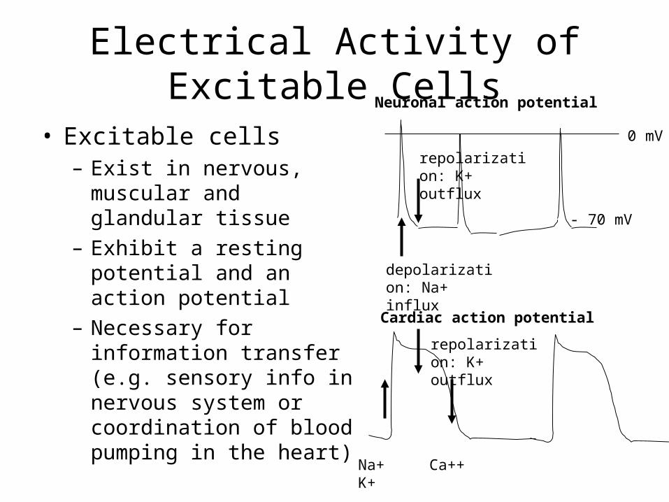
• Excitable cells – Exist in nervous, muscular
and glandular tissue– Exhibit a resting potential
and an action potential– Necessary for information
transfer (e.g. sensory info in nervous system or coordination of blood pumping in the heart)
0 mV
- 70 mV
depolarization: Na+ influx
repolarization: K+ outflux
Na+ Ca++ K+
Neuronal action potential
Cardiac action potential
repolarization: K+ outflux
Resting vs. Active State
• Resting State– Steady electrical potential of difference
between internal and external environments– Typically between -70 to -90mV, relative to
the external medium
• Active State– Electrical response to adequate stimulation– Consists of “all-or-none” action potential after
the cell threshold potential has been reached
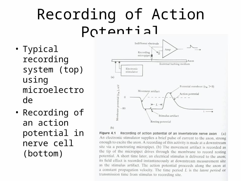
Recording of Action Potential
• Typical recording system (top) using microelectrode
• Recording of an action potential in nerve cell (bottom)
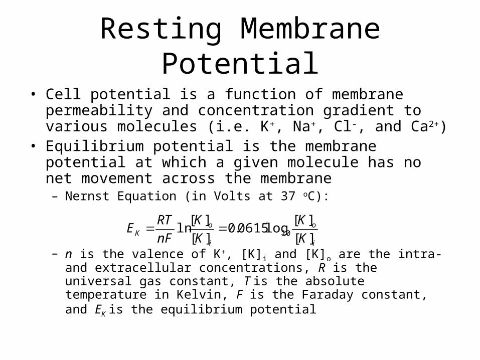
Resting Membrane Potential
• Cell potential is a function of membrane permeability and concentration gradient to various molecules (i.e. K+, Na+, Cl-, and Ca2+)
• Equilibrium potential is the membrane potential at which a given molecule has no net movement across the membrane– Nernst Equation (in Volts at 37 oC):
– n is the valence of K+, [K]i and [K]o are the intra- and extracellular concentrations, R is the universal gas constant, T is the absolute temperature in Kelvin, F is the Faraday constant, and EK is the equilibrium potential
i
o
i
oK K
K
K
K
nF
RTE
][
][log0615.0
][
][ln 10
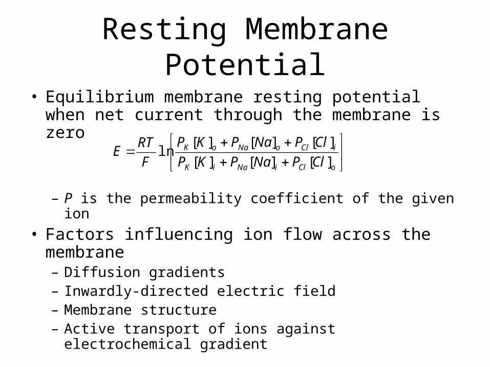
Resting Membrane Potential
• Equilibrium membrane resting potential when net current through the membrane is zero
– P is the permeability coefficient of the given ion
• Factors influencing ion flow across the membrane– Diffusion gradients– Inwardly-directed electric field– Membrane structure– Active transport of ions against electrochemical
gradient
oCliNaiK
iCloNaoK
ClPNaPKP
ClPNaPKP
F
RTE
][][][
][][][ln
Action Potential
• Stimulation of excitable cells causes “all-or-none” response
• At threshold, the membrane potential rapidly depolarizes due to a change in membrane permeability– PNa significantly increases causing the membrane
potential to approach ENa (+60mV)
• A delayed increase in PK causes hyperpolarization and a return to resting potential
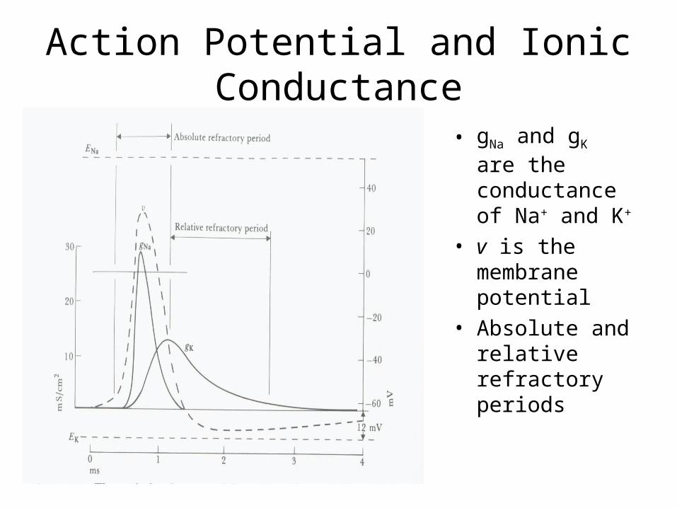
Action Potential and Ionic Conductance
• gNa and gK are the conductance of Na+ and K+
• v is the membrane potential
• Absolute and relative refractory periods
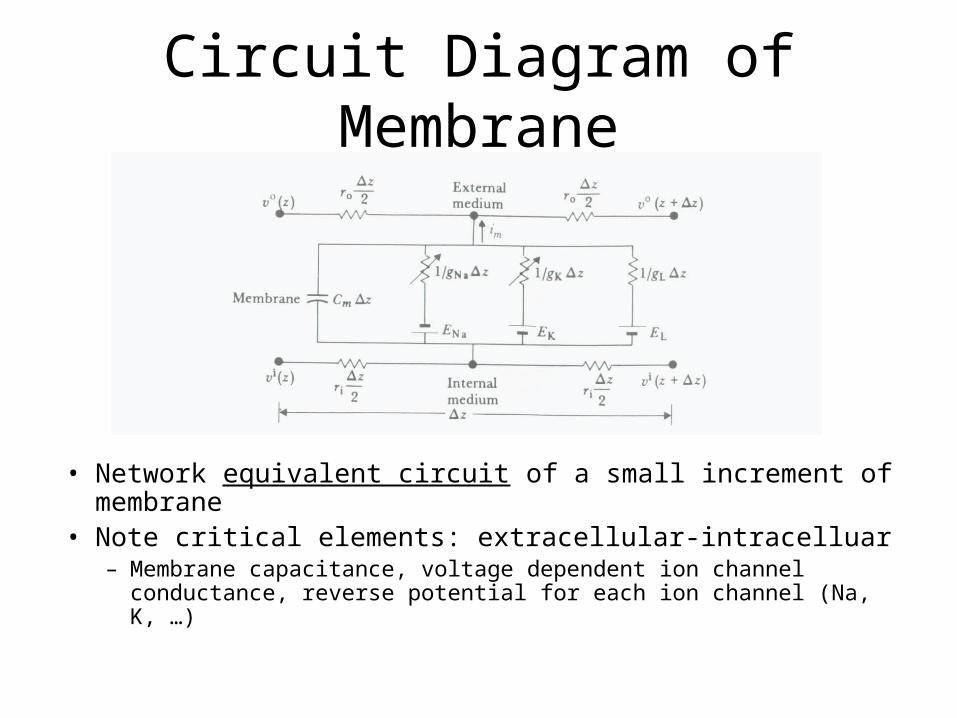
Circuit Diagram of Membrane
• Network equivalent circuit of a small increment of membrane
• Note critical elements: extracellular-intracelluar– Membrane capacitance, voltage dependent ion channel
conductance, reverse potential for each ion channel (Na, K, …)
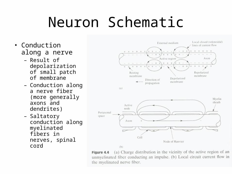
Neuron Schematic
• Conduction along a nerve– Result of
depolarization of small patch of membrane
– Conduction along a nerve fiber (more generally axons and dendrites)
– Saltatory conduction along myelinated fibers in nerves, spinal cord
Organization of Peripheral Nervous System
• Reflex arc– Sense organ (e.g. receptors)– Sensory nerve (transfers information from receptor to
CNS)– CNS (i.e. information processing station)– Motor nerve (transfers information from CNS to
effector organ)– Effector Organ (i.e. muscles)
• Simplest example– Knee reflex
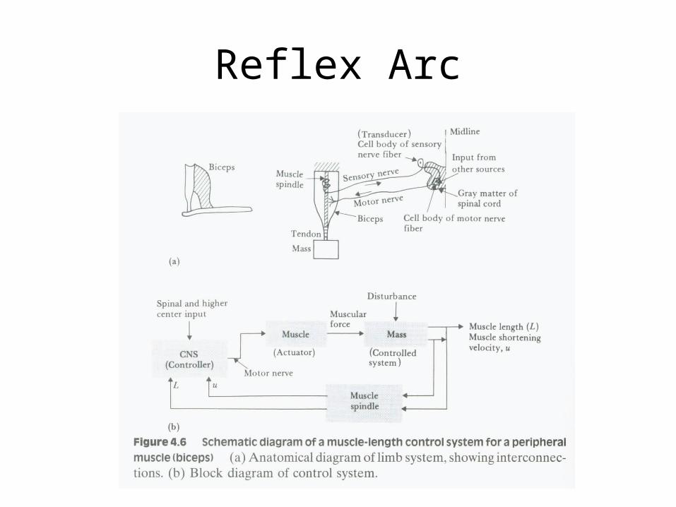
Reflex Arc
Organization of Peripheral Nervous System
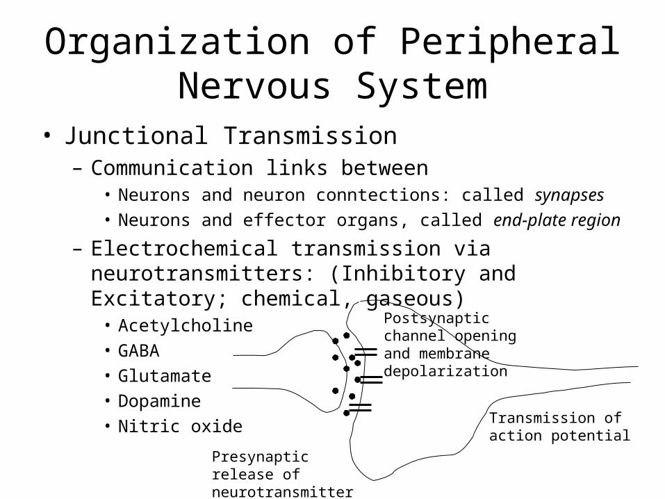
• Junctional Transmission– Communication links between
• Neurons and neuron conntections: called synapses• Neurons and effector organs, called end-plate region
– Electrochemical transmission via neurotransmitters: (Inhibitory and Excitatory; chemical, gaseous)
• Acetylcholine• GABA• Glutamate• Dopamine• Nitric oxide
Presynaptic release of neurotransmitter
Postsynaptic channel opening and membrane depolarization
Transmission of action potential
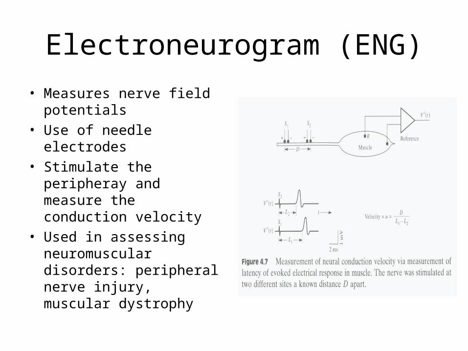
Electroneurogram (ENG)
• Measures nerve field potentials
• Use of needle electrodes• Stimulate the peripheray
and measure the conduction velocity
• Used in assessing neuromuscular disorders: peripheral nerve injury, muscular dystrophy
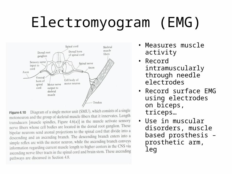
Electromyogram (EMG)
• Measures muscle activity
• Record intramuscularly through needle electrodes
• Record surface EMG using electrodes on biceps, triceps…
• Use in muscular disorders, muscle based prosthesis – prosthetic arm, leg
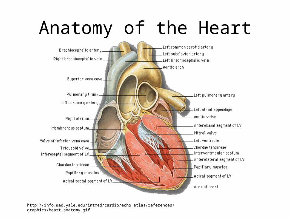
Anatomy of the Heart
http://info.med.yale.edu/intmed/cardio/echo_atlas/references/graphics/heart_anatomy.gif
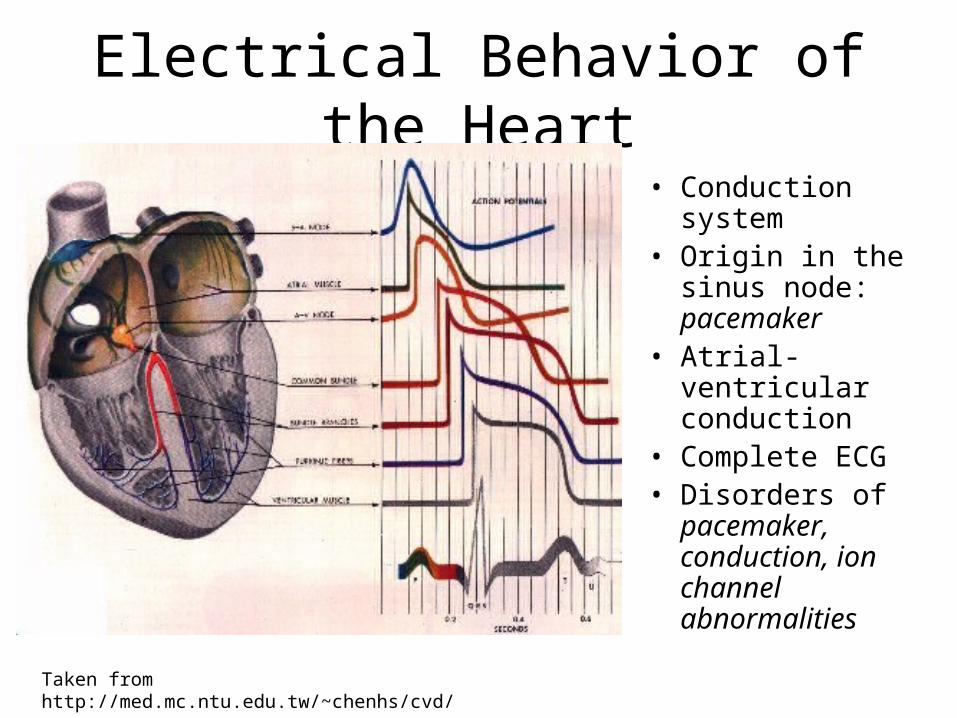
Electrical Behavior of the Heart
• Conduction system
• Origin in the sinus node: pacemaker
• Atrial-ventricular conduction
• Complete ECG• Disorders of
pacemaker, conduction, ion channel abnormalities
Taken from http://med.mc.ntu.edu.tw/~chenhs/cvd/
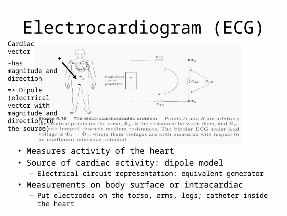
Electrocardiogram (ECG)
• Measures activity of the heart• Source of cardiac activity: dipole model
– Electrical circuit representation: equivalent generator
• Measurements on body surface or intracardiac– Put electrodes on the torso, arms, legs; catheter inside the heart
Cardiac vector
-has magnitude and direction
=> Dipole (electrical vector with magnitude and direction to the source)
+
-
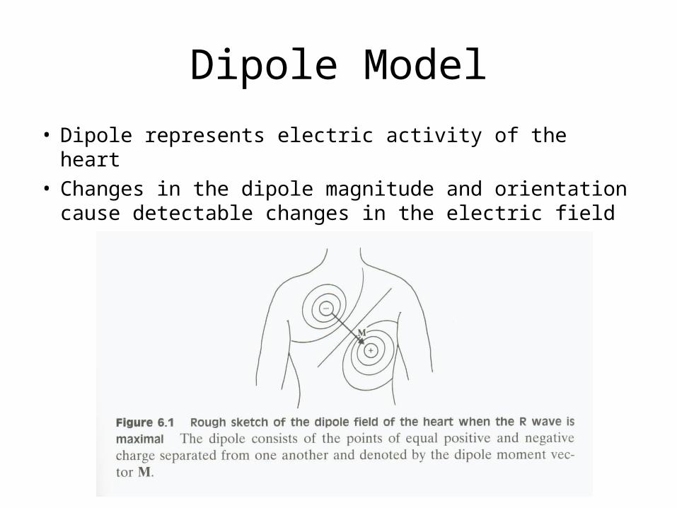
Dipole Model
• Dipole represents electric activity of the heart• Changes in the dipole magnitude and orientation
cause detectable changes in the electric field

Vector Algebra
• Dot product of vectors, where va1 is a scalar voltage:
• When the vector is perpendicular to M, va1 is zero
cos11 MaM av
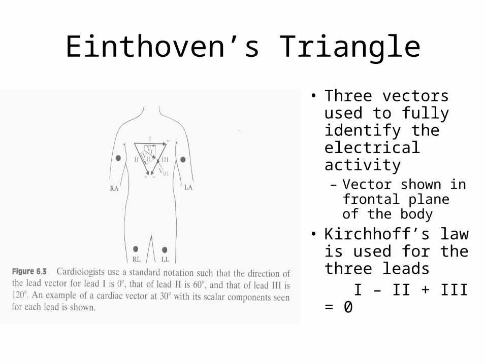
Einthoven’s Triangle
• Three vectors used to fully identify the electrical activity – Vector shown in
frontal plane of the body
• Kirchhoff’s law is used for the three leads I – II + III = 0
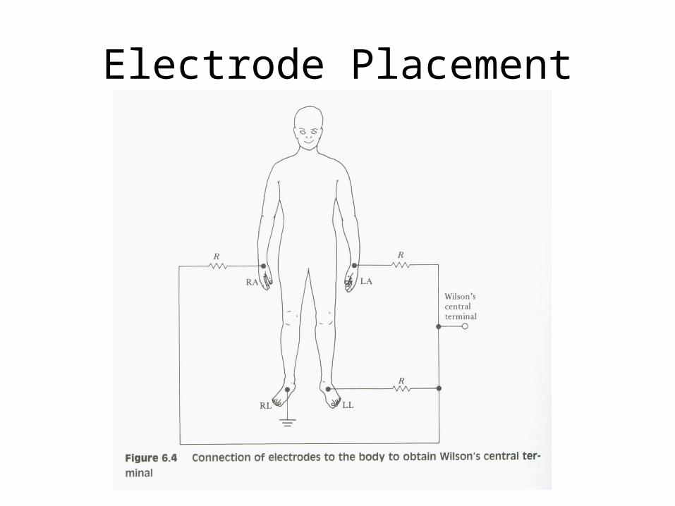
Electrode Placement
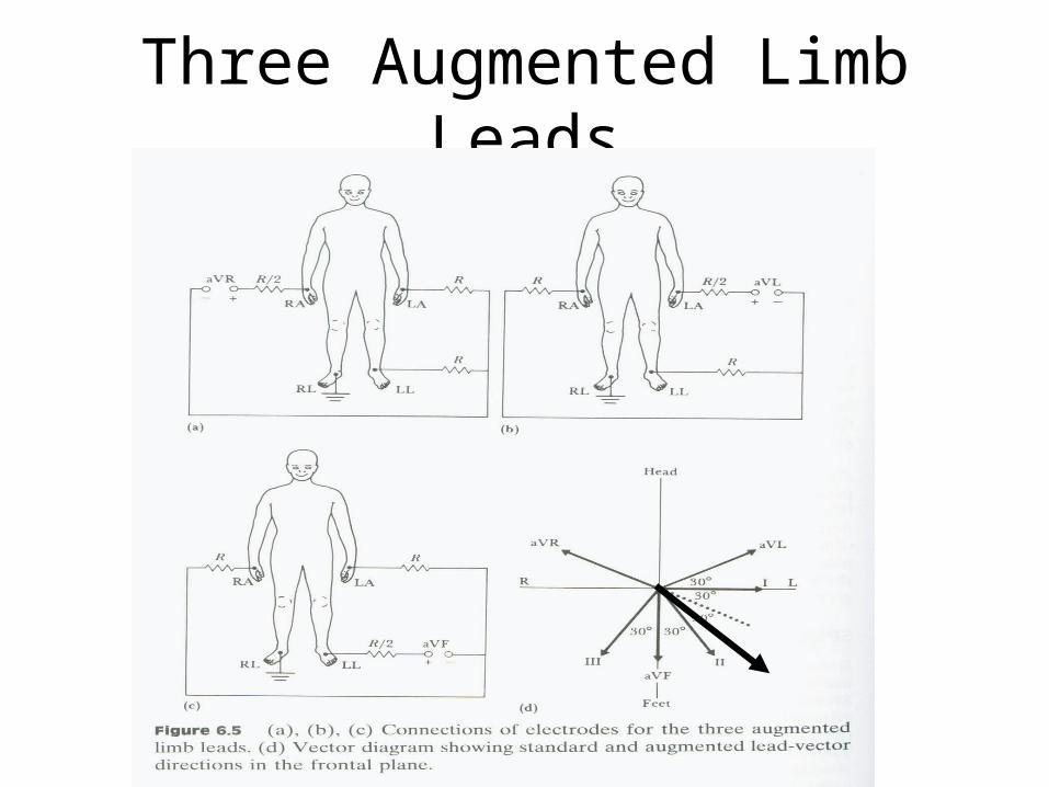
Three Augmented Limb Leads
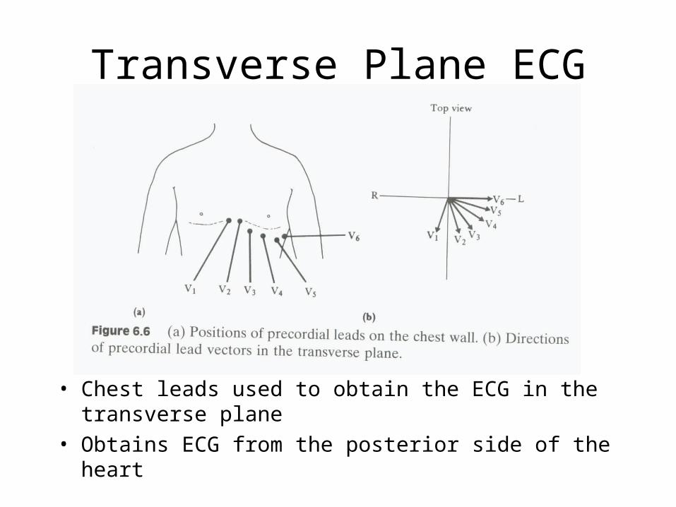
Transverse Plane ECG
• Chest leads used to obtain the ECG in the transverse plane
• Obtains ECG from the posterior side of the heart
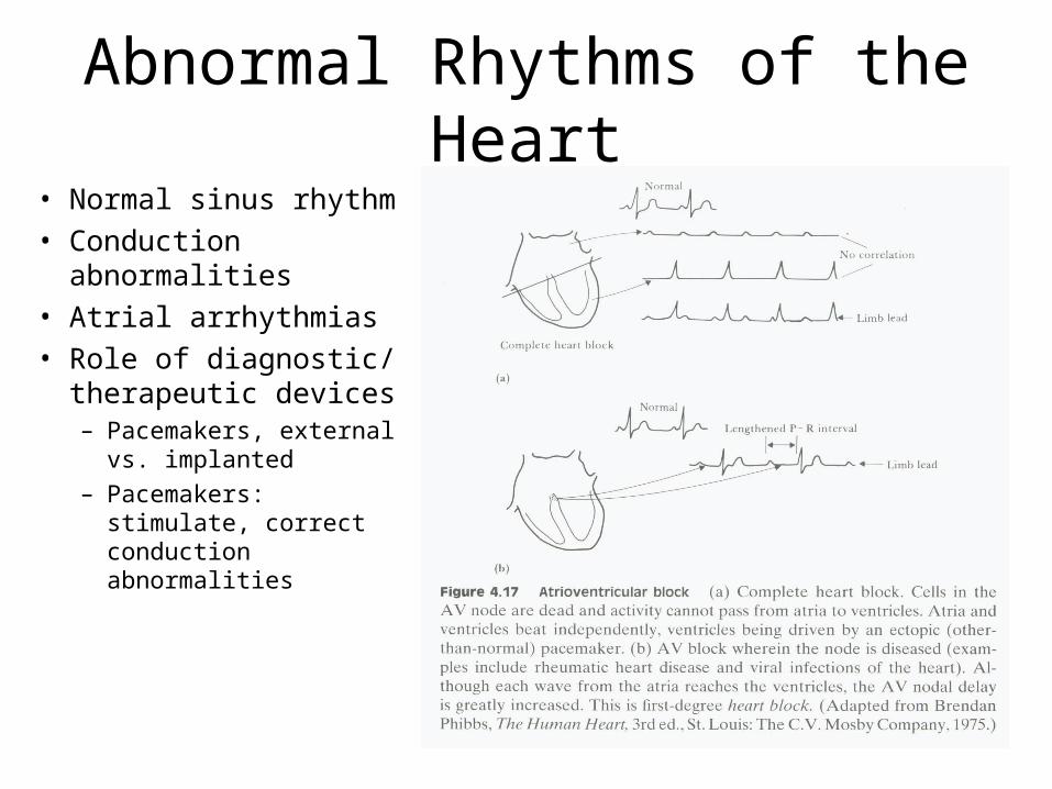
Abnormal Rhythms of the Heart
• Normal sinus rhythm• Conduction
abnormalities• Atrial arrhythmias• Role of diagnostic/
therapeutic devices– Pacemakers, external
vs. implanted– Pacemakers:
stimulate, correct conduction abnormalities

Abnormal Rhythms of the Heart
• PVCs are premonitory Ventricular
• Ventricular arrhythmias are more lethal
• Role of diagnostic monitoring in CCU
• Role of therapeutic devices (implantable cardioverter)
Atrial tachycardia
Ventricular tachycardia
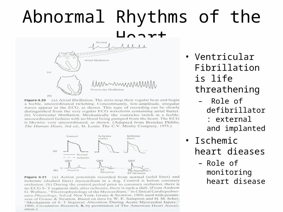
Abnormal Rhythms of the Heart
• Ventricular Fibrillation is life threathening– Role of
defibrillator: external and implanted
• Ischemic heart dieases– Role of
monitoring heart disease
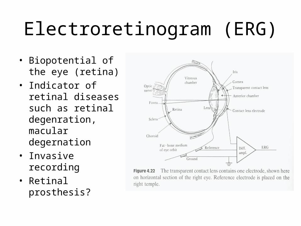
Electroretinogram (ERG)
• Biopotential of the eye (retina)
• Indicator of retinal diseases such as retinal degenration, macular degernation
• Invasive recording• Retinal prosthesis?
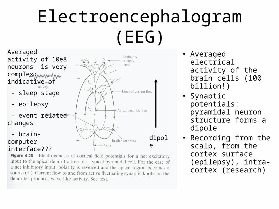
Electroencephalogram (EEG)
• Averaged electrical activity of the brain cells (100 billion!)
• Synaptic potentials: pyramidal neuron structure forms a dipole
• Recording from the scalp, from the cortex surface (epilepsy), intra-cortex (research)
dipole
Averaged activity of 10e8 neurons is very complex: indicative of
- sleep stage
- epilepsy
- event related changes
- brain-computer interface???
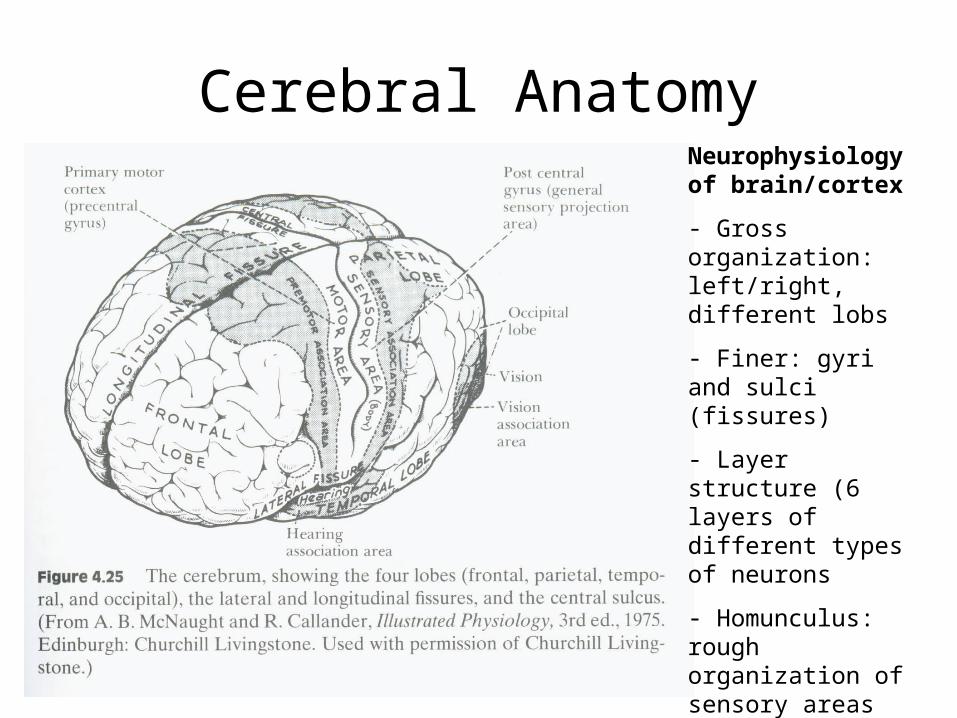
Cerebral AnatomyNeurophysiology of brain/cortex
- Gross organization: left/right, different lobs
- Finer: gyri and sulci (fissures)
- Layer structure (6 layers of different types of neurons
- Homunculus: rough organization of sensory areas along the sensory-motor cortex
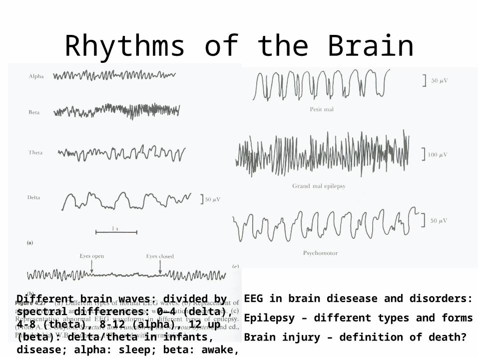
Rhythms of the Brain
Different brain waves: divided by spectral differences: 0—4 (delta), 4-8 (theta), 8-12 (alpha), 12 up (beta): delta/theta in infants, disease; alpha: sleep; beta: awake, eyes open
EEG in brain diesease and disorders:
Epilepsy – different types and forms
Brain injury – definition of death?
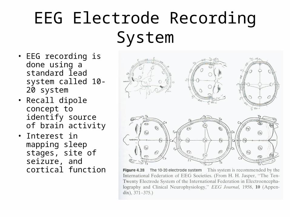
EEG Electrode Recording System
• EEG recording is done using a standard lead system called 10-20 system
• Recall dipole concept to identify source of brain activity
• Interest in mapping sleep stages, site of seizure, and cortical function
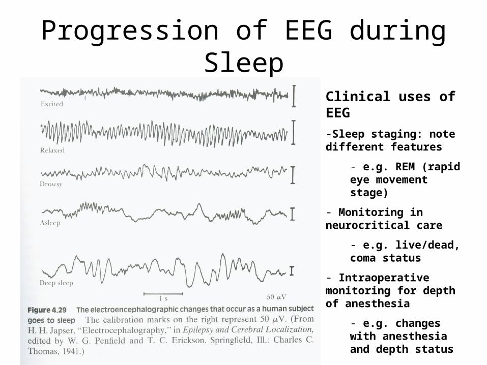
Progression of EEG during Sleep
Clinical uses of EEG
-Sleep staging: note different features
- e.g. REM (rapid eye movement stage)
- Monitoring in neurocritical care
- e.g. live/dead, coma status
- Intraoperative monitoring for depth of anesthesia
- e.g. changes with anesthesia and depth status
Reference
• Webster, JG (1998). Medical Instrumentation. John Wiley & Sons, Inc., New York, NY. Chapter 4.

Problems and self-study
1 A) Hodgkin and Huxley received a Nobel prize for their work with Squid axon to decipher the role of ion channels and formation of action potential. Research original papers and a) present graphics of their recording technique, b) describe the voltage clamp method and its use, c) optionally: research and present/describe the voltage clamp circuit
B) Bert Sakman and Erwin Neher received a Nobel prize for their development of a patch pipette electrode recording technique for measurement of ion channel activity. Show the schematic of a patch pipette attached to a) cell and b) membrane. In each case, what is the source of the current being measured? Optionally design the patch clamp circuit.
C) Draw the different ion channels and currents active during a cardiac action potential. Research how pacemaker potential arizes (repolarization of the action potential), and how ischemia might alter the action potentials
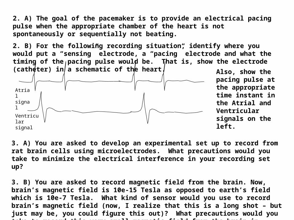
Atrial signal
Ventricular signal
3. A) You are asked to develop an experimental set up to record from rat brain cells using microelectrodes. What precautions would you take to minimize the electrical interference in your recording set up? 3. B) You are asked to record magnetic field from the brain. Now, brain’s magnetic field is 10e-15 Tesla as opposed to earth’s field which is 10e-7 Tesla. What kind of sensor would you use to record brain’s magnetic field (now, I realize that this is a long shot – but just may be, you could figure this out)? What precautions would you take to record this very small magnetic field from the brain in presence of other interference?
Also, show the pacing pulse at the appropriate time instant in the Atrial and Ventricular signals on the left.
2. A) The goal of the pacemaker is to provide an electrical pacing pulse when the appropriate chamber of the heart is not spontaneously or sequentially not beating.
2. B) For the following recording situation, identify where you would put a “sensing” electrode, a “pacing” electrode and what the timing of the pacing pulse would be. That is, show the electrode (catheter) in a schematic of the heart.
4. A) What does the 12-lead ECG system comprise of (sketch the different leads)? Is it superior or inferior to an orthogonal system (X, Y, and Z leads)? the different leads)? Is it superior or inferior to an orthogonal system (X, Y, and Z leads)? B) The ECG signal generating from the heart can be 6.2 A) What does the 12-lead ECG system comprise of (sketch modeled quite simply as a dipole. If a cardiac dipole has a magnitude of 1 mV and orientation of –45o with respect to Lead I, then calculate, using the Einthoven triangle, the magnitude of the signal in Lead I, II, and III. Show the geometric presentation as well as the trigonometric calculations.
5. A) Imagine it is the beginning of the 20th century. Cardiac activity is suspected as an electrical source inside the torso. Let us say that you were a contemporary of Prof. Einthoven. Prof. Einthoven recommends that to record ECG from the torso using a triangular formulation with what you now know at three leads, I, II, and III (respectively LA-RA, RA-LL, and LA-LL). However, you claim have a different theory of better presenting the cardiac vector on a different lead system (for example, you prefer not to use 3 leads arranged in the form of a triangle). Demonstrate superiority of your lead idea. B) After Einthoven’s original idea, a number of solutions were suggested. One of these was to put 6 leads (V1-V6) around the left ventricle. a) why around left ventricle? b) for the 6 differential amplifiers, each with one input being V1..V6 what is the other “neutral” input source?
6. A) Explain the origin of EEG signal in terms of its sources in the brain. Describe briefly the neural generator and the electrical field/vector representation that explains how an internal source produces an external EEG. B) What are the advantages and disadvantages of putting EEG electrodes on the scalp versus directly on the brain? Under what clinical condition is either procedure recommended? What kinds of electrodes are used for direct cortical recording? What are the design considerations? How does a neurologist identify an epileptic spike or seizure? How does a surgeon determine where to “cut” the brain to remove the focus?
C) What kind of a lead system would you use to record EEG from the scalp and for localizing the source of epileptic seizure? Sketch it. Now, putting electrodes on the scalp may not help localize the seizure focus better. Surgeons now put electrodes directly on brain. Research direct cortical recording of seizure and describe/Illustrate the technology.
D) i) What instrument is used to measure the magnetic field from the brain? ii) What are the possible advantages and disadvantages of the magnetic versus electrical measurement? iii) To your knowledge, what breakthroughs in the scientific world that have are occurred (or ought to occur?) that would make magnetic field measurement more feasible and affordable? iv) If you had a cheap magnetic field sensor (with a relatively lower sensitivity) available what other biomedical application would you think of (other than biopotential measurements).
7. A) We would like to record ECG of a fetus while in the womb. The main problem here is that when electrodes are placed on the mother’s stomach to capture the fetal ECG, a large maternal ECG signal pulse is also picked up. A) Draw a schematic of the mother and her heart dipole/vector and fetus and its heart dipole/vector. Now, show how mother’s ECG might corrupt the fetal ECG. B) How would you eliminate the maternal ECG artifact from the stomach recording? C) Someone suggests that at the most critical moment in labor, as the head of the fetus presents itself first , attach the ECG electrode to fetal scalp. Would you succeed or not in getting fetal ECG from an electrode placed on the scalp and why/why not? D) During the time of the late stage labor, what would be more likely to succeed – electrodes on the mother’s stomach or an electrode on fetus’s head? B) Show (draw) the possible current distribution between an electrosurgical electrode, body and the return ground electrode. What would be the desirable properties of the ground reference electrode?
C) Students in the past have proposed two methods for monitoring eye movements as a way to provide a command/control signal for a quadriplegic (e.g. eye movement command may be used to move a cursor on the computer screen). What might be two such methods (Hint: one is optical and other is based on biopotentials)?
Body Power!!!
• Power for an implanted stimulator (BION)– Battery, induction (radio frequency)– Heat, chemical, flow, mechanical, …
• What is your energy transduction principle– What sensor/actuator would you use– What circuit principle? E.g. piezo->electricity to power
the stimulator– What physical/chemical/ electrochem/optical principle?– What’s wrong/bad about this (e.g. effluent, gas, …)– What kind of energy density you can obtain? What is
the conversion efficiency of each of these?
• Real world examples, patents, products
More questions
• How many electrodes/lead in a 12 lead system?• What are the sources of interference in
biopotential recording?• How do you increase the likelihood of recording
event-related potential? How do you increase the signal-to-noise ratio? (averaging)
• Research and describe neurochemical sensor technologies