kuliah radiologi emergensi
DESCRIPTION
MEDICALTRANSCRIPT
Radiologi Trauma dan Emergensi
Bachtiar Murtala

Introduction:• Since Prof.W.C. Roentgen (1895) in Germany/ Europe
discovered x-ray, medical science developed rapidly both in diagnosis and treatment.
• The role of x-ray and other radiology modalities in emergency cases, as well as traumatology, also increased.
• It is very difficult to asses and make precise diagnosis without radiology examinations.

Emergency and trauma cases may come from the head and neck, chest, abdomen , and extremities regions.
We focus to discuss about conventional x-ray and the most common cases in the daily practice.
US is the tool of choise in detecting free fluid / blood in the abdominal and chect cavities.

Trauma
• Can cause fracture,dislocation• Fracture : discontinuity of bone, cartilage or
both, associated with soft tissue injury• Closed X open fracture• Complete X incomplete fracture

• More important is the impact of the fracture to the soft tissue i.e. spinal cord, brain tissue, etc

Types of shaft fracture

Fracture/dislocation of cervical spine:
• This fracture is very dangerous because of the risk to the spinal cord injure /pressure
• Routine position : AP and lateral view• Fracture of C1 or C7 –T1, usually needed
special view :- C 1 by Open mouth position
- C7 –T 1 by swimmer’s position

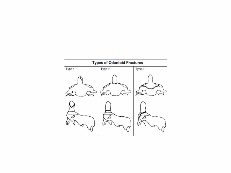

C1 fracture
• Fracture of C1 , as in Jefferson fr. Can be identified by open mouth position
• Fraktur Jefferson:
• Normal

Jefferson fracture


Burst fracture
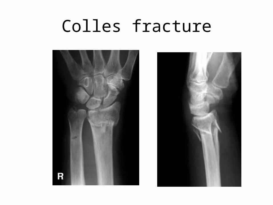
Colles fracture

Galeazzi fracture
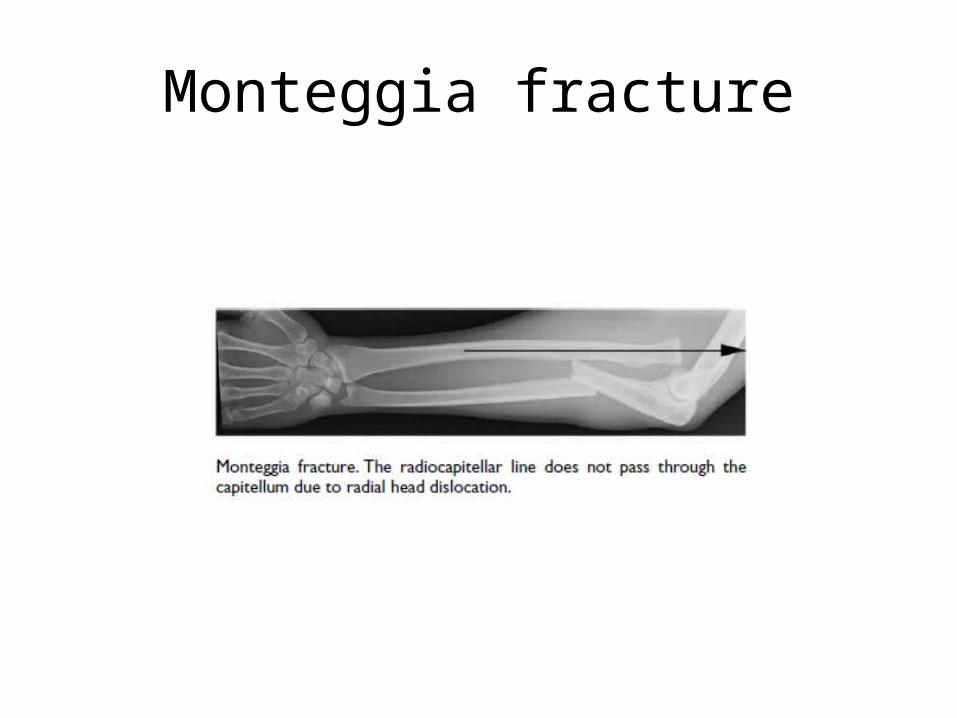
Monteggia fracture

Ruptur Ginjal

Ruptur uretra


Ruptur Buli

Ruptur Buli

Torsio testis

Placenta Previa

Plasenta Previa

Incomplete abortus

K E T

Molahidatidosa
