kidney’sthere are 1.2 million nephrons per kidney. the renal corpuscle begins the nephron. it has...
TRANSCRIPT
1
2
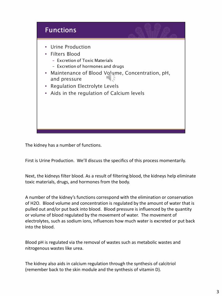
The kidney has a number of functions.
First is Urine Production. We’ll discuss the specifics of this process momentarily.
Next, the kidneys filter blood. As a result of filtering blood, the kidneys help eliminate toxic materials, drugs, and hormones from the body.
A number of the kidney’s functions correspond with the elimination or conservation of H2O. Blood volume and concentration is regulated by the amount of water that is pulled out and/or put back into blood. Blood pressure is influenced by the quantity or volume of blood regulated by the movement of water. The movement of electrolytes, such as sodium ions, influences how much water is excreted or put back into the blood.
Blood pH is regulated via the removal of wastes such as metabolic wastes and nitrogenous wastes like urea.
The kidney also aids in calcium regulation through the synthesis of calcitriol (remember back to the skin module and the synthesis of vitamin D).
3
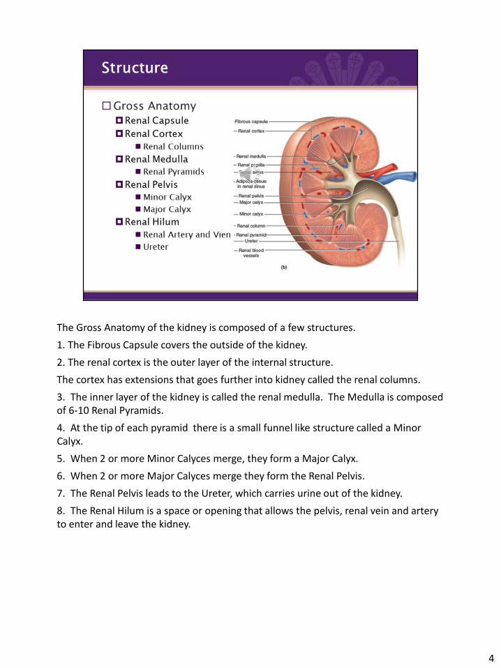
The Gross Anatomy of the kidney is composed of a few structures.
1. The Fibrous Capsule covers the outside of the kidney.
2. The renal cortex is the outer layer of the internal structure.
The cortex has extensions that goes further into kidney called the renal columns.
3. The inner layer of the kidney is called the renal medulla. The Medulla is composed of 6-10 Renal Pyramids.
4. At the tip of each pyramid there is a small funnel like structure called a Minor Calyx.
5. When 2 or more Minor Calyces merge, they form a Major Calyx.
6. When 2 or more Major Calyces merge they form the Renal Pelvis.
7. The Renal Pelvis leads to the Ureter, which carries urine out of the kidney.
8. The Renal Hilum is a space or opening that allows the pelvis, renal vein and artery to enter and leave the kidney.
4

The functional unit of the kidney is called the nephron. There are 1.2 million nephrons per kidney.
The renal corpuscle begins the nephron. It has the glomerulus which is a network of capillaries that filters blood plasma. The afferent arterioles are the blood vessels that come into the corpuscle and the efferent arterioles leave the glomerulus. The glomerular capsule is a double layered capsule that encloses the glomerulus and wraps around the capillaries.
5
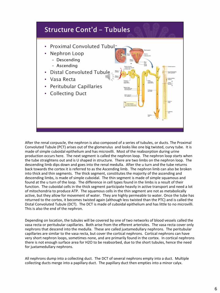
After the renal corpuscle, the nephron is also composed of a series of tubules, or ducts. The Proximal Convoluted Tubule (PCT) arises out of the glomerulus and looks like one big twisted, curvy tube. It is made of simple cuboidal epithelium and has microvilli. Most of the reabsorption during urine production occurs here. The next segment is called the nephron loop. The nephron loop starts when the tube straightens out and is U shaped in structure. There are two limbs on the nephron loop. The descending limb dips down and goes into the renal medulla. After the u turn and the tube returns back towards the cortex it is referred to as the Ascending limb. The nephron limb can also be broken into thick and thin segments. The thick segment, constitutes the majority of the ascending and descending limbs, is made of simple cuboidal. The thin segment is made of simple squamous and found at the u turn of the loop. The difference in cell types found in the limbs is a result of their function. The cuboidal cells in the thick segment participate heavily in active transport and need a lot of mitochondria to produce ATP. The squamous cells in the thin segment are not as metabolically active, but they allow for movement of water. They are highly permeable to water. Once the tube has returned to the cortex, it becomes twisted again (although less twisted than the PTC) and is called the Distal Convoluted Tubule (DCT). The DCT is made of cuboidal epithelium and has little to no microvilli. This is also the end of the nephron.
Depending on location, the tubules will be covered by one of two networks of blood vessels called the vasa recta or peritubular capillaries. Both arise from the efferent arterioles. The vasa recta cover only nephrons that descend into the medulla. These are called juxtamedullary nephrons. The peritubular capillaries are similar to the vasa recta, but cover the cortical nephrons. Cortical nephrons can have very short nephron loops, sometimes none, and are primarily found in the cortex. In cortical nephrons there is not enough surface area for H2O to be reabsorbed, due to the short tubules, hence the need for juxtamedullary nephrons.
All nephrons dump into a collecting duct. The DCT of several nephrons empty into a duct. Multiple collecting ducts merge into a papillary duct. The papillary duct then empties into a minor calyx.
6
7
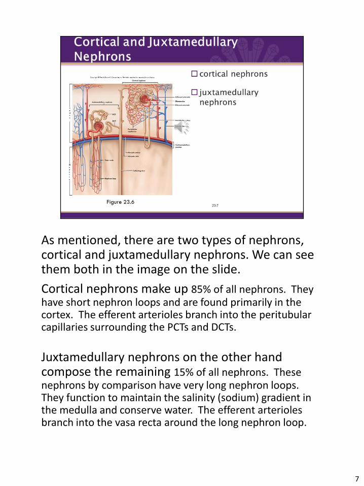
As mentioned, there are two types of nephrons,cortical and juxtamedullary nephrons. We can see them both in the image on the slide.
Cortical nephrons make up 85% of all nephrons. They have short nephron loops and are found primarily in the cortex. The efferent arterioles branch into the peritubularcapillaries surrounding the PCTs and DCTs.
Juxtamedullary nephrons on the other hand compose the remaining 15% of all nephrons. These nephrons by comparison have very long nephron loops.They function to maintain the salinity (sodium) gradient in the medulla and conserve water. The efferent arterioles branch into the vasa recta around the long nephron loop.

The formation of urine is a three step process. It begins by making filtrate in the glomerulus.
Glomerular filtrate is the fluid that has been filtered from the blood plasma in the glomerulus and throughout the nephron.
Fluid passes from glomerulus (ie capillaries) into the glomerular capsule via a filtration membrane.
The Filtration membrane is made of 1. fenestrated endothelium of the capillary, 2. a shared basement membrane, 3. and filtrations slits in the capsule wall.
The fenestrated endothelium is honeycombed shaped with large filtration pores, like a colander.
Particles are pushed out by an increase in blood pressure. The increase in blood pressure is due to dramatic decrease in vessel size of the afferent arteriole. The restriction of size causes pressure to build up and moves particles through the filtration membrane. This is similar to putting your thumb over the end of a garden hose to spray water. At this stage the things that get filtered out are water, electrolytes, glucose, fatty acids, amino acids, nitrogenous wastes, vitamins, and ions.
Glomerular Filtration Rate is the amount of filtrate formed in 1 minute by both kidneys. Healthcare providers can use the glomerular filtration rate to help determine if the kidneys are working properly. Males produce about 180 liters per day and women produce about 150 liters per day. Approximately 99% of this filtrate is reabsorbed back into the blood as we will see in the following steps.
8
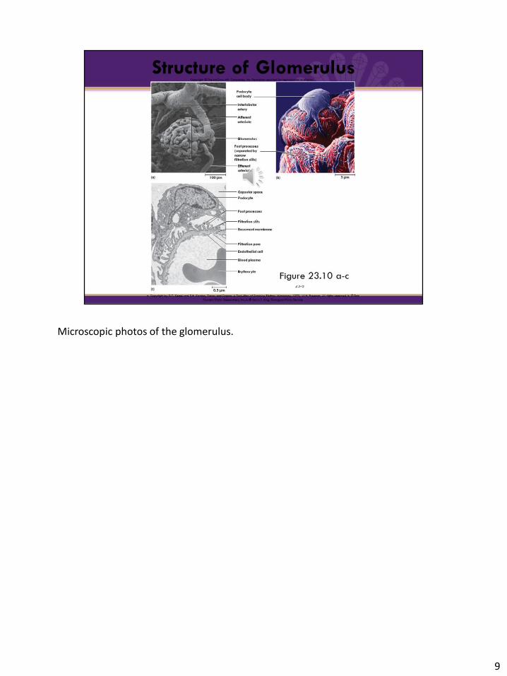
Microscopic photos of the glomerulus.
9

The fenestrated endothelium of the capillary on the left, the shared, thin basement membrane in the middle, and then the slotted epithelial wall of the glomerular capsule. Notice in the diagram the substances that are allowed to pass through and those that are not and remain in the blood.
10
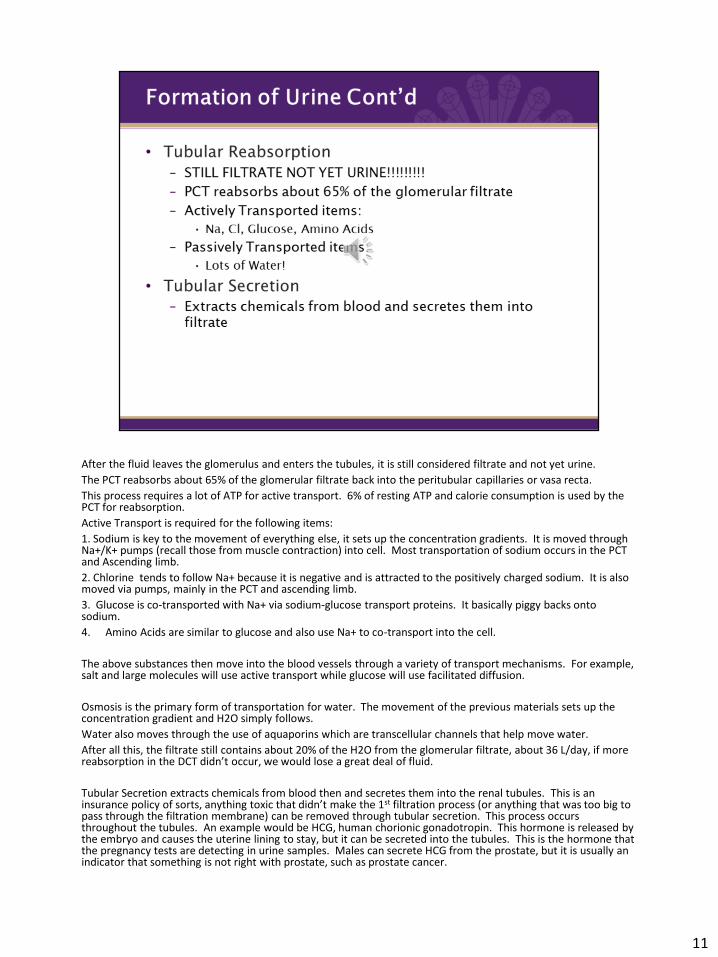
After the fluid leaves the glomerulus and enters the tubules, it is still considered filtrate and not yet urine.
The PCT reabsorbs about 65% of the glomerular filtrate back into the peritubular capillaries or vasa recta.
This process requires a lot of ATP for active transport. 6% of resting ATP and calorie consumption is used by the PCT for reabsorption.
Active Transport is required for the following items:
1. Sodium is key to the movement of everything else, it sets up the concentration gradients. It is moved through Na+/K+ pumps (recall those from muscle contraction) into cell. Most transportation of sodium occurs in the PCT and Ascending limb.
2. Chlorine tends to follow Na+ because it is negative and is attracted to the positively charged sodium. It is also moved via pumps, mainly in the PCT and ascending limb.
3. Glucose is co-transported with Na+ via sodium-glucose transport proteins. It basically piggy backs onto sodium.
4. Amino Acids are similar to glucose and also use Na+ to co-transport into the cell.
The above substances then move into the blood vessels through a variety of transport mechanisms. For example, salt and large molecules will use active transport while glucose will use facilitated diffusion.
Osmosis is the primary form of transportation for water. The movement of the previous materials sets up the concentration gradient and H2O simply follows.
Water also moves through the use of aquaporins which are transcellular channels that help move water.
After all this, the filtrate still contains about 20% of the H2O from the glomerular filtrate, about 36 L/day, if more reabsorption in the DCT didn’t occur, we would lose a great deal of fluid.
Tubular Secretion extracts chemicals from blood then and secretes them into the renal tubules. This is an insurance policy of sorts, anything toxic that didn’t make the 1st filtration process (or anything that was too big to pass through the filtration membrane) can be removed through tubular secretion. This process occurs throughout the tubules. An example would be HCG, human chorionic gonadotropin. This hormone is released by the embryo and causes the uterine lining to stay, but it can be secreted into the tubules. This is the hormone that the pregnancy tests are detecting in urine samples. Males can secrete HCG from the prostate, but it is usually an indicator that something is not right with prostate, such as prostate cancer.
11
12
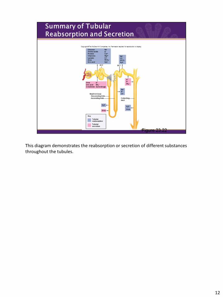
This diagram demonstrates the reabsorption or secretion of different substances throughout the tubules.

The main purpose of the collecting duct is to concentrate the filtrate, this is where filtrate becomes urine. The collecting duct can also be a last ditch effort to reabsorb water.
Urine concentration is controlled by a number of factors.
1. Hydration levels. The more hydrated you are, the less water that will be reabsorbed. If you are dehydrated, more water will be pulled for the filtrate (and urine in the collecting duct).
2. Antidiuretic Hormone (ADH) is a hormone that is secreted by the pituitary gland in response to dehydration. It promotes H2O reabsorption in the collecting duct. It can act on the DCT, but mainly works with the collecting duct.
3. Salt levels: Na+ sets everything up, so it influences the osmotic gradient and flow of water. This is regulated by the hormone Aldosterone. We will talk in greater detail about this particular hormone in the Endocrine system.
4. Diuretics: these are substances that increase urine volume. They are often used with heart meds to thin out blood volume and act as a quick way to lower bloodpressure. Diuretics work either by increasing glomerular filtration or decreasing reabsorption. We can also consume diuretics in our diet. Caffeine dilates the afferent arteriole which increases glomerular filtration. Alcohol is another example, it inhibits ADH and prevents the hormone for stimulating H2O reabsorption.
13

14
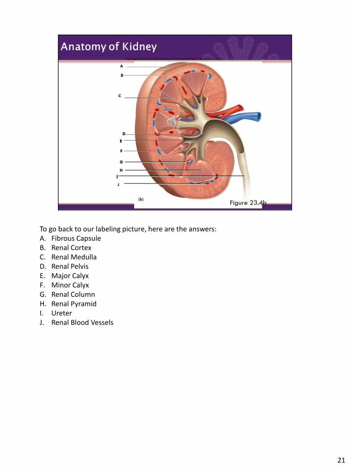
Before we move on, take a minute to see if you can label the structures on this diagram. I will have the answers at the end of the presentation.

15
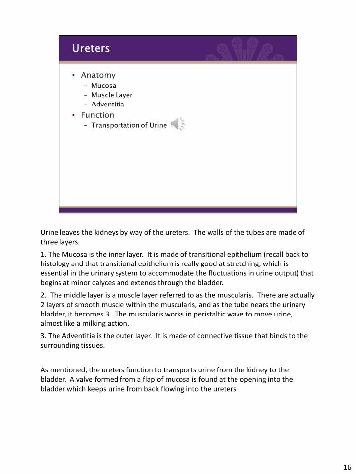
Urine leaves the kidneys by way of the ureters. The walls of the tubes are made of three layers.
1. The Mucosa is the inner layer. It is made of transitional epithelium (recall back to histology and that transitional epithelium is really good at stretching, which is essential in the urinary system to accommodate the fluctuations in urine output) that begins at minor calyces and extends through the bladder.
2. The middle layer is a muscle layer referred to as the muscularis. There are actually 2 layers of smooth muscle within the muscularis, and as the tube nears the urinary bladder, it becomes 3. The muscularis works in peristaltic wave to move urine, almost like a milking action.
3. The Adventitia is the outer layer. It is made of connective tissue that binds to the surrounding tissues.
As mentioned, the ureters function to transports urine from the kidney to the bladder. A valve formed from a flap of mucosa is found at the opening into the bladder which keeps urine from back flowing into the ureters.
16
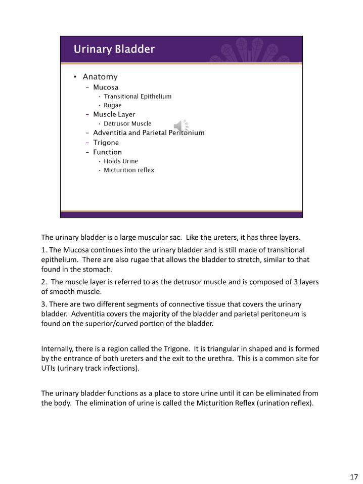
The urinary bladder is a large muscular sac. Like the ureters, it has three layers.
1. The Mucosa continues into the urinary bladder and is still made of transitional epithelium. There are also rugae that allows the bladder to stretch, similar to that found in the stomach.
2. The muscle layer is referred to as the detrusor muscle and is composed of 3 layers of smooth muscle.
3. There are two different segments of connective tissue that covers the urinary bladder. Adventitia covers the majority of the bladder and parietal peritoneum is found on the superior/curved portion of the bladder.
Internally, there is a region called the Trigone. It is triangular in shaped and is formed by the entrance of both ureters and the exit to the urethra. This is a common site for UTIs (urinary track infections).
The urinary bladder functions as a place to store urine until it can be eliminated from the body. The elimination of urine is called the Micturition Reflex (urination reflex).
17

18
The steps of the Micturition reflex (urination reflex) are simplified in the diagram.
1. Filling of the bladder excites stretch receptors which sends a message to the spinal cord.
2. The spinal cord responds and sends out a signal via parasympathetic nerve fibers.
3. The signal excites the detrusor muscles to contract.
4. The signal also stimulates the internal urethral sphincter to relax.
At this point one of two paths can be followed. The pons regulates voluntary control of the elimination of urine. It will give the go ahead signal or not.
If timely, the pons stops regulating signals which allows the external urethral sphincter to relax and urine to leave the body. If it is not timely, the external sphincter will not relax. When toddlers are potty training, this is the reflex control that they are developing.
Of course, there are situations when it may be necessary for the body to eliminate urine and it will override the voluntary control.

As you may have surmised from the previous slide, the urethra is a tube that conveys urine from the urinary bladder out of the body. The external urethral orifice is found between the vaginal opening and the clitoris. There are two sphincters, the external and internal, that allow for control of the passage of urine. The external urethra orifice is the opening to the external environment. The external opening is slightly different in females and males, which we’ll see when we do the reproductive unit. Also, in males, the urethra is used in both the urinary and reproductive systems. In females, at least in human females, this tube is used solely by the urinary system.
19

20
21
To go back to our labeling picture, here are the answers:A. Fibrous CapsuleB. Renal CortexC. Renal MedullaD. Renal PelvisE. Major CalyxF. Minor CalyxG. Renal ColumnH. Renal PyramidI. UreterJ. Renal Blood Vessels