journal of immunological methods · 2018-04-26 · (pierce/thermo scientific) were prepared by...
TRANSCRIPT

Contents lists available at ScienceDirect
Journal of Immunological Methods
journal homepage: www.elsevier.com/locate/jim
Research paper
Neutrophils mediate HIV-specific antibody-dependent phagocytosis andADCC
Matthew J. Worleya, Kuangyu Feia,b, Adam J. Lopez-Denmana, Anthony D. Kelleherc,Stephen J. Kenta,d,e, Amy W. Chunga,⁎
a Department of Microbiology and Immunology, Peter Doherty Institute for Infection and Immunity, University of Melbourne, Melbourne, Australiab School of Medicine, Tsinghua University, Beijing, Chinac The Kirby Institute, UNSW Australia, Sydney, NSW, AustraliadMelbourne Sexual Health Centre, Department of Infectious Diseases, Central Clinical School, Monash University, Melbourne, Australiae ARC Centre of Excellence in Convergent Bio-Nano Science and Technology, University of Melbourne, Melbourne, Australia
A R T I C L E I N F O
Keywords:HIV-1NeutrophilsHL-60ADCCPhagocytosisAntibody
A B S T R A C T
There is growing evidence to support the role of Fc-mediated effector functions, such as Antibody-DependentCellular cytotoxicity (ADCC) and Antibody-Dependent Phagocytosis (ADP) in the protection and control of HIV.The RV144 trial and other recent HIV vaccine studies have highlighted the importance of ADCC responses inprotection against HIV. The role of neutrophils, the most abundant leukocyte in the blood, has not been thor-oughly evaluated for Fc-mediated effector functions to HIV. We optimized HIV-specific neutrophil ADCC andAntibody-Dependent Neutrophil Phagocytosis (ADNP) assays using freshly isolated primary human neutrophilsfrom blood. We also developed methods to study ADP using the neutrophil-like HL-60 cell line. We found thatneutrophils mediate both HIV-specific ADP and ADCC responses. In vitro, neutrophil-mediated ADCC responsespeaked at 4 h, much faster than primary NK cell or monocyte-mediated responses. We detected a wide range ofresponses in the ADNP, HL-60 mediated ADP and ADCC across a cohort of 41 viremic antiretroviral therapynaïve HIV positive subjects. HL-60 and Neutrophil-mediated ADP and ADCC responses correlated well with eachother, suggesting that they measure overlapping functions. The ADNP and HL-60 ADP inversely correlated withHIV viral load, suggesting that these antibody-mediated neutrophil-based assays should prove useful in dis-secting HIV-specific immunity.
1. Introduction
HIV-specific antibodies with Fc-mediated functions may play animportant role in protection from HIV. Recent human and non-humanprimate HIV vaccine studies have identified Fc receptor (FcR) mediatedresponses as potential correlates of protective immunity (Haynes et al.,2012; Bradley et al., 2017; Chung et al., 2015; Barouch et al., 2013,2015). In non-human primate vaccine studies ADP and ADCC responseshave correlated with protection (Bradley et al., 2017; Barouch et al.,2012, 2013). The importance of ADCC and ADP mediating antibodiesduring HIV infection has also been highlighted by numerous studiescorrelating ADCC and ADP with decreased disease progression(Lambotte et al., 2009; Baum et al., 1996; Wren et al., 2013; Chung
et al., 2011a; Vaine et al., 2010; Ana-Sosa-Batiz et al., 2014; Ackermanet al., 2013). In addition, the moderately efficacious RV144 humanPhase III vaccine trial demonstrated the potentially protective cap-abilities of FcR mediated responses, with ADCC responses correlatingwith reduced risk of infection in the absence of IgA (Haynes et al.,2012). Multiple different effector cells, including plasmacytoid den-dritic cells (pDC) (Tjiam et al., 2015), NK cells (Chung et al., 2011a),monocytes/macrophages (Kramski et al., 2012a), other dendritic cellsubsets (Altfeld et al., 2011) and neutrophils (Smalls-Mantey et al.,2013), have the capacity to mediate potent anti-viral Fc-effector re-sponses against HIV. Recent research suggests that polyfunctional Fc-effector responses (i.e. the capacity to engage and recruit multipledifferent effector cells and functions) may be associated with protection
https://doi.org/10.1016/j.jim.2018.03.007Received 19 December 2017; Received in revised form 28 February 2018; Accepted 20 March 2018
⁎ Corresponding author at: Department of Microbiology and Immunology, University of Melbourne, Peter Doherty Institute for Infection and Immunity, 792 Elizabeth St., Melbourne3000, Australia.
E-mail address: [email protected] (A.W. Chung).
Abbreviations: HIV-1, human immunodeficiency virus 1; ADP, antibody-dependent phagocytosis; ADNP, antibody dependent neutrophil phagocytosis; ADCC, antibody dependentcellular cytotoxicity; RFADCC, rapid-fluorometric Antibody dependent cellular cytotoxicity; CFSE, carboxyfluorescein succinimidyl ester; FcR, Fc receptor; DMSO, dimethyl sulfoxide;ART, antiretroviral therapy; SHIP, specific hybridisation internalization probe

and control of HIV infection (Ackerman et al., 2016, Chung et al.,2014). However, the majority of Fc-effector studies in HIV focus uponexamining NK cells and/or monocytes responses, while other effectorcells such as neutrophils remain understudied.
Neutrophils are the most abundant circulating leukocyte in theblood (30–70%) and can rapidly migrate to sites of infection and canmediate a range of effector responses (Kolaczkowska and Kubes, 2013;Mantovani et al., 2011; Palmer et al., 2006). Although neutrophilfunctions have long been associated with the killing and control ofbacterial and fungal infections, there is growing interest in their role incontrol of viral infections (Mocsai, 2013; Galani and Andreakos, 2015;Naumenko et al., 2018). In the context of HIV infection, which is pre-dominantly a mucosally acquired infection, it is important to note thatneutrophils are abundantly present at mucosal surfaces, especially va-ginal tissues and their presence is upregulated during HIV infection(Somsouk et al., 2015; Sips et al., 2016). Neutrophils express FcγRI(induced by cytokines) (Bovolenta et al., 1998), FcγRII and FcγRIII andcan mediate a range of antibody-dependent effector functions, in-cluding ADP and phagocytosis-independent ADCC responses, howeverlittle is known of their importance in HIV infection (Bradley et al.,2017; Ackerman et al., 2016; Sips et al., 2016). Furthermore, whileneutrophils have previously been reported to mediate ADCC killing ofHIV infected cells, it remains unclear if this was in part mediated byphagocytosis (Baldwin et al., 1989; Jenkins et al., 1993).
NK cells have been extensively studied for ADCC responses (Isitmanet al., 2012; Seidel et al., 2013), and mediate target cell lysis throughthe release of perforin and granzymes (Bryceson et al., 2006). In con-trast, the mechanisms that neutrophils utilize to mediate ADCC remainscontroversial, as they lack perforin and granzyme (Grossman and Ley,2004; Metkar and Froelich, 2004). Neutrophils release reactive oxygenintermediates following crosslinking of FcR which have been associatedwith ADCC responses (Horner et al., 2007). However, neutrophils iso-lated from chronic granulomatous disease patients lack reactive oxygenintermediates but are still able to mediate ADCC responses (Robertset al., 1993). This indicates that reactive oxygen intermediates, con-tribute to, but are not the sole mechanism of neutrophil ADCC re-sponses.
The rapid-fluorometric ADCC (RFADCC) has been used extensivelyto evaluate ADCC responses with monocytes, NK cells and PBMCs(Vaine et al., 2010; Gomez-Roman et al., 2006; Chung et al., 2009,2011b; Ruiz et al., 2016; Lai et al., 2014). The short half-life of freshprimary neutrophils (6–8 h) means that it would be useful to developcell-line based neutrophil assays (Summers et al., 2010). The humanpromyelocytic leukemia HL-60 cell line has the capacity to be differ-entiated into neutrophil-like CD11b+ cells after culture with dimethylsulfoxide (DMSO) (Chang et al., 2006; Collins et al., 1978), and can beused to evaluate neutrophil effector functions (Yaseen et al., 2017;Fleck et al., 2005; Kim et al., 2015).
In this study, we optimized and validated HIV-specific neutrophil-mediated RFADCC assays and ADP assays and developed a neutrophil-like cell-line based HL-60 ADP assay. IgG purified from plasma of 41viremic antiretroviral therapy (ART) naïve HIV positive subjects werereadily able to mediate ADNP, HL-60 ADP and neutrophil RFADCCresponses. Furthermore, these Fc-mediated neutrophil responses in-versely correlated with viral load, suggesting that these optimized as-says should prove useful in the evaluation of immune responses, wherefunctional antibodies and neutrophils may play an important role.
2. Material and methods
2.1. Study subjects/plasma samples
Plasma was collected from HIV positive (n=41) subjects, pre-viously described (Wren et al., 2013; Chung et al., 2011a), and HIVnegative healthy donors (n=13). All HIV positive subjects were anti-retroviral therapy (ART) naive. HIV positive subjects had a median CD4
T cell count of 520 cells/μl (range 296–1156 cells/μl) and a medianplasma HIV-1 RNA level of 26,700 copies/ml, (range 399–339,000 co-pies/ml) to reflect the spectrum of HIV disease states (Table 1). Allsubjects provided written informed consent and the studies were ap-proved by the relevant institutional ethics committees.
2.2. Primary cell isolation and culture
Neutrophils were isolated by adapting previously publishedmethods (Bowers et al., 2014; Nauseef, 2007). Briefly, fresh heparinizedblood was obtained from HIV negative donor leukocytes, separatedusing Ficoll density centrifugation. The granulocytes were enriched forby using 3% dextran sedimentation for 25min at room temperature.The remaining cells were washed in and suspended in RPMI 1640media supplemented with 10% FCS and penicillin (100 U/ml)/Strep-tomycin/(100 μg/ml) and L-Glutamine (2 nM) (Gibco, 10378-16)(RF10). The enriched cells were then collected and treated with a hy-potonoic lysis reagent to remove any remaining red blood cells. Thepurity of the isolated cells was evaluated by staining with anti-CD16BV605, CD3 PerCP (Biolegend), CD32 FITC, CD89 APC, CD64 BV510,CD66 BV421, CD14 APC-H7 and CD56 PE (BD biosciences). Monocytesand NK cells were isolated with RosetteSep kits (Stem Cell Technolo-gies) as per manufacturer's instructions.
2.3. HL-60 cells maintenance and differentiation
HL-60 cells (ATCC) were cultured in Iscove's Modified Dulbecco'sMedium (Sigma) with 20% heat-inactivated FCS and penicillin (100 U/ml)/Streptomycin/(100 μg/ml) and L-Glutamine (2 nM). To differ-entiate the HL-60 cell into a neutrophil-like subset, sterile DMSO(Sigma) was added into media at a final concentration of 1.3%, cul-turing for five days, as previously described (Collins et al., 1978; Martinet al., 1990; Birnie, 1988). The generation of neutrophil-like cells wasassessed by staining with CD11b (Chang et al., 2006; Collins et al.,1978), a marker that has previously been identified as essential forneutrophil Fc receptor-mediated cytotoxicity (van Spriel et al., 2001).
2.4. IgG antibody purification
Total IgG was purified using Melon gel resin (Thermo Scientific)following the manufacturer's instructions. Briefly, purification columns(Pierce/Thermo Scientific) were prepared by loading 500 μl of Melongel resin and washed with Melon gel purification buffer (ThermoScientific). Plasma samples were diluted 1:5 in purification buffer,added to columns and incubated at room temperature on an orbitalrotator for 5min. The flow-through was collected then placed back intothe column and incubate at room temperature for a further 5min on theorbital rotator. The columns were centrifuged for 1min at 3000g toelute the purified IgG.
The purified antibodies were quantified using an anti-IgG ELISA kit(Mabtech) following the manufacturer's instructions. Briefly, Maxisorb96 wells plates (Nunc) were coated with the MT145 (2 μg/ml) captureantibody overnight at 4 °C. The plate was washed with PBST (PBS with0.05% Tween 20) and blocked with 1% BSA/PBST for 2 h. The platewas washed and purified IgG antibodies diluted 1:20000 and 1:50,000in 1% BSA/PBST were added for 2 h, alongside the IgG standard. Theplate was washed and the MT78-ALP secondary added to each well and
Table 1Clinical characteristics of study cohort.
HIV positive cohort
Number of subjects 41Median CD4 count entry, cells/μl (range) 520 (296–1156)Median plasma HIV-1 RNA copies/ml (range) 26,700 (399–339,000)
M.J. Worley et al.

incubated for 1 h at room temperature. The plate was washed five timesand developed using of p-nitrophenyl-phosphate (pNPP) and detectedfor optical density at 405 nm on a Thermo Fisher Multiskan Ascent platereader.
2.5. Rapid fluorometric antibody-dependent cellular cytotoxicity(RFADCC)
The RFADCC was performed using similar methods to previouslypublished methods (Kramski et al., 2012b) with different effector cells.Briefly, 1× 10⁶ CEM.NKr-CCR5 cells in 50 μl of RF10 medium werecoated in 3 μg of HIV-1BAL envelope protein gp120 (NIH AIDS reagentprogram) by incubation at room temperature for 1 h. UncoatedCEM.NKr-CCR5 cells were treated identically but without the additionof the gp120BAL. The cell membrane of coated and uncoated CEM.NKr-CCR5 cells were initially stained with 7.5× 10⁻⁷ M of PKH26 (Sigma)diluted in diluent-C for 5min, stopped with 500 μl of FCS and washedtwice with PBS. The cytosols of coated and uncoated CEM.NKr-CCR5cells were then stained with 1× 10⁻⁶ M CFSE (Sigma) for 3min,stopped with 500 μl of FCS and washed twice with PBS, then suspendedin RF10 media. 2× 10⁴ PKH26+ CFSE+ coated and uncoatedCEM.NKr-CCR5 cells per well were incubated with the purified IgG for15min at room temperature to allow for opsonization of target cells.2× 105 effector cells (freshly isolated neutrophils, monocytes, NK cellsor PBMCs) were then added to the target cells and incubated for a rangeof times between 0 and 5-5 h at 37 °C with CO2. Post incubation, thecells were immediately placed on ice and then fixed with a final con-centration of 2% formaldehyde and the proportion of PKH26+ cellsthat had lost intracellular CFSE staining (lysed target cells) was de-termined using flow cytometry on a BD LSR Fortessa with high-throughput sampler.
2.6. Antibody-dependent neutrophil phagocytosis (ADNP)
HIV-1BAL gp120 proteins were biotinylated using the EZ-Link Sulfo-NHS-LC biotinylation kit (Thermo scientific) using a 50mmol excessbiotin according to manufacturer's instructions. After the reaction, thefree biotin was removed by buffer exchange using Amicon 30 kDacentrifugal filters (EMD millipore). The biotinylated gp120BAL was usedto coat the binding sites of 1 μm fluorescent NeutrAvidin Fluosphere(Invitrogen) overnight at 4 °C. The excess antigen was removed bywashing the beads with 2% BSA/PBS and diluted 1:100 in 2% BSA/PBS.Then 10 μl of the diluted beads were incubated with the purified IgG for2 h at 37 °C. The purified neutrophils were added to the bead/antibodymix and incubated for a range of times between 0.5 and 5 h at 37 °Cwith CO2 to allow for phagocytic uptake. The cells were then fixed witha final concentration of 2% formaldehyde and cells were acquired byflow cytometry on a BD LSR Fortessa with a high-throughput samplerattachment. The data was analyzed using FlowJo version 9.8.5 and thephagocytic score (% bead positive cells×mean fluorescent intensity)/104 was calculated as previously described (Darrah et al., 2007) andwas used to compare between conditions.
2.7. HL-60 antibody-dependent phagocytosis
The HL-60 ADP was performed using a similar method to the ADNP.gp120BAL coated NeutrAvidin Fluospheres were prepared in the samemanner as the ADNP. The diluted beads were then incubated with thepurified IgG for 2 h at 37 °C. The differentiated HL-60 were washed andresuspended in fresh media then 1× 10⁵ HL-60 cells were added to thebeads/IgG mix and incubated for 20 h overnight. The HL-60's were thenstained with CD11b BV785 (1:100) and fixed with a final concentrationof 2% formaldehyde and acquired by flow cytometry on a BD LSRFortessa.
2.8. HL-60 ADP-SHIP (specific hybridisation internalization probe) assay
To confirm that the fluorescent beads were completely phagocy-tosed, the HL-60 ADP-SHIP (specific hybridization internalizationprobe) assay was performed as previously described for THP-1 cells(Ana-Sosa-Batiz et al., 2014). Briefly, 3 μg biotinylated gp120BAL (NIHAIDS Reagent Program) were incubated with 1 μl NeutrAvidin Fluo-sphere 1 μm beads (Invitrogen) and 1 μl of 150mM biotin- and Cy5-labelled florescent internalization probe (FIPCy5) (5′ Cy5-TCAGTTCAGGACCCTCGGCT-N3 3′, Integrated DNA Technologies) overnight at4°C. The gp120-coated beads were washed twice with sterile 2% PBS-BSA and diluted in 100 μl 2% PBS-BSA. Then 10 μl of the diluted beadswere incubated with the purified IgG for 2 h at 37 °C. Opsonized beadswere incubated with 1× 10⁵ DMSO-stimulated HL-60 cells in a totalvolume of 100 μl in fresh media. After incubation for a range of timesbetween 1 and 20 h at 37°C, cells were stained with CD11b BV785.Surface-bound beads were quenched by adding 1 μg/ml−1 of thecomplementary quenching probe (QPC) (5′ -AGCCGAGGGTCCTGAACTGA-BHQ2- 3′ Integrated DNA Technologies) for 10min at 4°C andwere subsequently washed with PBS and fixed in 2% formaldehyde.Cells were acquired on a Fortessa LSRII (BD Bioscience) and analyzedusing Flowjo.
2.9. Confocal imaging
Samples used for microscopy were treated the same way as de-scribed for the neutrophil RFADCC and the ADNP assay with the ex-ception that a membrane staining step was performed before fixing.Following the neutrophil RFADCC for 4 h, the cells were stained withCD66b APC, CD89 APC and CD11b APC. While the ADNP samples werestained with CD16 APC and CD32 APC. The samples were washed andfixed with 2% formaldehyde, then loaded on to poly-L-lysine (Sigma)coated slides and cover slips mounted with Ultramount no.4 (Fronine).The Cells were visualized on Zeiss LSM710 laser scanning confocalmicroscope and images were analyzed using ImageJ.
2.10. Statistical analysis
Statistical analyses were completed using Prism GraphPad version7.0a (GraphPad Software, San Diego, CA). A Kruskal-Wallis multiplecomparisons analysis was used to compare the responses of PBMCs,neutrophils, monocytes and NK cells at each time point. A Mann-Whitney U test was used to determine the difference between the HIVpositive IgG responses and the HIV negative IgG (healthy donors) forthe neutrophil RFADCC, ADNP and the HL-60 ADP. NonparametricSpearman correlation analyses were used to test for correlations. A p-value of< 0.05 was considered to indicate a significant difference.
3. Results
3.1. Purified primary blood neutrophils express a range of Fc receptors
Antibody-mediated neutrophil functions are of interest in HIV im-munity since neutrophils can be present at mucosal surfaces in highfrequency (Sips et al., 2016) and antibody-mediated functions cancontribute to control and protective immunity to HIV (Wren et al.,2013; Chung et al., 2011a; Rerks-Ngarm et al., 2009). To evaluatemodels of neutrophil-mediated antibody functions, primary neutrophilswere isolated from freshly collected heparinized blood by density gra-dient and dextran sedimentation from normal healthy subjects. Neu-trophils were defined as small, highly granular, FSC low, SSC high,CD66b+, CD32+ and were isolated with a mean purity of 99%(Fig. 1A). The Fc Receptor expression on freshly isolated neutrophilswas evaluated, with the median MFI for FcαRI (3471), FcγRI (7882),FcγRII (7079) and FcγRIII (34992) determined (Fig. 1B, C). Neutrophilswere isolated from 6 different healthy donors had only slightly varying
M.J. Worley et al.

levels of Fc receptors present on the surface (Fig. 1C).
3.2. Neutrophil-mediated ADCC as measured by the RFADCC assay
Neutrophils have previously been shown to mediate ADCC in re-sponse to antibody opsonized target cells (van der et al., 2002; Peippet al., 2008) and cancer/tumor cells (Schneider-Merck et al., 2010;Albanesi et al., 2013; Keler et al., 1997), but their ability to mediate
ADCC responses against HIV expressing targets has been rarely in-vestigated (Baldwin et al., 1989). To set up a model of neutrophil-de-pendent HIV-specific ADCC activity, fresh neutrophils were evaluatedfor their ability to mediate ADCC responses using the rapid fluorometricADCC (RFADCC) assay (Gomez-Roman et al., 2006). Neutrophils werecultured in the presence of HIV antibodies and HIV-1 envelope proteingp120 BAL coated PKH and CFSE labelled CEM.NKr-CCR5 target cells.Lysis of target cells were defined as PKH+ and CFSE− cells, with a
Fig. 1. Purity of isolated neutrophils and Fc receptor expression. A. Representative flow cytometry plots of staining for neutrophil purity for CD66b+ and CD32+.The isolated cells were confirmed to have typical neutrophil phenotypic markers including CD11b+ and CD56- with low levels of CD14. B. Representative flowcytometry plots of isolated neutrophils for the Fc receptors: CD16 (FcγRIII), CD32 (FcγRII), CD64 (FcγRI), CD89 (FcαRI) expression. C. The MFI for Fc receptorsurface expression of CD16 (FcγRIII), CD32 (FcγRII), CD64 (FcγRI) and CD89 (FcαRI) on the isolated neutrophils for 6 donors.
M.J. Worley et al.

Fig. 2. Neutrophil-mediated RFADCC assay. A. Representative gating strategy for the neutrophil RFADCC. The target cells were stained with PKH membrane dye,CFSE cytoplasmic dye and coated with HIV-1BAL gp120 and incubated with isolated neutrophils. Target cell lysis was identified as cells that were PKH+ and CFSE-with responses only observed for the HIV-1BAL gp120 coated target cells in the presence of HIV positive IgG. B. The %PKH+CFSE- cells at time points 0, 0.5, 1, 2, 3,4and 5 h utilising 25 μg/ml of a pooled purified IgG from HIV positive donors with 6 neutrophil donors, grey lines each represent different donors, while black linerepresents the mean response. C. The %PKH+CFSE− cells at different concentrations (100, 50, 25 10 μg/ml) of pooled purified IgG from HIV positive donors with 6neutrophil donors, grey lines represent different donors, while black line represents the mean response. D. The difference in RFADCC %PKH+CFSE− cell responseswith different effectors cells utilising neutrophils, monocytes, NK cells and PBMCs. A Kruskal-Wallis multiple comparisons analysis was performed and showedneutrophils killed more target cells at 0.5 h than NK cells (p= .0008) and PBMCs (p= .0001), at 1 h NK cells (p≤.0001), PBMC (p= .0032), 2 h (p≤.0001), PBMC(p= .0032), 3 h NK cells (p≤.0001), PBMC (p= .0003), 4 h NK cells (p≤.0001), PBMC (p= .0041). The monocytes responded higher than the NK cells at 0.5, 1, 2,3, 4 h (P= .0018, 0.023, 0.018, 0.023 respectively).
M.J. Worley et al.
representative gating strategy shown in Fig. 2A. To optimize the assay,we evaluated 5 different healthy neutrophil donors. We found thatneutrophils across all donors could readily mediate HIV-specific ADCCand optimal ADCC responses were observed at 4 h, with a median lysisof 72.2% (Fig. 2B).
The neutrophil RFADCC assay was further evaluated to determinethe optimal concentration of purified plasma IgG antibody. Purified IgGfrom 11 different HIV positive donors were evaluated at 4 differentconcentrations (100, 50, 25 and 10 μg/ml) and an EC50 was de-termined. A spearman nonparametric correlation (r value) comparedthe 1/EC50 to the percent lysis for each IgG concentration. For thedifferent concentrations of antibody we found the following results:100 μg/ml r=0.2091 and p= .5393; 50 μg/ml r=0.5818 andp= .0656; 25 μg/ml r=0.8182 and p= .0033 and 10 μg/ml r=0.900and p= .0004 (Fig. S1). We used 25 μg/ml of IgG in future experimentswith the HIV positive cohort as this level had the maximum dynamicrange of responses. The neutrophil variability (donors n=6) at dif-ferent IgG concentrations were evaluated with maximal responses oc-curring at 50 μg/ml and decreasing at 100 μg/ml showing a prozoneeffect (Fig. 2C).
A potential advantage of neutrophils as ADCC effectors compared toother immune cells is their ability to rapidly mediate effector functionsand thereby rapidly limiting viral spread. Isolated neutrophils wereevaluated against other isolated immune cells for their potency inmediating lysis of the target cells over 4 h in the RFADCC assay(Fig. 2D). We found in vitro that neutrophils exhibited higher levels ofADCC at all time points compared to NK cells (p≤ .001) and PBMC(p≤ .005), while the monocytes responded higher than the NK cells atall time points (p≤ .05). Neutrophils also induced higher activity thanmonocytes, however the responses were not significant. This demon-strates the rapid ability of neutrophils to kill HIV-1 gp120 coated targetcells.
3.3. Multiple mechanisms of target cell cytotoxicity are measured by theRFADCC assay
The RFADCC assay has been used widely to evaluate HIV-specificlysis of target cells. Interestingly, Kramski et al. showed that theRFADCC also measures phagocytosis of the labelled lysed target cells bymonocytes (Kramski et al., 2012b). We hypothesized such a phenom-enon may also occur with neutrophils. The neutrophil-mediatedRFADCC assay was therefore adapted to include the CD66b granulocytemarker surface staining to determine if the target cell lysis was medi-ated by ADCC and/or phagocytosis of the target cells using gatingstrategy in Fig. 3A. The neutrophil-mediated RFADCC at early time-points of 0.5 h observed high levels of PKH+ CFSE+ CD66b+ popu-lations (47.4%), suggesting the potential early phagocytosis of un-lysedPKH+ CFSE+ target cells by the CD66b+ neutrophils. While at thesame time-point low levels of PKH+ CFSE− CD66b+ were observed(10.1%) indicating the rapid lysis of phagocytosed target cells or po-tentially uptake of lysed target cell membranes. This PKH+ CFSE−CD66b+ population rapidly increased to 48.8% at the 1 hour timepoint, coinciding with the decrease of PKH+ CFSE+ CD66b+ popu-lations (3.74%) (Fig. 3B). Collectively this early association of theneutrophil and target cell (CD66b+ PKH+ CFSE+) changing to aneutrophil and lysed target cell (CD66b+ PKH+ CFSE−) may indicatethat the target cell has been phagocytosed.
To further interrogate the mechanism behind the PKH+ CFSE−target cell lysis, confocal microscopy imaging was used to evaluate theneutrophil-mediated RFADCC assay. Fig. 3C presents PKH+ membrane(red) of target cells internalized in the neutrophil (blue) in the presence(Fig. 3C ii) and absence (Fig. 3C iii) of the CFSE (green) showingphagocytosis of a lysed target cell. There is also evidence that tradi-tional ADCC responses are present, with PKH+CFSE− target cells inclose proximity to neutrophils (Fig. 3Ci). In addition, different levels ofPKH+ membrane were observed within neutrophils which may
indicate the neutrophils are phagocytosing PKH+ membranes of deadcells killed by traditional ADCC responses (Fig. 3Cvi).
3.4. HIV-specific ADNP assay
The ADNP has been previously published (Ackerman et al., 2016)but has not been directly compared to neutrophil RFADCC assays. Weemployed a modification of the previously reported gp120-coated bead-based phagocytosis assays to study ADNP by flow cytometry using thegating strategy shown in Fig. 4A. ADNP was evaluated using a pool ofHIV-specific polyclonal antibodies (HIVIG) at different concentrationsmeasuring phagocytosis with different neutrophil donors (n=6)(Fig. 4B). There was little difference between the 50 and 100 μg/mlconcentrations over the course of 5 h. However, when ADNP variability(donor n=6) testing for different IgG concentrations were evaluated at4 h incubation, similar to the RFADCC assay we saw a plateauing ofresponses at concentrations of 50 μg/ml or higher (Fig. 4C). To de-termine that the beads were phagocytosed and not associated with thesurface cell membrane, bead internalization was confirmed by confocalmicroscopy. Fig. 4D illustrates bead (green) surround by membrane(blue) showing the beads are phagocytosed. Z-stack movie confirmedcomplete phagocytosis (Fig. S2). There was no uptake of the beadsobserved in the absence of HIV positive IgG.
3.5. HIV-specific HL-60 ADP assay
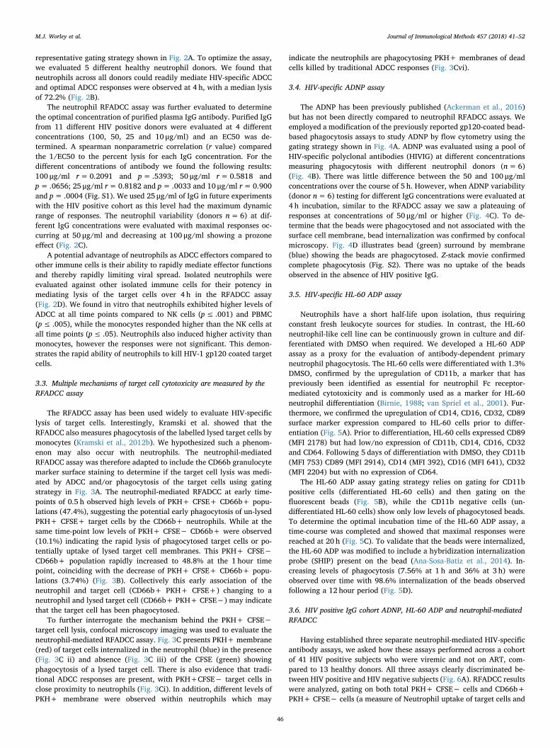
Neutrophils have a short half-life upon isolation, thus requiringconstant fresh leukocyte sources for studies. In contrast, the HL-60neutrophil-like cell line can be continuously grown in culture and dif-ferentiated with DMSO when required. We developed a HL-60 ADPassay as a proxy for the evaluation of antibody-dependent primaryneutrophil phagocytosis. The HL-60 cells were differentiated with 1.3%DMSO, confirmed by the upregulation of CD11b, a marker that haspreviously been identified as essential for neutrophil Fc receptor-mediated cytotoxicity and is commonly used as a marker for HL-60neutrophil differentiation (Birnie, 1988; van Spriel et al., 2001). Fur-thermore, we confirmed the upregulation of CD14, CD16, CD32, CD89surface marker expression compared to HL-60 cells prior to differ-entiation (Fig. 5A). Prior to differentiation, HL-60 cells expressed CD89(MFI 2178) but had low/no expression of CD11b, CD14, CD16, CD32and CD64. Following 5 days of differentiation with DMSO, they CD11b(MFI 753) CD89 (MFI 2914), CD14 (MFI 392), CD16 (MFI 641), CD32(MFI 2204) but with no expression of CD64.
The HL-60 ADP assay gating strategy relies on gating for CD11bpositive cells (differentiated HL-60 cells) and then gating on thefluorescent beads (Fig. 5B), while the CD11b negative cells (un-differentiated HL-60 cells) show only low levels of phagocytosed beads.To determine the optimal incubation time of the HL-60 ADP assay, atime-course was completed and showed that maximal responses werereached at 20 h (Fig. 5C). To validate that the beads were internalized,the HL-60 ADP was modified to include a hybridization internalizationprobe (SHIP) present on the bead (Ana-Sosa-Batiz et al., 2014). In-creasing levels of phagocytosis (7.56% at 1 h and 36% at 3 h) wereobserved over time with 98.6% internalization of the beads observedfollowing a 12 hour period (Fig. 5D).
3.6. HIV positive IgG cohort ADNP, HL-60 ADP and neutrophil-mediatedRFADCC
Having established three separate neutrophil-mediated HIV-specificantibody assays, we asked how these assays performed across a cohortof 41 HIV positive subjects who were viremic and not on ART, com-pared to 13 healthy donors. All three assays clearly discriminated be-tween HIV positive and HIV negative subjects (Fig. 6A). RFADCC resultswere analyzed, gating on both total PKH+ CFSE− cells and CD66b+PKH+ CFSE− cells (a measure of Neutrophil uptake of target cells and
M.J. Worley et al.

target cell membranes). There were only small marginal differencesbetween the responses with these 2 gating strategies (Fig. 6A).
The ADNP and HL-60 ADP both had a large dynamic range of
responses, however, the HL-60 ADP had somewhat greater non-specificuptake of the gp120-coated beads using HIV negative IgG. None-the-less, the ADNP and HL-60 ADP assay exhibited a strong positive
(caption on next page)
M.J. Worley et al.

correlation (r=0.7718, p≤ .0001, Fig. 6B, right panel). Interestingly,we observed that the neutrophil RFADCC with CD66b gating alsoshowed a significant correlation with both the ADNP (r=0.5692,p≤ .0001) and the HL-60 ADP (r=0.4524, p= .0030) (Fig. 6B). Inaddition, the neutrophil RFADCC without CD66b gating also showed asignificant correlation with both the ADNP (r=0.5655 p≤ .0001) and
the HL-60 ADP (r=0.4551, p= .0028) (Fig. S3). The same 5 (out of41) HIV positive donors responded very weakly in all 3 assays.
Since all 41 subjects were ART naive and had a range of HIV virallevels, we were able to evaluated the neutrophil RFADCC, ADNP andHL-60 ADP assays for clinical relevance. We found that there was asignificant negative correlation with vial load and ADNP (r=−0.32,
Fig. 3. Neutrophil- mediated RFADCC is a measure of both extracellular cytotoxicity and phagocytosis. A. Alternative gating strategy for the neutrophil RFADCC.Gating on PKH+ CD66b+ and CFSE− shows that CD66b+ neutrophils acquire target cell PKH and this occurs only in the presence of HIV-1BAL gp120 coated targetcells and HIV positive IgG. B. Gating on PKH+ cells and then CD66b and CFSE over 0, 0.5, 1, 2, 3 and 4 h utilising 25 μg/ml of a pooled purified IgG from HIVpositive donors. C. Confocal microscopy imaging of the neutrophil RFADCC with the neutrophils stained with CD66b APC, CD89 APC and CD11b APC (PKH: red,CFSE: green, Neutrophil markers: blue). Inset panels show i) Dead target cell (PKH+ CFSE−) next to neutrophils, ii) live target cell inside neutrophil, iii) Dead targetcell inside of neutrophil, iv) Neutrophil alone, v) CEM.NKr-CCR5 (target) cell alone, vi) small amount of target cell membrane inside of neutrophil. (For interpretationof the references to colour in this figure legend, the reader is referred to the web version of this article.)
Fig. 4. ADNP responses to HIV positive IgG. A. Representative gating strategy for the ADNP assay. The panels 3 and 4 show minimum uptake of the FITC+ beads inthe absence of antibody and in the presence of HIV negative IgG. Panel 5 shows that beads coated with HIV-1BAL gp120 in the presence HIV positive IgG can be takenup by neutrophils. Phagocytic score=%beads positive cells×mean fluorescence intensity/104. B. ADNP at time points 0, 0.5, 1, 2, 3, 4 and 5 h utilising differentconcentrations (100, 50, 25 10 μg/ml) of pooled purified IgG from HIV positive donors with 6 neutrophil donors. C. ADNP responses at 4 h using different con-centrations (100, 50, 25 10 μg/ml) of pooled purified IgG from HIV positive donors with 6 neutrophil donors; grey lines represent each donor and black line is mean.D. Confocal microscopy showing the internationalisation of 2 FITC+ beads following a 4 h incubation (neutrophils stained with CD16 APC and CD32 APC), scalebar= 20 μm.
M.J. Worley et al.
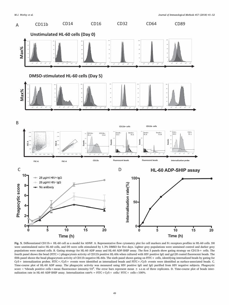
Fig. 5. Differentiated CD11b+ HL-60 cell as a model for ADNP. A. Representative flow cytometry plot for cell markers and Fc receptors profiles in HL-60 cells. D0were unstimulated naïve HL-60 cells, and D5 were cells stimulated by 1.3% DMSO for five days. Lighter grey populations were unstained control and darker greypopulations were stained cells. B. Gating strategy for HL-60 ADP assay and HL-60 ADP-SHIP assay. The first 3 panels show gating strategy on CD11b+ cells. Thefourth panel shows the bead (FITC+) phagocytosis activity of CD11b positive HL-60s when cultured with HIV positive IgG and gp120 coated fluorescent beads. Thefifth panel shows the bead phagocytosis activity of CD11b negative HL-60s. The sixth panel shows gating on FITC+ cells, identifying internalized beads by gating forCy5+ internalization probes. FITC+/Cy5+ events were identified as internalized beads and FITC+/Cy5- events were identified as surface-associated beads. C.Time-course plot of HL-60 ADP assay. The phagocytic activity was measured using HIV positive IgG and IgG purified from HIV negative subjects. Phagocyticscore=%beads positive cells×mean fluorescence intensity/104. The error bars represent mean ± s.e.m of three replicates. D. Time-course plot of beads inter-nalization rate in HL-60 ADP-SHIP assay. Internalization rate%=FITC+Cy5+ cells/ FITC+ cells ∗ 100%.
M.J. Worley et al.
p= .043) and the HL-60 ADP (r=−0.41, p= .008), with the RFADCCapproaching significance (r=−0.31, p= .051) (Fig. 6C). This mayindicate that neutrophils play a role in controlling HIV viral load insome patients.
4. Discussion
There is a growing interest in the role of Fc-mediated effectorfunctions, such as ADCC and ADP in the protection and control of HIV.However, there has been limited investigation into the role of neu-trophil-mediated Fc-responses in HIV infections. We show that neu-trophils are efficient Fc-effector cells and they respond faster thanmonocytes or NK cells when specifically measured by the RFADCCassay. These differences may be due to the different effector mechanisminvolved, as neutrophils express a range of FcγRs and can mediate bothphagocytosis and ADCC responses, whereas NK cells only expressFcγRIIIa and mediate only ADCC responses (Sips et al., 2016; Selvarajet al., 1988). Since the neutrophil RFADCC is measuring in part pha-gocytosis responses, it is unsurprising that these responses correlatewith the primary neutrophil ADNP assay and neutrophil-like cell lineHL-60 ADP assay. The HL-60 neutrophil cell line assay correlatedstrongly with the primary neutrophil ADNP assay and provides a moretractable model for primary neutrophils in the ADP assays. Importantly,
the neutrophil RFADCC, ADNP and HL-60-ADP assays inversely corre-lated with the viral load of the HIV positive IgG donors at the time ofplasma collections, suggesting the possibility that these assays are ofbiological relevance.
The RFADCC assay has been widely used to assess FcγR mediatedresponses of PBMCs (Vaine et al., 2010; Gomez-Roman et al., 2006;Chung et al., 2009). While NK cells have been shown to mediate ADCCresponses, monocyte responses have largely been attributed to trogo-cytosis and phagocytosis of the target cells (Kramski et al., 2012b). Ourdata indicates that the neutrophil RFADCC assay more representsphagocytosis of whole target cells, phagocytosis of killed target celldebris, or trogocytosis. When comparing HIV positive versus negativesample responses, there is an increase in the population of the PKH+CFSE+ CD66b+ cells (ie neutrophils internalizing live target cells) atearly time points (0.5 h) that are higher on the FSC and SSC, which mayindicate the target cells have been phagocytosed (Fig. S4). While at thesame early time point, there is also a small population of PKH+ CFSE−CD66b+ cells (i.e. neutrophils that have internalized target cellmembrane only) which have smaller FSC vs SSC profiles, similar to HIVnegative samples, suggesting that the neutrophils have ingested smallerfragments of dead target cells killed by more traditional extracellularADCC mechanisms. We also observe more PKH+ cells when testingHIV positive IgG samples compared to HIV negative IgG samples,
Fig. 6. Neutrophil Fc-Effector responses of HIV positive cohort. A. 41 HIV positive plasma samples and 13 HIV negative plasma samples were purified for IgG andwere then assess for responses in the neutrophil RFADCC, neutrophil RFADCC with CD66b gating, ADNP and the HL-60 ADP. B. The HIV positive IgG cohortresponses were correlated (spearman) between the 3 assays showing strong significant correlations between the assays. C. The HIV positive IgG cohort responses werecorrelated (spearman) with viral load (HIV-1 RNA copies/ml plasma) and showed significant negative correlations with the ADNP and HL-ADP and approachingsignificance with the neutrophil RFADCC with the CD66b gating.
M.J. Worley et al.
despite the same number of target cells being added to each well (Fig.S5), which may be caused by neutrophils mediating traditional extra-cellular ADCC target cell lysis prior to the neutrophil phagocytosingfragments of dead target cells. Alternatively, multiple neutrophils mayinteract with the same target cell and compete to phagocytose resultingin the sharing of target cell membrane between effector cells.
The neutrophil RFADCC, HL-60-ADP and ADNP responses inverselycorrelated with the viral load of the patients. ADNP responses haverecently been shown not to differ between elite controllers, viremiccontrollers or patients on antiretroviral therapy (Ackerman et al.,2016). It will be of interest to evaluate the differences in clinical po-pulations in the antibody-mediated neutrophil assays in future studies.In addition, neutrophil phagocytic activity is progressively impairedduring HIV infection despite antiretroviral therapy (Tsachouridou et al.,2017). In future studies it will be of interest to investigate neutrophilsisolated from different clinical populations of HIV positive donors fortheir ability to mediated antibody-dependent functions, although thesestudies are technically demanding due to the need for fresh neutrophils.Our and other studies (Bradley et al., 2017; Smalls-Mantey et al., 2013;Ackerman et al., 2016) have utilized circulating neutrophils isolatedfrom blood, however, it is known that neutrophils residing in tissuesand mucosal surfaces can have altered FcγR expression profiles, whichcould potentially alter the Fc-effector potentials of neutrophils at dif-ferent sites (Sips et al., 2016) and should be taken into considerationwhen evaluating FcγR responses. While this study focused exclusivelyon exploring neutrophil mediated Fc-effector responses, multiple otherFcγR bearing innate immune effector cells including pDC, NK cells andmonocytes/macrophages may also participate in antiviral control andtheir respective contributions also deserve further evaluation.
The strong correlation between the ADP and HL-60 ADP assaysuggest that HL-60 cells can represent a useful tool in the evaluation ofother neutrophil effector functions in the future. Due to the difficulty ofworking with primary isolated neutrophils that have short half lives andrequire fresh blood for assays, as neutrophils cannot be reliably cryo-preserved without severely effecting functionality (Boonlayangooret al., 1980), usage of neutrophil-differentiated HL-60 cell lines mayallow for easier, more robust, high throughput evaluation of Fc-medi-ated effector responses. However, HL-60 have several differencescompared to primary neutrophils including differences in gene ex-pression and the lack of several neutrophil proteins including theCD66b surface marker (Ozeki and Shively, 2008). In addition, HL-60neutrophils lack the development of the secondary and tertiary granuleswhich are hall marks of neutrophils, required for some effector func-tions upon cell activation (Gaines et al., 2005). HL-60 neutrophils havebeen shown to mediate lower levels of antimicrobial activity and lowerreactive oxygen production compared to primary blood derived neu-trophils (Yaseen et al., 2017; Watson et al., 1997). These differencesshould be taken into consideration when utilising HL-60 cells to eval-uate alternative Fc-effector functions.
In summary, the neutrophil RFADCC, ADNP or HL-60 ADP werevalidated, evaluated and compared for neutrophil effector functions. Asneutrophil Fc-mediated responses were associated with reduced viralloads in HIV ART naïve subjects, this suggests that neutrophil-mediatedeffector responses should be investigated in future HIV vaccine trials fortheir potential to control viremia, especially due to their rapid responsecompared to other FcγR innate immune cells and their abundance atkey sites of HIV transmission. The assays described should help formthe foundation for future neutrophil Fc-effector studies in HIV infectionand vaccination.
Acknowledgments
We thank Dr. Ivan Stratov for providing HIV positive plasma sam-ples. We would also like to thank Ansari Shaik for assisting with thecohort and clinical information. We acknowledge the NIH AIDS re-agents program for supplying the gp120 BAL (cat# 4961), CEM.NKr-
CCR5 cells (cat# 4376) and HIV-IG (cat# 3957). We also acknowledgethe facilities of the biological optical microscopy platform (BOMP) ofthe University of Melbourne.
Funding
This work was supported by the Australia National Health &Medical Research Center (APP1125164) and the American Foundationfor AIDS Research (amfAR) Mathilde Krim Fellowship (109499-61-RKVA).
Appendix A. Supplementary data
Supplementary data to this article can be found online at https://doi.org/10.1016/j.jim.2018.03.007.
References
Ackerman, M.E., et al., 2013. Enhanced phagocytic activity of HIV-specific antibodiescorrelates with natural production of immunoglobulins with skewed affinity forFcgammaR2a and FcgammaR2b. J. Virol. 87 (10), 5468–5476.
Ackerman, M.E., et al., 2016. Polyfunctional HIV-specific antibody responses are asso-ciated with spontaneous HIV control. PLoS Pathog. 12 (1), e1005315.
Albanesi, M., et al., 2013. Neutrophils mediate antibody-induced antitumor effects inmice. Blood 122 (18), 3160–3164.
Altfeld, M., et al., 2011. DCs and NK cells: critical effectors in the immune response toHIV-1. Nat. Rev. Immunol. 11 (3), 176–186.
Ana-Sosa-Batiz, F., et al., 2014. HIV-specific antibody-dependent phagocytosis maturesduring HIV infection. Immunol. Cell Biol. 92 (8), 679–687.
Baldwin, G.C., et al., 1989. Granulocyte- and granulocyte-macrophage colony-stimulatingfactors enhance neutrophil cytotoxicity toward HIV-infected cells. Blood 74 (5),1673–1677.
Barouch, D.H., et al., 2012. Vaccine protection against acquisition of neutralization-re-sistant SIV challenges in rhesus monkeys. Nature 482 (7383), 89–93.
Barouch, D.H., et al., 2013. Protective efficacy of a global HIV-1 mosaic vaccine againstheterologous SHIV challenges in rhesus monkeys. Cell 155 (3), 531–539.
Barouch, D.H., et al., 2015. Protective efficacy of adenovirus/protein vaccines against SIVchallenges in rhesus monkeys. Science 349 (6245), 320–324.
Baum, L.L., et al., 1996. HIV-1 gp120-specific antibody-dependent cell-mediated cyto-toxicity correlates with rate of disease progression. J. Immunol. 157 (5), 2168–2173.
Birnie, G.D., 1988. The HL60 cell line: a model system for studying human myeloid celldifferentiation. Br. J. Cancer Suppl. 9, 41–45.
Boonlayangoor, P., et al., 1980. Cryopreservation of human granulocytes: study ofgranulocyte function and ultrastructure. Blood 56 (2), 237–245.
Bovolenta, C., et al., 1998. High affinity receptor for IgG (Fc gamma RI/CD64) gene andSTAT protein binding to the IFN-gamma response region (GRR) are regulated dif-ferentially in human neutrophils and monocytes by IL-10. J. Immunol. 160 (2),911–919.
Bowers, N.L., et al., 2014. Immune suppression by neutrophils in HIV-1 infection: role ofPD-L1/PD-1 pathway. PLoS Pathog. 10 (3), e1003993.
Bradley, T., et al., 2017. Pentavalent HIV-1 vaccine protects against simian-human im-munodeficiency virus challenge. Nat. Commun. 8, 15711.
Bryceson, Y.T., et al., 2006. Synergy among receptors on resting NK cells for the acti-vation of natural cytotoxicity and cytokine secretion. Blood 107 (1), 159–166.
Chang, H.H., et al., 2006. Multistable and multistep dynamics in neutrophil differentia-tion. BMC Cell Biol. 7, 11.
Chung, A.W., et al., 2009. Rapid degranulation of NK cells following activation by HIV-specific antibodies. J. Immunol. 182 (2), 1202–1210.
Chung, A.W., et al., 2011a. Activation of NK cells by ADCC antibodies and HIV diseaseprogression. J. Acquir. Immune Defic. Syndr. 58 (2), 127–131.
Chung, A.W., et al., 2011b. Immune escape from HIV-specific antibody-dependent cel-lular cytotoxicity (ADCC) pressure. Proc. Natl. Acad. Sci. 108 (18), 7505–7510.
Chung, A.W., et al., 2014. Polyfunctional Fc-effector profiles mediated by IgG subclassselection distinguish RV144 and VAX003 vaccines. Sci. Transl. Med. 6 (228),228ra38.
Chung, A.W., et al., 2015. Dissecting polyclonal vaccine-induced humoral immunityagainst HIV using systems serology. Cell 163 (4), 988–998.
Collins, S.J., et al., 1978. Terminal differentiation of human promyelocytic leukemia cellsinduced by dimethyl sulfoxide and other polar compounds. Proc. Natl. Acad. Sci. U. S.A. 75 (5), 2458–2462.
Darrah, P.A., et al., 2007. Multifunctional TH1 cells define a correlate of vaccine-medi-ated protection against Leishmania major. Nat. Med. 13 (7), 843–850.
Fleck, R.A., Romero-Steiner, S., Nahm, M.H., 2005. Use of HL-60 cell line to measureopsonic capacity of pneumococcal antibodies. Clin. Diagn. Lab. Immunol. 12 (1),19–27.
Gaines, P., Chi, J., Berliner, N., 2005. Heterogeneity of functional responses in differ-entiated myeloid cell lines reveals EPRO cells as a valid model of murine neutrophilfunctional activation. J. Leukoc. Biol. 77 (5), 669–679.
Galani, I.E., Andreakos, E., 2015. Neutrophils in viral infections: current concepts andcaveats. J. Leukoc. Biol. 98 (4), 557–564.
M.J. Worley et al.

Gomez-Roman, V.R., et al., 2006. A simplified method for the rapid fluorometric as-sessment of antibody-dependent cell-mediated cytotoxicity. J. Immunol. Methods308 (1–2), 53–67.
Grossman, W.J., Ley, T.J., 2004. Granzymes A and B are not expressed in human neu-trophils. Blood 104 (3), 906–907 (author reply 907-8).
Haynes, B.F., et al., 2012. Immune-correlates analysis of an HIV-1 vaccine efficacy trial.N. Engl. J. Med. 366 (14), 1275–1286.
Horner, H., et al., 2007. Intimate cell conjugate formation and exchange of membranelipids precede apoptosis induction in target cells during antibody-dependent, gran-ulocyte-mediated cytotoxicity. J. Immunol. 179 (1), 337–345.
Isitman, G., Stratov, I., Kent, S.J., 2012. Antibody-dependent cellular cytotoxicity and NKcell-driven immune escape in HIV infection: implications for HIV vaccine develop-ment. Adv. Virol. 2012, 637208.
Jenkins, M., Mills, J., Kohl, S., 1993. Natural killer cytotoxicity and antibody-dependentcellular cytotoxicity of human immunodeficiency virus-infected cells by leukocytesfrom human neonates and adults. Pediatr. Res. 33 (5), 469–474.
Keler, T., et al., 1997. Bispecific antibody-dependent cellular cytotoxicity of HER2/neu-overexpressing tumor cells by Fc gamma receptor type I-expressing effector cells.Cancer Res. 57 (18), 4008–4014.
Kim, K.H., Seoh, J.Y., Cho, S.J., 2015. Phenotypic and functional analysis of HL-60 cellsused in opsonophagocytic-killing assay for Streptococcus pneumoniae. J. Korean Med.Sci. 30 (2), 145–150.
Kolaczkowska, E., Kubes, P., 2013. Neutrophil recruitment and function in health andinflammation. Nat. Rev. Immunol. 13 (3), 159–175.
Kramski, M., et al., 2012a. Critical role for monocytes in mediating HIV-specific antibody-dependent cellular cytotoxicity. Retrovirology 9.
Kramski, M., et al., 2012b. Role of monocytes in mediating HIV-specific antibody-de-pendent cellular cytotoxicity. J. Immunol. Methods 384 (1–2), 51–61.
Lai, J.I., et al., 2014. Divergent antibody subclass and specificity profiles but not pro-tective HLA-B alleles are associated with variable antibody effector function amongHIV-1 controllers. J. Virol. 88 (5), 2799–2809.
Lambotte, O., et al., 2009. Heterogeneous neutralizing antibody and antibody-dependentcell cytotoxicity responses in HIV-1 elite controllers. AIDS 23 (8), 897–906.
Mantovani, A., et al., 2011. Neutrophils in the activation and regulation of innate andadaptive immunity. Nat. Rev. Immunol. 11 (8), 519–531.
Martin, S.J., Bradley, J.G., Cotter, T.G., 1990. HL-60 cells induced to differentiate towardsneutrophils subsequently die via apoptosis. Clin. Exp. Immunol. 79 (3), 448–453.
Metkar, S.S., Froelich, C.J., 2004. Human neutrophils lack granzyme A, granzyme B, andperforin. Blood 104 (3), 905–906 (author reply 907-8).
Mocsai, A., 2013. Diverse novel functions of neutrophils in immunity, inflammation, andbeyond. J. Exp. Med. 210 (7), 1283–1299.
Naumenko, V., et al., 2018. Neutrophils in viral infection. Cell Tissue Res. 371 (3),505–516.
Nauseef, W.M., 2007. Isolation of human neutrophils from venous blood. Methods Mol.Biol. 412, 15–20.
Ozeki, M., Shively, J.E., 2008. Differential cell fates induced by all-trans retinoic acid-treated HL-60 human leukemia cells. J. Leukoc. Biol. 84 (3), 769–779.
Palmer, C., et al., 2006. Cell-type specific gene expression profiles of leukocytes in human
peripheral blood. BMC Genomics 7, 115.Peipp, M., et al., 2008. Antibody fucosylation differentially impacts cytotoxicity mediated
by NK and PMN effector cells. Blood 112 (6), 2390–2399.Rerks-Ngarm, S., et al., 2009. Vaccination with ALVAC and AIDSVAX to prevent HIV-1
infection in Thailand. N. Engl. J. Med. 361, 2209–2220.Roberts, R.L., et al., 1993. Role of oxygen intermediates in cytotoxicity: studies in chronic
granulomatous disease. Inflammation 17 (1), 77–92.Ruiz, M.J., et al., 2016. Env-specific IgA from viremic HIV-infected subjects compromises
antibody-dependent cellular cytotoxicity. J. Virol. 90 (2), 670–681.Schneider-Merck, T., et al., 2010. Human IgG2 antibodies against epidermal growth
factor receptor effectively trigger antibody-dependent cellular cytotoxicity but, incontrast to IgG1, only by cells of myeloid lineage. J. Immunol. 184 (1), 512–520.
Seidel, U.J., Schlegel, P., Lang, P., 2013. Natural killer cell mediated antibody-dependentcellular cytotoxicity in tumor immunotherapy with therapeutic antibodies. Front.Immunol. 4, 76.
Selvaraj, P., et al., 1988. The major fc receptor in blood has a phosphatidylinositol anchorand is deficient in paroxysmal-nocturnal hemoglobinuria. Nature 333 (6173),565–567.
Sips, M., et al., 2016. Fc receptor-mediated phagocytosis in tissues as a potent mechanismfor preventive and therapeutic HIV vaccine strategies. Mucosal Immunol. 9,1584–1595.
Smalls-Mantey, A., Connors, M., Sattentau, Q.J., 2013. Comparative efficiency of HIV-1-infected T cell killing by NK cells, monocytes and neutrophils. PLoS One 8 (9),e74858.
Somsouk, M., et al., 2015. Gut epithelial barrier and systemic inflammation duringchronic HIV infection. AIDS 29 (1), 43–51.
Summers, C., et al., 2010. Neutrophil kinetics in health and disease. Trends Immunol. 31(8), 318–324.
Tjiam, M.C., et al., 2015. Viremic HIV controllers exhibit high plasmacytoid dendriticcell-reactive opsonophagocytic IgG antibody responses against HIV-1 p24 associatedwith greater antibody isotype diversification. J. Immunol. 194 (11), 5320–5328.
Tsachouridou, O., et al., 2017. Deficient phagocytosis among HIV-1 infected adults overtime even in HAART setting. Curr. HIV Res. 15 (4), 285–290.
Vaine, M., et al., 2010. Profiles of human serum antibody responses elicited by threeleading HIV vaccines focusing on the induction of Env-specific antibodies. PLoS One5 (11), e13916.
van der Kolk, L.E., et al., 2002. Analysis of CD20-dependent cellular cytotoxicity by G-CSF-stimulated neutrophils. Leukemia 16 (4), 693–699.
van Spriel, A.B., et al., 2001. Mac-1 (CD11b/CD18) is essential for Fc receptor-mediatedneutrophil cytotoxicity and immunologic synapse formation. Blood 97 (8),2478–2486.
Watson, R.W.G., et al., 1997. Granulocytic differentiation of HL-60 cells results inspontaneous apoptosis mediated by increased caspase expression. FEBS Lett. 412 (3),603–609.
Wren, L.H., et al., 2013. Specific antibody-dependent cellular cytotoxicity responses as-sociated with slow progression of HIV infection. Immunology 138 (2), 116–123.
Yaseen, R., et al., 2017. Antimicrobial activity of HL-60 cells compared to primary blood-derived neutrophils against Staphylococcus aureus. J. Negat. Results Biomed. 16.
M.J. Worley et al.