jianghong rao- shedding light on tumors using nanoparticles
TRANSCRIPT

8/3/2019 Jianghong Rao- Shedding Light on Tumors Using Nanoparticles
http://slidepdf.com/reader/full/jianghong-rao-shedding-light-on-tumors-using-nanoparticles 1/4
Subscriber access provided by Stanford University
ACS Nano is published by the American Chemical Society. 1155 Sixteenth Street
N.W., Washington, DC 20036
Perspective
Shedding Light on Tumors Using NanoparticlesJianghong Rao
ACS Nano , 2008, 2 (10), 1984-1986• DOI: 10.1021/nn800669n • Publication Date (Web): 28 October 2008
Downloaded from http://pubs.acs.org on February 23, 2009
More About This Article
Additional resources and features associated with this article are available within the HTML version:
• Supporting Information• Access to high resolution figures• Links to articles and content related to this article• Copyright permission to reproduce figures and/or text from this article

8/3/2019 Jianghong Rao- Shedding Light on Tumors Using Nanoparticles
http://slidepdf.com/reader/full/jianghong-rao-shedding-light-on-tumors-using-nanoparticles 2/4
Shedding Light on Tumors UsingNanoparticlesJianghong Rao*
Molecular Imaging Program at Stanford, Department of Radiology & Bio-X Program, Cancer Biology Program, Stanford University School of Medicine, 1201 Welch Road,
Stanford, California 94305-5484
A cohort of nanoparticles with vari-
ous shapes such as quantum dots,
nanotubes, nanohorns, and nano-
cages and made of different materials, from
organic dendrimers, liposomes, gold, car-
bon, semiconductors, silicon to iron oxide,
have already been fabricated and explored
for cancer imaging and therapeutic applica-
tions (e.g., see Figure 1).17
However, thereare various concerns associated with their
use as the carrier system, including the in
vivo safety profile, stability, drug releasing
efficiency, and clearance kinetics.1,2,812
Consequently, development of nontoxic
biocompatible nanoparticles with favorable
in vivo pharmacokinetics and efficient deliv-
ery to tumors is still much needed for medi-
cal applications.
In this issue of ACS Nano, Adair and col-
leagues explored calcium phosphate nano-
particles (CPNPs) as the carrier system fornear-infrared fluorescence imaging of
breast cancer tumors.13 Calcium phosphate
is the principle building component of hard
tissues such as bone and tooth enamel in
the body. Unlike most other nanoparticles,
the biodegradation products of
CPNPs O calcium and phosphate
ions O already exist in the body at millimo-
lar concentrations and are thus presumed
to be nontoxic.
While the composition of the nanoparti-
cles is important for cytotoxicity, other pa-
rameters like size and surface groups are
mainly responsible for their in vivo behav-
ior.14 After introduction into the blood-
stream via intravenous administration, the
particles travel to the organs and peripheral
tissues of the body through the blood ves-
sels. Since the average effective pore size in
normal intact endothelium is5 nm, nano-
particles with a hydrodynamic diameter of 5 nm rapidly extravasate out across the
endothelium, resulting in a short blood cir-
culation time.15
The clearance pathway is also depend-
ent on the particle size. The glomerular fil-
tration in kidney has a size threshold: par-
ticles with a hydrodynamic diameter (HD) of
6 nm are typically filtered and undergo re-
nal clearance while those with a HD 8
nm do not.12,15 Particles undergoing renal
clearance will have a short blood circulation
time. Large particles commonly undergohepatic clearance in liver where Kupffer
cells (macrophages located in the liver) and
hepatocytes capture foreign nanoparticles
for further processing: breaking down or
trapping in Kupffer cells, or undergoing bil-
iary excretion by hepatocytes.
In addition to the size, the surface
groups on the nanoparticles are also criti-
cal. Depending on their surface properties
such as charge and hydrophobicity, they
may undergo adsorption or opsonization
by serum proteins, which results in an in-crease in the particle size.14
Highly charged (either posi-
tively or negatively) nanopar-
ticles often are taken up by
the macrophages in the retic-
uloendothelial system (RES).
To minimize the nonspecific
interactions and increase the
nanoparticle targeting at the
tumor site, a widely used
method is to attach polyethyl-
ene glycol (PEG) to the sur-
See the accompanying Article by
Altinoglu et al . on p 2075.
*Address correspondence to
Published onlineOctober 28, 2008.
10.1021/nn800669n CCC: $40.75
© 2008 American Chemical Society
Figure 1. Schematic of a multifunctional nanoparticle with im-aging probes and/or anticancer drugs encapsulated inside andtumor-specific ligands and/or antibodies presenting on thesurface.
ABSTRACT The scaffold of
nanoparticles (broadly defined as
having a size range of 1100 nm)
presents a convenient platform to
incorporate multiple functionalities
into one single particle for cancer
imaging and therapeutics. Whetherhollow inside or not, a single
nanoparticle can encapsulate a large
payload of imaging probes, anticancer
drug molecules, or both. On the
surface, tumor-specific targeting
molecules (e. g., receptor-binding
ligands or antibodies) may be
immobilized to facilitate active tumor
targeting and drug delivery. This
versatile nanoplatform promises more
efficient delivery of payloads to
tumors for improving cancer detection
and treatment.
P E
R S P E C
T I V E
VOL. 2 ▪ NO. 10 ▪ RAO www.acsnano.org1984
8/3/2019 Jianghong Rao- Shedding Light on Tumors Using Nanoparticles
http://slidepdf.com/reader/full/jianghong-rao-shedding-light-on-tumors-using-nanoparticles 3/4
face which can provide both
steric stabilization and sur-
face charge neutralization.
In the Adair study, the
synthesized calcium phos-
phate nanoparticles have a
spherical shape with a HD of
16 nm.13 Carboxylate groups
are present at the surface,
but some (likely not all) have
been conjugated with PEG
to inhibit the opsonization
and improve the in vivo phar-
macokinetics of particles.
Each CPNP is doped with a
near-infrared dye indocya-
nine green (ICG), which al-
lows real-time noninvasive
imaging of the CPNP distri-
bution and localization in liv-
ing animals by in vivo fluo-rescence imaging.
With the estimate of
600 copies of ICG encapsulated
in each CPNP, the nanoparticles
have a high loading capacity. Fur-
thermore, the authors found that
each embedded ICG displayed im-
proved chemical stability and quan-
tum efficiency, suggesting the pro-
tective effect of the encapsulation
by CPNPs. These results illustrate
the advantages of using CPNPs as
the carrier to deliver the imaging
probes.
Adair and colleagues observed
the uptake of the particles from the
blood to the liver and then to the
gastrointestinal tract, suggesting
the mechanism of heptobiliary
clearance. This clearance mecha-
nism is different from many other
nanoparticles that are taken up by
Kupffer cells in RES. The heptobiliary
clearance of CPNPs avoids long-
term accumulation in the liver and
thus limits potential hepatic toxicity.
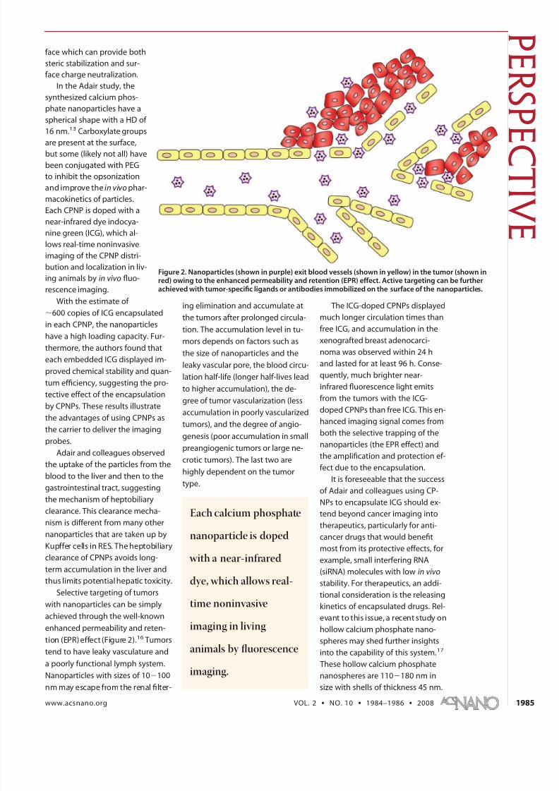
Selective targeting of tumors
with nanoparticles can be simply
achieved through the well-known
enhanced permeability and reten-
tion (EPR) effect (Figure 2).16 Tumors
tend to have leaky vasculature and
a poorly functional lymph system.
Nanoparticles with sizes of 10100
nm may escape from the renal filter-
ing elimination and accumulate at
the tumors after prolonged circula-
tion. The accumulation level in tu-
mors depends on factors such as
the size of nanoparticles and the
leaky vascular pore, the blood circu-
lation half-life (longer half-lives lead
to higher accumulation), the de-
gree of tumor vascularization (less
accumulation in poorly vascularizedtumors), and the degree of angio-
genesis (poor accumulation in small
preangiogenic tumors or large ne-
crotic tumors). The last two are
highly dependent on the tumor
type.
The ICG-doped CPNPs displayed
much longer circulation times than
free ICG, and accumulation in the
xenografted breast adenocarci-
noma was observed within 24 h
and lasted for at least 96 h. Conse-
quently, much brighter near-
infrared fluorescence light emits
from the tumors with the ICG-
doped CPNPs than free ICG. This en-
hanced imaging signal comes from
both the selective trapping of the
nanoparticles (the EPR effect) and
the amplification and protection ef-
fect due to the encapsulation.
It is foreseeable that the success
of Adair and colleagues using CP-
NPs to encapsulate ICG should ex-
tend beyond cancer imaging into
therapeutics, particularly for anti-
cancer drugs that would benefit
most from its protective effects, for
example, small interfering RNA
(siRNA) molecules with low in vivo
stability. For therapeutics, an addi-
tional consideration is the releasing
kinetics of encapsulated drugs. Rel-
evant to this issue, a recent study on
hollow calcium phosphate nano-
spheres may shed further insights
into the capability of this system.17
These hollow calcium phosphate
nanospheres are 110180 nm in
size with shells of thickness 45 nm.
Figure 2. Nanoparticles (shown in purple) exit blood vessels (shown in yellow) in the tumor (shown inred) owing to the enhanced permeability and retention (EPR) effect. Active targeting can be furtherachieved with tumor-specific ligands or antibodies immobilized on the surface of the nanoparticles.
Each calcium phosphate
nanoparticle is doped
with a near-infrared
dye, which allows real-
time noninvasive
imaging in living
animals by fluorescence
imaging.
P E R S P
E C T I V E
www.acsnano.org VOL. 2 ▪ NO. 10 ▪ 1984–1986 ▪ 2008 1985
8/3/2019 Jianghong Rao- Shedding Light on Tumors Using Nanoparticles
http://slidepdf.com/reader/full/jianghong-rao-shedding-light-on-tumors-using-nanoparticles 4/4
The cavities (60 nm) are loaded
with a model drug amylose. Under
the irradiation of ultrasound energy,the hollow structures collapse to
form pinlike nanocrystallites of cal-
cium phosphate and the encapsu-
lated drug molecules are released in
a few minutes in vitro. This
ultrasound-directed release of the
drug payload is fast and may pro-
vide both temporal and spatial con-
trol of the releasing process.
The current set of experiments
with the CPNP system relies on pas-
sive tumor accumulation based onthe EPR effect. Alternatively, ligands
or antibodies that bind specifically
to the receptor at the tumor surface
may be immobilized at the surface
of the nanoparticles to realize active
tumor targeting. The presence of
multiple copies of tumor-specific
ligands or antibodies would in-
crease the binding avidity and en-
hance tumor-targeting specificity,
rendering minimal drug toxicity at
the normal tissues. However, thesize of the nanoparticles will un-
avoidably increase after the ligand
or antibody conjugation. Whether
the resultant nanoparticles retain
the same biodistribution property
and clearance mechanism remains
to be tested.
Similar to the CPNPs reported by
Adair and co-workers, newer and
more complex multifunctional nano-
particles will continue to emerge
from research. These nanoparticles
may contain both imaging probes
and therapeutic drugs, allowing si-
multaneous real-time tracking of the
drug location (Figure 1). A single par-
ticle may even encapsulate two or
more drugs with different cancer-
killing mechanisms; ligands target-
ing two or more different receptors
on the same tumor surface may
present on one single particle, which
should further enhance the specific-
ity of targeted nanoparticles. With
well-designed new nanoparticles and
further addition of these sophisti-
cated multifunctions, nanoparticle-
based imaging and therapeutics are
beginning to have a genuine impact
on cancer diagnostics and treatment
in clinics.
REFERENCES AND NOTES
1. Davis, M. E.; Chen, Z.; Shin, D. M.
Nanoparticles Therapeutics: An
Emerging Treatment Modality for
Cancer. Nat. Rev. Drug Discovery
2008, 7 , 771–782.
2. Sanvicens, N.; Marco, M. P.
Multifunctional Nanoparticles O
Properties and Prospects for Their
Use in Human Medicine. Trends
Biotechnol.2008, 26, 425–433.
3. Ajima, K.; Murakami, T.; Mizoguchi,
Y.; Tsuchida, K.; Ichihashi, T.; Iijima,
S.; Yudasaka, M. Enhancement of In
Vivo Anticancer Effects of Cisplatin
by Incorporation Inside Single-WallCarbon Nanohorns. ACS Nano 2008,
2, 2057–2064.
4. Lam, R.; Chen, M.; Pierstorff, E.;
Huang, H.; Osawa, E.; Ho, D.
Nanodiamond-Embedded
Microfilm Devices for Localized
Chemotherapeutic Elution. ACS
Nano 2008, 2, 2085–2094.
5. Erogbogbo, F.; Yong, K.; Roy, I.; Xu,
G.; Prasad, P. N.; Swihart, M. T.
Biocompatible Luminescent Silicon
Quantum Dots for Imaging of
Cancer Cells. ACS Nano 2008, 2,
873–878.
6. Al-Jamal, W. T.; Al-Jamal, K. T.; Tian,
B.; Lacerda, L.; Bomans, P. H.;Frederik, P. M.; Kostarelos, K. Lipid-
Quantum Dot Bilayer Vesicles
Enhance Tumor Cell Uptake and
Retention in Vitro and in Vivo. ACS
Nano 2008, 2, 408–418.
7. Kobayashi, H.; Koyama, Y.; Barrett,
T.; Hama, Y.; Regino, C. A.; Shin, I. S.;
Jang, B.; Le, N.; Paik, C. H.; Choyke,
P. L.; Urano, Y. Multimodal
Nanoprobes for Radionuclide and
Five-Color Near-Infrared Optical
Lymphatic Imaging. ACS Nano2007, 1, 258–264.
8. Colvin, V. L. The Potential
Environmental Impact of
Engineered Nanomaterials. Nat.Biotechnol.2003, 21, 1166–1170.
9. Pan, Y.; Neuss, S.; Leifert, A.; Fischler,M.; Wen, F.; Simon, U.; Schmid, G.;Brandau, W.; Jahnen-Dechent, W.Size-Dependent Cytotoxicity of Gold Nanoparticles. Small 2007, 3,9141–1949.
10. Jeng, H. A.; Swanson, J. Toxicity of Metal Oxide Nanoparticles in
Mammalian Cells. J. Environ. Sci.Health, A 2006, 41, 2699–2711.
11. Schipper, M. L.; Cheng, Z; Lee, S.-W.;Keren, S.; Bentolila, L. A.;Sundaresan, G.; Iyer, G.; Gheysens,O.; Ebenstein, Y.; Li, J.; Rao, J.; Chen,X.; Wu, A. M.; Weiss, S. S.; Gambhir,S. S. MicroPET-Based Biodistributionof Quantum Dots in Living Mice.
J. Nucl. Med. 2007, 48, 1511–1518.12. Choi, H. S.; Liu, W.; Misra, P.; Tanaka,
E.; Zimmer, J. P.; Ipe, B. I.; Bawendi,M. G.; Frangioni, J. V. RenalClearance of Quantum Dots. Nat.Biotechnol.2007, 25, 1165–1170.
13. Altınoglu, E. I.; Russin, T. J.; Kaiser,J. M.; Barth, B. M.; Eklund, P. C.;Kester, M.; Adair, J. H. Near-InfraredEmitting Fluorophore-DopedCalcium Phosphate Nanoparticlesfor In Vivo Imaging of Human BreastCancer. ACS Nano 2008, 2,2075–2084.
14. Moghimi, S. M.; Hunter, A. C.;Murray, J. C. Long-Circulating and Target-Specific Nanoparticles: Theory to Practice. Pharmaco. Rev.2001, 53, 283–318.
15. Longmire, M. L.; Choyke, P. L.;Kobayashi, H. Clearance Propertiesof Nano-Sized Particles andMolecules Used as Imaging Agents:Considerations and Caveats.
Nanomedicine 2008, 3, 703–717.16. Maeda, H.; Wu, J.; Sawa, T.;
Matsumura, Y.; Hori, K. TumorVascular Permeability and the EPREffect in Macromolecular Therapeutics: A Review. J.Controlled Release 2000, 65,271–284.
17. Cai, Y.; Pan, H.; Xu, X.; Hu, Q.; Li, L.; Tang, R. Ultrasound ControlledMorphology Transformation of Hollow Calcium PhosphateNanospheres: A Smart andBiocompatible Drug ReleaseSystem. Chem. Mater. 2007, 19,3081–3083.
It is foreseeable that the
success of Adair and
colleagues using calcium
phosphate nanoparticlesshould extend beyond
cancer imaging into
therapeutics, particularly
for anticancer drugs.
P E R S P E
C T I V E
VOL. 2 ▪ NO. 10 ▪ RAO www.acsnano.org1986