j. biol. chem.-2008-lin-15003-14
TRANSCRIPT
Transforming Growth Factor � Up-regulates Cysteine-richProtein 2 in Vascular Smooth Muscle Cells via ActivatingTranscription Factor 2*
Received for publication, February 28, 2008, and in revised form, March 27, 2008 Published, JBC Papers in Press, April 3, 2008, DOI 10.1074/jbc.M801621200
Da-Wei Lin‡, Il-Chi Chang§, Alan Tseng‡, Meng-Ling Wu§, Chung-Huang Chen§, Cassandra A. Patenaude¶,Matthew D. Layne‡¶, and Shaw-Fang Yet‡§1
From the ‡Department of Medicine, Brigham and Women’s Hospital and Harvard Medical School, Boston, Massachusetts 02115,the §Cardiovascular and Blood Medical Research Center, National Health Research Institutes, Zhunan Town, Miaoli County 35053, Taiwan,and the ¶Department of Biochemistry, Boston University School of Medicine, Boston, Massachusetts 02118
CRP2 (cysteine-rich protein) is a vascular smooth muscle cell(VSMC)-expressed LIM-only protein. CRP2 associates with theactin cytoskeleton and interactswith transcription factors in thenucleus to mediate smooth muscle cell gene expression. UsingCsrp2 (gene symbol of themouseCRP2 gene)-deficientmice, wepreviously demonstrated that an absence of CRP2 enhancesVSMC migration and increases neointima formation followingarterial injury. Despite its importance in vascular injury, themolecular mechanisms controlling CRP2 expression in VSMCare largely unknown. Transforming growth factor � (TGF�), akey factor present in the vessel wall in the early phases ofarterial response to injury, plays an important role in modu-lating lesion formation. Because both CRP2 and TGF� aremediators of VSMC responses, we examined the possibilitythat TGF� might regulate CRP2 expression. TGF� signifi-cantly induced CRP2 mRNA and protein expression inVSMCs. Promoter analysis identified a conserved cAMP-re-sponsive element (CRE)-like site (TAACGTCA) in the Csrp2promoter that was critical for basal promoter activity andresponse to TGF�. Gel mobility shift assays revealed thatmainly ATF2 bound to this CRE-like element, and mutationof the CRE sequences abolished binding. TGF� enhanced theactivation of ATF2, leading to increased phospho-ATF2 lev-els within the DNA-protein complexes. Furthermore, ATF2-transactivated Csrp2 promoter activity and TGF� enhancedthis activation. In addition, a phosphorylation-negativeATF2 mutant construct decreased basal and TGF�-mediatedCsrp2 promoter activity. Our results show for the first time inVSMC that TGF� activates ATF2 phosphorylation and Csrp2gene expression via a CRE promoter element.
Vascular smooth muscle cells (VSMCs)2 of the arterial wallplay a critical role in the development of occlusive vascularlesions. In normal vessels, VSMCs are quiescent, differentiated,and contractile and regulate vascular tone and blood pressure.In response to arterial injury, VSMCs undergo a phenotypictransitionwhereby theymigrate and proliferate from themediainto the intima, contributing to vascular lesion formation andarteriosclerosis (1).The LIM domain is a double zinc finger structure that func-
tions as amodular protein-binding interface thatmediates pro-tein-protein interactions (2–6). By binding to target proteinsand serving as an adaptor molecule in the assembly of multi-protein complexes, LIM proteins participate in diverse biolog-ical processes (6, 7). The LIM-only cysteine-rich protein (CRP)family, which includes CRP1 (8, 9), CRP2/SmLIM (10), andCRP3/MLP (muscle LIM protein) (11), is characterized by twotandem LIM domains, each followed by a short glycine-richrepeat (12, 13). CRP1 is expressed in several cell types, includingvascular and visceral smooth muscle cells (SMCs) (8, 14),whereas CRP2 ismainly expressed in VSMCs, particularly arte-rial SMC (10, 15, 16). CRP3 is specifically expressed in cardiacand skeletal muscle (11, 17). CRPs are involved in promotingprotein assembly along the actin-based cytoskeleton (7, 13, 18).CRPs associate with the actin cytoskeleton via interacting withthe actin-cross-linking protein �-actinin and adhesion plaqueprotein zyxin (4, 13, 19). In addition, CRPs also interact withtranscription factors to activate gene transcription in thenucleus (20, 21). Given their prominent cytoskeletal associationand presence in the nucleus, CRPs have both a major cytoskel-etal function and a role controlling gene expression (6). Acytoskeletal function of CRP3 was demonstrated in mice lack-ing CRP3; those mice developed dilated cardiomyopathy withhypertrophy and heart failure after birth (17). Ultrastructuralanalysis revealed dramatic disruption of cardiomyocyte cytoar-chitecture (17). Further supporting a function of CRP3 inmain-taining the cytoarchitecture of striated muscle cells, the Dro-sophila homolog of muscle LIM protein, Mlp84B, was recently
* This work was supported, in whole or in part, by National Institutes of HealthGrants HL-057977 (to S.-F. Y.) and HL-078869 (to M. D. L.). This work wasalso supported by National Health Research Institutes (Taiwan) Grant97A1-CVPP02-014 (to S.-F. Y.) and National Science Council (Taiwan) Grant96-2321-B-400-004-MY2 (to S.-F. Y.). The costs of publication of this articlewere defrayed in part by the payment of page charges. This article musttherefore be hereby marked “advertisement” in accordance with 18 U.S.C.Section 1734 solely to indicate this fact.
1 To whom correspondence should be addressed: National Health ResearchInst., 35 Keyan Rd., R2-5021, Zhunan Town, Miaoli County 35053, Taiwan.Tel.: 886-37-246166 (ext. 38311); Fax: 886-37-587408; E-mail:[email protected].
2 The abbreviations used are: VSMC, vascular smooth muscle cell; CRP, cys-teine-rich protein; SMC, smooth muscle cell; TGF�, transforming growthfactor �; CRE, cAMP-responsive element; CREB, cAMP-responsive element-binding protein; p-CREB, phosphorylated CREB; PDGF, platelet-derivedgrowth factor; AcD, actinomycin D; EMSA, electrophoretic mobility shiftassay; ChIP, chromatin immunoprecipitation.
THE JOURNAL OF BIOLOGICAL CHEMISTRY VOL. 283, NO. 22, pp. 15003–15014, May 30, 2008© 2008 by The American Society for Biochemistry and Molecular Biology, Inc. Printed in the U.S.A.
MAY 30, 2008 • VOLUME 283 • NUMBER 22 JOURNAL OF BIOLOGICAL CHEMISTRY 15003
at Univ of C
alifornia - Irvine on February 14, 2017http://w
ww
.jbc.org/D
ownloaded from

demonstrated to cooperate with D-titin to maintain musclestructural integrity (22).We recently identified a key role for the LIM domain-con-
taining protein, CRP2, in the development of arteriosclerosis(16). Following femoral artery wire injury, CRP2 expressionpersisted in the first few days and then decreased but was notabolished in the vessel wall by 14 days (16), implicating a role forCRP2 in vascular injury. By gene deletion experiments, we dem-onstrated that a lack of CRP2 increased neointima formationfollowing arterial injury inmice (16). Importantly, the increasedintimal thickening in Csrp2 (gene symbol of the mouse CRP2gene)-deficient mice correlated with enhanced VSMC migra-tion into the intima (16). Thismigratory role of CRP2 in arterialSMC emphasizes a cytoskeleton-associated function of CRP2protein.In response to injury, many cytokines and growth factors are
released within injured blood vessels (1, 23, 24). These media-tors in turn alter gene expression patterns of vascular cells,including SMC marker genes, leading to phenotypic modula-tion of VSMC and neointima formation. Despite its importantfunction in the pathogenesis of arteriosclerosis, the molecularmechanisms controlling CRP2 expression in the context of vas-cular injury remain largely unknown. We showed previouslythat one of these factors, PDGF-BB, down-regulates CRP2mRNA expression (10). Given that an absence of CRP2 pro-motes VSMCmigration both in vitro and in vivo (16), inductionof CRP2 by factors present in the injured vessels might serve asa protectivemechanism and reducesVSMCmigration and sub-sequent neointima formation.In the current study, we sought to identify factors, present in
the context of vascular injury, that up-regulated CRP2. CRP2regulation by these factors might ultimately protect againstintimal thickening. TGF�, a key factor present in the vessel wallin the early phases of the arterial response to injury plays animportant role in modulating lesion formation. We identifiedthat TGF� induces CRP2 protein andmRNA expression. Anal-ysis of theCsrp2 promoter by reporter gene transfection exper-iments revealed that a conserved CRE-like motif located at bp�461 to�454 of theCsrp2 genewas required for both basal andTGF�-induced activation of the Csrp2 promoter. We furtherdemonstrated that TGF� activates ATF2 phosphorylation andCsrp2 gene expression via a CRE promoter element.
EXPERIMENTAL PROCEDURES
Cell Culture and Reagents—Primary VSMCs were isolatedfrom mouse or rat aortas and cultured in Dulbecco’s modifiedEagle’smedium as described previously (16, 25). Cells were pas-saged every 3–5 days, and experiments were performed on cells6–8 passages from primary culture. Actinomycin D and cyclo-heximide were purchased from Sigma.Western Blot Analysis—Approximately 1� 106 VSMCswere
plated on 100-mm cell culture dishes and incubated overnight.Cells were treated with human recombinant TGF� (10 ng/ml;PeproTech, Rocky Hill, NJ) for various times and lysed inextraction buffer (25 mM Tris, pH 7.4, 150 mM NaCl, 0.5%sodium deoxycholate, 2% Nonidet P-40, and 0.2% SDS) con-taining protease inhibitors (CompleteTM; Roche Applied Sci-ence). Cell extracts were cleared by centrifugation and sub-
jected to SDS-PAGE using Novex gels (Invitrogen). Proteinconcentrationswere determined by BCAprotein assay (Pierce),and 20 �g of protein were loaded per lane. Separated proteinswere then transferred to Protran nitrocellulose membranes(Whatman Schleicher & Schuell), followed by immunoblotting(26). Membranes were incubated with CRP2-(91–98) anti-serum (16) and monoclonal �-tubulin antibody (Sigma) to ver-ify equivalent loading. To assess phosphorylation of ATF2 andCREB, cells were plated, incubated overnight, serum-starved(0.5% fetal bovine serum in Dulbecco’s modified Eagle’smedium) for 24 h, and then stimulated with TGF�. Proteinextracts were prepared in extraction buffer containing proteaseinhibitor CompleteTM and phosphatase inhibitor mixture 1and 2 (Sigma) at the indicated time points. Membranes wereincubated with antibodies for phospho-ATF2 (Thr71) (phos-phorylated Thr71 of human ATF2; Cell Signaling Technology,Danvers, MA) that recognizes both Thr51/Thr53 dually phos-phorylated andThr51 singly phosphorylatedmouseATF2, totalATF2 (Santa Cruz Biotechnology, Inc., Santa Cruz, CA), phos-pho-CREB that recognizes phosphorylated Ser133 of CREB(Upstate Cell Signaling Solutions, Charlottesville, VA), or totalCREB (Santa Cruz Biotechnology).Northern Blot Analysis and Real Time Quantitative Poly-
merase Chain Reaction—Total RNA was isolated from VSMCsusing the RNeasy Mini RNA isolation kit (Qiagen, Valencia,CA), and Northern blot analysis was performed as described(26). The filters were hybridized with a random primed 32P-labeled mouse CRP2 cDNA open reading frame probe (15). Tocorrect for loading differences, the filters were hybridized to a32P-labeled oligonucleotide complementary to 18 S ribosomalRNA. The signal intensity was measured on a PhosphorImagerusing ImageQuant software (GEHealthcare). In cyclohexamideexperiments, 1 �g of RNA was reverse transcribed to cDNAusing the SuperScript III first strand synthesis system (Invitro-gen). Real time quantitative PCR was performed with the 7500real time PCR system (ABI) using 2� SYBR Green PCRmastermix (ABI) and 1%of the starting 1�g of RNAwith the followingprimers for CRP2: forward, 5�-CTGACTGAGAAAGAAGG-CGAAATC-3�; reverse, 5�-TGCTGGCTGTTTCACAGTA-GTGA-3�. �-Actin was used as the internal control with thefollowing primers: forward, 5�-GAGAGGTATCCTGACCCTG-AAG-3�; reverse, 5�-TGATCTGGGTCATCTTTTCACGG-3�.Quantification was performed by the comparative CT method.Migration Assays—To assess migration, wild type and Csrp2
null (Csrp2�/�) VSMCs (16) were serum-starved (0.5% fetalbovine serum in Dulbecco’s modified Eagle’s medium) for 24 hand treated with or without TGF� (10 ng/ml) for 12 h, and thenmigration assays were performed as described (16). Cells wereplaced in the upper chamber of 6-well transwell plates (8-�mpore size; Costar) in triplicate (300,000 cells/well). The bottomchambers were filled with 0.5% fetal bovine serum mediumcontaining PDGF-BB (10 ng/ml) (Peprotech) as a chemoat-tractant. Cells migrating through the filters after 2 h werequantified and normalized to the cell number of wild typewithout TGF� treatment.Luciferase Reporter and Expression Plasmid Constructs—
The mouse Csrp2 luciferase reporter plasmid �795Csrp2-lucwas described previously (27). To generate a series of 5�-dele-
CRP2 Induction by TGF�
15004 JOURNAL OF BIOLOGICAL CHEMISTRY VOLUME 283 • NUMBER 22 • MAY 30, 2008
at Univ of C
alifornia - Irvine on February 14, 2017http://w
ww
.jbc.org/D
ownloaded from

tion constructs, Csrp2 promoter deletion fragments were gen-erated by PCR with the use of Pfu polymerase (Stratagene, LaJolla, CA) and cloned into the luciferase reporter pGL2-Basic(Promega, Madison, WI). These constructs share a common3�-end at bp �40 but differ at the 5�-end at bp �573, �549,�480, and �438. With the �795Csrp2-luc construct as a tem-plate, site-directed mutagenesis was performed using Pfu poly-merase to create an internal deletion of bp �480 to �438 togenerate the �795�(480/438)Csrp2-luc construct. Site-di-rectedmutagenesis was performed tomutate the CRE site from�461TAACGTCA to CACCGTAA (mutated bases are under-lined) to generate the �795CREmutCsrp2-luc construct. Allconstructs were confirmed by DNA sequencing. The mouseATF2 expression plasmid pCMV�SPORT6-ATF2 was pur-chased from Invitrogen. The open reading frame of ATF2 wasamplified with Pfu polymerase using pCMV�SPORT6-ATF2 asa template and cloned into pFLAG-CMV5 vector (Sigma) togenerate pFLAG-CMV5-ATF2. Site-directed mutagenesis wasthen performed using Pfu polymerase to mutate the threepotential phosphorylation sites (Thr51, Thr53, and Ser72) to ala-nines (T51A, T53A, and S72A) (28) to generate phosphoryla-tion-negative mutant pFLAG-CMV5-ATF2-AAA, which alsofunctions as a dominant negative mutant (28).Transient Transfection Assays—Approximately 1.6 � 105
VSMCs were plated onto each well of 6-well plates and allowedto attach overnight. Cells were then transfected using FuGENE6 reagent (Roche Applied Science) (21). To correct for differ-ences in transfection efficiency, 500 ng each of the luciferaseplasmids and pCMV� (Clontech) were cotransfected into cells.Two hours following transfection, cells were treated with orwithout TGF� (10 ng/ml). Luciferase and �-galactosidaseactivities were measured 24 h later; luciferase activity was nor-malized to �-galactosidase activity. For cotransfection experi-ments, �795Csrp2-luc (0.33 �g/well) and expression plasmid(0.67 �g/well) pCMV-CREB (Clontech), pCMV�SPORT6-ATF2, or empty vector were cotransfected into VSMCs.Similarly, for phosphorylation-negative ATF2 experiments,�795Csrp2-luc and pFLAG-CMV5-ATF2-AAA or empty vec-tor pFLAG-CMV5 were cotransfected into VSMCs. For domi-nant negative Smad3 experiments, �795Csrp2-luc or 3TP-Lux(29) was cotransfected with pRK5-Smad3�C, which lacksC-terminal phosphorylation and activation sites (30) or emptypRK5 vector into VSMCs. Two hours following transfection,the indicatedwells were treatedwith orwithoutTGF�. BecausepCMV� contains a consensus CRE site for potential ATF2binding (data not shown), we omitted pCMV� for transfectionefficiency control and normalized luciferase activity to totalprotein content as described by Owens and co-workers (31).Each construct was transfected at least three times, and eachtransfection was performed in triplicate.Electrophoretic Mobility Shift Assays—Nuclear extracts were
prepared from VSMCs, and DNA-binding assays were per-formed as described (27). Complementary oligonucleotidesfrom the Csrp2 promoter bp �467 to �448 containing theCRE-like sequence (in boldface type), 5�-TTTTCATA-ACGTCATTTGTT-3� or three bases mutated (underlined)in the core sequence 5�-TTTTCACACCGTAATTTGTT-3�,and the CRE consensus sequence 5�-ATTGCCTGACGTCA-
GAGAGC-3� (32) were synthesized. Additional mutant oligo-nucleotides with one or two bases mutated (underlined) in thecore sequence (in boldface type) were also synthesized: mut1,5�-TTTTCACAACGTCATTTGTT-3� and mut2 5�-TTTTC-ACAACGCCATTTGTT-3�. Annealed complementary oligo-nucleotides were end-labeled with [�-32P]ATP and T4 polynu-cleotide kinase (New England Biolabs). To determine thespecificity of the DNA-protein complexes, competition assayswere performed with a 200-foldmolar excess of unlabeled dou-ble-stranded oligonucleotides. To characterize the specificDNA-binding proteins, nuclear proteins were incubated withantibodies to ATF-2, CREB, or c-Jun (Santa Cruz Biotechnol-ogy). To detect phosphorylated proteins in the DNA-proteincomplexes, nuclear proteins were incubated with antibodies tophospho-ATF2 (Thr71) (Cell Signaling Technology) or phospho-CREB (Ser133) (Upstate Cell Signaling Solutions).Chromatin Immunoprecipitation Assays—Mouse VSMCs
were cultured in 150-mm dishes and fixed with formaldehydeas described in the instructions of the EZ ChIP chromatinimmunoprecipitation kit (Upstate Cell Signaling Solutions).The fixed cells were harvested and prepared for chromatinimmunoprecipitation according to the manufacturer’s instruc-tions. Following DNA fragmentation by sonication, aliquots ofthe samples equivalent to 2 � 106 cells were used in each reac-tion and subsequently incubated with 5 �g of CREB antibody(Santa Cruz Biotechnology), phospho-CREB (Ser133) antibody(Upstate Cell Signaling Solutions), or normal rabbit IgG(Upstate Cell Signaling Solutions). Quantitative PCR (asdescribed under “Northern Blot Analysis and Real TimeQuan-titative Polymerase Chain Reaction”) was subsequently per-formed using immunoprecipitated DNA. Aliquots of samplesequivalent to 1% of initial cell lysate for each reaction wereprocessed, and DNAwas purified to use as input DNA control.The primers used to amplify a 165-bp fragment of the mouseCsrp2-CRE were 5�-GTGGGTTTCTTCCCCCCTCAG-3�(forward) and 5�-CAGGATAGTCTCTGGTCAGAATC-3�(reverse), and the primers used to amplify a 92-bp fragment ofthe mouse cyclin D1-CRE were 5�-CTGCCCGGCTTT-GATCTCT-3� (forward) and 5�-CTCTGGAGGCTGCAG-GAC-3� (reverse). As an additional negative control, a 164-bpfragment within intron 1 of the Csrp2 gene was also amplifiedby quantitative PCR with the following primers: forward,5�-TTCCCTTACTGCGGTGTCTCTC-3�; reverse, 5�-ACAC-ATATCCTGGGGGCTGAAG-3�.
RESULTS
TGF� Induces CRP2 Expression in Vascular Smooth MuscleCells—TGF�, a key factor present in the vessel wall in the earlyphases of arterial response to injury, plays important roles inmodulating lesion formation (33). Because CRP2 also has a sig-nificant role inmodulating this response, we examined the pos-sibility that TGF� might regulate CRP2 expression.We treatedVSMCswithTGF� and evaluatedCRP2protein levels byWest-ern blot analysis. The physiological concentrations of TGF�have been reported to be �2–3 ng/ml in plasma and cells (34,35). However, a higher concentration of 10 ng/ml has been rou-tinely used in cultured cells (34, 36, 37). To mimic the likelyhigher concentrations of cytokines released at the injured arte-
CRP2 Induction by TGF�
MAY 30, 2008 • VOLUME 283 • NUMBER 22 JOURNAL OF BIOLOGICAL CHEMISTRY 15005
at Univ of C
alifornia - Irvine on February 14, 2017http://w
ww
.jbc.org/D
ownloaded from
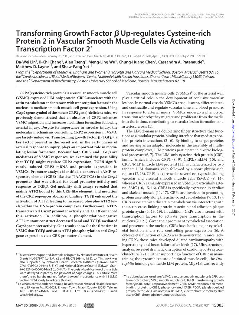
rial microenvironment we used 10 ng/ml TGF� for our studies.TGF� induced CRP2 protein expression as early as 2 h, and theinduction continued at the 4 h time point (Fig. 1A). A 3-foldinduction was observed at 8 h, and this level of induction wasmaintained at 24 h when compared with untreated controls(Fig. 1A). Similar to the protein results, Northern blot analysisrevealed that TGF� induced CRP2 mRNA expression in a time-dependent manner (Fig. 1B). A maximal 3-fold induction wasobserved at 4 h, which precededmaximal protein induction at 8 h(Fig. 1A), and was maintained at 24 h when compared withuntreated controls (Fig. 1B). To determine the significance ofCRP2 induction, we treated wild type and Csrp2�/� VSMC withorwithoutTGF� for 12 h to obtainmaximal induction ofCRP2 inwild type cells and thenperformedmigration assays. Interestingly,comparedwith untreated controls, TGF� treatment reducedwildtypeVSMCmigrationby�50%,whereasCsrp2�/�VSMCmigra-tion was not affected (Fig. 1C), indicating that up-regulation ofCRP2 correlated with a blunted VSMCmotility.TGF� Does Not Alter the Half-life of CRP2mRNA—To begin
to elucidate the mechanisms underlying CRP2 mRNA induc-tion, we performed experiments with the transcriptional inhib-itor actinomycin D. VSMCs were preincubated with vehicle oractinomycin D for 30 min and then treated for 4 h with orwithout TGF�. Northern analysis revealed that in the absenceof actinomycin D, TGF� substantially induced CRP2 mRNAexpression, whereas actinomycin D blocked induction of CRP2mRNA (Fig. 2A). Given that TGF� can regulatemRNA levels ofmany target genes by alteringmRNA stability and half-life (38–40), we measured the half-life of CRP2 mRNA in VSMCs stim-ulated with or without TGF�. In the absence of TGF�, the half-life of CRP2 mRNA was �14 h (Fig. 2B). The half-life of CRP2mRNA in TGF�-treated cells was also �14 h, indicating thatTGF� has no effect on CRP2 mRNA half-life (Fig. 2B).CRP2 Induction by TGF� Does Not Require New Protein
Synthesis—To further examine whether CRP2 mRNA induc-tion by TGF� required protein synthesis, we preincubatedVSMCs with protein synthesis inhibitor cyclohexamide for 30min and then treated for 4 h with or without TGF�. Real time
FIGURE 1. TGF� increases CRP2 expression in VSMCs. A, TGF� inducesCRP2 protein expression. VSMCs were exposed to TGF� (10 ng/ml), and pro-tein extracts were harvested at the indicated time points. Total protein was
also extracted from unstimulated control cells. Western blot analyses wereperformed using 20 �g of total protein/lane. After electrophoresis, proteinswere transferred to nitrocellulose membranes and incubated with a poly-clonal primary antiserum specific for CRP2-(91–98) and a horseradish perox-idase-conjugated goat anti-rabbit secondary antibody. The blot was visual-ized with enhanced chemiluminescence and exposed to film. Blots weresubsequently probed for �-tubulin for normalization. CRP2 protein inductionis expressed relative to control without TGF� stimulation at 0 h. Values aremean � S.E. of three experiments. B, up-regulation of CRP2 mRNA by TGF�.VSMCs were treated with TGF� (10 ng/ml) for the indicated times. Northernblot analysis was performed with 10 �g of total RNA/lane. After electrophore-sis, RNA was transferred to nitrocellulose filters and hybridized with a randomprimed 32P-labeled mouse CRP2 cDNA probe that hybridized to a 1.0-kb CRP2message. The blots were subsequently hybridized with a 32P-labeled 18 Soligonucleotide to verify loading. The signal intensity of each RNA samplehybridized to the CRP2 probe was divided by that hybridized to the 18 Sprobe. The normalized intensities were expressed relative to control withoutTGF� treatment at 0 h. Values are mean � S.E. of 3–5 experiments. C, TGF�treatment reduces wild type but not Csrp2�/� VSMC migration in response toPDGF-BB. Wild type (�/�) and Csrp2�/� (�/�) VSMCs were serum-starvedfor 24 h, treated without or with TGF� (10 ng/ml) for 12 h and then plated intriplicate in 6-well transwell plates for migration assays using PDGF-BB (10ng/ml) as a chemoattractant. Cells migrating through the filters after 2 h werequantified and normalized to the cell number of wild type without TGF�treatment. Values are mean � S.D. of two experiments.
CRP2 Induction by TGF�
15006 JOURNAL OF BIOLOGICAL CHEMISTRY VOLUME 283 • NUMBER 22 • MAY 30, 2008
at Univ of C
alifornia - Irvine on February 14, 2017http://w
ww
.jbc.org/D
ownloaded from

quantitative PCR analysis showed that cyclohexamide did notprevent CRP2 mRNA induction by TGF� (Fig. 2C), demon-strating that transcriptional activation of Csrp2 by TGF� doesnot require new protein synthesis.TheCsrp2 Promoter Region bp�480 to�438 Is Important for
TGF� Induction—To determine whether elements responsiblefor TGF� induction were present in the Csrp2 promoter, wetransiently transfected VSMCs with luciferase plasmid�795Csrp2-luc containing�795 bp of theCsrp2 promoter.Wedemonstrated previously that this region is sufficient to drivelacZ reporter gene expression in VSMCs of blood vessels intransgenic mice (21). TGF� increased �795Csrp2 promoteractivity by�4-fold (Fig. 3A), indicating that the�795 proximal
FIGURE 2. TGF� induction of CRP2 mRNA does not alter CRP2 mRNAhalf-life or require new protein synthesis. A, TGF� does not alter CRP2mRNA half-life. VSMCs were pretreated with vehicle (95% ethanol) or tran-scriptional inhibitor actinomycin D (AcD) (10 �g/ml) for 30 min and thenstimulated without or with TGF� (10 ng/ml) for 4 h and analyzed as in Fig.1B. TGF� induced CRP2 mRNA expression in the absence of AcD. In com-parison, AcD blocked the CRP2 mRNA induction by TGF�. B, TGF� does notalter CRP2 mRNA half-life. VSMCs were stimulated with or without TGF�(10 ng/ml) for 4 h, and then AcD (10 �g/ml) was administered to the cells.Total RNA was extracted at the indicated times after administration ofAcD, and Northern blot analyses were performed as in Fig. 1B. The normal-ized intensity was then plotted as a percentage of the 0 h value (in logscale) against time. C, CRP2 induction by TGF� does not require new pro-tein synthesis. VSMCs were pretreated with vehicle (DMSO) or proteinsynthesis inhibitor cyclohexamide (CHX) (10 �g/ml) for 30 min and thenstimulated without or with TGF� (10 ng/ml) for 4 h. RNA was harvested,reverse transcribed, and analyzed by real time quantitative PCR assays.Cyclohexamide did not prevent the induction of CRP2 mRNA by TGF�.Values are mean � S.E. of three experiments.
FIGURE 3. The Csrp2 promoter region bp �480 to �438 is important forTGF� induction. VSMCs were transiently transfected with Csrp2 promoter-luciferase reporter plasmids (500 ng/well) in triplicate using FuGENE 6. Allwells received 500 ng of pCMV� to normalize for transfection efficiency. Twohours after transfection, cells were treated with or without TGF� (10 ng/ml)for 24 h and harvested for luciferase and �-galactosidase activity assays. A, 5�deletion constructs of Csrp2 promoter-luciferase reporter plasmids. TGF�induction is expressed relative to control without TGF� of each construct.Values are mean � S.E. of three experiments. B, the �795 Csrp2 wild type�795 and mutant �795�(480/438) (an internal deletion of bp �480 to �438)promoter constructs are schematically depicted in the top panel. Luciferaseactivity is expressed relative to �795 without TGF� treatment. Values aremean � S.E. of three experiments.
CRP2 Induction by TGF�
MAY 30, 2008 • VOLUME 283 • NUMBER 22 JOURNAL OF BIOLOGICAL CHEMISTRY 15007
at Univ of C
alifornia - Irvine on February 14, 2017http://w
ww
.jbc.org/D
ownloaded from
promoter contained TGF�-responsive elements for the induc-tion of CRP2. To identify the elements, a series of Csrp2 5�-de-letion promoter-luciferase constructs were generated and tran-siently transfected into VSMCs. Similar to the �795 construct,the �573 construct showed a 3.8-fold induction by TGF� (Fig.3A). Deletion of the 5�-sequences to bp �549 retained TGF�inducibility (Fig. 3A). Additional deletion to bp �480 onlyslightly diminished the responsiveness to TGF�. Significantly,further deletion to bp �438 abolished the promoter inductionby TGF� (Fig. 3A), indicating that the region between bp �480and�438 was required for the TGF� stimulation ofCsrp2 pro-moter activity. To determine the functional importance of thisregion in the Csrp2 promoter, we generated a �795�(480/438)Csrp2-luc construct that had a 43-bp internal deletionfrom bp �480 to �438 within the context of �795Csrp2-lucconstruct. Transient transfection experiments revealed thatdeletion of this region reduced basal promoter activity by�50%(Fig. 3B, white bars). Furthermore, internal deletion of thisregion also abolished the TGF� induction (Fig. 3B, filled bars),consistent with the 5�-deletion studies (Fig. 3A).The CRE-like Element Is Critical for Basal and Inducible
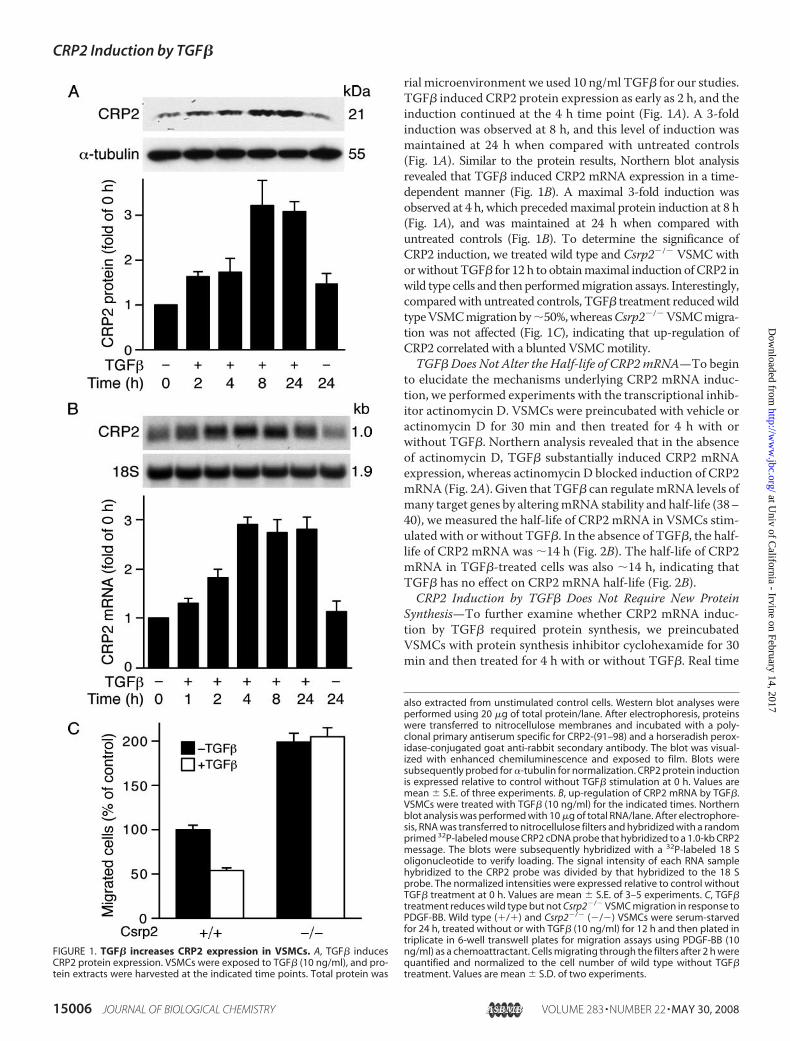
Activity of the Csrp2 Promoter—Comparison of the sequencesfrom the mouse promoter bp �485 to �435 with human andrat promoters (GenBankTM accession numbers U95017 andNW04774, respectively) revealed that this region is highly con-served across species (Fig. 4A). Through transcription factordata bases (BCM Search Launcher; TRANSFAC) searches andcloser examination of the sequences, we identified a putativeCRE-like element (TAACGTCA) (Fig. 4A, boldface type). Totest the potential function of this conserved CRE-like site,which is one base divergent from the consensus CRE site(TGACGTCA), we generated a luciferase construct,
�795CREmutCsrp2-luc, with threebases mutated (underlined) withinthe putative CRE site (CACCG-TAA). Compared with the control,the �795CREmutCsrp2-luc con-struct substantially reduced basalpromoter activity, similar to that ofthe internal deletion construct,�795�(480/438)Csrp2-luc (Fig. 4B,white bars). Most importantly,mutation of this site abrogatedTGF�-mediated induction of theCsrp2 promoter (Fig. 4B, blackbars).Nuclear Proteins Binding to the
CRE-like Site of the Csrp2 Pro-moter—Given the functional im-portance of the CRE-like site in reg-ulating Csrp2 promoter activity, wewere interested in characterizingthe transcription factors that boundto this site. We first tested whethernuclear proteins from controlVSMCs (i.e. not treated with TGF�)bound to the Csrp2-CRE site by gelmobility shift assays. Oligonucleo-
tide sequences used in the gel shift assays are indicated (Fig.5A). Incubation of VSMCnuclear extracts with an oligonucleo-tide probe encoding bp �467 to �448 of the Csrp2 promoterresulted in two prominent DNA-protein complexes, a domi-nant upper complex I and a minor lower complex II (Fig. 5A,lane 2). These two complexes were specific, because they werecompeted by excess unlabeled identical probe (Fig. 5A, lane 3)but not by an unrelated probe (data not shown). The mutantoligonucleotides (Fig. 5A, mut) with three bases mutated(CACCGTAA) failed to compete away the binding complexes(Fig. 5A, lane 4). Conversely, no specific complex formationwasobserved when themutant was used as a probe (Fig. 5A, lane 7),further indicating that nuclear factors bound specifically to thisCRE-like site. The consensus CRE oligonucleotides also com-peted away the complexes (Fig. 5A, lane 5), indicating that thisis a bona fide CRE site. When 32P-labeled consensus CRE wasused as a probe, the intensity of complex I relative to complex IIwas reduced compared with the Csrp2 probe (Fig. 5A, lane 9versus lane 2). The identical unlabeled CRE competitor abol-ished the complexes, confirming the specificity of the two com-plexes (Fig. 5A, lane 10). Intriguingly, the Csrp2 competitorprimarily abolished binding of complex I and to a lesser degreewith complex II (Fig. 5A, lane 11). Taken together, these resultssuggest that nuclear factors that bound to Csrp2-CRE differfrom that bound to consensus CRE. To examine whether lesssevere mutations are sufficient to disrupt nuclear protein bind-ing, we performed additional gel shift assays using oligonucleo-tides with one or two bases mutated in the Csrp2-CRE coresequence (Fig. 5B). Consistently, incubation of VSMC nuclearextracts withCsrp2 oligonucleotide probe resulted in complex Iand II formation (Fig. 5B, lane 2). Complexes I and II wereabolished by the addition of unlabeled identical (Fig. 5B, lane 3)
FIGURE 4. The CRE-like site is functionally important for basal and TGF� induction of Csrp2 promoteractivity. A, conservation of the putative CRE site among species. Sequence alignment of correspondingregions of human and rat promoter sequences to the mouse promoter. A putative CRE site (TAACGTCA) is inboldface type and underlined in the mouse sequence. B, the putative CRE site is important for Csrp2 promoteractivity. The �795 Csrp2 wild type (wt) and CRE mutant (mut) promoter constructs are schematically depictedin the left panel. VSMCs were transiently transfected with Csrp2 luciferase reporter constructs (500 ng/well)containing �795 or �795CREmut with putative CRE site mutated and pCMV� (500 ng/well) to normalize fortransfection efficiency in triplicate using FuGENE 6 transfection reagent. Two hours after transfection, cellswere treated with or without TGF� (10 ng/ml) for 24 h. Cells were then harvested for luciferase and �-galacto-sidase activity assays. Luciferase activity is expressed relative to �795 without TGF� treatment.
CRP2 Induction by TGF�
15008 JOURNAL OF BIOLOGICAL CHEMISTRY VOLUME 283 • NUMBER 22 • MAY 30, 2008
at Univ of C
alifornia - Irvine on February 14, 2017http://w
ww
.jbc.org/D
ownloaded from

oligonucleotides as competitors. mut1 with one base mutatedat the core partially competed away complexes (Fig. 5B, lane 4),whereas mut2 with two bases mutated did not compete awaythe complexes (Fig. 5B, lane 5). These data suggested thatmutation at the first base of the CRE core partially retained theability to bind to nuclear proteins. In comparison, a two-basemutation rendered the oligonucleotides unable to bind to thecognate nuclear proteins. In agreement with these results, gelshift assays using 32P-labeledmut1 oligonucleotides resulted inminimal complex formation (lane 6), whereas 32P-labeledmut2oligonucleotides did not result in any specific complex forma-tion (lane 7), demonstrating the importance ofCsrp2-CRE coresequences for nuclear protein binding.To identify the composition of these complexes, we
screened several known CRE-binding proteins for their abil-ity to supershift the Csrp2-CRE-protein complex. Antibod-ies specific for ATF2, CREB, or c-Jun were added to thebinding reactions and incubated with VSMC nuclearextracts (Fig. 5C). ATF2 antibodies completely supershiftedcomplex I, as indicated by the disappearance of complex I,and the appearance of a larger complex near the top of the gel(Fig. 5C, lane 3, asterisk). The lower complex II was not super-shifted by ATF2 antibodies. These data suggested that ATF2 wasthe main factor present in complex I. CREB antibodies super-shifted complex II to a larger complex (Fig. 5C, lane 4,dot) but notcomplex I, indicating the complex II containedCREB. In compar-ison, c-Junwasnot present in either complex, since c-Jun antibod-ies didnot supershift complex I or II (Fig. 5C, lane 5), although thisantibodyhasbeenpreviously shownto supershift c-Juncomplexes(41).We next determined whether TGF� affected levels of ATF2/
CREB binding to the CRE site. Since CRP2mRNA induction byTGF� was evident at 1 h (Fig. 1B), and DNA-protein complexformation would precede mRNA induction, we used nuclearextracts from VSMCs treated with TGF� for 15 and 30 min.Interestingly, gel mobility shift assays using nuclear extractsfrom VSMCs treated with TGF� for 15 min revealed similarintensity of complexes I and II (Fig. 5C, lanes 6–9) with control
FIGURE 5. Nuclear proteins binding to the CRE-like element of Csrp2promoter. A, oligonucleotide sequences used in the electrophoretic mobilityshift assays (EMSAs) are shown. The core sequence of CRE-like and consensusCRE sites are in boldface type, and mutated sequence is underlined. EMSAswere performed with double-stranded oligonucleotides corresponding to bp�467 to �448 of the Csrp2 promoter. The addition of nuclear extracts (10 �g)from VSMCs to the 32P-labeled Csrp2 probe resulted in two major retardedDNA-protein complexes, designated I and II (arrows on left) (lane 2). A non-specific band (ns) is indicated. Complexes I and II were abolished by the
addition of unlabeled identical (Csrp2, lane 3) or consensus CRE (lane 5) oligo-nucleotides as competitors but not by the addition of three bases mutated(mut) oligonucleotides (lane 4). Conversely, EMSAs using 32P-labeled mutatedoligonucleotides did not result in specific complex formation (lane 7). As acomparison, EMSAs using 32P-labeled CRE oligonucleotides were performed.The addition of nuclear extracts to the CRE probe resulted in the formation ofcomplexes I and II (lane 9), which were competed away by identical unlabeledoligonucleotides (CRE; lane 10). The addition of unlabeled Csrp2 mainly abol-ished complex I and to a lesser degree complex II (lane 11). B, oligonucleotidesused in the EMSAs are indicated. Csrp2 oligonucleotides contain the coresequence of CRE-like site (in boldface type), whereas mut1 has a one-basemutation (underlined) in the core and mut2 has two bases mutated (under-lined). As in A, complex I and II were abolished by the addition of unlabeledidentical (Csrp2, lane 3) oligonucleotides as competitors. mut1 partially com-peted away the complexes (lane 4), whereas mut2 did not compete away thecomplexes (lane 5). EMSAs using 32P-labeled mut1 oligonucleotides resultedin low intensity complex I and II formation (lane 6), whereas 32P-labeled mut2oligonucleotides did not result in specific complex formation (lane 7).C, nuclear extracts from control (lanes 2–5) or TGF� treated for 15 min (lanes6 –9) VSMCs were incubated with 32P-labeled Csrp2 probe without the addi-tion of antibodies (lanes 2 and 6) or antibodies specific to ATF2 (lanes 3 and 7),CREB (lanes 4 and 8), or c-Jun (lanes 5 and 9). ATF2 antibody completely super-shifted complex I to an upper band (*, lanes 3 and 7), whereas CREB antibodysupershifted complex II to an upper complex (F, lanes 4 and 8). Incubationwith c-Jun antibody did not produce supershifted bands (lanes 5 and 9).
CRP2 Induction by TGF�
MAY 30, 2008 • VOLUME 283 • NUMBER 22 JOURNAL OF BIOLOGICAL CHEMISTRY 15009
at Univ of C
alifornia - Irvine on February 14, 2017http://w
ww
.jbc.org/D
ownloaded from
nuclear extracts (Fig. 5C, lanes 2–5). Using nuclear extractsfromVSMCs treatedwithTGF� for 30min also showed similarDNA-protein complex patterns as controls (data not shown).TGF� Increases Phosphorylation Levels of ATF-2 But Not
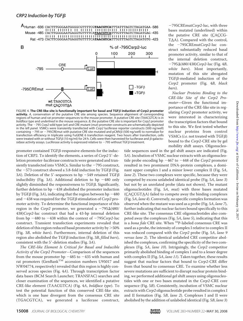
CREB—CREB/ATF2 can bind to CRE in an unphosphorylatedand transcriptionally inactive form (42–44). Phosphorylationof ATF2 or CREB within the activation domain leads to theiractivation of gene expression (45, 46). Thus, we hypothesizedthat TGF� might increase the phosphorylation of ATF2 andCREB in VSMCs. To investigate this possibility, VSMCs weretreated with TGF� and protein extracts harvested 5–30 minafter treatment. Western blot analysis revealed that TGF�increased ATF2 phosphorylation as early as 5 min after stimu-lation but did not alter total ATF2 levels (Fig. 6). In contrast,TGF� did not affect either phosphorylated CREB levels, whichwere present at baseline, or total amounts of CREB (Fig. 6). Totest the hypothesis that ATF2 and its phosphorylation levelsmight be responsible for TGF� induction of CRP2, we per-formed supershift assays using antibodies specific for phospho-ATF2 or phospho-CREB. Phospho-ATF2 antibodies partiallysupershifted complex I (Fig. 7A, lane 3) when control nuclearextracts were used. When nuclear extracts from VSMCstreated with TGF� were used in the reactions, most of complexI was supershifted (Fig. 7A, lane 6). TGF� increased the ratio ofphospho-ATF2 to unphosphorylated ATF2 within complex Ifrom 1.35 � 0.06 of controls to 3.09 � 0.29 (n 3), indicatingthat TGF� enhanced phosphorylation of ATF2 within theDNA-protein complex. Phospho-CREB antibodies appeared todisrupt complex II from both the control and TGF�-treatednuclear extracts (Fig. 7A, lanes 4 and 7); however, the resultswere not conclusive. To investigate further whether CREB andp-CREBbound toCsrp2-CRE in the intact native chromatin,weperformed quantitative ChIP assays. The CRE of the cyclin D1promoterwith the core sequence identical to that ofCsrp2-CREhas been shown mainly to bind CREB (46); thus, mouse cyclinD1-CRE was used as a positive control. CREB binding to thecyclin D1 promoter was substantially higher than to the Csrp2promoter (52 and 6%, respectively; Fig. 7B), indicating muchlower levels of CREB bound toCsrp2-CRE than cyclin D1-CRE.ChIP assays using p-CREB antibodies revealed that, comparedwith 22% of p-CREB bound to cyclin D1-CRE, p-CREB bindingto Csrp2 was barely detectable (Fig. 7B). Taken together, the
results of gel shifts and ChIP assays suggested that low levels ofCREB bound to Csrp2-CRE; however, the bound CREB wereprobably not phosphorylated.Transcriptional Activation of the Csrp2 Promoter by ATF2
via the CRE Site—To evaluate the functional role of CREB andATF2 in the induction of CRP2, we cotransfected �795Csrp2-luc constructwith expression plasmids in transient transfectionassays. CREBdid not increaseCsrp2promoter activity (Fig. 8A).In contrast, ATF2 expression increased Csrp2 promoter activ-ity by �3-fold (Fig. 8A), and TGF� further enhanced the tran-scriptional activation by ATF2 7-fold (Fig. 8A). To further con-firm the function of ATF2 and the importance of itsphosphorylation, we cotransfected �795Csrp2-luc with aphosphorylation-negative ATF2 mutant (ATF2-AAA), inwhich the three important phosphorylation residues Thr51,Thr53, and Ser72 were mutated to alanines; thus, it also func-
FIGURE 6. TGF� increases the phosphorylation of ATF2 but not CREB.VSMCs were stimulated with TGF� (10 ng/ml) and activation of ATF2 andCREB was determined using cell lysates harvested at the indicated time pointsby Western blot analysis. Phosphorylation of ATF2 and CREB was detected byusing phospho-ATF2 (p-ATF2) and p-CREB antibodies, respectively. To verifyequal loading, the blots were probed with total ATF2 and CREB antibodies. Arepresentative of three independent experiments is shown.
FIGURE 7. TGF� enhances phosphorylation of ATF2 within the DNA-pro-tein complex. A, EMSAs were performed with double-stranded oligonucleo-tides corresponding to bp �467 to �448 of the Csrp2 promoter. The additionof nuclear extracts (NE; 10 �g) from control or TGF�-treated for 15 min VSMCsto the 32P-labeled Csrp2 probe resulted in retarded DNA-protein complex Iand II (lanes 2 and 5). To detect phosphorylation of ATF2 and CREB within thecomplexes, nuclear extracts were incubated with antibodies (Ab) specific tophospho-ATF2 or phospho-CREB before the reactions. Phospho-ATF2(p-ATF2) antibody supershifted complex I to a larger complex (p-ATF23,lanes 3 and 6), whereas phospho-CREB antibody disrupted complex II (lanes 4and 7). B, quantitative ChIP assays. VSMCs were cross-linked with formalde-hyde and DNA fragmented by sonication. Chromatin was immunoprecipi-tated with normal rabbit IgG, CREB, or p-CREB antibodies. Aliquots of samplesequivalent to 1% of initial cell lysate for each reaction were processed, andDNA was purified to use as input DNA control. Quantitative PCR was per-formed to amplify a 165-bp fragment flanking the mouse Csrp2-CRE (Csrp2)and a 92-bp fragment flanking the mouse cylin D1-CRE (cyclin D1). As anadditional negative control, a 164-bp fragment within intron 1 of the Csrp2gene (intron 1) was also amplified by quantitative PCR. Binding activity isexpressed as percentage relative to input DNA. Values are mean � S.D. of twoor three experiments.
CRP2 Induction by TGF�
15010 JOURNAL OF BIOLOGICAL CHEMISTRY VOLUME 283 • NUMBER 22 • MAY 30, 2008
at Univ of C
alifornia - Irvine on February 14, 2017http://w
ww
.jbc.org/D
ownloaded from
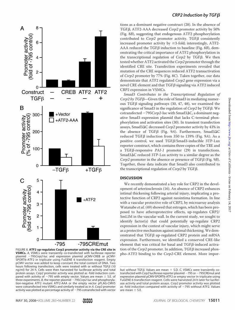
tions as a dominant negative construct (28). In the absence ofTGF�, ATF2-AAA decreased Csrp2 promoter activity by 50%(Fig. 8B), suggesting that endogenous ATF2 phosphorylationcontributed to Csrp2 promoter activity. TGF� consistentlyincreased promoter activity by �3-fold; interestingly, ATF2-AAA reduced the TGF� induction to baseline (Fig. 8B), dem-onstrating the critical importance of ATF2 phosphorylation inthe transcriptional regulation of Csrp2 by TGF�. We thentestedwhetherATF2 activated theCsrp2 promoter through theidentified CRE site. Transfection experiments revealed thatmutation of the CRE sequences reduced ATF2 transactivationof Csrp2 promoter by 77% (Fig. 8C). Taken together, our datademonstrate that ATF2 regulated Csrp2 gene expression via anovel CRE element and that TGF� signaling via ATF2 inducedCRP2 expression in VSMCs.Smad3 Contributes to the Transcriptional Regulation of
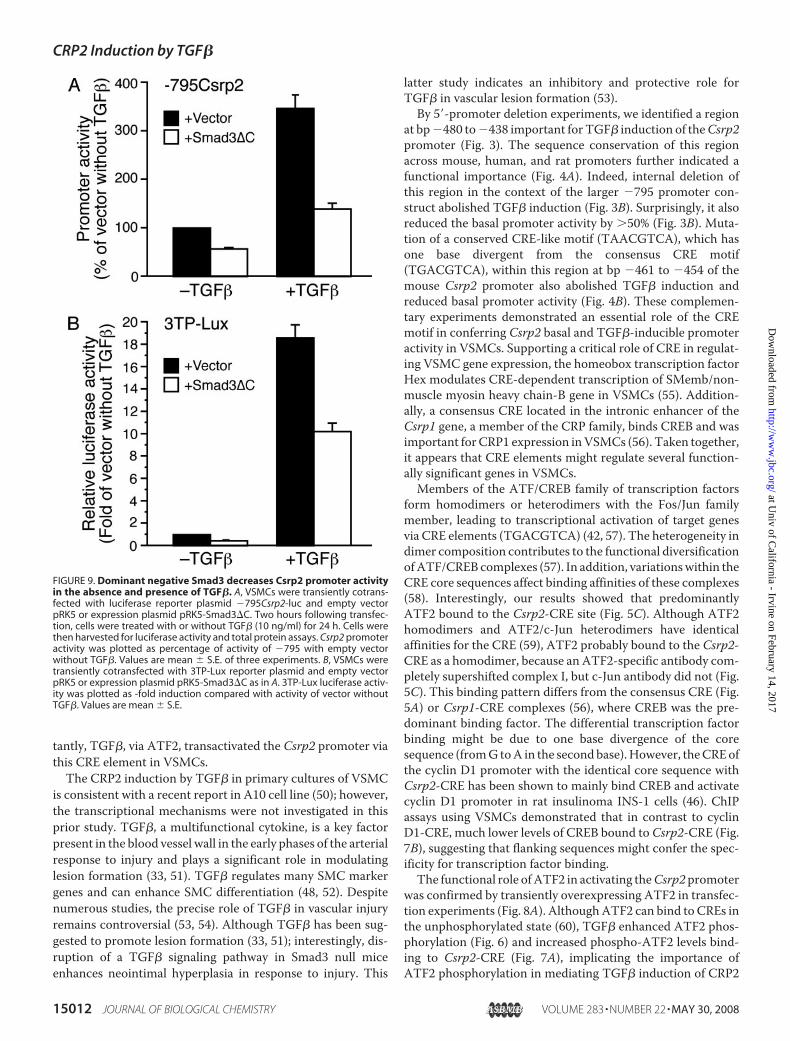
Csrp2 by TGF�—Given the role of Smad3 inmediating numer-ous TGF� signaling pathways (30, 47, 48), we examined thesignificance of Smad3 in the regulation of Csrp2 by TGF�. Wecotransfected�795Csrp2-luc with Smad3�C, a dominant neg-ative Smad3 expression plasmid that lacks C-terminal phos-phorylation and activation sites (30). In transient transfectionassays, Smad3�C decreased Csrp2 promoter activity by 43% inthe absence of TGF� (Fig. 9A). Furthermore, Smad3�Creduced TGF� induction from 350 to 139% (Fig. 9A). As apositive control, we used TGF�/Smad3-inducible 3TP-Luxreporter construct, which contains three copies of the TRE anda TGF�-responsive PAI-1 promoter (29) in transfections.Smad3�C reduced 3TP-Lux activity to a similar degree as theCsrp2 promoter in the absence or presence of TGF� (Fig. 9B).Together, these data indicate that Smad3 also contributed tothe transcriptional regulation of Csrp2 by TGF�.
DISCUSSION
We recently demonstrated a key role for CRP2 in the devel-opment of arteriosclerosis (16). An absence of CRP2 enhancesintimal thickening following arterial injury, implicating a pro-tective function of CRP2 against neointima formation. In linewith a vascular protective role of CRP2, by microarray analysisWatanabe et al. (49) showed that estrogen, which has been pro-posed to have atheroprotective effects, up-regulates CRP2/SmLIM in the vascular wall. In the current study, we sought toidentify factor(s) that could potentially up-regulate CRP2expression in the context of vascular injury, which might serveas a protectivemechanism against intimal thickening.Wedem-onstrated that TGF� up-regulated CRP2 protein and mRNAexpression. Furthermore, we identified a conserved CRE-likeelement that was critical for basal and TGF�-induced activa-tion of the Csrp2 promoter. In addition, TGF� increased phos-pho-ATF2 binding to the Csrp2-CRE element. More impor-
FIGURE 8. ATF2 up-regulates Csrp2 promoter activity via the CRE site inVSMCs. A, VSMCs were transiently co-transfected with luciferase reporterplasmid �795Csrp2-luc and expression plasmid pCMV-CREB or pCMV�SPORT6-ATF2 in triplicate using FuGENE 6 transfection reagent. EmptypCMV vector was added to keep constant the total content of DNA. Twohours following transfection, cells were treated with or without TGF� (10ng/ml) for 24 h. Cells were then harvested for luciferase activity and totalprotein assays. Csrp2 promoter activity was plotted as -fold induction com-pared with activity of �795 with empty vector. Values are mean � S.E. ofthree experiments. B, the reporter plasmid �795Csrp2-luc and phosphoryla-tion-negative ATF2 mutant ATF2-AAA or the empty vector pFLAG-CMV5were cotransfected into VSMCs and similarly treated as in A. Csrp2 promoteractivity was plotted as percentage activity of �795 cotransfected with vector
but without TGF�. Values are mean � S.D. C, VSMCs were transiently co-transfected with Csrp2 luciferase reporter plasmid �795 or �795CREmut andexpression plasmid pCMV�SPORT6-ATF2 or empty vector in triplicate usingFuGENE 6 transfection reagent. Cells were harvested 24 h later for lucifer-ase activity and total protein assays. Csrp2 promoter activity was plottedas -fold induction compared with activity of �795 without ATF2. Valuesare mean � S.E.
CRP2 Induction by TGF�
MAY 30, 2008 • VOLUME 283 • NUMBER 22 JOURNAL OF BIOLOGICAL CHEMISTRY 15011
at Univ of C
alifornia - Irvine on February 14, 2017http://w
ww
.jbc.org/D
ownloaded from
tantly, TGF�, via ATF2, transactivated the Csrp2 promoter viathis CRE element in VSMCs.The CRP2 induction by TGF� in primary cultures of VSMC
is consistent with a recent report in A10 cell line (50); however,the transcriptional mechanisms were not investigated in thisprior study. TGF�, a multifunctional cytokine, is a key factorpresent in the blood vessel wall in the early phases of the arterialresponse to injury and plays a significant role in modulatinglesion formation (33, 51). TGF� regulates many SMC markergenes and can enhance SMC differentiation (48, 52). Despitenumerous studies, the precise role of TGF� in vascular injuryremains controversial (53, 54). Although TGF� has been sug-gested to promote lesion formation (33, 51); interestingly, dis-ruption of a TGF� signaling pathway in Smad3 null miceenhances neointimal hyperplasia in response to injury. This
latter study indicates an inhibitory and protective role forTGF� in vascular lesion formation (53).
By 5�-promoter deletion experiments, we identified a regionat bp�480 to�438 important for TGF� induction of theCsrp2promoter (Fig. 3). The sequence conservation of this regionacross mouse, human, and rat promoters further indicated afunctional importance (Fig. 4A). Indeed, internal deletion ofthis region in the context of the larger �795 promoter con-struct abolished TGF� induction (Fig. 3B). Surprisingly, it alsoreduced the basal promoter activity by �50% (Fig. 3B). Muta-tion of a conserved CRE-like motif (TAACGTCA), which hasone base divergent from the consensus CRE motif(TGACGTCA), within this region at bp �461 to �454 of themouse Csrp2 promoter also abolished TGF� induction andreduced basal promoter activity (Fig. 4B). These complemen-tary experiments demonstrated an essential role of the CREmotif in conferring Csrp2 basal and TGF�-inducible promoteractivity in VSMCs. Supporting a critical role of CRE in regulat-ing VSMC gene expression, the homeobox transcription factorHex modulates CRE-dependent transcription of SMemb/non-muscle myosin heavy chain-B gene in VSMCs (55). Addition-ally, a consensus CRE located in the intronic enhancer of theCsrp1 gene, a member of the CRP family, binds CREB and wasimportant forCRP1 expression inVSMCs (56). Taken together,it appears that CRE elements might regulate several function-ally significant genes in VSMCs.Members of the ATF/CREB family of transcription factors
form homodimers or heterodimers with the Fos/Jun familymember, leading to transcriptional activation of target genesvia CRE elements (TGACGTCA) (42, 57). The heterogeneity indimer composition contributes to the functional diversificationofATF/CREB complexes (57). In addition, variationswithin theCRE core sequences affect binding affinities of these complexes(58). Interestingly, our results showed that predominantlyATF2 bound to the Csrp2-CRE site (Fig. 5C). Although ATF2homodimers and ATF2/c-Jun heterodimers have identicalaffinities for the CRE (59), ATF2 probably bound to the Csrp2-CRE as a homodimer, because an ATF2-specific antibody com-pletely supershifted complex I, but c-Jun antibody did not (Fig.5C). This binding pattern differs from the consensus CRE (Fig.5A) or Csrp1-CRE complexes (56), where CREB was the pre-dominant binding factor. The differential transcription factorbinding might be due to one base divergence of the coresequence (fromG toA in the secondbase).However, theCREofthe cyclin D1 promoter with the identical core sequence withCsrp2-CRE has been shown to mainly bind CREB and activatecyclin D1 promoter in rat insulinoma INS-1 cells (46). ChIPassays using VSMCs demonstrated that in contrast to cyclinD1-CRE, much lower levels of CREB bound toCsrp2-CRE (Fig.7B), suggesting that flanking sequences might confer the spec-ificity for transcription factor binding.The functional role ofATF2 in activating theCsrp2promoter
was confirmed by transiently overexpressing ATF2 in transfec-tion experiments (Fig. 8A). AlthoughATF2 can bind to CREs inthe unphosphorylated state (60), TGF� enhanced ATF2 phos-phorylation (Fig. 6) and increased phospho-ATF2 levels bind-ing to Csrp2-CRE (Fig. 7A), implicating the importance ofATF2 phosphorylation in mediating TGF� induction of CRP2
FIGURE 9. Dominant negative Smad3 decreases Csrp2 promoter activityin the absence and presence of TGF�. A, VSMCs were transiently cotrans-fected with luciferase reporter plasmid �795Csrp2-luc and empty vectorpRK5 or expression plasmid pRK5-Smad3�C. Two hours following transfec-tion, cells were treated with or without TGF� (10 ng/ml) for 24 h. Cells werethen harvested for luciferase activity and total protein assays. Csrp2 promoteractivity was plotted as percentage of activity of �795 with empty vectorwithout TGF�. Values are mean � S.E. of three experiments. B, VSMCs weretransiently cotransfected with 3TP-Lux reporter plasmid and empty vectorpRK5 or expression plasmid pRK5-Smad3�C as in A. 3TP-Lux luciferase activ-ity was plotted as -fold induction compared with activity of vector withoutTGF�. Values are mean � S.E.
CRP2 Induction by TGF�
15012 JOURNAL OF BIOLOGICAL CHEMISTRY VOLUME 283 • NUMBER 22 • MAY 30, 2008
at Univ of C
alifornia - Irvine on February 14, 2017http://w
ww
.jbc.org/D
ownloaded from
expression. The significance of post-translational modificationrather thannewprotein synthesis inTGF�-mediated transcrip-tional activation of Csrp2 was supported by the findings thatprotein synthesis inhibitor cyclohexamide did not preventCRP2mRNA induction by TGF� (Fig. 2C). A phosphorylation-negative mutant, which also functions as a dominant negativemutant, reduced basal and TGF�-mediated Csrp2 promoteractivity (Fig. 8B), further demonstrating the critical importanceof ATF2 phosphorylation in regulating CRP2 expression. Incontrast to ATF2, CREB probably played a minimal role in reg-ulating Csrp2 gene expression, given its very low levels of bind-ing (particularly p-CREB) to Csrp2-CRE both in vitro and invivo and that cotransfection of CREB did not increase Csrp2promoter activity (Fig. 8A). Interestingly, TGF� signaling mol-ecule Smad3 also contributed to the transcriptional regulationof Csrp2 (Fig. 9). However, further studies are needed to inves-tigate the underlying mechanisms.In conclusion, we identified a conserved CRE element
(TAACGTCA) in theCsrp2 promoter that was critical for basalpromoter activity and responsiveness to TGF�. Our resultsshow for the first time in VSMC that TGF� activates ATF2phosphorylation and Csrp2 gene expression via a CRE pro-moter element, whichmay represent a previously unrecognizedmechanismofVSMCgene expression.Understanding the tran-scriptional activation of CRP2may help to elucidate themolec-ular mechanisms that control VSMC gene expression in vascu-lar disease.
REFERENCES1. Owens, G. K., Kumar, M. S., and Wamhoff, B. R. (2004) Physiol. Rev. 84,
767–8012. Schmeichel, K. L., and Beckerle, M. C. (1994) Cell 79, 211–2193. Feuerstein, R., Wang, X., Song, D., Cooke, N. E., and Liebhaber, S. A.
(1994) Proc. Natl. Acad. Sci. U. S. A. 91, 10655–106594. Arber, S., and Caroni, P. (1996) Genes Dev. 10, 289–3005. Dawid, I. B., Breen, J. J., and Toyama, R. (1998)Trends Genet. 14, 156–1626. Kadrmas, J. L., and Beckerle, M. C. (2004) Nat. Rev. Mol. Cell. Biol. 5,
920–9317. Weiskirchen, R., and Gunther, K. (2003) BioEssays 25, 152–1628. Weiskirchen, R., and Bister, K. (1993) Oncogene 8, 2317–23249. Crawford, A. W., Pino, J. D., and Beckerle, M. C. (1994) J. Cell Biol. 124,
117–12710. Jain, M. K., Fujita, K. P., Hsieh, C.-M., Endege, W. O., Sibinga, N. E., Yet,
S.-F., Kashiki, S., Lee, W.-S., Perrella, M. A., Haber, E., and Lee, M.-E.(1996) J. Biol. Chem. 271, 10194–10199
11. Arber, S., Halder, G., and Caroni, P. (1994) Cell 79, 221–23112. Weiskirchen, R., Pino, J. D., Macalma, T., Bister, K., and Beckerle, M. C.
(1995) J. Biol. Chem. 270, 28946–2895413. Louis, H. A., Pino, J. D., Schmeichel, K. L., Pomies, P., and Beckerle, M. C.
(1997) J. Biol. Chem. 272, 27484–2749114. Henderson, J. R., Brown, D., Richardson, J. A., Olson, E. N., and Beckerle,
M. C. (2002) J. Histochem. Cytochem. 50, 107–11115. Yet, S.-F., Folta, S. C., Jain,M. K., Hsieh, C.-M.,Maemura, K., Layne,M.D.,
Zhang, D., Marria, P. B., Yoshizumi, M., Chin, M. T., Perrella, M. A., andLee, M.-E. (1998) J. Biol. Chem. 273, 10530–10537
16. Wei, J., Gorman, T. E., Liu, X., Ith, B., Tseng, A., Chen, Z., Simon, D. I.,Layne, M. D., and Yet, S.-F. (2005) Circ. Res. 97, 1323–1331
17. Arber, S., Hunter, J. J., Ross, J., Jr., Hongo, M., Sansig, G., Borg, J., Perriard,J. C., Chien, K. R., and Caroni, P. (1997) Cell 88, 393–403
18. Pomies, P., Louis, H. A., and Beckerle, M. C. (1997) J. Cell Biol. 139,157–168
19. Schmeichel, K. L., and Beckerle, M. C. (1998) Biochem. J. 331, 885–89220. Kong, Y., Flick, M. J., Kudla, A. J., and Konieczny, S. F. (1997) Mol. Cell.
Biol. 17, 4750–476021. Chang, D. F., Belaguli, N. S., Iyer, D., Roberts,W. B.,Wu, S. P., Dong, X. R.,
Marx, J. G., Moore, M. S., Beckerle, M. C., Majesky, M.W., and Schwartz,R. J. (2003) Dev. Cell 4, 107–118
22. Clark, K. A., Bland, J. M., and Beckerle, M. C. (2007) J. Cell Sci. 120,2066–2077
23. Ross, R. (1993) Nature 362, 801–80924. Tedgui, A., and Mallat, Z. (2006) Physiol. Rev. 86, 515–58125. Gunther, S., Alexander, R. W., Atkinson, W. J., and Gimbrone, M. A.
(1982) J. Cell Biol. 92, 289–29826. Yet, S.-F., Perrella, M. A., Layne, M. D., Hsieh, C.-M., Maemura, K.,
Kobzik, L., Wiesel, P., Christou, H., Kourembanas, S., and Lee, M.-E.(1999) J. Clin. Invest. 103, R23–R29
27. Chang, Y.-F., Wei, J., Liu, X., Chen, Y.-H., Layne, M. D., and Yet, S.-F.(2003) Am. J. Physiol. 285, H1675–1683
28. Gupta, S., Campbell, D., Derijard, B., and Davis, R. J. (1995) Science 267,389–393
29. Wrana, J. L., Attisano, L., Carcamo, J., Zentella, A., Doody, J., Laiho, M.,Wang, X. F., and Massague, J. (1992) Cell 71, 1003–1014
30. Zhang, Y., Feng, X., We, R., and Derynck, R. (1996) Nature 383,168–172
31. Yoshida, T., Sinha, S., Dandre, F., Wamhoff, B. R., Hoofnagle, M. H., Kre-mer, B. E.,Wang, D. Z., Olson, E. N., andOwens, G. K. (2003)Circ. Res. 92,856–864
32. Ishida, A., Fujita,N., Kitazawa, R., andTsuruo, T. (2002) J. Biol. Chem.277,26217–26224
33. Majesky, M. W., Lindner, V., Twardzik, D. R., Schwartz, S. M., and Reidy,M. A. (1991) J. Clin. Invest. 88, 904–910
34. Meng, J., Thongngarm, T., Nakajima, M., Yamashita, N., Ohta, K., Bates,C. A., Grunwald, G. K., and Rosenwasser, L. J. (2005) Int. Arch. AllergyImmunol. 138, 151–160
35. Fogel-Petrovic,M., Long, J. A.,Misso, N. L., Foster, P. S., Bhoola, K. D., andThompson, P. J. (2007) Int. Immunopharmacol. 7, 1924–1933
36. Feinberg, M. W., Jain, M. K., Werner, F., Sibinga, N. E., Wiesel, P., Wang,H., Topper, J. N., Perrella, M. A., and Lee, M. E. (2000) J. Biol. Chem. 275,25766–25773
37. Liu, G., Ding,W., Neiman, J., andMulder, K. M. (2006) J. Biol. Chem. 281,29479–29490
38. Tsukada, S., Westwick, J. K., Ikejima, K., Sato, N., and Rippe, R. A. (2005)J. Biol. Chem. 280, 10055–10064
39. DiCamillo, S. J., Yang, S., Panchenko, M. V., Toselli, P. A., Naggar, E. F.,Rich, C. B., Stone, P. J., Nugent, M. A., and Panchenko, M. P. (2006)Am. J.Physiol. 291, L232–L243
40. Dibrov, A., Kashour, T., and Amara, F.M. (2006)Growth Factors 24, 1–1141. Sreeramaneni, R., Chaudhry, A., McMahon, M., Sherr, C. J., and Inoue, K.
(2005)Mol. Cell. Biol. 25, 220–23242. Hai, T., andCurran, T. (1991)Proc. Natl. Acad. Sci. U. S. A. 88, 3720–372443. Herr, I., van Dam, H., and Angel, P. (1994) Carcinogenesis 15, 1105–111344. Li, X. Y., and Green, M. R. (1996) Genes Dev. 10, 517–52745. Livingstone, C., Patel, G., and Jones, N. (1995) EMBO J. 14, 1785–179746. Kim, M. J., Kang, J. H., Park, Y. G., Ryu, G. R., Ko, S. H., Jeong, I. K., Koh,
K. H., Rhie, D. J., Yoon, S. H., Hahn, S. J., Kim, M. S., and Jo, Y. H. (2006) JEndocrinol 188, 623–633
47. Qing, J., Liu, C., Choy, L., Wu, R. Y., Pagano, J. S., and Derynck, R. (2004)Mol. Cell. Biol. 24, 1411–1425
48. Sinha, S., Hoofnagle, M. H., Kingston, P. A., McCanna, M. E., and Owens,G. K. (2004) Am. J. Physiol. 287, C1560–C1568
49. Watanabe, T., Akishita, M., Nakaoka, T., He, H., Miyahara, Y., Yamashita,N., Wada, Y., Aburatani, H., Yoshizumi, M., Kozaki, K., and Ouchi, Y.(2004) Life Sci. 75, 1219–1229
50. Herrmann, J., Borkham-Kamphorst, E., Haas, U., Van de Leur, E., Fraga,M. F., Esteller, M., Gressner, A. M., and Weiskirchen, R. (2006) Biochem.Biophys. Res. Commun. 345, 1526–1535
51. Nikol, S., Isner, J.M., Pickering, J. G., Kearney,M., Leclerc, G., andWeir, L.(1992) J. Clin. Invest. 90, 1582–1592
52. Shah, N. M., Groves, A. K., and Anderson, D. J. (1996) Cell 85, 331–34353. Kobayashi, K., Yokote, K., Fujimoto, M., Yamashita, K., Sakamoto, A.,
Kitahara,M., Kawamura, H.,Maezawa, Y., Asaumi, S., Tokuhisa, T.,Mori,
CRP2 Induction by TGF�
MAY 30, 2008 • VOLUME 283 • NUMBER 22 JOURNAL OF BIOLOGICAL CHEMISTRY 15013
at Univ of C
alifornia - Irvine on February 14, 2017http://w
ww
.jbc.org/D
ownloaded from

S., and Saito, Y. (2005) Circ. Res. 96, 904–91254. Yokote, K., Kobayashi, K., and Saito, Y. (2006) Trends Cardiovasc. Med.
16, 240–24555. Sekiguchi, K., Kurabayashi, M., Oyama, Y., Aihara, Y., Tanaka, T., Saka-
moto,H.,Hoshino, Y., Kanda, T., Yokoyama, T., Shimomura, Y., Iijima,H.,Ohyama, Y., and Nagai, R. (2001) Circ. Res. 88, 52–58
56. Najwer, I., and Lilly, B. (2005) Am. J. Physiol. 289, C785–C79357. van Dam, H., and Castellazzi, M. (2001) Oncogene 20, 2453–246458. Benbrook, D.M., and Jones, N. C. (1994)Nucleic Acids Res. 22, 1463–146959. Benbrook, D. M., and Jones, N. C. (1990) Oncogene 5, 295–30260. Maekawa, T., Sakura, H., Kanei-Ishii, C., Sudo, T., Yoshimura, T., Fuji-
sawa, J., Yoshida, M., and Ishii, S. (1989) EMBO J. 8, 2023–2028
CRP2 Induction by TGF�
15014 JOURNAL OF BIOLOGICAL CHEMISTRY VOLUME 283 • NUMBER 22 • MAY 30, 2008
at Univ of C
alifornia - Irvine on February 14, 2017http://w
ww
.jbc.org/D
ownloaded from
Cassandra A. Patenaude, Matthew D. Layne and Shaw-Fang YetDa-Wei Lin, Il-Chi Chang, Alan Tseng, Meng-Ling Wu, Chung-Huang Chen,
Smooth Muscle Cells via Activating Transcription Factor 2 Up-regulates Cysteine-rich Protein 2 in VascularβTransforming Growth Factor
doi: 10.1074/jbc.M801621200 originally published online April 3, 20082008, 283:15003-15014.J. Biol. Chem.
10.1074/jbc.M801621200Access the most updated version of this article at doi:
Alerts:
When a correction for this article is posted•
When this article is cited•
to choose from all of JBC's e-mail alertsClick here
http://www.jbc.org/content/283/22/15003.full.html#ref-list-1
This article cites 60 references, 31 of which can be accessed free at
at Univ of C
alifornia - Irvine on February 14, 2017http://w
ww
.jbc.org/D
ownloaded from