issn 1996 - 0808 volume 11 number 28 28 july, 2017
TRANSCRIPT

African Journal of
Microbiology Research
Volume 11 Number 28 28 July, 2017
ISSN 1996-0808

The African Journal of Microbiology Research (AJMR) is published weekly (one volume per year) by Academic Journals.
provides rapid publication (weekly) of articles in all areas of Microbiology such as: Environmental Microbiology, Clinical Microbiology, Immunology, Virology, Bacteriology, Phycology, Mycology and Parasitology, Protozoology, Microbial Ecology, Probiotics and Prebiotics, Molecular Microbiology, Biotechnology, Food Microbiology, Industrial Microbiology, Cell Physiology, Environmental Biotechnology, Genetics, Enzymology, Molecular and Cellular Biology, Plant Pathology, Entomology, Biomedical Sciences, Botany and Plant Sciences, Soil and Environmental Sciences, Zoology, Endocrinology, Toxicology. The Journal welcomes the submission of manuscripts that meet the general criteria of significance and scientific excellence. Papers will be published shortly after acceptance. All articles are peer-reviewed.
Contact Us
Editorial Office: [email protected]
Help Desk: [email protected]
Website: http://www.academicjournals.org/journal/AJMR
Submit manuscript online http://ms.academicjournals.me/

Editors
Prof. Stefan Schmidt Applied and Environmental Microbiology School of Biochemistry, Genetics and Microbiology University of KwaZulu-Natal Pietermaritzburg, South Africa. Prof. Fukai Bao Department of Microbiology and Immunology Kunming Medical University Kunming, China. Dr. Jianfeng Wu Dept. of Environmental Health Sciences School of Public Health University of Michigan USA. Dr. Ahmet Yilmaz Coban OMU Medical School Department of Medical Microbiology Samsun, Turkey. Dr. Seyed Davar Siadat Pasteur Institute of Iran Pasteur Square, Pasteur Avenue Tehran, Iran. Dr. J. Stefan Rokem The Hebrew University of Jerusalem Department of Microbiology and Molecular Genetics Jerusalem, Israel. Prof. Long-Liu Lin National Chiayi University Chiayi, Taiwan.
Dr. Thaddeus Ezeji Fermentation and Biotechnology Unit Department of Animal Sciences The Ohio State University USA. Dr. Mamadou Gueye MIRCEN/Laboratoire commun de microbiologie IRD-ISRA-UCAD Dakar, Senegal. Dr. Caroline Mary Knox Department of Biochemistry, Microbiology and Biotechnology Rhodes University Grahamstown, South Africa. Dr. Hesham Elsayed Mostafa Genetic Engineering and Biotechnology Research Institute (GEBRI) Mubarak City For Scientific Research Alexandria, Egypt. Dr. Wael Abbas El-Naggar Microbiology Department Faculty of Pharmacy Mansoura University Mansoura, Egypt. Dr. Barakat S.M. Mahmoud Food Safety/Microbiology Experimental Seafood Processing Laboratory Costal Research and Extension Center Mississippi State University Pascagoula, USA. Prof. Mohamed Mahrous Amer Faculty of Veterinary Medicine Department of Poultry Diseases Cairo university Giza, Egypt.

Editors Dr. R. Balaji Raja Department of Biotechnology School of Bioengineering SRM University Chennai, India. Dr. Aly E Abo-Amer Division of Microbiology Botany Department Faculty of Science Sohag University Egypt.
Dr. Haoyu Mao Department of Molecular Genetics and Microbiology College of Medicine University of Florida Florida, USA. Dr. Yongxu Sun Department of Medicinal Chemistry and Biomacromolecules Qiqihar Medical University Heilongjiang P.R. China. Dr. Ramesh Chand Kasana Institute of Himalayan Bioresource Technology Palampur, India. Dr. Pagano Marcela Claudia Department of Biology, Federal University of Ceará - UFC Brazil. Dr. Pongsak Rattanachaikunsopon Department of Biological Science Faculty of Science Ubon Ratchathani University Thailand. Dr. Gokul Shankar Sabesan Microbiology Unit, Faculty of Medicine AIMST University Kedah, Malaysia.
Dr. Kamel Belhamel Faculty of Technology University of Bejaia Algeria. Dr. Sladjana Jevremovic Institute for Biological Research Belgrade, Serbia. Dr. Tamer Edirne Dept. of Family Medicine Univ. of Pamukkale Turkey. Dr. Mohd Fuat ABD Razak Institute for Medical Research Malaysia. Dr. Minglei Wang University of Illinois at Urbana-Champaign USA. Dr. Davide Pacifico Istituto di Virologia Vegetale – CNR Italy. Prof. N. S. Alzoreky Food Science & Nutrition Department College of Agricultural Sciences & Food King Faisal University Saudi Arabia. Dr. Chen Ding College of Material Science and Engineering Hunan University China. Dr. Sivakumar Swaminathan Department of Agronomy College of Agriculture and Life Sciences Iowa State University USA. Dr. Alfredo J. Anceno School of Environment, Resources and Development (SERD) Asian Institute of Technology Thailand. Dr. Iqbal Ahmad Aligarh Muslim University Aligrah, India.

Dr. Juliane Elisa Welke UFRGS – Universidade Federal do Rio Grande do Sul Brazil. Dr. Iheanyi Omezuruike Okonko Department of Virology Faculty of Basic Medical Sciences University of Ibadan Ibadan, Nigeria. Dr. Giuliana Noratto Texas A&M University USA. Dr. Babak Mostafazadeh Shaheed Beheshty University of Medical Sciences Iran. Dr. Mehdi Azami Parasitology & Mycology Department Baghaeei Lab. Isfahan, Iran. Dr. Rafel Socias CITA de Aragón Spain. Dr. Anderson de Souza Sant’Ana University of São Paulo Brazil. Dr. Juliane Elisa Welke UFRGS – Universidade Federal do Rio Grande do Sul Brazil. Dr. Paul Shapshak USF Health Depts. Medicine and Psychiatry & Beh Med. Div. Infect. Disease & Internat Med USA. Dr. Jorge Reinheimer Universidad Nacional del Litoral (Santa Fe) Argentina. Dr. Qin Liu East China University of Science and Technology China. Dr. Samuel K Ameyaw Civista Medical Center USA.
Dr. Xiao-Qing Hu State Key Lab of Food Science and Technology Jiangnan University China. Prof. Branislava Kocic University of Nis School of Medicine Institute for Public Health Nis, Serbia. Prof. Kamal I. Mohamed State University of New York Oswego, USA. Dr. Adriano Cruz Faculty of Food Engineering-FEA University of Campinas (UNICAMP) Brazil. Dr. Mike Agenbag Municipal Health Services, Joe Gqabi, South Africa. Dr. D. V. L. Sarada Department of Biotechnology SRM University Chennai India. Prof. Huaizhi Wang Institute of Hepatopancreatobiliary Surgery of PLA Southwest Hospital Third Military Medical University Chongqing China. Prof. A. O. Bakhiet College of Veterinary Medicine Sudan University of Science and Technology Sudan. Dr. Saba F. Hussain Community, Orthodontics and Peadiatric Dentistry Department Faculty of Dentistry Universiti Teknologi MARA Selangor, Malaysia.

Prof. Zohair I. F. Rahemo Department of Microbiology and Parasitology Clinical Center of Serbia Belgrade, Serbia. Dr. Afework Kassu University of Gondar Ethiopia. Dr. How-Yee Lai Taylor’s University College Malaysia. Dr. Nidheesh Dadheech MS. University of Baroda, Vadodara, India. Dr. Franco Mutinelli Istituto Zooprofilattico Sperimentale delle Venezie Italy. Dr. Chanpen Chanchao Department of Biology, Faculty of Science, Chulalongkorn University Thailand. Dr. Tsuyoshi Kasama Division of Rheumatology, Showa University Japan. Dr. Kuender D. Yang Chang Gung Memorial Hospital Taiwan. Dr. Liane Raluca Stan University Politehnica of Bucharest Department of Organic Chemistry Romania. Dr. Mohammad Feizabadi Tehran University of Medical Sciences Iran. Prof. Ahmed H Mitwalli Medical School King Saud University Riyadh, Saudi Arabia.
Dr. Mazyar Yazdani Department of Biology University of Oslo Blindern, Norway. Dr. Babak Khalili Hadad Department of Biological Sciences Islamic Azad University Roudehen, Iran. Dr. Ehsan Sari Department of Plant Pathology Iranian Research Institute of Plant Protection Tehran, Iran. Dr. Snjezana Zidovec Lepej University Hospital for Infectious Diseases Zagreb, Croatia. Dr. Dilshad Ahmad King Saud University Saudi Arabia. Dr. Adriano Gomes da Cruz University of Campinas (UNICAMP) Brazil Dr. Hsin-Mei Ku Agronomy Dept. NCHU Taichung,Taiwan. Dr. Fereshteh Naderi Islamic Azad University Iran. Dr. Adibe Maxwell Ogochukwu Department of Clinical Pharmacy and Pharmacy Management, University of Nigeria Nsukka, Nigeria. Dr. William M. Shafer Emory University School of Medicine USA. Dr. Michelle Bull CSIRO Food and Nutritional Sciences Australia.

Prof. Márcio Garcia Ribeiro School of Veterinary Medicine and Animal Science- UNESP, Dept. Veterinary Hygiene and Public Health, State of Sao Paulo Brazil. Prof. Sheila Nathan National University of Malaysia (UKM) Malaysia. Prof. Ebiamadon Andi Brisibe University of Calabar, Calabar, Nigeria. Dr. Julie Wang Burnet Institute Australia. Dr. Jean-Marc Chobert INRA- BIA, FIPL France. Dr. Zhilong Yang Laboratory of Viral Diseases National Institute of Allergy and Infectious Diseases, National Institutes of Health USA. Dr. Dele Raheem University of Helsinki Finland. Dr. Biljana Miljkovic-Selimovic School of Medicine, University in Nis, Serbia. Dr. Xinan Jiao Yangzhou University China. Dr. Endang Sri Lestari, MD. Department of Clinical Microbiology, Medical Faculty, Diponegoro University/Dr. Kariadi Teaching Hospital, Semarang Indonesia. Dr. Hojin Shin Pusan National University Hospital South Korea.
Dr. Yi Wang Center for Vector Biology Rutgers University New Brunswick USA. Prof. Natasha Potgieter University of Venda South Africa. Dr. Sonia Arriaga Instituto Potosino de Investigación Científicay Tecnológica/ División de Ciencias Ambientales Mexico. Dr. Armando Gonzalez-Sanchez Universidad Autonoma Metropolitana Cuajimalpa Mexico. Dr. Pradeep Parihar Lovely Professional University Punjab, India. Dr. William H Roldán Department of Medical Microbiology Faculty of Medicine Peru. Dr. Kanzaki, L. I. B. Laboratory of Bioprospection University of Brasilia Brazil. Prof. Philippe Dorchies National Veterinary School of Toulouse, France. Dr. C. Ganesh Kumar Indian Institute of Chemical Technology, Hyderabad India. Dr. Zainab Z. Ismail Dept. of Environmental Engineering University of Baghdad Iraq. Dr. Ary Fernandes Junior Universidade Estadual Paulista (UNESP) Brasil.

Dr. Fangyou Yu The first Affiliated Hospital of Wenzhou Medical College China. Dr. Galba Maria de Campos Takaki Catholic University of Pernambuco Brazil. Dr Kwabena Ofori-Kwakye Department of Pharmaceutics Kwame Nkrumah University of Science & Technology Kumasi, Ghana. Prof. Liesel Brenda Gende Arthropods Laboratory, School of Natural and Exact Sciences, National University of Mar del Plata Buenos Aires, Argentina. Dr. Hare Krishna Central Institute for Arid Horticulture Rajasthan, India. Dr. Sabiha Yusuf Essack Department of Pharmaceutical Sciences University of KwaZulu-Natal South Africa. Dr. Anna Mensuali Life Science Scuola Superiore Sant’Anna Italy. Dr. Ghada Sameh Hafez Hassan Pharmaceutical Chemistry Department Faculty of Pharmacy Mansoura University Egypt.
Dr. Kátia Flávia Fernandes Department of Biochemistry and Molecular Biology Universidade Federal de Goiás Brasil. Dr. Abdel-Hady El-Gilany Department of Public Health & Community Medicine Faculty of Medicine Mansoura University Egypt. Dr. Radhika Gopal Cell and Molecular Biology The Scripps Research Institute San Diego, CA USA. Dr. Mutukumira Tony Institute of Food Nutrition and Human Health Massey University New Zealand. Dr. Habip Gedik Department of Infectious Diseases and Clinical Microbiology Ministry of Health Bakırköy Sadi Konuk Training and Research Hospital Istanbul, Turkey. Dr. Annalisa Serio Faculty of Bioscience and Technology for Food Agriculture and Environment University of Teramo Teramo, Italy.

African Journal of Microbiology Research
Table of Contents: Volume 11 Number 28 28 July, 2017
ARTICLES
Antagonistic effect of Anabaena fertilissima CCC597 on pathogenic Vibrio cholerae propagating in association with cyanobacterial community in freshwater bodies of Eastern Madhya Pradesh 1127 Trashi Singh, Prashant Chaturvedi and Suvendra Nath Bagchi Detection of Chikungunya and West Nile viruses in febrile patients in Ile-Ife Osun State, Nigeria using real time reverse transcription-polymerase chain reaction (RT-PCR) 1136 Adesina O. A., Japhet M. O. and Omilabu S. A. Microbiota sampled from a polluted stream in Recife-PE, Brazil and its importance to public health 1142 Antonio Fernando da Purificação Júnior, Lívia Caroline Alexandre de Araújo, Ana Catarina de Souza Lopes, Marcela de Araújo Sobral, Gláucia Manoella de Souza Lima, Márcia Vanusa da Silva, Maria Tereza dos Santos Correia and Maria Betânia Melo de Oliveira

Vol. 11(28), pp. 1127-1135, 28 July, 2017
DOI: 10.5897/AJMR2017. 8603
Article Number: BAAF75E65353
ISSN 1996-0808
Copyright © 2017
Author(s) retain the copyright of this article
http://www.academicjournals.org/AJMR
African Journal of Microbiology Research
Full Length Research Paper
Antagonistic effect of Anabaena fertilissima CCC597 on pathogenic Vibrio cholerae propagating in association with cyanobacterial community in freshwater bodies of
Eastern Madhya Pradesh
Trashi Singh, Prashant Chaturvedi and Suvendra Nath Bagchi*
Department of Biological Science, Rani Durgavati University, Jabalpur-482001 (M.P.), India
Received 26 May, 2017; Accepted 30 June, 2017
Various biodiversity indices revealed that Microcystis aeruginosa is a major bloom forming colonial cyanobacterium dominantly present in the examined two districts of Eastern Madhya Pradesh. Microcystis viridis, Microcystis panniformis and Microcystis botrys along with filamentous cyanobacteria Anabaena spp., Arthrospira major and Oscillatoria limosa/O. laetevirens were the other species present. Amplification of VCO1 and VCO139 choleragenic Vibrio cholerae strains in phytoplankton material revealed their association with cyanobacteria. VCO1 gene was amplified in five water bodies, and among them, one reservoir also displayed amplification of VCO139 gene. VCO1 and VCO139 genes were not amplified in three water bodies. All of them were infested with Anabaena spp. as the second largest phytoplankton constituent. It was hypothesized that Anabaena spp. produced some antibacterial metabolites with antagonistic property against V. cholerae. To prove this, colonies of V. cholerae on TCBS agar were isolated from those water bodies which displayed VCO1 and VCO139 gene amplification. Methyl Red test, Voges-Prauskaur test and arginine dehydrolase tests confirmed Vibrio. Further identification of V. cholerae was carried out by amplification of VCO1 and VCO139 genes in genomic DNA isolated from V. cholerae colonies. A hexane extractable metabolite extracted from lab culture of Anabaena fertilissima CCC597, a native of these lakes, was tested for its antagonizing effect on growth of V. cholerae strains O1 and O139. A “closed water system” was used to examine the effect of A. fertilissima cell mass on time-dependent population size of Vibrio. Upon such incubation, there was a steady decrease in the viable colony counts of V. cholerae. Key words: Antibacterial effect, cyanobacterial population, Vibrio cholerae O1 and 139, important value index.
INTRODUCTION Cyanobacteria are primitive prokaryotic organisms dwelling in both freshwater as well as marine ecosystems. They are goldmines as they produce a wide variety of economically important compounds (Whitton and Potts, 2000). In the course of evolution, these ancient organisms have undergone many adaptations (Kumar et al., 2010; Khairy and El-Kassas, 2010; Sethubathi and
Prabu, 2010; Battu et al., 2011; Mhadhebi et al., 2012). A large number of microalgal compounds have been found to exhibit antibacterial activity, which includes an array of alkaloids, depsipeptides, undecapeptides, linear and lipopeptides and fatty acids (Swain et al., 2017). Recently, amongst the biologically active peptides, microginins have been shown originating from planktic cyanobacteria

1128 Afr. J. Microbiol. Res. that inhibits growth of a number of bacteria (Silva-Stenico et al., 2010). Studies further indicate the presence of bioactive compounds in freshwater cyanobacteria, that exhibit anticancer, antimicrobial, anti-inflammatory and other pharmacological activities (Borowitzka and Borowitzka, 1992; Gul and Hamann, 2005; Mayer and Hamann, 2005). Mundt et al. (2003) observed fatty acids produced by Oscillatoria redekei to possess antibacterial activity. Pedersen and Dasilva (1973) reported bromophenols with antibacterial activity, produced from a cyanobacterium Calothrix brevissima. In the application front, some of the bioactive metabolites were used as biocontrol agents. Chaudhary et al. (2012), for e.g., reported about eco-friendly bio-control options against soil borne fungal diseases of tomato and evaluated the fungicidal potential of a cyanobacterium, Anabaena.
Vibrio cholerae is a gamma Proteobacteria present in freshwaters and marine waters. V. cholerae strains VCO1 and VCO139 are predominantly responsible for the cause of cholera epidemic (Sack et al., 2004). V. cholerae are known to attach with phytoplankton, zooplankton and other aquatic organisms develop in many freshwaters resources such as ponds, lakes and reservoirs (Ahmad et al., 2007; Berg et al., 2009; Chaturvedi et al., 2015). Vibrio is reported to produce extracellular enzymes chitinase and mucinase for adherence and attachment to obtain nutrients for the rapid growth on phytoplankton, zooplankton and other aquatic organism (Schets et al., 2011; Neogi et al., 2012). Chitin is a major constituent of the exoskeleton in zooplankton and many species of bloom forming cyanobacterial phytoplankton. Vibrio develops biofilm on the plankton for survival in the aquatic environment (Cottingham et al., 2003; Bag et al., 2008; Givens et al., 2014).
The present study was aimed to understand the underlying mechanism of interaction between cyanobacterial populations and associated heterotrophic bacteria. A biodiversity parameter, Important Value Index was used to examine cyanobacterial dominance in the water bodies of some locations in Eastern Madhya Pradesh. Distribution of phytoplankton-anchored V. cholerae was also profiled. Finally, antagonistic effect of A. fertilissima CCC597 was demonstrated on isolated colonies of V. cholerae. MATERIALS AND METHODS
Chemicals
All general purpose chemicals were procured from HiMedia (India) and Sigma-Aldrich (USA). The primers were procured from Imperial Life Sciences Pvt. Ltd. (India).
Survey and sampling The study was conducted in the Eastern Madhya Pradesh. Jabalpur and Dindori districts (longitude 79ºE - 81ºE and latitude 22ºN - 24ºN) were surveyed and eight water bodies were examined for prevalence of planktic cyanobacterial populations within January, 2013 to January, 2015. These phytoplankton materials were collected by skimming over the surface of water and transferred to sterilized wide-mouth plastic bottles. The buoyant bloom/scum floated on the surface were collected and brought to the laboratory in ice box. Identification and diversity of cyanobacterial species present in water bodies
The cyanobacteria present in the bloom/scum samples were identified up to species level by following the keys as described by Desikachary (1959), Via-Ordorika et al. (2004) and Jain (2015). The various biodiversity indices namely abundance, frequency and biovolume were calculated according to Jayatissa et al. (2006). The diversity of cyanobacteria species was denoted in terms of Important Value Index (IVI) which is sum of the percentages of the abundance, frequency and biovolume (Jayatissa et al., 2006).
DNA extraction from bloom/scum
The bloom/scum materials were lyophilized at -20ºC until it turned into powder and became brittle. This was stored in cryo-vials at 4ºC. Samples collected at locations were dried and stored. In a method described by Jungblut and Neilan (2006), 25 mg of lyophilized bloom material was heated at 65ºC for 2 h in 3.0 ml of DNA extraction buffer containing 800 mM ammonium acetate, 20 mM EDTA, 100 mM Tris-HCl (pH 8.0), 1% SDS and 1% freshly prepared lysozyme. Thereafter, 50 µl of RNase from a stock of 10 mg ml-1 was added and further incubated at 37ºC for 30 min. To stop the reaction, mixture was chilled in ice bath for 10 min and centrifuged at 12000 × g for 10 min at 4ºC. To one volume of cell extract was added one volume of ice cold isopropanol and 0.1 volume of 4 M ammonium acetate and centrifuged at 12000 × g for 10 min at 4ºC to precipitate the DNA. The precipitated DNA was resuspended in 100 µl of sterile water. Approximately 10 µl of DNA sample obtained as above was added to 990 µl of sterile double distilled water. Their purity was checked by taking the ratio of their absorbance at A260/A280 nm. The yield of each sample was also calculated by using the following formula:
A260 × dilution factor × 50 µg ml-1
PCR amplification reaction for detecting VCO1/ VCO139 genes of V. cholera
Amplification reaction was carried out for V. cholerae O1 and O139 strains associated with bloom/scum materials using above DNA preparations and primer pairs specific for VCO1 and VCO139 genes as detailed subsequently (Binsztein et al., 2004), procured from Imperial Life Sciences, India. Reaction mixture was prepared according to Jungblut and Neilan (2006) and Kumar et al. (2011) and thermal cycling was performed according to the Binsztein et al. (2004) with an initial denaturation step at 94°C for 5 min, followed
*Corresponding author: E-mail: [email protected]. Tel: +91-761-2608704. Fax: +91-7661-4045389.
Author(s) agree that this article remains permanently open access under the terms of the Creative Commons Attribution
License 4.0 International License

by 40 cycles of 94°C for 1 min, 56°C for 1 min, and 72°C for 3 min, with a final extension for 10 min at 72°C. The amplified product was then subjected to agarose gel electrophoresis. VCO1: Forward 5’-CAACAGAATAGACTCAAGAA-3’; reverse 5’-TATCTTCTGATACTTTTCTAC-3’. VCO139: Forward 5’-TTACCAGTCTACATTGCC-3’; reverse 5’-CGTTTCGGTAGTTTTTCTGG-3’. Extraction of DNA from colonies of V. cholerae For the isolation of V. cholerae, the water bodies, wherein the amplification of VCO1/ VCO139 genes was observed in blooms/scum material, were shortlisted. The fresh phytoplankton material was collected from these water bodies as above and was diluted by 1/10 and poured on to Petri dishes containing agar-solidified thiosulfate-citrate-bile-salts-sucrose (TCBS) medium (Tulip Diagnostics) and incubated at 37°C for 48 h. One representative isolated colony from each plate were picked using inoculation loop and used for extraction of DNA according to Bag et al. (2008). The colony was homogenized in 100 µl of autoclaved normal saline. Bacterial suspension was pelleted by centrifugation at 12000 × g for 10 min at 4°C. The pellet was re-suspended in 100 µl of double distilled autoclaved water and it was boiled for 10 min. Debris was removed by centrifugation at 12000 × g for 10 min at 4°C. The supernatant containing DNA was transferred in fresh microcentrifuge tubes (Eppendorf) and stored at 4°C in a refrigerator for further use.
PCR amplification reaction was carried out using DNA isolated from V. cholerae grown on TCBS agar medium. Reaction mixture was prepared according to Jungblut and Neilan (2006) and Kumar et al. (2011). Thermal cycling and subsequent electrophoresis of amplified products was carried out according to the Binsztein et al. (2004) using the primer pairs selective for VCO1 and VCO139 genes. Biochemical tests for identification of V. cholera The colonies as recovered above were first characterized by colony appearance and colour according to the Handbook of Culture Media (Atlas and Parks, 1997), and the colony forming units were also calculated. These were subjected to the following standard examinations: For Methyl Red-Voges Proskauer (MR-VP) test (Aneja, 2010), 10 ml MRVP broth (peptone 7 g l-1, glucose 5.0 g l-1, potassium phosphate 5 g l-1 and 1000 ml of distilled water; pH 6.9) was placed in sterilized test tubes in two sets. The tubes were inoculated with colonies of Vibrio. One tube was left uninoculated and kept as control. The cultures/control were incubated at 35°C for 48 h. The tubes were divided in two sets. In the first set, 5 drops of methyl red reagent was added. In the other set, 12 drops of V-P reagent I (naphthol solution) and subsequently 2 to 3 drops of V-P reagent II (40% potassium hydroxide) were added. Appearance of red colour within 15 min gives positive test for MR or VP.
For L - arginine dehydrolase test (Choopun et al., 2002), Luria Bertani (LB) broth containing 1% arginine was prepared, autoclaved and poured in tubes. The medium was inoculated with colonies of Vibrio, and one tube was left uninoculated as control. This culture/control was incubated at 37°C for 48 h. Five to six drops of phenol red was added in the tubes. Appearance of red colour indicates positive test, whose visible intensity was arbitrarily determined as pale, moderate or intense.
Extraction of antibacterial metabolite from A. fertilissima lab cultures Large scale cultivation of a rice field cyanobacterium from Jabalpur,
Singh et al. 1129 A. fertilissima in BG-11 medium was carried out under the conditions as described in Banerjee et al. (2013). About 1 L of culture was centrifuged at 1,000 × g for 15 min to harvest the cells which were air dried at 37°C. The pellet was homogenized in 10 ml of 10% aqueous methanol. This crude extract was passed through previously equilibrated LiChrolut RP-18 (ODS) cartridges (Merck, Germany, 500 mg sorbent). After having washed with 10% methanol the bound material was eluted in 100% methanol. Methanolic extract was then mixed with equal volume of hexane in a separating funnel and the hexane phase was separated. Hexane phase was subjected to evaporation at room temperature and the final residue was dissolved in 10% of methanol. This extract was passed through 1 g of animal charcoal in Whatman filter paper. The filtrate was termed as hexane-extractable metabolite. Screening of the hexane-extractable metabolite on V. cholerae lawns V. cholerae was subcultured on TCBS medium by repeated streaking. A lawn was prepared by mixing one such segregated colony with agar medium and was poured on the Petri dishes. The antibacterial activity of the hexane-extractable metabolite was determined by well-diffusion method. For this, wells were dug using sterilized cork borer and inside about 0.5 ml of 10% methanolic solution of A. fertilissima extract was poured. After allowing the bacterium to grow, the antibacterial activity was determined as diameter of the clearing zone produced around the wells. 10% methanol (0.5 ml) at equivalent volume was used as a negative control. For positive control 0.5 ml solution of 150 mg ml-1 of azithromycin in 10% methanol was used.
Antibacterial activity was determined by following well diffusion technique. Suspension of V. cholerae wells (5 mm) was prepared in these plates using cork-borer, by maintaining sterile conditions. 0.5 ml of the extract (concentration 125 mg ml-1) was poured into the wells. All the plates were incubated at 37°C for 24 h. Growth inhibition zones produced by the extract were examined and the diameter (mm) was measured.
In situ analysis of the effect of A. fertilissima cell mass on colony counts of V. cholera First a “closed water system” was fabricated to conduct the experiment. For this, about 50 L of clear water from Pariyat reservoir (Jabalpur) was poured into 61 cm × 30 cm × 38 cm aquarium covered with acrylic cover, and placed under regular day-night regime. In batches, A. fertilissima cells from about 10 L of cultures in BG 11 medium were harvested by coarse filtration and pooled up for building a large mass of the cells. The inoculum was raised in 1 L of BG 11 medium in 5-L Erlenmeyer flasks kept under growth conditions as described previously (Banerjee et al., 2013). After 25-days of growth the cell mass equivalent to 10 g fresh weight was air-dried and placed in pouches prepared of three layers of muslin cloth and then tied from top. These pouches were suspended in the aquarium and were left for 20 days. Control sets were without the pouches being dipped in the water. Manually water was percolated twice a day for aeration. Aliquots of water from the tanks were diluted and from a series, V. cholera cells were enumerated on TCBS agar medium from colony specific viable counts.
RESULTS
Diversity of cyanobacterial population
During this study, eight water bodies were found to be

1130 Afr. J. Microbiol. Res.
Table 1. Cyanobacterial diversity in eight water bodies of Eastern Madhya Pradesh (India) in terms of relative frequencies and mean biovolume.
Water body Cyanobacterial frequencies (%) Mean biovolume (mm3 L
-1)
Khairy lake, Jabalpur Microcystis aeruginosa (92), Arthrospira major (8) 16.2
Shahpura lake, Dindori Oscillatoria limosa/ O. laetevirens (100) 16.0
Gangasagar lake, Jabalpur O. limosa (68), Nostoc spp. (32) 6.0
Salondi lake, Jabalpur O. limosa (76), Anabaena spp. (24) 204.0
Pariyat reservoir, Jabalpur M. aeruginosa (57), M. viridis (22), M. panniformis (21) 10.0
Bahela lake, Jabalpur O. limosa (64), Nostoc spp. (36) 33.2
Mahegong lake, Jabalpur O. limosa (78), Anabaena spp. (22) 98.2
Samnapur lake, Jabalpur O. limosa (87), Anabaena spp. (13) 27.8
Figure 1. Important value index (IVI) for all cyanobacterial genera present in the water bodies.
heavily infested with cyanobacterial bloom/scum. Analysis of indices that determine cyanobacterial diversity showed that the Microcystis aeruginosa was dominantly present as bloom forming cyanobacteria in water bodies. Maximum frequency of M. aeruginosa (92%) was in Khairy lake, Jabalpur (Table 1). Oscillatoria limosa/O. laetevirens were identified as being the sole or major proportion of scum material collected from different lakes. The other forms of colonial cyanobacteria present sub-dominantly were Microcystis viridis, M. botrys, M. panniformis and some filamentous cyanobacteria, viz. Arthrospira major and different species of Nostoc and Anabaena. Cyanobacterial diversity showed that A. major was present in one water body; O. limosa and O. laetevirens in six water bodies and Anabaena in three water bodies (Table 1).
In terms of biovolume of total cyanobacteria, the highest biomass was recovered from Salondi lake
(Jabalpur), while minimum from Gangasagar lake (Jabalpur) both predominantly harbouring O. limosa/O. laetevirens scums (Table 1). The IVI scores (Figure 1) clearly indicate that Pariyat reservoir (Jabalpur) and Shahpura lake (Dindori) had unicyanobacterial populations of Microcystis and Oscillatoria respectively, whereas the rest of the surveyed lakes exhibited presence of other cyanobacteria as sub-dominant genera. Amplification of VCO1 and VCO139 genes of V. cholerae adhered to cyanobacterial blooms/scum For PCR amplification reaction, the recovery of DNA in the dried bloom/scum material was between 100 to 200 µg ml
–1, as determined from ratio A260/280 which was 1.6 to
1.7. Subsequent PCR amplification results (Figure 2)
(%)

Singh et al. 1131
Figure 2. Amplification of (a) VCO1 and (b) VCO139 genes in dry bloom/scum samples. (M) 1000-100 bp DNA ladder (1) Khairy lake, Jabalpur; (2) Shahpura lake, Dindori; (3) Gangasagar lake, Jabalpur; (4) Salondi lake, Jabalpur; (5) Pariyat reservoir, Jabalpur; (6) Bahela lake, Jabalpur; (7) Mahegong lake, Jabalpur; (8) Samnapur lake, Jabalpur.
Figure 3. Bacterial colonies grown on TCBS agar medium isolated from Pariyat reservoir, Jabalpur. Large yellow colonies represent growth of V. cholerae and small green colonies represent V. parahaemolyticus.
indicate that VCO1 gene was amplified at 647 bp position from the DNA isolated from the scum processed from five lakes, namely Khairy lake (Jabalpur), Shahpura lake (Dindori), Gangasagar lake (Jabalpur), Pariyat reservoir (Jabalpur) and Bahela lake (Jabalpur). VCO139 gene was resolved at 741 bp position only in one water body, Pariyat reservoir (Jabalpur). However, in comparison to the above water resources, when DNA preparations from the remaining three water bodies, that is, Salondi lake (Jabalpur), Mahegong lake (Jabalpur), and Samnapur lake (Jabalpur) were used for amplification, there was no band discernable at either 647 bp or at 741 bp positions, suggesting that neither VCO1 nor VCO139 gene was amplified.
Morphological appearance of colonies of Vibrio In Figure 3, two different morphologically distinct colonies were observed on TCBS agar. Based on the colony morphology (flat, diameter 3 to 4 mm, yellow) the isolates were designated as V. cholerae. The other colonies were small green with dark green center, and were identified as of V. parahaemolyticus.
The results of the biochemical tests are shown in Table 2, in which green coloured V. parahaemolyticus colonies gave pink red colour in methyl red test and dark pink colour in arginine dihydrolase test, suggestive of positive reactions. VP test turned out to be negative. On the other hand, yellow coloured V. cholerae colonies presented

1132 Afr. J. Microbiol. Res.
Table 2. Biochemical tests’ results for identification of V. cholerae.
Name of the test Uninoculated Inoculated with
Control Yellow colour colonies Green colour colonies
Methyl Red test Yellow – Yellow + Pink - red
Voges Proskauer test Yellow + Pink red – Yellow
L-arginine dehydrolase test Light pink – Light pink + Dark pink
+, Positive test (colour change to pink to red); -, Negative test (no change in colour).
Figure 4. Amplification of (a) VCO1 and (b) VCO139 genes for identification of V. cholerae growing in TCBS agar medium cultivated from bloom/scum samples. (M) DNA ladder (1) Khairy lake, Jabalpur; (2) Gangasagar lake, Jabalpur; (3) Shahpura lake, Dindori; (4) Bahela lake, Jabalpur; (5) Pariyat reservoir, Jabalpur.
pink red colouration in VP test, and for the remaining two tests the strain gave negative results. Amplification of VCO1 and VCO139 genes of isolated V. cholerae strains Isolated colonies of V. cholerae were obtained from five water bodies, namely, Khairy lake (Jabalpur), Shahpura lake (Dindori), Gangasagar lake (Jabalpur), Pariyat reservoir (Jabalpur), and Bahela lake (Jabalpur). A single colony was used to isolate DNA for PCR amplification of VCO1/ 139 genes. Separate PCR’s were run for randomly picked colonies and were used for amplification of the above genes. The results of representative colonies as presented in Figure 4 highlight that in DNA preparations from colonies of Pariyat reservoir, Jabalpur, there was an amplification seen at a position of 741 bp, suggesting presence of VCO139 strains. The same colonies from the same lake also exhibited visualization of another prominent band at ca. 647 bp, indicative of presence of VCO1 strains. In the rest of the DNA
samples of all the randomly picked colonies from lakes other than Pariyat reservoir, Jabalpur, amplification was seen only at ca. 647, bp representing that only VCO1 strain of V. cholerae was isolated. Effect of a metabolite extracted from A. fertilissima on V. cholerae strains O1 and O139 While surveying for presence of VCO1 and VCO139 strains in the lakes, it was found that in three lakes in which Anabaena scum was prevalent throughout the year, neither VCO1 nor VCO139 was detected in amplification experiments of the corresponding genes (cf. Figure 2). A hexane-extractable metabolite from A. fertilissima was used to determine if the cyanobacterium exert any antagonistic effect on aforementioned strains of Vibrio. The metabolite was incubated with the two strains of V. cholerae, O1 and O139. A clear zone of inhibition (6-9 mm) was observed in both the strains. In negative control, zone of inhibition was not observed (Figure 5), whereas in azithromycin positive control, it was ~9 mm.

Singh et al. 1133
Figure 5. Effect of a hexane extracted metabolite of A. fertilissima on growth of V. cholerae O1 and O139 in bacterial lawns prepared on TCBS agar medium. Here well numbers 1, 2, 3 and 4 are the controls receiving solvent (10% methanol), 5 and 6 are the tests with cyanobacterial extracts in 10% methanol; and 7 and 8 are the positive control, with azithromycin in 10% methanol. The clearing zones are visible in positive control and with cyanobacterial extracts.
Effect of A. fertilissima cell mass on fate of V. cholerae counts in the “closed water system” A simulated “closed water system” was designed in which lake water was poured, and to this added was a thick cell mass of A. fertilissima culture. Upon addition of A. fertilissima cell mass, there was a steady and significant reduction observed in the viable V. cholerae counts in the aliquots taken from the water system (Figure 6). Interestingly, within the same time period of experimen-tation, upon omission of A. fertilissima, the Vibrio counts remained steady and did not change significantly from what was the original value at start of the experiment (Figure 6). DISCUSSION In this study, data on a chemical-based antagonistic interaction between choleragenic V. cholerae strains and a natural isolate of a cyanobacterium from the same region was presented. Usually water blooms/scum provide ample substrate and nutrients for promoting bacterial growth but in certain situations they also exhibit deterrent effects as well. One such situation could be found in which in all those water resources e.g., Salondi lake (Jabalpur), Mahegong lake (Jabalpur), and Samnapur lake (Jabalpur) harboring Anabaena spp., there was no trace of culturable and non-culturable V.
cholerae cells, as detected by (a) absence of colonies on TCBS medium and (b) negative amplification reaction for VCO1 and VCO139 genes. While, in the other water bodies, such as Khairy lake (Jabalpur), Shahpura lake (Dindori), Gangasagar lake (Jabalpur), Pariyat reservoir (Jabalpur) and Bahela lake (Jabalpur), with different cyanobacteria in their phytoplankton composition, both V. cholerae and V. parahaemolyticus was observed to thrive. The O1 and O139 antigens of V. cholerae are the etiological agents for epidemic and pandemic disease in this region (Sack et al., 2004) and therefore, it became important to examine as to whether A. fertilissima, an isolate from this region, can actually reduce the population size of this dreaded bacterium. A “closed water system” experiment gave reasonable proof that a lab-grown A. fertilissima cell mass indeed negatively affected the V. cholerae natural population size. A standard method was followed for bioassay-guided extraction of antibacterial metabolites from A. fertilissima. A hexane-extractable fraction showed strong antagonistic effect on the two pathogenic strains of V. cholerae. Although the nature of chemical-based interaction between the organisms is unknown as the putative antibacterial compound is yet to be purified, it can be predicted that one such candidate could be the cyclic peptide, called microginin previously isolated from lab-grown cultures of A. fertilissima along with a desmethyl- variant of microginin (Bagchi et al., 2016). Among other functions, microginins are also known for their

1134 Afr. J. Microbiol. Res.
Figure 6. Effect of incubation with cell mass of A. fertilissima for 20 days on viable counts of V. cholerae in lake water in a “closed water system”. Control panel, without and TEST panel, with cell mass suspended in the lake water. Periodically water aliquots were diluted and plated on TCBS agar to count the colonies that represent V. cholerae.
antibacterial effects (Silva-Stenico et al., 2010). Notwithstanding, compound(s) other than microginin could be responsible for the antibacterial effect. Future investigation is aimed at finding the exact chemical nature of the anti-Vibrio compound(s).
CONFLICT OF INTERESTS The authors have not declared any conflict of interests.
AKNOWLEDGEMENT The authors wish to thank the Head of the Department of Biological Science, Rani Durgavati University, Jabalpur for providing lab facilities. REFERENCES Ahmad MS, Raknuzzaman M, Akther H, Ahmad S (2007). The role of
cyanobacteria blooms in cholera epidemic in Bangladesh. J. Appl. Sci. 7(13):1785-1789.
Aneja KR (2010). Experiments in Microbiology, Plant Pathology and Biotechnology. New Age International Publishers. pp. 269-273.
Atlas RM, Parks LC (1997). Handbook of Microbiological Media. CRC Press Boca Raton (Florida). pp. 1351-1352.
Bag PK, Bhowmik P, Hajra TK, Ramamurthy T, Sarkar P, Majumder M, Chowdhury G, Das SC (2008). Putative virulence traits and pathogenicity of Vibrio cholerae non-O1, non-O139 isolates from
surface waters in Kolkata India. Appl. Environ. Microbiol. 74(18):5635-5644.
Bagchi SN, Sondhia S, Agrawal MK, Banerjee S (2016). An angiotensin-converting enzyme-inhibitory metabolite with partial structure of microginin in a cyanobacterium Anabaena fertilissima CCC597, producing fibrinolytic protease. J. Appl. Phycol. 28:177-180.
Banerjee S, Prasanna R, Bagchi SN (2013). Purification and characterization of a fibrino(geno)lytic protease from cultured natural isolate of cyanobacterium, Anabaena fertilissima. J. Appl. Phycol. 25:1111-1122.
Battu GR, Ethadi S, Murthy P, Praneeth VS, Rao M (2011). In-vitro antibacterial activity and preliminary phytochemical screening of three algae from Visakhapatnam coast, Andhra Pradesh, India. Int. J. Pharm. Pharm. Sci. 3(4):399-401.
Berg KA, Lyra C, Sivonen K, Paulin L, Suomalainen S, Tuomi P, Rapala J (2009). High diversity of cultivable heterotrophic bacteria in association with cyanobacterial water blooms. ISME J. 3(3):314-325.
Binsztein N, Costagliola MC, Pichel M, Jurquiza V, Ramirez FC, Akselman R, Vacchino M, Huq A, Colwell R (2004). Viable but non culturable Vibrio cholerae O1 in the aquatic environment of Argentina. Appl. Environ. Microbiol. 70(12):7481-7486.
Borowitzka MA, Borowitzka LJ (1992). Microalgal Biotechnology, Cambridge University Press. P 179.
Chaturvedi P, Agrawal MK, Bagchi SN (2015). Microcystin producing and non-producing cyanobacterial blooms collected from the Central India harbor potentially pathogenic Vibrio cholerae. Ecotoxicol. Environ. Saf. 115:67-74.
Chaudhary V, Prasanna R, Nain L, Dubey SC, Gupta V, Singh R, Jaggi S, Bhatnagar AK (2012). Bioefficiency of novel cyanobacteria-amended formulations in suppressing damping off disease in tomato seedlings. World J. Microbiol. Biotechnol. 28(12):3301-3310.
Choopun N, Louis V, Huq A, Colwell RR (2002). Simple procedure for rapid identification of Vibrio cholerae from the aquatic environment. Appl. Environ. Microbiol. 68(2):995-998.
Cottingham KL, Chiavelli DA, Taylor RK (2003). Environmental microbe
Incubation (days)

and human pathogen: the ecology and microbiology of Vibrio cholerae. Front. Ecol. Environ. 1(2):80-86.
Desikachary TV (1959). Cyanophyta-Monographs on algae. I.C.A.R., New Delhi.
Givens CE, Bowers JC, DePaola A, Hollibaugh JT, Jones JL (2014). Occurrence and distribution of Vibrio vulnificus and Vibrio parahaemolyticus– potential roles for fish, oyster, sediment and water. Lett. Appl. Microbiol. 58(6):503-510.
Gul W, Hamann MT (2005). Indole alkaloid marine natural products: An established source of cancer drug leads with considerable promise for the control of parasitic, neurological and other diseases. Life Sci. 78(5):442-453.
Jain DS (2015). A systematic account of Oscillatoria species from sonvad dam and devbhane dam of Dhule, Maharashtra. J. Exp. Biol. Agric. Sci. 3:2.
Jayatissa LP, Silva EIL, McElhiney J, Lawton LA (2006). Occurrence of toxigenic cyanobacterial blooms in fresh waters of Sri Lanka. Syst. Appl. Microbiol. 29(2):156-164.
Jungblut A-D, Neilan BA (2006). Molecular identification and evolution of cyclic peptide hepatotoxin, microcystin and nodularin, synthetase genes in three orders of cyanobacteria. Arch. Microbiol. 185(2):107-114.
Khairy HM, El-Kassas HY (2010). Active substance from some blue green algal species used as antimicrobial agents. Afr. J. Biotechnol. 9(19):2789-2800.
Kumar A, Kumar A, Rai AK, Tyagi M (2011). PCR based detection of mcy genes in blooms of Microcystis and extracellular DNA of pond water. Afr. J. Microbiol. 5(4):374-381.
Kumar RS, Thajuddin N, Venkateswari C (2010). Antibacterial activity of cyanolichen and symbiotic cyanobacteria against some selected microorganisms. Afr. J. Microbiol. 4:1408-1411.
Mayer AMS, Hamann MT (2005). Marine Pharmacology in 2001-2002: marine compounds with anthelmintic, antibacterial, anticoagulant, antidiabetic, antifungal, anti-inflammatory, antimalarial, antiplatelet, antiprotozoal, anti-tuberculosis and antiviral activities, affecting the cardiovascular, immune and nervous systems and other miscellaneous mechanisms of action. Comp. Biochem. Physiol. C Toxicol. Pharmacol. 140(3-4):265-286.
Singh et al. 1135 Mhadhebi L, Chaieb K, Bouraoui A (2012). Evaluation of antimicrobial
activity of organic extracts of six marine algae from Tunisian Mediterranean Coasts. Int. J. Pharm. Pharm. Sci. 4(1):534-537.
Mundt S, Kreitlow S, Jansen R, (2003). Fatty acids with antibacterial activity from the cyanobacterium Oscillatoria redekei HUB 051. J. Appl. Phycol. 15:263-267.
Neogi SB, Islam MS, Nair GB, Yamasaki S, Lara RJ (2012). Occurrence and distribution of plankton – associated and free – living toxigenic Vibrio cholerae in a tropical estuary of a cholera endemic zone. Wetl. Ecol. Manage. 20(3):271-285.
Pedersen M, Dasilva EJ (1973). Simple brominated phenols in the blue green alga Calothrix brevissima West. Planta 115(1):83-96.
Sack DA, Sack RB, Nair GB, Siddique AK (2004). Cholera. Lancet 363(9404):223-233. https://www.ncbi.nlm.nih.gov/pubmed/14738797
Schets FMH, Berg VD, Marchese A, Garb S, De Roda Husman AM (2011). Potentially human pathogenic Vibrios in marine and fresh bathing waters related to environmental conditions and disease outcome. Int. J. Hyg. Environ. Health 214(5):399-406.
Sethubathi GVB, Prabu VA (2010). Antibacterial activity of cyanobacterial species from Adirampattinam coast, Southeast coast of Palk Bay. Curr. Res. J. Biol. 2:24-26.
Silva-Stenico ME, Silva CSP, Lorwenzi AS, Shishido TK, Etchegaray A, Lira SP, Maraes LAB, Fiore MF (2010). Non-ribosomal peptides by Brazilian cyanobacterial isolates with antimicrobial activity. Microbiol. Res. 166(3):161-175.
Swain SS, Paidesetty SK, Padhy RN (2017). Antibacterial, antifungal and antimycobacterial compounds from cyanobacteria. Biomed. Pharmacother. 90:760-766.
Via-Ordorika L, Fatner J, Kurmayer R, Hisburgues M, Dittmann E, Komark J, Erhard M, Chorus I (2004). Distribution of microcystin producing and non-microcystin-producing Microcystis sp.in European freshwater bodies: detection of microcystins and microcystin gene in individual colonies. Syst. Appl. Microbiol. 27(5):592-602.
Whitton BA, Potts M (2000). The ecology of cyanobacteria: their diversity in time and space. Kluwer, Dordrecht, The Netherlands. pp. 1-11.

Vol. 11(28), pp. 1136-1141, 28 July, 2017
DOI: 10.5897/AJMR2017.8501
Article Number: A487B4165355
ISSN 1996-0808
Copyright © 2017
Author(s) retain the copyright of this article
http://www.academicjournals.org/AJMR
African Journal of Microbiology Research
Full Length Research Paper
Detection of Chikungunya and West Nile viruses in febrile patients in Ile-Ife Osun State, Nigeria using real time reverse transcription-polymerase chain reaction
(RT-PCR)
Adesina O. A.1*, Japhet M. O.1 and Omilabu S. A.2
1Department of Microbiology, Obafemi Awolowo University, Ile-Ife, Osun State, Nigeria.
2Department of Medical Microbiology and Parasitology, College of Medicine, University of Lagos, Lagos State, Nigeria.
Received 7 March, 2017; Accepted 26 April, 2017
Most patients presenting with febrile conditions are often treated for malaria, especially in the developing world, whereas some of them may be of arboviral origin as they also present with similar symptoms. Reverse transcription-polymerase chain reaction (RT-PCR) has been found to be the method of choice for the early detection and confirmation of virus in clinical samples, especially where there is an overlap of symptoms. The objective of this study was to detect the involvement of some arboviruses in febrile conditions in humans visiting two health institutions in Ile-Ife, Nigeria. Consenting febrile patients numbering one hundred and sixty five that were to be screened for malaria parasites at the hospitals were recruited for the study. From each patient, 2 ml venous blood was collected and processed for RNA extraction using QiAmp RNA Extraction kit (Qiagen, Hilden, Germany) and amplified using one step RT-PCR and appropriate primers for West Nile, Chikungunya, Dengue and Rift valley fever viruses. The detection was done in 2% agarose gel electrophoresis and viewed using a gel imager. The study reports the detection of West Nile RNA in 6 (3.6%) patients and Chikungunya RNA in 3 (1.8%) out of the 165 serum samples which have been pre-screened for malaria parasite by the hospital where the samples were collected from. Rift valley fever virus RNA and Dengue virus RNA were not detected in any of the samples. Out of the malaria parasite negative patients, 3 tested positive for the West Nile RNA and 1 showed detectable Chikungunya virus RNA, thus suggesting the role of these arboviruses in febrile conditions, first to be reported in Osun state, Nigeria. The involvement of viruses in febrile conditions as shown by this study has buttressed the need to extend laboratory examination of febrile conditions beyond malaria parasite to some of these arboviruses whose vectors are abundant and extending geographical coverage.
Key words: Febrile, malaria, Chikungunya virus, West Nile virus, Dengue virus, reverse transcription-polymerase chain reaction (RT-PCR).
INTRODUCTION
Arboviruses, as far back as 2004, have been considered as a global threat to human and public health (WHO,
2004). This status has not changed as their complex vector-virus-host cycle is leading to unpredictable

epidemiological patterns (Gan and Leo, 2014). They produce conditions ranging from asymptomatic infections to severe undifferentiated fever. In humans, arboviruses can produce three major syndromes which are systemic febrile illness (fever often associated with joint pain or rash), neuroinvasive disease (encephalitis or other infection of the central nervous system) and haemorrhagic fever (fever, generalized bleeding and shock). They can also progress to much more complex secondary conditions, or sequelae, which result in long-term physical and cognitive impairment or in early death. They have been able to progress in their geographical spread through urbanization, migration and climatic change, thereby increasing their impact on both humans and animals. While animal species, other than humans, are for the maintenance of many of these zoonotic viruses (Karabatsos, 2001), humans are incidental or dead end hosts to many of them, yet approximately 134 out of the over 534 viruses known to be transmitted by arthropod vectors have been shown to cause diseases in humans with mosquitoes and ticks being the principal transmitters. Most of these viruses are of public health importance especially members of the Flavivirus, Alphavirus and Bunyavirus genera. Emerging Flaviviruses of particular importance are West Nile (WN), Japanese encephalitis (JE), Chikungunya (CHIK) and Dengue (DEN) viruses which affect many countries of the whole world. Chikungunya virus (CHIKV) is an Alphavirus of importance that emerged in the Indian Ocean regions but is now spreading fast in Africa. Although, the vectors of both arboviruses and malaria are abundant in Africa, reports of coinfections have been scarce in scientific literatures, probably due to the limited number of laboratories capable of diagnosing arboviral infections or because there has not been major epidemic in Africa. Due to the presentation of similar symptoms which is mainly fever, arboviral infections are often taken for malaria thereby having the chance to be propagated the more among humans. Consequently, this may result in the slow identification of an arboviral disease outbreak and potentially high morbidity and mortality (Monlun et al., 1993; WHO, 2010; Baba et al., 2013). Arboviral and malaria parasite co-infections have previously been reported in Papua New Guinea (Senn et al., 2011), Senegal (Robin et al., 1980) and in European travellers in Senegal, Guinea and Sierra Leone (Charrel et al., 2005) but little has been said and known about arboviruses only in patients presenting with fever especially in Africa. Case definition and adequate surveillance, therefore, are major challenges. Treatment for arboviral diseases is mainly supportive (Domingues, 2009; WHO, 2011).
There have been more records of concurrent infection with malaria and dengue (Arya et al., 2005; Deresinski et
Adesina et al. 1137 al., 2006; Carme et al., 2009) after it was first reported in 2005 by Charrel et al. (2005). Although, it was a retrospective study that should be interpreted with caution, Epelboin et al. (2012) opined that, concurrent dengue and malaria infection tends to be more severe than single infections as they were characterized by haematologic abnormalities, such as thrombocytopaenia and anaemia, which are known risk factors of severe dengue fever and/or malaria. In Africa, the impact of most of these arboviruses on the public health has not been properly understood probably because the viruses have not been studied extensively and there has not been any major epidemic known or documented. In view of this, this study was designed to determine the involvement of four arboviruses namely CHIKV, DENV, RVFV and WNV in febrile conditions of patients visiting a private and a public hospital in Ile-Ife, Osun state, Nigeria. MATERIALS AND METHODS Sample collection For the study, 165 venous blood samples from consenting patients presenting with fever in the last two weeks and those going for malaria parasite test at Obafemi Awolowo University Health Centre and Seventh Day Adventist Hospital, Ile-Ife, Osun State, Nigeria were used. Those whose presentation was more than two weeks were excluded from the study. There was neither age limit nor bias for a gender. From each patient, 2 mL of venous blood was collected into plain sample bottles, separated into serum and packed cells and stored at -20°C until analysed. Questionnaires were administered to the patients and the data obtained analysed using SPSS version 20. RNA extraction The 165 samples were randomly pooled to 33 with each pool containing five samples. For the extraction, 28 µl of each sample was pipetted into Eppendorf tube to give 140 µl in each pool. The genomic viral RNA extraction procedure was carried out using Qiagen-QIAamp Viral RNA mini spin column extraction kit (Qiagen, Hilden, Germany). The RNA was eluted from the spin columns in a final volume of 60 µl of the elution buffer following the manufacturer’s procedure. Amplification Being a one-step real time RT-PCR procedure, the master mix was prepared using 4 µl dNTPs, 5 µl of one-step buffer, 0.5 µl probes, 1 µl enzymes and 1.5 µl forward and reverse primers for each of the viruses being screened for as stated in the QIAGEN One-Step RT-PCR Kit procedure used. All these were mixed in 9.5 µl of sterile (RNase free) water in 1.5 ml Eppendorf tube. The template used was 5 µl of the RNA extracted. The initial reverse transcription of RNA to cDNA (at 48 - 50°C for 20 min) and the amplification was done in Applied Biosystem 7300 real time PCR System in 50 cycles
*Corresponding author. E-mail: [email protected]. Tel: +2348034135898.
Author(s) agree that this article remains permanently open access under the terms of the Creative Commons Attribution
License 4.0 International License
1138 Afr. J. Microbiol. Res.
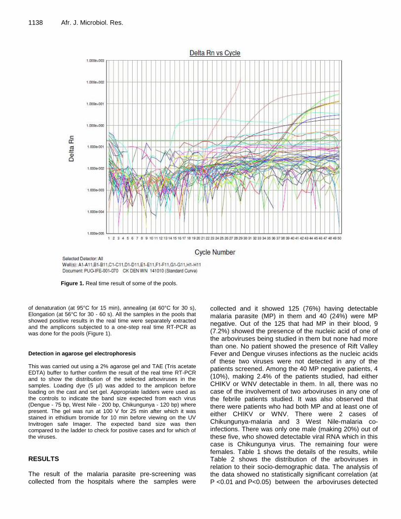
Figure 1. Real time result of some of the pools.
of denaturation (at 95°C for 15 min), annealing (at 60°C for 30 s), Elongation (at 56°C for 30 - 60 s). All the samples in the pools that showed positive results in the real time were separately extracted and the amplicons subjected to a one-step real time RT-PCR as was done for the pools (Figure 1).
Detection in agarose gel electrophoresis This was carried out using a 2% agarose gel and TAE (Tris acetate EDTA) buffer to further confirm the result of the real time RT-PCR and to show the distribution of the selected arboviruses in the samples. Loading dye (5 µl) was added to the amplicon before loading on the cast and set gel. Appropriate ladders were used as the controls to indicate the band size expected from each virus (Dengue - 75 bp, West Nile - 200 bp, Chikungunya - 120 bp) where present. The gel was run at 100 V for 25 min after which it was stained in ethidium bromide for 10 min before viewing on the UV Invitrogen safe Imager. The expected band size was then compared to the ladder to check for positive cases and for which of the viruses.
RESULTS The result of the malaria parasite pre-screening was collected from the hospitals where the samples were
collected and it showed 125 (76%) having detectable malaria parasite (MP) in them and 40 (24%) were MP negative. Out of the 125 that had MP in their blood, 9 (7.2%) showed the presence of the nucleic acid of one of the arboviruses being studied in them but none had more than one. No patient showed the presence of Rift Valley Fever and Dengue viruses infections as the nucleic acids of these two viruses were not detected in any of the patients screened. Among the 40 MP negative patients, 4 (10%), making 2.4% of the patients studied, had either CHIKV or WNV detectable in them. In all, there was no case of the involvement of two arboviruses in any one of the febrile patients studied. It was also observed that there were patients who had both MP and at least one of either CHIKV or WNV. There were 2 cases of Chikungunya-malaria and 3 West Nile-malaria co-infections. There was only one male (making 20%) out of these five, who showed detectable viral RNA which in this case is Chikungunya virus. The remaining four were females. Table 1 shows the details of the results, while Table 2 shows the distribution of the arboviruses in relation to their socio-demographic data. The analysis of the data showed no statistically significant correlation (at P <0.01 and P<0.05) between the arboviruses detected

Adesina et al. 1139
Table 1. Summary of the distribution of CHIKV, WNV and malaria in febrile patients.
Sample code Sex Age range (years) Occupation CHIKV +VE WNV +VE Malaria Parasite
+ve -ve
HC041 Male 21-30 Student
HC077 Male 11-20 Student
HC125 Female 51-60 Private Business
HC123 Female 11-20 Student
HC071 Female 31-40 Teaching
HC080 Female 21-30 Student
HC094 Male 21-30 Student
SD002 Female 61-70 None
SD008 Female 41-50 Private Business
CHIKV – Chikungunya virus; WNV – West Nile virus; MP– Malaria parasite.
Table 2. Socio-demography and the distribution of the detected arboviruses.
Parameter WNV result CHIK V Result
+ ve (%) -ve (%) Total +ve (%) -ve (%) Total
Month of sample collection
August 2010 0 (0) 69 (41.8) 69 1 (0.6) 68 (41.2) 69
September 2010 5 (3.0) 72 (43.6) 77 2 (1.2) 75 (45.5) 77
October 2010 1 (0.6) 18 (10.9) 19 0 (0.0) 19 (11.5) 19
Age category (years)
Under 1 0 (0) 1 (0.6) 01 0 (0.0) 1 (0.6) 01
1 - 10 0 (0) 4 (2.4) 04 0 (0) 4 (2.4) 04
11 - 20 1 (0.6) 24 (14.5) 25 1 (0.6) 24 (14.5) 25
21 - 30 2 (1.2) 71 (43.0) 73 1 (0.6) 72 (43.6) 73
31 - 40 1 (0.6) 26 (15.8) 27 1 (0.6) 26 (15.8) 27
41 - 50 1 (0.6) 18 (10.9) 19 0 (0) 19 (11.5) 19
51 - 60 0 (0) 10 (6.1) 10 0 (0) 10 (6.1) 10
61 - 70 1 (0.6) 5 (3.0) 06 0 (0) 6 (3.6) 06
Sex Female 5 (3.0) 85 (51.5) 90 1 (0.6) 89 (54.0) 90
Male 1 (0.6) 74 (44.8) 75 2 (1.2) 73 (44.2) 75
Occupation
Students 3 (1.8) 97 (58.8) 100 2 (1.2) 98 (59.4) 100
Health worker 0 (0) 2 (1.2) 2 0 (0) 2 (1.2) 02
Teacher 1 (0.6) 9 (5.4) 10 0 (0) 10 (6.1) 10
Other civil servants
0 (0) 29 (17.6) 29 1 (0.6) 28 (17.0) 29
Retiree 0 (0) 3 (1.8) 3 0 (0) 3 (1.8) 03
Private business 1 (0.6) 14 (8.5) 15 0 (0) 15 (9.1) 15
Artisan 0 (0) 2 (1.2) 2 0 (0) 2 (1.2) 2
None 1 (0.6) 3 (1.8) 4 0 (0) 4 (2.4) 4
Highest academic qualification
None 2 (1.2) 8 (4.8) 10 0 (0) 10 (6.1) 10
Primary 0 (0) 18 (10.9) 18 0 (0) 18 (10.9) 18
Secondary 3 (1.8) 85 (51.5) 88 1 (0.6) 87 (52.7) 88
Tertiary 1 (0.6) 48 (29.1) 49 2 (1.2) 47 (28.5) 49
Location Within Osun 6 (3.6) 153 (92.7) 159 3 (1.8) 156 (94.5) 159
Outside Osun 0 (0) 6 (3.6) 6 0 (0) 6 (3.6) 6

1140 Afr. J. Microbiol. Res. and the sociodemographic factors considered. DISCUSSION The concerted efforts to combat malaria has resulted in its mortality falling by 42% globally since 2000, and by 49% in the WHO African Region as well as mortality rates among children in Africa have been reduced by an estimated 54% since 2000 (WHO, 2014). However, the issue of the involvement of and co-infection with one or more arboviruses has called for taking febrile conditions more serious than before. Sow et al. (2016) carried out a study to investigate co-infection with malaria among arbovirus-infected patients in Senegal. Out of these patients, they reported that 48.7 % (20/41) were co-infected with malaria parasites with CHIKV having 18.7% (3/16) among other arboviruses with fever being the only sign or symptom associated with the dual malaria parasite/arbovirus infection. This is higher than 9 (7.2%) that is being reported in this study. In a study to ascertain the etiologic agent causing an outbreak of febrile illness with symptoms similar to chikungunya fever in Chiapas State, Mexico, Kautz et al. (2015) found that 79% of febrile illness cases with polyarthralgia in Chiapas State during late 2014 were caused by CHIKV. This is a further confirmation of the need to extend diagnosis of fever/febrile conditions beyond malaria as the pathogen as well as the vector seem to be thriving in virtually all the continents of the world.
The global spread of mosquito vectors of these pathogens via global demographic and societal changes, and modern transportation have provided the mechanisms for the vectors as well as the pathogens they transmit to break out of their natural ecology and become established in new geographic locations where susceptible arthropod vectors and hosts provide permissive conditions for them to cause major epidemics (Bonizzoni et al., 2013).
Presently in Africa, the epidemiology and public health impact of CHIKV and WNV is still unclear owing to the scanty information available but the current geographical distribution of their primary vectors and the likely further spread, increasing human population growth, unplanned urbanisation especially in developing world and increased international travel have all made transmission likely and successful (Amarasinghe et al., 2011 and Caglioti et al., 2013). Also, they present with similar symptoms thereby making it possible for them to be misdiagnosed. Furthermore, where malaria is endemic and the majority of febrile illnesses are diagnosed as such, often without laboratory confirmation, both viral infections may go undetected and so continue to perpetuate themselves (Amexo et al., 2004). It is very important that more extensive studies be carried out on the diagnosis and pathogenesis of arboviruses as some of them namely DFV, Zika virus and CHIKV infections have shown ocular
manifestations which can be present at the time of fever or may manifest after many weeks. Anterior uveitis, optic neuritis and retinitis are the most common manifestations during the acute infection of these infections (de Andrade et al., 2017).
It is interesting to know that all the age groups are represented in those who were malaria parasite negative but positive for one arbovirus or the other. Arbovirus-Plasmodium infections can be said therefore to have nothing to do with age as all groups are susceptible. This is another reason why arboviruses should be suspected in febrile conditions. It was observed in this study that all the samples that had one arbovirus or the other but void of malaria parasite were collected in September. Although, more studies need to be conducted to establish the reason for this, but it is in agreement with what Forshey et al. (2010) reported where the arboviral infections showed a rise in the number of people infected in September. It is important to improve on the research capacity of many of the affected countries so as to correctly diagnose and manage these arboviral infections. The prevalence reported in this study could be higher if a larger population is studied and if patients with febrile conditions report early enough and their sample collected in the earlier stage of the infection than two weeks used in this study. This is calling for a more extensive study of the involvement of arboviruses in febrile conditions as a way to control their further spread since it appears that the spread of the vectors of these arboviruses, mosquitoes, has not been properly checked. A more involvement of the laboratory in the diagnosis of febrile conditions as a way to minimize the effect and spread of the pathogens is therefore necessary. It is also important that these arboviruses should be considered in conditions with symptoms similar to their infections.
CONFLICT OF INTERESTS
The authors hereby declare that there is no conflict of interest. REFERENCES Amarasinghe A, Kuritsk JN, Letson GW, Margolis HS (2011). Dengue
virus infection in Africa. Emerg Infect. Dis. 17(8):1349-1354. Amexo M, Tolhurst R, Barnish G, Bates I (2004). Malaria misdiagnosis:
Effects on the poor and vulnerable. Lancet 364 (9448):1896-1898. Arya CS, Mehta KL, Agarwal N, Agarwal BK, Mathai G, Moondhara A
(2005). Episodes of concurrent dengue and malaria. Dengue Bull. 29:208-209.
Baba M, Logue CH, Oderinde B, Abdulmaleek H, Williams J, Lewis J, Laws TR, Hewson R, Marcello A, D' Agaro P (2013). Evidence of arbovirus co-infection in suspected febrile malaria and typhoid patients in Nigeria. J. Infect. Dev. Ctries. 7:51-59.
Bonizzoni M, Gasperi G, Chen X, James AA (2013). The invasive mosquito species Aedes albopictus: Current knowledge and future perspectives. Trends Parasitol. 29(9):460-468.
Caglioti C, Lalle E, Castilletti C, Carletti F, Capobianchi MR, Bordi L (2013). Chikungunya virus infection: an overview. New Microbiol. 36(3):211-27.
Carme B, Matheus S, Donutil G, Raulin O, Nacher M, Morvan J (2009).
Concurrent dengue and malaria in Cayenne Hospital, French Guiana. Emerg Infect Dis. 15:668-671.
Charrel RN, Brouqui P, Foucault C, De Lamballerie X (2005). Concurrent dengue and malaria. Emerg. Infect Dis. 11:1153-1154.
de Andrade GC, Ventura CV, Mello Filho PA, Maia M, Vianello S, Rodrigues EB (2017). Arboviruses and the eye. Int. J. Retina Vitreous 3:4.
Deresinski S (2006). Concurrent Plasmodium vivax malaria and dengue. Emerg. Infect. Dis. 12(11):1802.
Domingues RB (2009). Treatment of viral encephalitis. Central Nervous Syst Agents Med. Chem. 9:56-62.
Epelboin L, Hanf M, Dussart P, Ouar-Epelboin S, Djossou F, Nacher M, Carme B (2012). Is dengue and malaria co-infection more severe than single infections? A retrospective matched-pair study in French Guiana. Malar. J. 11:142
Forshey BM, Guevara C, Laguna-Torres VA, Cespedes M, Vargas J, et al (2010). Arboviral Etiologies of Acute Febrile Illnesses in Western South America, 2000-2007. PLoS Negl. Trop. Dis. 4(8): e787.
Gan VCH, Leo YS (2014). Current epidemiology and clinical practice in arboviral infections - implications on blood supply in South-East Asia. ISBT Science Series 9:262-267
Karabatsos N (2001). International catalogue of arboviruses, including certain other viruses of vertebrates. Am. Soc Trop. Med. Hyg. (San Antonio, TX, USA); 1985. 2001 update.
Kautz TF, Díaz-González EE, Erasmus JH, Malo-García IR, Langsjoen RM, Patterson EI, Auguste DI, Forrester NL, Sanchez-Casas RM, Hernández-Ávila M, Alpuche-Aranda CM (2015). Chikungunya Virus as Cause of Febrile Illness Outbreak, Chiapas, Mexico, 2014. Emerg. Infect. Dis. Vol. 21, No. 11.
Monlun E, Zeller H, Le Guenno B, Traoré-Lamizana M, Hervy JP, Adam F, Ferrara L, Fontenille D, Sylla R, Mondo M, Digoutte JP (1993). Surveillance of the circulation of arbovirus of medical interest in the region of eastern Senegal (1988-1991)] (In French). Bull. Soc. Pathol. Exot. 86:21-28.
Robin Y, Cornet M, Heme G, Le Gonidec G (1980). Isolement du virus de la dengue au Sénégal. Ann. Virol. (Inst. Pasteur). 131E:149-54.
Adesina et al. 1141 Senn N, Suarkia DL, Manong D, Siba PM, McBride WJH (2011).
Contribution of dengue fever to the burden of acute febrile illnesses in Papua New Guinea: an age-specific prospective study. Am. J. Trop. Med. Hyg. 85:132-137.
Sow A, Loucoubar C, Diallo D, Faye O, Ndiaye Y, Senghor CS, Dia AT, Faye O, Weaver SC, Diallo M, Malvy D, Sall AA (2016). Concurrent malaria and arbovirus infections in Kedougou, southeastern Senegal. Malar. J. 15:47.
WHO (2011). Comprehensive guidelines for prevention and control of dengue and dengue haemorrhagic fever - revised and expanded edition. New Delhi: WHO Regional Office for South East Asia 2011; p. xiv+196.
WHO (2014). Malaria Fact sheet N°94 Updated March 2014 http://www.who.int/mediacentre/factsheets/fs094/en.
WHO global burden disease 2004 update. Available from: http://www.who.int/healthinfo/global burden disease/2004 report update/en/index.html
WHO (2010). World malaria report 2010. Geneva: Word Health Organization.

Vol. 11(28), pp. 1142-1149, 28 July, 2017
DOI: 10.5897/AJMR2017.8577
Article Number: C59282265357
ISSN 1996-0808
Copyright © 2017
Author(s) retain the copyright of this article
http://www.academicjournals.org/AJMR
African Journal of Microbiology Research
Full Length Research Paper
Microbiota sampled from a polluted stream in Recife-PE, Brazil and its importance to public health
Antonio Fernando da Purificação Júnior1, Lívia Caroline Alexandre de Araújo2, Ana Catarina de Souza Lopes3, Marcela de Araújo Sobral4, Gláucia Manoella de Souza Lima4, Márcia
Vanusa da Silva5, Maria Tereza dos Santos Correia5 and Maria Betânia Melo de Oliveira5*
1Genetic, Federal University of Pernambuco (UFPE), Recife, PE, Brazil.
2Biological Science, Federal University of Pernambuco (UFPE), Recife, PE, Brazil.
3Department of Tropical Medicine, Federal University of Pernambuco (UFPE), Recife, PE, Brazil.
4Department of Antibiotics, Federal University of Pernambuco (UFPE), Recife, PE, Brazil.
5Department of Biochemistry, Federal University of Pernambuco (UFPE), Recife, PE, Brazil.
Received 3 May, 2017; Accepted 7 July, 2017
Pollution of water bodies can cause environmental and public health problems. The Cavouco stream is a tributary of the Capibaribe River, one of the main rivers in the state of Pernambuco, Brazil, and receives a high pollution load from residential, laboratory and hospital effluents. The aim of the present study was to perform phenotypic and molecular characterization in this stream, and evaluate the water quality using microbiological parameters. Water was collected from five sampling points, and bacterial species were identified using biochemical and molecular methods through 16S rRNA gene sequence analysis. Total and thermotolerant coliforms were also quantified. Fermenting Gram-negative bacilli from the family Enterobacteriaceae (Escherichia coli, Klebsiella pneumoniae and Proteus mirabilis), non-fermenting bacilli (Pseudomonas aeruginosa and Pseudomonas putida) and Gram-positive bacilli (Bacillus cereus, Bacillus licheniformis, Bacillus pumilus and Staphylococcus hominis) were identified. A total of 25 bacterial isolates were phenotypically identified. All phenotypic identifications were confirmed by molecular analysis, except for S. hominis, which was molecularly identified as Exiguobacterium. Regarding water quality, all analyzed samples were positive for total and thermotolerant coliforms. The results obtained suggest that the Cavouco stream presents a potential risk for transmission of water-borne diseases, because of the presence of pathogenic bacteria. In addition, the current state of the stream also threatens the conservation of its native species. Keywords: Public health, Enterobacteriaceae, thermotolerant, Bacilli.
INTRODUCTION Aquatic ecosystems have suffered significant changes due to multiple environmental effects, resulting from the release of large quantities of effluent without prior treatment (International Joint Commission, 2015; USGS, 2015). This discharge can cause physical, chemical and
biological deterioration, and endangers both the resident aquatic organisms and public health. Scientific, technological and epidemiological advances have provided new tools for the assessment of water quality, both for human consumption and environmental
purposes (Tanchou, 2014).
In 2015, the Centers for Disease Control and Prevention (CDC) reported that approximately 780 million people had no access to drinking water around the world. The consumption of contaminated water and lack of basic sanitation is estimated to cause 842,000 deaths per year worldwide, and 1,000 children under the age of five years die every day (WHO, 2014; WHO and UNICEF, 2015). Contaminated water carries pathogens that cause diarrhea, gastrointestinal disorders and systemic diseases, approximately 70% of diarrheal diseases could be avoided by improving basic sanitation (WHO, 2014). According to the Brazilian Ministry of Health, 6,715 deaths caused by diarrhea or gastroenteritis, presumably resulting from infection, were recorded between 2010 and 2015 (BRASIL, 2015).
Some studies have used traditional methods of selective isolation and cultivation to characterize the microbial communities of the affected environments (Skariyachan et al., 2013). However, taxonomic classification by these methods can be difficult because of variations in phenotypic characteristics (Woo et al., 2008). For this reason, molecular methods that allow fast and reliable confirmation of microbial identity have been developed (Ramírez-Castillo et al., 2015). Among these, methods using 16S rRNA gene sequencing are predominant. This gene is used as a phylogenetic marker because its sequences are highly conserved (Srinivasan et al., 2015).
The aim of the present study was to evaluate the water quality by the isolation and identification of representative bacterial species present in this environment, using biochemical and molecular methods. METHODS Study area The Cavouco stream, located at latitude 8°2'52.05"S and longitude 34°57'10.33" W, state of Pernambuco (UFPE), Brazil, is approximately 6 km long, and flows into the right margin of the Capibaribe River, one of the main rivers of state. Along its course, it receives pollutants from residential, laboratory and hospital waste, which reduces the water quality and threatens the aquatic life (Araujo and Oliveira, 2013; Freitas et al., 2016). Water samples (200 mL) were collected from five points (Figure 1) along the stream, according to the methods of Araújo and Oliveira (2013), and stored between 1 and 4°C until subsequent bacteriological analysis. Isolation and phenotypic identification of bacterial isolates For bacterial isolation, 50 µL of water was inoculated onto eosin
da Purificação Júnior et al. 1143 methylene blue (EMB) agar and 5% bovine blood agar (to count colony forming units), and incubated at 37°C for 24 to 48 h. Gram staining was then performed to make presumptive identifications of the bacteria found according to the technique described by Koneman and Winn (2006).
Gram-negative isolates were preliminarily identified using the following biochemical tests: glucose, lactose and sucrose fermentation, hydrogen sulfide production, motility, indole production and citrate, lysine and urea degradation. Species identification was confirmed using the Kit API 20E (Biomérieux), according to the manufacturer’s instructions.
Gram-positive isolates were preliminarily identified through colony morphology, hemolysis in blood agar, and presence/absence and position of spores, visualized through Gram staining. Species identification was confirmed using an automated system (BD Phoenix™ Automated Microbiology System). Analysis of total and thermotolerant coliforms For analysis of total and thermotolerant coliforms, water samples were collected from the water surface at a depth of 30 cm, from each of the five sampling points. The samples were stored between 1 and 4°C until analysis.
The presence and number of total and thermotolerant coliforms were determined using the multiple-tube fermentation method, and the results were expressed as most probable number (MPN) per 100 mL of sample, according to APHA (2015). Molecular identification using 16S rRNA gene sequence analysis The bacterial samples were inoculated into 5 mL brain heart infusion (BHI) broth for 24 h at 37°C for to DNA extraction. Chromosomal DNA was extracted using the phenol-chloroform method (Sambrook and Russell, 2001). DNA quality was evaluated by electrophoresis in a 0.8% agarose gel using 0.5× TBE buffer and run at 100 V for 1 h; gels were analyzed using a UV transilluminator and photographed. DNA concentration was quantified using a Nanodrop spectrophotometer (Thermo Scientific).
The 16S rRNA gene was amplified by polymerase chain reaction (PCR), using the primers fD1 (5´-AGAGTTTGATCCTGGCTCAG-3´) and rD1 (5´-AAGGAGGTGATCCAGCC-3´) (Weisburg et al., 1991). PCR was performed in a final volume of 25 μL, containing 1× buffer, 200 µM dNTPs, 1.5 mM MgCl2, Taq DNA-polymerase (1 U/µL; Invitrogen), 10 pmol of each primer, and 10 ng of DNA template. The PCR was performed using a thermocycler (C1000 Thermal Cycler – BioRad), and the PCR program consisted of 95°C for 5 min, 30 cycles at 95°C for 45 s, primer annealing at 54°C for 45 s, extension at 72°C for 2 min, and a final extension at 72°C for 5 min.
PCR products were purified using the Pure Link purification kit (Invitrogen) according to the manufacturer’s instructions, and sequenced using the Big Dye Kit (Applied Biosystems) on an automated DNA sequencer (ABI 3100). The 16S rRNA gene sequences obtained was compared with sequences deposited in the GenBank database (NCBI). The dendrogram was constructed using multiple sequence alignment, based on genetic distances, maximum parsimony, and maximum likelihood, using Molecular Evolutionary Genetic Analysis 5.2 software (MEGA5).
*Corresponding author: E-mail: [email protected] or [email protected]. Tel: +55 (81) 2126 8547 R. 229
Fax: +55 (81) 2126 8576.
Author(s) agree that this article remains permanently open access under the terms of the Creative Commons Attribution
License 4.0 International License

1144 Afr. J. Microbiol. Res.
Figure 1. Representation of Cavouco creek area, Brazil, showing the five collection points (P1-P5) and microbial representatives obtained from each point.
Bacterial library of Cavouco stream – UFPE Identified bacteria were stored in 80% glycerol (150 µL of glycerol and 850 µL of bacterial culture) at -80°C, and under pure mineral oil (Isofar) at ambient temperature. The isolates were labeled according to: C (Cavouco), P (sampling point), the number of sampling points, and the number of isolations. RESULTS Microscopic and biochemical identification of bacterial colonies Inoculated blood agar presented innumerable colony forming units (CFU), except for water samples collected at the stream source (Point 1), for which only a few colonies were observed. Due to EMB medium selectivity, these plates presented an average of 200 CFU, with a lower number of CFUs observed for Point 1, and higher number for point 5 EMB plates presented colonies with metallic green sheen, dark center, some with bright or pink edges, and mucoid appearance. In blood agar, some colonies were shiny or gray, with or without beta-hemolysis, and with irregular edges. Microscopic examination of Gram-stained colonies revealed the presence of Gram-
negative and positive bacilli. Some Gram-positive bacilli were observed to be arranged in chains, filaments and spherical bodies. Approximately, 22 and 25 colonies were selected from the most frequent colonies present on EMB medium, and from blood agar, respectively.
A total of 25 isolates were identified phenotypically, belonging to seven genera (Bacillus, Enterobacter, Escherichia, Klebsiella, Proteus, Pseudomonas and Staphylococcus). Enterobacteria were present in all sampling points. Proteus mirabilis was highly prevalent among Gram-negative species (with eight isolates mostly from point 5), followed by Escherichia coli (six isolates), and Klebsiella pneumoniae (five isolates, from points 2, 3, and 4). Among the Gram-positive bacilli, the genus Bacillus was prevalent, with two species (Bacillus licheniformis and Bacillus pumilus) identified from point 4, and one (Bacillus cereus) from point 1. Another Gram-positive species, S. hominis, was identified from point 4 (Table 1). Presence of total and thermotolerant coliforms All analyzed samples were positive to total and thermotolerant coliforms, presenting an MPN > 1.4 ×

da Purificação Júnior et al. 1145 Table 1. Phenotypic and molecular identification of isolated bacteria from Cavouco stream collection points. ID: Identity. *Access number refer to sequences deposited in GenBank.
Points Isolates Phenotypic Identification 16S rRNA gene sequencing
API 20E (ID%) BD Phoenix Species Similarity (%) Accession*
P1 CP11S - Bacillus cereus Bacillus cereus 99 KT719668.1
P2 CP23P Escherichia coli (99.8%) - Escherichia coli 99 CP014225.1 CP24P Escherichia coli (94.8%) - Escherichia coli 99 CP014225.1 CP22P Klebsiella pneumoniae (97.8%) - Klebsiella pneumoniae 99 KM233642.1
P3
CP35S Proteus mirabilis (99.9%) - Proteus mirabilis 99 KR150991.1 CP36S Pseudomonas putida (44.5%) - Pseudomonassp. 99 JQ994361.1 CP38S Klebsiella pneumoniae (97.9) - Klebsiella pneumoniae 99 AB680212.1 CP39P Klebsiella pneumoniae (98.1%) - Klebsiella pneumoniae 99 AB680212.1 CP313P Klebsiella pneumoniae (97.7%) - Klebsiella pneumoniae 99 AB680212.11 CP37S Escherichia coli (99.8%) - Escherichia coli 99 CP014225.1 CP310P Escherichia coli (99.8%) - Escherichia coli 99 CP014225.1 CP311P Escherichia coli (99.8%) - Escherichia coli 99 CP014225.1
P4
CP414S Escherichia coli (99.9%) - Escherichia coli 98 CP013837.1 CP416S Klebsiella pneumoniae (97.7%) - Klebsiella pneumoniae 99 KC524425.1 CP415S Enterobacter clocae (99.4%) - Enterobacter clocae 99 GU191924c.1 CP418S - Bacillus licheniformis Bacillus licheniformis 99 KJ26873.1 CP419S - Bacillus pumilis Bacillus pumilis 99 KJ526890.1 CP420P Staphylococcus hominis Exiquobacterium spp. 99 KT074375.1
P5
CP522S Proteus mirabilis (99.9%) - Proteus mirabilis 99 KF811051.1 CP523S Proteus mirabilis (99.9%) - Proteus mirabilis 99 KR150991.1 CP525S Proteus mirabilis (99.9%) - Proteus mirabilis 99 KR150991.1 CP526P Proteus mirabilis (99.9%) - Proteus mirabilis 99 KR150991.1 CP527P Proteus mirabilis (99.9%) - Proteus mirabilis 99 KR150991.1 CP528P Proteus mirabilis (99.9%) - Proteus mirabilis 99 HQ169118.1 CP527P Proteus mirabilis (99.9%) - Proteus mirabilis 99 KR150991.1
10
3/100 mL.
Molecular identification using 16S rRNA gene analysis The 16S rRNA gene sequences (approximately 1.500 bp) for the 25 selected isolates were aligned, and compared with sequences deposited in the GenBank database. The obtained sequences were found to have a high degree of genetic similarity (98-99%) with deposited sequences for the same species, confirming their phenotypic identification. Of the 25 isolates, only S. hominis did not display concordance between the phenotypic and molecular identification. This isolate was identified phenotypically as belonging to the genus Staphylococcus, but through molecular analysis, it revealed 99% similarity with the genus Exiguobacterium (Table 1). The phylogenetic tree showed clustering of 16S rRNA gene sequences obtained from the studied isolates with those from GenBank, confirming molecular identification (Figure 2).
Construction of the bacterial library of Cavouco The bacterial library of Cavouco currently includes 21 Gram-negative and four Gram-positive species isolated from five sampling points, stored in frozen stocks under mineral oil. These are the first bacterial isolates of this environment, and the first representatives of the Bacterial Library of Impacted Environments of UFPE. DISCUSSION The analyses showed high water contamination of the Cavouco stream, located in Recife-PE, Brazil, with all samples containing thermotolerant coliforms. The presence of coliforms is a parameter evaluated for water quality monitoring programs and indicates the presence of potentially pathogenic microorganisms (WHO, 2014).
In the present study, the quantification of microbiological parameters indicated that all samples were unfit for human consumption and recreational use. The observed coliform levels were higher than those considered safe by

1146 Afr. J. Microbiol. Res.
Figure 2. Similarity dendrogram generated from the comparison between the sequences of 16S rRNA strains isolated from collection points at Cavouco and the deposited ones in GenBank database.
the US Environmental Protection Agency (EPA), 2011 and the Brazilian Environmental Council (Normative Resolution N° 357/2005) (CONAMA, 2005), representing risk for human health.
Indications of significant pollution in the studied regions of the Cavouco stream had been previously observed. Araujo and Oliveira (2013) analyzed Cavouco stream water, and found changes in the levels of dissolved oxygen, ions and ammonia, as well as values of the Index of Water Quality for the Protection of Aquatic Life (IQAPVA) indicating low capacity for the maintenance of aquatic life.
Enterobacteria were present at all sampling points in the present study. Several studies have shown that most enterobacteria can cause infections because of their
ability to survive in hostile environments and their ability to develop resistance to antimicrobial drugs (Irenge et al., 2015; Tajbakhsh et al., 2015; Patel et al., 2016).
Several pipes located along the margins of the Cavouco stream discharge residential, hospital, and laboratory effluents into the stream, which explains the presence of bacteria of fecal origin. In addition, the existence of pastures for animal grazing and the development of other activities, together with the poor conservation state of the local riparian forest, could contribute to this microbiological water pollution.
Previous studies of affected environments have reported similar results. Rodrigues et al. (2009), evaluated the water quality of the Piracuama River, located in the state of São Paulo (SP), Brazil, and identified enterobacteria of

genera Citrobacter, Edwardsiella, Enterobacter, Escherichia, Klebsiella, Morganella, Salmonella and Shigella. A study performed on a contaminated river in India also identified the presence of important pathogens for public health, namely Edwardsiella spp., Enterobacter spp., E. coli, Morganella spp., Proteus spp., Pseudomonas spp., Serratia spp. and Staphylococcus spp. (Skariyachan et al., 2013), with fecal coliforms being the most common pathogens in contaminated rivers and streams (Kim et al., 2013; Liu et al., 2015).
Escherichia coli was isolated from several sampling points of stream, is used as a specific indicator of fecal contamination in tropical and temperate regions (Páll et al., 2013). This species is considered one of the main causes of diarrhea in adults and children in developing countries, infection most often occurring through contact with contaminated water (Walker et al., 2007; Isidean et al., 2011) and is an important cause of urinary tract and wound infections and pneumonia in immunosuppressed hospitalized patients, and meningitis in newborn children (Wijetunge et al., 2015; Martelius et al., 2016).
Proteus spp. has been described as an important infectious agent in hospital environments (Chen et al., 2014; Murray et al., 2015). Bacteria from the genus, are commonly found in the environment, especially in locations with water pollution and soils with degraded
material (Drzewiecka, 2016). A study performed in hospitals located in northeast Brazil, the same region as the present study, isolated P. mirabilis, which produces extended spectrum beta-lactamase (ESBL), have been reported to be an important cause of nosocomial infection in worldwide (Abreu et al., 2011). In the present study, this species was isolated in an area where there is discharge of hospital waste.
The genus Klebsiella is widely distributed in nature and in the gastro intestinal tract of humans and animals.. K. pneumonia can also be found in the oropharynx of hospitalized patients, constituting a source of pulmonary infections, and usually occurring in patients with debilitating conditions such as alcoholism and diabetes (Distel et al., 2013). This species can also infect the urinary tract, cutaneous wounds, and blood, causing bacteremia, meningitis in infants, hepatic abscess, and urinary tract infections (Siu et al., 2012). This species has gained importance due to the development and inter-species and intra-species dis-semination of several antimicrobial resistance mechanisms, namely, the production of beta-lactamases such as ESBLs and KPC (K. pneumoniae Carbapenemase), which degrade beta-lactam antibiotics, frequently detected in hospitalized patients in Recife, PE, Brazil (Lopes et al., 2010; Cabral et al., 2012; Melo et al., 2014).
The presence of the genus Bacillus in the Cavouco stream is worrisome. The presence of Bacillus in food in
amounts higher than 10 cells per gram indicates multiplication, and indicates a high health risk (Germano and Germano, 2003). Another interesting aspect was the
da Purificação Júnior et al. 1147 observed diversity of Bacillus species. This might indicate that the environmental conditions (nutrients, temperature, humidity, oxygen concentration, and pH) were favorable for the multiplication and maintenance of these species at the studied site.
Bacillus species can sporulate, and the resistance conferred by these spores constitutes an important problem for the epidemiology of associated infections. Because the Cavouco stream is a tributary of the Capibaribe River, which is used as a water source for local agriculture and fishing, the presence of Bacillus could contribute to food contamination.
Of the five sampling points, points 3 and 4 presented the highest microbial diversity, whereas point 5 yielded only one species, P. mirabilis. The absence of diversity at point 5, which receives the discharge of effluents from a morgue and hospital, may have been due to difficulty of isolating Gram-positive bacteria and the lack of selectivity of the blood agar used for bacterial cultures. Another possible explanation for the difficulty in isolating Gram-positive bacteria was the presence of high concentrations of toxic substances in hospital effluents, such as antibiotics, cytostatic agents, heavy metals, disinfectants and hormones, which could have a genotoxic effect on these bacteria (Jean et al., 2012; Devarajan et al., 2015).
Molecular tools were used in the present study to confirm the phenotypic identification of the 25 isolates. The 16S rRNA gene is widely used as phylogenetic marker, and it has been sequenced for a large number of bacterial lineages (Srinivasan et al., 2015). Most of these sequences are deposited in the GenBank database (Benson et al., 2012), and can therefore be compared with sequences of new isolates.
For isolates belonging to different species, their 16S rRNA gene sequences must share less than 97% similarity (Goebel and Stackebrant, 1994). The sequences obtained in the present study shared 98% or higher similarity with sequences deposited in the GenBank database, confirming the phenotypic identification of all isolates, except for S. hominis, which presented 98% similarity with genus Exiguobacterium. This can be explained by the fact that both genera have very similar morphological and biochemical characteristics, such as motility, positive catalase and urease activity, negative phosphatase and coagulase activity, presence or absence of nitrate reduction, and acid production under aerobic conditions, and both contain aerobic and facultative anaerobic species (Stieglmeier et al., 2009). These colonies are spherical, opaque, butyrous or yellow-orange, growing at temperatures varying between 20 and 45°C (Schleifer et al., 1979; Collins et al., 1983). The similar morphological and biochemical characteristics of the two genera might have contributed to its initial identification as Staphylococcus. Similar results were reported by Elmaci et al. (2015), who observed a divergence in identification at the species level for 53.3% (81/152) of the tested lactic
1148 Afr. J. Microbiol. Res. acid bacteria isolates, identified phenotypically using the API CHL method and 16S rRNA gene sequence analysis. Bosshard et al. (2006) observed a discrepancy at the genus and species level for 20.6% (12/58) of the tested non-fermenting Gram-negative bacterial isolates, identified using API 20 NE and 16S rRNA gene sequence analysis. Both studies attributed this divergence to the similarity of morphological and biochemical characteristics of different taxonomic groups, making identification using phenotypical methods difficult, and confirming the specificity of molecular analysis.
The present results indicate that the Cavouco stream could significantly contribute to an increase in microbial pollution, presenting a potential risk of waterborne disease transmission, as genera of pathogenic bacteria were identified. In addition, the current state the Cavouco stream also threatens the conservation of its native species. Environmental cleaning actions such as the establishment of a sewage collection and treatment systems are urgently needed to improve the water quality of this and other affected aquatic environments, such as rivers, lakes and coastal areas. The obtained data enabled the establishment of a bacteria library, which will help to understand the evolution of impacted environment, in terms of environmental quality, overtime. The present study characterized a sample of cultured bacteria isolated from the studied area. In spite of the importance and relevance of this data for public health, it does not represent the totality of organisms present in the studied area. CONFLICT OF INTERESTS The authors have not declared any conflict of interests. REFERENCES Abreu AG, Marques SG, Monteiro-Neto V, Carvalho RM, Gonçalves AG
(2011). Nosocomial infection and characterization of extended-spectrum β-lactamases-producing Enterobacteriaceae in Northeast Brazil. Rev. Soc. Bras. Med. Trop. 44(4):441-446.
APHA-American Public Health Association. Standard Methods for the Examination of Water and Wastewater (2015). Multiple tube fermentation technique for members of the coliform group. American Public Health Association, 22 ed. Washington, DC, USA.
Araujo MC, Oliveira MBM (2013). Monitoramento da qualidade das águas de um riacho da Universidade Federal de Pernambuco, Brasil. Rev. Ambient. Água 8(3):247-257.
Benson DA, Karsch-Mizrachi I, Clark K, Lipman DJ, Ostell J, Sayers EW (2012). GenBank. Nucleic Acids Res. 40 (D1):D48-D53.
Bosshard PP, Zbinden R, Abels S, Böddinghaus B, Altweeg M, Bötter EC (2006). 16S rRNA Gene Sequencing versus API 20 NE System and the VITEK 2 ID-GNB Card for identification of non-fermenting gram-negative bacteria in the clinical laboratory. J. Clin. Microbiol. 44(4):1359-1366.
BRASIL Ministério da Saúde (2015). DATASUS, informações de Saúde- Brasil. http://tabnet.datasus.gov.br
Cabral AB, Melo RCA, Maciel MA, Lopes AC (2012). Multidrug resistance genes, including bla (KPC) and bla (CTX)-M-2, among Klebsiella pneumoniae isolated in Recife, Brazil. Rev. Soc. Bras. Med. Trop. 45:572-578.
Centers for Disease Control and Prevention- CDC (2015). Global
Water, Sanitation, and Hygiene (WASH). http://www.cdc.gov/healthywater/global/.
Chen P, Zhang L, Meng B (2014). Correlation between urinary stones and urinary tract infections. Zhonghua Liu Xing Bing Xue Za Zhi. 35:597-599.
Collins MD, Lund BM, Farrow JAE, Schleifer KH (1983). Chemotaxonomic study of an alkalophilic bacterium, Exiguobacterium aurantiacum gen. nov., sp. nov. J. Gen. Microbiol. 129:2037-2042.
Conselho Nacional do Meio Ambiente (CONAMA) (2005). Resolução 357. Diário Oficial da União, n. 53, 17 de março de 2005, Seção 1. Available in. P 58. http://www.mma.gov.br/port/conama/res/res05/res35705.pdf.Conam.
Devarajan N, Laffite A, Ngelikoto P, Elongo V, Prabakar K, Mubedi JI, Piana PT, Wildi W, Poté J (2015). Hospital and urban effluent waters as a source of accumulation of toxic metals in the sediment receiving system of the Cauvery River, Tiruchirappalli, Tamil Nadu, India. Environ. Sci. Pollut. Res. Int. 22:12941-12950.
Distel C, Jacobson S, Tille PM (2013). Alcohol induced diabetic ketoacidosis exacerbated by an acute respiratory infection with Klebsiella pneumoniae. Clin. Lab. Sci. 26(2):68-71.
Drzewiecka D (2016). Significance and roles of Proteus spp. bacteria in natural environments. Microb. Ecol. 72(4):741-758.
Elmaci SB, Tokath M, Dursun D, Özçelik F, Şanlibaba P (2015). Phenotypic and genotypic identification of lactic acid bactéria isolated from traditional pickles of the Çubuk region in Turkey. Folia Microbiol. 60:241-251.
Environmental Protection Agency- EPA (2011). National Characteristics of Drinking Water Systems Serving 10,000 or Fewer People. http://water.epa.gov/type/drink/pws/smallsystems/upload/REVFINAL-Nat-Characte-July-2011-508-compliant.pdf
Freitas JHES, Santana KV, Nascimento ACC, Paiva SC, Moura MC, Coelho LCCB et al. (2016). Evaluation of using aluminum sulfate and water-soluble Moringa oleifera seed lectin to reduce turbidity and toxicity of polluted stream water. Chemosphere 163:133-141.
Germano PML, Germano MIS (2003). Higiene E Vigilância Sanitária de Alimentos, Qualidade das Matérias Primas, Doenças transmitidas por alimentos e Treinamento de recursos humanos, 2nd ed. Varela, São Paulo, Brasil. 655p.
Goebel BM, Stackebrandt E (1994). Cultural and phylogenetic analysis of mixed microbial populations found in natural and commercial bioleaching environments. Appl. Environ. Microbiol. 60(5):1614-1621.
International Joint Commission (2015). The Impact of Urban Development on Water Quality. http://www.ijc.org/php/publications/html/12br/english/report/physical/iudwq.html.
Irenge LM, Kabego L, Kinunu FB, Itongwa M, Mitangala PN, Gala JL, Chirimwami RB (2015). Antimicrobial resistance of bacteria isolated from patients with bloodstream infections at a tertiary care hospital in the Democratic Republic of the Congo. S. Afr. Med. J. 105(9):752-755.
Isidean SD, Riddle MS, Savarino SJ, Porter CK (2011). Porter A systematic review of ETEC epidemiology focusing on colonization factor and toxin expression. Vaccine 29:6167-6178.
Jean J, Perrodin Y, Pivot C, Trepo D, Perraud M, Droguet J, Tissot-Guerraz F, Locher F (2012). Identification and prioritization of bioaccumulable pharmaceutical substances discharged in hospital effluents. J. Environ. Manage.103:113-121.
Kim JY, Lee H, Lee JE, Chung MS, Ko GP (2013). Identification of human and animal fecal contamination after rainfall in the Han River, Korea. Microbes Environ. 28:187-194.
Koneman EW, Winn WC (2006). Color Atlas and Textbook of Diagnostic Microbiology, 6
th edn. Williams & Wilkins, Philadelphia:
Lippincott-Raven Publishers. 1565p. Liu WC, Chan WT, Young CC (2015). Modeling fecal coliform
contamination in a tidal Danshuei River estuarine system. Sci. Total. Environ. 502:632-640.
Lopes AC, Veras DL, Lima AM, Melo RD, Ayala J (2010). bla(CTX-M-2) and bla(CTX-M-28) extended-spectrum beta-lactamase genes and class 1integrons in clinical isolates of Klebsiella pneumoniae from Brazil. Mem. Inst. Oswaldo Cruz. 105:163-167.
Martelius T, Jalava J, Kärki T, Möttönen T, Ollgren J, Lyytikäinen O

(2016). Nosocomial bloodstream infections caused by Escherichia coli and Klebsiella pneumoniae resistant to third-generation cephalosporins, Finland, 1999-2013: Trends, patient characteristics and mortality. Infect. Dis.(Lond). 48:229-234.
Melo RCA, Barros EM, Loureiro NG, de Melo HR, Maciel MA, Lopes AC (2014). Presence of fimH, mrkD, and irp2 virulence genes in KPC-2-producing Klebsiella pneumoniae isolates in Recife-PE, Brazil. Curr. Microbiol. 69:824-831.
Murray EC, Marek A, Thomson PC, Coia JE (2015). Gram-negative bacteraemia in haemodialysis. Nephrol. Dial. Transplant. 30(7):1202-1208.
Páll E, Niculae M, Kiss T, Şandru CD, Spînu M (2013). Human impact on the microbiological water quality of the rivers. J. Med. Microbiol. 62:1635-1640.
Patel CB, Shanker R, Gupta VK, Upadhyay RS (2016). Q-PCR based culture-independent enumeration and detection of Enterobacter: an emerging environmental human pathogen in riverine systems and potable Water. Front. Microbiol. 7:172.
Ramírez-Castillo FY, Loera-Muro A, Jacques M, Garneau P, Avelar-González FJ, Harel J, Guerrero-Barrera AL (2015). Waterborne pathogens: Detection methods and challenges. Pathogens 4(2):307-334.
Rodrigues JDD, Jorge AOC, Ueno M (2009). Avaliação da qualidade das águas de dias áreas utilizadas para recreação do Rio Piraciama-SP. Revista Biociências. 15:88-94.
Sambrook J, Russell DW (2001). Molecular Cloning: A laboratory manual. Cold Spring Harbor Laboratory; 3 ed.
Schleifer KH, Meyer SA, Rupprecht M (1979). Relatedness among coagulase-negative staphylococci: Deoxyribonucleic acid reassociation and comparative immunological studies. Arch. Microbiol. 122:93-101.
Siu LK, Yeh KM, Lin JC, Fung CP, Chang FY (2012). Klebsiella pneumoniae liver abscess: a new invasive syndrome. Lancet Infect. Dis. 12:881-887.
Skariyachan S, Lokesh P, Rao R, Kumar AU, Vasist KS, Narayanappa R(2013).A pilot study on water pollution and characterization of multidrugresistant superbugs from Byramangala tank,Ramanagara district, Karnataka, India. Environ. Monit. Assess. 185:5483-5495.
Srinivasan R, Karaoz U, Volegova M, MacKichan J, Kato-Maeda M, Miller S, Nadarajan R, Brodie EL, Lynch SV (2015). Use of 16S rRNA gene for identification of a broad range of clinically relevant bacterial pathogens. PLoS ONE 10:e0117617.
Stieglmeier M, Wirth R, Kminek G, Moissl-Eichinger C (2009). Cultivation of Anaerobic and Facultatively Anaerobic Bacteria from Spacecraft-Associated Clean Rooms. Appl. Environ. Microbiol. 75(11):3484-3491.
Tajbakhsh E, Tajbakhsh S, Khamesipour F (2015). Isolation and molecular detection of Gram negative bacteria causing urinary tract infection in patients referred to Shahrekord hospitals, Iran. Iran Red Crescent Med. J. 17(5):e24779.
Tanchou V (2014). Review of Methods for the Rapid Identification of Pathogens in Water Samples—ERNCIP Thematic A = πr
2 Chemical
& Biological Risks in the Water Sector—Task 7, Deliverable 1. Institute for the Protection and Security of the Citizen Publications Office of the European Union, http://publications.jrc.ec.europa.eu/repository/bitstream/JRC92395/lbna26881enn.pdf.
da Purificação Júnior et al. 1149 USGS-Science for a Changing World (2015). The Effects of
Urbanization on Water Quality: Waterborne Pathogens. http://water.usgs.gov/edu/urbanpath.html.
Walker RI, Steele D, Aguado T, Ad Hoc ETEC Technical Expert Committee (2007). Analysis of strategies to successfully vaccinate infants in developing countries against enterotoxigenic E. coli (ETEC) disease. Vaccine 25:2545-2566.
Weisburg WG, Barns SM, Pelletier DA, Lane DJ (1991). 16S ribosomal DNA amplification for phylogenetic study. J. Bacteriol. 173:697-703.
WHO (2014). Water Sanitation and Health. http://www.who.int/water_sanitation_health/diseases.
WHO and UNICEF (2015). Lack of sanitation for 2.4 billion people undermining health improvements. http://www.unicef.org.mz/en/unicef-who-lack-of-sanitation-for-2-4-billion-people-undermining-health-improvements.
Wijetunge DS, Gongati S, DebRoy C, Kim KS, Couraud PO, Romero IA, Weksler B, Kariyawasam S (2015). Characterizing the pathotype of neonatal meningitis causing Escherichia coli (NMEC). BMC Microbiol. 15(1):211.
Woo PCY, Lau SKP, Teng JLL, Tse H, Yuen KY (2008). Then and now: use of 16S rDNA gene sequencing for bacterial identification and discovery of novel bactéria in clinical microbiology. Clin. Microbiol. Infect. 14:908-934.

African Journal of
Microbiology Research
Related Journals Published by Academic Journals African Journal of Biotechnology African Journal of Biochemistry Research Journal of Bacteriology Research Journal of Evolutionary Biology Research Journal of Yeast and Fungal Research Journal of Brewing and Distilling