investigation of morphological changes to staphylococcus ...rhaverka/anderson fems 2004.pdf · the...
TRANSCRIPT

www.fems-microbiology.org
FEMS Microbiology Letters 240 (2004) 105–110
Investigation of morphological changes to Staphylococcus aureusinduced by ovine-derived antimicrobial peptides using
TEM and AFM
Rachel Claire Anderson, Richard G. Haverkamp, Pak-Lam Yu *
Institute of Technology and Engineering, Massey University, Private Bag 11-222, Palmerston North, New Zealand
Received 18 July 2004; received in revised form 9 September 2004; accepted 15 September 2004
First published online 2 October 2004
Edited by A.M. George
Abstract
The effect of two ovine-derived peptides on the morphology of Staphylococcus aureus NCTC 4163 cells were investigated using
transmission electron microscopy and atomic force microscopy. Both techniques showed that SMAP29, an a-helical peptide,
induced cell lysis, whereas OaBac5mini, a proline/arginine-rich peptide, caused no observable morphological changes. This is con-
sistent with previous experimental work which indicated that SMAP29 caused cell death and induced cell lysis, whereas OaBac5mini
acted by interacting with the inner cellular contents.
� 2004 Federation of European Microbiological Societies. Published by Elsevier B.V. All rights reserved.
Keywords: Antimicrobial peptides; Cathelicidins; Transmission electron microscopy; Atomic force microscopy; Ovine blood
1. Introduction
Antimicrobial peptides are part of the innate immune
system of all living things. These peptides act as the first
line of defence against invading micro-organisms. This
work focuses on ovine antimicrobial peptides becauseovine blood is readily available in New Zealand and
has the potential to be separated into a number of
high-value products, including the antimicrobial pep-
tides isolated from the blood neutrophils [1].
For this work two synthesised ovine-derived peptides
were used (Table 1). SMAP29 is a very potent a-helicalpeptide [2] that was predicted from ovine cDNA [3,4],
but has not yet been isolated from ovine cells. OaBac5-mini is a truncated version of OaBac5, comprising the
0378-1097/$22.00 � 2004 Federation of European Microbiological Societies
doi:10.1016/j.femsle.2004.09.027
* Corresponding author. Tel.: +64 6 350 5027; fax: +64 6 350 5604.
E-mail address: [email protected] (P.-L. Yu).
first 24 N-terminal residues. OaBac5 is a 51-residue, pro-
line and arginine-rich peptide that was inferred based on
a cDNA sequence [5]. Since then, three variants of Oa-
Bac5, as well as the originally predicted molecule, have
been isolated from sheep neutrophils [6,7]. OaBac5 is
made up of a 6-residue N-terminus, followed by twocopies of a 16-residue repeat and a 5-residue C-terminus
[7]. OaBac5mini, the truncated version of OaBac5, is
made up of the six N-terminal residues, one copy of
the 16-residue repeat, and the first two residues of the
second repeat. The full-length molecule could not be
synthesised, so this truncation was chosen because it
has been shown that the bovine peptide Bac7 retained
its activity when truncated similarly [8]. It has previouslybeen shown that OaBac5mini displays similar antimicro-
bial activity to the parent molecule, OaBac5, so it is an
appropriate model to use to represent the naturally
occurring peptide [9].
. Published by Elsevier B.V. All rights reserved.

Table 1
Sequences of ovine-derived antimicrobial peptides used for this
research
Peptide Sequence
SMAP29 RGLRRLGRKIAHGVKKYGPTVLRIIRIA–NH2
OaBac5mini RFRPPIRRPPIRPPFRPPFRPPVR–NH2
106 R.C. Anderson et al. / FEMS Microbiology Letters 240 (2004) 105–110
Previous worked showed that SMAP29 and OaBac5-
mini had different mechanisms-of-action [9]. The aim of
this research was to investigate whether the test peptides
induced changes in the bacterial cell morphology to help
further understand the mechanisms used by the pep-
tides. The previous results indicated that SMAP29
caused rapid cell death by depolarising the cytoplasmic
membrane, which resulted in leakage of the cellular con-tents, and some complete cell lysis. In contrast, OaBac5-
mini appeared to inhibit cell division by interacting with
the inner cellular contents. Therefore, it was hypothe-
sised that SMAP29 would cause the morphology of
the cells to change significantly, whereas OaBac5mini
would cause little or no change.
To get a good understanding of the morphology of
the peptide treated cells two techniques, transmissionelectron microscopy (TEM) and atomic force micros-
copy (AFM), were used. TEM gives an electron density
image of cell cross sections, whereas AFM gives topo-
logical images of the cell surface. Together these tech-
niques should give a good overall model of the cell
morphology.
2. Materials and methods
2.1. Peptide synthesis
The two ovine-derived peptides were synthesised, by
N-(9-fluorenyl)methoxy carbonyl solid-phase peptide
synthesis using a model 432A synthesiser (Applied Bio-
systems Inc, Foster City, CA), at the University of
British Columbia Nucleic Acid/Protein Service facility.
The purities of the peptides were confirmed to be at
least 99% using high-performance liquid chromatogra-
phy and mass spectroscopy analysis. SMAP29 was syn-thesised as a 28-amino acid amidated peptide because
this is thought to be its natural form [2]. The full
OaBac5 molecule could not be synthesised, due to
the large number of proline residues, so a shortened
version (the first 24 amino acids) called OaBac5mini
was created. Both peptides were amidated at their
C-termini.
2.2. Transmission electron microscopy
A log phase culture of Staphylococcus aureus NCTC
4163 in Mueller–Hinton broth was split into 1.5 mL
aliquots. The cells were collected by centrifugation
(10000 rpm, 5 min) and resuspended in peptone water.
Six samples were prepared; two untreated, two treated
with SMAP29 (4 lg/mL) and two treated with OaBac5-
mini (64 lg/mL). The samples were incubated at 37 �Cfor 1 h. The cells were collected by centrifugation(10000 rpm, 5 min) to remove the peptone water. To
fix the cells, 2% glutaraldehyde in 0.1 M cacodylate
was added and the samples were incubated at 4 �Cfor 1 h. The cells were collected by centrifugation
(10000 rpm, 5 min) and washed twice with 0.1 M phos-
phate buffer. To postfix the cells, 1% osmium tetraox-
ide was added and the samples were left at room
temperature for 1 h. The samples were dehydrated withgraded ethanol solutions (30% ethanol for 10 min, 60%
ethanol for 10 min, 90% ethanol for 10 min, 100% eth-
anol for 10 min, 100% ethanol for 1 h), embedded in
Procure 812 resin and left to polymerise (over the
weekend). From each sample 10 thin slices (approxi-
mately 100 nm) were cut with a diamond knife and
stained with uranyl acetate and lead citrate on grids.
Each of these sections was examined with a PhilipsEM201 80 kV Transmission Electron Microscope and
images were taken with a 35-mm camera.
2.3. Atomic force microscopy
A log phase culture of S. aureus NCTC 4163 in
Mueller–Hinton broth was split into 1 mL aliquots.
The cells were collected by centrifugation (10000 rpm,5 min) and resuspended in peptone water. Three sam-
ples were prepared; one was untreated, one was treated
with SMAP29 (4 lg/mL for both organisms) and one
was treated with OaBac5mini (64 lg/mL). The samples
were incubated at 37 �C for 30 min. The cells were col-
lected by centrifugation (10000 rpm, 5 min) to remove
the peptone water. A number of different methods were
tried to secure the cells for AFM imaging. These in-cluded passing the samples through polycarbonate
membranes (SPI-Pore Filter, 13 mm diameter, 0.8 lmpores) to entrap the cells in the pores and immobilising
the cells in agarose gel (5% agarose). The most success-
ful method involved drying the culture on a glass slide.
The treated samples were washed with and resuspended
in water, and then diluted 100-fold. About 10 lL of the
sample was dropped onto a glass microscope slide andallowed to dry at room temperature. For each sample
duplicate slides were prepared. The dried samples typi-
cally had a diameter of 5–8 mm. To image the samples
an Asylum Research MFP-3D scanning probe micro-
scope was used. The tips used were either NT-MDT
CSG01 or Olympus TR400PSA which typically have
a spring constant in the range 0.01–0.08 N/m. The areas
for imaging were chosen randomly from the totalsample area.

R.C. Anderson et al. / FEMS Microbiology Letters 240 (2004) 105–110 107
3. Results and discussion
3.1. Transmission electron microscopy
S. aureus 4163 NCTC cells were treated with
SMAP29 or OaBac5mini at concentrations four timestheir minimal inhibitory concentrations (MICs) [9], be-
fore they were fixed, cut into sections and examined
using TEM. For each treatment duplicate samples were
prepared and from each sample 10 sections were cut
and processed. The electron density images given in
Fig. 1 shows the typical results. The dark areas show
where the sample had a high electron density and the
light areas show where the sample had a low electrondensity.
Fig. 1. Transmission electron microscope images taken of S. aureus 4163 N
arrows point to cell walls that have separated from the cytoplasm. The green
interpretation of the references to colour in this figure legend, the reader is
The images of the samples treated with SMAP29 were
greatly different to those of the untreated cells. The un-
treated cells were uniformly shaped, with intact cell
walls; whereas many of the SMAP29 treated cells had
their cell walls missing. In Fig. 1 the green arrows point
to cells without cell walls, called ghost cells, and the yel-low arrows point to cell walls that have separated from
the cells. The images show that in many cases the cyto-
plasmic contents were leaking out of the cells. Previous
experiments showed that SMAP29 causes cell death
and induces lysis of Gram-negative cells [9]. These re-
sults indicate that SMAP29 acts on Gram-positive cells
in a similar way to how it acts on Gram-negative cells.
In contrast, the images of the samples treated withOaBac5mini show fewer damages than those treated
CTC cells treated with SMAP29 and OaBac5mini for 1 h. The yellow
arrows point to ghost cells that are not surrounded by a cell wall. (For
referred to the web version of this article.)

108 R.C. Anderson et al. / FEMS Microbiology Letters 240 (2004) 105–110
with SMAP29 (Fig. 1). Unlike the SMAP29 treated
sample, there were no ghost cells without cell walls, or
cells that appeared to be leaking their cellular contents.
However, some cell wall debris was present (yellow ar-
rows). This is consistent with the idea that OaBac5mini
does not cause cell death and induce lysis [9]. However,the cells treated with OaBac5mini appeared to be swol-
len and the samples had a high proportion of cells con-
taining septa, compared with the control samples. This
may be due to OaBac5mini stopping the cell division
process at this point, whereas the control cells moved
on from this state and continued to divide as normal.
Fig. 2. Far away AFM images of S. aureus NCTC 4163 cells on a glass slide
left are the height images and those on the right are the deflection images.
This is consistent with previous work that indicated that
the mechanism-of-action of OaBac5mini involved the
inhibition of cell division [9].
3.2. Atomic force microscopy
S. aureus NCTC 4163 cells were treated with
SMAP29 or OaBac5mini at four times their minimal
inhibitory concentrations (MICs) [9], before they were
dried at room temperature on glass slides and imaged
using AFM. For each sample duplicate slides were made
and numerous randomly chosen parts of each slide were
treated with SMAP29 and OaBac5mini for 30 min. The images on the
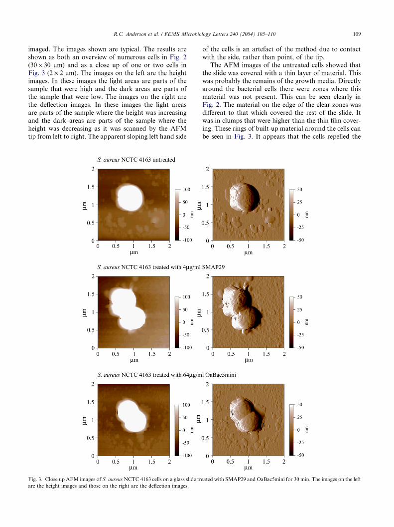
R.C. Anderson et al. / FEMS Microbiology Letters 240 (2004) 105–110 109
imaged. The images shown are typical. The results are
shown as both an overview of numerous cells in Fig. 2
(30 · 30 lm) and as a close up of one or two cells in
Fig. 3 (2 · 2 lm). The images on the left are the height
images. In these images the light areas are parts of the
sample that were high and the dark areas are parts ofthe sample that were low. The images on the right are
the deflection images. In these images the light areas
are parts of the sample where the height was increasing
and the dark areas are parts of the sample where the
height was decreasing as it was scanned by the AFM
tip from left to right. The apparent sloping left hand side
Fig. 3. Close up AFM images of S. aureusNCTC 4163 cells on a glass slide tr
are the height images and those on the right are the deflection images.
of the cells is an artefact of the method due to contact
with the side, rather than point, of the tip.
The AFM images of the untreated cells showed that
the slide was covered with a thin layer of material. This
was probably the remains of the growth media. Directly
around the bacterial cells there were zones where thismaterial was not present. This can be seen clearly in
Fig. 2. The material on the edge of the clear zones was
different to that which covered the rest of the slide. It
was in clumps that were higher than the thin film cover-
ing. These rings of built-up material around the cells can
be seen in Fig. 3. It appears that the cells repelled the
eated with SMAP29 and OaBac5mini for 30 min. The images on the left

110 R.C. Anderson et al. / FEMS Microbiology Letters 240 (2004) 105–110
media solids during the drying process, but the reason
for this is unknown.
The AFM images of the cells treated with OaBac5mi-
ni were similar to those of the untreated cells. The clear-
ings directly around the cells (Fig. 2) and the clumpy
material in rings were both present (Fig. 3). This is con-sistent with the idea that the mechanism-of-action of
OaBac5mini does not induce the bacterial cells to lyse
[9].
In contrast, the AFM images of the cells treated with
SMAP29 were different to those of the cells that were
untreated or treated with OaBac5mini. There were a
lot fewer whole cells present on the slide (Fig. 2). In
the case of the untreated and OaBac5mini treated sam-ples, each of the areas of the sample chosen randomly
for imaging contained numerous cells, whereas for the
SMAP29 treated samples, the randomly chosen areas
for imaging contained fewer, and in some cases no cells.
This indicated that the majority of the cells had been
lysed. This high proportion of lysed cells may be the re-
sult of the drying process. SMAP29 may have weakened
the cell walls and caused the cells to be leaky, whichmade them prone to lysis during drying.
Unlike the other samples, which only had built up
material in rings around the cells, the SMAP29 treated
sample contained a number of piles of material. This
material was probably cell debris from lysis induced
by the peptide. The cells themselves did not appear to
be morphologically different to those of the untreated
sample. Interestingly, around the cells that were intactthere were no clearings like those in the other samples.
The reason for this is unknown.
As predicted there were significant differences in the
culture treated with SMAP29 to that treated with Oa-
Bac5mini. This gives further evidence to support the re-
sults in the previous work [9] which showed that the two
ovine-derived peptides had different mechanisms of
action.
Acknowledgements
This work was supported by MeatNZ and the Mas-
sey University Research Fund (MURF). R.C.A. was a
recipient of the MeatNZ Doctoral Scholarship. The syn-
thesised peptides were kindly supplied by the R.E.W.Hancock Laboratory (Department of Microbiology
and Immunology, University of British Columbia, Van-
couver, Canada). We thank HortResearch (Palmerston
North, New Zealand) for the use of the TEM and Aaron
Hicks (Institute of Veterinary, Animal and Biomedical
Sciences) for helping with the sample preparation.
References
[1] Anderson, R.C. and Yu, P.L. (2004) Ovine antimicrobial peptides:
new products from an age-old industry. Aust. J. Agr. Res. 15, 69–75.
[2] Skerlavaj, B., Benincasa, M., Risso, A., Zanetti, M. and Gennaro,
R. (1999) SMAP-29: a potent antibacterial and antifungal peptide
from sheep leukocytes. FEBS Lett. 463, 58–62.
[3] Bagella, L., Scocchi, M. and Zanetti, M. (1995) cDNA sequences
of three sheep myeloid cathelicidins. FEBS Lett. 376, 225–228.
[4] Mahoney, M.M., Lee, A.Y., Brezinski-Caliguri, D.J. and Huttner,
K.M. (1995) Molecular analysis of the sheep cathelin family reveals
a novel antimicrobial peptide. FEBS Lett. 337, 519–522.
[5] Huttner, K.M., Lambeth, M.R., Burkin, H.R., Burkin, D.J. and
Broad, T.E. (1998) Localisation and genomic organisation of sheep
antimicrobial peptide genes. Gene 206, 85–91.
[6] Shamova, O.V., Brogden, K.A., Zhao, C., Nguyen, T., Kok-
ryakov, V.N. and Lehrer, R.I. (1999) Purification and properties of
proline-rich antimicrobial peptides from sheep and goat leuko-
cytes. Infect. Immun. 67, 4106–4111.
[7] Anderson, R.C. and Yu, P.L. (2003) Isolation and characterisation
of proline/arginine-rich cathelicidin peptides from ovine neutroph-
ils. Biochem. Biophys. Res. Commun. 312, 1139–1146.
[8] Sadler, K., Eom, K.D., Yang, J.L., Dimitrova, Y. and Tam, J.P.
(2002) Translocating proline-rich peptides from the antimicrobial
peptide bactenecin 7. Biochemistry 41, 14150–14157.
[9] Anderson, R.C., Hancock, R.E.W. and Yu, P.L. (2004) Antimi-
crobial activity and bacterial membrane interaction of ovine-
derived cathelicidins. Antimicrob. Agents Chemother. 48, 673–676.