introduction to themes of ap...
TRANSCRIPT
Cells
Unit 4

A Tour of the Cell

Cell Theory
• Hooke, Leeuwenhoek, Schleiden, Schwann, Virchow
• All living things or organisms are made of cells and their products.
• New cells are created by old cells dividing into two.
• Cells are the basic building units of life.
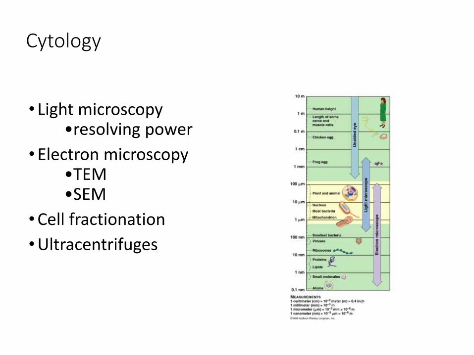
Cytology
• Light microscopy•resolving power
• Electron microscopy•TEM•SEM
• Cell fractionation
• Ultracentrifuges

Dissecting Microscope Images

Compound Light Microscope Images
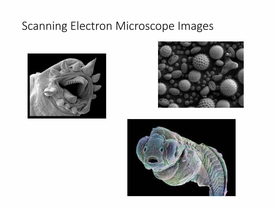
Scanning Electron Microscope Images

Transmission Electron Microscope Images

Cell Types: Prokaryotic
• Nucleoid Region: DNA concentration
• No organelles with membranes
• Ribosomes: protein synthesis
• Plasma membrane: semi-permeable
• Cytoplasm/cytosol (all cells)
• Archea (no peptidoglycan)
• Eubacteria (with peptidoglycan)
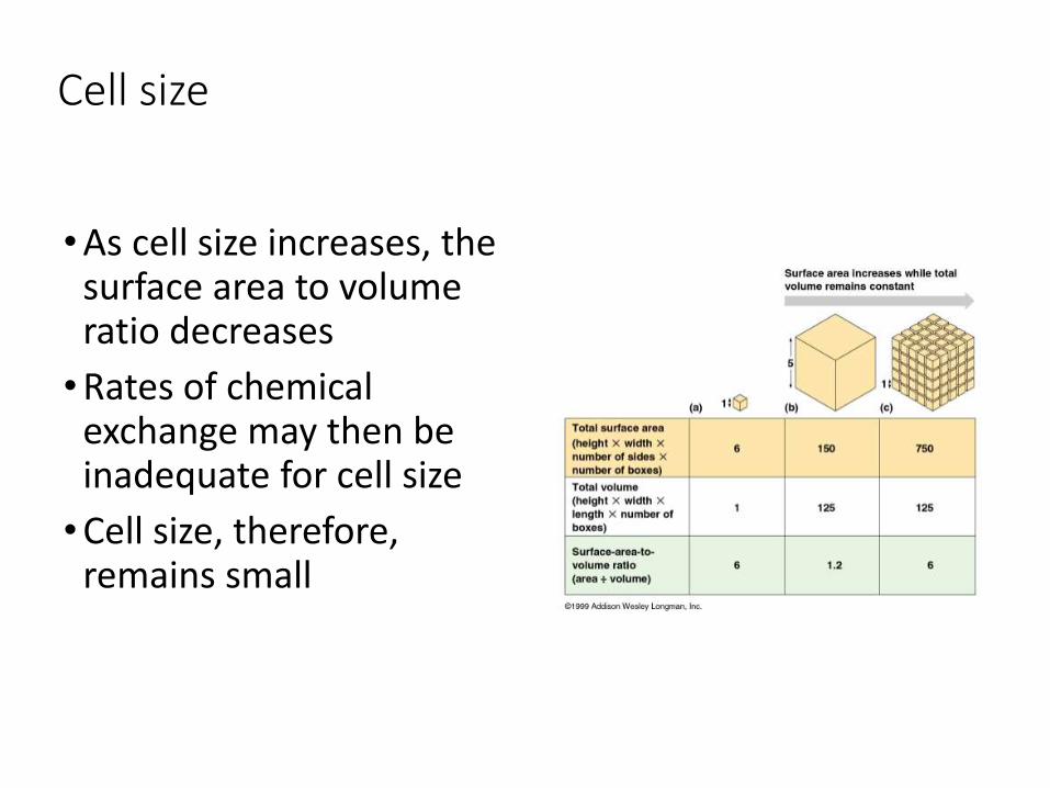
Cell size
• As cell size increases, the surface area to volume ratio decreases
• Rates of chemical exchange may then be inadequate for cell size
• Cell size, therefore, remains small
Organelle Chart
1. Ribosomes
2. Endoplasmic reticulum
3. Golgi apparatus
4. Vacuoles
5. Cytoskeleton
6. Flagella
7. Cilia
8. Mitochondria
9. Chloroplast
10. Lysosome
Place each
organelle in chart
with the following
information:
structure
function
drawing
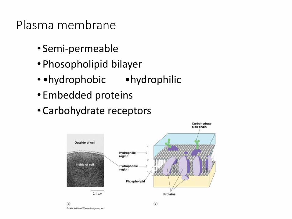
Plasma membrane
• Semi-permeable
• Phosopholipid bilayer
• •hydrophobic •hydrophilic
• Embedded proteins
• Carbohydrate receptors
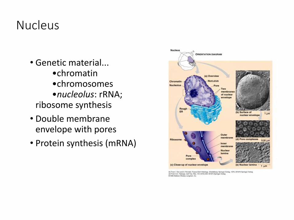
Nucleus
• Genetic material...•chromatin•chromosomes•nucleolus: rRNA;
ribosome synthesis
• Double membrane envelope with pores
• Protein synthesis (mRNA)

Endosymbiosis
•Sonderia sp.
(a ciliate that preys upon various algae, diatoms, and cyanobacteria)
Endosymbiosis
• https://www.youtube.com/watch?v=FGnS-Xk0ZqU
• Chapter 28~ The Origins of Eukaryotic Diversity
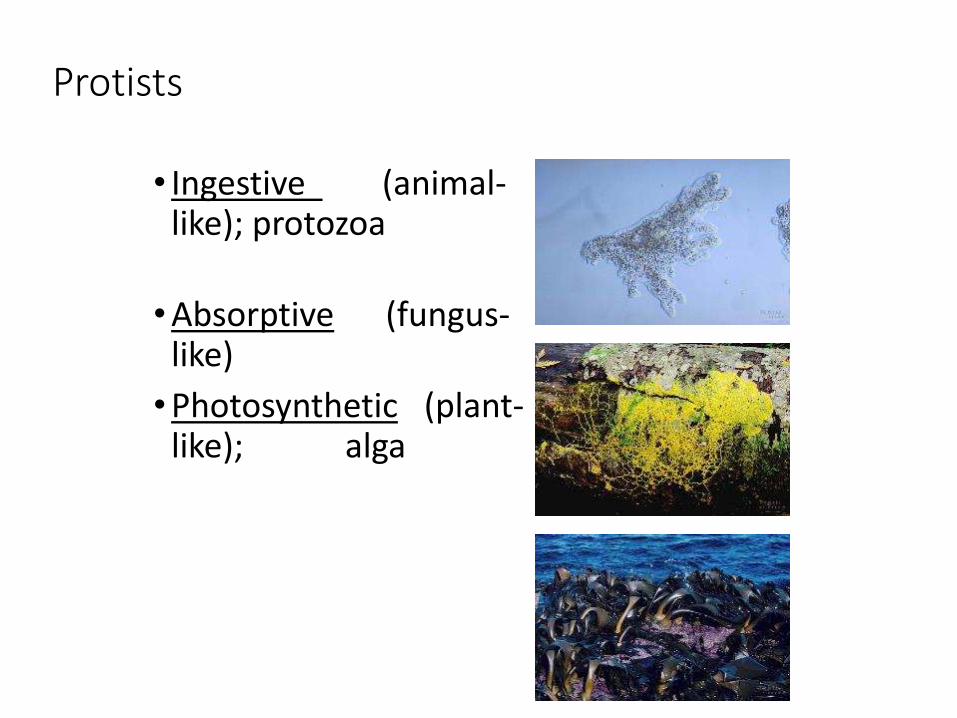
Protists
• Ingestive (animal-like); protozoa
• Absorptive (fungus-like)
• Photosynthetic (plant-like); alga
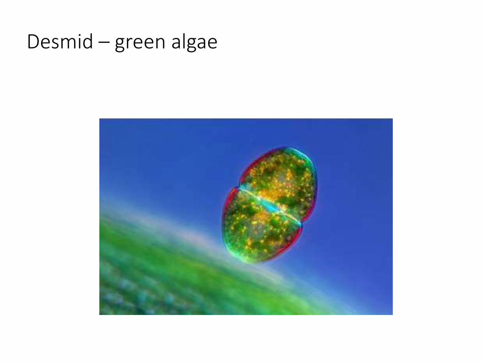
Desmid – green algae

The Endosymbionic Theory
• Mitochondria and chloroplasts were formerly from small prokaryotes living within larger cells (Margulis)
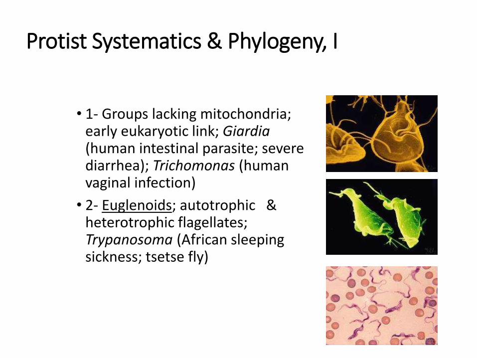
Protist Systematics & Phylogeny, I
• 1- Groups lacking mitochondria; early eukaryotic link; Giardia (human intestinal parasite; severe diarrhea); Trichomonas (human vaginal infection)
• 2- Euglenoids; autotrophic & heterotrophic flagellates; Trypanosoma (African sleeping sickness; tsetse fly)
Protist Systematics & Phylogeny, II
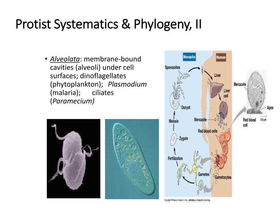
• Alveolata: membrane-boundcavities (alveoli) under cell surfaces; dinoflagellates (phytoplankton); Plasmodium(malaria); ciliates (Paramecium)
Protist Systematics & Phylogeny, III
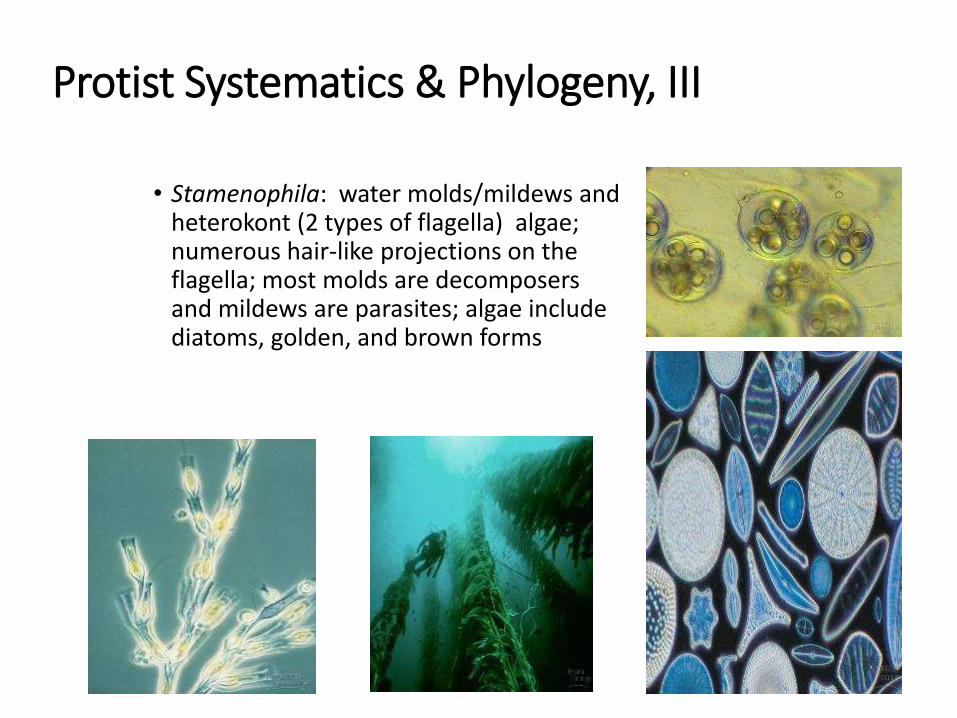
• Stamenophila: water molds/mildews and heterokont (2 types of flagella) algae; numerous hair-like projections on the flagella; most molds are decomposers and mildews are parasites; algae include diatoms, golden, and brown forms
Protist Systematics & Phylogeny, IV
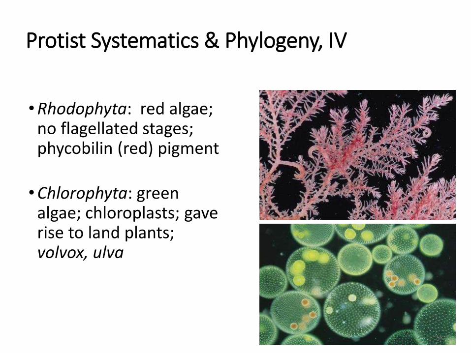
• Rhodophyta: red algae; no flagellated stages; phycobilin (red) pigment
• Chlorophyta: green algae; chloroplasts; gave rise to land plants; volvox, ulva
Protist Systematics & Phylogeny, V
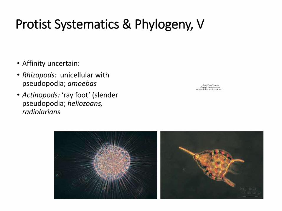
• Affinity uncertain:
• Rhizopods: unicellular with pseudopodia; amoebas
• Actinopods: ‘ray foot’ (slender pseudopodia; heliozoans, radiolarians
QuickTime™ and aCinepak decompressor
are needed to see this picture.
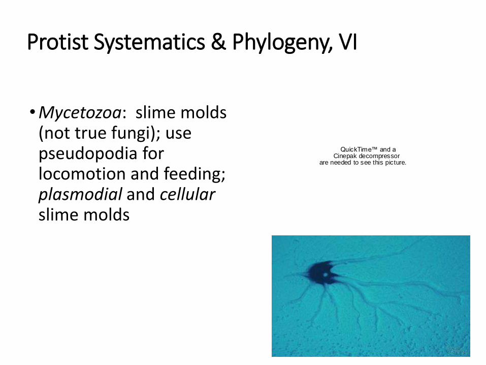
Protist Systematics & Phylogeny, VI
• Mycetozoa: slime molds (not true fungi); use pseudopodia for locomotion and feeding; plasmodial and cellularslime molds
QuickTime™ and aCinepak decompressor
are needed to see this picture.
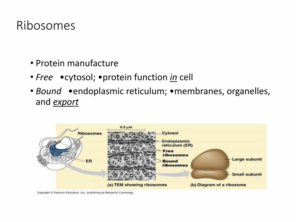
Ribosomes
• Protein manufacture
• Free •cytosol; •protein function in cell
• Bound •endoplasmic reticulum; •membranes, organelles, and export
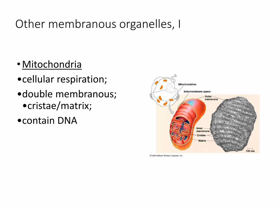
Other membranous organelles, I
• Mitochondria
•cellular respiration;
•double membranous; •cristae/matrix;
•contain DNA
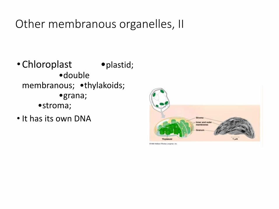
Other membranous organelles, II
• Chloroplast •plastid;•double
membranous; •thylakoids; •grana;
•stroma;
• It has its own DNA
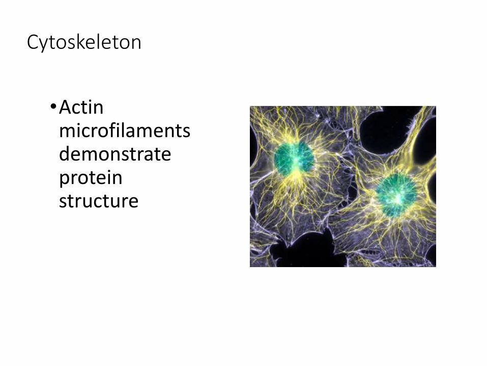
Cytoskeleton
•Actin microfilaments demonstrate protein structure
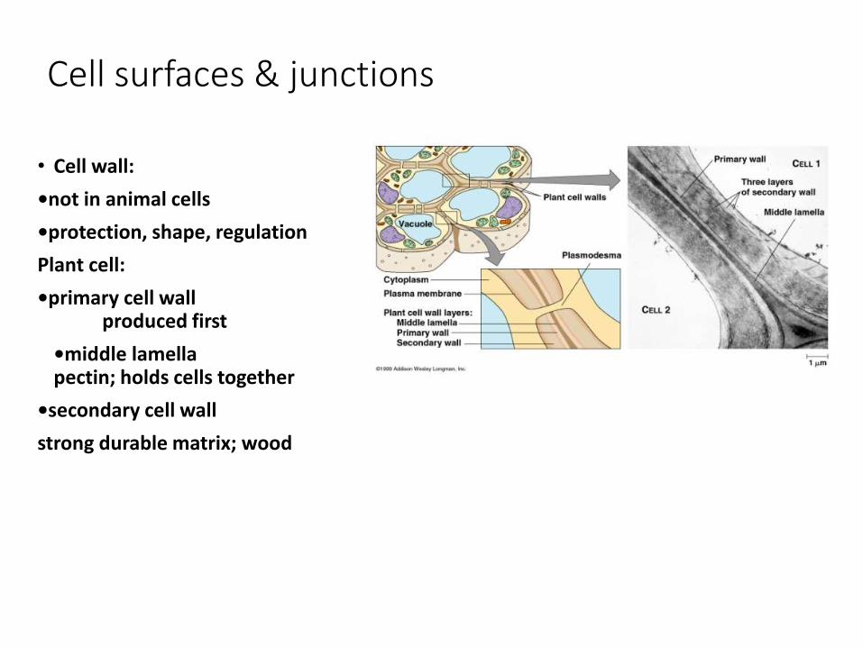
Cell surfaces & junctions
• Cell wall:
•not in animal cells
•protection, shape, regulation
Plant cell:
•primary cell wallproduced first
•middle lamellapectin; holds cells together
•secondary cell wall
strong durable matrix; wood
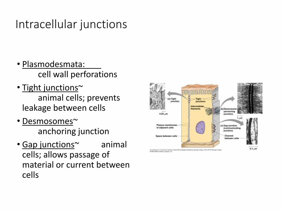
Intracellular junctions
• Plasmodesmata: cell wall perforations
• Tight junctions~animal cells; prevents
leakage between cells
• Desmosomes~anchoring junction
• Gap junctions~ animal cells; allows passage of material or current between cells
Intracellular Junctions
1. Desmosomes
2. Hemidesmosomes
3. Tight Junction
4. Gap Junctions
5. Adherens
• Build a 3-D representation of your assigned intracellular junction
• Label your model
• Include a written description which includes
• How the junction is formed
• When this type of junction is used
• Benefits of this junction for the cell
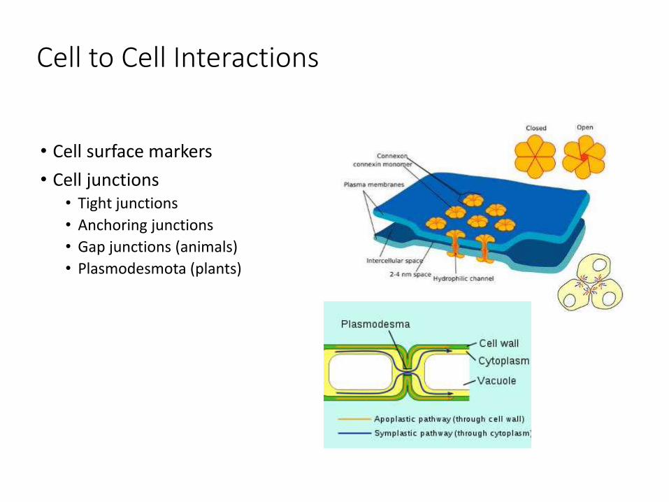
Cell to Cell Interactions
• Cell surface markers
• Cell junctions• Tight junctions
• Anchoring junctions
• Gap junctions (animals)
• Plasmodesmota (plants)

Extracellular matrix (ECM)
• Glycoproteins: • proteins covalently bonded to carbohydrate
• Collagen (50% of protein in human body) •embedded in proteoglycan (another glycoprotein-95% carb)
• Fibronectins •bind to receptor proteins in plasma membrane called integrins (cell communication?)
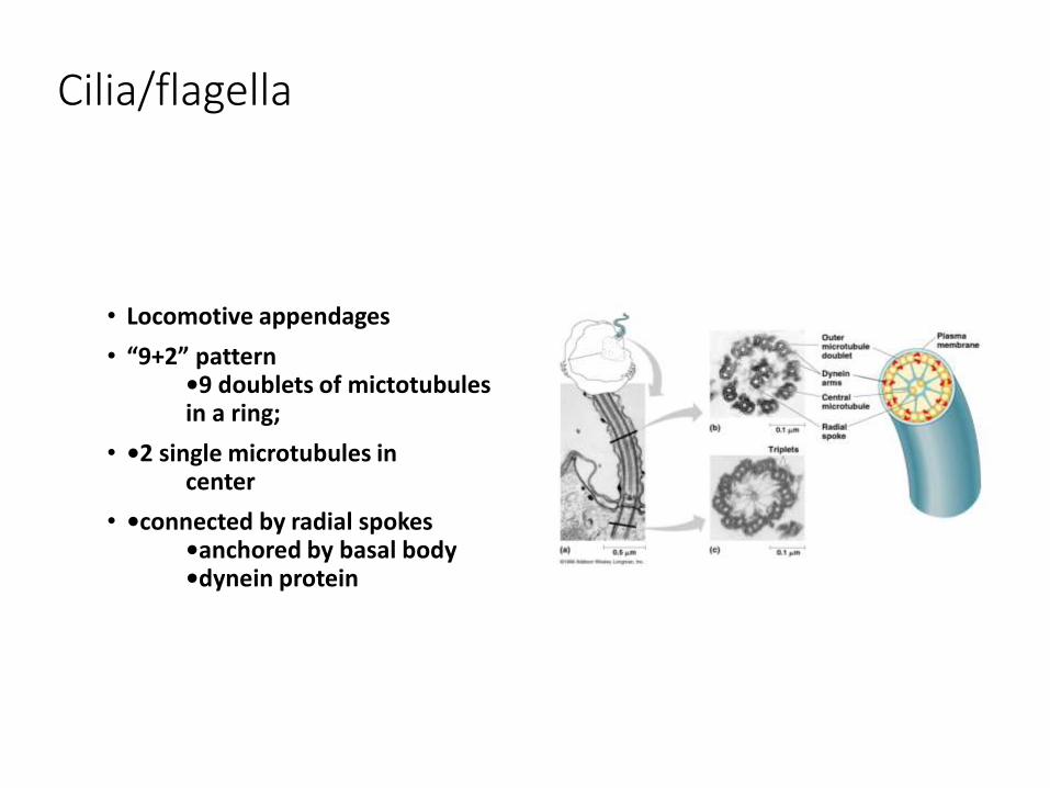
Cilia/flagella
• Locomotive appendages
• “9+2” pattern•9 doublets of mictotubules in a ring;
• •2 single microtubules in center
• •connected by radial spokes•anchored by basal body•dynein protein

Centrosomes/centrioles
• Centrosome: •region near nucleus
• Centrioles: •9 sets of triplet microtubules in a ring •used in cell
replication •only in animal cells
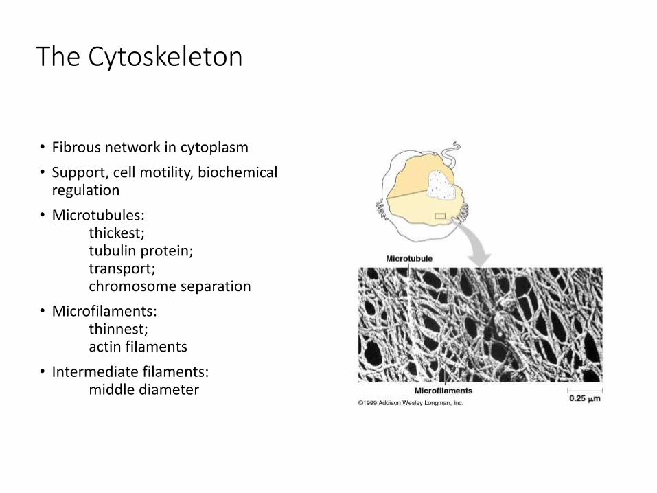
The Cytoskeleton
• Fibrous network in cytoplasm
• Support, cell motility, biochemical regulation
• Microtubules:thickest;tubulin protein;transport;chromosome separation
• Microfilaments:thinnest;actin filaments
• Intermediate filaments:middle diameter

Cell Movement
• Internal via cytoskeleton
• Flagella• Prokaryotic
• Eukaryotic• 9 + 2 structure
• Cilia

Organelles for Quiz:
11. Peroxisome
12. Centrosome
13. Vesicle
14. Cell Membrane
15. Cell Wall
16. Nucleus
17. Nucleolus
18. Plasmid
19. Chromosome
20. Smooth ER
1. Ribosomes
2. Rough Endoplasmic reticulum
3. Golgi apparatus
4. Vacuoles
5. Cytoskeleton
6. Flagella
7. Cilia
8. Mitochondria
9. Chloroplast
10. Lysosome
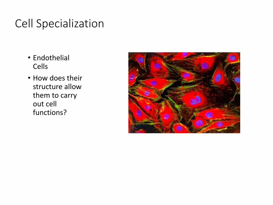
Cell Specialization
• Endothelial Cells
• How does their structure allow them to carry out cell functions?
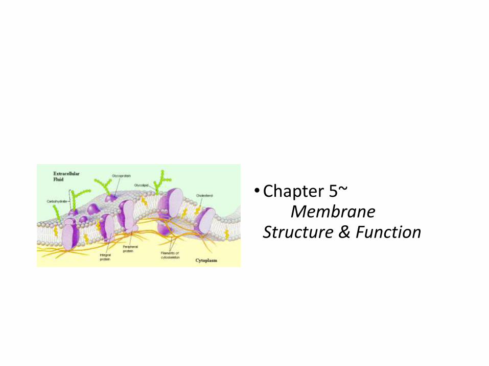
• Chapter 5~Membrane
Structure & Function

Phospholipids
• Phosphate head plus lipid tail
• Phosphate is hydrophilic
• Lipid is hydrophobic

• Three main structures formed by phospholipids in water
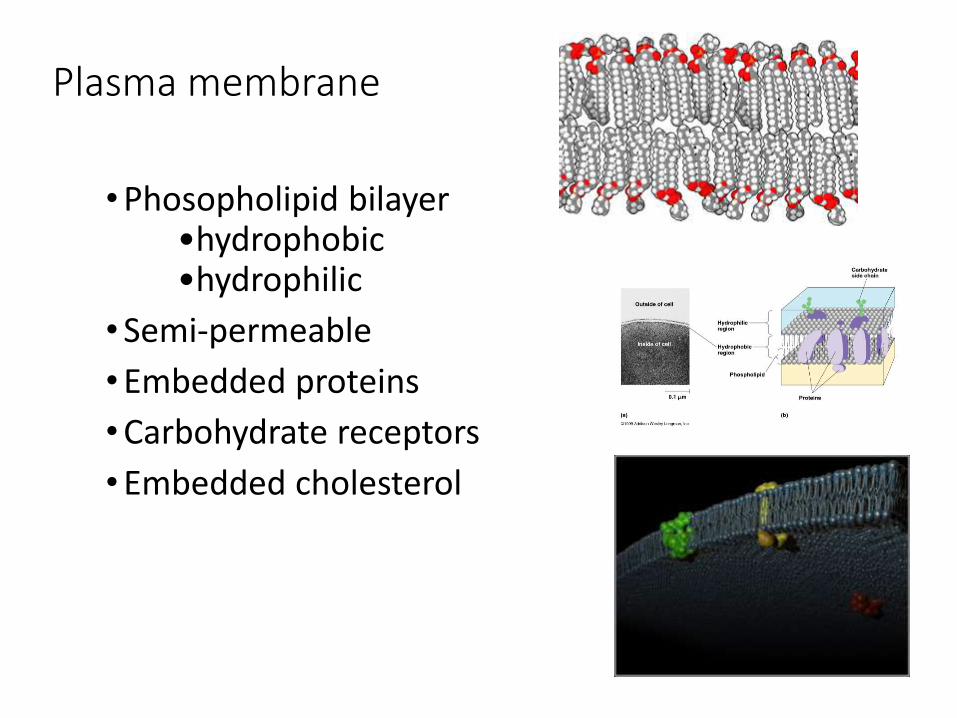
Plasma membrane
• Phosopholipid bilayer•hydrophobic•hydrophilic
• Semi-permeable
• Embedded proteins
• Carbohydrate receptors
• Embedded cholesterol

Membrane structure, I
• Selective permeability
• Amphipathic~
• hydrophobic & hydrophilic regions
• Singer-Nicolson:
• fluid mosaic model
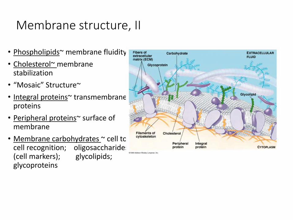
Membrane structure, II
• Phospholipids~ membrane fluidity
• Cholesterol~ membrane stabilization
• “Mosaic” Structure~
• Integral proteins~ transmembraneproteins
• Peripheral proteins~ surface of membrane
• Membrane carbohydrates ~ cell to cell recognition; oligosaccharides (cell markers); glycolipids; glycoproteins

Membrane structure, III
• Membrane protein function:
•transport•enzymatic activity•signal transduction•intercellular joining•cell-cell recognition•ECM attachment
Phospholipid Bilayer
• Video 1: https://www.youtube.com/watch?v=Qqsf_UJcfBc
• Video 2: https://www.youtube.com/watch?v=moPJkCbKjBs&feature=related
• Video 3: https://www.youtube.com/watch?v=LXaPt9i9hqk
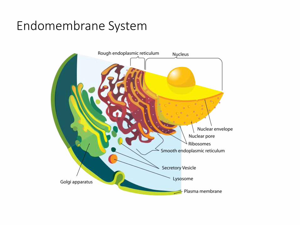
Endomembrane System
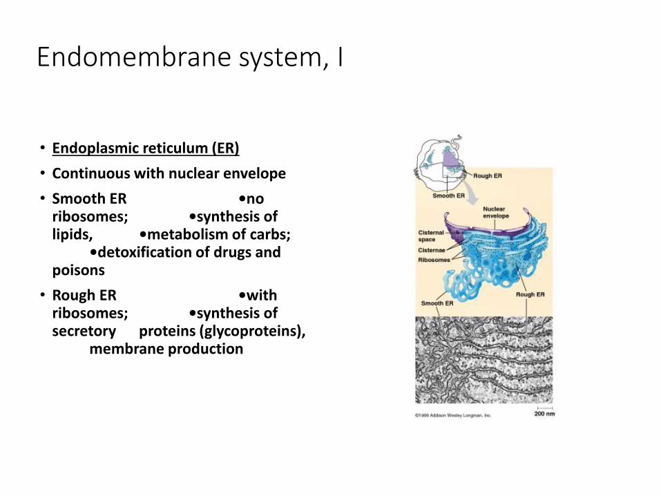
Endomembrane system, I
• Endoplasmic reticulum (ER)
• Continuous with nuclear envelope
• Smooth ER •no ribosomes; •synthesis of lipids, •metabolism of carbs;
•detoxification of drugs and poisons
• Rough ER •with ribosomes; •synthesis of secretory proteins (glycoproteins),
membrane production
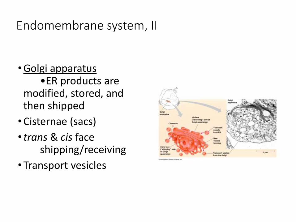
Endomembrane system, II
• Golgi apparatus•ER products are
modified, stored, and then shipped
• Cisternae (sacs)
• trans & cis faceshipping/receiving
• Transport vesicles
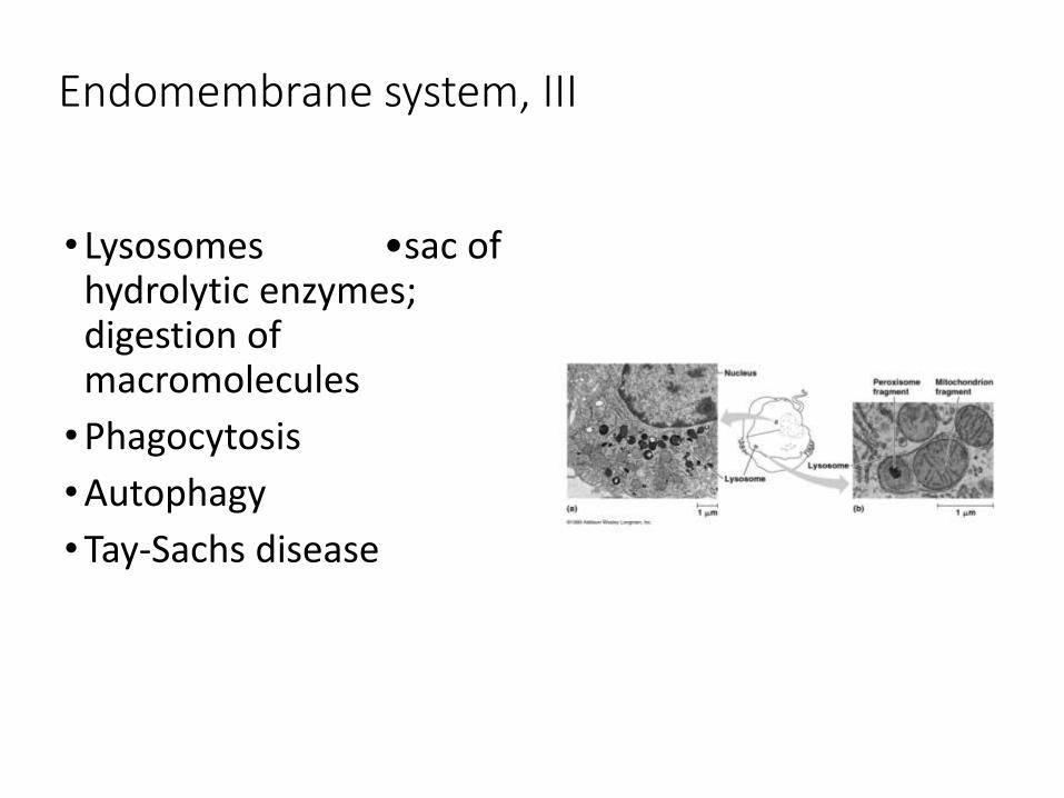
Endomembrane system, III
• Lysosomes •sac of hydrolytic enzymes; digestion of macromolecules
• Phagocytosis
• Autophagy
• Tay-Sachs disease
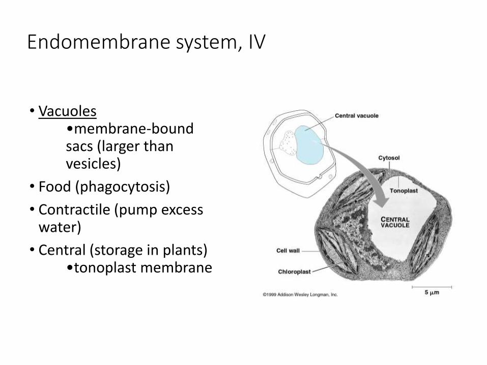
Endomembrane system, IV
• Vacuoles•membrane-bound sacs (larger than vesicles)
• Food (phagocytosis)
• Contractile (pump excess water)
• Central (storage in plants)•tonoplast membrane
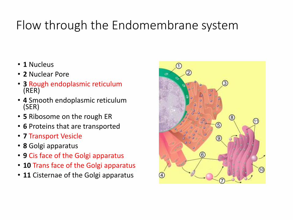
Flow through the Endomembrane system
• 1 Nucleus
• 2 Nuclear Pore
• 3 Rough endoplasmic reticulum (RER)
• 4 Smooth endoplasmic reticulum (SER)
• 5 Ribosome on the rough ER
• 6 Proteins that are transported
• 7 Transport Vesicle
• 8 Golgi apparatus
• 9 Cis face of the Golgi apparatus
• 10 Trans face of the Golgi apparatus
• 11 Cisternae of the Golgi apparatus
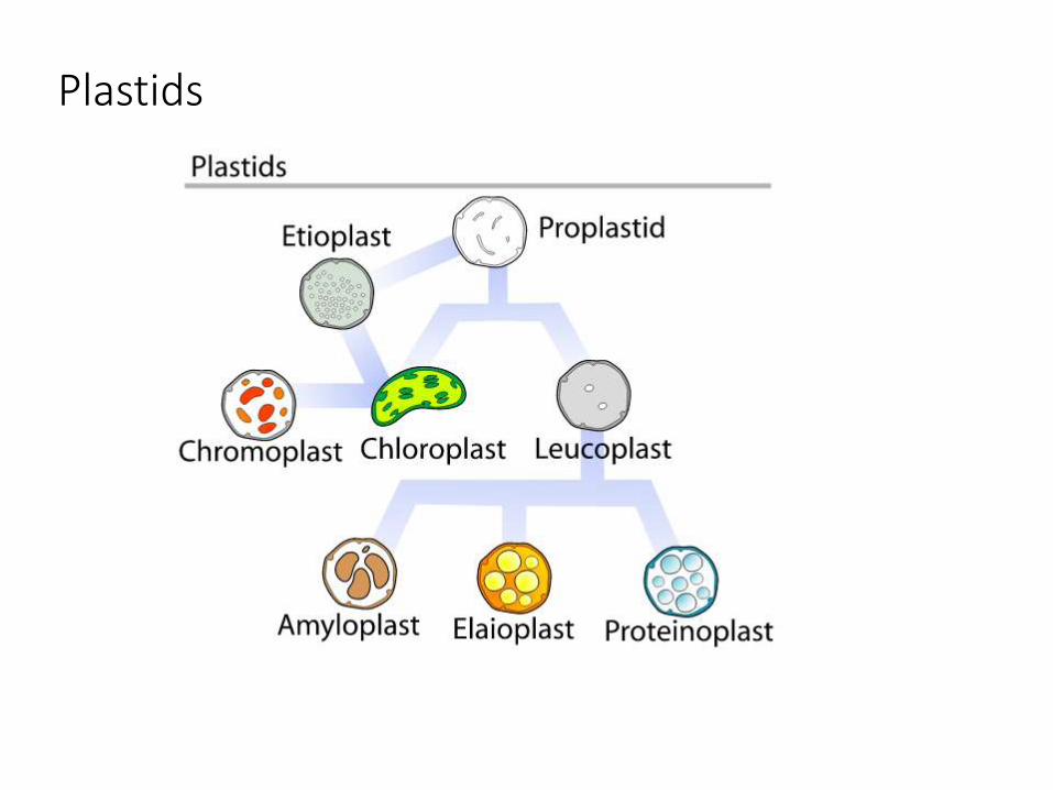
Plastids
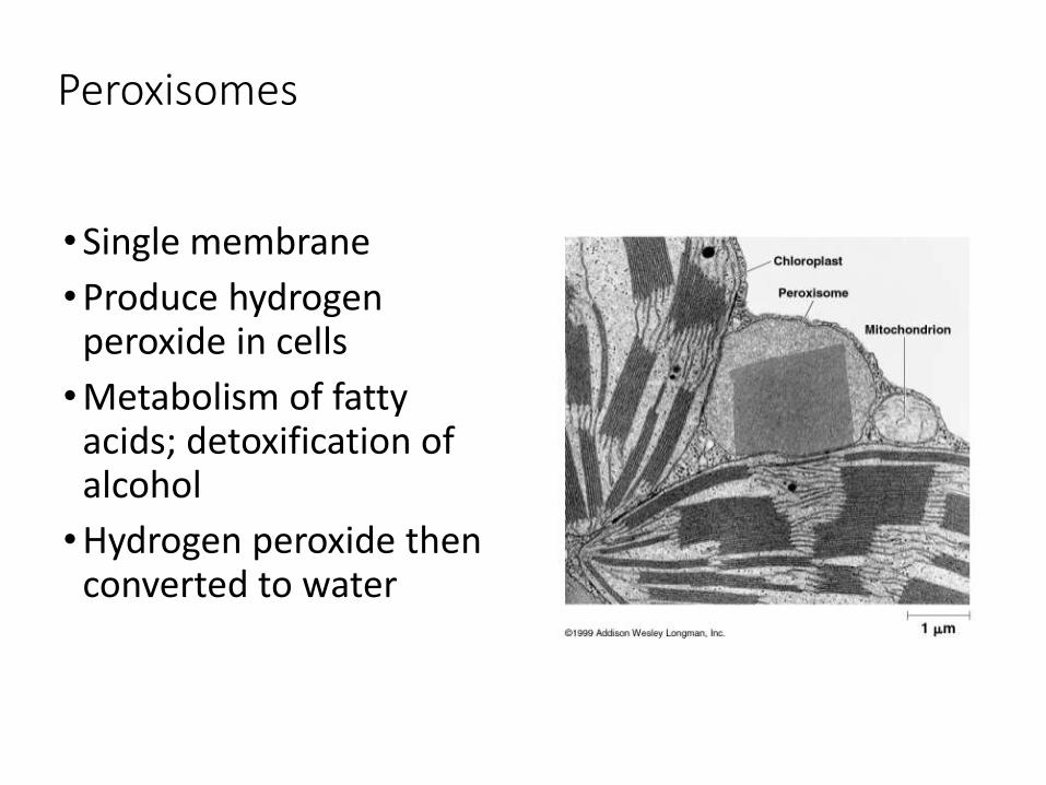
Peroxisomes
• Single membrane
• Produce hydrogen peroxide in cells
• Metabolism of fatty acids; detoxification of alcohol
• Hydrogen peroxide then converted to water

Membrane traffic
• Diffusion
• Concentration gradient
• Passive transport
• Osmosis
• Transport proteins
• Facilitated transport
• Active transport

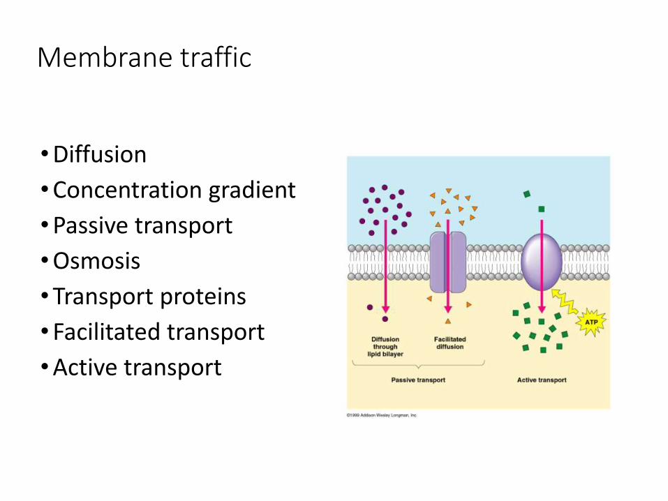
Membrane traffic
• Diffusion~ tendency of any molecule to spread out into available space
• Concentration gradient
• Passive transport~ diffusion of a substance across a biological membrane
• Osmosis~ the diffusion of water across a selectively permeable membrane
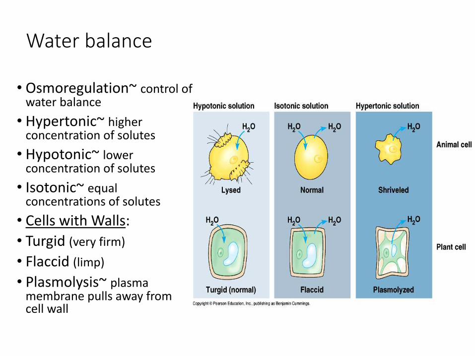
Water balance
• Osmoregulation~ control of water balance
• Hypertonic~ higher concentration of solutes
• Hypotonic~ lower concentration of solutes
• Isotonic~ equal concentrations of solutes
• Cells with Walls:
• Turgid (very firm)
• Flaccid (limp)
• Plasmolysis~ plasma membrane pulls away from cell wall
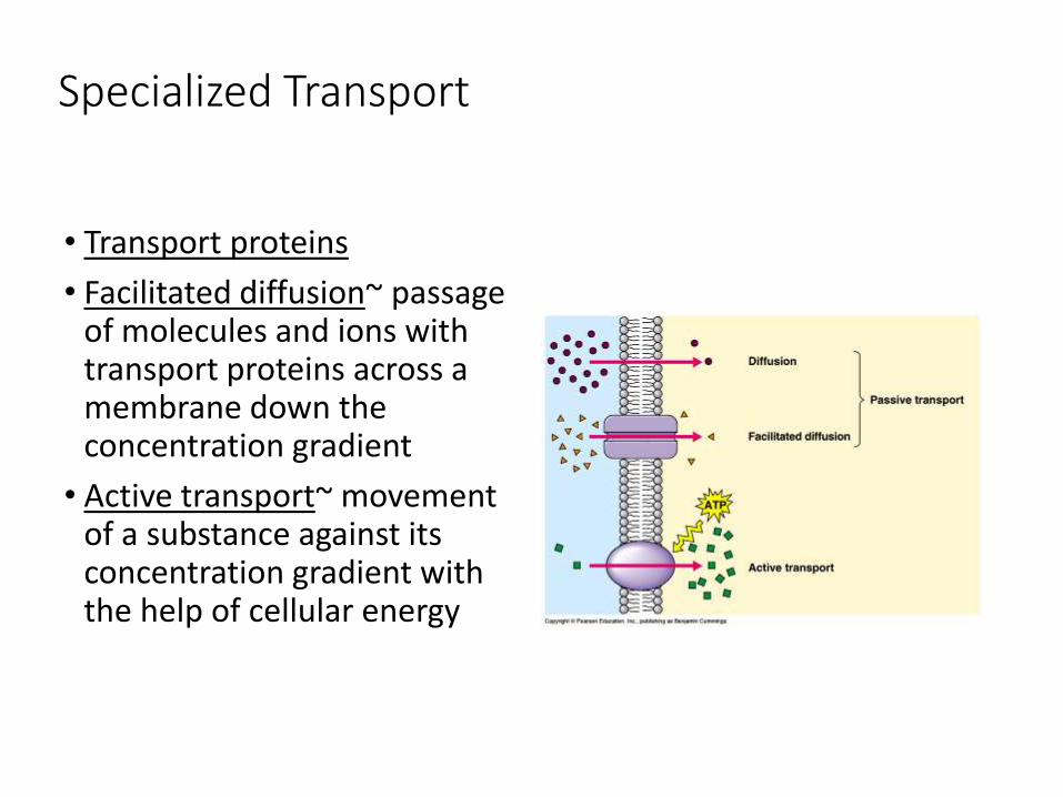
Specialized Transport
• Transport proteins
• Facilitated diffusion~ passage of molecules and ions with transport proteins across a membrane down the concentration gradient
• Active transport~ movement of a substance against its concentration gradient with the help of cellular energy

Active transport
Membrane traffic
• Diffusion
• Concentration gradient
• Passive transport
• Osmosis
• Transport proteins
• Facilitated transport
• Active transport
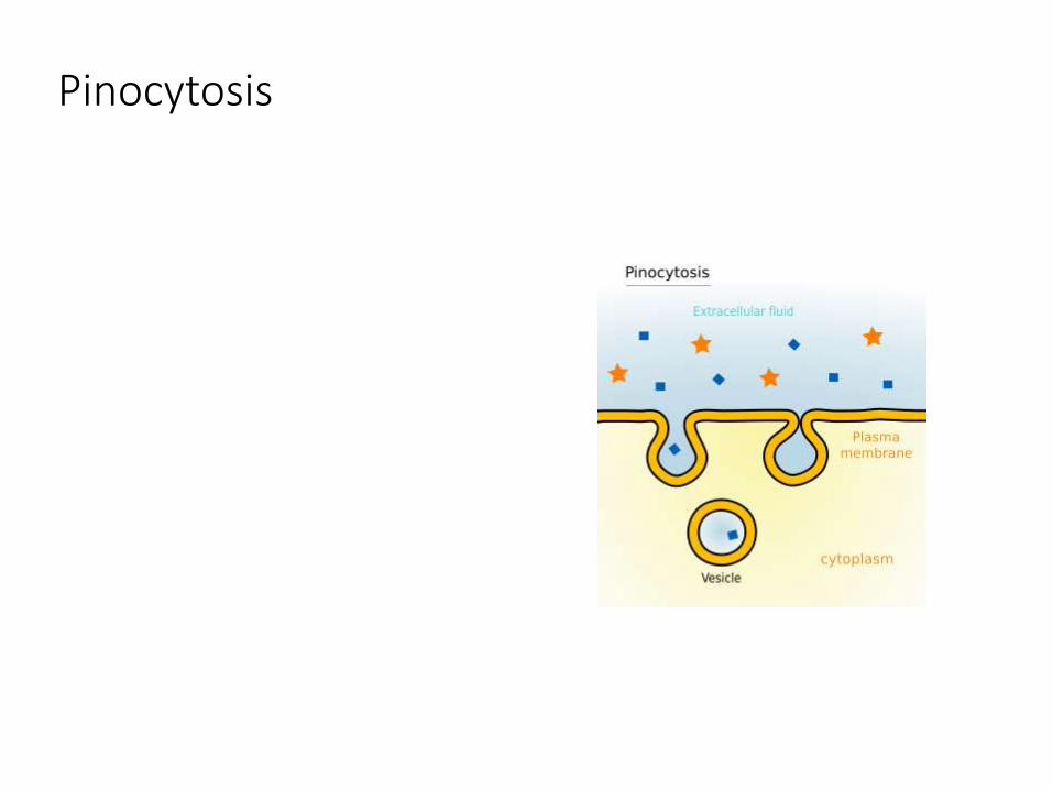
Pinocytosis

Exocytosis
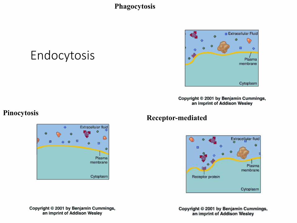
Endocytosis
Phagocytosis
Receptor-mediatedPinocytosis
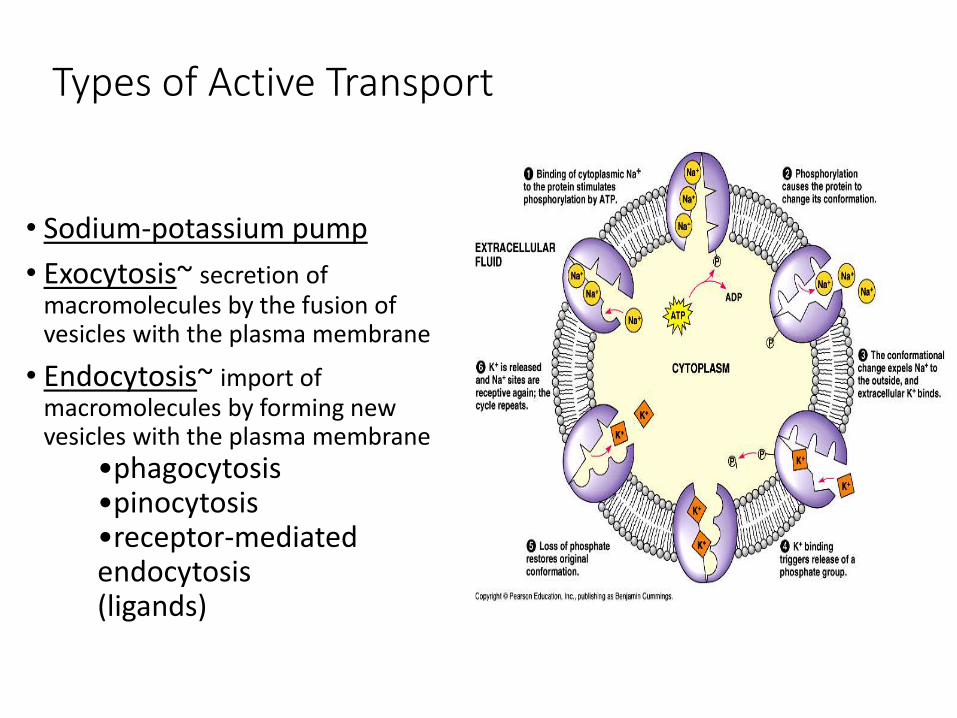
Types of Active Transport
• Sodium-potassium pump
• Exocytosis~ secretion of macromolecules by the fusion of vesicles with the plasma membrane
• Endocytosis~ import of macromolecules by forming new vesicles with the plasma membrane
•phagocytosis•pinocytosis•receptor-mediated endocytosis (ligands)
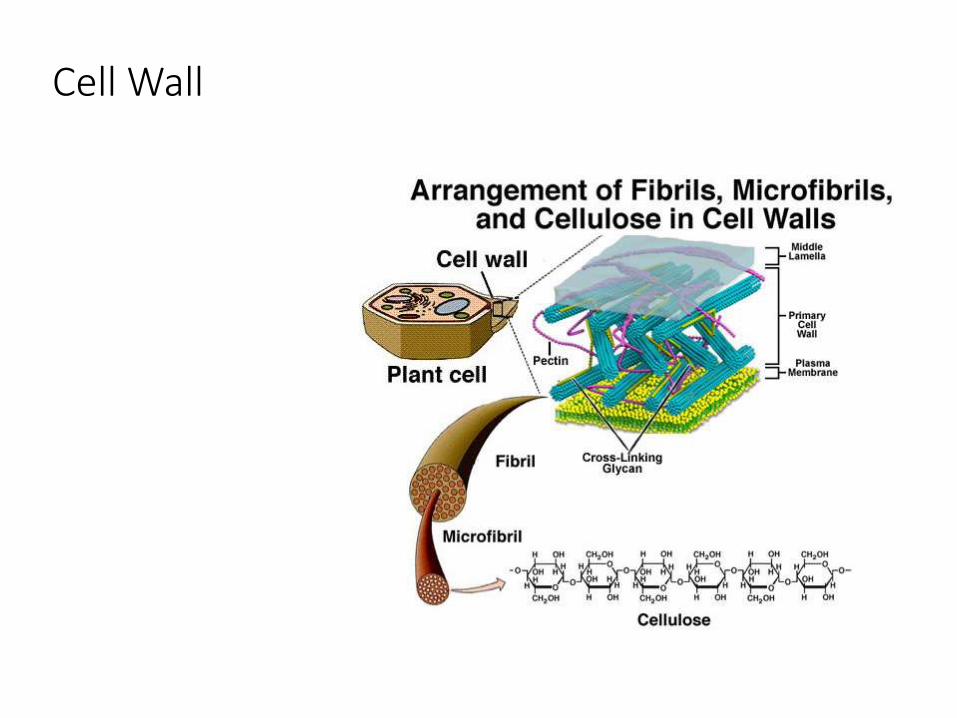
Cell Wall
Chapter 51
• Osmotic Regulation and the Urinary System

Homeostasis and the Big Bang Theory
• https://www.youtube.com/watch?v=9RLnlXNlfdk

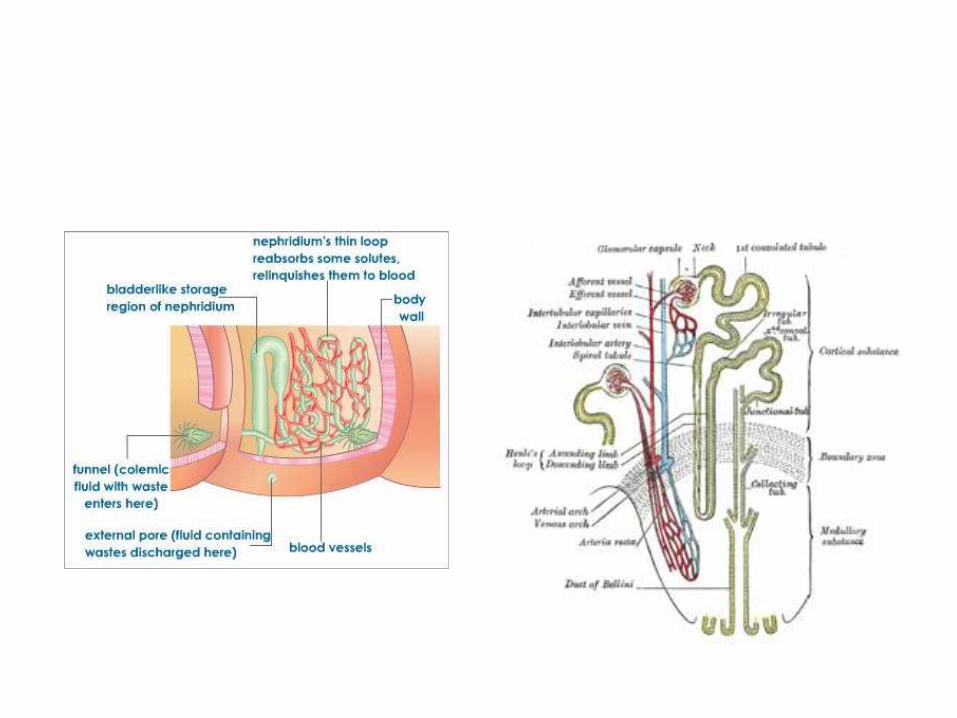
QOD •Animals have a wide variety of excretory organs. Though they all serve the same basic purpose of maintaining osmotic balance, they have significant structural differences. Compare and contrast nephridiaand the nephron.
Homeostasis: regulation of internal environment
• Thermoregulation internal temperature
• Osmoregulation solute and water balance
• Excretion nitrogen containing waste
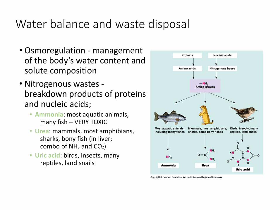
Water balance and waste disposal
• Osmoregulation - management of the body’s water content and solute composition
• Nitrogenous wastes -breakdown products of proteins and nucleic acids;
• Ammonia: most aquatic animals, many fish – VERY TOXIC
• Urea: mammals, most amphibians, sharks, bony fish (in liver; combo of NH3 and CO2)
• Uric acid: birds, insects, many reptiles, land snails
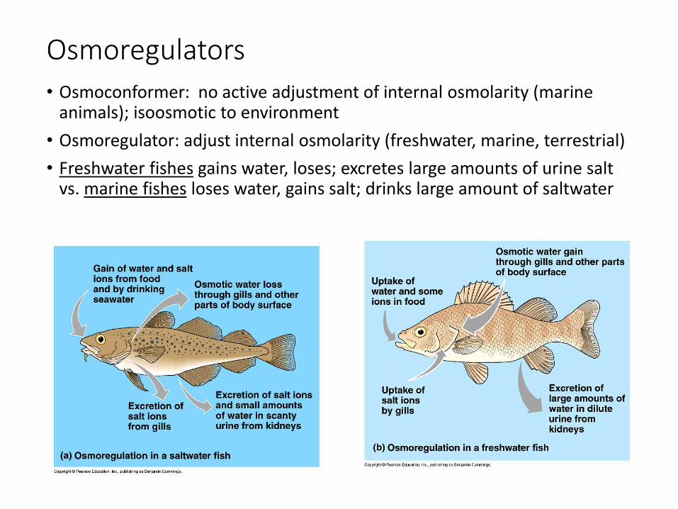
Osmoregulators• Osmoconformer: no active adjustment of internal osmolarity (marine
animals); isoosmotic to environment
• Osmoregulator: adjust internal osmolarity (freshwater, marine, terrestrial)
• Freshwater fishes gains water, loses; excretes large amounts of urine salt vs. marine fishes loses water, gains salt; drinks large amount of saltwater
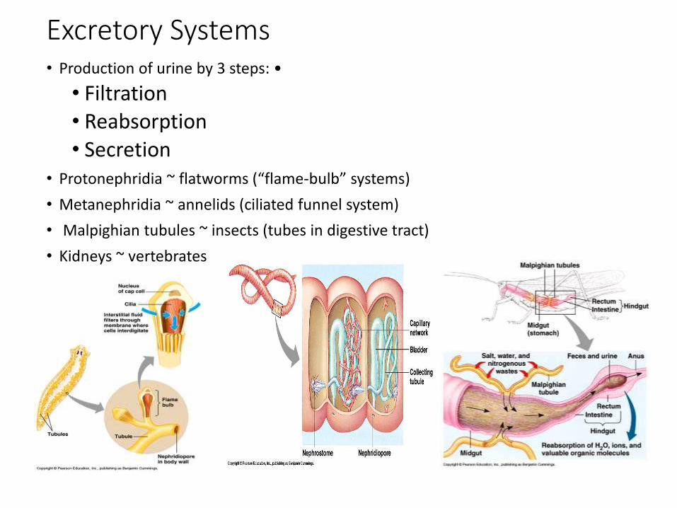
Excretory Systems• Production of urine by 3 steps: •
• Filtration• Reabsorption• Secretion
• Protonephridia ~ flatworms (“flame-bulb” systems)
• Metanephridia ~ annelids (ciliated funnel system)
• Malpighian tubules ~ insects (tubes in digestive tract)
• Kidneys ~ vertebrates
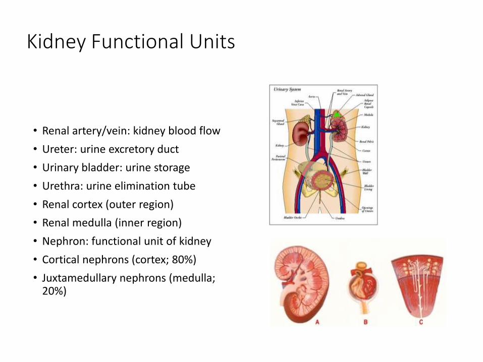
Kidney Functional Units
• Renal artery/vein: kidney blood flow
• Ureter: urine excretory duct
• Urinary bladder: urine storage
• Urethra: urine elimination tube
• Renal cortex (outer region)
• Renal medulla (inner region)
• Nephron: functional unit of kidney
• Cortical nephrons (cortex; 80%)
• Juxtamedullary nephrons (medulla; 20%)

Nephron Structure
• Afferent arteriole: supplies blood to nephron from renal
artery
• Glomerulus: ball of capillaries
• Efferent arteriole: blood from glomerulus
• Bowman’s capsule: surrounds glomerulus
• Proximal tubule: secretion & reabsorption
• Loop of Henle: water & salt balance
• Distal tubule: secretion & reabsorption
• Collecting duct: carries filtrate to renal pelvis
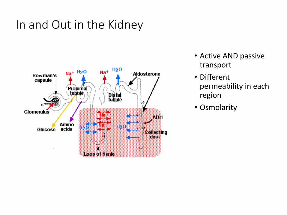
In and Out in the Kidney
• Active AND passive transport
• Different permeability in each region
• Osmolarity

Kidney regulation: hormones
• Antidiuretic hormone (ADH) ~ secretion increases permeability of distal tubules and collecting ducts to water (H2O back to body); inhibited by alcohol and coffee
• Juxtaglomerular apparatus (JGA) ~ reduced salt intake--->enzyme renin initiates conversion of angiotension (plasma protein) to angiotension II (peptide); increase blood pressure and blood volume by constricting capillaries
• Angiotension II also stimulates adrenal glands to secrete aldosterone; acts on distal tubules to reabsorb more sodium, thereby increasing blood pressure (renin-angiotension-aldosterone system; RAAS)
• Atrial natriuretic factor (ANF) ~ walls of atria; inhibits release of renin, salt reabsorption, and aldosterone release

Osmolality
• https://www.youtube.com/watch?v=UA6FeVHAqoc
• https://www.youtube.com/watch?v=Dtsen_YNwVk
Sheldon Cleans Penny’s Apartment
• https://www.youtube.com/watch?v=WFQ7Nzlzgbk
• https://www.youtube.com/watch?v=_5RVXYEX2L4