interstitial lung change in pre-radiation therapy … · purpose we examined clinical and...
TRANSCRIPT
│ http://www.e-crt.org │676 Copyright ⓒ 2015 by the Korean Cancer AssociationThis is an Open-Access article distributed under the terms of the Creative Commons Attribution Non-Commercial License (http://creativecommons.org/licenses/by-nc/3.0/)
which permits unrestricted non-commercial use, distribution, and reproduction in any medium, provided the original work is properly cited.
Cancer Res Treat. 2015;47(4):676-686
pISSN 1598-2998, eISSN 2005-9256
http://dx.doi.org/10.4143/crt.2014.180
Open Access
Interstitial Lung Change in Pre-radiation Therapy Computed Tomography Is a Risk Factor for Severe Radiation Pneumonitis
Original Article
PurposeWe examined clinical and dosimetric factors as predictors of symptomatic radiation pneu-monitis (RP) in lung cancer patients and evaluated the relationship between interstitial lungchanges in the pre-radiotherapy (RT) computed tomography (CT) and symptomatic RP.
Materials and MethodsMedical records and dose volume histogram data of 60 lung cancer patients from August2005 to July 2006 were analyzed. All patients were treated with three dimensional (3D)conformal RT of median 56.9 Gy. We assessed the association of symptomatic RP with clin-ical and dosimetric factors.
ResultsWith a median follow-up of 15.5 months (range, 6.1 to 40.9 months), Radiation TherapyOncology Group grade ! 2 RP was observed in 14 patients (23.3%). Five patients (8.3%)died from RP. The interstitial changes in the pre-RT chest CT, mean lung dose (MLD), andV30 significantly predicted RP in multivariable analysis (p=0.009, p < 0.001, and p < 0.001,respectively). MLD, V20, V30, and normal tissue complication probability normal tissue com-plication probability (NTCP) were associated with the RP grade but less so for grade 5 RP.The risk of RP grade ! 2, ! 3, or ! 4 was higher in the patients with interstitial lung change(grade 2, 15.6% to 46.7%, p=0.03; grade 3, 4.4% to 40%, p=0.002; grade 4, 4.4% to 33.3%,p=0.008). Four of the grade 5 RP patients had diffuse interstitial change in pre-RT CT andreceived chemoradiotherapy.
ConclusionOur study identified diffuse interstitial disease as a significant clinical risk for RP, particularlyfatal RP. We showed the usefulness of MLD, V20, V30, and NTCP in predicting the incidenceand severity of RP.
Key wordsRadiation pneumonitis, Interstitial lung diseases, Lung neoplasms, Radiotherapy
Introduction
Thoracic radiation therapy (RT) is an important compo-nent of treatment for locally advanced non-small cell lungcancer and limited stage small cell lung cancer [1-4]. The fundamental goal of RT is to deliver an effective dose for
local control and keep toxicity to an acceptable level in thesurrounding normal tissues. However, irradiation of the thorax is frequently accompanied by harmful side effects.One major concern is radiation pneumonitis (RP), which canlead to severe respiratory dysfunction, including chronic respiratory insufficiency caused by the development of pulmonary fibrosis, and even death [5,6].
+ + + + + + + + + + + + + + + + + + + + + + + + + + + + + + + + + + + + + + + + + + + + + + + + + + + + + + + + + + + ++ + + + + + + + + + + + + + + + + + + + + + + + + + + + + + + + + + + + + + + + + + + + + + + + + + + + + + + + + + + ++ + + + + + + + + + + + + + + + + + + + + + + + + + + + + + + + + + + + + + + ++ + + + + + + + + + + + + + + + + + + ++ + + + + + + + + + + + + + + + + + + + + + + + + + + + + + + + + + + + + + + ++ + + + + + + + + + + + + + + + + + + ++ + + + + + + + + + + + + + + + + + + + + + + + + + + + + + + + + + + + + + + +
Correspondence: Yeon Sil Kim, MD Department of Radiation Oncology, Seoul St. Mary’s Hospital, College of Medicine,The Catholic University of Korea, 222 Banpo-daero, Seocho-gu, Seoul 06591, Korea Tel: 82-2-2258-6259Fax: 82-2-592-1532E-mail: [email protected]
Received July 11, 2014Accepted September 13, 2014Published online February 13, 2015
Yun Hee Lee, MD1
Yeon Sil Kim, MD1
Sang Nam Lee, MD1
Hyo Chun Lee, MD1
Se Jin Oh, MD1
Seoung Joon Kim, MD2
Young Kyoon Kim, MD2
Dae Hee Han, MD3
Ie Ryung Yoo, MD4
Jin Hyung Kang, MD5
Suk Hee Hong, MD5
Departments of 1Radiation Oncology, 2Internal Medicine, 3Radiology, 4Nuclear Medicine, and 5Medical Oncology,Seoul St. Mary’s Hospital, College of Medicine, The Catholic University of Korea, Seoul, Korea
Yun Hee Lee, Interstitial Lung Change in Pre-RT CT for Severe RP
VOLUME 47 NUMBER 4 OCTOBER 2015 677
Clinical risk factors for the development of RP include olderage, disease location in the mid-lower lung, chemothera-pyschedule, and the presence of comorbidity [7,8]. Addition-ally, many studies suggest dosimetric factors relate to RP [9-12].
Several studies reported that patients with interstitial lungdisease (ILD) developed severe RP after thoracic stereotacticbody radiotherapy (SBRT) [13,14]. Acute exacerbation of ILDcan cause respiratory failure and death. Severe ILD was regarded as a relative contraindication in the clinical guide-lines for SBRT published by the Japanese Society for Thera-peutic Radiation and Oncology [15]. There is limited datashowing an association of subclinical ILD with RP [16,17],and whether subclinical ILD is a risk factor for thoracic RT isa matter of speculation.
We examined clinical and dosimetric factors as predictorsof symptomatic RP in lung cancer patients and further eval-uated the relationship of pre-radiation interstitial changes inthe computed tomography (CT) and symptomatic RP.
Materials and Methods
1. Patients
The medical records and dose volume histogram (DVH)data of 60 lung cancer patients, treated with three dimen-sional (3D) conformal RT from August 2005 to July 2006 atSeoul St. Mary's Hospital, were retrospectively collected andanalyzed. At least 6 months of follow-up was required forinclusion in this study. Patients who died within 6 monthsafter completion of RT, were lost to follow-up, or did notcomplete treatment, were excluded from the analysis.
Patient characteristics are summarized in Table 1. All 60patients were treated with curative intent, using 3D confor-mal RT. Forty-eight patients (80%) received chemotherapyas a part of their definitive treatment. Twenty-seven patients(45%) received chemotherapy prior to RT and 21 patients(35%) received concurrent chemoradiotherapy. The regimensused for concurrent chemotherapy were etoposide and cisplatin (8 patients), docetaxel and cisplatin (8 patients),taxol (4 patients), and cisplatin (1 patient). Fourteen patients(23.3%) received RT or concurrent chemoradiotherapy as definitive salvage treatment following recurrent disease aftersurgical intervention (lobectomy eight patients, wedge resec-tion two patients, pneumonectomy one patient, and othersthree patients). Fifty-two patients (86.7%) showed a goodperformance status of more than Eastern Cooperative Oncol-ogy Group (ECOG) 2. Each patient’s pre-RT chest CT scanswere reviewed by two radiation oncologists and one diag-nostic radiologist who were not informed of which patients
developed symptomatic RP. Fifteen patients (25%) had inter-stitial changes on their chest CT scans. Interstitial changeswere recorded if any of the following criteria were met: subpleural ground glass opacity (GGO), fibrous reticulation,traction bronchiectasis, micronodules, honey-comb appear-ance, or volume loss, especially in both lower lobes [18]. Tho-racic RT was not performed in patients with clinical ILD,which was defined as a status of symptomatic disease orpost-treatment with high probability of acute exacerbation.The patients showing interstitial change on pre-RT CT with-out related symptom or history of a treatment were includedin this study as subclinical ILD. This retrospective study wasapproved by the hospital’s institutional review board.
2. Radiation therapy
A planning CT scan with contrast enhancement of the entire lung volume was acquired with a vac-lock immobi-lization device at shallow normal breathing. Four dimen-sional CT (4D-CT) scans were also acquired in all patients toevaluate respiratory motion. All patients underwent virtualsimulation and treatment planning with the 3D treatmentplanning system (Pinnacle, ADAC Laboratories, Milpitas,CA). The calculation was performed with lung heterogeneitycorrections by radiological path algorithm. The gross tumorvolume (GTV) included the tumor and the clinically involvednodes. The GTV node was determined by chest CT and/orpositron emission tomography–computed tomography forthe majority of patients; the node was determined by medi-astinoscopy in a small number of patients. The clinical targetvolume included the GTV with an additional uniform 5-mmexpansion and high risk, uninvolved mediastinal lymphnodes as well as the ipsilateral hilar lymph nodes. The highrisk, involved mediastinal lymph nodes were defined asthose lymph nodes adjacent to the GTV node with a highprobability of microscopic involvement. We added a differ-ent panning target volume (PTV) margin based on an assess-ment of each tumor’s motion using the Leonardo system(Siemens, Concord, CA) in 4D-CT. The lung, esophagus,spinal cord, and heart were contoured. The lung volume wascontoured and defined excluding the GTV.
Most patients were treated with conventional beamarrangements (i.e., initial AP/PA fields to include the PTV)for the first 30-40 Gy, followed by an off-cord oblique beamto the PTV, usually composed of three to five beams. Somepatients with small PTVs were treated with multiple beamarrangements from the beginning. The median radiationdose was 56.9 Gy (range, 45.0 to 64.8 Gy), generally deliveredat 1.8-2 Gy/fraction, five fractions per week. RT was deliv-ered with 10 MV X-ray from a LINAC (Siemens). Generally,the radiation dose was prescribed such that 95% of PTV wascovered by 95% of the prescription dose. The lung DVH was
Cancer Res Treat. 2015;47(4):676-686
678 CANCER RESEARCH AND TREATMENT
calculated directly from the physical dose distribution (i.e.,adjustments were not made for fraction size or overall treat-ment time). The following dosimetric factors were extractedfrom the lung DVH: V10, V20, V30, V40, mean lung dose (MLD),and normal tissue complication probability (NTCP) as derived from the Lyman and Kutcher models.
3. Evaluation of RP
A diagnosis of RP was made by the clinical symptoms ofnonproductive cough, fever, exertional dyspnea and charac-teristic chest radiographs and CT findings. Patients were
evaluated by radiation oncologists 3 to 6 weeks after com-pletion of RT and at 1- to 3-month intervals thereafter. RPgrading was recorded using Radiation Therapy OncologyGroup (RTOG) scale, based on the severity of clinical symp-toms and radiographic changes. Two radiation oncologistsand one diagnostic radiologist who were blinded to RP statusreviewed the medical records, chest X-ray and/or chest CTscan of all patients with suspected RP, and ruled out othercauses of pneumonia that were confirmed as infectious pneu-monia or disease progression.
4. Statistical analysis
All statistical analysis in this study was performed withthe SPSS ver. 18.0 (SPSS Inc., Chicago, IL). The chi-squaredtest and Fisher exact test were used to compare RP risk insubgroups and Student’s t test was used for normally distributed continuous variables. To identify independentpredictors of RP, all factors were included in a multivariablelinear regression model. Using a stepwise procedure, vari-ables with a p-value of 0.05 or higher were dropped from themodel so that only significant predictors remained. All statistical tests were two sided and a p-value of less than 0.05was considered statistically significant.
Results
1. RP incidence
With a median follow-up of 15.5 months (range, 6.1 to 40.9months), 22 patients (36.7%) developed RP: eight (13.3%)with grade 1, six (10.0%) with grade 2, one (1.6%) with grade3, 2 (3.3%) with grade 4, and five (8.3%) with grade 5. RTOGgrade 2 or higher RP was observed in 14 patients (23.3%).Five patients (8.3%) died from RP. The median time to theonset of symptoms was 33 days (range, 6 to 160 days) fromthe completion of RT.
2. Clinical and dosimetric factors
Table 2 shows the relationship between clinical factors andRP. There was a trend of increased risk of RP in patientstreated with combined chemoradiation compared with patients treated with RT alone (41.7% vs. 16.7%, p=0.108).However, this difference was not statistically significant. Interstitial change in the pre-RT chest CT was the only clini-cal factor associated with RP (p=0.033). In multivariableanalysis, interstitial changes in the pre-RT chest CT were significantly associated with RP (p=0.009; odds ratio, 10.24;
Table 1. Patient characteristics (n=60)
Characteristic No. (%)Median age (range, yr) 62 (40-80)Gender (male:female) 48:12ECOG PS 0 4 (6.7)
1 33 (55.0)2 15 (25.0)3 8 (13.3)
Smoking Yes 46 (76.7)No 12 (20.0)Unknown 2 (3.3)
Weight loss Yes 21 (35.0)No 39 (65.0)
COPD Yes 24 (40.0)No 36 (60.0)
Interstitial change in CT Yes 15 (25.0)No 45 (75.0)
Histology NSCLC 39 (65.0)SCLC 18 (30.0)Other 3 (5.0)
Stage of NSCLC I 1 (1.7)II 2 (3.3)III 33 (55.0)IV 3 (5.0)
Tumor location Central 53 (88.3)Periphery 7 (11.7)Upper 39 (65.0)Lower 21 (35.0)
Surgery Yes 14 (23.3)No 46 (76.7)
Chemotherapy Yes 48 (80.0)No 12 (20.0)Sequential 27 (45.0)Concurrent 21 (35.0)
ECOG PS, Eastern Cooperative Oncology Group perform-ance status; COPD, chronic obstructive pulmonary disease;CT, computed tomography; NSCLC, non-small cell lungcancer; SCLC, small cell lung cancer.
Yun Hee Lee, Interstitial Lung Change in Pre-RT CT for Severe RP
VOLUME 47 NUMBER 4 OCTOBER 2015 679
95% confidence interval, 1.787 to 58.679).The statistically significant dosimetric factors for predict-
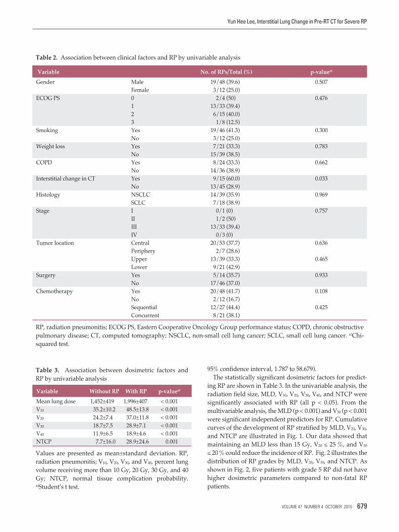
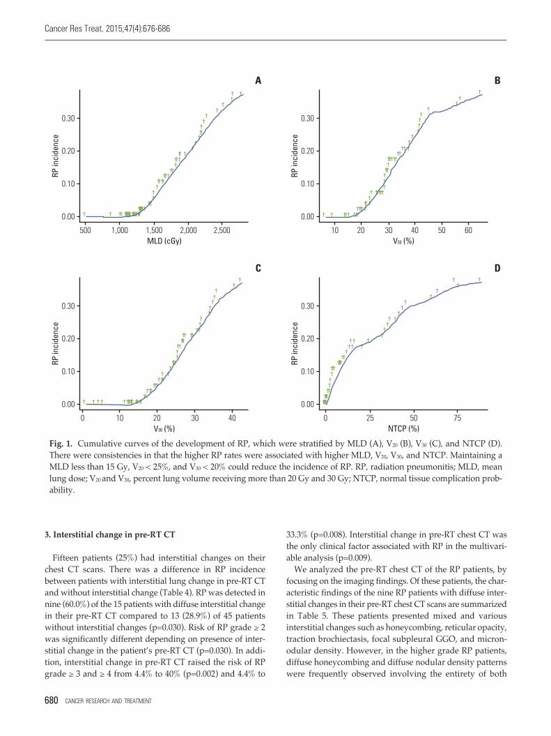
ing RP are shown in Table 3. In the univariable analysis, theradiation field size, MLD, V10, V20, V30, V40, and NTCP weresignificantly associated with RP (all p < 0.05). From the multivariable analysis, the MLD (p < 0.001) and V30 (p < 0.001were significant independent predictors for RP. Cumulativecurves of the development of RP stratified by MLD, V20, V30,and NTCP are illustrated in Fig. 1. Our data showed thatmaintaining an MLD less than 15 Gy, V20 ! 25 %, and V30
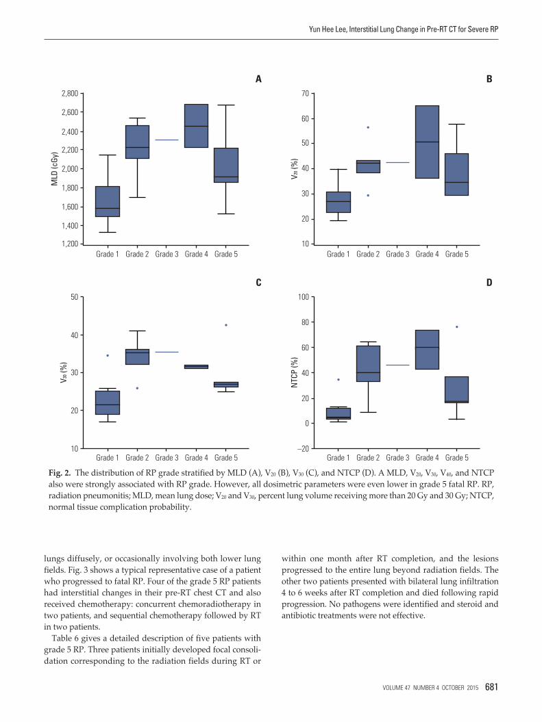
! 20 % could reduce the incidence of RP. Fig. 2 illustrates thedistribution of RP grades by MLD, V20, V30, and NTCP. Asshown in Fig. 2, five patients with grade 5 RP did not havehigher dosimetric parameters compared to non-fatal RP patients.
Table 2. Association between clinical factors and RP by univariable analysis
Variable No. of RPs/Total (%) p-valuea)
Gender Male 19/48 (39.6) 0.507Female 3/12 (25.0)
ECOG PS 0 2/4 (50) 0.4761 13/33 (39.4)2 6/15 (40.0)3 1/8 (12.5)
Smoking Yes 19/46 (41.3) 0.300No 3/12 (25.0)
Weight loss Yes 7/21 (33.3) 0.783No 15/39 (38.5)
COPD Yes 8/24 (33.3) 0.662No 14/36 (38.9)
Interstitial change in CT Yes 9/15 (60.0) 0.033No 13/45 (28.9)
Histology NSCLC 14/39 (35.9) 0.969SCLC 7/18 (38.9)
Stage I 0/1 (0) 0.757II 1/2 (50)III 13/33 (39.4)IV 0/3 (0)
Tumor location Central 20/53 (37.7) 0.636Periphery 2/7 (28.6)Upper 13/39 (33.3) 0.465Lower 9/21 (42.9)
Surgery Yes 5/14 (35.7) 0.933No 17/46 (37.0)
Chemotherapy Yes 20/48 (41.7) 0.108No 2/12 (16.7)Sequential 12/27 (44.4) 0.425Concurrent 8/21 (38.1)
RP, radiation pneumonitis; ECOG PS, Eastern Cooperative Oncology Group performance status; COPD, chronic obstructivepulmonary disease; CT, computed tomography; NSCLC, non-small cell lung cancer; SCLC, small cell lung cancer. a)Chi-squared test.
Table 3. Association between dosimetric factors and RP by univariable analysis
Variable Without RP With RP p-valuea)
Mean lung dose 1,452±419 1,996±407 < 0.001V10 35.2±10.2 48.5±13.8 < 0.001V20 24.2±7.4 37.0±11.8 < 0.001V30 18.7±7.5 28.9±7.1 < 0.001V40 11.9±6.5 18.9±4.6 < 0.001NTCP 7.7±16.0 28.9±24.6 0.001
Values are presented as mean±standard deviation. RP, radiation pneumonitis; V10, V20, V30, and V40, percent lungvolume receiving more than 10 Gy, 20 Gy, 30 Gy, and 40Gy; NTCP, normal tissue complication probability. a)Student’s t test.
Cancer Res Treat. 2015;47(4):676-686
680 CANCER RESEARCH AND TREATMENT
3. Interstitial change in pre-RT CT
Fifteen patients (25%) had interstitial changes on theirchest CT scans. There was a difference in RP incidence between patients with interstitial lung change in pre-RT CTand without interstitial change (Table 4). RP was detected innine (60.0%) of the 15 patients with diffuse interstitial changein their pre-RT CT compared to 13 (28.9%) of 45 patientswithout interstitial changes (p=0.030). Risk of RP grade " 2was significantly different depending on presence of inter-stitial change in the patient’s pre-RT CT (p=0.030). In addi-tion, interstitial change in pre-RT CT raised the risk of RPgrade " 3 and " 4 from 4.4% to 40% (p=0.002) and 4.4% to
33.3% (p=0.008). Interstitial change in pre-RT chest CT wasthe only clinical factor associated with RP in the multivari-able analysis (p=0.009).
We analyzed the pre-RT chest CT of the RP patients, by focusing on the imaging findings. Of these patients, the char-acteristic findings of the nine RP patients with diffuse inter-stitial changes in their pre-RT chest CT scans are summarizedin Table 5. These patients presented mixed and various interstitial changes such as honeycombing, reticular opacity,traction brochiectasis, focal subpleural GGO, and micron-odular density. However, in the higher grade RP patients,diffuse honeycombing and diffuse nodular density patternswere frequently observed involving the entirety of both
RP in
cide
nce
0.30
0.20
0.10
0.00
500MLD (cGy)
1,000 1,500 2,000 2,500
A B
RP in
cide
nce
0.30
0.20
0.10
0.00
10V20 (%)
20 30 40 50 60
RP in
cide
nce
0.30
0.20
0.10
0.00
0V30 (%)
10 20 30 40
C D
RP in
cide
nce
0.30
0.20
0.10
0.00
0NTCP (%)
25 50 75
Fig. 1. Cumulative curves of the development of RP, which were stratified by MLD (A), V20 (B), V30 (C), and NTCP (D).There were consistencies in that the higher RP rates were associated with higher MLD, V20, V30, and NTCP. Maintaining aMLD less than 15 Gy, V20 < 25%, and V30 < 20% could reduce the incidence of RP. RP, radiation pneumonitis; MLD, meanlung dose; V20 and V30, percent lung volume receiving more than 20 Gy and 30 Gy; NTCP, normal tissue complication prob-ability.
Yun Hee Lee, Interstitial Lung Change in Pre-RT CT for Severe RP
VOLUME 47 NUMBER 4 OCTOBER 2015 681
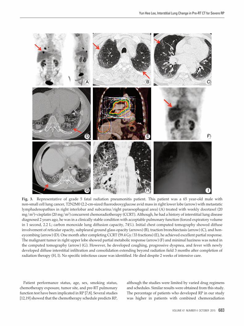
lungs diffusely, or occasionally involving both lower lungfields. Fig. 3 shows a typical representative case of a patientwho progressed to fatal RP. Four of the grade 5 RP patientshad interstitial changes in their pre-RT chest CT and also received chemotherapy: concurrent chemoradiotherapy intwo patients, and sequential chemotherapy followed by RTin two patients.
Table 6 gives a detailed description of five patients withgrade 5 RP. Three patients initially developed focal consoli-dation corresponding to the radiation fields during RT or
within one month after RT completion, and the lesions progressed to the entire lung beyond radiation fields. Theother two patients presented with bilateral lung infiltration4 to 6 weeks after RT completion and died following rapidprogression. No pathogens were identified and steroid andantibiotic treatments were not effective.
MLD
(cGy
)
1,200
1,400
1,600
1,800
2,000
2,200
2,400
2,600
2,800
Grade 1
A B
Grade 2 Grade 3 Grade 4 Grade 5
C DNT
CP (%
)
–20
0
20
40
60
80
100
Grade 1 Grade 2 Grade 3 Grade 4 Grade 5
V20 (
%)
10
20
30
40
50
60
70
Grade 1 Grade 2 Grade 3 Grade 4 Grade 5
V30 (
%)
10
20
30
40
50
Grade 1 Grade 2 Grade 3 Grade 4 Grade 5
Fig. 2. The distribution of RP grade stratified by MLD (A), V20 (B), V30 (C), and NTCP (D). A MLD, V20, V30, V40, and NTCPalso were strongly associated with RP grade. However, all dosimetric parameters were even lower in grade 5 fatal RP. RP,radiation pneumonitis; MLD, mean lung dose; V20 and V30, percent lung volume receiving more than 20 Gy and 30 Gy; NTCP,normal tissue complication probability.
Cancer Res Treat. 2015;47(4):676-686
682 CANCER RESEARCH AND TREATMENT
Discussion
In this study, we examined clinical and dosimetric factorsas predictors of symptomatic RP in lung cancer patients andfurther evaluated the relationship of pre-radiation interstitialchanges in the CT with symptomatic RP, as there are limited
data for interstitial change in pre-RT CT regarding RP. TheRP risk differed significantly by the presence of interstitiallung change in the pre-RT CT. The risk of RP grade " 2 or " 3 was elevated in patients with interstitial change in theirpre-RT CT scans. Interstitial change in the pre-RT chest CTscan was the only clinical factor associated with RP in themultivariable analysis.
Table 4. Risk of RP by interstitial lung disease in pre-RT CT
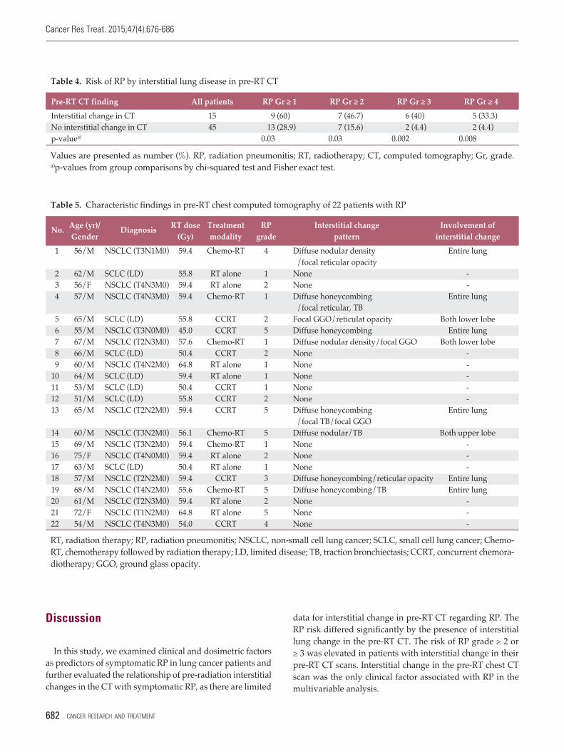
Pre-RT CT finding All patients RP Gr ! 1 RP Gr ! 2 RP Gr ! 3 RP Gr ! 4Interstitial change in CT 15 9 (60) 7 (46.7) 6 (40) 5 (33.3)No interstitial change in CT 45 13 (28.9) 7 (15.6) 2 (4.4) 2 (4.4)p-valuea) 0.03 ( 0.03 ( 0.002 ( 0.008 (
Values are presented as number (%). RP, radiation pneumonitis; RT, radiotherapy; CT, computed tomography; Gr, grade.a)p-values from group comparisons by chi-squared test and Fisher exact test.
Table 5. Characteristic findings in pre-RT chest computed tomography of 22 patients with RP
No. Age (yr)/ Diagnosis RT dose Treatment RP Interstitial change Involvement of Gender (Gy) modality grade pattern interstitial change
1 56/M NSCLC (T3N1M0) 59.4 Chemo-RT 4 Diffuse nodular density Entire lung/focal reticular opacity
2 62/M SCLC (LD) 55.8 RT alone 1 None -3 56/F NSCLC (T4N3M0) 59.4 RT alone 2 None -4 57/M NSCLC (T4N3M0) 59.4 Chemo-RT 1 Diffuse honeycombing Entire lung
/focal reticular, TB 5 65/M SCLC (LD) 55.8 CCRT 2 Focal GGO/reticulat opacity Both lower lobe6 55/M NSCLC (T3N0M0) 45.0 CCRT 5 Diffuse honeycombing Entire lung7 67/M NSCLC (T2N3M0) 57.6 Chemo-RT 1 Diffuse nodular density/focal GGO Both lower lobe8 66/M SCLC (LD) 50.4 CCRT 2 None -9 60/M NSCLC (T4N2M0) 64.8 RT alone 1 None -
10 64/M SCLC (LD) 59.4 RT alone 1 None -11 53/M SCLC (LD) 50.4 CCRT 1 None -12 51/M SCLC (LD) 55.8 CCRT 2 None -13 65/M NSCLC (T2N2M0) 59.4 CCRT 5 Diffuse honeycombing Entire lung
/focal TB/focal GGO 14 60/M NSCLC (T3N2M0) 56.1 Chemo-RT 5 Diffuse nodular/TB Both upper lobe 15 69/M NSCLC (T3N2M0) 59.4 Chemo-RT 1 None -16 75/F NSCLC (T4N0M0) 59.4 RT alone 2 None -17 63/M SCLC (LD) 50.4 RT alone 1 None -18 57/M NSCLC (T2N2M0) 59.4 CCRT 3 Diffuse honeycombing/reticular opacity Entire lung19 68/M NSCLC (T4N2M0) 55.6 Chemo-RT 5 Diffuse honeycombing/TB Entire lung20 61/M NSCLC (T2N3M0) 59.4 RT alone 2 None -21 72/F NSCLC (T1N2M0) 64.8 RT alone 5 None -22 54/M NSCLC (T4N3M0) 54.0 CCRT 4 None -
RT, radiation therapy; RP, radiation pneumonitis; NSCLC, non-small cell lung cancer; SCLC, small cell lung cancer; Chemo-RT, chemotherapy followed by radiation therapy; LD, limited disease; TB, traction bronchiectasis; CCRT, concurrent chemora-diotherapy; GGO, ground glass opacity.
Yun Hee Lee, Interstitial Lung Change in Pre-RT CT for Severe RP
VOLUME 47 NUMBER 4 OCTOBER 2015 683
Patient performance status, age, sex, smoking status,chemotherapy exposure, tumor site, and pre-RT pulmonaryfunction test have been implicated in RP [7,8]. Several studies[12,19] showed that the chemotherapy schedule predicts RP,
although the studies were limited by varied drug regimensand schedules. Similar results were obtained from this study.The percentage of patients who developed RP in our studywas higher in patients with combined chemoradiation
Fig. 3. Representative of grade 5 fatal radiation pneumonitis patient. This patient was a 65 year-old male with non-small cell lung cancer, T2N2M0 (2.2-cm-sized fluorodeoxyglucose avid mass in right lower lobe (arrow) with metastatic lymphadenopathies in right interlobar and subcarina/right paraesophageal area) (A) treated with weekly docetaxel (20mg/m2)+cisplatin (20 mg/m2) concurrent chemoradiotherapy (CCRT). Although, he had a history of interstitial lung diseasediagnosed 2 years ago, he was in a clinically stable condition with acceptable pulmonary function (forced expiratory volumein 1 second, 2.2 L; carbon monoxide lung diffusion capacity, 74%). Initial chest computed tomography showed diffuse involvement of reticular opacity, subpleural ground glass opacity (arrows) (B), traction bronchiectasis (arrow) (C), and hon-eycombing (arrow) (D). One month after completing CCRT (59.4 Gy/33 fractions) (E), he achieved excellent partial response.The malignant tumor in right upper lobe showed partial metabolic response (arrow) (F) and minimal haziness was noted inthe computed tomography (arrow) (G). However, he developed coughing, progressive dyspnea, and fever with newly developed diffuse interstitial infiltration and consolidation extending beyond radiation field 3 months after completion ofradiation therapy (H, I). No specific infectious cause was identified. He died despite 2 weeks of intensive care.
A B C
D E F
G H I

Cancer Res Treat. 2015;47(4):676-686
684 CANCER RESEARCH AND TREATMENT
compared with RT alone (41.7% vs. 16.7%, respectively), butthis difference did not reach statistical significance (p=0.108),perhaps due to small sample size.
We noted that MLD and V30 significantly predicted RP. Inour studies, all RP grades were associated with dosimetricfactors, such as MLD V20, V30, V40, and NTCP, similar to otherreports [9-12], except in the cases of grade 5 fatal RP. The fivepatients with grade 5 RP in our study did not have worsedosimetric parameters than those with non-fatal RP. RP isusually limited to the irradiation field; however, in grade 5RP patients of our study, their chest CT scans were charac-terized by diffuse interstitial infiltration and consolidationeven outside the radiation field in the absence of an identifi-able infectious organism. This suggests that other biologicfactors might have contributed to RP deaths.
We analyzed the pre-RT chest CT scans of the RP patientsand frequently observed diffuse honeycombing and diffusenodular density patterns in high grade RP. Patients whosepre-RT chest CT scans revealed diffuse interstitial changes ineither the entirety of both lungs or both lower lung fields hada greater risk of high grade RP. These patients must be care-fully watched during chemoradiation and closely monitoredfollowing RT due to the increased risk. This is the essentialpoint: the physician must monitor carefully even patientswho are within the acceptable range results for the pulmonary function test prior to RT, even those with no history of symptomatic ILDs. Several Japanese studies havesuggested that subclinical ILD found in CT scans before
thoracic RT is related to fatal RP, and those patients have tobe carefully evaluated [16,17,20]. Our study found that patients with interstitial lung change in CT have a signifi-cantly higher risk of severe RP, and that the interstitialchange is a statistically significant prognostic factor for severe RP. Yamaguchi et al. [16] suggested that extensive interstitial change in pre-RT CT is a contraindication for thoracic RT. In our study, we obtained similar results. In addition we showed that five patients with grade 5 RP didnot have higher dosimetric parameters compared to the non-fatal RP patients. Even if patients have reasonable dosi-metric results in their radiotherapy planning, it is necessaryto carefully evaluate and follow up with the patient due totheir high risk of severe RP.
Several studies suggest that certain biological or physio-logical responses to radiation could explain RP and that theseare not well predicted by the empiric dose-volume relation-ship. Further research that takes into consideration biologicmarkers may be necessary [21]. Tumor growth factor !1, interleukins 1, 6, 8, and 10, Krebs von den Lungen-6 (KL-6)and surfactant proteins (SP) are important biologic markersin the development of symptomatic radiation-induced lunginjury. Interstitial change on the CT and high levels of biologic markers (KL-6, SP-D) were associated with an increased risk of severe RP [22]. Further study with a highernumber of patients and several biologic factors is necessaryto determine the relationships of interstitial change in pre-RT CT, biologic markers, and severe RP. It is hoped that
Table 6. Characteristics of the five patients with grade 5 RP
Age (yr)/ Diagnosis COPD Pre-RT Treatment Chemotherapy RT dose MLD V20 V30 RP onset Death Gender (stage) CT finding modality regimen (Gy) (Gy) (%) (%) (mo) (mo)55/M NSCLC No Diffuse CCRT Cisplatin 45 15.22 29.48 24.82 0 3
(T3N0M0) honeycombing 65/M NSCLC No Diffuse CCRT Docetaxel 59.4 26.73 57.67 42.56 1 3
(T2N2M0) honeycombing +cisplatin/focal TB/focal GGO
60/M NSCLC Yes Diffuse nodular Chemo-RT Cisplatin 56.1 22.22 46.02 27.53 0 1(T3N2M0) /TB +gemcitabine
68/M NSCLC No Diffuse Chemo-RT Cisplatin 55.6 19.12 34.57 27.1 1 1(T4N2M0) honeycombing +gemcitabine
/TB72/F NSCLC Yes None RT alone None 64.8 18.54 29.47 26.17 1.5 1.5
(T1N2M0)
RP, radiation pneumonitis; COPD, chronic obstructive pulmonary disease; RT, radiation therapy; CT, computed tomography;MLD, mean lung dose; V20 and V30, percent lung volume receiving more than 20 Gy and 30 Gy; NSCLC, non-small cell lungcancer; CCRT, concurrent chemoradiotherapy; TB, traction bronchiectasis; GGO, ground glass opacity; Chemo-RT, chemother-apy followed by radiation therapy.

Yun Hee Lee, Interstitial Lung Change in Pre-RT CT for Severe RP
VOLUME 47 NUMBER 4 OCTOBER 2015 685
further research will identify biomarkers that will allow usto tailor our treatment for lung cancer patients to avoid thedevelopment of severe RP.
There are several limitations in this study. As a retrospec-tive study, this study has the possibility of selection bias. Asmall number of patients with short inclusion were analyzedin this study, because we explored this study as the extensionof another study for RP. Patient characteristics and radiationtreatment were heterogeneous. A prospective study with alarger number of patients will improve the result of thisstudy. Despite these limitations, our findings are helpful toidentify and follow the high risk patients of severe RP.
Conclusion
Our study identified diffuse interstitial disease as a signif-icant clinical factor for the occurrence of RP and also showedthe usefulness of dosimetric factors such as MLD, V20, V30,and NTCP in predicting the incidence and severity of RP.Four of our five grade 5 RP patients had interstitial changes
in their pre-RT chest CTs. When image findings were analyzed in RP patients, diffuse honeycombing and diffusenodular patterns were frequently observed in high grade RP.The patients with interstitial change in pre-RT CT have to becarefully evaluated and managed for severe RP after thoracicRT.
Conflicts of Interest
Conflict of interest relevant to this article was not reported.
Acknowledgments
The authors would like to acknowledge the statistical assistance of Catholic Medical Center Clinical Research Coordinating Center.
1. Choy H, Akerley W, Safran H, Graziano S, Chung C, WilliamsT, et al. Multiinstitutional phase II trial of paclitaxel, carbo-platin, and concurrent radiation therapy for locally advancednon-small-cell lung cancer. J Clin Oncol. 1998;16:3316-22.
2. Toschi L, Cappuzzo F, Janne PA. Evolution and future perspectives in the treatment of locally advanced non-smallcell lung cancer. Ann Oncol. 2007;18 Suppl 9:ix150-5.
3. Pignon JP, Arriagada R, Ihde DC, Johnson DH, Perry MC,Souhami RL, et al. A meta-analysis of thoracic radiotherapyfor small-cell lung cancer. N Engl J Med. 1992;327:1618-24.
4. De Ruysscher D, Pijls-Johannesma M, Vansteenkiste J, KesterA, Rutten I, Lambin P. Systematic review and meta-analysisof randomised, controlled trials of the timing of chest radio-therapy in patients with limited-stage, small-cell lung cancer.Ann Oncol. 2006;17:543-52.
5. Morgan GW, Breit SN. Radiation and the lung: a reevaluationof the mechanisms mediating pulmonary injury. Int J RadiatOncol Biol Phys. 1995;31:361-9.
6. McDonald S, Rubin P, Phillips TL, Marks LB. Injury to thelung from cancer therapy: clinical syndromes, measurableendpoints, and potential scoring systems. Int J Radiat OncolBiol Phys. 1995;31:1187-203.
7. Vogelius IR, Bentzen SM. A literature-based meta-analysis ofclinical risk factors for development of radiation inducedpneumonitis. Acta Oncol. 2012;51:975-83.
8. Palma DA, Senan S, Tsujino K, Barriger RB, Rengan R,Moreno M, et al. Predicting radiation pneumonitis afterchemoradiation therapy for lung cancer: an international individual patient data meta-analysis. Int J Radiat Oncol BiolPhys. 2013;85:444-50.
9. Jin H, Tucker SL, Liu HH, Wei X, Yom SS, Wang S, et al. Dose-volume thresholds and smoking status for the risk of treat-ment-related pneumonitis in inoperable non-small cell lungcancer treated with definitive radiotherapy. Radiother Oncol.2009;91:427-32.
10. Baker R, Han G, Sarangkasiri S, DeMarco M, Turke C, StevensCW, et al. Clinical and dosimetric predictors of radiationpneumonitis in a large series of patients treated with stereo-tactic body radiation therapy to the lung. Int J Radiat OncolBiol Phys. 2013;85:190-5.
11. Dang J, Li G, Ma L, Diao R, Zang S, Han C, et al. Predictors ofgrade " 2 and grade " 3 radiation pneumonitis in patients withlocally advanced non-small cell lung cancer treated with three-dimensional conformal radiotherapy. Acta Oncol. 2013;52:1175-80.
12. Park YH, Kim JS. Predictors of radiation pneumonitis and pulmonary function changes after concurrent chemoradiother-apy of non-small cell lung cancer. Radiat Oncol J. 2013;31:34-40.
13. Togashi Y, Masago K, Handa T, Tanizawa K, Okuda C,
References
Cancer Res Treat. 2015;47(4):676-686
686 CANCER RESEARCH AND TREATMENT
Sakamori Y, et al. Prognostic significance of preexisting inter-stitial lung disease in Japanese patients with small-cell lungcancer. Clin Lung Cancer. 2012;13:304-11.
14. Yamaguchi S, Ohguri T, Ide S, Aoki T, Imada H, Yahara K, etal. Stereotactic body radiotherapy for lung tumors in patientswith subclinical interstitial lung disease: the potential risk ofextensive radiation pneumonitis. Lung Cancer. 2013;82:260-5.
15. Nagata Y, Hiraoka M, Mizowaki T, Narita Y, Matsuo Y, Nori-hisa Y, et al. Survey of stereotactic body radiation therapy inJapan by the Japan 3-D Conformal External Beam Radiother-apy Group. Int J Radiat Oncol Biol Phys. 2009;75:343-7.
16. Yamaguchi S, Ohguri T, Matsuki Y, Yahara K, Oki H, ImadaH, et al. Radiotherapy for thoracic tumors: association betweensubclinical interstitial lung disease and fatal radiation pneu-monitis. Int J Clin Oncol. 2015;20:45-52.
17. Sanuki N, Ono A, Komatsu E, Kamei N, Akamine S, YamazakiT, et al. Association of computed tomography-detected pul-monary interstitial changes with severe radiation pneumonitisfor patients treated with thoracic radiotherapy. J Radiat Res.2012;53:110-6.
18. Mueller-Mang C, Grosse C, Schmid K, Stiebellehner L, BankierAA. What every radiologist should know about idiopathic interstitial pneumonias. Radiographics. 2007;27:595-615.
19. Das SK, Zhou S, Zhang J, Yin FF, Dewhirst MW, Marks LB.Predicting lung radiotherapy-induced pneumonitis using amodel combining parametric Lyman probit with nonparamet-ric decision trees. Int J Radiat Oncol Biol Phys. 2007;68:1212-21.
20. Makimoto T, Tsuchiya S, Hayakawa K, Saitoh R, Mori M. Riskfactors for severe radiation pneumonitis in lung cancer. Jpn JClin Oncol. 1999;29:192-7.
21. Graves PR, Siddiqui F, Anscher MS, Movsas B. Radiation pulmonary toxicity: from mechanisms to management. SeminRadiat Oncol. 2010;20:201-7.
22. Yamashita H, Kobayashi-Shibata S, Terahara A, Okuma K,Haga A, Wakui R, et al. Prescreening based on the presence ofCT-scan abnormalities and biomarkers (KL-6 and SP-D) mayreduce severe radiation pneumonitis after stereotactic radio-therapy. Radiat Oncol. 2010;5:32.