insights into the osteoblast precursor differentiation towards mature
TRANSCRIPT

Insights into the osteoblast precursor differentiationtowards mature osteoblasts induced by continuousBMP-2 signaling
Omar F. Zouani1,*, Lila Rami1, Yifeng Lei2 and Marie-Christine Durrieu1,2
1Bioingenierie Tissulaire (BioTis), INSERM U1026, Universite de Bordeaux, 146 rue Leo Saignat, 33076 Bordeaux, France2Institut Europeen de Chimie et Biologie (IECB), CNRS, UMR 5248, Universite de Bordeaux I, 2 rue Robert Escarpit, 33607 Pessac, France
*Author for correspondence ([email protected]). O.F.Z. is the principal investigator
Biology Open 2, 872–881doi: 10.1242/bio.20134986Received 7th April 2013Accepted 7th June 2013
SummaryMature osteoblasts are the cells responsible for bone formation
and are derived from precursor osteoblasts. However, the
mechanisms that control this differentiation are poorly
understood. In fact, unlike the majority of organs in the body,
which are composed of ‘‘soft’’ tissue from which cells can easily
be isolated and studied, the ‘‘hard’’ mineralized tissue of bone
has made it difficult to study the function of bone cells. Here, we
established an in vitro model that mimics this differentiation
under physiological conditions. We obtained mature osteoblasts
and characterized them on the basis of the following parameters:
the strong expression of osteoblastic markers, such as Runx2 and
Col-I; the achievement of specific dimensions (the cell volume
increases 26-fold compared to the osteoblast precursors); and the
production of an abundant extracellular matrix also called
osteoid. We demonstrated that the differentiation of osteoblast
precursors into mature osteoblasts requires the continuous
activation of Bone Morphogenetic Protein (BMP) receptors,
which we established with the immobilization of a
BMP-2mimetic peptide on a synthetic matrix mimicking in vivo
microenvironment. Importantly, we demonstrated that
the organization of the F-actin network and acetylated
microtubules of the cells were modified during the
differentiation process. We showed that the perturbation of the
F-actin cytoskeleton organization abolished the differentiation
process. In addition, we demonstrated that expression of the
Runx2 gene is required for this differentiation. These findings
demonstrate the retro-regulation of cytoplasmic and genic
components due to the continuous induction of BMP-2 and
also provide more detailed insights into the correct signaling of
BMPs for cell differentiation in bone tissue.
� 2013. Published by The Company of Biologists Ltd. This is an
Open Access article distributed under the terms of the Creative
Commons Attribution License (http://creativecommons.org/
licenses/by/3.0), which permits unrestricted use, distribution
and reproduction in any medium provided that the original
work is properly attributed.
Key words: Mature osteoblasts, BMP-2, Runx2, Actin cytoskeleton,
Super-resolution optical profilometry
IntroductionUnlike the majority of organs in the body, which are composed of‘‘soft’’ tissue from which cells can easily be isolated and studied,
the ‘‘hard’’ mineralized tissue of bone has made it difficult to
study the function of bone cells. Moreover, we have the lack oftools to manipulate these bone cells (Bonewald, 2011; Lutolf and
Blau, 2009). Furthermore, the complexity of the spatial
arrangement and the composition of the microenvironment of
the bone cells add to the challenge. Several strategies have beendeveloped for the design of in vitro culture systems to mimic the
extracellular microenvironment of bone cells (Lutolf and Blau,
2009). In bone tissue, the extracellular matrix (ECM) iscomposed of proteins that promote cell adhesion, and growth
factors such as BMPs that promote cell differentiation (Chen et
al., 2004; Wagner et al., 2010). These various proteins exhibitdifferent distributions (Marie, 2009). In addition to the
extracellular antagonists controlling the binding of the ligand to
receptor complexes to regulate BMP signaling (Sengle et al.,2008), different adhesion components of ECM modulate BMP
signaling (Wang et al., 2008; Hynes, 2009). Therefore, the
retention of BMPs by antagonists or ECM adhesion proteins may
be an important mechanism for controlling receptor availability
at the cell surface and inducing the attenuation of signal
transduction, thereby delaying the internalization of the BMP–
receptor complex. Thus is the ‘‘solid induction mode’’ (Crouzier
et al., 2011; Zouani et al., 2013a). The concept of presenting a
growth factor ‘‘in the solid induction mode,’’ which we refer to as
‘‘matrix-bound,’’ has been proposed in pioneering work
performed by Kuhl et al. on tethered epidermal growth factor
(EGF); however, this work has been neglected for many years
(Kuhl and Griffith-Cima, 1996). This matrix-bound mode of
growth factor presentation is closer to physiological conditions
because most growth factors in the ECM are bound to proteins
and glycosaminoglycans. Recent experimental studies have
shown that the matrix-bound form of presentation improves the
efficacy of different growth factors when compared to the soluble
form and that their biological function is enhanced (Chen et al.,
2010; Davis et al., 2006; Fan et al., 2007). The ‘‘punctual
induction mode’’ is the most commonly studied and best known
method for the presentation of BMPs in the soluble state (Shah
872 Research Article
Bio
logy
Open
by guest on November 24, 2018http://bio.biologists.org/Downloaded from

et al., 1999; Sieber et al., 2009; Wagner et al., 2010)(supplementary material Fig. S1A). In bone tissue, because of
the presence of high levels of collagens containing BMPsinteraction specific site, these BMPs are sequestered in the ECM,and the solid induction mode is favored over the punctualinduction mode. Diseases in different tissues are caused by
malfunctions in the BMP signaling pathways (Ramirez andRifkin, 2009; Maciel et al., 2010; Mazzaferro et al., 2010; Sethiand Kang, 2011).
BMPs are secretory signaling molecules belonging to the TGF-b superfamily of growth factors. BMPs play important roles inthe induction of bone formation (Wagner et al., 2010). These
growth factors have the potential to induce different bone celldifferentiation processes at different stages (Carragee et al.,2011). BMPs bind to dimeric receptor complexes composed oftype I and type II transmembrane serine/threonine kinase
receptors (Wagner et al., 2010; Sieber et al., 2009). Thereceptors form homomeric and heteromeric complexes indistinct membrane areas and are differentially modulated by
their ligands. BMP-2 binds to preformed heterocomplexes of thetype I and type II receptors, initiating Smad-dependent signaling.The Smad pathway is initiated by the phosphorylation of
regulatory Smad1/5/8, which associate with the commonmediator Smad (Smad4), translocate into the nucleus, andregulate the transcription of specific BMP target genes, such as
Runx2, by recruiting additional activators and repressors (Sieberet al., 2009; Komori, 2010).
In the osteoblast lineage, cells progress through various stages ofdifferentiation and maturation. Osteoblast progenitors are derived
from adult mesenchymal stem cells, followed by osteoblastprecursors, mature osteoblasts and osteocytes (Kato et al., 2001).In vivo, mature osteoblasts are distinguished by their dimensions in
the bone (Gohel et al., 1995; Maes et al., 2010) and are found onthe ECM (referred to as osteoid), which is produced by theosteoblasts themselves. BMPs (particularly BMP-2, which is more
abundant in the ECM of bone tissue) play an important role duringthese differentiation stages and are particularly important for thegeneration of mature osteoblasts in vivo (Sieber et al., 2009)(supplementary material Fig. S1B,C). However, the mechanisms
that control this differentiation (osteoblast precursors into matureosteoblasts) are poorly understood. Recently, the solid inductionmode was suggested for BMPs to induce the correct intracellular
signals promoting precursor osteoblasts differentiation into matureosteoblasts (Zouani et al., 2010). In the present study, we used amature osteoblast differentiation model using a modified polymer
that mimics the osteo-induced ECM and that is based on the solidBMP-2 induction mode. With this approach, the polymerpolyethylene terephthalate (PET) was functionalized using a
BMP-2mimetic peptide favoring osteogenesis. Using this system, theactivation of BMPr-1A is continuous, mimicking the physiologicalECM-containing BMP-2 that surrounds the osteoblast precursors,because of the arrest of the BMP-2/BMP receptor complex
process. In addition, with the generation of mature osteoblasts, theregulation of the osteoblast precursor differentiation can becharacterized, giving an indication at the physiological level.
Using this differentiation model, we showed that the matureosteoblasts generated in vitro are characterized by cell swelling.This study of cell size, providing a three-dimensional view, was
performed using an optimized optical profilometry (Schmidt andBlack, 1992; Chen et al., 2009; Xiao et al., 2010; Zouani et al.,2012; Pan et al., 2011; Lei et al., 2013) and a specific cell
treatment protocol, which allowed for a vertical nanometerresolution. Other features that are characteristic of a mature
osteoblast include (i) an increase in Runx2 and Col-I expressionand (ii) an increase in ECM (osteoid-like matrix) production.These features signify a transformation from precursor to mature
osteoblasts. Next, we were interested in characterizing therelationship between the genes that characterize the differentiationof the cells and the increased volume of the mature osteoblasts.We investigated the major components of the cytoskeleton,
such as F-actin organization and the presence of acetylatedtubulin. Therefore, we showed that whereas osteoblastdifferentiation is regulated by Runx2 gene, it is also
dependent on cytoskeletal components that are responsible forthe size and shape of the cells during their transformation. Insummary, this study demonstrates that the continuous induction
mode of BMP-2 (the correct BMP-2 signaling mode in bonetissue) is essential for the generation of mature osteoblasts. Ourfindings suggest that this phenomenon is likely common to allBMPs signaling that are regulated by the ECM on bone,
thereby regulating a number of bone–cell differentiationprocesses.
ResultsGenerating mature osteoblasts
The interaction between the ECM components and the inducers(e.g., BMP-2) is essential for correct signaling. Here, a syntheticECM-BMP-2 analogs in two-dimensional (2D) culture
systems controlling cell fate was created and their effects havebeen evaluated on the behavior of a mouse osteoblasticprecursor cell line (MC3T3-E1). After culturing for 24 h, the
osteoblast precursors were seeded onto the BMP-2mimetic peptides
functionalized polymer and transformed into mature osteoblasts.The cells exhibited a radically changed phenotype with an
increased morphology, volume and height (Fig. 1A). In ourmodel, the activated signaling pathway is the Smad1/5/8(Fig. 1B). In addition, the ECM quantity synthesized (theosteoid-like matrix) by the cells was studied on this novel cell
type. The differentiated osteoblast precursors, characterized bytheir increased height, produced intensive amounts of ECMcompared to the osteoblast precursors cultured on a native
polymer (on plastic or on a modified polymer grafted only withRGD peptides derived from natural ECM proteins; data notshown) (Fig. 1C,D). Finally, the osteoblast precursors
transformation into mature osteoblasts was verified byanalyzing the expression of the osteogenic biomarker corebinding factor a1 (Cbfa-1), also called Runx2, as well as the
expression of Col-I. We observed an increase in Runx2 and Col-Igene expression after 24 h in culture (Fig. 1E).
Mature osteoblasts are characterized by cell swelling
Accumulating evidence suggests that cell volume alterationsare a component of a wide variety of cellular functions, including
epithelial transport, metabolism, hormone release, migration,cell proliferation and cell growth and cell death (al-Habori,1995; Hoffmann et al., 2009). Conceivably, cell volume
regulation may play an essential role in cell growth andproliferation (Hoffmann et al., 2009); however, the regulationof cell volume in the osteoblastic lineage is unclear. We
demonstrated the characterization of osteoblast precursordifferentiation into mature osteoblasts by cell volumemodification (Fig. 2A; supplementary material Fig. S2). The
Osteoblast differentiation 873
Bio
logy
Open
by guest on November 24, 2018http://bio.biologists.org/Downloaded from

optical profilometry technique demonstrated that the cell height
and cell volume increased approximately 26-fold in the
osteoblast precursors cultured on the polymer functionalized
with the BMP-2mimetic peptide after 24 h (Fig. 2B,C). Surprisingly,
osteoblast precursors treated with BMP-2 in ‘‘the soluble state’’
do not induce the differentiation from precursors to mature
osteoblasts (supplementary material Fig. S3). The data shown
here (Figs 1, 2; supplementary material Figs S1–3) demonstrate
that BMP-2 retention by covalent binding to the matrix is
essential for the induction of osteoblast precursor differentiation
into mature osteoblasts.
Mature osteoblasts are characterized by the novel organization
of the cytoskeleton
The role of the cytoskeleton in cell volume regulation has been
studied in various cell types (Pedersen et al., 2001). For example,
Fig. 1. The generation of mature osteoblasts. (A) SEM micrographs showing cultured osteoblast precursors on different modified polymer surfaces and theirtransformation. Scale bar: 20 mm. (B) Smad1/5/8 pathway has been activated after 24 hours for osteoblast precursors cultured on polymer grafted with
BMP-2mimetic peptide as seen by phospho-Smad1/5/8 blotting. (C) An example of an OPS micrograph showing the ECM formed by the cells after 24 h culture on thepolymer surface grafted with the BMP-2mimetic peptide. The yellow square represents a zoom of a particular region. 7 to 10 measurements were performed in order todetermine the thickness of the newly synthesized ECM. Down, right, a 3D reconstruction image of the region in the yellow square. (D) A graph quantifying theevolution of the ECM thickness at 24 h after cell seeding. Note that the ECM protein aggregates are scarce on the native PET and are abundant on the polymersurfaces grafted with RGD and/or the BMP-2mimetic peptide (*P,0.001). (E) The quantitative PCR analysis for Runx2 and Col-I; significant differences were observedbetween the native polymer (control) and the polymer grafted with the BMP-2mimetic peptide alone (P,0.005).
Osteoblast differentiation 874
Bio
logy
Open
by guest on November 24, 2018http://bio.biologists.org/Downloaded from

in Ehrlich ascites tumor cells (EATC), cell shrinkage is
associated with an increase in cell swelling and a decrease in
F-actin content (Pedersen et al., 1999; Pedersen and Hoffmann,
2002). This observation suggests that the effect of F-actin
polymerization/depolymerization regulates the process of volume
decrease/increase in cells.
F-actin organization was observed in mature osteoblasts, and a
critical decrease in the percentage of stress fiber compared to
osteoblast precursors was observed (Fig. 3A,B). This decrease in
the F-actin stress fibers correlated with an increase in the cell
volume. The shape of the mature osteoblast changed compared to
the osteoblast precursor cells (Fig. 3A), and it has been
demonstrated that the cell shape and contractility regulate
ciliogenesis (Pitaval et al., 2010). To confirm the association
between the change in cell shape and dimension with
ciliogenesis, we labeled the acetylated tubulin (previously
demonstrated to exhibit primary cilia structures) and observed
the disappearance of these cytoskeletal structures on the mature
osteoblasts (Fig. 3A,C). Short cilia (approximately 1 mm) were
observed only on osteoblast precursors (Fig. 3C). Thus, there is
evidence that mature osteoblasts play a specific role in bone, and
the presence or absence of a number of cytoskeletal structures
reflect the commitment of bone cells to a specific function.
Cell proliferation and nuclear morphology strongly correlate
with osteoblast status
The various osteoblastic cell functions interact; for example,
previous studies have shown that when cells migrate, the
inducting factor (such as BMP-2) induces the cells to migrate
and not to differentiate (de Jesus Perez et al., 2009).
The differentiation of osteoblast precursors to mature
osteoblasts is characterized by the inhibition of proliferation
(Fig. 4A; supplementary material Fig. S4). These results confirm
the in vivo data suggesting that the function of mature osteoblasts
Fig. 2. A mature osteoblast characterized by a substantial increase in cell height and volume. (A) OPS micrographs showing a single cell on different polymersurfaces after culturing for 24 h. (B) A graph quantifying the cell height registered by the OPS after culturing for 24 h. The cell height varies with the level of
osteoblast precursor differentiation. (C) Volume changes in a single cultured cell.
Osteoblast differentiation 875
Bio
logy
Open
by guest on November 24, 2018http://bio.biologists.org/Downloaded from

Fig. 3. A mature osteoblast characterized by the novel
organization of the F-actin cytoskeleton.
(A) Fluorescence staining of cells under different polymer
conditions after culturing for 24 h; the cells were stainedfor visualization of the actin filaments (green), acetylatedtubulin (red) and the nucleus (blue, using DAPI). Scalebars: 10 mm. (B,C) Graphs quantifying the percentage ofstress fibers and cilium length, respectively.
Fig. 4. Nuclear morphology strongly correlates with osteoblast status. (A) The inhibition of cell proliferation on polymer surfaces promoted the differentiationof osteoblast precursors into mature osteoblasts. (B) Top panels: fluorescence staining of the cell nucleus under the two different polymer conditions (nativepolymer and polymer grafted with the BMP-2mimetic peptide) after 24 h. Bottom panels: 3D volumes obtained from the serial confocal sections to confirm thatthe changes in the diameter represent the changes in the volume. Scale bars: 10 mm. (C) A graph quantifying the nuclear volume during differentiation.
Osteoblast differentiation 876
Bio
logy
Open
by guest on November 24, 2018http://bio.biologists.org/Downloaded from

producing ECM to build bone inhibits the cells from performing
other functions, such as migration and proliferation. Indeed,precursor osteoblasts, but not mature osteoblasts, move andmigrate into developing bones along with invading blood vessels
that stabilize the bone (Maes et al., 2010). Interestingly,differentiating osteoblasts are also characterized by changes intheir nuclear volume, which increases in mature osteoblasts(Fig. 4B,C). These findings suggest the importance of nuclear
morphology on the transformation of osteoblast precursors intomature osteoblasts. These results may also explain the changes inthe organization of both the cytoskeleton and the nucleus, which
are likely essential for stabilizing the novel cell architecture.
Runx2 gene expression, integrin engagement and dynamicactin networks regulate osteoblast precursor differentiation intomature osteoblasts
In adherent mature osteoblastic cells, the relationship between thecytoskeleton and the nuclei is suggested by subtle genetic cues;
however, this relationship has not been studied extensively and isunclear. Here, we demonstrated that during osteoblast precursorsdifferentiate into mature osteoblasts, the F-actin cytoskeleton isstrongly correlated with cell volume (cell height and dimension).
Therefore, the next step was to investigate the components thatplay key roles in this differentiation process and identify theregulators of mature osteoblast functions. The potential of two
components was investigated: a cytoplasmic component (F-actinorganization) and a genetic component (Runx2 gene expression).In our model, we activated the Smad1/5/8 pathway using BMP-2
mimetic peptides and induced Runx2 gene expression along withother mature osteoblastic markers, such as Col-I. Next, weobserved that cell swelling strongly correlated with F-actin
cytoskeleton organization and with increased ECM formation.This reasoning suggested that Runx2 gene regulates thereorganization of the actin networks.
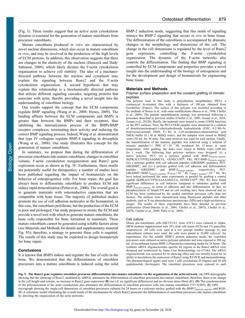
To confirm this hypothesis, RNAi was used to silence the
Runx2 expression. The silencing of Runx2 gene expression usingshRNA inhibited the differentiation of osteoblast precursors intomature osteoblasts (Fig. 5A–C). No significant Runx2 gene over-
expression was observed (Fig. 5B); the initial F-actin organizationwas preserved (stress fiber organization) (Fig. 5C), and there wasno cell swelling compared to the induced osteoblasts (Fig. 5A,induced osteoblasts are osteoblast precursors cultured on polymer
grafted with BMP-2mimetic peptide). These results indicate thatRunx2 is required for the differentiation of osteoblast precursorsinto mature osteoblasts. However, the organization of the F-actin
cytoskeleton is also important for the differentiation process.Indeed, jasplakinolide, which stabilizes actin filaments byinhibiting their depolymerization, and cytochalasin D, which
inhibits actin polymerization, both inhibit the differentiation ofosteoblast precursors into mature osteoblasts (Fig. 5D). The datasuggest that the modification of the dynamic of actin networks
perturbed the differentiation of the osteoblast precursors intomature osteoblasts.
The addition of adhesion component (RGD peptide) to thedifferentiation culture model (the polymer grafted with the
BMP-2mimetic peptide) promotes the differentiation of osteoblastprecursors into mature osteoblasts. Indeed, the mature osteoblastsexhibited a higher volume, increased Runx2 gene expression and
the highest level of ECM production under these conditions(Fig. 5C,D,E). A decrease in the levels of F-actin stress fibers andthe disappearance of acetylated tubulin was also observed
(supplementary material Fig. S5). These data suggest that
integrin engagement (via RGD ligand here) intensifies thedifferentiation of osteoblast precursors into mature osteoblasts.
DiscussionThe mechanisms that direct the differentiation of osteoblastprecursor cells into mature osteoblasts are of interest because ofthe potential for developing biomaterials that promote bone
reparation and regeneration. In this present study, we determinedthat BMP-2-mediated signaling induces the differentiation ofprecursor osteoblasts, suggesting the continuous activation of theBMP receptors. Furthermore, we demonstrated the essential role
of Runx2 in the differentiation of osteoblast precursors intomature osteoblasts and showed that this differentiation is relatedto the dynamics of the entire F-actin network.
In this study, we first validated our osteoblast differentiationmodel by the identification of the following three physiologicalparameters, which are typical of mature osteoblasts: (i) thesignificant increase in cell height; (ii) the expression of
osteoblastic markers, such as Runx2 and Col-I; and (iii) theproduction of a large quantity of ECM proteins (Figs 1, 2). Next,we investigated the organization of the F-actin cytoskeleton
networks in osteoblast precursors and in mature osteoblasts;mature osteoblasts are characterized by the absence of stressfibers (Fig. 3). It is likely that the reorganization of the F-actin
cytoskeleton that promotes the volume changes is accompaniedby effects on other cytoskeletal components, such as myosin II,as suggested in other studies (Hoffmann and Pedersen, 2007).
However, this may only play a role under extreme conditions ofswelling or shear stress. A change in the volume of the cell couldcertainly initiate different signaling pathways (Pedersen et al.,1999); for example, small G-proteins (the Rho family GTPases
Rac1, RhoA and Cdc42, in particular) reversibly associate withthe actin cytoskeleton, and this likely plays a role in regulatingtheir activity. Conversely, the Rho GTPases are themselves
important regulators of cytoskeletal organization and integrinsignaling pathways (Pedersen and Hoffmann, 2002). In ourmodel, when the RGD peptides (derived from natural ECM
proteins i.e., fibronectin) and the BMP-2mimetic peptides are graftedonto a polymer surface, the mature osteoblasts exhibit a greatercell volume than the cells cultured on polymers grafted with theBMP-2mimetic peptide alone (Fig. 5). This result supports several
studies suggesting that integrin activation also plays a role insensing the cell volume changes, thus confirming that theengagement of integrins coupled with inductors on a matrix
induces the differentiation of osteoblast precursors. The cellswelling during the osteoblast precursor differentiation intomature osteoblasts is associated with a decrease in F-actin stress
fiber content (Fig. 3), a process that is observed in many differentcell types (Pedersen et al., 1999; Pedersen et al., 2001; Hoffmannand Pedersen, 2007; Hoffmann et al., 2009).
We next investigated whether the differentiation of osteoblast
precursors into mature osteoblasts is regulated by Runx2 (geneticregulation) alone or by another cell regulator, such as F-actin.The specific silencing of Runx2 blocked the differentiation of
osteoblast precursor cells into mature osteoblasts (Fig. 5). The F-actin networks were synchronized using pharmacological agentsduring the differentiation of osteoblast precursors on different
matrices. This perturbation of the F-actin network dynamic(inhibiting depolymerization/polymerization) blocked thedifferentiation of osteoblast precursors into mature osteoblasts
Osteoblast differentiation 877
Bio
logy
Open
by guest on November 24, 2018http://bio.biologists.org/Downloaded from

Fig. 5. See next page for legend.
Osteoblast differentiation 878
Bio
logy
Open
by guest on November 24, 2018http://bio.biologists.org/Downloaded from
(Fig. 5). These results suggest that an active actin cytoskeleton
dynamic is essential for the generation of mature osteoblasts from
precursor osteoblasts.
Mature osteoblasts produced in vitro are characterized by
novel nuclear dimensions, which also occur in mature osteoblasts
in vivo, and may be involved in the production of the high levels
of ECM proteins. In addition, this observation suggests that there
are changes in the elasticity of the nucleus (Hancock and Hadj-
Sahraoui, 2009), which likely dictates the F-actin cytoskeleton
organization to achieve cell stability. The idea of a mechano-
directed pathway between the nucleus and cytoplasm may
explain the signaling between Runx2 and the F-actin
cytoskeleton organization. A second hypothesis that may
explain this relationship is a biochemically directed pathway
that utilizes different signaling cascades, targeting proteins that
associate with actin, thereby providing a novel insight into the
understanding of osteoblast biology.
Our results support the concept that the ECM components
regulate BMP signaling (Wang et al., 2008; Hynes, 2009). The
binding affinity between the ECM components and BMPs is
greater than between the BMPs and their receptors, thus
inhibiting the internalization process of the BMPs/BMP
receptor complexes, terminating their activity and inducing the
correct BMP signaling process. Indeed, Wang et al. demonstrated
that type IV collagens regulate BMP-4 signaling in drosophila
(Wang et al., 2008). Our study illustrates this concept for the
generation of mature osteoblasts.
In summary, we propose that during the differentiation of
precursor osteoblasts into mature osteoblasts, changes in osteoblast
volume, F-actin cytoskeleton reorganization and Runx2 gene
expression occur as shown in Fig. 5F. Furthermore, these results
are potentially useful for therapeutics; a number of studies have
been published regarding the impact of biomaterials on the
behavior of osteoprogenitor cells. For bone repair, the goal has
always been to differentiate osteoblasts to produce ECM and
induce rapid mineralization (Petite et al., 2000). The overall goal is
to generate materials with osteoinductive capacities that are
compatible with bone tissue. The majority of published studies
promote the use of cell adhesion molecules in the biomaterial; in
this case, the osteoblasts proliferate, but the production of the ECM
is poor and prolonged. Our study proposes to mimic the ECM and
provide a novel tool with which to generate mature osteoblasts, the
bone cells responsible for bone formation in mammals. These
mature osteoblasts cannot be generated using soluble factors alone
(see Materials and Methods for details and supplementary material
Fig. S5); therefore, a strategy to generate these cells is required.
The results of this study may be exploited to design biomaterials
for bone repair.
ConclusionsIt is known that BMPs induce and regulate the fate of cells in the
bone. We demonstrated that the differentiation of osteoblast
precursors into a mature osteoblasts is induced using the solid
BMP-2 induction mode, suggesting that this mode of signaling
mimics the BMP-2 signaling that occurs in vivo in bone tissue.
The differentiation of the osteoblasts is accompanied by dramatic
changes in the morphology and dimensions of the cell. The
change in the cell dimensions is regulated by the level of Runx2
gene expression, controlling the F-actin cytoskeleton
organization. The dynamic of the F-actin networks also
controls the differentiation. The finding that BMP signaling is
controlled by ECM components and properties provides a novel
insight into the understanding of the biology of osteogenesis and
for the development and design of biomaterials for engineering
bone tissue.
Materials and MethodsPolymer surface preparation and the covalent grafting of mimeticpeptidesThe polymer used in this study is poly(ethylene terephthalate) (PET), acommercial bi-oriented film with a thickness of 100 mm obtained fromGoodfellow (France). The surface of this polymer was modified according tothe methods of Boxus et al., with some modifications (Boxus et al., 1996; Cholletet al., 2009). The peptide immobilization strategy was performed following aprocedure described in previous studies (Chollet et al., 2009; Zouani et al., 2010;Zouani et al., 2013b). Briefly, the materials were treated to create COOH functionson the PET surfaces. Next, the PET-COOH samples were immersed in a solutionof dimethylaminopropyl-3-ethylcarbodiimide hydrochloride (EDC, 0.2 M)+N-hydroxysuccinimide (NHS, 0.1 M) in 2-(N-morpholino)-ethanesulfonic acid(MES) buffer (0.1 M in MilliQ water), and the samples were rinsed in MilliQwater (50 mL for 30 min). The same protocol was used for each surface. Finally,the immobilization of the mimetic peptides was achieved using a solution ofmimetic peptides/16 PBS (C51023 M) incubated for 15 hours at roomtemperature. After grafting, the disks were rinsed in MilliQ water (100 ml)for 1 week. The following four polymer surfaces were designed: (i) anative polymer, PET; (ii) a polymer grafted with BMP-2mimetic peptides
(KIPKACCVPTELSAISMLYL, GENECUSTH, FR), PET-BMP-2mimetic peptides;(iii) a polymer grafted with cell adhesion peptides (GRGDSPC peptides), PET-GRGDSPC; and (iv) a polymer grafted with cell adhesion and differentiationpeptides (GRGDSPC and BMP-2mimetic peptides, respectively), PET-GRGDSPC+BMP-2mimetic peptides (CRGD51023 M; CBMP-2 peptides51023 M). Wehave indeed performed the same experiments in parallel by grafting a controlpeptide (KIPKACCVPTEAAAAAMAYA, GENECUSTH, FR). We have foundsignificant differences in cell behavior compared to substrates grafted withBMP-2mimetic peptides in terms of adhesion and also differentiation. In fact, nophosphorylation of Smad1/5/8 and no cell swelling have been observed and noECM has been synthesized by the seeded osteoblast precursor cells (data notshown). The surfaces were characterized using fluorescent peptides and othermethods, such as X-ray photoelectron spectroscopy (XPS) and a high-resolution m-imager. The results of these experiments have been detailed in previouspublications (Porte-Durrieu et al., 2004; Chollet et al., 2007a; Chollet et al.,2007b; Chollet et al., 2009; Pallu et al., 2005).
Cell cultureMouse pre-osteoblastic cells (MC3T3-E1, from ATCC) were cultured in Alpha-MEM medium supplemented with 10% fetal calf serum (FCS) and 1% penicillin/streptomycin. All cells were used at a low passage number (passage 4), andsubconfluent cultures were used; the cells were plated at 10,000 cells/cm2 forexperiments. For the soluble BMP-2 protein induction mode, the osteoblastprecursors were cultured on native polymer substrates and were exposed to 300 ng/mL of recombinant human BMP-2 (Peprotech)-containing media for 24 hours. Thesynthetic siRNA oligonucleotides specific for regions in the Runx2 mRNA weredesigned and synthesized by Santa Cruz Biotechnology (sc-37146). The siRNAoligonucleotide was screened for its silencing effect and was initially tested for itsability to knockdown the expression of Runx2 using RT-PCR and immunoblotting.The pharmacological agents used were 1 mM cytochalasin D (Sigma) and 50 nMjasplakinolide (Invitrogen). The osteoblast precursors were exposed to each
Fig. 5. The Runx2 gene regulates osteoblast precursor differentiation into mature osteoblasts via the organization of the actin network. (A) OPS micrographsshowing that the silencing of Runx2, mediated by shRNA, attenuates the differentiation of osteoblast precursors into mature osteoblasts; therefore, there is no changein the cell height and volume, no increase in Runx2 gene expression (B) (no s.d: no significant differences) and no obvious stress fibers (C). (D) The synchronizationof the polymerization of the actin cytoskeleton also attenuates the differentiation of osteoblast precursor cells into mature osteoblasts (*P,0.005). (E) OPS
micrograph showing the single-cell dimensions of osteoblast precursors cultured for 24 hours on a polymer surface grafted with the BMP-2mimetic peptides and RGD.(F) A schematic model illustrating the overall trends of the mechanisms by which Runx2 promotes the differentiation of osteoblast precursors into mature osteoblastsby directing the organization of the actin networks.
Osteoblast differentiation 879
Bio
logy
Open
by guest on November 24, 2018http://bio.biologists.org/Downloaded from

pharmacological agent for 1 hour, 4 h after seeding on a modified polymer. Thecell proliferation was measured using a fluorometric Hoechst 33258 DNA assay.
We cannot compare the results of the osteoblast precursors cultured on thepolymers grafted with the BMP-2mimetic peptide and the osteoblast precursorscultured on the native polymer or on plastic treated with soluble BMP-2 protein.
First, in our differentiation model, we used a BMP-2mimetic peptide that mimics themonomeric form of the BMP-2 protein. In its soluble state, the BMP-2 protein is
mostly homodimeric (supplementary material Fig. S6). Second, there is adifference in the spatial concentration of the BMP-2 in the two systems. In ourdifferentiation model, the osteoblast precursors do not choose their induction
intensity because these cells (osteoblast precursors) need to adhere to avoid death.In the BMP-2 soluble mode, the growth factor can remain in the culture media andwait for the availability of a specific BMP receptor. Therefore, we have presented
the results of the soluble BMP-2 protein mode in a separate figure (supplementarymaterial Fig. S3).
Real-time PCR analysis of gene expressionRT-PCR was performed as previously described (Zouani et al., 2010; Zouani et al.,2012). Briefly, total RNA was extracted using the RNeasy total RNA kit fromQiagen in accordance with the manufacturer’s instructions. Purified total RNA was
used to make cDNA by the reverse transcription reaction (Gibco BRL) usingrandom primers (Invitrogen). Real-time PCR was performed using SYBR greenreagents (Bio-Rad). The data were analyzed using iCycler IQTM software. The
cDNA samples (1 mL in a total volume of 20 mL) were analyzed for the gene ofinterest and for the house-keeping gene HPRT. The comparison test of the cycle-
threshold point was used to quantify the gene expression level in each sample. Theprimers used for the amplification are listed in supplementary material Table S1.
Western blottingAfter 24 hours, cells were permeabilized (10% SDS, 25 mM NaCl, 10 nM
pepstatin and 10 nM leupeptin in distilled water and loading buffer), boiled for10 minutes and resolved by reducing PAGE (Invitrogen). Proteins were transferredonto nitrocellulose, blocked, and labeled with HRP-conjugated antibodies
(Invitrogen). Phosphor-Smad1/5/8 was blotted by treating the nitrocellulose withmonoclonal anti-p-Smad1/5/8 (Santa Cruz Biotechnology). Our western blot was
run in triplicate, along with an additional blot for actin and Coomassie Bluestaining to ensure consistent protein load between samples.
ImmunostainingAfter 24 hours in culture, the cells on the polymer surfaces were fixed for 20 min
in 4% paraformaldehyde/PBS at 4 C. After fixation, the cells were permeabilizedin PBS containing 1% Triton X-100 for 15 min. The cytoskeletal filamentous actin(F-actin) was visualized by treating the cells with 5 U/mL Alexa FluorH 488
phalloidin (Sigma, France) for 1 h at 37 C. The acetylated tubulin was visualizedby treating the cells with 1% (v/v) monoclonal anti-acetylated tubulin (produced in
mice, Santa Cruz Biotechnology) for 1 hour at 37 C. The F(ab)2 fragment of rabbitanti-mouse IgG(H+L) was coupled with Alexa FluorH 568 for 30 min at roomtemperature. The cell nuclei were counterstained in 20 ng/mL DAPI for 10 min at
room temperature. The images for this experiment were produced using a LeicaSP5 confocal microscope and MetaMorph software. The Z-series scans wereobtained to visualize the staining at different depths. The free Edit 3D software
was used for the 3D reconstruction of the confocal images and the nuclear volumecalculations. For the quantification of the percentage of stress fibers (F-actin), weused the Image J freeware (Image J, U.S. National Institute of Health, Bethesda,
Maryland, USA, 1997–2007, W.S. Rasband, http://rsb.info.nih.gov). Aftersmoothing, the resulting image, which is similar to the original photomicrograph
but with minimal background, was converted to a binary image by setting athreshold. The threshold values were determined empirically by selecting a setting(Chollet et al., 2009).
Histological analysisFor histologic analyses (Monfoulet et al., 2011), undecalcified bones were fixed,dehydrated in increasing ethanol solutions at 4 C, and embedded in methyl
methacrylate according to standard protocols. For each right femur, 7-mm-thicklongitudinal sections, parallel to the sagittal plane, were obtained using a LeicaPolycut E microtome (Leica, Glattburg, Switzerland) equipped with tungsten
carbide 50j knives. Sections were stained with aniline blue for osteoblastic cellanalysis.
Scanning electron microscopy (SEM)Initially, the cells were seeded on the substrates at a density of 16104 cells/mL.
After 24 h in culture under different polymer conditions, the cells were washedwith 16 PBS and were fixed with paraformaldehyde in PBS (4%) for 20 min at
4 C. The samples were dehydrated in increasing concentrations of ethanol (30, 70,80, 90, 95 and 100%) and critical-point dried. The replicas were gold coated and
were examined using a scanning electron microscope (SEM Hitachi S2500) at10 kV.
Optical profilometryThe Wyko surface profiler systems (Veeco-NT1100) are non-contact opticalprofilers that use two technologies to measure a wide range of surface heights. Thephase-shifting interferometry (PSI) mode allows for the measurement of smoothsurfaces and small steps, whereas the vertical scanning interferometry (VSI) modeallows for the measurement of rough surfaces and steps up to several micrometershigh. We used the VSI mode to measure the thickness of the extracellular matrixproduced by the cells. To compare the surface of PET or PET functionalized withthe formed extracellular matrix, we used a spatula to scratch the surfaces. The PSIand VSI modes were used to measure the evolution of cell thickness (celltopography) as follows: the cells were fixed with paraformaldehyde in PBS (4%)for 30 minutes at 4 C and dehydrated with ethanol, and the samples weremetallized with gold or titanium. For the detailed calculation of the cell height andcell volume, see supplementary material Fig. S7.
Cell volume calculationThe cell imaging with OPS involves raster scanning as follows: the profilometerimage consists of a three-dimensional map of the apical cell surface with a limitednumber of points, n, where each raster scan point represents a cell height. Beforeassessing the cell volume using the OPS, we measured the vertical position (ZREF)of the probe tip on the substrate where the cells were grown (supplementarymaterial Fig. S7). This process allows us to calculate the real cell height Z(x,y) bysubtracting ZREF from the measured height value (Z(x,y)). The cell volume (VCell)can be estimated using the equation:
VCell~Xn
1
Z x,yð Þ|dx|dy
where n is a number of scan points per cell, Z(x,y) is the cell height at each rasterscan point, and dx and dy are scan increments (steps of 1 mm) in the x and y
directions.
Statistical analysisIn terms of cell thickness, cell volume, fluorescence intensity, and real-time PCRassay, the data were expressed as the mean 6 standard error, and were analyzedusing the paired Student’s t-test method. Significant differences were designatedfor P values of at least ,0.01.
AcknowledgementsThe authors sincerely thank Dr V. Gocheva (Centre de BiochimieStructurale, Universite Montpellier II, France) for useful discussionsand for a careful and insightful review of the manuscript. We alsothank Dr L.E. Monfoulet (UMR 1019 INRA, Universite Clermont 1)for providing us with bone in vivo section. This work was supportedby the ‘‘Region Aquitaine’’.
Competing InterestsThe authors have no competing interests to declare.
Referencesal-Habori, M. (1995). Microcompartmentation, metabolic channelling and carbohydrate
metabolism. Int. J. Biochem. Cell Biol. 27, 123-132.
Bonewald, L. F. (2011). The amazing osteocyte. J. Bone Miner. Res. 26, 229-238.
Boxus, T., Deldime-Rubbens, M., Mougenot, P., Schneider, Y.-J. and Marchand-
Brynaert, J. (1996). Chemical assays of end-groups displayed on the surface of
poly(ethylene terephthalate) (PET) films and membranes by radiolabeling. Polym.
Adv. Technol. 7, 589-598.
Carragee, E. J., Hurwitz, E. L. and Weiner, B. K. (2011). A critical review of
recombinant human bone morphogenetic protein-2 trials in spinal surgery: emerging
safety concerns and lessons learned. Spine J. 11, 471-491.
Chen, D., Zhao, M. and Mundy, G. R. (2004). Bone morphogenetic proteins. Growth
Factors 22, 233-241.
Chen, C. H., Tsai, F. C., Wang, C. C. and Lee, C. H. (2009). Three-dimensional
characterization of active membrane waves on living cells. Phys. Rev. Lett. 103,
238101.
Chen, T. T., Luque, A., Lee, S., Anderson, S. M., Segura, T. and Iruela-Arispe,
M. L. (2010). Anchorage of VEGF to the extracellular matrix conveys differential
signaling responses to endothelial cells. J. Cell Biol. 188, 595-609.
Chollet, C., Chanseau, C., Brouillaud, B. and Durrieu, M. C. (2007a). RGD peptides
grafting onto poly(ethylene terephthalate) with well controlled densities. Biomol. Eng.
24, 477-482.
Osteoblast differentiation 880
Bio
logy
Open
by guest on November 24, 2018http://bio.biologists.org/Downloaded from

Chollet, C., Labrugere, C. and Durrieu, M. C. (2007b). Impact of RGD peptidedensity grafted onto Poly(ethylene terephthalate) on MC3T3 cell attachment.Conf. Proc. IEEE Eng. Med. Biol. Soc. 2007, 5123-5126.
Chollet, C., Chanseau, C., Remy, M., Guignandon, A., Bareille, R., Labrugere, C.,
Bordenave, L. and Durrieu, M. C. (2009). The effect of RGD density on osteoblastand endothelial cell behavior on RGD-grafted polyethylene terephthalate surfaces.Biomaterials 30, 711-720.
Crouzier, T., Fourel, L., Boudou, T., Albiges-Rizo, C. and Picart, C. (2011).Presentation of BMP-2 from a soft biopolymeric film unveils its activity on celladhesion and migration. Adv. Mater. 23, H111-H118.
Davis, M. E., Hsieh, P. C., Takahashi, T., Song, Q., Zhang, S., Kamm, R. D.,Grodzinsky, A. J., Anversa, P. and Lee, R. T. (2006). Local myocardial insulin-likegrowth factor 1 (IGF-1) delivery with biotinylated peptide nanofibers improves celltherapy for myocardial infarction. Proc. Natl. Acad. Sci. USA 103, 8155-8160.
de Jesus Perez, V. A., Alastalo, T. P., Wu, J. C., Axelrod, J. D., Cooke, J. P., Amieva,
M. and Rabinovitch, M. (2009). Bone morphogenetic protein 2 induces pulmonaryangiogenesis via Wnt-beta-catenin and Wnt-RhoA-Rac1 pathways. J. Cell Biol. 184,83-99.
Fan, V. H., Tamama, K., Au, A., Littrell, R., Richardson, L. B., Wright, J. W.,
Wells, A. and Griffith, L. G. (2007). Tethered epidermal growth factor provides asurvival advantage to mesenchymal stem cells. Stem Cells 25, 1241-1251.
Gohel, A. R., Hand, A. R. and Gronowicz, G. A. (1995). Immunogold localization ofbeta 1-integrin in bone: effect of glucocorticoids and insulin-like growth factor I onintegrins and osteocyte formation. J. Histochem. Cytochem. 43, 1085-1096.
Hancock, R. and Hadj-Sahraoui, Y. (2009). Isolation of cell nuclei using inertmacromolecules to mimic the crowded cytoplasm. PLoS ONE 4, e7560.
Hoffmann, E. K. and Pedersen, S. F. (2007). Shrinkage insensitivity of NKCC1 inmyosin II-depleted cytoplasts from Ehrlich ascites tumor cells. Am. J. Physiol. 292,C1854-C1866.
Hoffmann, E. K., Lambert, I. H. and Pedersen, S. F. (2009). Physiology of cellvolume regulation in vertebrates. Physiol. Rev. 89, 193-277.
Hynes, R. O. (2009). The extracellular matrix: not just pretty fibrils. Science 326, 1216-1219.
Kato, Y., Boskey, A., Spevak, L., Dallas, M., Hori, M. and Bonewald, L. F. (2001).Establishment of an osteoid preosteocyte-like cell MLO-A5 that spontaneouslymineralizes in culture. J. Bone Miner. Res. 16, 1622-1633.
Komori, T. (2010). Regulation of bone development and extracellular matrix proteingenes by RUNX2. Cell Tissue Res. 339, 189-195.
Kuhl, P. R. and Griffith-Cima, L. G. (1996). Tethered epidermal growth factor as aparadigm for growth factor-induced stimulation from the solid phase. Nat. Med. 2,1022-1027.
Lei, Y., Zouani, O. F., Rami, L., Chanseau, C. and Durrieu, M. C. (2013).Modulation of lumen formation by microgeometrical bioactive cues and migrationmode of actin machinery. Small 9, 1086-1095.
Lutolf, M. P. and Blau, H. M. (2009). Artificial stem cell niches. Adv. Mater. 21, 3255-3268.
Maciel, T. T., Kempf, H. and Campos, A. H. (2010). Targeting bone morphogeneticprotein signaling on renal and vascular diseases. Curr. Opin. Nephrol. Hypertens. 19,26-31.
Maes, C., Kobayashi, T., Selig, M. K., Torrekens, S., Roth, S. I., Mackem, S.,Carmeliet, G. and Kronenberg, H. M. (2010). Osteoblast precursors, but not matureosteoblasts, move into developing and fractured bones along with invading bloodvessels. Dev. Cell 19, 329-344.
Marie, P. J. (2009). Bone cell-matrix protein interactions. Osteoporos. Int. 20, 1037-1042.
Mazzaferro, S., Pasquali, M., Pirro, G., Rotondi, S. and Tartaglione, L. (2010). Thebone and the kidney. Arch. Biochem. Biophys. 503, 95-102.
Monfoulet, L. E., Rabier, B., Dacquin, R., Anginot, A., Photsavang, J., Jurdic, P.,
Vico, L., Malaval, L. and Chassande, O. (2011). Thyroid hormone receptor b
mediates thyroid hormone effects on bone remodeling and bone mass. J. Bone Miner.
Res. 26, 2036-2044.Pallu, S., Bourget, C., Bareille, R., Labrugere, C., Dard, M., Sewing, A., Jonczyk,
A., Vernizeau, M., Christine Durrieu, M. and Amedee-Vilamitjana, J. (2005). Theeffect of cyclo-DfKRG peptide immobilization on titanium on the adhesion anddifferentiation of human osteoprogenitor cells. Biomaterials 26, 6932-6940.
Pan, S. H., Chao, Y. C., Hung, P. F., Chen, H. Y., Yang, S. C., Chang, Y. L., Wu,
C. T., Chang, C. C., Wang, W. L., Chan, W. K. et al. (2011). The ability ofLCRMP-1 to promote cancer invasion by enhancing filopodia formation isantagonized by CRMP-1. J. Clin. Invest. 121, 3189-3205.
Pedersen, S. F. and Hoffmann, E. K. (2002). Possible interrelationship betweenchanges in F-actin and myosin II, protein phosphorylation, and cell volume regulationin Ehrlich ascites tumor cells. Exp. Cell Res. 277, 57-73.
Pedersen, S. F., Mills, J. W. and Hoffmann, E. K. (1999). Role of the F-actincytoskeleton in the RVD and RVI processes in Ehrlich ascites tumor cells. Exp. Cell
Res. 252, 63-74.Pedersen, S. F., Hoffmann, E. K. and Mills, J. W. (2001). The cytoskeleton and cell
volume regulation. Comp. Biochem. Physiol. 130A, 385-399.Petite, H., Viateau, V., Bensaıd, W., Meunier, A., de Pollak, C., Bourguignon, M.,
Oudina, K., Sedel, L. and Guillemin, G. (2000). Tissue-engineered boneregeneration. Nat. Biotechnol. 18, 959-963.
Pitaval, A., Tseng, Q., Bornens, M. and Thery, M. (2010). Cell shape and contractilityregulate ciliogenesis in cell cycle-arrested cells. J. Cell Biol. 191, 303-312.
Porte-Durrieu, M. C., Guillemot, F., Pallu, S., Labrugere, C., Brouillaud, B.,
Bareille, R., Amedee, J., Barthe, N., Dard, M. and Baquey, C. (2004). Cyclo-(DfKRG) peptide grafting onto Ti-6Al-4V: physical characterization and interesttowards human osteoprogenitor cells adhesion. Biomaterials 25, 4837-4846.
Ramirez, F. and Rifkin, D. B. (2009). Extracellular microfibrils: contextual platformsfor TGFbeta and BMP signaling. Curr. Opin. Cell Biol. 21, 616-622.
Schmidt, J. A. and Black, J. (1992). Determination of three-dimensional morphometryof adherent cells by surface profilometry. Biomaterials 13, 483-487.
Sengle, G., Charbonneau, N. L., Ono, R. N., Sasaki, T., Alvarez, J., Keene, D. R.,Bachinger, H. P. and Sakai, L. Y. (2008). Targeting of bone morphogenetic proteingrowth factor complexes to fibrillin. J. Biol. Chem. 283, 13874-13888.
Sethi, N. and Kang, Y. (2011). Dysregulation of developmental pathways in bonemetastasis. Bone 48, 16-22.
Shah, A. K., Lazatin, J., Sinha, R. K., Lennox, T., Hickok, N. J. and Tuan, R. S.(1999). Mechanism of BMP-2 stimulated adhesion of osteoblastic cells to titaniumalloy. Biol. Cell 91, 131-142.
Sieber, C., Kopf, J., Hiepen, C. and Knaus, P. (2009). Recent advances in BMPreceptor signaling. Cytokine Growth Factor Rev. 20, 343-355.
Wagner, D. O., Sieber, C., Bhushan, R., Borgermann, J. H., Graf, D. and Knaus,P. (2010). BMPs: from bone to body morphogenetic proteins. Sci. Signal. 3, mr1.
Wang, X., Harris, R. E., Bayston, L. J. and Ashe, H. L. (2008). Type IV collagensregulate BMP signalling in Drosophila. Nature 455, 72-77.
Xiao, J.-L., Hsu, T.-H., Hsu, P.-Y., Yang, W.-J., Kuo, P.-L. and Lee, C.-H. (2010).Motion of cancer-cell lamellipodia perturbed by laser light of two wavelengths. Appl.
Phys. Lett. 97, 203702.Zouani, O. F., Chollet, C., Guillotin, B. and Durrieu, M. C. (2010). Differentiation of
pre-osteoblast cells on poly(ethylene terephthalate) grafted with RGD and/or BMPsmimetic peptides. Biomaterials 31, 8245-8253.
Zouani, O. F., Chanseau, C., Brouillaud, B., Bareille, R., Deliane, F., Foulc, M. P.,Mehdi, A. and Durrieu, M. C. (2012). Altered nanofeature size dictates stem celldifferentiation. J. Cell Sci. 125, 1217-1224.
Zouani, O. F., Kalisky, J., Ibarboure, E. and Durrieu, M. C. (2013a). Effect of BMP-2 from matrices of different stiffnesses for the modulation of stem cell fate.Biomaterials 34, 2157-2166.
Zouani, O. F., Lei, Y. and Durrieu, M. C. (2013b). Pericytes, stem-cell-like cells, butnot mesenchymal stem cells are recruited to support microvascular tube stabilization.Small [Epub ahead of print].
Osteoblast differentiation 881
Bio
logy
Open
by guest on November 24, 2018http://bio.biologists.org/Downloaded from