innate fear-induced weight regulation in the c57bl/6j … · · 2017-04-13(tmt; brechbühl et al...
TRANSCRIPT

ORIGINAL RESEARCHpublished: 04 July 2016
doi: 10.3389/fnbeh.2016.00132
Innate Fear-Induced WeightRegulation in the C57BL/6J MouseElizabeth A. Genné-Bacon 1,2, Joseph R. Trinko 1 and Ralph J. DiLeone 1*
1 Division of Molecular Psychiatry, Ribicoff Research Facilities, Department of Psychiatry, Yale University School of Medicine,New Haven, CT, USA, 2 Department of Genetics, Yale University School of Medicine, New Haven, CT, USA
Edited by:Nuno Sousa,
University of Minho, Portugal
Reviewed by:Carlos Tomaz,
University CEUMA, BrazilJohn Roger Speakman,
Chinese Academy of SciencesBeijing, China
*Correspondence:Ralph J. DiLeone
Received: 26 January 2016Accepted: 13 June 2016Published: 04 July 2016
Citation:Genné-Bacon EA, Trinko JR and
DiLeone RJ (2016) InnateFear-Induced Weight Regulation in
the C57BL/6J Mouse.Front. Behav. Neurosci. 10:132.doi: 10.3389/fnbeh.2016.00132
Regulation of body weight is an important strategy for small prey animals to avoidcapture. Field and laboratory studies have shown that prey animals reduce bodysize when subjected to long-term predator stimuli. However, the causes of predator-induced weight regulation are highly variable and the underlying mechanisms remainunclear. Understanding this phenomenon is important for gaining a better understandingof how animals regulate body weight under ethologically relevant conditions andhas implications for obesity. Here we expose inbred C57BL/6J mice to a fear-inducing odorant (2,4,5-trimethylthiazole; mT) to model predation-induced weightregulation. Eight week-old mice were put on a 45% high fat diet (HFD) or chowdiet (5% fat) and exposed daily to mT, an equally aversive dose of butyric acid(BA), or a neutral control scent (almond). mT-exposed mice in both diet groupsgained significantly less weight over a 6-week period than BA-exposed mice. Thisdifferential weight gain appears unlikely to be due to differences in food intake andactivity level, or brown adipose thermogenesis between the mT and BA groups.However, following chronic mT exposure we find increases in ∆FosB protein, amarker for long-term neural plasticity, in the dorsomedial hypothalamus (DMH)—anarea previously implicated in chronic stress and defensive responses, as well asweight regulation. This study establishes a simplified and robust laboratory model ofpredation-mediated weight regulation with inbred lab mice and fear-inducing odor,and suggests a likely, yet undetermined, metabolic adaptation as contributing to thisresponse.
Keywords: hypothalamus, weight regulation, mouse models, predator, obesity, ∆FosB, dorsomedial hypothalamicnucleus
INTRODUCTION
Proper body weight regulation is key to the survival of any animal. This is particularlytrue for small prey animals, which live under the continuous threat of predation. It iswell-documented that prey animals, particularly birds and mammals, reduce body size whenpredators are present in their environment (Gosler et al., 1995; Lilliendahl, 1997; Carlsenet al., 1999; Gentle and Gosler, 2001; McNamara et al., 2005; Tidhar et al., 2007; Monarcaet al., 2015b). This has been hypothesized to be an adaptive response, as smaller animals areable to move faster (Hedenstrom, 1992) and fit into a diverse array of hideaways (Sundell andNorrdahl, 2002), generally making them more difficult prey targets (Lima, 1986; Speakman,2007). However, the mechanisms underlying this weight response are unclear.
Frontiers in Behavioral Neuroscience | www.frontiersin.org 1 July 2016 | Volume 10 | Article 132

Genné-Bacon et al. Innate Fear-Induced Weight Regulation
Both field and laboratory studies have produced mixed resultson the behavioral and physiological mechanisms responsiblefor changes in weight after predator exposure. For example,field research (Sundell et al., 2004) has been inconsistent onthe role of reduced foraging and food intake in contributingto weight loss (Lima and Bednekoff, 1999). This uncertaintymay be due to the challenges of field methodology wheremany factors remain uncontrolled. Laboratory studies allowfor better control of conditions, but have also yielded mixedresults on the mechanisms underlying weight changes. Forexample, a laboratory study of predator scent exposurein field voles found a significant reduction of weight inpredator-exposed animals compared to control animals, butno difference in food intake between groups (Carlsen et al.,1999). In contrast, Monarca’s studies of predatory birdcalls in wood mice (Monarca et al., 2015b) and C57BL/6Jlaboratory mice (Monarca et al., 2015a) found that weightdifferences were likely driven by alterations in food intake.Additionally, Tidhar’s study of weasel feces exposure in bankvoles found increased daily energy expenditure associatedwith reduced body weight, but variability in food intakedifferences (Tidhar et al., 2007). While these studies haveadvanced our understanding of predation-mediated weightregulation, there is still great need to standardize protocolsto resolve discrepancies and better define the mechanismsinvolved.
Choice of predator stimulus is likely to influence themechanisms underlying predator-mediated weight regulation.Scent is a particularly salient sense in rodents (Doty, 2012),and therefore most studies use the natural scent of predatorfeces, urine, or fur as a stimulus. While this strategy closelyimitates conditions prey animals might encounter in thewild, it is difficult to fully control and replicate thesenatural scents between studies and subjects, making thisapproach somewhat limiting. Modeling predator stimulus-mediated weight regulation with an easily controlled andreplicable predator-like scent could help clarify mechanisms andcompliment the work already done with other stimuli. 2,4,5-trimethylthiazole (mT) is a chemical structurally very similarto the red-fox derived predator scent 2,4,5-trimethylthiazoline(TMT; Brechbühl et al., 2013a; see ‘‘Materials and Methods’’Section), and produces several innate fear responses such asavoidance (Brechbühl et al., 2013a) and freezing (Kobayakawaand Kobayakawa, 2011). Both TMT and mT are syntheticcompounds, making them easily quantified and controlled,greatly reducing problems with variability encountered withnatural predator stimuli.
Selection of animal model is also essential for reducingvariation and standardizing protocols for studying predatorresponses. The C57BL/6J mouse is a commonly used laboratorymouse strain that offers an extensive genetic toolkit formore detailed neural studies of mechanisms that influencepredator responses. Despite separation from wild conditions,it is known that C57BL/6J mice do respond to a varietyof predator stimuli (e.g., Cohen et al., 2008; Janitzky et al.,2015; Monarca et al., 2015a). Additionally, C57BL/6J micegain weight on a high-fat diet, and are commonly used as
a model for obesity and body weight regulation. Because ofthis, establishing the C57BL/6J mouse as a model for predator-mediated weight regulation allows us to bridge knowledgegained from ecological studies with biomedical studies of weightregulation.
Defining mechanisms governing predator-mediated weightregulation are of both ecological and biomedical relevance.Understanding the neurobiological basis of predator responseswill help to inform our view of predator-prey interactions inwild animals. Additionally, defining physiological and behavioralchanges that regulate body weight under ethologically relevantconditions may improve our understanding of the etiology ofobesity in these important model animals.
MATERIALS AND METHODS
AnimalsAll animals used were inbred male C57BL/6J andpurchased from The Jackson Laboratory (Bar Harbor,ME, USA) between 7 and 8 weeks of age. Mice weregroup housed (five or three to a cage, except whereindicated otherwise) in standard mouse cages, kept on a12-h light/dark cycle (lights off at 7 pm), and providedad libitum access to food and water. Mice were allowedto acclimate to our facility for at least 1 week prior toany manipulations and were between 8 and 10 weeksof age at the onset of experimentation. All experimentswere carried out in accordance with Yale UniversityInstitutional Animal Care and Use Committee (IACUC)protocol.
Scents and ControlsNote on TMT and mTThere are several distinct chemicals commonly referred to as‘‘fox odor’’ in the literature, including: the cis- and trans-isomers of TMT (e.g., Vernet-Maury, 1980; Wallace and Rosen,2000; Endres et al., 2005); 2,5-dihydro-2,4,5-trimethylthiazole,an isomer of the first chemicals, differing in placement ofthe ring double bond, (e.g., Vernet-Maury et al., 1984; Iharaet al., 2013); and mT, an unsaturated variant of the firsttwo chemicals (e.g., Mueller and Bale, 2008; Howerton andBale, 2014). However, only the TMTs have been directlyfound in the secretions of the red fox, a natural predator ofrodents (Vernet-Maury, 1980). Nonetheless, other researchershave sought to improve the predator fear-like response tofox odor by creating a variety of thiazole/ine-related fearodors, which produce similar or greater degrees of threatresponses (Kobayakawa and Kobayakawa, 2011; Isosaka et al.,2015). mT was shown to produce similar levels of freezingcompared to TMT (Kobayakawa and Kobayakawa, 2011). mTalso activates the Grueneberg ganglion (Brechbühl et al., 2013a),an olfactory sub-system which is important for responsesto alarm pheromones and predator scents, and required forTMT-induced freezing (Brechbühl et al., 2013b). Thus, thoughmT is itself not a predator odor, it is an innate fear-inducing odor,
Frontiers in Behavioral Neuroscience | www.frontiersin.org 2 July 2016 | Volume 10 | Article 132
Genné-Bacon et al. Innate Fear-Induced Weight Regulation
eliciting many of the behavioral and physiological responsesof TMT.
The fear-inducing odor mT (Sigma Aldrich, 98%) wasused as a model of predator fear. Mice were exposed toall scents by pipetting liquid odorant onto a small squareof KimWipe in a small (4.6 × 4.6 cm) weigh boat andplacing it in a standard mouse cage (or other apparatuswhere described) with the mice. Diluted concentrations of mT(see‘‘Center Avoidance Test’’ Section below) were made bycreating an emulsion with distilled water. Undiluted butyricacid (‘‘BA’’, ≥99%, Sigma-Aldrich, St. Louis, MO, USA), oralmond scent (Badia Spices, Doral, FL, USA) were used forcontrols.
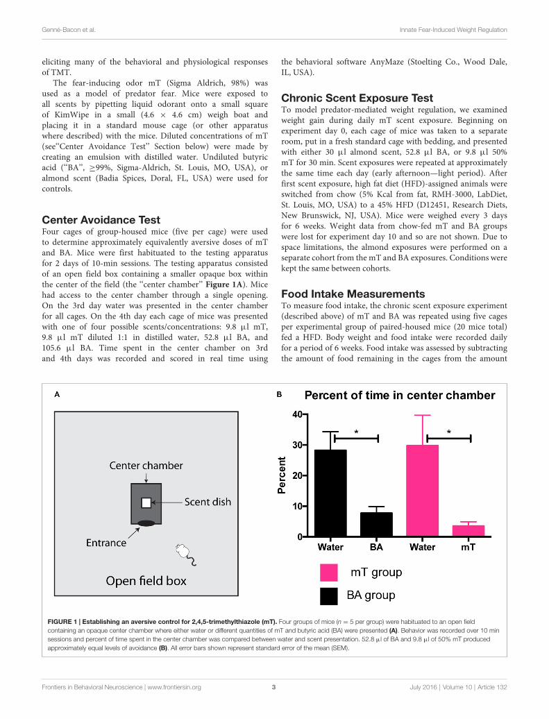
Center Avoidance TestFour cages of group-housed mice (five per cage) were usedto determine approximately equivalently aversive doses of mTand BA. Mice were first habituated to the testing apparatusfor 2 days of 10-min sessions. The testing apparatus consistedof an open field box containing a smaller opaque box withinthe center of the field (the ‘‘center chamber’’ Figure 1A). Micehad access to the center chamber through a single opening.On the 3rd day water was presented in the center chamberfor all cages. On the 4th day each cage of mice was presentedwith one of four possible scents/concentrations: 9.8 µl mT,9.8 µl mT diluted 1:1 in distilled water, 52.8 µl BA, and105.6 µl BA. Time spent in the center chamber on 3rdand 4th days was recorded and scored in real time using
the behavioral software AnyMaze (Stoelting Co., Wood Dale,IL, USA).
Chronic Scent Exposure TestTo model predator-mediated weight regulation, we examinedweight gain during daily mT scent exposure. Beginning onexperiment day 0, each cage of mice was taken to a separateroom, put in a fresh standard cage with bedding, and presentedwith either 30 µl almond scent, 52.8 µl BA, or 9.8 µl 50%mT for 30 min. Scent exposures were repeated at approximatelythe same time each day (early afternoon—light period). Afterfirst scent exposure, high fat diet (HFD)-assigned animals wereswitched from chow (5% Kcal from fat, RMH-3000, LabDiet,St. Louis, MO, USA) to a 45% HFD (D12451, Research Diets,New Brunswick, NJ, USA). Mice were weighed every 3 daysfor 6 weeks. Weight data from chow-fed mT and BA groupswere lost for experiment day 10 and so are not shown. Due tospace limitations, the almond exposures were performed on aseparate cohort from the mT and BA exposures. Conditions werekept the same between cohorts.
Food Intake MeasurementsTo measure food intake, the chronic scent exposure experiment(described above) of mT and BA was repeated using five cagesper experimental group of paired-housed mice (20 mice total)fed a HFD. Body weight and food intake were recorded dailyfor a period of 6 weeks. Food intake was assessed by subtractingthe amount of food remaining in the cages from the amount
FIGURE 1 | Establishing an aversive control for 2,4,5-trimethylthiazole (mT). Four groups of mice (n = 5 per group) were habituated to an open fieldcontaining an opaque center chamber where either water or different quantities of mT and butyric acid (BA) were presented (A). Behavior was recorded over 10 minsessions and percent of time spent in the center chamber was compared between water and scent presentation. 52.8 µl of BA and 9.8 µl of 50% mT producedapproximately equal levels of avoidance (B). All error bars shown represent standard error of the mean (SEM).
Frontiers in Behavioral Neuroscience | www.frontiersin.org 3 July 2016 | Volume 10 | Article 132

Genné-Bacon et al. Innate Fear-Induced Weight Regulation
provided to the animals the previous day. Food spillage wasminimal, and was assessed by visual inspection and accountedfor when necessary.
Locomotor Activity MeasurementsTo measure locomotor activity, one mouse from each cage fromthe above food intake experiment cohort was removed fromits home cage after 21 days of scent exposure and moved toindividual cages containing food, water, and bedding, and placedin locomotor boxes (Med Associates, St Albans, VT, USA) at theonset of the night cycle. Locomotor activity counts were definedas consecutive beam breaks, and recorded using Med-PC IVsoftware for a period of 22 h.
To study locomotor activity in more detail, the chronicexposure protocol was again repeated, using group-housed mice(five mice per cage, one cage per scent group). To obtain abaseline reading prior to beginning of scent exposure and HFD,mice were placed in locomotor boxes (as described above) atapproximately 3 pm and removed 22 h later. Following the startof daily scent exposure, locomotor activity was reassessed weeklyfor a period of 4 weeks. On the days of locomotor activity testing,scent exposure was conducted during the 2-h period prior to thebeginning of locomotor testing. Locomotor boxes were located ina separate room.
Corticosterone MeasurementsAcute ExposureGroup housed mice (five per cage) were habituated to thescent exposure setup for 2 days prior to sacrifice. On day ofsacrifice, animals were exposed to 52.8 µl BA, 9.8 µl 50% mT,or 30 µl almond scent for 30 min, and then sacrificed by rapiddecapitation and trunk blood was collected. Scent exposure andsacrifice was performed on separate days for different scentgroups, at the same time each day (early afternoon). Twohundred fifty microliter of trunk blood was immediately addedto 100 µl of 2% EDTA (Sigma-Aldrich) and spun for 15 minat 2000 g at room temperature; plasma was collected as thesupernatant.
Chronic ExposureAnimals from a separate cohort (n = 6 per scent group, twocages of three mice each per group) of the chronic scent exposureexperiment were sacrificed by rapid decapitation following thefinal scent exposure session (21 scent exposures total). Trunkblood was collected and processed as described above.
Plasma CORT MeasurementsPlasma corticosterone (CORT) was measured with AssayDesigns ELISA kits according to manufacturer’s protocol, asdescribed previously (Guarnieri et al., 2012). Briefly, 1 µl ofdiluted plasma (1:50), was compared to known concentrations.An OD reading at 405 nm with correction at 570 nm was taken, astandard curve was generated, and unknowns were extrapolatedusing the Prism statistical software (GraphPad Software, La Jolla,CA, USA).
Temperature MeasurementsTo evaluate changes in body temperature, mice from thelocomotor activity cohort (see above) were briefly anesthetizedwith isoflurane and implanted with IPTT-300 temperaturetransponders (Bio Medic Data Systems, Seaford, DE, USA).Temperatures were recorded with a non-invasive probe (DAS-5007 IPTT pocket scanner, Bio Medic Data Systems) prior todaily scent exposure.
Brown Adipose Tissue Dissection and qPCRTo search for evidence of metabolic changes, uncouplingprotein 1 (Ucp1) mRNA from brown adipose tissues (BAT)was measured in both chronic and acute scent-exposed mice.Chronic-exposure mice were sacrificed on the 36th day ofscent exposure. Acute exposure tissue collection timing (5 hpost-scent) was based on peak Ucp1 mRNA expression timingfrom cold challenge studies (Nedergaard and Cannon, 2013).Following sacrifice by rapid decapitation, whole interscapularBAT was collected and quickly frozen on dry ice and storedat −80◦C. RNA samples were purified from approximately0.1 g of frozen BAT using Trizol reagent (Invitrogen, Carlsbad,CA, USA); a Nanodrop ND-100 (Thermo Fisher Scientific,Waltham, MA, USA) was used to quantify purified totalRNA. cDNA samples were made from 1000 ng RNA usingSuperscript III Reverse Transcriptase kit and qPCR was carriedout using Taqman gene expression kits. The 2−∆∆Ct methodwas used for calculating fold change compared to TATA-binding protein (Tbp) control (Livak, 2001). Primer kitsused:
– Mm01244861_m1 Ucp1 TaqManr Gene Expression Assay(Thermo Fisher).
– Mm01277042_m1 Tbp TaqManr Gene Expression Assay(Thermo Fisher).
Brain Dissection and 1FosB MeasurementTo identify brain regions that may be involved in threatresponse, ∆FosB, a marker of neural plasticity, was measuredafter chronic scent exposure. Brains were collected after rapiddecapitation and quickly frozen and stored at −80◦C. Brainswere then briefly partially thawed and then sectioned usinga 1 mm brain block (BrainTree Scientific, Braintree, MA,USA) and each region was microdissected with guidancefrom the Mouse Brain Atlas (Paxinos and Franklin, 2004)using a scalpel, except for the nucleus accumbens (NAc)and Amygdala, where a 12 gauge circular punch was used.Protein extraction and analysis was carried out as describedpreviously (Sears et al., 2010). Briefly, frozen tissue sampleswere sonicated and then boiled for 20 min in a 1% SDSlysis buffer solution containing 1:100 protease inhibitor (SigmaP8340) and 1:100 phosphatase inhibitors (Sigma P5726 andP0044). Total protein concentration was assessed with a Piercebicinchoninic acid (BCA) protein assay (Thermo Fisher). Onesample (BA#1) from the medial hypothalamus (mHyp) hadinsufficient protein concentration and was excluded fromfurther analysis. Equal quantities of protein (55 µg) wereseparated by SDS polyacrylamide gel electrophoresis (Bio-Rad
Frontiers in Behavioral Neuroscience | www.frontiersin.org 4 July 2016 | Volume 10 | Article 132

Genné-Bacon et al. Innate Fear-Induced Weight Regulation
mini-PROTEAN TGX precast 12% polyacrylamide gels), andtransferred to nitrocellulose membranes (0.2 µm, Bio-Rad,Hercules, CA, USA). Themembranes were immunoblotted usinga 1:1000 dilution rabbit anti-FosB (Cell Signaling Technology,Danvers, MA, USA) and a 1:2000 dilution of mouse anti-β-actin (Cell Signaling). Antibody binding to FosB and ∆FosB wasvisualized by incubation with a 1:10,000 dilution of donkey anti-rabbit horseradish peroxidase-linked IgG (Vector Laboratories,Burlingame, CA, USA) and developed using Western LightningPlus ECL chemiluminescence HRP substrate (PerkinElmer,Waltham, MA, USA) and a ChemiDoc imager (Bio-Rad).Antibody binding to β-actin was revealed using the LI-COROdyssey quantitative infrared western blot detection system(IRDye 680LT secondary antibody, LI-COR, Lincoln, NE, USA).Band densities were analyzed using the Gel Analysis tool inImageJ.
FosB Immunofluorescence and Cell CountingImmunoflourescent labeling was used to visualize FosB/∆FosBprotein expression in hypothalamic nuclei. Chow fed micewere exposed to 36 days of daily scent exposure (six miceper scent group) as described above. Ninety minutes followingthe final scent exposure animals were sacrificed throughintracardial perfusion of 10% formalin, and brains werecollected, processed, and cut to 40 µm sections as describedpreviously (Land et al., 2014). Staining for FosB (rabbit-anti-FosB, Cell Signaling; 1:500) and secondary antibody (donkey-anti-rabbit Alexa 555; 1:500) was done in 3% normal donkeyserum and 0.3% Triton X-100. Tissue was visualized andimages were captured using a fluorescent microscope, asdescribed previously (Land et al., 2014). Images were takenat 10× magnification, with three 40 µm sections quantifiedper animal. FosB labeling was quantified using ImageJ onmatched sections by a researcher blinded to the experimentalgroups.
Statistics and AnalysesAll statistical analyses were carried out using the GraphPad Prism software package (version 6). Unpaired two-tailedt-tests were used for comparisons between two groups. One-way ANOVA was used for comparison between more thantwo groups. Two-way repeated measures ANOVAs wereused for comparisons of two or more groups over time.Tukey corrections for multiple comparisons were used to testfor significance among individual groups within ANOVAs.Significance threshold (alpha) was set at 0.05 for most tests withlevels of significance defined as follows: ∗p ≤ 0.05, ∗∗p ≤ 0.01,∗∗∗p ≤ 0.001. For ∆FosB measurements in the brain, theBenjamini–Hochberg procedure (with a false discovery rate setat Q = 0.2) was used to correct for multiple testing (adjustedalpha= 0.0286).
RESULTS
Establishing an Aversive Control for mTBoth mT and TMT, in addition to being innate fear-inducingodors, are also aversive and noxious scents. BA is commonly used
as a control for TMT due to its strong non-predator associatednoxious odor (e.g., Morrow et al., 2000; Endres et al., 2005).In order to determine doses of mT and BA that were similarlyaversive, we set up a test of avoidance of a center chamber insidean open field. Four separate groups of mice were exposed todifferent scent quantities (see Figure 1A), and time spent inthe center chamber was scored and compared to a no-scentcontrol session. We found 52.8 µl of undiluted BA producedapproximately equivalent levels of avoidance to 9.8 µl of a 50%mT dilution (see Figure 1B). To examine the fear-inducingpotential of these quantities of BA and mT, we conducted atest for freezing (a common behavioral threat response; seeSupplementary Methods). Although these quantities of BA andmT produce equal aversive behavior, we found mT significantlyelevated freezing (a threat response) compared to BA, as wellas to no-scent (water) and neutral scent controls (p < 0.0001).BA did not significantly elevate freezing compared to no-scent or neutral scent control (see Supplementary Figure 1).These quantities were used in all subsequent scent exposureexperiments.
Chronic mT Exposure Attenuates WeightGain in Both Low and High Fat DietsTo determine if mT exposure will affect weight gain in C57BL/6Jmice, six cages of group-housed mice (n = 5 per group)were exposed daily to mT, BA, or almond scent and givenad libitum access to either a 45% HFD or a standard chow(5% fat) diet. For the HFD-fed groups, there was a significanteffect of scent (p = 0.0024, F = 10.43; 2-way ANOVA), day(p< 0.0001, F= 366.8), and a significant interaction (p< 0.0001,F = 15.27). Animals exposed daily to mT gained significantlyless weight over time than mice exposed to almond scentcontrol (p < 0.05) or equally-aversive BA control (p < 0.01;see Figures 2A,B). The BA-exposed group was not significantlydifferent from the almond scent control in the HFD fed group. Inthe chow-fed group, differences were less robust. While a 2-wayANOVA revealed a significant main effect of scent (p = 0.0212,F = 5.401), as well as a significant interaction between scentand day (p < 0.0001, F = 4.290), testing for differences betweenindividual scent groups revealed only significant differencesbetween BA and mT groups (p < 0.05), while the almondgroup did not significantly differ from either (Figure 2C). Totalweight gained over the 6-week period was significantly lower inthe mT exposed animals compared to BA-exposed animals inboth diet groups (Figure 2D), with BA-exposed mice gainingan average of 3.32 g (±1.023 g) more than mT-exposed miceover the 6 week testing period for the HFD-fed group and1.54 g (±0.4512 g) for the chow-fed group. As differences inweight gain between scent groups were more robust in HFDanimals, we chose to focus primarily on this diet group for furtherexperiments.
mT Elevates Plasma Corticosterone AfterAcute and Repeated ExposuresTo determine approximate stress levels provoked by mT andBA, we measured plasma CORT, a common marker of stress
Frontiers in Behavioral Neuroscience | www.frontiersin.org 5 July 2016 | Volume 10 | Article 132

Genné-Bacon et al. Innate Fear-Induced Weight Regulation
FIGURE 2 | Chronic mT exposure attenuates weight gain in both low and high fat diets (HFDs). Six cages of group housed (n = 5 per cage) were exposeddaily to 9.8 µl mT, 52.8 µl BA, or 30 µl almond scent for 6 weeks while on a chow (5% fat) or high fat (45% fat, HFD) diet. (A) Weight gain on HFD presented as foldchange from baseline weight. (B) Total weight gained on HFD at the end of the 6-week experiment. mT-exposed mice gained significantly less weight than BA orAlmond exposed groups. (C) Weight gain on chow diet presented as fold change from baseline weight. (D) Total weight gained on chow diet. mT-exposed micegained significantly less weight than BA-exposed mice on a chow diet. (E) Plasma corticosterone (CORT) levels from trunk blood after a single 30-min exposure to9.8 µl mT, 52.8 µl BA, or 30 µl almond scent. (F) Plasma CORT levels from trunk blood after 3 weeks of daily scent exposure (and immediately after the final 30 minscent exposure). All error bars shown represent SEM.
in rodents, after acute (one time) and repeated scent exposure(21 days of daily exposure). Plasma CORT was significantlyelevated (p = 0.0026, F = 10.74; 1-way ANOVA) following asingle exposure to 9.8 µl 50% mT compared to 52.8 µl BA or30 µl almond scent (BA = 51.62 ng/ml, Almond = 49.48 ng/ml,mT= 104.46 ng/ml). Plasma CORT levels did not differ between
BA and almond scent exposures, suggesting differential stressresponses despite equivalent aversion levels (See Figure 2E).Plasma CORT levels after repeated daily exposure to mT(Figure 2F) were also elevated compared to BA (p = 0.0248,BA = 24.08 ng/ml, mT = 39.45 ng/ml), however plasmaCORT levels following repeated mT exposure were significantly
Frontiers in Behavioral Neuroscience | www.frontiersin.org 6 July 2016 | Volume 10 | Article 132

Genné-Bacon et al. Innate Fear-Induced Weight Regulation
FIGURE 3 | Weight attenuation following chronic mT exposure is notexplained by differences in food intake or locomotor activity. (A) Dailyfood intake per cage (two mice per cage, five cages per group) of mice onHFD and experiencing daily scent exposure. (B) Total food intake per groupover the 6-week experiment. (C) No differences were seen in food intakedespite a significant attenuation of weight gain in mT-exposed mice.(D) Locomotor activity for mice undergoing daily scent exposure. Mice wereplaced in new locomotor boxes with bedding at the onset of the night cycle(∼5 h after daily scent exposure). Locomotor counts measured as twoconsecutive beam breaks. Black and white bar represents night/day cycle.(E) Cumulative locomotor activity counts over 22-h periods for a new cohortof chronic scent-exposed mice tested for locomotor activity weekly.(F) Cumulative locomotor counts divided by a pre-scent baseline day. All errorbars shown represent SEM.
decreased (p = 0.0007, mean difference of 35.16 ± 7.01 ng/mlcomparing Figures 2E,F) compared to after only a singleexposure.
Chronic mT does not Significantly ChangeFood IntakeTo determine if the difference in weight gain between mT andBA-exposed mice could be driven by differences in food intake,an independent cohort of animals housed two to a cage (n = 5cages per experimental group and n = 10 animals per grouptotal) was put on 45% HFD and exposed daily to mT or BA fora period of 6 weeks with food intake and body weight measureddaily. As expected, mT-exposed animals gained significantly lessweight than BA-exposed animals when put on HFD (2-tailedt-test: p= 0.0028, Figure 3C). However, there was no significantdifference between groups for food intake over time (scent:p = 0.1179, F = 3.068; 2-way ANOVA, see Figure 3A) ortotal food eaten between groups (p = 0.1148, 2-tailed t test; seeFigure 3B).
Chronic mT Exposure does not AffectLocomotor ActivityTo determine if changes in physical activity could be drivingdifferences in weight gain, this cohort of animals was also testedfor locomotor activity over a 22-h period. Following daily scentexposure, one mouse from each of the double-housed cages wasplaced in a locomotor box and activity was recorded over a22-h period. No differences in the pattern of locomotion (asmeasured by consecutive beam breaks) were detected betweengroups (Figure 3D). To further assess potential differencesin physical activity, an independent cohort was subjected tothe chronic scent exposure test. Locomotor activity in thesemice was recorded for 22-h periods once per week, includinga baseline reading prior to any scent exposure. Again, nodifferences in the hourly circadian rhythm of locomotor activitywas observed between groups, nor were changes observed overtime (Figures 3E,F), despite a significant difference in weightgain between groups (BA: 6.86 g gained, mT: 3.6 g gained;p= 0.0008, 2-tailed t-test, see Supplementary Figure 2).
Chronic mT does not Change BodyTemperature or Uncoupling Protein mRNALevelsTo investigate a potential thermogenic effect of stresscontributing to differences in weight gain, we recorded dailytemperatures in chronic mT exposed animals. Transponderswere inserted subdermally between the scapulae and nomain effect of mT on temperature was detected (p = 0.4495,F = 0.6322; Figure 4A). We also tested a molecular marker ofthermogenesis: UCP1 mRNA from BAT using qPCR. A newchronic scent exposure cohort (with both HFD and chow-fed groups, n = 6 per treatment/food group) was sacrificedand BAT collected from each animal after 36 days of scentexposure. No differences in Ucp1 mRNA level were detectedbetween scent groups in either HFD (Figure 4B) or chow(Figure 4C) fed groups. We additionally measured Ucp1 mRNAfrom BAT following an acute (one-time) scent exposure innaive animals, but again did not see any changes in Ucp1mRNA expression between scent groups (See SupplementaryFigure 3).
Frontiers in Behavioral Neuroscience | www.frontiersin.org 7 July 2016 | Volume 10 | Article 132

Genné-Bacon et al. Innate Fear-Induced Weight Regulation
FIGURE 4 | No evidence for changes in thermogenesis after mT exposure. (A) Daily temperature readings relative to pre-scent baseline period. Mice wereimplanted with subdermal temperature transponders and exposed to daily scents while on a HFD. No main effect of scent was observed. Uncoupling protein 1(Ucp1) mRNA levels measured with qPCR from brown adipose tissue (BAT) for HFD (B) and chow (C) fed animals exposed to 36 days of daily scent. Fold changecompared with TATA-binding protein (Tbp) and calculated using the 2−∆∆Ct method. All error bars shown represent SEM.
Changes in a Marker of Neural PlasticityFollowing Repeated mT ExposureTo investigate which regions of the brain may be involved inthe response to chronic mT exposure, we measured ∆FosB, amarker of long-term neural plasticity, in a survey of severalbrain regions previously implicated in TMT and other predatorthreat responses. Following 6 weeks of mT or BA scent exposure(n = 5 per scent group), chow-fed mice were sacrificed andbrains sectioned in a brain block to 1 mm slices. Braintissue from the septal nucleus (LS), NAc (core and shell), bednucleus of the stria terminalis (BNST), anterior hypothalamus(AHN), mHyp (encompassing both the dorsomedial andventromedial hypothalamic nuclei), amygdala (basolateral), andwhole hippocampus was dissected (Supplementary Figure 4),isolated, and FosB and ∆FosB protein levels measured with
western blot. We found significant elevation of ∆FosB proteinbetween mT and BA exposed mice in the mHyp (p = 0.024,2-tailed t test), despite no change in total FosB (p = 0.256) orβ-actin (p = 0.692; Figure 5). No differences in FosB or ∆FosBprotein levels were detected in any other brain region.
To visualize which fine structure(s) in the mHyp maybe contributing to differential ∆FosB protein expression,immunofluorescent staining of total FosB/∆FosB protein in themHyp was carried out on perfused brain tissue from chronicallyscent exposed, chow fed mice. We found a significantly highernumber of FosB/∆FosB positive cells in the dorsomedialhypothalamus (DMH) in mT-exposed mice (2-tailed t-test,p = 0.0047, see Figure 6). Very little, if any, FosB/∆FosBexpression was observed in the ventromedial hypothalamus(VMH) in either scent group.
Frontiers in Behavioral Neuroscience | www.frontiersin.org 8 July 2016 | Volume 10 | Article 132

Genné-Bacon et al. Innate Fear-Induced Weight Regulation
FIGURE 5 | ∆∆∆FosB levels are increased in the medial hypothalamus (mHyp) following repeated mT exposure. Western blots for FosB and β-actin (loadingcontrol) were carried out on fresh-frozen brain tissue dissected from the mHyp (A). Protein bands were distinguished by size (B,C). Band intensities were calculatedusing ImageJ gel analysis tools. ∆FosB protein levels were significantly elevated (D) in the mHyp following 6 weeks of daily mT exposure compared to BA-exposedmice. Neither FosB levels (E), nor β-actin (F) differed between scent groups. All error bars shown represent SEM.
DISCUSSION
In this study we find significant attenuation of weight gainin C57BL/6J mice on both a high fat and low fat diet whenexposed daily to a threatening scent compared to a non-
threatening, but equally aversive, scent. We find no evidence forthe involvement of differences in food intake, locomotor activity,or brown adipose thermogenesis in mediating this differentialweight gain. We do find significant increases in a marker ofneural plasticity (∆FosB) in the mHyp, a brain region thought
Frontiers in Behavioral Neuroscience | www.frontiersin.org 9 July 2016 | Volume 10 | Article 132

Genné-Bacon et al. Innate Fear-Induced Weight Regulation
FIGURE 6 | FosB/∆∆∆FosB levels are increased specifically in the dorsomedial hypothalamus (DMH) following repeated mT exposure. FosB/∆FosBimmunofluorescent staining was carried out on perfused brain tissue from chronic scent-exposed mice. (A,B) Representative images of FosB/∆FosB expression inthe DMH in a BA or mT-exposed mouse brain, respectively. Dotted line represents DMH based on approximate brain region boundaries (C) from Paxinos andFranklin (2004); 3V, third ventricle, f, fornix, VMH, ventromedial hypothalamus, Arc, arcuate nucleus. Significantly more FosB/∆FosB positive cells were counted inthe DMH of chronically mT-exposed mice, compared to BA-exposed (D). Error bars shown represent SEM.
to be involved in processing predator threat (Canteras, 2002).This study provides additional evidence for the role of predationstimuli in regulation of body weight in rodents, and demonstratesthat predation-mediated weight reduction can be modeled usinginbred laboratory mice and a synthetic fear-inducing olfactorystimulus.
We find that mT-exposure attenuates weight gain in miceon both high and low fat diets, with HFD showing more robustand consistent changes. This is likely due to the relatively smalloverall weight gain seen in each scent group for the chow fedanimals. HFD provokes a larger change in weight, making thedifferences between scent groups more distinct. Additionallythis study uses only relatively young, male mice, and differentresponses may be seen in different age groups or sexes.
Previous studies have implicated alterations in food intakeand physical activity to explain predator-mediated weight
regulation (Tidhar et al., 2007;Monarca et al., 2015a,b). However,we find no evidence to support the contribution of thesebehaviors to the difference in weight with mT exposure. Itis possible for the differences we see in weight gain to bedue to very small changes in food intake that we lack thepower to detect. However, our results are in contrast to asimilar study with auditory cues, where changes in weightof C57BL/6J mice were clearly driven by differences in foodintake (Monarca et al., 2015a). This conflict is not withoutprecedent, as other studies have found no difference or mixeddifferences in food intake or foraging behavior with exposureto predator stimuli (Carlsen et al., 1999; Sundell et al., 2004). Itis likely that these discrepancies are due to differences betweenexperimental protocol, species used, or chosen stimulus. Forexample, diurnal timing of scent exposure may affect factorssuch as food intake, and presenting scent during the animals’
Frontiers in Behavioral Neuroscience | www.frontiersin.org 10 July 2016 | Volume 10 | Article 132

Genné-Bacon et al. Innate Fear-Induced Weight Regulation
natural feeding period could result in greater changes in foodintake than observed here. Further studies controlling for eachof these variables of predator stimulus presentation will help totease apart the complex factors that encompass predator threatresponse.
This study uses mT, a fear-inducing odor structurallyvery similar to the predator odor TMT, which is found inthe excretions of red fox anal glands (Vernet-Maury, 1980;Brechbühl et al., 2013a). We are able to recapitulate severalpreviously observed components of predator-mediated weightregulation (e.g., reduced body weight, increases in ∆FosBin the mHyp), while others (e.g., food intake changes) areabsent. It is possible that mT is not a close enough mimicof predator stimuli, and further studies should be done todirectly compare mT with TMT and with other predatorodors. There is also evidence that single-molecule odorants,such as TMT, do not elicit the full range of predator threatresponses in rodents (Staples et al., 2008; Pagani and Rosen,2009). It is possible that we are observing a very specificcomponent of the complete predator scent response repertoireand other responses require different stimuli. Several othersingle-molecule predator-associated odorants have now beendiscovered (Rosen et al., 2015), and these may elicit otherpredator-threat responses seen in response to natural predatorodors but not to TMT or mT.
The underlying mechanism for the mT-provoked weightresponse seen in our animals remains unclear. Because therewere no detectable changes observed in food intake orphysical activity, metabolic changes are suspected. Metabolicchanges have been previously implicated in weight responseto chronic stress (Michel et al., 2005; Harris, 2015), as wellas specifically to predator-mediated weight regulation (Tidharet al., 2007). We find equally aversive fear-inducing odorand non-fear inducing control odors elicit different CORTresponses, with mT-exposed mice showing elevated CORTlevels both acutely and chronically, indicating that our mT-exposed animals are under chronic stress (Tidhar et al.,2007).
Stress-induced hyperthermia has been proposed as theprimary metabolic component of stress-induced weight loss(Arase et al., 1988). BAT is the body’s primary source ofnon-shivering thermogenesis and known to be activated bycertain kinds of stress (Ricquier and Mory, 1984; Cannon andNedergaard, 2004). Because of this, we measured expressionof the primary heat-producing molecule of BAT, UCP1, fromintrascapular BAT through qPCR of Ucp1 mRNA in chronicallymT-exposed mice. We found no differences in BATUcp1mRNAexpression between scent groups after repeated mT exposure.Though mRNA expression is only a proxy-measurement for thethermogenic capacity of BAT (Nedergaard and Cannon, 2013),we also did not see corresponding changes in body temperaturefrom intrascapularly placed temperature transponders, leadingus to conclude that local thermogenesis is likely not a majorcontributor to mT-induced weight attenuation.
This study finds increases in expression of ∆FosB protein, apersistent transcription factor involved in long-term regulationof gene expression, in the mHyp following 6 weeks of daily
exposure to a fear-inducing, predator-like stimulus. This isconsistent with increases in FosB/∆FosB mRNA seen in themHyp of Bradt’s voles after repeated exposure to cat feces,as well as changes in FosB/∆FosB immunoreactivity in theVMH (dorsomedial region-VMHdm) of rats after repeatedexposure to cat fur (Staples et al., 2009; Hegab et al., 2014).To our knowledge, our study is the first to confirm it is∆FosB protein, a truncated isoform of FosB known to mediatelong-term neural plasticity, that is elevated in this region, asno differences were detected for FosB protein between scentgroups. Additionally, the comparatively long time course usedfor our study indicates that this region is highly resistant tohabituation and/or continues to be important to long-term threatresponses.
Interestingly, previous evidence for the mHyp’s rolespecifically in TMT-response (as opposed to other predatorodors) has been mostly negative. While the role of the VMHdmin response to natural predator odors such as cat fur iswell established (Canteras, 2002), multiple studies show noimmediate early gene activation in the VMHdm followingTMT exposure (Staples et al., 2008; Pérez-Gómez et al., 2015).Similarly, we observed low expression of FosB/∆FosB in theVMH (including the VMHdm) of chronically mT-exposedmice, but do see a robust increase in FosB/∆FosB in theDMH in mT-exposed mice. Since protein expression fromour western blot shows no differences in FosB in the mHyp,the increased number of FosB+ immunofluorescent cells inthe DMH is likely driven by increases in ∆FosB. The DMHis a region known to be involved in stress response and bodyweight regulation (DiMicco et al., 2002; DiMicco and Zaretsky,2007), and increases in FosB/∆FosB protein expression havebeen previously observed in the DMH following repeatedrestraint stress (Flak et al., 2012). More specifically, the DMHmediates the metabolic, but not the anorectic, responses toleptin (Rezai-Zadeh et al., 2014). This is consistent with ourfinding that chronic mT exposure does not attenuate weight gainthrough changes in food intake, and suggests that changes inDMH neural activity may influence body weight via a metabolicmechanism.
In summary, the current data add to the complex picture ofpredator threat response andweight regulation in preymammals.We demonstrate that C57BL/6J laboratory mice will regulatetheir body weight in response to fear-inducing odor, but that thisresponse, at least for mT, is likely not mediated through changesin food intake. Interestingly, we find that this weight regulationoccurs regardless of dietary fat content, demonstrating the broadinfluence of innate threat in regulation of body weight even inthe context of obesogenic diet. More study of this ecologicallyrelevant and obesity-preventing neurophysiological pathway isimportant for gaining a broader picture of the biology of weightregulation.
AUTHOR CONTRIBUTIONS
EAG-B conceived, designed, and conducted experiments,analyzed data, interpreted results, and wrote the manuscript.JRT conducted experiments and edited manuscript. RJD helped
Frontiers in Behavioral Neuroscience | www.frontiersin.org 11 July 2016 | Volume 10 | Article 132

Genné-Bacon et al. Innate Fear-Induced Weight Regulation
conceive and design experiments, interpreted results, and revisedmanuscript. All authors approved the submitted version.
FUNDING
National Science Foundation Graduate Research Fellowship toEAG-B.
ACKNOWLEDGMENTS
We thank members of the DiLeone lab for help and technicaladvice. We also thank Carol Gianessi for laboratory help
when needed and Dr. Jane Taylor for advice and useof testing boxes. We also thank Dr. Elena Gracheva foruse of temperature transponders, Dr. Elise Jeffery fordemonstrating BAT dissections, and Dr. Louise Guardfor her help with understanding predator odor chemicalstructures.
SUPPLEMENTARY MATERIAL
The Supplementary Material for this article can be found onlineat: http://journal.frontiersin.org/article/10.3389/fnbeh.2016.00132/abstract
REFERENCES
Arase, K., York, D. A., Shimizu, H., Shargill, N., and Bray, G. A. (1988).Effects of corticotropin-releasing factor on food intake and brownadipose tissue thermogenesis in rats. Am. J. Physiol. 255, E255–E259.
Brechbühl, J., Moine, F., and Broillet, M.-C. (2013a). Mouse Gruenebergganglion neurons share molecular and functional features with C. elegansamphid neurons. Front. Behav. Neurosci. 7:193. doi: 10.3389/fnbeh.2013.00193
Brechbühl, J., Moine, F., Klaey, M., Nenniger-Tosato, M., Hurni, N., Sporkert, F.,et al. (2013b). Mouse alarm pheromone shares structural similarity withpredator scents. Proc. Natl. Acad. Sci. U S A 110, 4762–4767. doi: 10.1073/pnas.1214249110
Cannon, B., and Nedergaard, J. (2004). Brown adipose tissue: function andphysiological significance. Physiol. Rev. 84, 277–359. doi: 10.1152/physrev.00015.2003
Canteras, N. S. (2002). The medial hypothalamic defensive system: hodologicalorganization and functional implications. Pharmacol. Biochem. Behav. 71,481–491. doi: 10.1016/s0091-3057(01)00685-2
Carlsen, M., Lodal, J., Leirs, H., and Secher Jensen, T. (1999). The effect ofpredationrisk on body weight in the field vole, Microtus agrestis. Oikos 87,277–285. doi: 10.2307/3546742
Cohen, H., Geva, A. B., Matar, M. A., Zohar, J., and Kaplan, Z. (2008). Post-traumatic stress behavioural responses in inbred mouse strains: can geneticpredisposition explain phenotypic vulnerability? Int. J. Neuropsychopharmacol.11, 331–349. doi: 10.1017/s1461145707007912
DiMicco, J. A., Samuels, B. C., Zaretskaia, M. V., and Zaretsky, D. V. (2002).The dorsomedial hypothalamus and the response to stress: part renaissance,part revolution. Pharmacol. Biochem. Behav. 71, 469–480. doi: 10.1016/s0091-3057(01)00689-x
DiMicco, J. A., and Zaretsky, D. V. (2007). The dorsomedial hypothalamus: a newplayer in thermoregulation. Am. J. Physiol. Regul. Integr. Comp. Physiol. 292,R47–R63. doi: 10.1152/ajpregu.00498.2006
Doty, R. (2012). Mammalian Olfaction, Reproductive Processes, and Behavior.Amsterdam: Elsevier.
Endres, T., Apfelbach, R., and Fendt,M. (2005). Behavioral changes induced in ratsby exposure to trimethylthiazoline, a component of fox odor. Behav. Neurosci.119, 1004–1010. doi: 10.1037/0735-7044.119.4.1004
Flak, J. N., Solomon, M. B., Jankord, R., Krause, E. G., and Herman, J. P. (2012).Identification of chronic stress-activated regions reveals a potential recruitedcircuit in rat brain. Eur. J. Neurosci. 36, 2547–2555. doi: 10.1111/j.1460-9568.2012.08161.x
Gentle, L. K., and Gosler, A. G. (2001). Fat reserves and perceived predation risk inthe great tit, Parus major. Proc. Biol. Sci. 268, 487–491. doi: 10.1098/rspb.2000.1405
Gosler, A. G., Greenwood, J., and Perrins, C. (1995). Predation risk and the cost ofbeing fat. Nature 377, 621–623. doi: 10.1038/377621a0
Guarnieri, D. J., Brayton, C. E., Richards, S. M., Maldonado-Aviles, J., Trinko, J. R.,Nelson, J., et al. (2012). Gene profiling reveals a role for stress hormones inthe molecular and behavioral response to food restriction. Biol. Psychiatry 71,358–365. doi: 10.1016/j.biopsych.2011.06.028
Harris, R. B. S. (2015). Chronic and acute effects of stress on energy balance: arethere appropriate animal models? Am. J. Physiol. Regul. Integr. Comp. Physiol.308, R250–R265. doi: 10.1152/ajpregu.00361.2014
Hedenstrom, A. (1992). Flight performance in relation to fuel load in birds.J. Theor. Biol. 158, 535–537. doi: 10.1016/s0022-5193(05)80714-3
Hegab, I. M., Shang, G., Ye, M., Jin, Y., Wang, A., Yin, B., et al. (2014). Defensiveresponses of Brandt’s voles (Lasiopodomys brandtii) to chronic predatorystress. Physiol. Behav. 126, 1–7. doi: 10.1016/j.physbeh.2013.12.001
Howerton, C. L., and Bale, T. L. (2014). Targeted placental deletion ofOGT recapitulates the prenatal stress phenotype including hypothalamicmitochondrial dysfunction. Proc. Natl. Acad. Sci. U S A 111, 9639–9644. doi: 10.1073/pnas.1401203111
Ihara, S., Yoshikawa, K., and Touhara, K. (2013). Chemosensory signals and theirreceptors in the olfactory neural system. Neuroscience 254, 45–60. doi: 10.1016/j.neuroscience.2013.08.063
Isosaka, T., Matsuo, T., Yamaguchi, T., Funabiki, K., Nakanishi, S.,Kobayakawa, R., et al. (2015). Htr2a-Expressing Cells in the Central AmygdalaControl the Hierarchy between Innate and Learned Fear. Cell 163, 1153–1164.doi: 10.1016/j.cell.2015.10.047
Janitzky, K., D’Hanis, W., Kröber, A., and Schwegler, H. (2015). TMT predatorodor activated neural circuit in C57BL/6J mice indicates TMT-stress as asuitable model for uncontrollable intense stress. Brain Res. 1599, 1–8. doi: 10.1016/j.brainres.2014.12.030
Kobayakawa, K., and Kobayakawa, R. (2011). Animal repellents. US Patent WO2011096575A1.
Land, B. B., Narayanan, N. S., Liu, R.-J., Gianessi, C. A., Brayton, C. E.,Grimaldi, D. M., et al. (2014). Medial prefrontal D1 dopamine neurons controlfood intake. Nat. Neurosci. 17, 248–253. doi: 10.1038/nn.3625
Lilliendahl, K. (1997). The effect of predator presence on body massin captive greenfinches. Anim. Behav. 53, 75–81. doi: 10.1006/anbe.1996.0279
Lima, S. L. (1986). Predation risk and unpredictable feeding conditions:determinants of body-mass in birds. Ecology 67, 377–385. doi: 10.2307/1938580
Lima, S. L., and Bednekoff, P. A. (1999). Temporal variation in danger drivesantipredator behavior: the predation risk allocation hypothesis. Am. Nat. 153,649–659. doi: 10.1086/303202
Livak, K. (2001). Analysis of relative gene expression data using real-timequantitative PCR and the 2−∆∆CT method. Methods 25, 402–408. doi: 10.1006/meth.2001.1262
McNamara, J. M., Barta, Z., Houston, A. I., and Race, P. (2005). A theoreticalinvestigation of the effect of predators on foraging behaviour and energyreserves. Proc. Biol. Sci. 272, 929–934. doi: 10.1098/rspb.2004.3037
Michel, C., Duclos, M., Cabanac, M., and Richard, D. (2005). Chronic stressreduces body fat content in both obesity-prone and obesity-resistant strains ofmice. Horm. Behav. 48, 172–179. doi: 10.1016/j.yhbeh.2005.02.004
Monarca, R. I., da Luz Mathias, M., Wang, D., and Speakman, J. R. (2015a).Predation risk modulates diet-induced obesity in male C57BL/6 mice. Obesity23, 2059–2065. doi: 10.1002/oby.21193
Monarca, R. I., Mathias, M. D. L., and Speakman, J. R. (2015b). Behavioural andphysiological responses of wood mice (Apodemus sylvaticus) to experimentalmanipulations of predation and starvation risk. Physiol. Behav. 149, 331–339.doi: 10.1016/j.physbeh.2015.06.037
Frontiers in Behavioral Neuroscience | www.frontiersin.org 12 July 2016 | Volume 10 | Article 132

Genné-Bacon et al. Innate Fear-Induced Weight Regulation
Morrow, B. A., Redmond, A. J., Roth, R. H., and Elsworth, J. D. (2000). Thepredator odor, TMT, displays a unique, stress-like pattern of dopaminergicand endocrinological activation in the rat. Brain Res. 864, 146–151. doi: 10.1016/s0006-8993(00)02174-0
Mueller, B. R., and Bale, T. L. (2008). Sex-specific programming of offspringemotionality after stress early in pregnancy. J. Neurosci. 28, 9055–9065. doi: 10.1523/JNEUROSCI.1424-08.2008
Nedergaard, J., and Cannon, B. (2013). UCP1 mRNA does not produceheat. Biochim. Biophys. Acta 1831, 943–949. doi: 10.1016/j.bbalip.2013.01.009
Pagani, J. H., and Rosen, J. B. (2009). The medial hypothalamic defensive circuitand 2,5-dihydro-2,4,5-trimethylthiazoline (TMT) induced fear: comparison ofelectrolytic and neurotoxic lesions. Brain Res. 1286, 133–146. doi: 10.1016/j.brainres.2009.06.062
Paxinos, G., and Franklin, K. B. J. (2004). The Mouse Brain in StereotaxicCoordinates. Amsterdam: Gulf Professional Publishing.
Pérez-Gómez, A., Bleymehl, K., Stein, B., Pyrski, M., Birnbaumer, L.,Munger, S. D., et al. (2015). Innate predator odor aversion driven by parallelolfactory subsystems that converge in the ventromedial hypothalamus. Curr.Biol. 25, 1340–1346. doi: 10.1016/j.cub.2015.03.026
Rezai-Zadeh, K., Yu, S., Jiang, Y., Laque, A., Schwartzenburg, C., Morrison, C. D.,et al. (2014). Leptin receptor neurons in the dorsomedial hypothalamus are keyregulators of energy expenditure and body weight, but not food intake. Mol.Metab. 3, 681–693. doi: 10.1016/j.molmet.2014.07.008
Ricquier, D., and Mory, G. (1984). Factors affecting brown adipose tissue activityin animals and man. Clin. Endocrinol. Metab. 13, 501–520. doi: 10.1016/s0300-595x(84)80035-3
Rosen, J. B., Asok, A., and Chakraborty, T. (2015). The smell of fear: innatethreat of 2,5-dihydro-2,4,5-trimethylthiazoline, a single molecule componentof a predator odor. Front. Neurosci. 9:292. doi: 10.3389/fnins.2015.00292
Sears, R. M., Liu, R.-J., Narayanan, N. S., Sharf, R., Yeckel, M. F., Laubach, M.,et al. (2010). Regulation of nucleus accumbens activity by the hypothalamicneuropeptide melanin-concentrating hormone. J. Neurosci. 30, 8263–8273.doi: 10.1523/JNEUROSCI.5858-09.2010
Speakman, J. R. (2007). A nonadaptive scenario explaining the geneticpredisposition to obesity: the ‘‘predation release’’ hypothesis. Cell Metab. 6,5–12. doi: 10.1016/j.cmet.2007.06.004
Staples, L. G., McGregor, I. S., Apfelbach, R., and Hunt, G. E. (2008). Catodor, but not trimethylthiazoline (fox odor), activates accessory olfactory anddefense-related brain regions in rats.Neuroscience 151, 937–947. doi: 10.1016/j.neuroscience.2007.11.039
Staples, L. G., McGregor, I. S., and Hunt, G. E. (2009). Long-lasting FosB/∆FosBimmunoreactivity in the rat brain after repeated cat odor exposure. Neurosci.Lett. 462, 157–161. doi: 10.1016/j.neulet.2009.06.069
Sundell, J., Dudek, D., Klemme, I., Koivisto, E., Pusenius, J., and Yl nen, H. (2004).Variation in predation risk and vole feeding behaviour: a field test of the riskallocation hypothesis.Oecologia 139, 157–162. doi: 10.1007/s00442-004-1490-x
Sundell, J., and Norrdahl, K. (2002). Body size-dependent refuges in voles: analternative explanation of the Chitty effect. Ann. Zool. Fennici. 39, 325–333.http://www.jstor.org/stable/23735831.
Tidhar, W. L., Bonier, F., and Speakman, J. R. (2007). Sex- and concentration-dependent effects of predator feces on seasonal regulation of body mass in thebank vole Clethrionomys glareolus. Horm. Behav. 52, 436–444. doi: 10.1016/j.yhbeh.2007.06.009
Vernet-Maury, E. (1980). ‘‘Trimethyl-thiazoline in fox feces: a natural alarmingsubstance for the rat,’’ in Olfaction Taste, Vol. VII (Washington, D.C.: IRLPress).
Vernet-Maury, E., Polak, E. H., and Demael, A. (1984). Structure-activityrelationship of stress-inducing odorants in the rat. J. Chem. Ecol. 10, 1007–1018.doi: 10.1007/BF00987509
Wallace, K. J., and Rosen, J. B. (2000). Predator odor as an unconditioned fearstimulus in rats: elicitation of freezing by trimethylthiazoline, a component offox feces. Behav. Neurosci. 114, 912–922. doi: 10.1037/0735-7044.114.5.912
Conflict of Interest Statement: The authors declare that the research wasconducted in the absence of any commercial or financial relationships that couldbe construed as a potential conflict of interest.
Copyright © 2016 Genné-Bacon, Trinko and DiLeone. This is an open-access articledistributed under the terms of the Creative Commons Attribution License (CC BY).The use, distribution and reproduction in other forums is permitted, provided theoriginal author(s) or licensor are credited and that the original publication in thisjournal is cited, in accordance with accepted academic practice. No use, distributionor reproduction is permitted which does not comply with these terms.
Frontiers in Behavioral Neuroscience | www.frontiersin.org 13 July 2016 | Volume 10 | Article 132