infectome of medicago truncatula changes in cell cycle ... · pocket formation requires localized...
TRANSCRIPT
LARGE-SCALE BIOLOGY ARTICLE
The Root Hair “Infectome” of Medicago truncatula UncoversChanges in Cell Cycle Genes and Reveals a Requirement forAuxin Signaling in Rhizobial InfectionW OPEN
Andrew Breakspear,a,1 Chengwu Liu,a,1 Sonali Roy,a Nicola Stacey,a Christian Rogers,a Martin Trick,b
Giulia Morieri,c Kirankumar S. Mysore,d Jiangqi Wen,d Giles E.D. Oldroyd,a J. Allan Downie,c
and Jeremy D. Murraya,2
a Cell and Developmental Biology, John Innes Centre, Norwich NR4 7UH, United KingdombComputational and Systems Biology, John Innes Centre, Norwich NR4 7UH, United KingdomcDepartment of Molecular Microbiology, John Innes Centre, Norwich NR4 7UH, United KingdomdDivision of Plant Biology, The Samuel Roberts Noble Foundation, Ardmore, Oklahoma 73401
ORCID ID: 0000-0003-3000-9199 (J.D.M.)
Nitrogen-fixing rhizobia colonize legume roots via plant-made intracellular infection threads. Genetics has identified somegenes involved but has not provided sufficient detail to understand requirements for infection thread development. Therefore,we transcriptionally profiled Medicago truncatula root hairs prior to and during the initial stages of infection. This revealedchanges in the responses to plant hormones, most notably auxin, strigolactone, gibberellic acid, and brassinosteroids.Several auxin responsive genes, including the ortholog of Arabidopsis thaliana Auxin Response Factor 16, were induced atinfection sites and in nodule primordia, and mutation of ARF16a reduced rhizobial infection. Associated with the induction ofauxin signaling genes, there was increased expression of cell cycle genes including an A-type cyclin and a subunit of theanaphase promoting complex. There was also induction of several chalcone O-methyltransferases involved in the synthesisof an inducer of Sinorhizobium meliloti nod genes, as well as a gene associated with Nod factor degradation, suggesting bothpositive and negative feedback loops that control Nod factor levels during rhizobial infection. We conclude that the onset ofinfection is associated with reactivation of the cell cycle as well as increased expression of genes required for hormone andflavonoid biosynthesis and that the regulation of auxin signaling is necessary for initiation of rhizobial infection threads.
INTRODUCTION
Nodulation in legumes requires two coordinated programs, rhizo-bial infection and nodule organogenesis, that converge to releaserhizobia into developing nodule cells where they fix nitrogen. Inmost legumes, rhizobia enter roots through plant-made infectionthreads, tubular invaginations, which initiate on growing root hairs.Following rhizobial entrapment within root hairs, infection threadsprovide a conduit through which rhizobia can colonize the rootcortex (Oldroyd et al., 2011). These structures are unique to nitrogen-fixing symbioses and represent a significant innovation in cel-lular growth and differentiation, but we know very little abouthow they are formed.
Rhizobia attach to the flank of the root hairs, and subsequentanisotropic growth of the root hair forms a tight curl enclosingrhizobia in an infection pocket formed by the apposed cell walls.
The nucleus then doubles in size and moves to a central positionin the cell (Dart, 1974) and a broad cytoplasmic bridge formsbetween the site where the infection thread will initiate and thenucleus (Timmers et al., 1999; Fournier et al., 2008). Once initi-ated, the infection thread grows intermittently and is colonizedby dividing rhizobia (Fournier et al., 2008). As the growing infectionthread nears the base of the root hair, the cell wall starts to weakenat the junction of the cytoplasmic bridge. The underlying outercortical cell then undergoes a similar series of events whereby thenucleus occupies a central position within a large anticlinal cyto-plasmic bridge that is aligned with the incoming infection thread(van Brussel et al., 1992). The infection thread progresses throughthis cell-cell junction and continues growing in this manner throughthe outer cortical cell layers. When it reaches the cells of the na-scent nodule, the infection thread ramifies, extending into thenodule cells, where rhizobia are taken up from the tips of the in-fection threads by endocytosis to form organelle-like structurescalled symbiosomes.Rhizobial infection and nodule organogenesis both require the
production of lipochitooligosaccharide Nod factors that are pro-duced by rhizobia in response to plant flavonoids and relatedcompounds (Peters et al., 1986; Subramanian et al., 2006). Infectionpocket formation requires localized release of Nod factors (vanBatenburg, et al., 1986; Esseling et al., 2003) and the changes in cellarchitecture that precede infection thread formation result from Nod
1 These authors contributed equally to this work.2 Address correspondence to [email protected] author responsible for distribution of materials integral to the findingspresented in this article in accordance with the policy described in theInstructions for Authors (www.plantcell.org) is: Jeremy D. Murray ([email protected]).W Online version contains Web-only data.OPENArticles can be viewed online without a subscription.www.plantcell.org/cgi/doi/10.1105/tpc.114.133496
The Plant Cell, Vol. 26: 4680–4701, December 2014, www.plantcell.org ã 2014 American Society of Plant Biologists. All rights reserved.
factor-induced rearrangements in actin filaments (Crdenas et al.,1998; de Ruijter et al., 1999). Perception of Nod factors inMedicagotruncatula and Lotus japonicus is mediated by the LysM receptor-like kinases NFP and LYK3 (Radutoiu et al., 2003; Geurts et al.,2005; Smit et al., 2007). Activation of the nodulation signalingpathway to induce gene expression requires several transcriptionfactors, including Nodule Inception (NIN), ERF Required forNodulation1 (ERN1), and the GRAS transcription factors Nodu-lation Signaling Pathway1 (NSP1) and NSP2 (Schauser et al.,1999; Oldroyd and Long, 2003; Kaló et al., 2005; Smit et al., 2005;Heckmann et al., 2006; Marsh et al., 2007; Middleton et al., 2007),all of which are involved in the formation of infection threads butnot root hair deformation. Some components induced have rolesin actin nucleation (Yokota et al., 2009; Miyahara et al., 2010;Hossain et al., 2012). NIN, NSP1, and NSP2 act in a signalingpathway required for both rhizobial colonization and nodule or-ganogenesis in L. japonicus (Madsen et al., 2010). NIN is requiredfor the expression of the flotillin encoding genes FLOT2 andFLOT4 in M. truncatula (Haney and Long, 2010) as well asNODULATION PECTATE LYASE1 (NPL1) in L. japonicus (Xieet al., 2012), which are necessary for bacterial infection. NIN isalso required for the induction of two CCAAT-box transcriptionfactors Lj-NFYA1 and Lj-NFYB1 in L. japonicus, which are requiredfor nodule organogenesis (Soyano et al., 2013). Mt-NFYA1 (theortholog of Lj-NFYA1, previously known as HAP2.1) also has a rolein infection in Medicago (Combier et al., 2006, 2008; Laporte et al.,2014).
In Medicago, LUMPY INFECTIONS, encoding an E3 ubiquitinligase is required for infection thread development and isneeded for the expression of the infection genes RHIZOBIUM-DIRECTED POLAR GROWTH (RPG; Arrighi et al., 2008; Kisset al., 2009) and VAPYRIN, a protein of unknown function also re-quired for mycorrhization (Pumplin et al., 2010; Murray et al., 2011).In soybean (Glycine max), knockdown of Nucleolar/Mitochondrialprotein involved in Nodulation a impaired infection (Libault et al.,2011). Other legume mutants that are impaired in the sensing ofethylene (sickle) or cytokinin (hyperinfected1) have hyperinfectionphenotypes (Penmetsa and Cook, 1997; Murray et al., 2007).However, the relationship of these downstream components re-quired for infection and the transcription factors that control themis not known.
From the first efforts at isolation of proteins and RNA from puri-fied root hairs (Röhm and Werner, 1987; Covitz et al., 1998; Ramosand Bisseling, 2003; Sauviac et al., 2005) to the recent application ofdifferent -omics technologies in soybean (Brechenmacher et al.,2009, 2010; Libault et al., 2010), the advantages of isolating roothairs have been recognized. We analyzed the root hair tran-scriptome of M. truncatula during rhizobial infection, studying geneexpression over a time course. By comparing gene induction byNod factors and Sinorhizobium meliloti in the wild type and thehyperinfected mutant sickle (skl), we identified infection-relatedgenes and processes previously undetected in whole-root studiesor in forward genetic analyses. Medicago spp have served asmodels for phenylpropanoid metabolism for over two decades, andwe extend these studies providing new insights into rhizobial in-fection. In addition, we identified other processes that are regulatedin root hairs undergoing rhizobial colonization. Among them arechanges in the cell cycle and in the plant hormones strigolactone
(SL), gibberellic acid (GA), brassinosteroids (BRs), and auxin, and wereveal a novel role for auxin signaling in rhizobial infection.
RESULTS
The Infectome
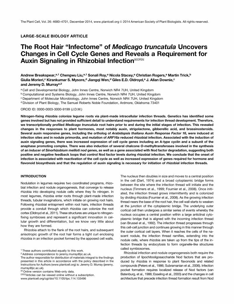
Our main goal was the identification of genes responsible for thedevelopmental changes occurring during rhizobial infection. Toaccomplish this, wild-type roots were inoculated with S. melilotiSm1021 or an isogenic mutant (SL44; S. meliloti nodDD1ABC)that is completely defective for Nod factor signaling, ensuringthat the effects we detect are specific for Nod factor signalingrather than other rhizobial-root interactions. We first monitoredthe progression of rhizobial infection under our selected growthconditions. One day postinoculation (dpi), no curled root hairs orinfections were evident; 3 dpi, root hair curling and microcolonyformation was observed but no infection threads were evident;5 dpi, many infection threads had initiated but few had extendedinto the outer cortex (Figure 1A).To enhance identification of infection genes, we also profiled
the skl mutant, which, due to a defect in ethylene signaling, hadat 5 dpi a 6-fold increase in rhizobial infections relative to the wildtype (P value = 0.00004, Student’s t test). We also analyzed wild-type root hairs 24 h after addition of 10 nM Nod factor, a concen-tration sufficient to induce both calcium spiking and a calcium influxin root hairs that is associated with rhizobial infection (Morieri et al.,2013). A file with all data from individual replicates normalizedacross treatments is provided in Supplemental Data Set 1.RNA extracted from root hairs from the different treatment
groups at specific time points was analyzed using microarrays(GeneChip Medicago Genome Array, first version). GeneChiphybridizations were performed on three biological replicates pertreatment (response of wild-type seedlings to S. meliloti strainSm1021 (wild type) versus strain SL44 (nodDD1ABC). All dataare available through the Medicago Gene Expression Atlas server(http://mtgea.noble.org/v3/). Comparing the responses of wild-type seedlings to S. meliloti wild type and SL44, we observeda significant and at least 2-fold increase in expression of 158, 161,and 339 genes 1, 3, and 5 dpi, respectively; 79 of these geneswere induced at all three time points (Figure 1B; see Methods fordescription of normalization and statistical analysis). Comparisonof the data from root hairs (5 dpi) to data from emerging noduleprimordia (Benedito et al., 2008) detected 230 genes that couldnot be identified in excised root tissue containing nodule pri-mordia (Supplemental Figure 1).In wild-type seedlings, 93% of the genes that responded to
rhizobial infection had increased expression (404 induced, 30 re-pressed). A larger proportion of the responding genes were re-pressed in skl (Figure 1E). Responses to Nod factors were morebalanced with almost as many genes being repressed as induced(Figure 1E). All data showing significant changes following in-oculation or Nod factor treatment are provided in SupplementalData Set 2.About 61% (96/158) of the genes induced 1 dpi with S. meliloti
1021 were also induced by Nod factors (Figure 1D), indicatingthat most of the gene expression changes that occur during
Auxin Signaling in Infection 4681
preinfection can be accounted for by Nod factors alone. Evenmore Nod factor-induced genes were also induced at the onsetof infection (5 dpi), but at this time point, about twice as manygenes were induced compared with the preinfection time points,so the proportion of Nod factor-responsive genes was reduced(41%, 140/339).
The skl Mutation Increases Detection ofInfection-Related Genes
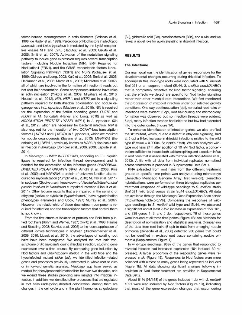
At 5 dpi, 87% (295/339) of the genes upregulated in the wildtype by strain 1021 were induced in the skl mutant, but an ad-ditional 453 upregulated genes were identified (Figure 1C). Genesknown to be required for, or associated with infection, revealedthat they were on average 2.2-fold more strongly induced in sklthan in the wild type (Figure 2) (e.g., NIN expression was induced26- and 117-fold in the wild type and skl, respectively). Of the 414genes induced by S. meliloti in the wild type (Figure 1B), 96% hadenhanced expression in the skl mutant, including the cytokininreceptor CRE1 (Figure 2). Most of the 410 additional genes dis-covered in skl also appeared to be slightly increased in the wildtype relative to the controls, although they did not show statisti-cally significant differences. Several (47) genes were induced by
S. meliloti 1021 in skl and in the wild type by Nod factor butwere not significantly induced by S. meliloti in the wild type atany stage; this may be a consequence of a stronger nodu-lation signaling response in skl compared with the wild type.This global effect of ethylene perception on infection-relatedgene expression is consistent with the ability of this hormoneto suppress Nod factor-induced calcium oscillations (Oldroydet al., 2001).
Highlights from the Infectome
In addition to the known infection-related genes mentionedabove (Figure 2), several other symbiosis-related genes wereseen to be regulated by S. meliloti 1021 and/or Nod factor. TheLysM receptor kinase genes LYK10 and LYE2 were induced byS. meliloti 1021 (Supplemental Figure 2B), whereas three LysM-RLKs (including the Nod factor receptors NFP and LYK3) andtwo LYK-related (LYR) genes were repressed. Extracellularperoxidases that produce reactive oxygen species in the apoplastare induced early during rhizobial colonization (Cook et al., 1995;Ramu et al., 2002) and are predicted to be secreted to the lumen ofthe developing infection thread where they may promote hardeningof thematrix (Wisniewski et al., 2000; Passardi et al., 2004). We found
Figure 1. Experimental Overview of Transcriptome Analysis Performed and Numbers of Significantly Regulated Genes Identified.
(A) Infection time course of wild-type (A17) M. truncatula root hairs from seedlings inoculated with S. meliloti expressing lacZ. Major developmentalmilestones of infection: 1 dpi represents preinfection before visible root hair curling, at 3 dpi microcolonies had formed within curled root hairs, and at5 dpi elongating infection threads were seen. Bar = 20 mm.(B) Genes upregulated in the wild type 1, 3, and 5 dpi with Sm1021.(C) Genes upregulated 5 dpi with Sm1021 in the wild type and in the hyperinfected skl mutant.(D) Genes upregulated in wild-type 1 dpi with Sm1021 and 1 d post-treatment with Nod factor.(E) Summary of up- and downregulated genes for each treatment. dpt, days post-treatment.Genes with significant regulation relative to control experiments are shown (>2-fold change; P < 0.05).
4682 The Plant Cell
10 peroxidase genes, including Rhizobium-Induced Peroxidase1(RIP1; Cook et al., 1995; Ramu et al., 2002) that were induced inroot hairs by both S. meliloti and Nod factors (SupplementalFigure 2A). Eight of these had predicted secretion signal pep-tides, suggesting increased production of reactive oxygenspecies in the lumen of the infection thread and/or aroundroot hairs. Several other genes that had not been previouslylinked to infection were identified. The sucrose synthasegene SUCS1 (Mtr.22018.1.S1_s_at: Medtr4g124660), whichis expressed in nodules and required for nitrogen fixation(Baier et al., 2007) and is required for colonization by ar-buscular mycorrhizal fungi (Hohnjec et al., 1999; Baier et al.,2010), had increased expression in skl after S. meliloti in-oculation. SWEET13, which encodes a sugar transporter(Chen et al., 2012), and a gene involved in actin nucleationABIL1 (Mtr.20281.1.S1_at: Medtr7g116710), which had verylow expression in root hairs in the absence of Nod factor-producing rhizobia, were also induced by S. meliloti. Someevidence for transient defense responses was seen. Anothergene encoding a subtilase (Medtr4g102400) was also tran-siently induced at 1 dpi. This gene does not respond topathogens and instead is highly expressed in mycorrhizedroots. These genes provide potentially useful markers fordefense responses and common symbiotic signaling.
Comparison with Soybean
The Medicago lineage diverged from that of Glycine ;54 millionyears ago (Lavin et al., 2005). To identify genes that are conservedin host responses to rhizobia, we compared our data with a similarstudy that monitored gene expression in root hairs from soybean 6to 48 h after inoculation with Bradyrhizobium japonicum (Libaultet al., 2010). Putative M. truncatula orthologs for all G. max genesreported to be increased or decreased by rhizobial inoculation wereidentified (see Methods), revealing 370 genes with conserved reg-ulation. Of these, 51% were orthologs based on their placement incollinear synteny blocks identified by Li et al. (2012) (SupplementalData Set 3, Conserved Gm). Among the genes induced in bothspecies were virtually all known genes required for infection, in-cluding NIN, PUB1, VPY, RPG, NSP1, NSP2, NPL1, FLOT4, RPG,ERN1, ERN2, NFYA1, and NMN1. As discussed below, inducedgenes involved in early auxin responses and SL and GA bio-synthesis were all conserved between soybean and M. truncatula.Also conserved was the induction of the SWEET13 sugar trans-porter and the Nod factor hydrolase1 (NFH1), the latter encoding anenzyme involved in specifically inactivating Nod factors in the rhi-zosphere (Tian et al., 2013). Other genes identified as being in-duced in both species include those encoding an expansin,peroxidases, proteases, pectinesterases, and pectinesterase in-hibitors. Another conserved gene strongly induced by Nod factorsand by S. meliloti at all time points tested is the ortholog of Arabi-dopsis thaliana DOWNY MILDEW RESISTANCE6 (DMR6), whichencodes a 2-oxoglutarate-Fe(II) oxygenase of unknown function.This is interesting because DMR6 expression is induced specificallyat infection sites, and dmr6 mutants can no longer support growthof the biotrophic oomycete pathogen Hyaloperonospora arabidop-sidis (nee H. parasitica; van Damme et al., 2008). The conservedinduction of this gene in compatible legumes suggests it hasa positive role in biotrophic interactions.Among the conserved genes suppressed during infection
were the JA receptor JAZ2 and several genes involved in poly-amine biosynthesis and transport (SPERMIDINE HYDROX-YCINNAMOYL TRANSFERASE, POLYAMINE OXIDASE2, andPOLYAMINE UPTAKE TRANSPORTER4). In addition, the soy-bean orthologs of Mt-LYK3, Gm-NFR1a and Gm-NFR1b, wererepressed 3 dpi by Bradyrhizobium japonicum (Libault et al.,2010), suggesting negative regulation of the Nod factor re-ceptors is widespread in legumes.The conservation in transcriptional regulation observed be-
tween soybean and Medicago suggests that many of the keygenes for symbiosis were recruited prior to the divergence ofthese lineages (Supplemental Data Set 3, Conserved Gm).
Identification of Root Hair-Specific Genes Inducedby Infection
To identify genes that are specific to root hairs during symbioticinfection, we subtracted all nodule-expressed genes (Beneditoet al., 2008) from the genes significantly induced to a level >2-fold by S. meliloti. This identified 17 genes induced in the wildtype by S. meliloti or Nod factors (Figure 3). Eleven genesmeeting these same criteria were also induced in mycorrhizalroots (Figure 3; data from Gomez et al., 2009), including a probeset that corresponded to two blue copper binding proteins,
Figure 2. Induction of Known Symbiotic Genes.
Fold induction of known symbiotic genes 5 dpi with Sm1021 or 1 d post-treatment with Nod factor. References for genes not cited elsewhere inthe text: ANN1 (De Carvalho-Niebel et al., 2002), ERN2 (Andriankajaet al., 2007; Cerri et al., 2012), and ENOD11 (Journet et al., 2001). Sig-nificant inductions relative to control experiments are shown (*P < 0.05,**P < 0.01). Error bars = SE (n = 3).
Auxin Signaling in Infection 4683
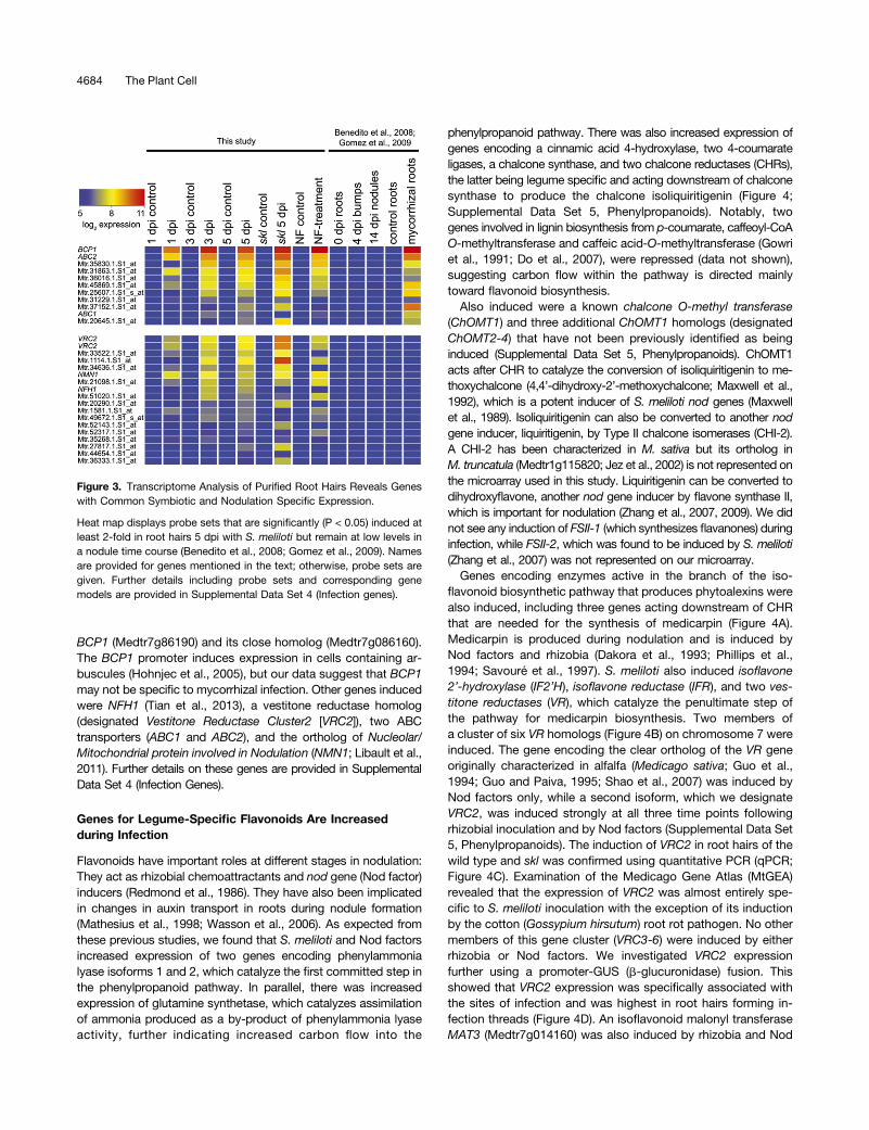
BCP1 (Medtr7g86190) and its close homolog (Medtr7g086160).The BCP1 promoter induces expression in cells containing ar-buscules (Hohnjec et al., 2005), but our data suggest that BCP1may not be specific to mycorrhizal infection. Other genes inducedwere NFH1 (Tian et al., 2013), a vestitone reductase homolog(designated Vestitone Reductase Cluster2 [VRC2]), two ABCtransporters (ABC1 and ABC2), and the ortholog of Nucleolar/Mitochondrial protein involved in Nodulation (NMN1; Libault et al.,2011). Further details on these genes are provided in SupplementalData Set 4 (Infection Genes).
Genes for Legume-Specific Flavonoids Are Increasedduring Infection
Flavonoids have important roles at different stages in nodulation:They act as rhizobial chemoattractants and nod gene (Nod factor)inducers (Redmond et al., 1986). They have also been implicatedin changes in auxin transport in roots during nodule formation(Mathesius et al., 1998; Wasson et al., 2006). As expected fromthese previous studies, we found that S. meliloti and Nod factorsincreased expression of two genes encoding phenylammonialyase isoforms 1 and 2, which catalyze the first committed step inthe phenylpropanoid pathway. In parallel, there was increasedexpression of glutamine synthetase, which catalyzes assimilationof ammonia produced as a by-product of phenylammonia lyaseactivity, further indicating increased carbon flow into the
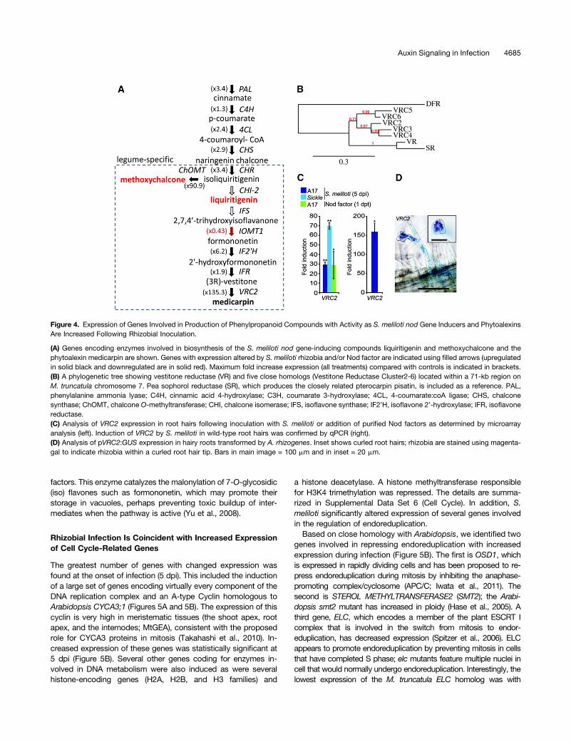
phenylpropanoid pathway. There was also increased expression ofgenes encoding a cinnamic acid 4-hydroxylase, two 4-coumarateligases, a chalcone synthase, and two chalcone reductases (CHRs),the latter being legume specific and acting downstream of chalconesynthase to produce the chalcone isoliquiritigenin (Figure 4;Supplemental Data Set 5, Phenylpropanoids). Notably, twogenes involved in lignin biosynthesis from p-coumarate, caffeoyl-CoAO-methyltransferase and caffeic acid-O-methyltransferase (Gowriet al., 1991; Do et al., 2007), were repressed (data not shown),suggesting carbon flow within the pathway is directed mainlytoward flavonoid biosynthesis.Also induced were a known chalcone O-methyl transferase
(ChOMT1) and three additional ChOMT1 homologs (designatedChOMT2-4) that have not been previously identified as beinginduced (Supplemental Data Set 5, Phenylpropanoids). ChOMT1acts after CHR to catalyze the conversion of isoliquiritigenin to me-thoxychalcone (4,4’-dihydroxy-2’-methoxychalcone; Maxwell et al.,1992), which is a potent inducer of S. meliloti nod genes (Maxwellet al., 1989). Isoliquiritigenin can also be converted to another nodgene inducer, liquiritigenin, by Type II chalcone isomerases (CHI-2).A CHI-2 has been characterized in M. sativa but its ortholog inM. truncatula (Medtr1g115820; Jez et al., 2002) is not represented onthe microarray used in this study. Liquiritigenin can be converted todihydroxyflavone, another nod gene inducer by flavone synthase II,which is important for nodulation (Zhang et al., 2007, 2009). We didnot see any induction of FSII-1 (which synthesizes flavanones) duringinfection, while FSII-2, which was found to be induced by S. meliloti(Zhang et al., 2007) was not represented on our microarray.Genes encoding enzymes active in the branch of the iso-
flavonoid biosynthetic pathway that produces phytoalexins werealso induced, including three genes acting downstream of CHRthat are needed for the synthesis of medicarpin (Figure 4A).Medicarpin is produced during nodulation and is induced byNod factors and rhizobia (Dakora et al., 1993; Phillips et al.,1994; Savouré et al., 1997). S. meliloti also induced isoflavone2’-hydroxylase (IF2’H), isoflavone reductase (IFR), and two ves-titone reductases (VR), which catalyze the penultimate step ofthe pathway for medicarpin biosynthesis. Two members ofa cluster of six VR homologs (Figure 4B) on chromosome 7 wereinduced. The gene encoding the clear ortholog of the VR geneoriginally characterized in alfalfa (Medicago sativa; Guo et al.,1994; Guo and Paiva, 1995; Shao et al., 2007) was induced byNod factors only, while a second isoform, which we designateVRC2, was induced strongly at all three time points followingrhizobial inoculation and by Nod factors (Supplemental Data Set5, Phenylpropanoids). The induction of VRC2 in root hairs of thewild type and skl was confirmed using quantitative PCR (qPCR;Figure 4C). Examination of the Medicago Gene Atlas (MtGEA)revealed that the expression of VRC2 was almost entirely spe-cific to S. meliloti inoculation with the exception of its inductionby the cotton (Gossypium hirsutum) root rot pathogen. No othermembers of this gene cluster (VRC3-6) were induced by eitherrhizobia or Nod factors. We investigated VRC2 expressionfurther using a promoter-GUS (b-glucuronidase) fusion. Thisshowed that VRC2 expression was specifically associated withthe sites of infection and was highest in root hairs forming in-fection threads (Figure 4D). An isoflavonoid malonyl transferaseMAT3 (Medtr7g014160) was also induced by rhizobia and Nod
Figure 3. Transcriptome Analysis of Purified Root Hairs Reveals Geneswith Common Symbiotic and Nodulation Specific Expression.
Heat map displays probe sets that are significantly (P < 0.05) induced atleast 2-fold in root hairs 5 dpi with S. meliloti but remain at low levels ina nodule time course (Benedito et al., 2008; Gomez et al., 2009). Namesare provided for genes mentioned in the text; otherwise, probe sets aregiven. Further details including probe sets and corresponding genemodels are provided in Supplemental Data Set 4 (Infection genes).
4684 The Plant Cell
factors. This enzyme catalyzes the malonylation of 7-O-glycosidic(iso) flavones such as formononetin, which may promote theirstorage in vacuoles, perhaps preventing toxic buildup of inter-mediates when the pathway is active (Yu et al., 2008).
Rhizobial Infection Is Coincident with Increased Expressionof Cell Cycle-Related Genes
The greatest number of genes with changed expression wasfound at the onset of infection (5 dpi). This included the inductionof a large set of genes encoding virtually every component of theDNA replication complex and an A-type Cyclin homologous toArabidopsis CYCA3;1 (Figures 5A and 5B). The expression of thiscyclin is very high in meristematic tissues (the shoot apex, rootapex, and the internodes; MtGEA), consistent with the proposedrole for CYCA3 proteins in mitosis (Takahashi et al., 2010). In-creased expression of these genes was statistically significant at5 dpi (Figure 5B). Several other genes coding for enzymes in-volved in DNA metabolism were also induced as were severalhistone-encoding genes (H2A, H2B, and H3 families) and
a histone deacetylase. A histone methyltransferase responsiblefor H3K4 trimethylation was repressed. The details are summa-rized in Supplemental Data Set 6 (Cell Cycle). In addition, S.meliloti significantly altered expression of several genes involvedin the regulation of endoreduplication.Based on close homology with Arabidopsis, we identified two
genes involved in repressing endoreduplication with increasedexpression during infection (Figure 5B). The first is OSD1, whichis expressed in rapidly dividing cells and has been proposed to re-press endoreduplication during mitosis by inhibiting the anaphase-promoting complex/cyclosome (APC/C; Iwata et al., 2011). Thesecond is STEROL METHYLTRANSFERASE2 (SMT2); the Arabi-dopsis smt2 mutant has increased in ploidy (Hase et al., 2005). Athird gene, ELC, which encodes a member of the plant ESCRT Icomplex that is involved in the switch from mitosis to endor-eduplication, has decreased expression (Spitzer et al., 2006). ELCappears to promote endoreduplication by preventing mitosis in cellsthat have completed S phase; elc mutants feature multiple nuclei incell that would normally undergo endoreduplication. Interestingly, thelowest expression of the M. truncatula ELC homolog was with
Figure 4. Expression of Genes Involved in Production of Phenylpropanoid Compounds with Activity as S. meliloti nod Gene Inducers and PhytoalexinsAre Increased Following Rhizobial Inoculation.
(A) Genes encoding enzymes involved in biosynthesis of the S. meliloti nod gene-inducing compounds liquiritigenin and methoxychalcone and thephytoalexin medicarpin are shown. Genes with expression altered by S. meliloti rhizobia and/or Nod factor are indicated using filled arrows (upregulatedin solid black and downregulated are in solid red). Maximum fold increase expression (all treatments) compared with controls is indicated in brackets.(B) A phylogenetic tree showing vestitone reductase (VR) and five close homologs (Vestitone Reductase Cluster2-6) located within a 71-kb region onM. truncatula chromosome 7. Pea sophorol reductase (SR), which produces the closely related pterocarpin pisatin, is included as a reference. PAL,phenylalanine ammonia lyase; C4H, cinnamic acid 4-hydroxylase; C3H, coumarate 3-hydroxylase; 4CL, 4-coumarate:coA ligase; CHS, chalconesynthase; ChOMT, chalcone O-methyltransferase; CHI, chalcone isomerase; IFS, isoflavone synthase; IF2’H, isoflavone 2’-hydroxylase; IFR, isoflavonereductase.(C) Analysis of VRC2 expression in root hairs following inoculation with S. meliloti or addition of purified Nod factors as determined by microarrayanalysis (left). Induction of VRC2 by S. meliloti in wild-type root hairs was confirmed by qPCR (right).(D) Analysis of pVRC2:GUS expression in hairy roots transformed by A. rhizogenes. Inset shows curled root hairs; rhizobia are stained using magenta-gal to indicate rhizobia within a curled root hair tip. Bars in main image = 100 mm and in inset = 20 mm.
Auxin Signaling in Infection 4685

S. meliloti 1021 in the skl mutant. This suggests a possible role forethylene in ELC regulation and is of interest due to a recent reportthat ethylene-insensitive mutants in L. japonicus have occasionalexamples where root hair cells infected by rhizobia undergo nucleardivision (Gresshoff et al., 2009). A homolog of Arabidopsis MEI-2 Like(AML2) was also repressed. AML2 is an RNA binding protein thatpromotes meiosis (Kaur et al., 2006).Nod factors did not significantly alter the expression of the
A-type cyclin or endoreduplication-related genes except for ELC,which it repressed, but they did induce three different D-typecyclins and the APC6 subunit of the APC/C (Figure 5C). Thesedata suggest that infection involves reactivation of the cell cycleand repression of endoreduplication.
Infection Alters Expression of Genes Involved in HormoneBiosynthesis and Signaling
GA Biosynthesis
Nod factor signaling appears to increase the production of GA inroot hairs cells because the GA biosynthesis genesGIBBERELLIN3 BETA-HYDROXYLASE1 (GA3OX1),GIBBERELLIN 2-OXIDASE6(GA2OX6), ENT-COPALYL DIPHOSPHATE SYNTHETASE1(CPS1), ENT-KAURENE OXIDASE1 (KO1), and ENT-KAURENOICACID OXIDASE2 (KAO2) were induced by S. meliloti and by pu-rified Nod factors. Two genes involved in GA regulation and sig-naling, GAST1 PROTEIN HOMOLOG1 (GASA1) and ZINCFINGER PROTEIN6 (ZFP6), were also induced by Nod factors aswas KO1, whereas the gene encoding the GA receptor GAINSENSITIVE DWARF1B (GID1B) was repressed (SupplementalData Set 7, GA). Many of these genes were also increased afterB. japonicum inoculation in soybean.
SL Biosynthesis
SLs are carotenoid-derived hormones that act as signaling mole-cules for arbuscular mycorrhiza (Akiyama et al., 2005). DWARF27(D27) and CAROTENOID CLEAVAGE DIOXYGENASE8 (CCD8)were induced by S. meliloti, suggesting SLs were being produced(Figures 6A and 6B; Supplemental Data Set 8, SLs). In addition, thecarotenoid biosynthesis genes encoding z-carotene desaturase(ZDS) and 15-cis-z-carotene isomerase (Z-ISO) were induced, aswas the ortholog of the cauliflower (Brassica oleracea) Orangegene, which promotes the formation of carotenoid-producingchromoplasts (Lu et al., 2006). These data support increased syn-thesis of SLs during infection. To assess the potential site of SLproduction, we fused the promoter of CCD8 to the GUS gene andintroduced it into M. truncatula using Agrobacterium rhizogenes-based hairy root transformation. Staining was evident at the sites ofS. meliloti infection (Figure 6C) and was initially restricted to colo-nized root hair cells and was later seen in developing nodule pri-mordia, implying a role for SLs during rhizobial infection.
BR Biosynthesis and Signaling
The BR biosynthesis gene DWARF1 (Mtr.10571.1.S1_at:Medtr4g074350) was induced by S. meliloti but not Nod factors.
Figure 5. Cell Cycle-Related Genes Are Regulated during Infection.
(A) Fold induction of replication fork components including mini chro-mosome maintenance (MCM) genes.(B) Fold induction of cell cycle regulation genes.(C) Nod factor treatments induce the expression of the anaphase pro-moting complex (APC6), a C-type cyclin, and genes encoding threeD-type cyclins.Significant inductions relative to control experiments are shown (*P <0.05, **P < 0.01). Error bars = SE (n = 3).
4686 The Plant Cell

Nod factors did affect BR-related gene expression, repressing theexpression of BRASSINAZOLE-RESISTANT1 (BZR1; Mtr.43192.1.S1_at: Medtr5g019550) and SHAGGY-LIKE PROTEIN KINASE23(SK23; Mtr.17959.1.S1_s_at: Medtr2g083940). BZR1 encodes atranscription factor that is required for normal responses to BR andacts directly to repress the expression of BR biosynthesis genes;SK23 encodes a kinase that phosphorylates BZR1, which excludes
it from the nucleus (Ryu et al., 2007). Together, these data suggestthat BR synthesis and signaling are triggered during rhizobialinfection.
Jasmonic Acid Biosynthesis and Signaling Genes AreRepressed by Nod Factors and by Rhizobial Inoculationin skl
The production of the defense hormone jasmonic acid (JA) ap-peared to be repressed by S. meliloti in the skl mutant becausethere was reduced expression of the JA-responsive geneJASMONATE-ZIM-DOMAIN PROTEIN2 (JAZ2), the JA receptorCORONATINE-INSENSITIVE1 (COI1), and the Allene OxideSynthase1 (AOS1) gene encoding an enzyme required for JAbiosynthesis (Supplemental Data Set 9, JA). The marked repressionof JA-related responses in skl during infection is presumably in partdue to interactions between JA and ethylene signaling. Notably,ethylene is known to induce AOS activity and accumulation of JA(Laudert and Weiler, 1998). However, Nod factors also repressedJA-related responses. Infection of the orthologous Arabidopsismutant ein2 by the oomycete pathogen Pythium irregular inducesmuch higher levels of JA production than in wild-type plants (Adie
Figure 6. Auxin Signaling and SL Biosynthesis Genes Are Induced byS. meliloti and by Nod Factors.
(A) Microarray-based quantification of expression of auxin signalinggenes SAUR1, GH3.1, ARF16a, and IAA9 and SL biosynthetic genesD27 and CCD8 at 5 dpi with S. meliloti or 1 d post-treatment with Nodfactors in the wild type and skl.(B) qPCR confirmation of the induction of auxin signaling and SL bio-synthetic genes at 5 dpi with S. meliloti.(C) Analysis of expression of the SL biosynthesis gene CCD8 usinga promoter-GUS fusion in A. rhizogenes-induced transgenic rootsstained for GUS activity (image on left). The image on the right showsstaining of both CCD8:GUS (blue) and S. meliloti (lacZ ) (magenta/purple).Bars in main images = 100 mm; insets = 20 mm. Significant inductionrelative to control experiments are shown (*P < 0.05, **P < 0.01, and***P < 0.001). Error bars = SE (n = 3).
Figure 7. DR5 Is Expressed throughout the Infection Zone after Rhizo-bial Inoculation.
DR5:GUS expression in stable transgenic M. truncatula seedlings in-oculated with S. meliloti. The blue color in (A) and (B) indicates stainingof GUS activity. Arrows (A) indicate infection pockets containing mi-crocolonies, and the arrowhead (B) indicates a root hair containing aninfection thread. Bars = 50 mm.(A) 3 dpi. The inset is a magnification of the boxed region.(B) 5 dpi.
Auxin Signaling in Infection 4687
et al., 2007). Together these data suggest that repression of JA isa specific feature of rhizobial interactions and not simply a conse-quence of altered ethylene signaling in skl.
Auxin Signaling
Auxin plays an important role during nodule organogenesis(Mathesius et al., 1998), but no role for auxin during rhizobialinfection has been demonstrated. Although there was no sig-nificant change in expression of genes for auxin biosynthesis ofauxin transport (PIN-FORMED [PIN]), S. meliloti induced ex-pression of the auxin-responsive genes AUXIN RESPONSEFACTOR 16a (ARF16a), Gretchen Hagen3.1 (GH3.1), INDOLE-3-ACETIC ACID INDUCIBLE9 (IAA9), and SMALL AUXIN UPRNA1 (SAUR1) (Figures 6A and 6B; Supplemental Data Set 10,Auxin). All of these belong to relatively large gene families butwere the only members that responded to infection.
To investigate the location of auxin signaling during infection,we usedM. truncatula stably transformed with the auxin reporterDR5:GUS (Zhou et al., 2011) and observed that S. meliloti in-duced DR5:GUS in both infected and uninfected root hairs overthe entire infection zone (root hair differentiation zone) 3 and 5dpi (Figures 7A and 7B). This increase in auxin signaling isconsistent with the observation that auxin signaling is initiallydownregulated and then increases at the site of inoculation(Mathesius et al., 1998).
We compared the expression patterns of GH3.1, SAUR1, andARF16a with that of the DR5 using promoter-GUS fusions inA. rhizogenes-induced hairy roots. GH3.1, SAUR1, and ARF16a
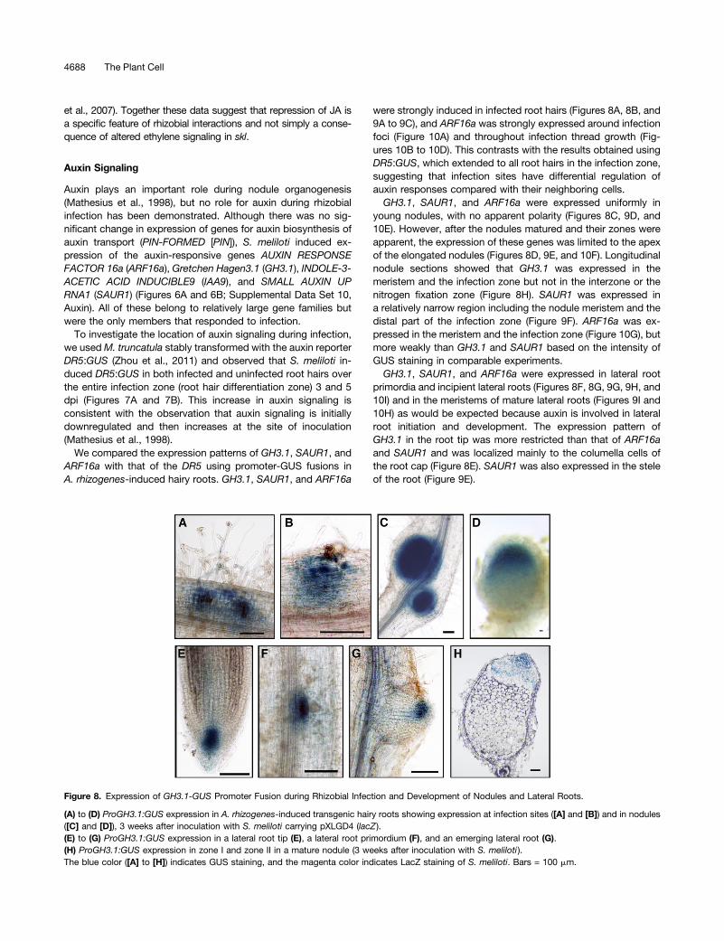
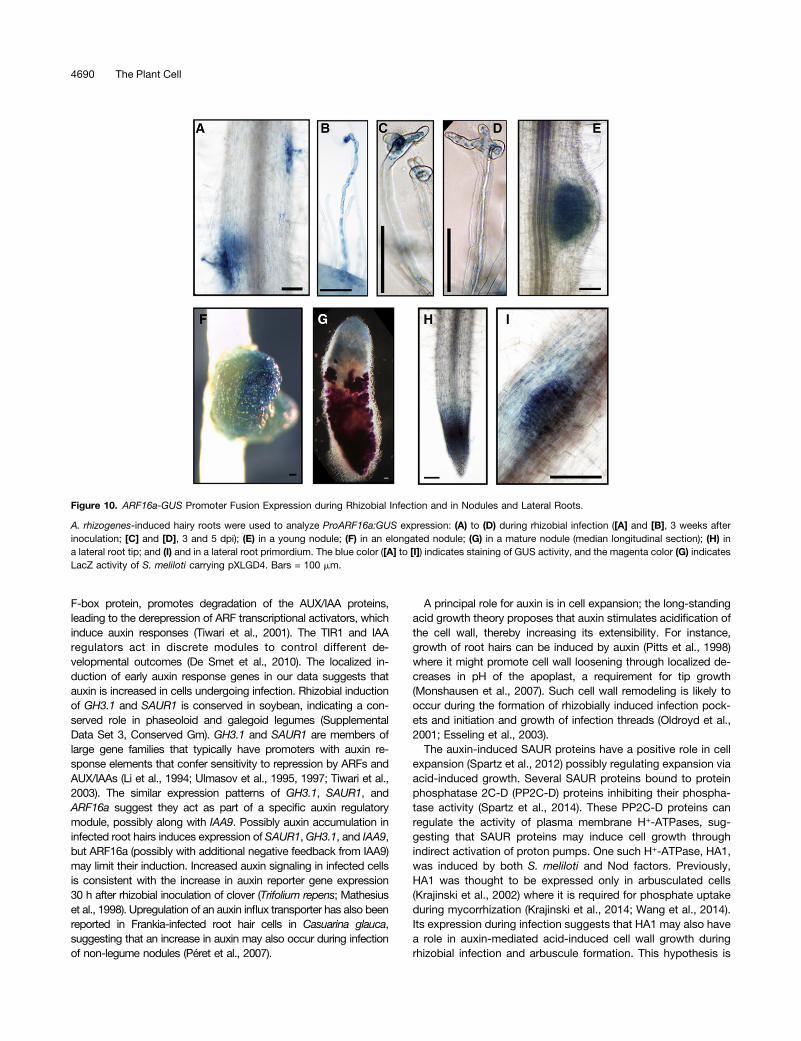
were strongly induced in infected root hairs (Figures 8A, 8B, and9A to 9C), and ARF16a was strongly expressed around infectionfoci (Figure 10A) and throughout infection thread growth (Fig-ures 10B to 10D). This contrasts with the results obtained usingDR5:GUS, which extended to all root hairs in the infection zone,suggesting that infection sites have differential regulation ofauxin responses compared with their neighboring cells.GH3.1, SAUR1, and ARF16a were expressed uniformly in
young nodules, with no apparent polarity (Figures 8C, 9D, and10E). However, after the nodules matured and their zones wereapparent, the expression of these genes was limited to the apexof the elongated nodules (Figures 8D, 9E, and 10F). Longitudinalnodule sections showed that GH3.1 was expressed in themeristem and the infection zone but not in the interzone or thenitrogen fixation zone (Figure 8H). SAUR1 was expressed ina relatively narrow region including the nodule meristem and thedistal part of the infection zone (Figure 9F). ARF16a was ex-pressed in the meristem and the infection zone (Figure 10G), butmore weakly than GH3.1 and SAUR1 based on the intensity ofGUS staining in comparable experiments.GH3.1, SAUR1, and ARF16a were expressed in lateral root
primordia and incipient lateral roots (Figures 8F, 8G, 9G, 9H, and10I) and in the meristems of mature lateral roots (Figures 9I and10H) as would be expected because auxin is involved in lateralroot initiation and development. The expression pattern ofGH3.1 in the root tip was more restricted than that of ARF16aand SAUR1 and was localized mainly to the columella cells ofthe root cap (Figure 8E). SAUR1 was also expressed in the steleof the root (Figure 9E).
Figure 8. Expression of GH3.1-GUS Promoter Fusion during Rhizobial Infection and Development of Nodules and Lateral Roots.
(A) to (D) ProGH3.1:GUS expression in A. rhizogenes-induced transgenic hairy roots showing expression at infection sites ([A] and [B]) and in nodules([C] and [D]), 3 weeks after inoculation with S. meliloti carrying pXLGD4 (lacZ ).(E) to (G) ProGH3.1:GUS expression in a lateral root tip (E), a lateral root primordium (F), and an emerging lateral root (G).(H) ProGH3.1:GUS expression in zone I and zone II in a mature nodule (3 weeks after inoculation with S. meliloti ).The blue color ([A] to [H]) indicates GUS staining, and the magenta color indicates LacZ staining of S. meliloti. Bars = 100 mm.
4688 The Plant Cell

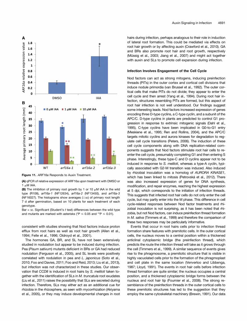
To test if ARF16 acts like its Arabidopsis counterpart, wemeasured (by quantitative RT-PCR) the auxin responsiveness ofARF16a in roots and saw about a 3-fold increase in expression(Figure 11A). This is consistent with M. truncatula transcriptomicsdata after NAA treatment in embryogenic leaf explant cultures(Imin et al., 2008) and induction of the Arabidopsis ARF16 or-tholog by indole-3-acetic acid (IAA) (Wang et al., 2005). Weidentified three arf16a:Tnt1 insertional mutants (Figure 12A)through PCR screening. The root growth of these mutants wasfound to be more sensitive to auxin inhibition than the wild type(Figure 11B), particularly at 10 mM IAA. This is consistent withwork in soybean that showed that overexpression of miR160,
which targets ARF16 and its close homolog ARF10, resulted inauxin hypersensitivity (Turner et al., 2013).
The arf16a Mutant Is Resistant to Infection by S. meliloti
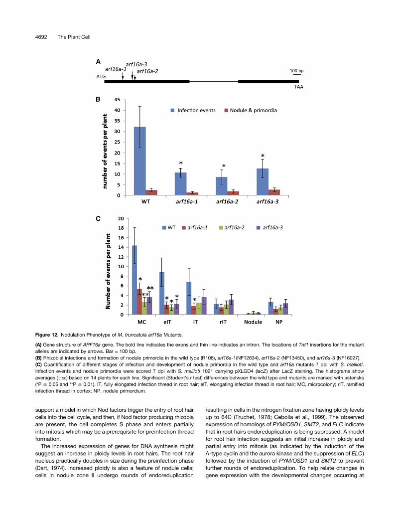
The localized expression patterns of GH3.1, SAUR1, and ARF16ain infected tissues suggested a role for auxin in infection and sothe arf16a mutants were examined for S. meliloti-induced in-fection and nodulation. The number of infection events observed7 dpi was reduced in the mutants compared with the wild type(Figure 12B). A few normal-looking infection threads were formed(Supplemental Figure 4) and these led to successful nodule col-onization (Supplemental Figures 5 and 6). Infection events wereclassified into four stages of infection thread progression (scoringkey provided in Supplemental Figure 3), showing that the re-duction in infections could be attributed to a decreased numbersof infection pockets containing microcolonies and elongatingthreads (Figure 12C). This suggests that the earliest stages ofinfection were impaired in the mutants. Nodules and nodule pri-mordia were found to be normal in morphology and number(Supplemental Figures 5 and 6). These results indicate a role forARF16a in the initiation of infection (i.e., formation of the infectionpocket and thread initiation) rather than in infection thread extension.
DISCUSSION
Our work has provided insights into the molecular crosstalk thatoccurs between the rhizobia and legume roots during initiationof infection and has identified some of the biological processesassociated with infection thread development.A key finding is a role for auxin signaling in infection. Four
auxin-responsive genes, GH3.1, SAUR1, ARF16a, and IAA9,were more strongly induced by S. meliloti in skl relative to thewild type (Figure 5), consistent with enhanced auxin transport inskl (Prayitno et al., 2006), and reveals an unexpected role forauxin in root-hair infection. Although S. meliloti induced DR5-GUS in all root hairs in the infection zone, expression of GH3.1,ARF16a, and SAUR1 was restricted to infected cells, indicatingthat auxin signaling in infected and noninfected root hairs isdistinct. This could result from an infected cell-specific modifi-cation of auxin signaling caused by Nod factors because GH3.1and ARF16a were induced by purified Nod factors. A local al-teration in auxin response must be important because mutationof ARF16a reduced the frequency of infection. Although thefunction of the putative repressor ARF16a has not been studieddirectly in legumes, the effect of overexpressing miR160 (whichtargets ARF16 as well as other closely related ARFs) has beenexamined in soybean and M. truncatula during nodulation. Over-expression of miR160 in soybean did not affect infections in roothairs; in a similar M. truncatula study, infections were not scored(Bustos-Sanmamed et al., 2013; Turner et al., 2013). The apparentdiscrepancy between our work and the soybean study could bedue to inefficient knockdown of ARF16 by miR160 or the differentialregulation of auxin in determinate versus indeterminate nodulation(Subramanian et al., 2006, 2007; Wasson et al. 2006).GH3, SAUR, and AUX/IAAs regulate auxin responses. Auxin
binds to the Transport Inhibitor Response 1 (TIR1) and AUX/IAAcoreceptor complex (Calderón Villalobos et al., 2012). TIR1, an
Figure 9. SAUR1-GUS Promoter Fusion Expression during RhizobialInfection, and Development of Nodules and Lateral Roots.
A. rhizogenes-induced hairy roots were used to analyze ProSAUR1:GUSexpression: (A) to (C) during rhizobial infection; (D) to (F) during noduledevelopment; (G) during the formation of a lateral root primordium; (H) inan emerging lateral root; and (I) in a root tip. The blue color ([A] to [I])indicates GUS staining of ProSAUR1:GUS activity, and the magentacolor (C) indicates LacZ staining of S. meliloti. Bars = 100 mm.
Auxin Signaling in Infection 4689
F-box protein, promotes degradation of the AUX/IAA proteins,leading to the derepression of ARF transcriptional activators, whichinduce auxin responses (Tiwari et al., 2001). The TIR1 and IAAregulators act in discrete modules to control different de-velopmental outcomes (De Smet et al., 2010). The localized in-duction of early auxin response genes in our data suggests thatauxin is increased in cells undergoing infection. Rhizobial inductionof GH3.1 and SAUR1 is conserved in soybean, indicating a con-served role in phaseoloid and galegoid legumes (SupplementalData Set 3, Conserved Gm). GH3.1 and SAUR1 are members oflarge gene families that typically have promoters with auxin re-sponse elements that confer sensitivity to repression by ARFs andAUX/IAAs (Li et al., 1994; Ulmasov et al., 1995, 1997; Tiwari et al.,2003). The similar expression patterns of GH3.1, SAUR1, andARF16a suggest they act as part of a specific auxin regulatorymodule, possibly along with IAA9. Possibly auxin accumulation ininfected root hairs induces expression of SAUR1,GH3.1, and IAA9,but ARF16a (possibly with additional negative feedback from IAA9)may limit their induction. Increased auxin signaling in infected cellsis consistent with the increase in auxin reporter gene expression30 h after rhizobial inoculation of clover (Trifolium repens; Mathesiuset al., 1998). Upregulation of an auxin influx transporter has also beenreported in Frankia-infected root hair cells in Casuarina glauca,suggesting that an increase in auxin may also occur during infectionof non-legume nodules (Péret et al., 2007).
A principal role for auxin is in cell expansion; the long-standingacid growth theory proposes that auxin stimulates acidification ofthe cell wall, thereby increasing its extensibility. For instance,growth of root hairs can be induced by auxin (Pitts et al., 1998)where it might promote cell wall loosening through localized de-creases in pH of the apoplast, a requirement for tip growth(Monshausen et al., 2007). Such cell wall remodeling is likely tooccur during the formation of rhizobially induced infection pock-ets and initiation and growth of infection threads (Oldroyd et al.,2001; Esseling et al., 2003).The auxin-induced SAUR proteins have a positive role in cell
expansion (Spartz et al., 2012) possibly regulating expansion viaacid-induced growth. Several SAUR proteins bound to proteinphosphatase 2C-D (PP2C-D) proteins inhibiting their phospha-tase activity (Spartz et al., 2014). These PP2C-D proteins canregulate the activity of plasma membrane H+-ATPases, sug-gesting that SAUR proteins may induce cell growth throughindirect activation of proton pumps. One such H+-ATPase, HA1,was induced by both S. meliloti and Nod factors. Previously,HA1 was thought to be expressed only in arbusculated cells(Krajinski et al., 2002) where it is required for phosphate uptakeduring mycorrhization (Krajinski et al., 2014; Wang et al., 2014).Its expression during infection suggests that HA1 may also havea role in auxin-mediated acid-induced cell wall growth duringrhizobial infection and arbuscule formation. This hypothesis is
Figure 10. ARF16a-GUS Promoter Fusion Expression during Rhizobial Infection and in Nodules and Lateral Roots.
A. rhizogenes-induced hairy roots were used to analyze ProARF16a:GUS expression: (A) to (D) during rhizobial infection ([A] and [B], 3 weeks afterinoculation; [C] and [D], 3 and 5 dpi); (E) in a young nodule; (F) in an elongated nodule; (G) in a mature nodule (median longitudinal section); (H) ina lateral root tip; and (I) and in a lateral root primordium. The blue color ([A] to [I]) indicates staining of GUS activity, and the magenta color (G) indicatesLacZ activity of S. meliloti carrying pXLGD4. Bars = 100 mm.
4690 The Plant Cell
consistent with studies showing that Nod factors induce protonefflux from root hairs as well as root hair growth (Allen et al.,1994; Felle et al., 1996; Oldroyd et al., 2001).
The hormones GA, BR, and SL have not been extensivelystudied in nodulation but appear to be induced during infection.Pea (Pisum sativum) mutants deficient in BRs or GA had reducednodulation (Ferguson et al., 2005), and SL levels were positivelycorrelated with nodulation in pea and L. japonicus (Soto et al.,2010; Foo and Davies, 2011; Foo and Reid, 2013; Liu et al., 2013),but infection was not characterized in these studies. Our obser-vation that CCD8 is induced in root hairs by S. meliloti taken to-gether with the identification of SLs inM. truncatula root exudates(Liu et al., 2011) raises the possibility that SLs are secreted duringinfection. Therefore, SLs may either act as an additional cue forrhizobia in the rhizosphere, as seen with mycorrhization (Akiyamaet al., 2005), or they may induce developmental changes in root
hairs during infection, perhaps analogous to their role in inductionof lateral root formation. This could be mediated via effects onroot hair growth or by affecting auxin (Crawford et al., 2010). GAand BRs also promote root hair and root growth, respectively(Müssig et al., 2003; Jiang et al., 2007) and might act togetherwith auxin and SLs to promote cell expansion during infection.
Infection Involves Engagement of the Cell Cycle
Nod factors can act as strong mitogens, inducing preinfectionthreads (PITs) in the outer cortex and cortical cell divisions thatinduce nodule primordia (van Brussel et al., 1992). The outer cor-tical cells that make PITs do not divide; they appear to enter thecell cycle and then arrest (Yang et al., 1994). During root hair in-fection, structures resembling PITs are formed, but this aspect ofroot hair infection is not well understood. Our findings suggestsome interesting leads. Nod factors increased expression of genesencoding three D-type cyclins, a C-type cyclin, and a subunit of theAPC/C. D-type cyclins in plants are predicted to control G1 pro-gression in response to extrinsic mitogenic signals (Dahl et al.,1995), C-type cyclins have been implicated in G0-to-G1 entry(Meskiene et al., 1995; Ren and Rollins, 2004), and the APC/Ctargets mitotic cyclins and aurora kinases for degradation to reg-ulate cell cycle transitions (Peters, 2006). The induction of thesecell cycle components along with DNA replication-related com-ponents suggests that Nod factors stimulate root hair cells to re-enter the cell cycle, presumably completing G1 and then entering Sphase. Interestingly, these type-C and D cyclins appear not to beinduced in response to S. meliloti, whereas a type-A cyclin, typi-cally associated with G2-M transition was induced. Also inducedby rhizobial inoculation was a homolog of AURORA KINASE1,which has been linked to mitosis (Petrovská et al., 2012). Therewas also increased expression of genes for DNA synthesis,modification, and repair enzymes, reaching the highest expressionat 5 dpi, which corresponds to the initiation of infection threads.This suggests that infected root hair cells do not only enter the cellcycle, but may partly enter into the M phase. This difference in cellcycle-related responses between Nod factor treatments and rhi-zobial inoculation is not surprising, as it has been noted that rhi-zobia, but not Nod factors, can induce preinfection thread formationin M. sativa (Timmers et al., 1999) and therefore the comparison ofthese two responses may be particularly informative.Events that occur in root hairs cells prior to infection thread
formation share features with premitotic cells. In the outer corticalcells, the nucleus moves to a central position within a thickenedanticlinal cytoplasmic bridge (the preinfection thread), whichpredicts the route the infection thread will take as it grows throughthe cell (Timmers et al., 1999). A similar sequence of events givesrise to the phragmosome, a premitotic structure that is visible inhighly vacuolated cells prior to the formation of the phragmoplastand cell plate in the same location (Venverloo and Libbenga,1987; Lloyd, 1991). The events in root hair cells before infectionthread formation are quite similar; the nucleus occupies a centralposition, and a thickened cytoplasmic bridge forms between thenucleus and root hair tip (Fournier et al., 2008). The strong re-semblance of the preinfection threads in the outer cortical cells tothese premitotic structures has led to the suggestion that theyemploy the same cytoskeletal machinery (Brewin, 1991). Our data
Figure 11. ARF16a Responds to Auxin Treatment.
(A) qPCR of relative expression of ARF16a upon treatment with DMSO or1 mM IAA.(B) The inhibition of primary root growth by 1 or 10 mM IAA in the wildtype (R108), arf16a-1 (NF12634), arf16a-2 (NF13450), and arf16a-3(NF16027). The histograms show averages (6SE) of primary root length7 d after germination, based on 10 plants for each treatment of eachgenotype.Bar = SE. Significant (Student’s t test) differences between the wild typeand mutants are marked with asterisks (*P # 0.05 and **P # 0.01).
Auxin Signaling in Infection 4691
support a model in which Nod factors trigger the entry of root haircells into the cell cycle, and then, if Nod factor producing rhizobiaare present, the cell completes S phase and enters partiallyinto mitosis which may be a prerequisite for preinfection threadformation.
The increased expression of genes for DNA synthesis mightsuggest an increase in ploidy levels in root hairs. The root hairnucleus practically doubles in size during the preinfection phase(Dart, 1974). Increased ploidy is also a feature of nodule cells;cells in nodule zone II undergo rounds of endoreduplication
resulting in cells in the nitrogen fixation zone having ploidy levelsup to 64C (Truchet, 1978; Cebolla et al., 1999). The observedexpression of homologs of PYM/OSD1, SMT2, and ELC indicatethat in root hairs endoreduplication is being supressed. A modelfor root hair infection suggests an initial increase in ploidy andpartial entry into mitosis (as indicated by the induction of theA-type cyclin and the aurora kinase and the suppression of ELC)followed by the induction of PYM/OSD1 and SMT2 to preventfurther rounds of endoreduplication. To help relate changes ingene expression with the developmental changes occurring at
Figure 12. Nodulation Phenotype of M. truncatula arf16a Mutants.
(A) Gene structure of ARF16a gene. The bold line indicates the exons and thin line indicates an intron. The locations of Tnt1 insertions for the mutantalleles are indicated by arrows. Bar = 100 bp.(B) Rhizobial infections and formation of nodule primordia in the wild type (R108), arf16a-1(NF12634), arf16a-2 (NF13450), and arf16a-3 (NF16027).(C) Quantification of different stages of infection and development of nodule primordia in the wild type and arf16a mutants 7 dpi with S. meliloti.Infection events and nodule primordia were scored 7 dpi with S. meliloti 1021 carrying pXLGD4 (lacZ ) after LacZ staining. The histograms showaverages (6SE) based on 14 plants for each line. Significant (Student’s t test) differences between the wild type and mutants are marked with asterisks(*P # 0.05 and **P # 0.01). IT, fully elongated infection thread in root hair; eIT, elongating infection thread in root hair; MC, microcolony; rIT, ramifiedinfection thread in cortex; NP, nodule primordium.
4692 The Plant Cell
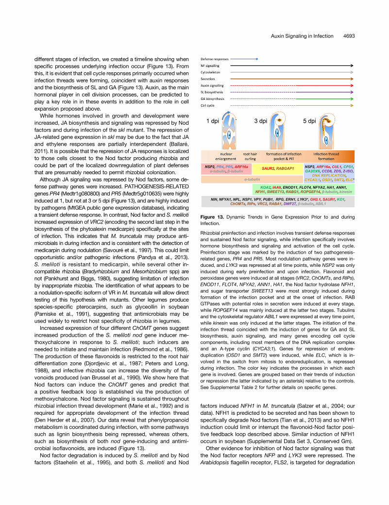
different stages of infection, we created a timeline showing whenspecific processes underlying infection occur (Figure 13). Fromthis, it is evident that cell cycle responses primarily occurred wheninfection threads were forming, coincident with auxin responsesand the biosynthesis of SL and GA (Figure 13). Auxin, as the mainhormonal player in cell division processes, can be predicted toplay a key role in in these events in addition to the role in cellexpansion proposed above.
While hormones involved in growth and development wereincreased, JA biosynthesis and signaling was repressed by Nodfactors and during infection of the skl mutant. The repression ofJA-related gene expression in skl may be due to the fact that JAand ethylene responses are partially interdependent (Ballaré,2011). It is possible that the repression of JA responses is localizedto those cells closest to the Nod factor producing rhizobia andcould be part of the localized downregulation of plant defensesthat are presumably needed to permit rhizobial colonization.
Although JA signaling was repressed by Nod factors, some de-fense pathway genes were increased. PATHOGENESIS-RELATEDgenes PR4 (Medtr1g080800) and PR5 (Medtr5g010635) were highlyinduced at 1, but not at 3 or 5 dpi (Figure 13), and are highly inducedby pathogens (MtGEA public gene expression database), indicatinga transient defense response. In contrast, Nod factor and S. melilotiincreased expression of VRC2 (encoding the second last step in thebiosynthesis of the phytoalexin medicarpin) specifically at the sitesof infection. This indicates that M. truncatula may produce anti-microbials in during infection and is consistent with the detection ofmedicarpin during nodulation (Savouré et al., 1997). This could limitopportunistic and/or pathogenic infections (Pandya et al., 2013).S. meliloti is resistant to medicarpin, while several other in-compatible rhizobia (Bradyrhizobium and Mesorhizobium spp) arenot (Pankhurst and Biggs, 1980), suggesting limitation of infectionby inappropriate rhizobia. The identification of what appears to bea nodulation-specific isoform of VR in M. truncatula will allow directtesting of this hypothesis with mutants. Other legumes producespecies-specific pterocarpins, such as glyceollin in soybean(Parniske et al., 1991), suggesting that antimicrobials may beused widely to restrict host specificity of rhizobia in legumes.
Increased expression of four different ChOMT genes suggestincreased production of the S. meliloti nod gene inducer me-thoxychalcone in response to S. meliloti; such inducers areneeded to initiate and maintain infection (Redmond et al., 1986).The production of these flavonoids is restricted to the root hairdifferentiation zone (Djordjevic et al., 1987; Peters and Long,1988), and infective rhizobia can increase the diversity of fla-vonoids produced (van Brussel et al., 1990). We show here thatNod factors can induce the ChOMT genes and predict thata positive feedback loop is established via the production ofmethoxychalcone. Nod factor signaling is sustained throughoutrhizobial infection thread development (Marie et al., 1992) and isrequired for appropriate development of the infection thread(Den Herder et al., 2007). Our data reveal that phenylpropanoidmetabolism is coordinated during infection, with some pathwayssuch as lignin biosynthesis being repressed, whereas others,such as biosynthesis of both nod gene-inducing and antimi-crobial isoflavonoids, are induced (Figure 13).
Nod factor degradation is induced by S. meliloti and by Nodfactors (Staehelin et al., 1995), and both S. meliloti and Nod
factors induced NFH1 in M. truncatula (Salzer et al., 2004; ourdata). NFH1 is predicted to be secreted and has been shown tospecifically degrade Nod factors (Tian et al., 2013) and so NFH1induction could limit or interrupt the flavonoid-Nod factor posi-tive feedback loop described above. Similar induction of NFH1occurs in soybean (Supplemental Data Set 3, Conserved Gm).Other evidence for inhibition of Nod factor signaling was that
the Nod factor receptors NFP and LYK3 were repressed. TheArabidopsis flagellin receptor, FLS2, is targeted for degradation
Figure 13. Dynamic Trends in Gene Expression Prior to and duringInfection.
Rhizobial preinfection and infection involves transient defense responsesand sustained Nod factor signaling, while infection specifically involveshormone biosynthesis and signaling and activation of the cell cycle.Preinfection stage was marked by the induction of two pathogenesis-related genes, PR4 and PR5. Most nodulation pathway genes were in-duced, and LYK3 was repressed at all time points, while NSP2 was onlyinduced during early preinfection and upon infection. Flavonoid andperoxidase genes were induced at all stages (VRC2, ChOMTs, and RIPs).ENOD11, FLOT4, NFYA2, ANN1, HA1, the Nod factor hydrolase NFH1,and sugar transporter SWEET13 were most strongly induced duringformation of the infection pocket and at the onset of infection. RABGTPases with potential roles in secretion were induced at every stage,while ROPGEF14 was mainly induced at the latter two stages. Tubulinsand the cytoskeletal regulator ABIL1 were expressed at every time point,while kinesin was only induced at the latter stages. The initiation of theinfection thread coincided with the induction of genes for GA and SLbiosynthesis, auxin signaling, and many genes encoding cell cyclecomponents, including most members of the DNA replication complexand an A-type cyclin (CYCA3;1). Genes for repression of endore-duplication (OSD1 and SMT2) were induced, while ELC, which is in-volved in the switch from mitosis to endoreduplication, is repressedduring infection. The color key indicates the processes in which eachgene is involved. Genes are grouped based on their trends of inductionor repression (the latter indicated by an asterisk) relative to the controls.See Supplemental Table 2 for further details on specific genes.
Auxin Signaling in Infection 4693
by the ubiquitin E3 ligases PUB12 and PUB13 upon treatmentwith flagellin (Lu et al., 2011), and LYK3 similarly is targeted bythe E3 ligase PUB1 (Mbengue et al., 2010). Taken together, theobservations imply that the host regulates Nod factor pro-duction, turnover, and perception.
In this analysis, the expression of 1500 genes was altered,and we can discuss only a few in this article. We highlightedgenes involved in secondary metabolism and hormone regula-tion, but many other genes of interest were noted. For example,ABIL1 has been proposed to mediate NAP1 and SCAR/WAVEbinding (Basu et al., 2005) so probably acts together with NAP1,PIR1, and ARPC1 to nucleate actin for the development of in-fection threads (Yokota et al., 2009; Miyahara et al., 2010;Hossain et al., 2012). Unlike other components of the SCAR/WAVE complex, ABIL1 expression in root hairs in the absence ofNod factors was very low, and its increased transcription in-dicates that regulation of ABIL expression is a potential controlfor the changes in actin that occur during infection. Also of in-terest were the SWEET sugar transporter and SUCS1 that arealso expressed in mycorrhized roots (Gomez et al., 2009);SWEET may provide sugars to support bacterial growth in theinfection thread, and SUCS has a potential role in uptake ofsucrose into infected cells. Two RAB GTPases (RABA1 andRABG1), a RAB GTPase activator (RABGAP1), and a ROP-Guanine nucleotide exchange factor (ROFGEF14) were inducedby infection; small GTPases feature play central roles in endo-membrane trafficking.
A key finding from this temporal analysis is that the onset ofinfection in root hairs appears to involve reactivation of the cellcycle and that this coincides with the localized activation of anauxin signaling module at infection sites which is required for theinitiation of infection. The data further suggest that SLs, BRs,and GAs are produced in root hairs undergoing rhizobial infection.Previous work implicated the role of some of these hormones innodule organogenesis, but a function for plant hormones duringinfection thread development had not been proposed. Altogetherthis work provides a transcriptional framework for infection:a tightly regulated flavonoid-Nod factor signaling loop supportedby continuous secretion of proteins and small molecules, in-cluding phytoalexins and nutrients (Figure 13). Along with theseinsights, our study helps interpret two decades of biochemicalresearch inMedicago and provides a foundation for future geneticstudies on host-symbiont interactions in the rhizosphere.
METHODS
Isolation of Root Hairs from Roots
Wild-type Medicago truncatula (Jemalong A17) or skl-1 (Penmetsa andCook, 1997) seeds were scarified with glasspaper and surface sterilized in10% sodium hypochlorite for 3 min. Seeds were washed five times withsterile distilled water and left to imbibe for 2 h. Seeds were then left oninverted agar plates for 72 h at 4°C before being transferred to 22°C for 16 h.The following day, seedlings with a radical length of;1 cmwere transferredto 1003 80-mm pieces ofWhatman Envelop Strip (VWR) placed on square(120 3 120 mm) Petri dishes (Fisher Scientific) containing 1.5% agar(10 plants per dish) and 100 nM aminoethoxyvinylglycine. An upperlayer of Whatman paper was then inoculated with 600 mL of eitherwild-type Sinorhizobium meliloti (Sm1021) or S. meliloti nodDD1ABC
(SL44): (OD600 = 0.03) or 500 mL of 1 mM Nod factors using a mucosalatomization device (Intavent Direct). Plates containing 50 mL agarwere used providing a final Nod factor concentration of 10 nM. Plateswere placed in black bags with only aerial parts exposed to light.Plants were grown in a controlled environment room (22°C; 16 h light,8 h dark) for the appropriate period of time.
A root hair harvesting protocol was adapted from Ramos and Bisseling(2003). Briefly, root tips were removed and discarded, and the roots wereplunged into liquid nitrogen contained in a Teflon-coated loaf tin (DunelmMill). A Daler Rowney number 2 filbert paint brush (Dunelm Mill) was thenused to break off root hairs that accumulated in the loaf tin. After theseedlings were processed, the tin was placed at a 25° angle until;40 mLnitrogen remained. The remaining nitrogen was then poured into a 45-mLPTFE-coated conical centrifuge tube (VWR) and the nitrogen was left toboil off. The purified root hair samplewas transferred immediately to280°C.
RNA Purification from Root Hairs
RNA was extracted using the Qiagen RNeasy micro kit according to themanufactures instructions with the exception of an additional 80% eth-anol wash prior to elution in 14 mL RNase free water. Contaminating DNAwas removed using the TURBO DNA-free Kit (Ambion). Quantity wasmeasured using a NanoDrop 1000 spectrophotometer (Thermo Scientific)and quality assessed using a Bioanalyzer 2100 (Agilent).
Microarray Analysis
GeneChip hybridizations were performed on cRNA samples derived fromthe root hair of 150 M. truncatula seedlings. Three biological replicateswere performed for each treatment using a total of 30 GeneChips.M. truncatula A17 was profiled at 1, 3, and 5 dpi following inoculation withS. meliloti Sm1021 or SL44 (S. meliloti nodDD1ABC). A17 was alsoprofiled at 1 d post-treatment following addition of Nod factors purifiedfrom Sm1021 or as control, an equivalent preparation from SL44. The sklmutant was profiled at 5 dpi following inoculation with Sm1021 or SL44.
Labeling was performed using the GeneChip 39 IVT Express Kit (Af-fymetrix P/N 901229) according to the manufacturer’s instructions using150 ng total RNA. Hybridization was done using the GeneChip Hybrid-ization, Wash, and Stain Kit (Affymetrix P/N 900720) with a GeneChipHybridization Oven 640 (Affymetrix) and a GeneChip Fluidics Station 450(Affymetrix; protocol FS450_0001). Finally, GeneChips were scannedusing a GeneChip Scanner 3000 (Affymetrix).
Normalization and statistical analysis were performed using Gene-spring 12.0 GX. Background correction, normalization, and probe sum-marization were performed using robust multichip averaging. Genes withsignificant changes in expression were identified for each comparisonusing unpaired t tests. Briefly, probe sets were first filtered by expression,retaining only those with a value of >50 for all three biological replicates inat least one of the two conditions being compared. P values were derivedasymptotically and multiple test corrected using Benjamini Hochbergfalse discovery rate. Normalized data were then exported and furtheranalyzed in Microsoft Excel.
Assessment of Sample Purity
Several of the isolated root hair pellets were visually inspected undera microscope and were found to be free of cells originating from root tips,this being themost likely source of contamination. As a further measure ofsample purity, we examined the microarray data for evidence of con-taminating transcripts from the root tip or border cells. To do this, weidentified genes highly expressed in root tips and border cells and of roothair-specific genes using the differential expression analysis tool availableon MtGEAv3. To identify root hair-specific genes, all probe sets with5-fold higher signal in root hair than in root that had an expression level
4694 The Plant Cell

of <40 in root (370 probe sets) were examined across 74 wild-type rootand shoot tissues (no chemical and biotic treatments were included).Expression in border cells was considered separately since they areanother type of epidermal cell. A total of 49 root hair specific genes wereidentified and an additional five genes that also had some expression inborder cells (Supplemental Data Set 11, root hair-specific genes). Weidentified many probe sets with very high levels of expression in root tipsand/or border cells with only background levels in root hairs (such asMtr.26895.1.S1_s_at and Mtr.14964.1.S1_at), indicating that our materialwas essentially free of contamination from these tissues.
Identification of Soybean Homologs
A similar data set obtained from root hairs of soybean and its symbiontBradyrhizobium japonicum (Libault et al., 2010) provides an opportunity toidentify genes with conserved roles in infection. Putative M. truncatulaorthologs for all G. max genes reported to be increased or decreased byrhizobial inoculation were identified using BLASTP. To allow for geneduplication in the M. truncatula lineage post-divergence, the two highestscoring hits for each gene model were retained. Genes that we de-termined as having significantly changed expression (data for all wild-typerhizobial time-points as well as the skl mutant and Nod factor treatmentwere used) that have homologs with changed expression as determinedby Libault et al. (2010) are listed in Supplemental Data Set 3 (ConservedGm). Syntenic genes were identified using previously identified syntenyblocks (Legume Information Portal web server, LegumeIP; Li et al. 2012).
qPCR
First-strand cDNA was synthesized from root hair RNA using SuperScriptII and oligo(dT)12-18 primer (Invitrogen) with 1 mg total RNA in a 20 mLreaction volume. Resulting cDNA was then diluted with 380 mL water andstored at 280°C prior to qPCR. qPCR was performed in 20-mL reactionsusing SYBRGreen JumpStart Taq ReadyMixwithoutMgCl2 (Sigma-Aldrich)and a Bio-Rad CFX96 real-time system. Initial denaturation was at 94°C for4 min and then 40 cycles of 94°C, 30 s; 60°C, 30 s; and 72°C, 30 s. Themeltcurves were analyzed between 65 and 90°C at 0.5°C intervals.
Primer3wasused to designprimers to generate ampliconsof 80 to 120bpusing the default settings. All primers generated a single product of thecorrect size. The efficiency of all primer pairs was calculated using a dilutionseries and linear regression of the resulting Ct data points. The stability of fivehousekeeping genes (protein phosphatase 2A, actin, histone H3, ubiquitin,and TIP41-like protein) was first assessed using the geNorm algorithm(Vandesompele et al., 2002) contained within qbasePLUS (Biogazelle)(Hellemans et al., 2007). Ubiquitin and TIP41-like protein were determined tobe the most stable references across all root hair samples and used forsubsequent normalization. Normalized relative quantities were calculatedusing the qBase model (Hellemans et al., 2007), which allows for multiplehousekeeping genes and primer specific efficiencies. A two-tailed, type 3t test was used to test for significant differences in expression using inMicrosoft Excel. Primers, primer efficiencies, Ct values, normalized relativequantities, and t tests are detailed in Supplemental Table 1 (Primers).
Agrobacterium rhizogenes Hairy Root Transformation
Plant roots transformed with promoter-GUS constructs were generatedinM. truncatula A17 background by hairy root transformation mediated byA. rhizogenes Arqua1 (Boisson-Dernier et al., 2001). The composite plantswere transferred to plates containing water agar (with 100 nM amino-ethoxyvinylglycine) or a mixture of equal amounts of sand and terra green4 weeks after transformation and inoculated with Sm1021 pXLGD4 (lacZ ).The roots were harvested at different time points for GUS staining, andsome samples were then stained with Magental-gal (Melford) for visuali-zation of lacZ-tagged rhizobia as previously reported (Pichon et al., 1994).
Promoter-GUS Analysis
A 2028-bp upstream fragment of CCD8 was amplified from M. truncatulaA17 genomic DNA using Phusion High-Fidelity DNA Polymerase (NEB).The fragment was cloned into pDONR207 using Gateway BP Clonase IIenzyme mix (Invitrogen). Then the fragment was introduced into a des-tination vector pKGWFS7 by a LR reaction to make the construct pCCD8:GUS. The other promoter constructs (VRC2, 2197 bp; ARF16a, 1883 bp;GH3.1, 2172 bp; SAUR1, 2269 bp) were made by the same method. Theprimers used are listed in Supplemental Table 1 (Primers). The DR5:GUSanalysis was performed in the R108 background using a stably trans-formed line (Zhou et al., 2011; Guan et al., 2013).
Insertional Mutant Screening, Genotyping, and Phenotyping
Tnt1 retrotransposon insertions in ARF16a were screened using a nestedPCR approach (Cheng et al., 2011, 2014). Subsequent genotyping in theR2 generation progeny was performed using primers ARF16_geno_F2and Tnt1F for the arf16a-1 allele and ARF16_geno_F2 and Tnt1R for thearf16a-2 and arf16a-3 alleles (Supplemental Table 1, Primers). Methodsfor nodulation assay and rhizobial infection assaywere described byGuanet al. (2013).
Phylogenetic Tree
Alignments were made with MUSCLE (Edgar, 2004), the phylogenetic treewas reconstructed using the maximum likelihood method implemented inthe PhyML program (v3.0; Guindon and Gascuel, 2003), and reliability forinternal branches was assessed using the aLRT test (SH-Like; Anisimovaand Gascuel, 2006).
Auxin Treatments
To study the responses of ARF16a to exogenous auxin,M. truncatula A17seedlings germinated overnight were grown vertically on agarose mediumfor 3 d at 23°C. The seedlings were then immersed in either 1 mM IAAdissolved in 10% DMSO or 10% DMSO alone for 24 h in the dark. Finally,root tips of the treated seedlings were removed, and only the root zonecontaining root hairs was collected for extraction of RNA. Eight seedlingswere used per replicate and three biological replicates were used forqPCR analysis.
To test auxin sensitivity, wild-type M. truncatula (R108) and arf16amutants were germinated overnight in the dark at 22°C and were thentransferred to BNM media plates containing 0, 1, or 10 mM IAA. Then theplants were then grown at 22°C in a controlled environment chamber with16 h/8 h (light/dark) photoperiod for 7 d, at which point the length of theprimary root was measured.
Supplemental Data
The following materials are available in the online version of this article.
Supplemental Figure 1. Differentially Expressed Genes (P < 0.05) inWild-Type Plants Infected by Sm1021.
Supplemental Figure 2. Regulation of Genes Encoding Peroxidases,and LYSM Proteins during Infection.
Supplemental Figure 3. Depiction of the Different Categories ofInfection Events Used to Characterize the Infection Phenotype ofmtarf16 Mutants.
Supplemental Figure 4. Infection Threads and Nodule Primordia fromthe Wild Type (R108) and arf16a Mutants.
Supplemental Figure 5. Nodule Numbers of arf16a Mutants Are NotDifferent from the Wild Type.
Auxin Signaling in Infection 4695

Supplemental Figure 6. Nodules of arf16a Mutants Have NormalMorphology.
Supplemental Table 1. Primers Used in This Study.
Supplemental Table 2. Details for Genes in Figure 13.
Supplemental Data Set 1. Complete Data Set for All Treatments andControls.
Supplemental Data Set 2. Genes Induced or Repressed afterRhizobial Inoculation or Nod Factor Treatment.
Supplemental Data Set 3. Genes Induced or Repressed in G. maxand M. truncatula Root Hairs by Rhizobia or Nod Factors.
Supplemental Data Set 4. Genes Expressed Only in Root Hairs ofSeedlings Inoculated with Rhizobia or by Rhizobia and in MycorrhizedRoots
Supplemental Data Set 5. Phenylpropanoid-Related Genes Regu-lated by Rhizobial Inoculation or Nod Factors.
Supplemental Data Set 6. Cell Cycle-Related Genes Regulated byRhizobial Inoculation or Nod Factors.
Supplemental Data Set 7. GA-Related Genes Regulated by RhizobialInoculation or Nod Factors.
Supplemental Data Set 8. SL-Related Genes Regulated by RhizobialInoculation or Nod Factors
Supplemental Data Set 9. JA-Related Genes Regulated by RhizobialInoculation or Nod Factors.
Supplemental Data Set 10. Auxin-Related Genes Regulated byRhizobial Inoculation or Nod Factors.
Supplemental Data Set 11. Genes Expressed Only in Root Hair Cells.
ACKNOWLEDGMENTS
We thank Michael Schultze for critical reading of the article, Julie Ellwoodfor help in formatting the article, and Graham McGrann, Grant Calder,and Ali Pendle for technical advice. This work was supported by theBiotechnology and Biological Sciences Research Council (Grants BB/G023832/1 and BB/L010305/1 [David Phillips Fellowship]) and the JohnInnes Foundation (to S.R. and J.A.D.). G.M. was funded by a Marie CurieEuropean Union grant (MRTN-CT-2006-035546) within the “Nodpercep-tion” Network to the John Innes Centre and CNRS.
AUTHOR CONTRIBUTIONS
A.B., C.L., J.A.D., and J.D.M. designed the research. A.B., C.L., G.M., N.S.,S.R., and C.R. performed the research. A.B., M.T., and J.D.M. analyzed thedata. A.B., C.L., G.E.D.O., J.A.D., and J.D.M. wrote the article.
Received October 23, 2014; revised October 23, 2014; acceptedDecember 3, 2014; published December 19, 2014.
REFERENCES
Adie, B.A., Pérez-Pérez, J., Pérez-Pérez, M.M., Godoy, M.,Sánchez-Serrano, J.J., Schmelz, E.A., and Solano, R. (2007).ABA is an essential signal for plant resistance to pathogens af-fecting JA biosynthesis and the activation of defenses in Arabi-dopsis. Plant Cell 19: 1665–1681.
Akiyama, K., Matsuzaki, K., and Hayashi, H. (2005). Plant sesqui-terpenes induce hyphal branching in arbuscular mycorrhizal fungi.Nature 435: 824–827.
Allen, N.S., Bennett, M.N., Cox, D.N., Shipley, A., Ehrhardt, D.W.,and Long, S.R. (1994). Effects of Nod factors on alfalfa root hairCa++ and H+ currents and on cytoskeletal behaviour. In Advancesin Molecular Genetics of Plant-Microbe Interactions, Vol. 3. M.J.Daniels, J.A. Downie, and A.E. Osbourn, eds (Dordrecht, The Netherlands:Kluwer Academic Publishers), pp. 107–113.
Andriankaja, A., Boisson-Dernier, A., Frances, L., Sauviac, L.,Jauneau, A., Barker, D.G., and de Carvalho-Niebel, F. (2007).AP2-ERF transcription factors mediate Nod factor dependent MtENOD11 activation in root hairs via a novel cis-regulatory motif.Plant Cell 19: 2866–2885.
Anisimova, M., and Gascuel, O. (2006). Approximate likelihood-ratiotest for branches: A fast, accurate, and powerful alternative. Syst.Biol. 55: 539–552.
Arrighi, J.F., Godfroy, O., de Billy, F., Saurat, O., Jauneau, A., andGough, C. (2008). The RPG gene of Medicago truncatula controlsRhizobium-directed polar growth during infection. Proc. Natl. Acad.Sci. USA 105: 9817–9822.
Baier, M.C., Barsch, A., Küster, H., and Hohnjec, N. (2007). Anti-sense repression of the Medicago truncatula nodule-enhanced su-crose synthase leads to a handicapped nitrogen fixation mirrored byspecific alterations in the symbiotic transcriptome and metabolome.Plant Physiol. 145: 1600–1618.
Baier, M.C., Keck, M., Gödde, V., Niehaus, K., Küster, H., andHohnjec, N. (2010). Knockdown of the symbiotic sucrose synthaseMtSucS1 affects arbuscule maturation and maintenance in mycor-rhizal roots of Medicago truncatula. Plant Physiol. 152: 1000–1014.
Ballaré, C.L. (2011). Jasmonate-induced defenses: a tale of intelligence,collaborators and rascals. Trends Plant Sci. 16: 249–257.
Basu, D., Le, J., El-Essal, Sel.-D., Huang, S., Zhang, C., Mallery, E.L.,Koliantz, G., Staiger, C.J., and Szymanski, D.B. (2005). DISTORTED3/SCAR2 is a putative Arabidopsis WAVE complex subunit that activatesthe Arp2/3 complex and is required for epidermal morphogenesis. PlantCell 17: 502–524.
Benedito, V.A., et al. (2008). A gene expression atlas of the modellegume Medicago truncatula. Plant J. 55: 504–513.
Boisson-Dernier, A., Chabaud, M., Garcia, F., Bécard, G., Rosenberg, C.,and Barker, D.G. (2001). Agrobacterium rhizogenes-transformed roots ofMedicago truncatula for the study of nitrogen-fixing and endomycorrhizalsymbiotic associations. Mol. Plant Microbe Interact. 14: 695–700.
Brechenmacher, L., Lee, J., Sachdev, S., Song, Z., Nguyen, T.H.,Joshi, T., Oehrle, N., Libault, M., Mooney, B., Xu, D., Cooper, B.,and Stacey, G. (2009). Establishment of a protein reference map forsoybean root hair cells. Plant Physiol. 149: 670–682.
Brechenmacher, L., Lei, Z., Libault, M., Findley, S., Sugawara, M.,Sadowsky, M.J., Sumner, L.W., and Stacey, G. (2010). Soybeanmetabolites regulated in root hairs in response to the symbioticbacterium Bradyrhizobium japonicum. Plant Physiol. 153: 1808–1822.
Brewin, N.J. (1991). Development of the legume root nodule. Annu.Rev. Cell Biol. 7: 191–226.
Bustos-Sanmamed, P., Mao, G., Deng, Y., Elouet, M., Abbas Khan,G., Bazin, J., Turner, M., Subramanian, S., Yu, O., Crespi, M., andLelandais-Brière, C. (2013). Overexpression of miR160 affects rootgrowth and nitrogen-fixing nodule number in Medicago truncatula.Funct. Plant Biol. 40: 1208–1220.
Calderón Villalobos, L.I., et al. (2012). A combinatorial TIR1/AFB-Aux/IAAco-receptor system for differential sensing of auxin. Nat. Chem. Biol. 8:477–485.
Cebolla, A., Vinardell, J.M., Kiss, E., Oláh, B., Roudier, F., Kondorosi, A.,and Kondorosi, E. (1999). The mitotic inhibitor ccs52 is required for
4696 The Plant Cell

endoreduplication and ploidy-dependent cell enlargement in plants.EMBO J. 18: 4476–4484.
Crdenas, L., Vidali, L., Domnguez, J., Prez, H., Snchez, F., Hepler,P.K., and Quinto, C. (1998). Rearrangement of actin microfilamentsin plant root hairs responding to rhizobium etli nodulation signals.Plant Physiol. 116: 871–877.
Cerri, M.R., Frances, L., Laloum, T., Auriac, M.C., Niebel, A.,Oldroyd, G.E.D., Barker, D.G., Fournier, J., and de Carvalho-Niebel, F. (2012). Medicago truncatula ERN transcription factors:regulatory interplay with NSP1/NSP2 GRAS factors and expressiondynamics throughout rhizobial infection. Plant Physiol. 160: 2155–2172.
Chen, L.Q., Qu, X.Q., Hou, B.H., Sosso, D., Osorio, S., Fernie, A.R.,and Frommer, W.B. (2012). Sucrose efflux mediated by SWEETproteins as a key step for phloem transport. Science 335: 207–211.
Cheng, X., Wang, M., Lee, H.K., Tadege, M., Ratet, P., Udvardi, M.,Mysore, K.S., and Wen, J. (2014). An efficient reverse geneticsplatform in the model legume Medicago truncatula. New Phytol.201: 1065–1076.
Cheng, X., Wen, J., Tadege, M., Ratet, P., and Mysore, K.S. (2011).Reverse genetics in Medicago truncatula using Tnt1 insertion mu-tants. Methods Mol. Biol. 678: 179–190.
Combier, J.P., de Billy, F., Gamas, P., Niebel, A., and Rivas, S.(2008). Trans-regulation of the expression of the transcription factorMtHAP2-1 by a uORF controls root nodule development. GenesDev. 22: 1549–1559.
Combier, J.P., Frugier, F., de Billy, F., Boualem, A., El-Yahyaoui, F.,Moreau, S., Vernié, T., Ott, T., Gamas, P., Crespi, M., and Niebel,A. (2006). MtHAP2-1 is a key transcriptional regulator of symbioticnodule development regulated by microRNA169 in Medicago trun-catula. Genes Dev. 20: 3084–3088.
Cook, D., Dreyer, D., Bonnet, D., Howell, M., Nony, E., andVandenBosch, K. (1995). Transient induction of a peroxidasegene in Medicago truncatula precedes infection by Rhizobium me-liloti. Plant Cell 7: 43–55.
Covitz, P.A., Smith, L.S., and Long, S.R. (1998). Expressed sequencetags from a root-hair-enriched Medicago truncatula cDNA library.Plant Physiol. 117: 1325–1332.
Crawford, S., Shinohara, N., Sieberer, T., Williamson, L., George, G.,Hepworth, J., Müller, D., Domagalska, M.A., and Leyser, O. (2010).Strigolactones enhance competition between shoot branches bydampening auxin transport. Development 137: 2905–2913.
Dahl, M., Meskiene, I., Bögre, L., Ha, D.T., Swoboda, I., Hubmann,R., Hirt, H., and Heberle-Bors, E. (1995). The D-type alfalfa cyclingene cycMs4 complements G1 cyclin-deficient yeast and is inducedin the G1 phase of the cell cycle. Plant Cell 7: 1847–1857.
Dakora, F.D., Joseph, C.M., and Phillips, D.A. (1993). Alfalfa (Med-icago sativa L.) root exudates contain isoflavonoids in the presenceof Rhizobium meliloti. Plant Physiol. 101: 819–824.
Dart, P.J. (1974). The infection process. In The Biology of NitrogenFixation, A. Quispel, ed (Amsterdam: North-Holland Publishing), pp.382–420.
De Carvalho-Niebel, F., Timmers, A.C., Chabaud, M., Defaux-Petras, A., and Barker, D.G. (2002). The Nod factor-elicited an-nexin MtAnn1 is preferentially localised at the nuclear periphery insymbiotically activated root tissues of Medicago truncatula. Plant J.32: 343–352.
Den Herder, J., Vanhee, C., De Rycke, R., Corich, V., Holsters, M.,and Goormachtig, S. (2007). Nod factor perception during infectionthread growth fine-tunes nodulation. Mol. Plant Microbe Interact.20: 129–137.
de Ruijter, N., Bisseling, T., and Emons, A. (1999). Rhizobium Nod fac-tors induce an increase in sub-apical fine bundles of actin filaments in
Vicia sativa root hairs within minutes. Mol. Plant Microbe Interact. 12:829–832.
Edgar, R.C. (2004). MUSCLE: multiple sequence alignment with highaccuracy and high throughput. Nucleic Acids Res. 32: 1792–1797.
De Smet, I., et al. (2010). Bimodular auxin response controls organo-genesis in Arabidopsis. Proc. Natl. Acad. Sci. USA 107: 2705–2710.
Djordjevic, M.A., Redmond, J.W., Batley, M., and Rolfe, B.G. (1987).Clovers secrete specific phenolic compounds which either stimu-late or repress nod gene expression in Rhizobium trifolii. EMBO J. 6:1173–1179.
Do, C.T., Pollet, B., Thévenin, J., Sibout, R., Denoue, D., Barrière,Y., Lapierre, C., and Jouanin, L. (2007). Both caffeoyl coenzyme A3-O-methyltransferase 1 and caffeic acid O-methyltransferase 1 areinvolved in redundant functions for lignin, flavonoids and sinapoylmalate biosynthesis in Arabidopsis. Planta 226: 1117–1129.
Esseling, J.J., Lhuissier, F.G., and Emons, A.M. (2003). Nod factor-induced root hair curling: continuous polar growth towards the pointof nod factor application. Plant Physiol. 132: 1982–1988.
Felle, H.H., Kondorosi, E., Kondorosi, A., and Schultze, M. (1996).Rapid alkalinization in alfalfa root hairs in response to rhizobiallipochitooligosaccharide signals. Plant J. 10: 295–301.
Ferguson, B.J., Ross, J.J., and Reid, J.B. (2005). Nodulation phe-notypes of gibberellin and brassinosteroid mutants of pea. PlantPhysiol. 138: 2396–2405.
Foo, E., and Davies, N.W. (2011). Strigolactones promote nodulationin pea. Planta 234: 1073–1081.
Foo, E., and Reid, J.B. (2013). Strigolactones: New physiologicalroles for an ancient signal. J. Plant Growth Regul. 32: 429–442.
Fournier, J., Timmers, A.C., Sieberer, B.J., Jauneau, A., Chabaud, M.,and Barker, D.G. (2008). Mechanism of infection thread elongation inroot hairs ofMedicago truncatula and dynamic interplay with associatedrhizobial colonization. Plant Physiol. 148: 1985–1995.
Geurts, R., Fedorova, E., and Bisseling, T. (2005). Nod factor sig-naling genes and their function in the early stages of Rhizobiuminfection. Curr. Opin. Plant Biol. 8: 346–352.
Gomez, S.K., Javot, H., Deewatthanawong, P., Torres-Jerez, I.,Tang, Y., Blancaflor, E.B., Udvardi, M.K., and Harrison, M.J. (2009).Medicago truncatula and Glomus intraradices gene expression in corti-cal cells harboring arbuscules in the arbuscular mycorrhizal symbiosis.BMC Plant Biol. 9: 10.
Gowri, G., Bugos, R.C., Campbell, W.H., Maxwell, C.A., and Dixon,R.A. (1991). Stress responses in alfalfa (Medicago sativa L.): X.Molecular cloning and expression of S-adenosyl-l-methionine:caffeic acid3-O-methyltransferase, a key enzyme of lignin biosynthesis. Plant Physiol.97: 7–14.
Gresshoff, P.M., Lohar, D., Chan, P.K., Biswas, B., Jiang, Q., Reid, D.,Ferguson, B., and Stacey, G. (2009). Genetic analysis of ethyleneregulation of legume nodulation. Plant Signal. Behav. 4: 818–823.
Guan, D., et al. (2013). Rhizobial infection is associated with the de-velopment of peripheral vasculature in nodules of Medicago trun-catula. Plant Physiol. 162: 107–115.
Guindon, S., and Gascuel, O. (2003). A simple, fast, and accuratealgorithm to estimate large phylogenies by maximum likelihood.Syst. Biol. 52: 696–704.
Guo, L., Dixon, R.A., and Paiva, N.L. (1994). Conversion of vestitoneto medicarpin in alfalfa (Medicago sativa L.) is catalyzed by twoindependent enzymes. Identification, purification, and character-ization of vestitone reductase and 7,29-dihydroxy-49-methoxyiso-flavanol dehydratase. J. Biol. Chem. 269: 22372–22378.
Guo, L., and Paiva, N.L. (1995). Molecular cloning and expression ofalfalfa (Medicago sativa L.) vestitone reductase, the penultimateenzyme in medicarpin biosynthesis. Arch. Biochem. Biophys. 320:353–360.
Auxin Signaling in Infection 4697
Haney, C.H., and Long, S.R. (2010). Plant flotillins are required forinfection by nitrogen-fixing bacteria. Proc. Natl. Acad. Sci. USA 107:478–483.
Hase, Y., Fujioka, S., Yoshida, S., Sun, G., Umeda, M., and Tanaka, A.(2005). Ectopic endoreduplication caused by sterol alteration results inserrated petals in Arabidopsis. J. Exp. Bot. 56: 1263–1268.
Heckmann, A.B., Lombardo, F., Miwa, H., Perry, J.A., Bunnewell, S.,Parniske, M., Wang, T.L., and Downie, J.A. (2006). Lotus japonicusnodulation requires two GRAS domain regulators, one of which isfunctionally conserved in a non-legume. Plant Physiol. 142: 1739–1750.
Hellemans, J., Mortier, G., De Paepe, A., Speleman, F., andVandesompele, J. (2007). qBase relative quantification frameworkand software for management and automated analysis of real-timequantitative PCR data. Genome Biol. 8: R19.
Hohnjec, N., Becker, J.D., Pühler, A., Perlick, A.M., and Küster, H.(1999). Genomic organization and expression properties of theMtSucS1 gene, which encodes a nodule-enhanced sucrose syn-thase in the model legume Medicago truncatula. Mol. Gen. Genet.261: 514–522.
Hohnjec, N., Vieweg, M.F., Pühler, A., Becker, A., and Küster, H.(2005). Overlaps in the transcriptional profiles of Medicago trunca-tula roots inoculated with two different Glomus fungi provide in-sights into the genetic program activated during arbuscular mycorrhiza.Plant Physiol. 137: 1283–1301.
Hossain, M.S., Liao, J., James, E.K., Sato, S., Tabata, S.,Jurkiewicz, A., Madsen, L.H., Stougaard, J., Ross, L., andSzczyglowski, K. (2012). Lotus japonicus ARPC1 is required forrhizobial infection. Plant Physiol. 160: 917–928.
Imin, N., Goffard, N., Nizamidin, M., and Rolfe, B.G. (2008). Ge-nome-wide transcriptional analysis of super-embryogenic Medi-cago truncatula explant cultures. BMC Plant Biol. 8: 110.
Iwata, E., Ikeda, S., Matsunaga, S., Kurata, M., Yoshioka, Y.,Criqui, M.C., Genschik, P., and Ito, M. (2011). GIGAS CELL1,a novel negative regulator of the anaphase-promoting complex/cyclosome, is required for proper mitotic progression and cell fatedetermination in Arabidopsis. Plant Cell 23: 4382–4393.
Jez, J.M., Bowman, M.E., and Noel, J.P. (2002). Role of hydrogenbonds in the reaction mechanism of chalcone isomerase. Bio-chemistry 41: 5168–5176.
Jiang, C., Gao, X., Liao, L., Harberd, N.P., and Fu, X. (2007).Phosphate starvation root architecture and anthocyanin accumu-lation responses are modulated by the gibberellin-DELLA signalingpathway in Arabidopsis. Plant Physiol. 145: 1460–1470.
Journet, E.P., El-Gachtouli, N., Vernoud, V., de Billy, F., Pichon, M.,Dedieu, A., Arnould, C., Morandi, D., Barker, D.G., and Gianinazzi-Pearson, V. (2001). Medicago truncatula ENOD11: a novel RPRP-encoding early nodulin gene expressed during mycorrhizationin arbuscule-containing cells. Mol. Plant Microbe Interact. 14:737–748.
Kaló, P., et al. (2005). Nodulation signaling in legumes requires NSP2,a member of the GRAS family of transcriptional regulators. Science308: 1786–1789.
Kaur, J., Sebastian, J., and Siddiqi, I. (2006). The Arabidopsis-mei2-like genes play a role in meiosis and vegetative growth in Arabi-dopsis. Plant Cell 18: 545–559.
Kiss, E., Oláh, B., Kaló, P., Morales, M., Heckmann, A.B., Borbola,A., Lózsa, A., Kontár, K., Middleton, P., Downie, J.A., Oldroyd,G.E.D., and Endre, G. (2009). LIN, a novel type of U-box/WD40protein, controls early infection by rhizobia in legumes. Plant Physiol.151: 1239–1249.
Krajinski, F., Courty, P.E., Sieh, D., Franken, P., Zhang, H., Bucher,M., Gerlach, N., Kryvoruchko, I., Zoeller, D., Udvardi, M., andHause, B. (2014). The H+-ATPase HA1 of Medicago truncatula is
essential for phosphate transport and plant growth during arbus-cular mycorrhizal symbiosis. Plant Cell 26: 1808–1817.
Krajinski, F., Hause, B., Gianinazzi-Pearson, V., and Franken, P.(2002). Mtha1, a plasma membrane H+-ATPase gene from Medi-cago truncatula, shows arbuscule-specific induced expression inmycorrhizal roots. Plant Biol. 4: 754–761.
Laporte, P., Lepage, A., Fournier, J., Catrice, O., Moreau, S.,Jardinaud, M.F., Mun, J.H., Larrainzar, E., Cook, D.R., Gamas,P., and Niebel, A. (2014). The CCAAT box-binding transcriptionfactor NF-YA1 controls rhizobial infection. J. Exp. Bot. 65: 481–494.
Laudert, D., and Weiler, E.W. (1998). Allene oxide synthase: a majorcontrol point in Arabidopsis thaliana octadecanoid signalling. PlantJ. 15: 675–684.
Lavin, M., Herendeen, P.S., and Wojciechowski, M.F. (2005). Evo-lutionary rates analysis of Leguminosae implicates a rapid di-versification of lineages during the tertiary. Syst. Biol. 54: 575–594.
Li, J., Dai, X., Liu, T., and Zhao, P.X. (2012). LegumeIP: an integrativedatabase for comparative genomics and transcriptomics of modellegumes. Nucleic Acids Res. 40: D1221–D1229.
Li, Y., Liu, Z.B., Shi, X., Hagen, G., and Guilfoyle, T.J. (1994). Anauxin-inducible element in soybean SAUR promoters. Plant Physiol.106: 37–43.
Libault, M., Farmer, A., Brechenmacher, L., Drnevich, J., Langley,R.J., Bilgin, D.D., Radwan, O., Neece, D.J., Clough, S.J., May,G.D., and Stacey, G. (2010). Complete transcriptome of the soybeanroot hair cell, a single-cell model, and its alteration in response to Bra-dyrhizobium japonicum infection. Plant Physiol. 152: 541–552.
Libault, M., Govindarajulu, M., Berg, R.H., Ong, Y.T., Puricelli, K.,Taylor, C.G., Xu, D., and Stacey, G. (2011). A dual-targeted soy-bean protein is involved in Bradyrhizobium japonicum infection ofsoybean root hair and cortical cells. Mol. Plant Microbe Interact. 24:1051–1060.
Liu, J., Novero, M., Charnikhova, T., Ferrandino, A., Schubert, A.,Ruyter-Spira, C., Bonfante, P., Lovisolo, C., Bouwmeester, H.J.,and Cardinale, F. (2013). Carotenoid cleavage dioxygenase 7modulates plant growth, reproduction, senescence, and de-terminate nodulation in the model legume Lotus japonicus. J.Exp. Bot. 64: 1967–1981.
Liu, W., et al. (2011). Strigolactone biosynthesis in Medicago trun-catula and rice requires the symbiotic GRAS-type transcriptionfactors NSP1 and NSP2. Plant Cell 23: 3853–3865.
Lloyd, C.W. (1991). Cytoskeletal elements of the phragmosome es-tablish the division plane in vacuolated higher plant cells. In TheCytoskeletal Basis of Plant Growth and Form, C.W. Lloyd, ed (SanDiego, CA: Academic Press), pp. 245–257.
Lu, S., et al. (2006). The cauliflower Or gene encodes a DnaJ cysteine-rich domain-containing protein that mediates high levels of b-car-otene accumulation. Plant Cell 18: 3594–3605.
Lu, D., Lin, W., Gao, X., Wu, S., Cheng, C., Avila, J., Heese, A.,Devarenne, T.P., He, P., and Shan, L. (2011). Direct ubiquitinationof pattern recognition receptor FLS2 attenuates plant innate im-munity. Science 332: 1439–1442.
Madsen, L.H., Tirichine, L., Jurkiewicz, A., Sullivan, J.T.,Heckmann, A.B., Bek, A.S., Ronson, C.W., James, E.K., andStougaard, J. (2010). The molecular network governing noduleorganogenesis and infection in the model legume Lotus japonicus.Nat. Commun. 1: 10.
Marie, C., Barny, M.A., and Downie, J.A. (1992). Rhizobium legu-minosarum has two glucosamine synthases, GlmS and NodM, re-quired for nodulation and development of nitrogen-fixing nodules.Mol. Microbiol. 6: 843–851.
Marsh, J.F., Rakocevic, A., Mitra, R.M., Brocard, L., Sun, J.,Eschstruth, A., Long, S.R., Schultze, M., Ratet, P., and
4698 The Plant Cell
Oldroyd, G.E.D. (2007). Medicago truncatula NIN is essential forrhizobial-independent nodule organogenesis induced by autoactivecalcium/calmodulin-dependent protein kinase. Plant Physiol. 144:324–335.
Mathesius, U., Schlaman, H.R., Spaink, H.P., Of Sautter, C., Rolfe,B.G., and Djordjevic, M.A. (1998). Auxin transport inhibition pre-cedes root nodule formation in white clover roots and is regulatedby flavonoids and derivatives of chitin oligosaccharides. Plant J. 14:23–34.
Maxwell, C.A., Edwards, R., and Dixon, R.A. (1992). Identification,purification, and characterization of S-adenosyl-L-methionine: iso-liquiritigenin 29-O-methyltransferase from alfalfa (Medicago sativaL.). Arch. Biochem. Biophys. 293: 158–166.
Maxwell, C.A., Hartwig, U.A., Joseph, C.M., and Phillips, D.A.(1989). A chalcone and two related flavonoids released from alfalfaroots induce nod genes of Rhizobium meliloti. Plant Physiol. 91:842–847.
Mbengue, M., Camut, S., de Carvalho-Niebel, F., Deslandes, L.,Froidure, S., Klaus-Heisen, D., Moreau, S., Rivas, S., Timmers,T., Hervé, C., Cullimore, J., and Lefebvre, B. (2010). The Medi-cago truncatula E3 ubiquitin ligase PUB1 interacts with the LYK3symbiotic receptor and negatively regulates infection and nodula-tion. Plant Cell 22: 3474–3488.
Meskiene, I., Bögre, L., Dahl, M., Pirck, M., Ha, D.T., Swoboda, I.,Heberle-Bors, E., Ammerer, G., and Hirt, H. (1995). cycMs3,a novel B-type alfalfa cyclin gene, is induced in the G0-to-G1transition of the cell cycle. Plant Cell 7: 759–771.
Middleton, P.H., et al. (2007). An ERF transcription factor in Medi-cago truncatula that is essential for Nod factor signal transduction.Plant Cell 19: 1221–1234.
Miyahara, A., Richens, J., Starker, C., Morieri, G., Smith, L., Long,S., Downie, J.A., and Oldroyd, G.E.D. (2010). Conservation infunction of a SCAR/WAVE component during infection thread androot hair growth in Medicago truncatula. Mol. Plant Microbe In-teract. 23: 1553–1562.
Monshausen, G.B., Bibikova, T.N., Messerli, M.A., Shi, C., andGilroy, S. (2007). Oscillations in extracellular pH and reactive oxy-gen species modulate tip growth of Arabidopsis root hairs. Proc.Natl. Acad. Sci. USA 104: 20996–21001.
Morieri, G., Martinez, E.A., Jarynowski, A., Driguez, H., Morris, R.,Oldroyd, G.E.D., and Downie, J.A. (2013). Host-specific Nod-factorsassociated with Medicago truncatula nodule infection differentially in-duce calcium influx and calcium spiking in root hairs. New Phytol. 200:656–662.
Murray, J.D., Karas, B.J., Sato, S., Tabata, S., Amyot, L., andSzczyglowski, K. (2007). A cytokinin perception mutant colonizedby Rhizobium in the absence of nodule organogenesis. Science315: 101–104.
Murray, J.D., et al. (2011). Vapyrin, a gene essential for intracellularprogression of arbuscular mycorrhizal symbiosis, is also essentialfor infection by rhizobia in the nodule symbiosis of Medicagotruncatula. Plant J. 65: 244–252.
Müssig, C., Shin, G.H., and Altmann, T. (2003). Brassinosteroidspromote root growth in Arabidopsis. Plant Physiol. 133: 1261–1271.
Oldroyd, G.E.D., Engstrom, E.M., and Long, S.R. (2001). Ethyleneinhibits the Nod factor signal transduction pathway of Medicagotruncatula. Plant Cell 13: 1835–1849.
Oldroyd, G.E.D., and Long, S.R. (2003). Identification and charac-terization of Nodulation-Signaling Pathway 2, a gene of Medicagotruncatula involved in Nod actor signaling. Plant Physiol. 131: 1027–1032.
Oldroyd, G.E.D., Murray, J.D., Poole, P.S., and Downie, J.A. (2011).The rules of engagement in the legume-rhizobial symbiosis. Annu.Rev. Genet. 45: 119–144.
Pandya, M., Kumar, G.N., and Rajkumar, S. (2013). Invasion of rhi-zobial infection thread by non-rhizobia for colonization of Vignaradiata root nodules. FEMS Microbiol. Lett. 348: 58–65.
Pankhurst, C.E., and Biggs, D.R. (1980). Sensitivity of Rhizobium toselected isoflavonoids. Can. J. Microbiol. 26: 542–545.
Parniske, M., Ahlborn, B., and Werner, D. (1991). Isoflavonoid-inducibleresistance to the phytoalexin glyceollin in soybean rhizobia. J. Bacteriol.173: 3432–3439.
Passardi, F., Penel, C., and Dunand, C. (2004). Performing the par-adoxical: how plant peroxidases modify the cell wall. Trends PlantSci. 9: 534–540.
Penmetsa, R.V., and Cook, D.R. (1997). A legume ethylene-insensitivemutant hyperinfected by its rhizobial symbiont. Science 275: 527–530.
Péret, B., Swarup, R., Jansen, L., Devos, G., Auguy, F., Collin, M.,Santi, C., Hocher, V., Franche, C., Bogusz, D., Bennett, M., andLaplaze, L. (2007). Auxin influx activity is associated with Frankiainfection during actinorhizal nodule formation in Casuarina glauca.Plant Physiol. 144: 1852–1862.
Peters, J.M. (2006). The anaphase promoting complex/cyclosome:a machine designed to destroy. Nat. Rev. Mol. Cell Biol. 7: 644–656.
Peters, N.K., Frost, J.W., and Long, S.R. (1986). A plant flavone,luteolin, induces expression of Rhizobium meliloti nodulation genes.Science 233: 977–980.
Peters, N.K., and Long, S.R. (1988). Alfalfa root exudates and com-pounds which promote or inhibit induction of Rhizobium melilotinodulation genes. Plant Physiol. 88: 396–400.
Petrovská, B., Cenklová, V., Pochylová, Z., Kourová, H., Dosko�cilová, A.,Plíhal, O., Binarová, L., and Binarová, P. (2012). Plant Aurora kinasesplay a role in maintenance of primary meristems and control of endor-eduplication. New Phytol. 193: 590–604.
Phillips, D.A., Dakora, F.D., Sande, E., Joseph, C.M., and Zo�n, J.(1994). Synthesis, release, and transmission of alfalfa signals torhizobial symbionts. Plant Soil 161: 69–80.
Pichon, M., Journet, E.P., de Billy, F., Dedieu, A., Truchet, G., andBarker, D.G. (1994). ENOD12 gene expression as a molecularmarker for comparing rhizobium-dependent and independent nod-ulation in alfalfa. Mol. Plant Microbe Interact. 7: 740–747.
Pitts, R.J., Cernac, A., and Estelle, M. (1998). Auxin and ethylenepromote root hair elongation in Arabidopsis. Plant J. 16: 553–560.
Prayitno, J., Rolfe, B.G., and Mathesius, U. (2006). The Ethylene-insensitive sickle mutant of Medicago truncatula shows alteredauxin transport regulation during nodulation. Plant Physiol. 142:168–180.
Pumplin, N., Mondo, S.J., Topp, S., Starker, C.G., Gantt, J.S., andHarrison, M.J. (2010). Medicago truncatula Vapyrin is a novelprotein required for arbuscular mycorrhizal symbiosis. Plant J. 61:482–494.
Radutoiu, S., Madsen, L.H., Madsen, E.B., Felle, H.H., Umehara, Y.,Grønlund, M., Sato, S., Nakamura, Y., Tabata, S., Sandal, N., andStougaard, J. (2003). Plant recognition of symbiotic bacteria re-quires two LysM receptor-like kinases. Nature 425: 585–592.
Ramos, J., and Bisseling, T. (2003). A method for the isolation of roothairs from the model legume Medicago truncatula. J. Exp. Bot. 54:2245–2250.
Ramu, S.K., Peng, H.M., and Cook, D.R. (2002). Nod factor inductionof reactive oxygen species production is correlated with expressionof the early nodulin gene rip1 in Medicago truncatula. Mol. PlantMicrobe Interact. 15: 522–528.
Redmond, J.R., Batley, M., Djordjevic, M.A., Innes, R.W., Keumpel,P.L., and Rolfe, B.G. (1986). Flavones induce expression of nodu-lation genes in Rhizobium. Nature 323: 632–635.
Ren, S., and Rollins, B.J. (2004). Cyclin C/cdk3 promotes Rb-dependentG0 exit. Cell 117: 239–251.
Auxin Signaling in Infection 4699

Röhm, M., and Werner, D. (1987). Isolation of root hairs from seed-lings of Pisum sativum. Identification of root hair specific proteins byin situ labelling. Physiol. Plant. 69: 129–136.
Ryu, H., Kim, K., Cho, H., Park, J., Choe, S., and Hwang, I. (2007).Nucleocytoplasmic shuttling of BZR1 mediated by phosphorylationis essential in Arabidopsis brassinosteroid signaling. Plant Cell 19:2749–2762.
Salzer, P., Feddermann, N., Wiemken, A., Boller, T., and Staehelin,C. (2004). Sinorhizobium meliloti-induced chitinase gene expressionin Medicago truncatula ecotype R108-1: a comparison betweensymbiosis-specific class V and defence-related class IV chitinases.Planta 219: 626–638.
Sauviac, L., Niebel, A., Boisson-Dernier, A., Barker, D.G., and deCarvalho-Niebel, F. (2005). Transcript enrichment of Nod factor-elicited early nodulin genes in purified root hair fractions of themodel legume Medicago truncatula. J. Exp. Bot. 56: 2507–2513.
Savouré, A., Sallaud, C., El-Turk, J., Zuanazzi, J., Ratet, P.,Schultze, M., Kondorosi, A., Esnault, R., and Kondorosi, E.(1997). Distinct response of Medicago suspension cultures androots to Nod factors and chitin oligomers in the elicitation of de-fense-related responses. Plant J. 11: 277–287.
Schauser, L., Roussis, A., Stiller, J., and Stougaard, J. (1999). Aplant regulator controlling development of symbiotic root nodules.Nature 402: 191–195.
Shao, H., Dixon, R.A., and Wang, X. (2007). Crystal structure ofvestitone reductase from alfalfa (Medicago sativa L.). J. Mol. Biol.369: 265–276.
Smit, P., Limpens, E., Geurts, R., Fedorova, E., Dolgikh, E., Gough,C., and Bisseling, T. (2007). Medicago LYK3, an entry receptor inrhizobial nodulation factor signaling. Plant Physiol. 145: 183–191.
Smit, P., Raedts, J., Portyanko, V., Debellé, F., Gough, C., Bisseling, T.,and Geurts, R. (2005). NSP1 of the GRAS protein family is essential forrhizobial Nod factor-induced transcription. Science 308: 1789–1791.
Soto, M.J., Fernandez-Aparicio, M., Castellanos-Morales, V.,Garcia-Garrido, J.M., Ocampo, J.A., Delgado, M.J., and Vierheilig, H.(2010). First indications for the involvement of strigolactones on noduleformation in alfalfa (Medicago sativa). Soil Biol. Biochem. 42: 383–385.
Soyano, T., Kouchi, H., Hirota, A., and Hayashi, M. (2013). Noduleinception directly targets NF-Y subunit genes to regulate essentialprocesses of root nodule development in Lotus japonicus. PLoSGenet. 9: e1003352.
Spartz, A.K., Lee, S.H., Wenger, J.P., Gonzalez, N., Itoh, H., Inzé,D., Peer, W.A., Murphy, A.S., Overvoorde, P.J., and Gray, W.M.(2012). The SAUR19 subfamily of SMALL AUXIN UP RNA genespromote cell expansion. Plant J. 70: 978–990.
Spartz, A.K., Ren, H., Park, M.Y., Grandt, K.N., Lee, S.H., Murphy,A.S., Sussman, M.R., Overvoorde, P.J., and Gray, W.M. (2014).SAUR inhibition of PP2C-D phosphatases activates plasma mem-brane H+-ATPases to promote cell expansion in Arabidopsis. PlantCell 26: 2129–2142.
Spitzer, C., Schellmann, S., Sabovljevic, A., Shahriari, M., Keshavaiah, C.,Bechtold, N., Herzog, M., Müller, S., Hanisch, F.G., and Hülskamp, M.(2006). The Arabidopsis elch mutant reveals functions of an ESCRTcomponent in cytokinesis. Development 133: 4679–4689.
Staehelin, C., Schultze, M., Kondorosi, E., and Kondorosi, A.(1995). Lipo-chitooligosaccharide nodulation signals from Rhizo-bium meliloti induce their rapid degradation by the host plant alfalfa.Plant Physiol. 108: 1607–1614.
Subramanian, S., Stacey, G., and Yu, O. (2006). Endogenous iso-flavones are essential for the establishment of symbiosis betweensoybean and Bradyrhizobium japonicum. Plant J. 48: 261–273.
Subramanian, S., Stacey, G., and Yu, O. (2007). Distinct, crucial rolesof flavonoids during legume nodulation. Trends Plant Sci. 12: 282–285.
Takahashi, I., Kojima, S., Sakaguchi, N., Umeda-Hara, C., andUmeda, M. (2010). Two Arabidopsis cyclin A3s possess G1 cyclin-like features. Plant Cell Rep. 29: 307–315.
Tian, Y., Liu, W., Cai, J., Zhang, L.Y., Wong, K.B., Feddermann, N.,Boller, T., Xie, Z.P., and Staehelin, C. (2013). The nodulation factorhydrolase of Medicago truncatula: characterization of an enzymespecifically cleaving rhizobial nodulation signals. Plant Physiol. 163:1179–1190.
Timmers, A.C., Auriac, M.C., and Truchet, G. (1999). Refined anal-ysis of early symbiotic steps of the Rhizobium-Medicago interactionin relationship with microtubular cytoskeleton rearrangements.Development 126: 3617–3628.
Tiwari, S.B., Hagen, G., and Guilfoyle, T. (2003). The roles of auxinresponse factor domains in auxin-responsive transcription. PlantCell 15: 533–543.
Tiwari, S.B., Wang, X.J., Hagen, G., and Guilfoyle, T.J. (2001). AUX/IAA proteins are active repressors, and their stability and activity aremodulated by auxin. Plant Cell 13: 2809–2822.
Truchet, G. (1978). Sur l’état diploide des cellules du méritème desnodules radiculaires des légumineuses. Ann. Sci. Nat. Bot. Paris 19:3–38.
Turner, M., Nizampatnam, N.R., Baron, M., Coppin, S.,Damodaran, S., Adhikari, S., Arunachalam, S.P., Yu, O., andSubramanian, S. (2013). Ectopic expression of miR160 results inauxin hypersensitivity, cytokinin hyposensitivity, and inhibition ofsymbiotic nodule development in soybean. Plant Physiol. 162:2042–2055.
Ulmasov, T., Liu, Z.B., Hagen, G., and Guilfoyle, T.J. (1995). Compositestructure of auxin response elements. Plant Cell 7: 1611–1623.
Ulmasov, T., Murfett, J., Hagen, G., and Guilfoyle, T.J. (1997). Aux/IAA proteins repress expression of reporter genes containing nat-ural and highly active synthetic auxin response elements. Plant Cell9: 1963–1971.
van Damme, M., Huibers, R.P., Elberse, J., and Van denAckerveken, G. (2008). Arabidopsis DMR6 encodes a putative2OG-Fe(II) oxygenase that is defense-associated but required forsusceptibility to downy mildew. Plant J. 54: 785–793.
van Brussel, A.A., Bakhuizen, R., van Spronsen, P.C., Spaink, H.P.,Tak, T., Lugtenberg, B.J., and Kijne, J.W. (1992). Induction of pre-infection thread structures in the leguminous host plant by mito-genic lipo-oligosaccharides of Rhizobium. Science 257: 70–72.
van Brussel, A.A., Recourt, K., Pees, E., Spaink, H.P., Tak, T.,Wijffelman, C.A., Kijne, J.W., and Lugtenberg, B.J. (1990). Abiovar-specific signal of Rhizobium leguminosarum bv. viciae in-duces increased nodulation gene-inducing activity in root exudateof Vicia sativa subsp. nigra. J. Bacteriol. 172: 5394–5401.
van Batenburg, F.H.D., Jonker, R., and Kijne, J. (1986). Rhizobiuminduces marked root hair curling by redirection of tip growth -a computer-simulation. Physiol. Plant. 66: 476–480.
Vandesompele, J., De Preter, K., Pattyn, F., Poppe, B., Van Roy, N.,De Paepe, A., and Speleman, F. (2002). Accurate normalization ofreal-time quantitative RT-PCR data by geometric averaging ofmultiple internal control genes. Genome Biol. 3: 0034.1.
Venverloo, C., and Libbenga, K. (1987). Regulation of the plane ofcell-division in vacuolated cells. 1. The function of nuclear positioningand phragmosome formation. J. Plant Physiol. 131: 267–284.
Wang, J.-W., Wang, L.-J., Mao, Y.-B., Cai, W.-J., Xue, H.-W., andChen, X.-Y. (2005). Control of root cap formation by MicroRNA-targeted auxin response factors in Arabidopsis. Plant Cell 17: 2204–2216.
Wang, E., et al. (2014). A H+-ATPase that energizes nutrient uptakeduring mycorrhizal symbioses in rice and Medicago truncatula.Plant Cell 26: 1818–1830.
4700 The Plant Cell
Wasson, A.P., Pellerone, F.I., and Mathesius, U. (2006). Silencingthe flavonoid pathway in Medicago truncatula inhibits root noduleformation and prevents auxin transport regulation by rhizobia. PlantCell 18: 1617–1629.
Wisniewski, J.P., Rathbun, E.A., Knox, J.P., and Brewin, N.J.(2000). Involvement of diamine oxidase and peroxidase in in-solubilization of the extracellular matrix: implications for pea noduleinitiation by Rhizobium leguminosarum. Mol. Plant Microbe Interact.13: 413–420.
Xie, F., Murray, J.D., Kim, J., Heckmann, A.B., Edwards, A., Oldroyd,G.E., and Downie, J.A. (2012). Legume pectate lyase required for rootinfection by rhizobia. Proc. Natl. Acad. Sci. USA 109: 633–638.
Yang, W.C., de Blank, C., Meskiene, I., Hirt, H., Bakker, J., vanKammen, A., Franssen, H., and Bisseling, T. (1994). Rhizobiumnod factors reactivate the cell cycle during infection and noduleprimordium formation, but the cycle is only completed in primor-dium formation. Plant Cell 6: 1415–1426.
Yokota, K., et al. (2009). Rearrangement of actin cytoskeleton me-diates invasion of Lotus japonicus roots by Mesorhizobium loti.Plant Cell 21: 267–284.
Yu, X.H., Chen, M.H., and Liu, C.J. (2008). Nucleocytoplasmic-localized acyltransferases catalyze the malonylation of 7-O-glycosidic(iso)flavones in Medicago truncatula. Plant J. 55: 382–396.
Zhang, J., Subramanian, S., Stacey, G., and Yu, O. (2009). Flavonesand flavonols play distinct critical roles during nodulation of Medi-cago truncatula by Sinorhizobium meliloti. Plant J. 57: 171–183.
Zhang, J., Subramanian, S., Zhang, Y., and Yu, O. (2007). Flavonesynthases from Medicago truncatula are flavanone-2-hydroxylasesand are important for nodulation. Plant Physiol. 144: 741–751.
Zhou, C., Han, L., Hou, C., Metelli, A., Qi, L., Tadege, M., Mysore,K.S., and Wang, Z.Y. (2011). Developmental analysis of a Medicagotruncatula smooth leaf margin1 mutant reveals context-dependenteffects on compound leaf development. Plant Cell 23: 2106–2124.
Auxin Signaling in Infection 4701

DOI 10.1105/tpc.114.133496; originally published online December 19, 2014; 2014;26;4680-4701Plant Cell
MurrayMorieri, Kirankumar S. Mysore, Jiangqi Wen, Giles E.D. Oldroyd, J. Allan Downie and Jeremy D.
Andrew Breakspear, Chengwu Liu, Sonali Roy, Nicola Stacey, Christian Rogers, Martin Trick, GiuliaReveals a Requirement for Auxin Signaling in Rhizobial Infection
Uncovers Changes in Cell Cycle Genes andMedicago truncatulaThe Root Hair ''Infectome'' of
This information is current as of January 4, 2020
Supplemental Data /content/suppl/2014/12/05/tpc.114.133496.DC1.html
References /content/26/12/4680.full.html#ref-list-1
This article cites 158 articles, 79 of which can be accessed free at:
Permissions https://www.copyright.com/ccc/openurl.do?sid=pd_hw1532298X&issn=1532298X&WT.mc_id=pd_hw1532298X
eTOCs http://www.plantcell.org/cgi/alerts/ctmain
Sign up for eTOCs at:
CiteTrack Alerts http://www.plantcell.org/cgi/alerts/ctmain
Sign up for CiteTrack Alerts at:
Subscription Information http://www.aspb.org/publications/subscriptions.cfm
is available at:Plant Physiology and The Plant CellSubscription Information for
ADVANCING THE SCIENCE OF PLANT BIOLOGY © American Society of Plant Biologists