research article open access medicago truncatula gene …
TRANSCRIPT

RESEARCH ARTICLE Open Access
The Medicago truncatula nodule identitygene MtNOOT1 is required for coordinatedapical-basal development of the rootDefeng Shen, Olga Kulikova, Kerstin Guhl, Henk Franssen, Wouter Kohlen, Ton Bisseling and René Geurts*
Abstract
Background: Legumes can utilize atmospheric nitrogen by hosting nitrogen-fixing bacteria in special lateral rootorgans, called nodules. Legume nodules have a unique ontology, despite similarities in the gene networkscontrolling nodule and lateral root development. It has been shown that Medicago truncatula NODULE ROOT1(MtNOOT1) is required for the maintenance of nodule identity, preventing the conversion to lateral rootdevelopment. MtNOOT1 and its orthologs in other plant species -collectively called the NOOT-BOP-COCH-LIKE(NBCL) family- specify boundary formation in various aerial organs. However, MtNOOT1 is not only expressed innodules and aerial organs, but also in developing roots, where its function remains elusive.
Results: We show that Mtnoot1 mutant seedlings display accelerated root elongation due to an enlarged rootapical meristem. Also, Mtnoot1 mutant roots are thinner than wild-type and are delayed in xylem cell differentiation.We provide molecular evidence that the affected spatial development of Mtnoot1 mutant roots correlates withdelayed induction of genes involved in xylem cell differentiation. This coincides with a basipetal shift of the rootzone that is susceptible to rhizobium-secreted symbiotic signal molecules.
Conclusions: Our data show that MtNOOT1 regulates the size of the root apical meristem and vasculardifferentiation. Our data demonstrate that MtNOOT1 not only functions as a homeotic gene in nodule developmentbut also coordinates the spatial development of the root.
Keywords: Medicago truncatula, NOOT1, NOOT-BOP-COCHLEATA-LIKE, NBCL, Xylem cell differentiation, Rhizobiumsusceptible zone, NIN
BackgroundLegume plants (Fabaceae) can form unique lateral rootorgans to host nitrogen-fixing rhizobium bacteria,known as nodules. Legume nodules originate from rootcells upon rhizobium-induced lipo-chitooligosaccharide(LCO) signalling. In the legume model Medicago trunca-tula (medicago) LCO signalling induces cell divisions inthe pericycle and endodermis, followed by a coordinatedmitotic activation of cortical cells. This will give rise tonodule primordia [1]. When fully developed, a medicagonodule possesses a large central zone with cells harbour-ing nitrogen-fixing rhizobia, surrounded by peripheral
vascular bundles and a meristem at the apex allowing in-determinate growth [1].Legume nodule formation is controlled by a network
of transcriptional regulators, among which NODULEINCEPTION (NIN) is a master regulator [2, 3]. NIN ex-pression is activated upon LCO signalling in a smallzone of the root with elongating root hairs [4–7]. Ex-pression of NIN in the root pericycle and dividing cor-tical cells is essential and sufficient to trigger noduleorganogenesis [6, 8, 9]. Legumes recruited a BTB/POZ-ankyrin domain containing protein of the NOOT-BOP-CHOCLEATA-LIKE (NBCL) family to maintain noduleidentity in the newly formed primordium [10–12].Knockout mutations in this gene – in medicago namedNODULE ROOT1 (MtNOOT1)- cause a homeotic switchfrom nodule organogenesis towards lateral root forma-tion [10]. This underlines the important functioning of
© The Author(s). 2019 Open Access This article is distributed under the terms of the Creative Commons Attribution 4.0International License (http://creativecommons.org/licenses/by/4.0/), which permits unrestricted use, distribution, andreproduction in any medium, provided you give appropriate credit to the original author(s) and the source, provide a link tothe Creative Commons license, and indicate if changes were made. The Creative Commons Public Domain Dedication waiver(http://creativecommons.org/publicdomain/zero/1.0/) applies to the data made available in this article, unless otherwise stated.
* Correspondence: [email protected] of Plant Science, Laboratory of Molecular Biology, WageningenUniversity, Droevendaalsesteeg 1, 6708, PB, Wageningen, The Netherlands
Shen et al. BMC Plant Biology (2019) 19:571 https://doi.org/10.1186/s12870-019-2194-z

MtNOOT1 in nodule development. Besides nodules,MtNOOT1 is also expressed in young root tissue [13,14]. However, its functioning during root developmentremains elusive.MtNOOT1 is orthologous to the Arabidopsis thaliana
(arabidopsis) BLADE-ON-PETIOLE1 (AtBOP1) andAtBOP2 genes. Studies in arabidopsis have revealed thatBOP proteins function as co-transcriptional regulatorsinvolved in plant boundary formation (Reviewed in [15–19]). For example, AtBOP1 and AtBOP2 can promoteexpression of LATERAL ORGAN BOUNDARIES (LOB)genes to repress brassinosteroid signalling, which subse-quently restricts cell growth and division in the bound-ary domain between the shoot apical meristem andlateral organs such as leaves [20, 21]. AtBOP1 andAtBOP2 also control proximal-distal leaf patterning byrepressing the expression of genes that promote meri-stematic activity [22, 23]. Knockout mutations inAtBOP1/AtBOP2 lead to ectopic outgrowths of blade tis-sue along the petioles of cotyledons and leaves, due tomisexpression of meristematic genes [22, 23]. Addition-ally, AtBOP1 and AtBOP2 are essential for abscissionzone (AZ) formation at the junction between the leavingorgan and the main plant body [24]. In line with this, acomplete loss of floral organ abscission is observed inthe arabidopsis Atbop1;Atbop2 double mutant [24].Similar to arabidopsis, mutations in orthologous BOP
genes in the legumes medicago MtNOOT1, pea (Pisumsativum) COCHLEATA1 (PsCOCH1), and lotus (Lotusjaponicus) LjNBCL1 affect leaf patterning and AZ forma-tion [25]. Arabidopsis Atbop1;Atbop2 double mutants donot form stipules [24, 26], which are also simplified orreduced in the medicago Mtnoot1 mutant and at earlynodes of the pea Pscoch1 mutant [10]. In the lotusLjnbcl1 mutant, nectary glands (proposed modified stip-ules) are completely absent [11]. Furthermore, in themedicago Mtnoot1, pea Pscoch1 and lotus Ljnbcl1 mu-tants, the abscission of petals is impaired [25], similar towhat is observed in arabidopsis Atbop1;Atbop2 [24]. Thisindicates that the function of NBCL proteins in bound-ary specification in the proximal region of the leaf andin AZ formation is well-conserved. In addition,MtNOOT1 and PsCOCH1 function during the rootnodule development by defining the boundary betweennodule meristem and nodule vasculature [10]. Taken to-gether, NBCL proteins play a conserved role in definingboundaries in various developmental contexts.Studies on NBCL genes in roots are limited. In arabi-
dopsis it was shown that AtBOP1 and AtBOP2 play anegative role in differentiation of lignified fibres in hypo-cotyl and tap root [27, 28]. In arabidopsis and medicago,AtBOP1, AtBOP2 and MtNOOT1 are expressed also inthe root tip [13, 14, 28], though their functioning in rootdevelopment has not been unveiled yet. Here, we
examined the function of MtNOOT1 in primary root de-velopment. We show that MtNOOT1 is involved in de-fining the position of the transition zone between theapical meristem and the elongation/differentiation zone,and is required for coordinated development of the rootalong the apical-basal axis.
ResultsThe primary root of the medicago Mtnoot1 mutant islongerAccording to Medicago truncatula Gene ExpressionAtlas [29], the MtNOOT1 gene (Medtr7g090020) ishighest expressed in root tips, surpassing the expressionin many nodule samples that have been analysed (Add-itional file 1: Figure S1). This suggests a non-symbioticfunction of MtNOOT1 in the root. To investigate this,we compared wild-type and Mtnoot1 mutant seedlings(tnk507) when grown in vitro, and observed that thegrowth of the Mtnoot1 primary root is accelerated com-pared to wild-type (Additional file 2: Figure S2). To ob-tain insight in the timing of the increase in primary rootgrowth of the Mtnoot1 mutant, we measured the rootlength at different time points (2 Days After Germin-ation (DAG), 4 DAG and 6 DAG). This showed that theprimary roots of the Mtnoot1 mutants are markedly lon-ger at 4 and 6 DAG when compared to wild-type seed-lings (Fig. 1a-b). At 2 DAG no differences in root lengthwas detected, suggesting that the observed differences inroot length of the Mtnoot1 mutant is not due to an earl-ier or faster germination. A similar result was obtainedby analysing a second Mtnoot1 mutant allele (NF2717)[10, 12] (Additional file 3: Figure S3a).Studies in arabidopsis show that accelerated primary
root growth can correlate with increased length of theroot apical meristem (RAM) [30, 31]. Therefore, wecompared the length of the RAM of medicago wild typeand Mtnoot1 seedlings. A difference in primary rootelongation is visible at 4 DAG, which was more signifi-cant at 6 DAG (Fig. 1b). As 6 DAG is the latest timepoint when lateral roots have not yet emerged, we fo-cused on this timepoint to analyse the length of theRAM. Since both Mtnoot1 mutant lines showed a similarroot length phenotype, our analysis was focused on asingle mutant allele; tnk507. We found that the RAM ofthis Mtnoot1 mutant is significantly larger than that ofwild-type seedlings (Fig. 1c). A larger RAM can be theresult of an increase in cell number or an increase in celllength. To quantify cell numbers in the medicago RAMis technically difficult due to the relatively thick rootwhen compared to arabidopsis. To distinguish betweenboth scenarios that can cause larger RAM, we thereforemeasured the length of 10 cortical cells at the proximalpart of the RAM. This is the upper limit of cell numberthat we can confidently measure. We found that the
Shen et al. BMC Plant Biology (2019) 19:571 Page 2 of 12
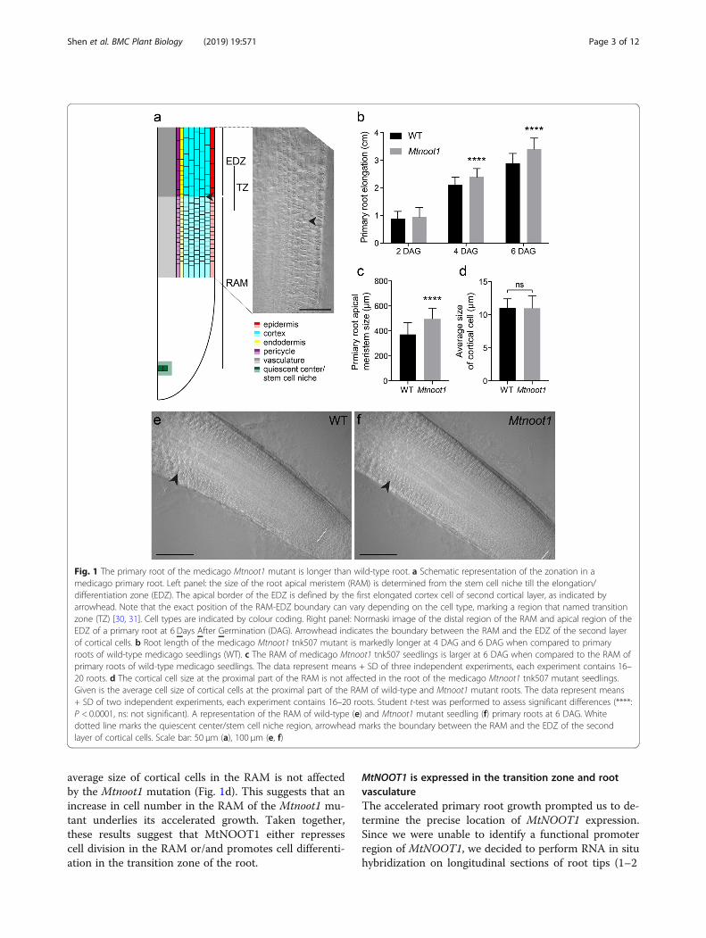
average size of cortical cells in the RAM is not affectedby the Mtnoot1 mutation (Fig. 1d). This suggests that anincrease in cell number in the RAM of the Mtnoot1 mu-tant underlies its accelerated growth. Taken together,these results suggest that MtNOOT1 either repressescell division in the RAM or/and promotes cell differenti-ation in the transition zone of the root.
MtNOOT1 is expressed in the transition zone and rootvasculatureThe accelerated primary root growth prompted us to de-termine the precise location of MtNOOT1 expression.Since we were unable to identify a functional promoterregion of MtNOOT1, we decided to perform RNA in situhybridization on longitudinal sections of root tips (1–2
Fig. 1 The primary root of the medicago Mtnoot1 mutant is longer than wild-type root. a Schematic representation of the zonation in amedicago primary root. Left panel: the size of the root apical meristem (RAM) is determined from the stem cell niche till the elongation/differentiation zone (EDZ). The apical border of the EDZ is defined by the first elongated cortex cell of second cortical layer, as indicated byarrowhead. Note that the exact position of the RAM-EDZ boundary can vary depending on the cell type, marking a region that named transitionzone (TZ) [30, 31]. Cell types are indicated by colour coding. Right panel: Normaski image of the distal region of the RAM and apical region of theEDZ of a primary root at 6 Days After Germination (DAG). Arrowhead indicates the boundary between the RAM and the EDZ of the second layerof cortical cells. b Root length of the medicago Mtnoot1 tnk507 mutant is markedly longer at 4 DAG and 6 DAG when compared to primaryroots of wild-type medicago seedlings (WT). c The RAM of medicago Mtnoot1 tnk507 seedlings is larger at 6 DAG when compared to the RAM ofprimary roots of wild-type medicago seedlings. The data represent means + SD of three independent experiments, each experiment contains 16–20 roots. d The cortical cell size at the proximal part of the RAM is not affected in the root of the medicago Mtnoot1 tnk507 mutant seedlings.Given is the average cell size of cortical cells at the proximal part of the RAM of wild-type and Mtnoot1 mutant roots. The data represent means+ SD of two independent experiments, each experiment contains 16–20 roots. Student t-test was performed to assess significant differences (****:P < 0.0001, ns: not significant). A representation of the RAM of wild-type (e) and Mtnoot1 mutant seedling (f) primary roots at 6 DAG. Whitedotted line marks the quiescent center/stem cell niche region, arrowhead marks the boundary between the RAM and the EDZ of the secondlayer of cortical cells. Scale bar: 50 μm (a), 100 μm (e, f)
Shen et al. BMC Plant Biology (2019) 19:571 Page 3 of 12
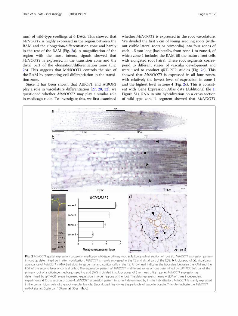
mm) of wild-type seedlings at 6 DAG. This showed thatMtNOOT1 is highly expressed in the region between theRAM and the elongation/differentiation zone and barelyin the rest of the RAM (Fig. 2a). A magnification of theregion with the most intense signals showed thatMtNOOT1 is expressed in the transition zone and thedistal part of the elongation/differentiation zone (Fig.2b). This suggests that MtNOOT1 controls the size ofthe RAM by promoting cell differentiation in the transi-tion zone.Since it has been shown that AtBOP1 and AtBOP2
play a role in vasculature differentiation [27, 28, 32], wequestioned whether MtNOOT1 may play a similar rolein medicago roots. To investigate this, we first examined
whether MtNOOT1 is expressed in the root vasculature.We divided the first 2 cm of young seedling roots (with-out visible lateral roots or primordia) into four zones ofeach ~ 5mm long (basipetally, from zone 1 to zone 4, ofwhich zone 1 includes the RAM till the mature root cellswith elongated root hairs). These root segments corres-pond to different stages of vascular development andwere used to conduct qRT-PCR studies (Fig. 2c). Thisshowed that MtNOOT1 is expressed in all four zones,with relatively the lowest level of expression in zone 1and the highest level in zone 4 (Fig. 2c). This is consist-ent with Gene Expression Atlas data (Additional file 1:Figure S1). RNA in situ hybridization on a cross sectionof wild-type zone 4 segment showed that MtNOOT1
Fig. 2 MtNOOT1 spatial expression pattern in medicago wild-type primary root. a, b Longitudinal section of root tip. MtNOOT1 expression patternin root tip determined by in situ hybridization. MtNOOT1 is mainly expressed in the TZ and distal part of the EDZ. b A close-up of (a), visualizingabundance of MtNOOT1 mRNA (red dots) in epidermal and cortical cells in the TZ. Arrowhead indicates the boundary between the RAM and theEDZ of the second layer of cortical cells. c The expression pattern of MtNOOT1 in different zones of root determined by qRT-PCR. Left panel: theprimary root of a wild-type medicago seedling at 6 DAG is divided into four zones of 5 mm each. Right panel: MtNOOT1 expression asdetermined by qRT-PCR reveals increased expression in older regions of the root. The data represent means + SEM of three independentexperiments. d Cross section of zone 4. MtNOOT1 expression pattern in zone 4 determined by in situ hybridization. MtNOOT1 is mainly expressedin the procambium cells of the root vascular bundle. Black dotted line circles the pericycle of vascular bundle. Triangles indicate the MtNOOT1mRNA signals. Scale bar: 100 μm (a), 50 μm (b, d)
Shen et al. BMC Plant Biology (2019) 19:571 Page 4 of 12

transcripts mainly occur in the procambium cells of thevasculature (Fig. 2d). This suggests that MtNOOT1could also play a role in root vasculature development inmedicago.
The primary root of the Mtnoot1 mutant is delayed inxylem cell differentiationAs MtNOOT1 is expressed in the root vasculature, weinvestigated whether the Mtnoot1 mutant is affected inroot vasculature development. Preliminary observationssuggested that Mtnoot1 mutant roots are thinner thanwild-type roots. To quantify this, cross sections onMtnoot1 mutant line tnk507 were made on the middleparts of zone 1 to zone 4 and the size of the cross-sectional area was determined. This showed that in zone2 to zone 4 the cross-sectional area of the Mtnoot1 mu-tant roots is significantly smaller when compared to thecounterparts of wild-type roots. The root cross-sectionalarea sizes of zone 3 and zone 4 of the Mtnoot1 mutantswere more comparable to those of zone 2 and zone 3 inwild-type, respectively (Fig. 3a). A similar reduction inroot cross-sectional area size was also observed in theMtnoot1 mutant line NF2717 (Additional file 3: FigureS3b). Further, the cross-sectional area sizes of the vascu-lature also showed that this is significantly smaller inMtnoot1 (tnk507) root zone 2 to zone 4 when comparedto wild-type roots. This suggests that the Mtnoot1 mu-tants have a delayed pattern of root radial growth, whichcorrelates with the growth defects in its vasculature.Vascular bundles are mainly built up of xylem and
phloem cells, in medicago roots in a four-arch constitu-tion (Fig. 3c, e). During the development of the root,vascular metaxylem cells differentiate and become ligni-fied [33]. Upon toluidine blue staining, lignified metaxy-lem cells gain a lighter blue colouration than non-lignified cells [34]. Upon toluidine blue staining of wild-type and Mtnoot1 tnk507 roots, we found that 32% (19/60) of wild-type zone 3 showed lignified central metaxy-lem cells, in contrast to only 2% (1/60) of Mtnoot1 zone3 showed lignification of central metaxylem cells. Fur-thermore, central metaxylem cells are lignified in 68%(41/60) of wild-type zone 4, but only in 27% (16/60) ofMtnoot1 zone 4, which is more similar to the lignifica-tion level of wild-type zone 3 (Fig. 4). A similar resultwas obtained by analysing the Mtnoot1 NF2717 mutantallele (Additional file 3: Figure S3c-g). These observa-tions demonstrate that in Mtnoot1 mutants the lignifica-tion level of vascular metaxylem cells in zone 3 and zone4 is lower when compared with wild-type, suggestingthat the differentiation of Mtnoot1 root vasculature isdelayed. This is consistent with the observation that thecross-sectional area sizes of Mtnoot1 root vasculature inzone 2 to zone 4 are significantly smaller when com-pared to wild-type (Fig. 3b).
The expression of genes involved in xylem celldifferentiation is delayed in Mtnoot1 mutant rootsIn arabidopsis, secondary cell wall formation and subse-quent programmed cell death (PCD) are two criticalsteps in the maturation of proto−/metaxylem and fibrecells [33]. Secondary cell wall biosynthesis is initiated bythe master regulators VASCULAR-RELATED NAC-DOMAIN 7 (AtVND7) for protoxylem, AtVND6 formetaxylem and SECONDARY WALL–ASSOCIATEDNAC DOMAIN PROTEIN 1 (AtSND1) for fibre differ-entiation [33]. These master regulators control second-ary cell wall biosynthesis via a network of genes, whichinclude the transcription factors AtMYB46, AtMYB58,AtMYB63, AtMYB83 and AtMYB85 that ultimately con-trol lignin biosynthesis genes, and the peptidase encod-ing gene XYLEM CYSTEINE PEPTIDASE 1 (AtXCP1)involved in the PCD during xylem cell development [33,35–37]. To find molecular support for the observationof the delayed vasculature development in the Mtnoot1mutant, we aimed to analyse the transcript levels of theputative medicago orthologues of the above-mentionedgenes. To identify such medicago orthologues we usedphylogenetic reconstruction (Additional files 4-9: Fig-ures. S4-S9). Subsequent qRT-PCR expression studieson medicago root zone 3 and zone 4 revealed that bothMtVND6 and MtVND7, but not MtSND1 are slightlylower expressed in zone 3 of Mtnoot1 mutant root (Add-itional file 10: Figure S10), while the putative down-stream targets MtMYB46–1, MtMYB46–2, MtMYB83,MtMYB58/63, and MtMYB85 and MtXCP1 are mark-edly lower expressed in the Mtnoot1 root (Fig. 5a). Inzone 4 of the Mtnoot1 mutant root, the expression of allthese genes was restored to the wild-type level (Fig. 5a),indicating a delayed transcriptional regulation of genescontrolling secondary cell wall biosynthesis in the apical-basal direction in the Mtnoot1 mutant roots. In contrast,the expression level of a phloem marker gene, ALTEREDPHLOEM DEVELOPMENT (MtAPL) [38], is not affectedin Mtnoot1 (Fig. 5b), suggesting that phloem cell devel-opment is not disturbed. Taken together, these resultssupport our observation that xylem cell differentiation inthe Mtnoot1 mutant is delayed.
The LCO susceptible zone of Mtnoot1 mutant roots isshifted basipetallyIn medicago, the site where the interaction with rhizobiaoccurs is tightly linked to the developmental status ofthe root tissue. At the start of the differentiation zone,where young elongating root hairs can be found is calledthe susceptible zone, and it is here where rhizobiumLCOs can trigger expression of symbiotic genes such asMtNIN [5–7]. The delayed root differentiation observedin the medicago Mtnoot1 mutant led us to questionwhether this may also affect the susceptibility of the root
Shen et al. BMC Plant Biology (2019) 19:571 Page 5 of 12

to rhizobium. To investigate this, we studied the expres-sion of MtNIN in the four root zones upon applicationof ~ 10− 9 M Sinorhizobium meliloti 2011 LCOs (dis-solved in 1% DMSO) or mock (1% DMSO). qRT-PCRexpression analysis showed that in wild-type roots thestrongest induction of MtNIN occurred in zone 1 and alower induction in zone 2. This indicates that the LCOsusceptible zone of wild-type predominately locates inzone 1 (Fig. 6). In contrast, in the medicago Mtnoot1mutant, the strongest MtNIN expression is observed inzone 2 (Fig. 6), implying that the Mtnoot1 mutation
caused the LCO susceptible zone to shift to zone 2. Thisis consistent with the delayed differentiation of theMtnoot1 root, which affects the LCO response by basip-etally shifting the LCO susceptible zone.
DiscussionHere, we demonstrate that MtNOOT1 is required for co-ordinated development of the primary root of medicagoalong the apical-basal axis. A knockout mutation in theMtNOOT1 gene causes two root developmental affects;(1) the primary root growth is faster due to a larger
Fig. 3 Medicago Mtnoot1 mutant seedlings have a thinner primary root at 6 DAG. The size of the cross-sectional area of (a) primary root and (b)vascular bundle of the medicago Mtnoot1 tnk507 mutant at zone 2, 3 and 4 are significantly smaller when compared to wild-type medicagoroots. The data represent means + SD of three independent experiments, each experiment contains 18–20 roots. Student t-test was performed toassess significant differences (**: P < 0.01, ***: P < 0.001, ****: P < 0.0001). Representative root cross sections of wild-type (c) and Mtnoot1 (d) roots,and wild-type (e) and Mtnoot1 (f) vascular bundle at zone 4. Black dotted line circles the pericycle of vascular bundle. Black arrow marksdifferentiated/lignified metaxylem cells, which are not found in the Mtnoot1 mutant. Scale bar: 100 μm (c, d), 50 μm (e, f)
Shen et al. BMC Plant Biology (2019) 19:571 Page 6 of 12

RAM, meaning more dividing cells, and (2) the primaryroot is thinner with delayed root differentiation. Thisdemonstrates that in roots MtNOOT1 not only functionsas a homeotic gene in nodule development, but also co-ordinates root development.In situ hybridization revealed that MtNOOT1 is
expressed in the transition zone, which is located be-tween the RAM and the elongation/differentiation zone.A larger RAM due to an increased number of meristem-atic cells indicates that the transition zone is positionedmore distal from the stem cell niche. This suggests thatMtNOOT1 controls the size of the RAM by defining theposition of the boundary region (i.e. the transition zone)between the RAM and the elongation//differentiationzone. This is consistent with the aerial function of itsorthologs in arabidopsis, where AtBOP1 and AtBOP2promote the boundary specification between groups ofcells with different fates [15–18]. In contrast to what wereport for medicago Mtnoot1, a root growth and differ-entiation phenotype has not been reported in the arabi-dopsis Atbop1;Atbop2 double mutant [28]. Therecruitment of NBCL family by legumes to maintainnodule symbiotic organ identity by defining nodule
territories may have required adaptations in proteinregulation and functioning [10]. Such adaptations couldbe causal for the difference in root functioning ofMtNOOT1 in medicago and AtBOP1 and AtBOP2 inarabidopsis. Further, it has been proposed that arabidop-sis has a closed RAM organization with distinct initialcells, while legumes have a RAM with a basic-openorganization. In the latter case, the identity of cell filesseems disorganized and cannot be predicted [39]. Thisdeviation in structure of the RAM may explain the dif-ferent functioning of BOP1/NOOT1 in arabidopsis andlegume roots.We also noted a basipetal shift of the root zone that is
susceptible for rhizobium LCO signal molecules in themedicago Mtnoot1 mutant. Such shift of susceptibility torhizobium can be explained by the delayed differenti-ation of root cells. In medicago, susceptibility to LCOsignalling can only occur in defined window of develop-ment. By elongating the RAM, the differentiation zonegets shifted basipetally. As a result, the susceptible zoneshifts basipetally as well.In the arabidopsis root the transition zone is deter-
mined by the antagonistic interaction between auxin and
Fig. 4 Vascular xylem differentiation is delayed in the primary root of Mtnoot1 at 6 DAG. The fraction of roots with lignified central metaxylemcells in zone 3 and zone 4 is decreased in the Mtnoot1 tnk507 mutant when compared with wild-type seedlings. The lignification level of thecentral metaxylem cells in zone 4 of the Mtnoot1 mutant is similar to that of wild-type medicago zone 3, indicating that the differentiation ofvascular xylem cells is delayed in the Mtnoot1 mutant. The presented data combines three independent experiments, each experimentcontains 20 roots
Shen et al. BMC Plant Biology (2019) 19:571 Page 7 of 12

cytokinin. A well-defined auxin minimum is establishedin the boundary region, which is mediated by an activecytokinin signalling in the transition zone [40]. Activa-tion of auxin signalling or inhibition of cytokinin signal-ling can lead to accelerated primary root elongation anda larger RAM with increased number of meristematic
cells, which are similar to the phenotype of Mtnoot1roots [30, 31]. Auxin and cytokinin are also involved inroot vasculature development (Reviewed in [41, 42]). Forexample, mutations in cytokinin biosynthetic genes leadto abolished cambium formation and reduced thickeningof the arabidopsis root [43], and auxin signalling is
Fig. 6 Rhizobium LCO-induced MtNIN expression is spatially different in the medicago Mtnoot1 roots in comparison with wild-type roots. MtNINexpression is quantified using qRT-PCR in four root zones at three hours post application of 10− 9 M S. meliloti LCOs. The data represent means +SEM of two independent experiments using the Mtnoot1 tnk507 mutant
Fig. 5 Genes putatively involved in xylem cell differentiation display reduced expression in Mtnoot1 roots. a, b Expression level of various genesputatively involved in xylem and phloem cell differentiation in zone 3 and zone 4 of wild-type and Mtnoot1 tnk507 roots determined by qRT-PCR.a The MYB-type transcription factors MtMYB46–1, MtMYB46–2, MtMYB83 and MtMYB85, and the XYLYME CYSTEINE PEPTIDASE 1 putative orthologMtXCP1 are lower expressed in zone 3 of Mtnoot1 mutant roots when compared to wild-type. c The expression level of phloem marker geneMtAPL is not affected in Mtnoot1 mutant roots. The data represent means + SEM of two (MtXCP1 and MtAPL) or three (MtMYBs) independentexperiments. Student t-test was performed to assess significant differences (*: P < 0.05, ****: P < 0.0001, ns: not significant)
Shen et al. BMC Plant Biology (2019) 19:571 Page 8 of 12
required for xylem differentiation and the organizeridentity of vascular cambium cells in arabidopsis root[44]. In legumes, auxin and cytokinin participate in theLCO response and nodule organogenesis (Reviewed in[45–47]). For example, a mutation in cytokinin percep-tion severally weakens the LCO response and perturbsnodule organogenesis [7, 48], whereas LCOs triggerauxin biosynthesis in the epidermis, leading to a rapidaccumulation of auxin locally [49]. Recent findings sug-gest that in Mtnoot1 nodules the interplay betweenauxin and cytokinin is imbalanced [12]. In line with that,we speculate that the auxin/cytokinin signalling pathwayis affected during the primary growth of Mtnoot1 roots,which will be targeted in future research.We also showed that the lignification of xylem cells is
delayed in Mtnoot1 primary roots, which correlates witha delayed induction of a cascade of transcription factorsinvolved in xylem cell differentiation. This can be ex-plained by the extended RAM, leading to delayed celldifferentiation in the basal region of primary root. How-ever, the expression of MtNOOT1 in the procambiumcells by in situ hybridization suggests that MtNOOT1can positively regulates vascular cell differentiation in acell-autonomous manner. This is in line with the func-tion of arabidopsis AtBOP1 and AtBOP2 in stem vascu-lature, where they can induce lignin deposition invascular cells by promoting the expression of lignin bio-synthetic genes. Ectopic expression of AtBOP1/AtBOP2in stem tissue leads to an expanded pattern of lignifica-tion [32]. Therefore, we hypothesize that MtNOOT1promotes root vasculature differentiation by activating acascade of genes involved in xylem cell differentiation.
ConclusionsHere, we showed that MtNOOT1 controls two aspectsof root development; (1) the positioning of the transitionzone, and (2) vasculature development by transcription-ally activating of a cascade of genes involved in xylemcell differentiation at a controlled distance from the roottip. Knockout of the MtNOOT1 gene not only leads to adelayed differentiation of the root, but also shift the rootregion susceptible to LCOs. Taken together, this demon-strates that MtNOOT1 not only maintains nodule iden-tity, but also coordinates the primary root developmentalong the apical-basal axis.
MethodsPlant materials and growth conditionsMedicago truncatula wild-type accession R108–1 plantsand Mtnoot1 mutant lines tnk507 and NF2717 wereused in this study [10, 12]. R108–1 was acquired fromToan Hanh Trinh [50]. tnk507 and NF2717 were ac-quired from Pascal Ratet (IPS2, CNRS, Gif-sur-Yvette,France), they were identified by a forward genetics
screen of Tnt1 insertion lines (Institut des Sciences duVégétal, France; Noble Foundation, Ardmore, USA). Thesurface-sterilization and germination of medicago seedswere performed as previously described by [51]. Notethat medicago seeds germinated at room temperaturefor one day, before growing on Fahraeus agar medium[52] (including 0.75 mM Ca (NO3)2) in square petri dish(9 cm × 9 cm) and exposed to light for another five days.Plants were grown in an environmentally controlledgrowth chamber at 21 °C with a 16-h light/8-h dark.
Microscopy and imagingFor microscopy studies, root segments were all collectedat 6 DAG. For measuring the length of the RAM, ~ 5mm root tips were cut and immersed in chloral hydratesolution at 4 °C overnight, analyzed under Axio ImagerA1 microscope (Zeiss) with Nomarski optics. For meas-uring cross-sectional areas, root segments were fixedwith 4% paraformaldehyde (w/v), 5% glutaraldehyde (v/v) in 0.05M sodium phosphate buffer (pH 7.2) at 4 °Covernight. The fixed material was dehydrated in an etha-nol series and subsequently embedded in Technovit7100 (Heraeus Kulzer) according to the manufacturer’sprotocol. Sections (5 μm) were made with a RJ2035microtome (Leica Microsystems) stained 1.5 min in0.05% toluidine blue O. For phloroglucinol-HCl staining,root segments were fixed as abovementioned. The fixedmaterial was washed with 1 x PBS (sodium phosphatebuffer), and directly embedded in 6% low melting agar-ose dissolved in 1 x PBS. Sections (50 μm) were madewith a VT1000 S vibratome (Leica Microsystems),stained with 2% phloroglucinol (in 95% ethanol) for 2min and applied with a few drops of 37% HCl. Sectionswere all analysed by using a DM5500B microscopeequipped with a DFC425C camera (Leica Microsystems).
In situ hybridizationHybridization was performed twice on root segments at 6DAG by using Invitrogen ViewRNA ISH Tissue 1-Plexassay kit (Thermo Fisher Scientific), as previously de-scribed by [9, 53]. For user manual, visit https://assets.thermofisher.com/TFS-Assets/LSG/manuals/MAN0018633_viewRNA_ISH_UG.pdf. The probe sets for MtNOOT1(catalogue number: VF1–16434, information is availableon request) were designed and synthesized by ThermoFisher Scientific. MtNOOT1 probe sets cover the region2–913 nucleotide (nt) of the coding sequence (1449 nt,Medtr7g090020.1). A typical probe set contains ~ 20oligonucleotide pairs of probes that hybridize to specificregions across the target mRNA. Each probe covers 20 nt,only a pair of two adjacent probes, which can target 40 nt,can form a site for signal amplification. By this principle,control probes are not needed [9, 53–56]. Sections wereimaged as mentioned above.
Shen et al. BMC Plant Biology (2019) 19:571 Page 9 of 12

Phylogenetic tree constructionThe protein sequences of different orthogroups were ob-tained from [57]. For phylogenetic reconstruction, fulllength (predicted) protein sequences of at least twoclosely related orthogroups were aligned using MAFFTv7.017 [58], implemented in Geneious R6 (Biomatters,Auckland, New Zealand), using default parameter set-tings. After manual inspection, alignments were used fortree building by using W-IQ-TREE [59] with best-fitsubstitution model [60]. Branch support was assessed byusing Ultrafast Bootstrap Approximation based on 1000replicates [61].
RNA isolation and qRT-PCR analysisRNA was isolated from root segments at 6 DAG usingthe EZNA Plant RNA mini kit (Omega), following thesupplier’s manual. 1 μg total RNA was used to synthesizecDNA using iScript cDNA synthesis kit (Bio-Rad). Equalamounts of cDNA were used for qPCR using SYBRGreen Super-mix (Bio-Rad) in a Bio-Rad CFX connectreal-time system qPCR machine. Cycling conditionswere: 95 °C for 3 min, [95 °C for 15 s, 60 °C for 30 s] (40cycles), 95 °C for 10 s, followed by melt curve analysis(from 65 °C to 95 °C, at an increment of 0.5 °C, for 5 s).The gene expression was normalized using MtACT2 asreference gene. Three technical replicates per biologicalreplicate. All primers used in this study are listed inAdditional file 11: Table S1.
Supplementary informationSupplementary information accompanies this paper at https://doi.org/10.1186/s12870-019-2194-z.
Additional file 1: Figure S1. Medicago MtNOOT1 is expressed in theroot tip. Expression profiles are derived from the Medicago truncatulaGene Expression Atlas [29]. MtNOOT1 is targeted by the probe-setsMtr.19586.1.S1_at, Mtr.27707.1.S1_s_at, and Mtr.39297.1.S1_s_at. Root 3mm tip: 3 mm root tip [14]; adj tip: 1 cm root segment adjacent to 3 mmroot tip [14]; Nod: nodules, all nodule samples are derived from [13]; dpi:days post inoculation. (DOCX)
Additional file 2: Figure S2. The primary root Mtnoot1 tnk507 mutantis longer than wild-type. Representative seedlings at 6 DAG are pre-sented. (DOCX)
Additional file 3: Figure S3. The Mtnoot1 NF2717 mutant allele showsa similar phenotype as the Mtnoot1 tnk507 allele. a Root length of themedicago Mtnoot1 mutant (NF2717) is markedly longer at 4 DAG and 6DAG when compared to primary roots of wild-type medicago seedlings(WT). b The cross-sectional area is significantly reduced in the medicagoMtnoot1 mutant (NF2717) at zone 3 and zone 4. The data representmeans + SD of two independent experiments, each experiment contains15–20 roots. Student t-test was performed to assess significant differences(****: P < 0.0001). Representative root cross sections of wild-type (c, e)and Mtnoot1 (NF2717) (d, f) vascular bundle at zone 3 stained withphloroglucinol-HCl to demonstrate lignin deposition at 6 DAG. Blackarrow marks lignified metaxylem cells, which are not found in theMtnoot1 mutant (NF2717). g Vascular xylem differentiation is delayed inthe Mtnoot1 (NF2717) primary root at 6 DAG. The fraction of roots withlignified central metaxylem cells in zone 3 and zone 4 is decreased in theMtnoot1 mutant when compared with wild-type seedlings. The presented
data combines two independent experiments, each experiment contains15–18 roots. Scale bar: 50 μm (c, d), 100 μm (e, f). (DOCX)
Additional file 4: Figure S4. Maximum likelihood tree of VND6, VND7and related proteins. The protein sequences of OG0006787 (red),OG0009959 (dark red), OG0004118 (purple), OG0001465 (green and blue)are obtained from van Velzen et al. (2018), except VND6, which was notincluded in OG0001465. Species include arabidopsis (Athaliana),Eucalyptus grandis (Egrandis), Fragaria vesca (Fvesca), Glycine max (Gmax),medicago (Mtruncatula), Populus trichocarpa (Ptrichocarpa), Parasponiaandersonii (Pan) and Trema orientalis (Tor). Numbers at to the branchesindicate support from 1000 ultrafast bootstrap replicates. OG0006787including NAC1 was used as outgroup. (DOCX)
Additional file 5: Figure S5. Maximum likelihood tree of SND1 andrelated proteins. The protein sequences of OG0009898 (red) andOG0001875 (green) are obtained from van Velzen et al. (2018). Speciesinclude arabidopsis (Athaliana), Eucalyptus grandis (Egrandis), Fragariavesca (Fvesca), Glycine max (Gmax), medicago (Mtruncatula), Populustrichocarpa (Ptrichocarpa), Parasponia andersonii (Pan) and Tremaorientalis (Tor). Numbers at to the branches indicate support from 1000ultrafast bootstrap replicates. OG0009898 containing SMB was used asoutgroup. (DOCX)
Additional file 6: Figure S6. Maximum likelihood tree of MYB46,MYB83 and related proteins. The protein sequences of OG0000857 (red)and OG0001270 (green) are obtained from van Velzen et al. (2018).Species include arabidopsis (Athaliana), Eucalyptus grandis (Egrandis),Fragaria vesca (Fvesca), Glycine max (Gmax), medicago (Mtruncatula),Populus trichocarpa (Ptrichocarpa), Parasponia andersonii (Pan) and Tremaorientalis (Tor). Numbers at the branches indicate support from 1000ultrafast bootstrap replicates. OG0000857 containing MYB50 was used asoutgroup. (DOCX)
Additional file 7: Figure S7. Maximum likelihood tree of MYB58,MYB63 and MYB85 proteins. The protein sequences of OG0005384 (red)and OG0002420 (green) are obtained from van Velzen et al. (2018).Species include arabidopsis (Athaliana), Eucalyptus grandis (Egrandis),Fragaria vesca (Fvesca), Glycine max (Gmax), medicago (Mtruncatula),Populus trichocarpa (Ptrichocarpa), Parasponia andersonii (Pan) and Tremaorientalis (Tor). Numbers at the branches indicate support from 1000ultrafast bootstrap replicates. OG0005384 containing MYB58 and MYB63was used as outgroup. (DOCX)
Additional file 8: Figure S8. Maximum likelihood tree of XCP1 andrelated proteins. The protein sequences of OG0003401 (red) andOG0003952 (green) are obtained from van Velzen et al. (2018). Speciesinclude arabidopsis (Athaliana), Eucalyptus grandis (Egrandis), Fragariavesca (Fvesca), Glycine max (Gmax), medicago (Mtruncatula), Populustrichocarpa (Ptrichocarpa), Parasponia andersonii (Pan) and Tremaorientalis (Tor). Numbers at the branches indicate support from 1000ultrafast bootstrap replicates. OG0003401 containing CEP1 was used asoutgroup. (DOCX)
Additional file 9: Figure S9. Maximum likelihood tree of APL andrelated proteins. The protein sequences of OG0009526 (red) andOG0006786 (green) are obtained from van Velzen et al. (2018). Speciesinclude arabidopsis (Athaliana), Eucalyptus grandis (Egrandis), Fragariavesca (Fvesca), Glycine max (Gmax), medicago (Mtruncatula), Populustrichocarpa (Ptrichocarpa), Parasponia andersonii (Pan) and Tremaorientalis (Tor). Numbers at the branches indicate support from 1000ultrafast bootstrap replicates. OG0009526 containing sequences highlyhomologous to MtAPL was used as outgroup. (DOCX)
Additional file 10: Figure S10. The NAC domain transcription factorsMtVND6 and MtVND7, but not MtSND1, are lower expressed in zone 3 ofMtnoot1 tnk507 roots when compared to wild-type. The data representmeans + SEM of three independent experiments. Student t-test was per-formed to assess significant differences (ns: not significant). (DOCX)
Additional file 11: Table S1. qRT-PCR primers used in this study.(DOCX)
AbbreviationsAZ: Abscission zone; DAG: Days after germination; DMSO: Dimethyl sulfoxide;EDZ: Elongation/differentiation zone; LCO: Lipo-chitooligosaccharide;
Shen et al. BMC Plant Biology (2019) 19:571 Page 10 of 12

nt: Nucleotide; PBS: Sodium phosphate buffer; qRT-PCR: Quantitative reversetranscriptase polymerase chain reaction; RAM: Root apical meristem;TZ: Transition zone
AcknowledgementsWe thank Pascal Ratet for providing the Mtnoot1 seeds (tnk507 and NF2717)and thank Huchen Li and Kévin Magne for their comments on themanuscript.
Authors’ contributionsRG and TB supervised this research. DS designed and performed most of theexperiments with help from KG and WK. OK and DS performed the in situhybridization. DS drafted the manuscript with input from HF. RG contributedto writing and revising the manuscript, with input from TB. All authors readand approved the final manuscript.
FundingThis research was supported by China Scholarship Council (201306040120) toDS, European Research Council (ERC-2011-AdG294790) to TB, and a NWO-VICI grant (865.13.001) to RG. These funding bodies did not play any roles inthe design of the study and collection, analysis, and interpretation of dataand in writing the manuscript.
Availability of data and materialsThe datasets supporting the conclusions of this research and materials usedin this research are available by contacting with the corresponding author([email protected]).
Ethics approval and consent to participateNot applicable.
Consent for publicationNot applicable.
Competing interestsThe authors declare that they have no competing interests. RG is a memberof the editorial board (Associate Editor) of BMC Plant Biology.
Received: 21 September 2019 Accepted: 10 December 2019
References1. Xiao TT, Schilderink S, Moling S, Deinum EE, Kondorosi E, Franssen H, et al.
Fate map of Medicago truncatula root nodules. Development. 2014;141:3517–28.
2. Schauser L, Roussis A, Stiller J, Stougaard J. A plant regulator controllingdevelopment of symbiotic root nodules. Nature. 1999;402:191–5.
3. Marsh JF, Rakocevic A, Mitra RM, Brocard L, Sun J, Eschstruth A, et al.Medicago truncatula NIN is essential for Rhizobial-independent noduleorganogenesis induced by autoactive calcium/Calmodulin-dependentprotein kinase. Plant Physiol. 2007;144:324–35.
4. Desbrosses GJ, Stougaard J. Root nodulation: a paradigm for how plant-microbe symbiosis influences host developmental pathways. Cell Host andMicrobe. 2011;10:348–58.
5. Yano K, Yoshida S, Müller J, Singh S, Banba M, Vickers K, et al. CYCLOPS, amediator of symbiotic intracellular accommodation. Proc Natl Acad Sci U SA. 2008;105:20540–5.
6. Vernié T, Kim J, Frances L, Ding Y, Sun J, Guan D, et al. The NIN transcriptionfactor coordinates diverse nodulation programs in different tissues of theMedicago truncatula root. Plant Cell. 2015;27:3410–24.
7. Van Zeijl A. Op Den camp RHM, Deinum EE, Charnikhova T, Franssen H, OpDen camp HJM, et al. rhizobium Lipo-chitooligosaccharide signaling triggersaccumulation of Cytokinins in Medicago truncatula roots. Mol Plant. 2015;8:1213–26.
8. Soyano T, Kouchi H, Hirota A, Hayashi M. NODULE INCEPTION directlytargets NF-Y subunit genes to regulate essential processes of root noduledevelopment in Lotus japonicus. PLoS Genet. 2013;9:e1003352.
9. Liu J, Rutten L, Limpens E, van der Molen T, van Velzen R, Chen R, et al. Aremote cis-regulatory region is required for NIN expression in the Pericycleto initiate nodule primordium formation in Medicago truncatula. Plant Cell.2019;31:68–83.
10. Couzigou J-MJ, Zhukov V, Mondy S, Abu el Heba G, Cosson V, THN E, et al.NODULE ROOT and COCHLEATA maintain nodule development and arelegume Orthologs of Arabidopsis BLADE-ON-PETIOLE genes. Plant Cell.2012;24:4498–510.
11. Magne K, George J, Berbel Tornero A, Broquet B, Madueño F, Andersen SU,et al. Lotus japonicus NOOT-BOP-COCH-LIKE1 is essential for nodule, nectary,leaf and flower development. Plant J. 2018;94:880–94.
12. Magne K, Couzigou J-M, Schiessl K, Liu S, George J, Zhukov V, et al.MtNODULE ROOT1 and MtNODULE ROOT2 are essential for indeterminatenodule identity. Plant Physiol. 2018;178:295–316.
13. Benedito VA, Torres-Jerez I, Murray JD, Andriankaja A, Allen S, Kakar K, et al.A gene expression atlas of the model legume Medicago truncatula. Plant J.2008;55:504–13.
14. Holmes P, Goffard N, Weiller GF, Rolfe BG, Imin N. Transcriptional profiling ofMedicago truncatula meristematic root cells. BMC Plant Biol. 2008;8:21.
15. Aida M, Tasaka M. Genetic control of shoot organ boundaries. Curr OpinPlant Biol. 2006;9:72–7.
16. Khan M, Xu H, Hepworth SR. BLADE-ON-PETIOLE genes: setting boundariesin development and defense. Plant Sci. 2014;215–216:157–71.
17. Žádníková P, Simon R. How boundaries control plant development. CurrOpin Plant Biol. 2014;17:116–25.
18. Hepworth SR, Pautot VA. Beyond the divide: boundaries for patterning andstem cell regulation in plants. Front Plant Sci. 2015;6:1052.
19. Wang Q, Hasson A, Rossmann S, Theres K. Divide et impera: boundariesshape the plant body and initiate new meristems. New Phytol. 2016;209:485–98.
20. Jun JH, Ha CM, Fletcher JC. BLADE-ON-PETIOLE1 coordinates organdeterminacy and axial polarity in Arabidopsis by directly activatingASYMMETRIC LEAVES2. Plant Cell. 2010;22:62–76.
21. Bell EM, Lin W -C., Husbands AY, Yu L, Jaganatha V, Jablonska B, et al.Arabidopsis LATERAL ORGAN BOUNDARIES negatively regulatesbrassinosteroid accumulation to limit growth in organ boundaries. Proc NatlAcad Sci 2012;109:21146–21151.
22. Ha CM, Kim G-T, Kim BC, Jun JH, Soh MS, Ueno Y, et al. The BLADE-ON-PETIOLE 1 gene controls leaf pattern formation through the modulation ofmeristematic activity in Arabidopsis. Development. 2003;130:161–72.
23. Ha CM, Jun JH, Nam HG, Fletcher JC. BLADE-ON-PETIOLE1 and 2 controlArabidopsis lateral organ fate through regulation of LOB domain andAdaxial-Abaxial polarity genes. Plant Cell. 2007;19:1809–25.
24. McKim SM, Stenvik G-E, Butenko MA, Kristiansen W, Cho SK, Hepworth SR,et al. The BLADE-ON-PETIOLE genes are essential for abscission zoneformation in Arabidopsis. Development. 2008;135:1537–46.
25. Couzigou JM, Magne K, Mondy S, Cosson V, Clements J, Ratet P. Thelegume NOOT-BOP-COCH-LIKE (NBCL) genes are conserved regulators ofabscission, a major agronomical trait in cultivated crops. New Phytol. 2016;209:228–40.
26. Ichihashi Y, Kawade K, Usami T, Horiguchi G, Takahashi T, Tsukaya H. Keyproliferative activity in the junction between the leaf blade and leaf Petioleof Arabidopsis. Plant Physiol. 2011;157:1151–62.
27. Liebsch D, Sunaryo W, Holmlund M, Norberg M, Zhang J, Hall HC, et al.Class I KNOX transcription factors promote differentiation of cambialderivatives into xylem fibers in the Arabidopsis hypocotyl. Development.2014;141:4311–9.
28. Woerlen N, Allam G, Popescu A, Corrigan L, Pautot V, Hepworth SR.Repression of BLADE-ON-PETIOLE genes by KNOX homeodomain proteinBREVIPEDICELLUS is essential for differentiation of secondary xylem inArabidopsis root. Planta. 2017;245:1079–90.
29. He J, Benedito VA, Wang M, Murray JD, Zhao PX, Tang Y, et al. The Medicagotruncatula gene expression atlas web server. BMC Bioinformatics. 2009;10:441.
30. Dello Ioio R, Linhares FS, Scacchi E, Casamitjana-Martinez E, Heidstra R,Costantino P, et al. Cytokinins determine Arabidopsis root-meristem size bycontrolling cell differentiation. Curr Biol. 2007;17:678–82.
31. Ioio RD, Nakamura K, Moubayidin L, Perilli S, Taniguchi M, Morita MT, et al. Agenetic framework for the control of cell division and differentiation in theroot meristem. Science. 2008;322:1380–4.
32. Khan M, Xu M, Murmu J, Tabb P, Liu Y, Storey K, et al. Antagonisticinteraction of BLADE-ON-PETIOLE1 and 2 with BREVIPEDICELLUS andPENNYWISE regulates Arabidopsis inflorescence architecture. Plant Physiol.2012;158:946–60.
33. Schuetz M, Smith R, Ellis B. Xylem tissue specification, patterning, anddifferentiation mechanisms. J Exp Bot. 2013;64:11–31.
Shen et al. BMC Plant Biology (2019) 19:571 Page 11 of 12

34. Lars Hennig, Köhler C. Plant Developmental Biology Methods and Protocols 2010.35. Zhong R, Lee C, Zhou J, McCarthy RL, Ye Z-H. A battery of transcription
factors involved in the regulation of secondary Cell Wall biosynthesis inArabidopsis. Plant Cell Online. 2008;20:2763–82.
36. Ohashi-Ito K, Oda Y, Fukuda H. Arabidopsis VASCULAR-RELATED NAC-DOMAIN6 directly regulates the genes that govern programmed cell deathand secondary wall formation during xylem differentiation. Plant Cell. 2010;22:3461–73.
37. Zhong R, Lee C, Ye Z-H. Global analysis of direct targets of secondary wallNAC master switches in Arabidopsis. Mol Plant. 2010;3:1087–103.
38. Bonke M, Thitamadee S, Mähönen AP, Hauser MT, Helariutta Y. APLregulates vascular tissue identity in Arabidopsis. Nature. 2003;426:181–6.
39. Rost TL. The organization of roots of dicotyledonous plants and thepositions of control points. Ann Bot. 2011;107:1213–22.
40. Kong X, Liu G, Liu J, Ding Z. The root transition zone: a hot spot for signalcrosstalk. Trends Plant Sci. 2018;23:403–9.
41. Campbell L, Turner S. Regulation of vascular cell division. J Exp Bot.2017;68:27–43.
42. Ruonala R, Ko D, Helariutta Y. Genetic networks in plant vasculardevelopment. Annu Rev Genet. 2017;51:335–59.
43. Matsumoto-Kitano M, Kusumoto T, Tarkowski P, Kinoshita-Tsujimura K,Vaclavikova K, Miyawaki K, et al. Cytokinins are central regulators of cambialactivity. Proc Natl Acad Sci. 2008;105:20027–31.
44. Smetana O, Mäkilä R, Lyu M, Amiryousefi A, Sánchez Rodríguez F, Wu M-F,et al. High levels of auxin signalling define the stem-cell organizer of thevascular cambium. Nature. 2019;565:485–9.
45. Boivin S, Fonouni-Farde C, Frugier F. How Auxin and Cytokinin PhytohormonesModulate Root Microbe Interactions. Front Plant Sci. 2016;7:1–12.
46. Gamas P, Brault M, Jardinaud MF, Frugier F. Cytokinins in symbioticnodulation: when, where, what for? Trends Plant Sci. 2017;22:792–802.
47. Kohlen W, Ng JLP, Deinum EE, Mathesius U. Auxin transport, metabolism,and signalling during nodule initiation: indeterminate and determinatenodules. J Exp Bot. 2018;69:229–44.
48. Gonzalez-Rizzo S, Crespi M, Frugier F. The Medicago truncatula CRE1cytokinin receptor regulates lateral root development and early symbioticinteraction with Sinorhizobium meliloti. Plant Cell. 2006;18:2680–93.
49. Nadzieja M, Kelly S, Stougaard J, Reid D. Epidermal auxin biosynthesisfacilitates rhizobial infection in Lotus japonicus. Plant J. 2018;95:101–11.
50. Hoffmann B, Trinh TH, Leung J, Kondorosi A, Kondorosi E. A new Medicagotruncatula line with superior in vitro regeneration, transformation, andsymbiotic properties isolated through cell culture selection. Mol Plant-Microbe Interact. 1997;10:307–15.
51. Limpens E, Ramos J, Franken C, Raz V, Compaan B, Franssen H, et al. RNAinterference in Agrobacterium rhizogenes-transformed roots of Arabidopsisand Medicago truncatula. J Exp Bot. 2004;55:983–92.
52. Fahraeus G. The infection of clover root hairs by nodule bacteria studied bya simple glass slide technique. J Gen Microbiol. 1957;16:374–81.
53. Kulikova O, Franken C, Bisseling T. Methods in Molecular Biology. In: In situhybridization method for localization of mRNA molecules in medicagotissue sections. New York, NY: Humana Press; 2018. p. 145–59.
54. Katsushima K, Natsume A, Ohka F, Shinjo K, Hatanaka A, Ichimura N, et al.Targeting the Notch-regulated non-coding RNA TUG1 for glioma treatment.Nat Commun. 2016;7:1–14.
55. Osteen JD, Herzig V, Gilchrist J, Emrick JJ, Zhang C, Wang X, et al. Selectivespider toxins reveal a role for the Nav1.1 channel in mechanical pain.Nature. 2016;534:494–9.
56. Roux B, Rodde N, Jardinaud MF, Timmers T, Sauviac L, Cottret L, et al. Anintegrated analysis of plant and bacterial gene expression in symbiotic rootnodules using laser-capture microdissection coupled to RNA sequencing.Plant J. 2014;77:817–37.
57. van Velzen R, Holmer R, Bu F, Rutten L, van Zeijl A, Liu W, et al. Comparativegenomics of the nonlegume Parasponia reveals insights into evolution ofnitrogen-fixing rhizobium symbioses. Proc Natl Acad Sci U S A. 2018;115:E4700–9.
58. Katoh K. MAFFT: a novel method for rapid multiple sequence alignmentbased on fast Fourier transform. Nucleic Acids Res. 2002;30:3059–66.
59. Trifinopoulos J, Nguyen LT, von Haeseler A, Minh BQ. W-IQ-TREE: a fastonline phylogenetic tool for maximum likelihood analysis. Nucleic AcidsRes. 2016;44:W232–5.
60. Kalyaanamoorthy S, Minh BQ, Wong TKF, Von Haeseler A, Jermiin LS.ModelFinder: fast model selection for accurate phylogenetic estimates. NatMethods. 2017;14:587–9.
61. Minh BQ, Nguyen MAT, Von Haeseler A. Ultrafast approximation forphylogenetic bootstrap. Mol Biol Evol. 2013;30:1188–95.
Publisher’s NoteSpringer Nature remains neutral with regard to jurisdictional claims inpublished maps and institutional affiliations.
Shen et al. BMC Plant Biology (2019) 19:571 Page 12 of 12