increased neuromuscular activity reduces … neuromuscular activity reduces sprouting in partially...
TRANSCRIPT

Increased Neuromuscular Activity Reduces Sprouting in PartiallyDenervated Muscles
Siu Lin Tam, Vey Archibald, Balvinder Jassar, Neil Tyreman, and Tessa Gordon
Department of Pharmacology, Division of Neuroscience, University of Alberta, Edmonton, Canada T6G 2S2
The effects of increasing neural activity on sprouting remainunclear and controversial. In a rat model of partial denervationof skeletal muscles, we investigated the effect of neuromuscu-lar activity on sprouting. Rat hindlimb muscles were partiallydenervated by avulsion of either L4 or L5 spinal root. Immedi-ately after partial denervation, the rats were divided into threegroups: (1) normal caged activity, (2) running exercise onwheels, 8 hr daily, and (3) functional electrical stimulation (FES)of sciatic nerves, 20 Hz for 8 hr daily. At 1 month, muscle unit(MU) enlargement was quantitated electrophysiologically andhistochemically. MU twitch force was increased by four- tofivefold by partial denervation in extensively denervated tibialisanterior (TA) and medial gastrocnemius (MG) and by approxi-mately twofold in moderately denervated plantaris (PL) andsoleus (SOL). For the extensively denervated TA and MG mus-
cles, MU enlargement, measured electrophysiologically, de-clined significantly after an average of 1757 6 310 m/d runningexercise and daily FES for 1 month. The detrimental effects onMU enlargement were much less but significant in the moder-ately denervated PL and did not reach statistical significance inthe moderately denervated SOL muscle. Histochemical evalu-ation of sprouting showed a reduction in the number of sproutsin the extensively denervated TA muscle, but not the moder-ately denervated PL and SOL muscles, by increased neuromus-cular activity. Thus, increased neuromuscular activity is detri-mental primarily in muscles that are extensively denervated,and the MUs are smaller than under conditions in which themuscles experience normal physiological levels of activation.
Key words: sprouting; motor unit; motoneuron disease; neu-romuscular activity; partial denervation; poliomyelitis
Poliomyelitis, the early stages of amyotrophic lateral sclerosis(ALS), spinal cord trauma, and motoneuron destruction associ-ated with cancer are only some of the neuromuscular conditionsresulting in compensatory axonal sprouting and, in turn, MUenlargement (Brown et al., 1981; Halstead and Wiechers, 1987).MU enlargement is unfortunately restricted to a limit of five- toeightfold such that sprouting compensates for up to 85% loss ofmuscle units (MUs) (Thompson and Jansen, 1977; Brown andIronton, 1978; Yang et al., 1990; Rafuse et al., 1992). Thus when,20% of intact MUs remain and sprouting cannot reinnervate alldenervated muscle fibers, muscle weakness becomes evident(Luff et al., 1988; Rafuse et al., 1992; Rafuse and Gordon,1996a,b).
The strong association of exercise with muscle strength andendurance has led naturally to attempts to optimize muscle func-tion with exercise. However, the effects of neuromuscular activityon sprouting are both unclear and controversial because of theconflicting findings of previous studies of these effects. Somestudies have shown that activity can promote sprouting or rein-nervation (Ribchester, 1988; Einsiedel and Luff, 1994) or that ithas no effect at all (Gardiner and Faltus, 1986; Michel andGardiner, 1989; Seburn and Gardiner, 1996), whereas others haveshown inhibitory effects of activity on sprouting (Brown and
Holland, 1979; Gardiner et al., 1984; Rafuse et al., 1992). In mostof these studies, the extent of partial denervation was moderate,possibly contributing to the variability in the effects of increasedneuromuscular activity on MU enlargement.
We have reexamined the issue in extensively denervated mus-cles using two functionally different muscles, tibialis anterior,flexor (TA) and medial gastrocnemius, extensor (MG) muscles,and compared the effectiveness of neuromuscular activity in mod-ulating MU enlargement in these muscles with the more fre-quently studied soleus (SOL) and plantaris (PL) muscles. Afurther modification in the experimental approach has been to (1)document the extent of partial denervation for each muscle, (2)use complementary force measurement and histochemicalmethod to quantitate MU enlargement and sprouting, respec-tively, and (3) compare the effect of natural (running on exercisewheels) and artificial [functional electrical stimulation (FES)]means of increasing neuromuscular activity on sprouting. Weshow that increased neuromuscular activity during the acutephase of sprouting is not beneficial for sprouting. In fact, in-creased neuromuscular activity is detrimental primarily in par-tially denervated muscles in which MUs enlarge by a factor of 2or more, and the MUs are smaller than under conditions in whichthe muscles experience normal physiological levels of activation.
The present results have been presented previously in abstractform (Tam et al., 1995, 1996, 1997).
MATERIALS AND METHODSSurgical proceduresA total of 55 female Sprague Dawley rats (body weight 180–200 gm) wereused for these studies. Rats were fed normal rat food and supplied withwater. Surgery was performed under surgical anesthesia (sodium pento-barbital administered intraperitoneally as 0.07 ml/g body weight) andaseptic conditions. A small incision near the L4 and L5 spinal roots was
Received April 26, 2000; revised Oct. 13, 2000; accepted Oct. 23, 2000.This work was supported by the Muscular Dystrophy Association of Canada. We
thank the Alberta Heritage Foundation of Medical Research for supporting T.G. asa research scientist, and the Rick Hansen Man in Motion Legacy Fund and AlbertaHeritage Foundation Medical Research for supporting S.L.T. as a research fellow.This work partially fulfilled the requirements for S.L.T.’s M.Sc. thesis.
Correspondence should be addressed to Dr. Tessa Gordon, Division of Neuro-science, 525 Heritage Medical Research Center, Faculty of Medicine, University ofAlberta, Edmonton, Alberta T6G 2S2, Canada. E-mail: [email protected] © 2001 Society for Neuroscience 0270-6474/01/210654-14$15.00/0
The Journal of Neuroscience, January 15, 2001, 21(2):654–667

made on the back of the rats. Either the L4 (n 5 23) or L5 (n 5 20) spinalroot was avulsed unilaterally to extensively (.80% partial denervation)denervate TA (L4 avulsion) or MG muscles (L5 avulsion) and moder-ately (;50% partial denervation) denervate PL and SOL muscles. Im-mediately after partial denervation, the rats were divided into threegroups. (1) Normal caged activity (n 5 20): rats were put back into theirnormal rat cages and allowed to continue their normal caged activity. (2)Natural running exercise on wheels (n 5 11): rats were allowed to runvoluntarily on exercise wheels for 8 hr/d. Throughout the 8 hr, runningdistance was recorded for individual rats. The running distance per day(i.e., 8 hr) for individual rats was calculated by dividing the total runningdistance by the total number of days. The average running distance perday was calculated by averaging the running distances per day of all rats.(3) FES (n 5 12): A 2 cm length of insulation was removed at the end ofthe two stainless steel wires to implant on either side of the sciatic nervein the experimental leg for chronic electrical stimulation. In some cases,the insulated wires were externalized and attached to an external stim-ulator (Grass SD9) via an implanted connector, which was placed andsecured on a pedestal at the back of the animal’s head. The externalizedwires were protected by a spring core on the outside such that the animalcould not chew on them, and they were long enough (;3 feet long) toallow the animals to move freely. In others, the insulated wires remainedinternalized and attached to an implantable and locally made electricalstimulator that was turned on and off by a flash of light. Supramaximalpulses of 100 msec were delivered at 20 Hz for 8 hr/d. Threshold voltagewas established to evoke muscle twitch contractions. The voltage was setat 23 threshold.
The number of MUs recruited during exercise varies according to themuscle such that daily voluntary exercise does not necessarily recruit allMUs. We therefore used supramaximal electrical stimulation of sciaticnerve to recruit all MUs. We chose a low-stimulation frequency of 20 Hz,the classical firing frequency of slow MUs in SOL muscle, which atcontinuous stimulation rates does not occlude muscle blood supply ineither fast or slow muscles (Pette and Vrbova, 1992). We used 8 hr ofelectrical stimulation in this study to ensure a high daily period ofactivity. Initially, stimulated muscles became fatigued rapidly (Pette andVrbova, 1992; Gordon, 1995), but when associated with stimulation ofangiogenesis and conversion of anaerobic to aerobic metabolism, themuscles became fatigue resistant, which was associated with conversionof muscle fiber type. The unoperated muscles of the left side hindlimb ofthe experimental rats served as contralateral controls.
One month later, implanted wires were removed from the animals ofthe FES group and checked visually to ensure that there was no corro-sion, which could cause potential tissue damage. Muscle force and MUenlargement were evaluated in all four muscles using muscle and MUforce measurements. Subsequently, TA and MG muscles were removedfor staining for acid or alkaline-myosin ATPase, whereas PL and SOLmuscles were removed for either toluidine blue histological staining orcombined silver/acetylcholinesterase (Ag/AChE) histochemical staining.
Both electrophysiological and histochemical evaluations were per-formed in the PL and SOL muscles, where L4 or L5 avulsion resulted inmoderate denervation with few exceptions of partial denervation thatexceeded 75%. Only electrophysiological evaluation was performed inthe TA and MG muscles because they were prepared for cross-sectionalhistochemical analysis (myosin ATPase) and muscle fiber cross-sectionalarea (CSA) measurement to correct the force measurements for changesin CSA. To obtain morphological evidence of sprouting to complementthe electrophysiological data in extensively denervated muscles, a furtherset of experiments was performed in a group of rats (n 5 12). Avulsionof L 4 spinal root was performed to extensively denervate the hindlimbflexor muscles (n 5 8). Equal numbers of rats were subjected to eithernormal cage activity (n 5 4) or natural running exercise on wheels (n 54, 8 hr daily). Unoperated rats (n 5 4) were used as normal controls. At4 weeks, rats were perfused transcardially with 4% buffered formalin,and fixed TA muscles were removed and prepared for Ag/AChE histo-chemical staining.
Muscle and MU force recordingsAt the end of the 4-week experimental treatment, muscle force and MUenlargement were evaluated by muscle and MU isometric force measure-ments in the final experiment. Rats were again anesthetized using so-dium pentobarbital. Hydration and blood volume were maintained byhourly intravenous injection of saline solution via external jugular can-nulas. The trachea was cannulated for mechanical ventilation. TA, MG,PL, and SOL muscles were isolated bilaterally by denervating all other
hip, tail, and hindlimb muscles (Fig. 1 A). Bipolar silver wires were placedon either side of the sciatic nerve for stimulation. Braided silk threads,2.0 gauge, were tied to the distal muscle tendons for attachment to a forcetransducer, and the skin around the incision was closed loosely. Alaminectomy from L3 to L6 spinal processes was performed to isolate theL4 and L5 spinal ventral roots bilaterally.
For each partially denervated and contralateral unoperated controlmuscle, isometric twitch and tetanic forces were recorded in response tosuprathreshold (23 threshold) sciatic nerve stimulation. Maximal evokedmuscle twitch and tetanic forces were measured in response to 1, 5, and21 pulses at 100 Hz at a repetition rate of 0.5–1 Hz. Recordings weremade sequentially in the TA, MG, SOL, and PL muscles. The musclesand their innervation were separated sufficiently so that the evokedmuscle contractions of the one muscle attached to the strain gauge wasnot interfered with by the contractions of the muscles that were notattached to the strain gauge. For each muscle, length for maximal twitchforce was determined before the force recordings. Muscle tetanic forceelicited by stimulation of each ventral root (L4 or L5) was recorded.Thereafter, MU force recordings were made sequentially in the samemuscles. The sciatic nerve was stimulated to evoke whole-muscle con-tractions at regular intervals throughout the MU recording to check theintegrity of the innervation. Recordings were discontinued if musclecontractions declined by .10%. The ventral roots were teased into small
Figure 1. Muscle and MU twitch forces recordings. A, Each rat hindlimbmuscle (TA, tibialis anterior; MG, medial gastrocnemius; PL, plantaris;SOL, soleus) was isolated by denervating all other hindlimb muscles andattached to a force transducer. Ventral roots L4 and L5 were isolated.Whole-muscle twitch force elicited by stimulation of each ventral root (L4or L5) was recorded. Ventral roots were then split into small filaments forstimulation of single motor axons. B, Single MU force was elicited bygradually increasing stimulation voltage to progressively recruit singleMUs as judged by all-or-none increments in twitch force. Isometric twitchcontractions (5–10) elicited at 1 Hz stimulation were digitized and aver-aged. Individual MU twitch force was obtained by digital subtraction.
Tam et al. • Neuromuscular Activity and Sprouting J. Neurosci., January 15, 2001, 21(2):654–667 655

filaments, each of which contained approximately 5–10 motor axons tothe muscle in which MU force recording was being performed. A singleMU was isolated by the all-or-none recruitment of a twitch contraction(Fig. 1 B) by increasing the stimulation voltage applied to the rootlet asshown in Figure 1 A and described previously (Totosy de Zepetnek et al.,1992a). The signal was sufficiently amplified for visualization of theisometric twitch contraction on an oscilloscope screen and a Gould penrecorder with a 5 V maximum. The voltage of the 10 msec stimulus pulseapplied to the rootlet was increased gradually until an all-or-none twitchcontraction was evoked 50% of the time. The stimulus voltage was thenincreased to 23 threshold for signal averaging of 5–10 contractionsrepeated at 1 Hz. The same method was repeated to recruit a second andup to seven MUs, using the same all-or-none criteria. The number ofMUs recruited in this fashion was limited to approximately seven so thatforce increments were always clearly delineated.
As previously demonstrated for MU populations with twitch andtetanic forces that vary over a 10-to 100-fold range, at least 40% of theMU population must be sampled to adequately represent the population(Rafuse et al., 1992). Hence we recorded twitch forces in up to 40% ofthe MUs in the contralateral control muscles and all MUs in the partiallydenervated muscles. MUs were isolated first in the TA (L4 root avulsion)or MG (L5 root avulsion) muscle by splitting the remaining root con-taining an average of 10 MUs for the TA and MG muscles beforeisolating MUs in the PL and SOL muscles. The corresponding roots wereteased on the contralateral side to record MUs in the correspondingintact muscles. In each experiment, MU recording from a single musclewas repeated after the nerves to the other three muscles were cut as afurther check of the validity of the method.
HistochemistryAcid or alkaline-myosin ATPase. After muscle and MU force recordings,TA and MG muscles were quickly removed, cut into three cross-sectionalblocks, and frozen in isopentane cooled in liquid nitrogen. Cryostat crosssections (mainly from the middle portion of the muscles) at 12 mm werecut and stained for acid or alkaline-myosin ATPase (Totosy de Zepetneket al., 1992b). Muscle fiber CSA was subsequently measured from thesesections.
Combined Ag/AChE histochemical staining. PL and SOL muscles wereremoved and then fixed in 4% formalin overnight and cryoprotected bysubsequent overnight incubation in gum sucrose solution. Muscles werethen frozen in isopentane at 274°C. Cryostat longitudinal sections at 100mm were cut and stained using combined Ag/AChE histochemical stain-ing to visualize the motor axons, sprouts, and endplates. For cholinest-erase staining, 100 mm cryostat longitudinal sections were collected indistilled water and incubated for 25 min at room temperature in amixture of 0.01 M Tris-HCl buffer, pH 7.2, bromoindoxyl acetate, 1.65%potassium ferricyanide, 2.11% potassium ferrocyanide, and 1.11% cal-cium chloride. For silver staining, sections from acetylcholinesterasestaining were (1) incubated in 20% silver nitrate for 15 min, then (2)incubated in 3% sodium sulfite for 10 min, and finally (3) developed in amixture of silver nitrate and physical developer. Washing in distilledwater was performed between steps. The number of sprouts and freeendplates were subsequently counted from the sections. Silver stainingrevealed the motor axons as brown. AChE staining revealed the locationof motor endplates by staining the enzyme, acetylcholinesterase, whichlocates at the synaptic lamina, greenish-blue. Unlike acetylcholine re-ceptors, acetylcholinesterase stays indefinitely after denervation.
Toluidine blue. Because sufficient sprout counts were obtained from arepresentative number of PL and SOL muscles, the remaining muscleswere prepared for toluidine blue histological staining and subsequentlyfor measurement of muscle fiber CSA. Cryostat cross sections (from themiddle portion of the muscles) at 12 mm were cut and stained using 5%toluidine blue solution. Muscle fiber CSA was then measured from thesesections.
Data analysisMuscle fiber CSA. We measured CSAs of sampled muscle fibers (at least40% of the total number of muscle fibers) in both the partially dener-vated and contralateral TA, MG, PL, and SOL muscles using a micro-computer digitizing software program (JAVA, Jandel Scientific). Musclefibers were sampled randomly throughout the muscle sections as de-scribed previously (Totosy de Zepetnek et al., 1992b). In the partiallydenervated muscles, we found two distinct populations of size of musclefibers: a smaller and a larger, which correspond to denervated andinnervated muscle fibers, respectively. The mean CSA of the larger
population, corresponding to the innervated muscle fibers in the partiallydenervated muscles, was used for correction of whole muscle twitch forceof each muscle and MU twitch force of each single MU in each muscle.A systematic error associated with possible shrinkage (;10%) caused bytissue freezing did not affect the normalization of forces because it wasdone by multiplying the force by the ratio of CSA of the contralateralmuscle to that of the experimental muscle. Because CSAs of bothmuscles were measured under the same conditions and criteria, anysystematic errors were canceled out. The mean muscle fiber CSAs ofboth the partially denervated and contralateral TA, MG, PL, and SOLmuscles for all experimental groups were calculated and are shown inTable 1.
MU number and size, and whole muscle force. All MUs in each partiallydenervated muscle and at least 40% of MUs in contralateral controlmuscles were counted and sampled to obtain a representative mean MUtwitch force in each case. The total number of MUs innervating eachmuscle was estimated by dividing the whole muscle twitch force by themean MU twitch force of each muscle. This estimation of MU numberswas in agreement with the MU counts, there being no significant differ-ence between them for any muscle. For the partially denervated TAmuscles, for example, the estimated and recorded numbers of remainingMUs were, respectively, 7 6 2 and 6 6 2 after caged activity, 8 6 2 and7 6 1 after running exercise, and 4 6 2 and 5 6 2 after FES for a totalof 4–7 animals per group. MU twitch force of each single MU in eachexperimental muscle was corrected for changes in CSAs by multiplyingthe recorded force value by the ratio of the mean muscle fiber CSA of allcontralateral muscles to that of all experimental muscles that belonged tothe same experimental condition. The forces, which were corrected byCSAs, are referred to as “corrected” throughout this paper. MU force isthe product of innervation ratio (IR; number of muscle fibers innervatedby one motoneuron), muscle fiber CSA, and specific force (force per unitarea of muscle fiber). Several studies have shown that MU force variessystematically with IR and CSA in both normal and reinnervated muscles(Kanda and Hashizume, 1992; Totosy de Zepetnek et al., 1992a), butspecific force does not change after reinnervation (Totosy de Zepetneket al., 1992a; Fu and Gordon, 1995a,b). Therefore, MU force correctedfor muscle fiber CSA reasonably reflects IR.
Extent of partial denervation. Our previous study has shown that thereis a bilateral symmetry of the left and right sides in the hindlimb muscleforces and innervation contributions of the ventral roots in normal rats(our unpublished data). The study showed that the mean differencebetween the two sides in the innervation contributions of L4 and L5spinal roots caused by experimental and physiological variability was wellwithin the acceptable limits. Similar findings have also been shown instudies using cats (Buller and Pope, 1977; Gordon et al., 1986). There-fore, we determined the contributions of L4 and L5 spinal roots to themuscles on the contralateral side to obtain a reasonable estimate ofextent of partial denervation of the muscles on the experimental side.Muscle tetanic force elicited by the stimulation of each spinal root andsciatic nerve on the contralateral side were measured. The ratio of muscletetanic force elicited by the stimulation of each spinal root to that elicitedby the stimulation of sciatic nerve was determined and used to representthe extent of partial denervation of the muscles on the experimental side.To ensure the accuracy of the estimation, the number of remaining MUson the experimental side was also counted electrophysiologically (seeabove).
Analysis of axonal sprouts and free endplates. Initially, we examined atleast 3500 endplates in 18–22 longitudinal sections of two PL and twoSOL muscles (.90% of all endplates). Subsequently, data from analysisof 500 endplates in the same muscles from a total of six sections from themiddle portion of the muscle were not significantly different from datafrom the analysis of the 3500 endplates per muscle in the same twomuscles. Hence, we analyzed 500 rather than the larger number ofendplates subsequently for all the remaining muscles. Endplates fromsections stained for combined Ag/AChE histochemical staining wereexamined under light microscopy at a total magnification of 1603 or4003 and classified as one of the following: (1) free endplates (endplateshaving no visible axonal attachment) and (2) endplates reinnervated byintranodal sprouts (axonal outgrowth coming out from a node of Ran-vier), preterminal sprouts (axonal outgrowth originating from themyelin-free region of an axon at the entry point to the motor endplates),or ultraterminal sprouts (axonal outgrowth from the myelin-free axonswithin the motor endplate region). The percentage of endplates thatwere reinnervated by either type of sprouts was determined. The quan-tity of free endplates was determined by the percentage of free endplates
656 J. Neurosci., January 15, 2001, 21(2):654–667 Tam et al. • Neuromuscular Activity and Sprouting

from the total number of endplates sampled. All counting was “blind” inthe sense that the investigator was not aware of the nature of the tissuesamples being examined. The identity of muscles from the differentexperimental groups was unknown at the time of counting and encoded.The identity of muscles was revealed only after the counting wascompleted.
StatisticsThroughout this paper, means with SEs are given. Statistical significanceof differences in the mean numbers of MUs, numbers of sprouts, and freeendplates between control, partially denervated muscles with and with-out neural activation was determined using one-way ANOVA and sub-sequently Tukey’s honestly significance difference (HSD) Test. One-wayANOVA was used to determine whether differences among the meanscores between and within experimental groups were statistically signif-icant. When significant differences existed, Tukey’s HSD test, one of thepost hoc tests, was performed to determine the loci of the significance.The Kolmogorov–Smirnov test (Daniel, 1995) was applied to examinestatistical significance of differences in cumulative distribution of MUtwitch force between control, partially denervated muscles with andwithout neural activation. For all above statistical analyses, p , 0.05 wasregarded as significant.
RESULTSThroughout this study, no significant difference was found in MUnumbers and MU twitch forces between the contralateral controlmuscles for different experimental groups. Thus, the results fromthe contralateral control muscles for different experimentalgroups were grouped and used as an overall control. As for thequantitation of sprout and free-endplate numbers, there was nosignificant difference between the normal and the contralateralcontrol muscles. Thus, the results from both groups were groupedand used as an overall control.
MU numbers and reduction by partial denervationWe reduced the number of intact MUs in TA, MG, PL, and SOLmuscles by avulsion of either L4 or L5 spinal roots. The contri-bution of each spinal root to motor innervation of the muscles isbilaterally symmetrical (Buller and Pope, 1977). Hence, the ex-tent of partial denervation of the experimental muscles was de-termined from the ratio of muscle tetanic force elicited by thestimulation of either L4 or L5 spinal root to that elicited by thestimulation of sciatic nerve on the contralateral unoperated con-trol side. As shown in Figure 2A, stimulation of L4 ventral rootevoked .80% of the force in TA muscle and stimulation L5ventral root evoked .80% of the force in MG muscle, indicatingthat L4 ventral root carries the majority of motor axons to the TAmuscle and L5 ventral root carries the majority of motor axons tothe MG muscle. In comparison, PL and SOL muscles demon-strated a much less preferential distribution of motor axons in theL4 and L5 ventral roots. Also shown in Figure 2 are the differ-ences in the muscles with respect to mean muscle twitch force(Fig. 2B), number of MUs (Fig. 2C), and MU twitch force (Fig.2D). Muscle twitch force correlated more directly with MUnumber than mean MU twitch force, which was very similar infast-twitch muscles and smaller in the slow-twitch SOL muscle,which contains predominantly slow MUs.
MU enlargement by sprouting in extensivelydenervated musclesAvulsion of L4 spinal roots axotomized .80% of the TA mo-toneurons, and avulsion of L5 spinal roots axotomized .80% ofthe MG motoneurons. After partial denervation, 3–20 MUs re-mained (.80% partial denervation) in the TA and MG muscles
Figure 2. Mean (6SE) percentage ofventral root L4 (open histograms) or L5( filled histograms) innervation (A),mean muscle twitch forces (B), meanMU numbers (C), and mean MUtwitch forces (D) of TA, MG, PL, andSOL muscles. Avulsion of L4 spinal rootresulted in .80% denervation in TAmuscle, ranging from 48 to 100% (aver-age 89 6 3%; n 5 21). Avulsion of L5spinal root resulted in .80% denerva-tion in MG muscle, ranging from 75 to99% (average 90 6 2%; n 5 20), butavulsion of either L4 or L5 spinal rootsresulted in less extensive denervation inPL muscle, ranging from 4 to 92% (av-erage 45 6 4%; n 5 39), and SOL mus-cle, ranging from 2 to 95% (average47 6 4%; n 5 38). TA, MG, PL, andSOL muscles had the mean MU num-bers of 141 6 17 (n 5 9), 108 6 8 (n 511), 53 6 6 (n 5 10), and 26 6 2 (n 511), respectively; the mean muscletwitch forces of 2040 6 156 (n 5 9),1770 6 161 (n 5 11), 836 6 52 (n 5 10),and 28 6 24 (11), respectively; and themean MU twitch forces of 15.8 6 0.7(n’ 5 245), 16.4 6 0.6 (n’ 5 303), 1601 60.7 (n’ 5 161), and 11.0 6 0.5 (n’ 5 105),respectively. Muscle force varies moredirectly with mean number than meanMU force (n 5 total number of musclessampled for the calculation of the meanMU numbers; n’ 5 total number ofMUs sampled for the calculation of themean MU twitch forces).
Tam et al. • Neuromuscular Activity and Sprouting J. Neurosci., January 15, 2001, 21(2):654–667 657

(MU 5 7 6 2 in both muscles) as compared with 133 6 14 and88 6 8 MUs in contralateral control TA and MG muscles,respectively. In contrast to the insignificant difference betweenmean MU twitch forces in the contralateral control muscles, themean twitch forces of the small number of remaining MUs variedwidely between partially denervated muscles, as illustrated for theMG muscle in Figure 3. Although it is clear that the mean forceswere generally significantly higher than forces in the contralateralcontrol muscles, and that the average MU forces declined dra-matically after exercise (Fig. 3), the mean values of MUs sampledfrom different rats varied widely from rat to rat. This variability isreadily understood by the fact that section of .80% of the motoraxons in one ventral root leaves behind ,20% of the motor axonsin the remaining intact ventral root. These remaining axonspreviously supplied MUs that developed forces randomly distrib-uted within the 10-fold range of forces in the normally innervatedmuscle (Fig. 4). In view of this distribution, comparisons of meanforce values after partial denervation do not adequately reflectthe MU population. Collation of the few MU data from differentrats in which muscles have been partially denervated provides ameans of better representing the normal distribution whether thedistribution is shown as the skewed distribution on arithmeticscales (Fig. 4A) or as semilogarithmic scales (Fig. 4B). Note howthe collated distributions for the partially denervated muscles aresimilar to the normal distributions, as demonstrated previouslyfor larger MU samples in cat muscles (Rafuse et al., 1992). Thescatter and differences between the mean twitch forces of the fewremaining MUs in the extensively partially denervated musclesdemonstrate the wide variability between animals in the distribu-tion of the MU forces in the L4 and L5 roots through which themotor axons exit the spinal cord (Fig. 3). Hence, the populationof MUs in the partially denervated muscles is better representedby sampling many MUs from several animals for collation anddirect comparison with the collated population of MU twitchforces from the control muscles, as shown in Figure 4. Thedistributions of MU forces in the contralateral control and par-tially denervated muscles were skewed to the left with more smallforce MUs than large (Fig. 4A). The distributions were normal-ized on semilogarithmic scales (Fig. 4B). Comparisons between
the distributions of normal and partially denervated muscles arevery clearly represented by cumulative frequency histograms onsemilogarithmic scales (Fig. 4C). A parallel shift in the distribu-tions, after partial denervation, denotes the enlargement of all
Figure 3. Average MU twitch force for MG muscle is plotted as afunction of the MU number for partially denervated muscles that expe-rienced caged activity (E) and running exercise (F), as compared withcontralateral control muscles (M).
Figure 4. Percentage histograms of the distributions of MU twitch forcesin partially denervated MG muscle (PD) as compared with contralateralcontrol (Con) MG muscle. The twitch force is plotted on a linear scale(A) and a logarithmic scale (B, C), and the distribution is designated asthe percentage of total in A and B and as cumulative percentage in C.Note that the MU force distributions are skewed to the lef t on linearscales (A) in contrast to a more normal distribution on semilogarithmicscales (B). The enlarged twitch forces in the partially denervated musclesare shifted in parallel to the forces of the contralateral control muscles inthe cumulative percentage histograms on semilogarithmic scales. Thisdemonstrates that all MUs were enlarged to the same extent after partialdenervation.
658 J. Neurosci., January 15, 2001, 21(2):654–667 Tam et al. • Neuromuscular Activity and Sprouting

MUs after partial denervation, as shown previously for popula-tions of MUs sampled from single animals (Rafuse et al., 1992).
The ,20% motoneurons that innervated the extensively de-nervated muscles enlarged their MU size by sprouting to com-pensate for the motoneuron loss. MU twitch forces, after nor-malization for muscle fiber CSAs (Table 1), increasedapproximately four- to fivefold on average in these extensivelydenervated muscles that experienced normal caged activity (Fig.5A,B). The parallel right-hand shift in the cumulative frequencyhistograms after partial denervation indicates that all MUs in thepopulation enlarged by the same factor as shown previously in catpartially denervated MG muscles (Rafuse et al., 1992).
Comparisons of MU twitch forces in normal and partiallydenervated muscles that experienced normal caged activity weremade after correcting for changes in muscle fiber CSAs. This isbecause meaningful comparisons of MU size in terms of IRs
could be made, given that specific force of muscle fiber is a smallcontributing factor (Totosy de Zepetnek et al., 1992a; Fu andGordon, 1995a,b).
MU enlargement by sprouting in moderatelydenervated musclesPL and SOL muscles suffered less severe partial denervation. MUtwitch forces in PL and SOL muscles, which experienced normalcaged activity, increased approximately twofold after moderatepartial denervation (Fig. 5C,D), and the rightward shift of distri-bution was much less than that for the partially denervated TAand MG muscles (Fig. 5A,B). Nevertheless, the MU twitch forcesin the partially denervated PL and SOL muscles, after normal-ization for muscle fiber CSAs (Table 1), were statistically higheras compared with control. These results are consistent with pre-vious studies of partially denervated muscles in which significant
Figure 5. Cumulative frequency histo-grams of distributions of MU twitch forceafter normalization for muscle fiber CSAsin partially denervated TA (A), MG (B), PL(C), and SOL (D) muscles [partial dener-vation (PD, E)] as compared with contralat-eral normally innervated muscles (Control,M) from rats that experienced normal cagedactivity without either FES or running ex-ercise on wheels. The cumulative distribu-tion of MU twitch forces was plotted on asemilogarithmic scale with the result thatlogarithmic values of force are more nor-mally distributed. In TA and MG muscles inwhich PD was .80%, MU twitch forceswere significantly larger than control asshown by the significant rightward shift ofthe MU twitch force distributions ( p ,0.001). The shift in the MU twitch forcedistributions was much less but significant( p , 0.01) for moderately denervated(PD , 80%) PL and SOL muscles. For TA,n’ 5 160 (Control ) and n’ 5 45 (PD); forMG, n’ 5 199 (Control ) and n’ 5 50 (PD);for PL, n’ 5 76 (Control ) and n’ 5 204 (PD);for SOL, n’ 5 57 (Control ) and n’ 5 169(PD) (n 5 total number of muscles sampledfor the calculation of the mean MU num-bers; n’ 5 total number of MUs sampled forMU twitch force measurement).
Table 1. Summary of mean 6 SE of muscle fiber CSA in TA, MG, PL, and SOL muscles in control,partially denervated muscles in rats experiencing normal caged activity (PD), partially denervatedmuscles after running exercise (PD 1 exercise), and partially denervated muscles with FES (PD 1 FES)
Experimentalgroups
Mean muscle fiber CSAs (mm2)
TA MG PL SOL
Control 3160 6 101 3481 6 191 1105 6 14 1991 6 23PD 1 caged activity 2396 6 292 2851 6 651 895 6 16 1377 6 25PD 1 exercise 1910 6 287 3729 6 178 1319 6 28 1919 6 29PD 1 FES 1850 6 196 2370 6 326 1426 6 22 1805 6 30
A total of 67 muscles (up to 11 muscles for each muscle type in each experimental group), including the contralateral controland partially denervated muscles, were sampled for muscle fiber CSA measurement.
Tam et al. • Neuromuscular Activity and Sprouting J. Neurosci., January 15, 2001, 21(2):654–667 659

increases in MU size were not always detected until MU numberwas reduced below 50% of normal (Rafuse et al., 1992). Vari-ability among animals in the extent of partial denervation may bea contributing factor.
Neural activity reduces MU enlargement in extensivelydenervated musclesEffects of running exercise (average 1757 6 310 m/d) or FES (20Hz, 8 hr daily) on sprouting were examined in the extensivelydenervated TA and MG muscles. Increased neuromuscular activ-ity severely reduced MU enlargement in the extensively dener-vated but not the moderately denervated muscles. Avulsion of L4or L5 spinal roots resulted in significant reduction in the numbersof MUs in all experimental groups of partially denervated TAand MG muscles (Figs. 5A,B, 6). There were no significantdifferences in the number of MUs between the groups of cagedactivity, running exercise, and FES for both extensively dener-vated TA and MG muscles. The cumulative distribution histo-grams were shifted far to the left of the partially denervated TAand MG muscles in rats that experienced normal caged activity.
More detailed analysis of the cumulative frequency histograms,which compare MUs from high and normal levels of daily activity,showed that the detrimental effect of neuromuscular activity inreducing MU size was most pronounced for the smaller MUs andless so for the MUs that developed larger forces. Hence the sizesof the MUs in the extensively denervated TA and MG muscles,which developed forces of ,60–80 mN under the normal controlcondition of “caged” activity, were either reduced considerablymore than the larger MUs after exercise (Fig. 6A,B) or evenenlarged after FES (Fig. 6C,D). The enlargement may indicate a
small beneficial effect of activity on the largest MUs. One mayargue that the larger MUs in partially denervated muscles mightnot be recruited during running exercise and therefore werespared from the detrimental effect of activity. This argument mayseem likely, especially when one considers the study of Walmsleyet al. (1978) in which treadmill locomotion required only 10–20%of the maximal force output of the MU pool in the cat MGmuscle and this relatively low force output was normally providedby slow and fatigued resistant units. In this study, however,denervation of TA and MG muscles was very extensive. Less than20% of MUs remained in these muscles after extensive denerva-tion. This remaining small number of MUs was likely recruitedduring running exercise. Most importantly, the sparing effect ofthe larger MUs was also evident in all partially denervatedmuscles with FES (Fig. 6C,D), which recruits all MUs. Given thatlow-threshold slow and fatigue-resistant units are involved inpostural maintenance and locomotion (Burke, 1981), smallerMUs might very well be maximally active in the partially dener-vated muscles even during normal level activity (normal cagedactivity). Further involvement of these relatively small units in theincreased level activity (running exercise or FES) might haveoverloaded the sprouting units and therefore resulted in reduc-tion of MU enlargement. Furthermore, because the levels ofrunning exercise for individual rats were similar, the possibilitythat different amounts of running exercise accounted for thedifferential effects on different MU types was discarded.
Our findings that activity might even promote MU enlarge-ment in the larger MUs are consistent with the findings ofEinsiedel and Luff (1994). In their study of the effect of 2 week
Figure 6. Cumulative frequency histo-grams of MU twitch force distributions af-ter normalization for CSAs in partially de-nervated TA (A, C) and MG (B, D) musclesof rats experiencing normal caged activity(Control, E) as compared with partially de-nervated muscles after running exercise(Exercise, A, B, F) and partially denervatedmuscles with FES (FES, C, D, f). Thedramatic effect of increased neuromuscularactivity in reducing MU enlargement wasseen as a shift in cumulative MU forcedistributions to the left of the extensivelydenervated TA and MG muscles in rats thatexperienced normal caged activity ( p ,0.05). For TA, n’ 5 45 (Caged), n’ 5 34(Exercise), and n’ 5 18 (FES); for MG, n’ 550 (Caged), n’ 5 54 (Exercise), and n’ 5 69(FES) (n 5 total number of muscles sam-pled for the calculation of the mean MUnumbers; n’ 5 total number of MUs sam-pled for MU twitch force measurement).
660 J. Neurosci., January 15, 2001, 21(2):654–667 Tam et al. • Neuromuscular Activity and Sprouting

treadmill walking on sprouting in partially denervated MG mus-cle suggested that there was a preferential effect of treadmillrunning exercise on promoting MU enlargement of fast-fatigableand fast-intermediate units on the basis of comparisons of meanMU forces in different MU types that were distinguished byfatigability test. However, because activity alters MU type tomore fatigue-resistant MUs (Pette and Vrbova, 1992), analysis interms of MU size, as in the present study, rather than type allowsthe differences in MU enlargement of different unit types to bedetected more readily.
In summary, MU twitch force reasonably reflects IR after nor-malization for muscle fiber CSA (Table 1), as explained earlier.Therefore, the detrimental effect of increased neuromuscular activ-ity on MU enlargement in extensively denervated TA and MGmuscles could not be accounted for by the reduced muscle fiber size.The remaining effect therefore must be caused by a reduction insprouting.
Effect of neural activity on MU enlargement inmoderately denervated musclesFor the moderately denervated PL and SOL, increased neuro-muscular activity by either running exercise (average 1757 6 310m/d) or FES (20 Hz, 8 hr daily) resulted in a small but significantdecrease in MU size in PL muscle (Fig. 7A,C), but the effect wasnot detected in SOL muscle (Fig. 7B,D). The small shift of thelargest MUs to even higher forces seen in the extensively dener-vated TA and MG muscles was also evident in these moderatelydenervated muscles (Fig. 7).
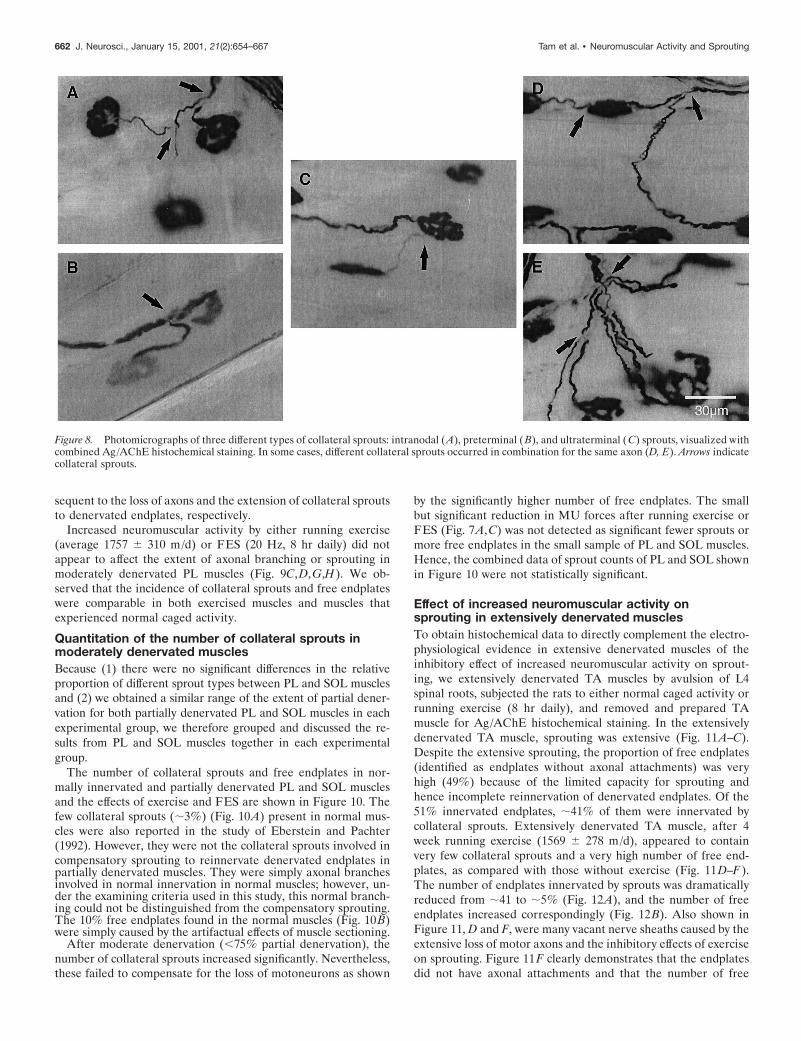
Sprouting and effect of increased neuromuscularactivity in moderately denervated musclesUsing combined Ag/AChE histochemical staining, three differenttypes of collateral sprouts were examined in this study. A singleaxonal outgrowth, for example, originated from the node ofRanvier (Fig. 8A) (intranodal sprout) or preterminal region (Fig.8B) (preterminal sprout). Another example is shown in Figure8C. A single axonal outgrowth originated from the endplate area(Fig. 8C) (ultraterminal sprout). There was usually only one typeof sprout per axon in moderately denervated muscle, with fewexceptions, such as the examples in Figure 8D. An ultraterminalsprout originated from the endplate region of the same axon fromwhence an intranodal sprout originated. We also occasionallyobserved that many intranodal sprouts originated from a singleaxon (Fig. 8E).
In normal PL muscle, the typical pattern of innervation isshown in Figure 9, A and E. The combined Ag/AChE histochem-ical staining on 100-mm-thick cryostat longitudinal sections re-vealed, at high magnification (Fig. 9E), an intramuscular nervetrunk composed of several axons branching down into singleaxons that innervated single motor endplates.
As compared with normal PL muscle (Fig. 9A,E), we foundthat moderately denervated PL muscle (,75% partial denerva-tion) contained considerable numbers of collateral sprouts (Fig.9B,F). Nevertheless, there were some free endplates at 1 month.These collateral sprouts appeared to be mainly intranodalsprouts. The number and size of intramuscular nerve trunks weredecreased, and the nerve branches became relatively longer con-
Figure 7. Cumulative frequency histogramsof MU twitch force distributions in partiallydenervated PL (A, C) and SOL (B, D) mus-cles of rats experiencing normal caged activ-ity (Control, E) as compared with partiallydenervated muscles after running exercise(Exercise, A, B, F) and partially denervatedmuscles with FES (FES, C, D, f). There wasa small but significant shift in cumulative MUforce distributions, after exercise or FES, tothe left of the moderately denervated PLmuscle in rats that experienced normal cagedactivity ( p , 0.01). For moderately dener-vated SOL muscle, there were no significantdifferences in the MU twitch force distribu-tion between the caged and activity groups.For PL, n’ 5 204 (Caged), n’ 5 139 (Exercise),and n’ 5 196 (FES); for SOL, n’ 5 169(Caged), n’ 5 79 (Exercise), and n’ 5 100(FES) (n 5 total number of muscles sampledfor the calculation of the mean MU numbers;n’ 5 total number of MUs sampled for MUtwitch force measurement).
Tam et al. • Neuromuscular Activity and Sprouting J. Neurosci., January 15, 2001, 21(2):654–667 661
sequent to the loss of axons and the extension of collateral sproutsto denervated endplates, respectively.
Increased neuromuscular activity by either running exercise(average 1757 6 310 m/d) or FES (20 Hz, 8 hr daily) did notappear to affect the extent of axonal branching or sprouting inmoderately denervated PL muscles (Fig. 9C,D,G,H). We ob-served that the incidence of collateral sprouts and free endplateswere comparable in both exercised muscles and muscles thatexperienced normal caged activity.
Quantitation of the number of collateral sprouts inmoderately denervated musclesBecause (1) there were no significant differences in the relativeproportion of different sprout types between PL and SOL musclesand (2) we obtained a similar range of the extent of partial dener-vation for both partially denervated PL and SOL muscles in eachexperimental group, we therefore grouped and discussed the re-sults from PL and SOL muscles together in each experimentalgroup.
The number of collateral sprouts and free endplates in nor-mally innervated and partially denervated PL and SOL musclesand the effects of exercise and FES are shown in Figure 10. Thefew collateral sprouts (;3%) (Fig. 10A) present in normal mus-cles were also reported in the study of Eberstein and Pachter(1992). However, they were not the collateral sprouts involved incompensatory sprouting to reinnervate denervated endplates inpartially denervated muscles. They were simply axonal branchesinvolved in normal innervation in normal muscles; however, un-der the examining criteria used in this study, this normal branch-ing could not be distinguished from the compensatory sprouting.The 10% free endplates found in the normal muscles (Fig. 10B)were simply caused by the artifactual effects of muscle sectioning.
After moderate denervation (,75% partial denervation), thenumber of collateral sprouts increased significantly. Nevertheless,these failed to compensate for the loss of motoneurons as shown
by the significantly higher number of free endplates. The smallbut significant reduction in MU forces after running exercise orFES (Fig. 7A,C) was not detected as significant fewer sprouts ormore free endplates in the small sample of PL and SOL muscles.Hence, the combined data of sprout counts of PL and SOL shownin Figure 10 were not statistically significant.
Effect of increased neuromuscular activity onsprouting in extensively denervated musclesTo obtain histochemical data to directly complement the electro-physiological evidence in extensive denervated muscles of theinhibitory effect of increased neuromuscular activity on sprout-ing, we extensively denervated TA muscles by avulsion of L4spinal roots, subjected the rats to either normal caged activity orrunning exercise (8 hr daily), and removed and prepared TAmuscle for Ag/AChE histochemical staining. In the extensivelydenervated TA muscle, sprouting was extensive (Fig. 11A–C).Despite the extensive sprouting, the proportion of free endplates(identified as endplates without axonal attachments) was veryhigh (49%) because of the limited capacity for sprouting andhence incomplete reinnervation of denervated endplates. Of the51% innervated endplates, ;41% of them were innervated bycollateral sprouts. Extensively denervated TA muscle, after 4week running exercise (1569 6 278 m/d), appeared to containvery few collateral sprouts and a very high number of free end-plates, as compared with those without exercise (Fig. 11D–F).The number of endplates innervated by sprouts was dramaticallyreduced from ;41 to ;5% (Fig. 12A), and the number of freeendplates increased correspondingly (Fig. 12B). Also shown inFigure 11, D and F, were many vacant nerve sheaths caused by theextensive loss of motor axons and the inhibitory effects of exerciseon sprouting. Figure 11F clearly demonstrates that the endplatesdid not have axonal attachments and that the number of free
Figure 8. Photomicrographs of three different types of collateral sprouts: intranodal (A), preterminal (B), and ultraterminal (C) sprouts, visualized withcombined Ag/AChE histochemical staining. In some cases, different collateral sprouts occurred in combination for the same axon (D, E). Arrows indicatecollateral sprouts.
662 J. Neurosci., January 15, 2001, 21(2):654–667 Tam et al. • Neuromuscular Activity and Sprouting

endplates was higher than in the muscles that experienced normalcaged activity. Both the electrophysiological and histochemicalresults show that the effects of increased neuromuscular activitycan be demonstrated at both the level of nerve innervation ofendplates and the recording of MU forces.
DISCUSSIONThe results of this study demonstrate that increased neuromus-cular activity during the acute phase of sprouting compromisessprouting, particularly in extensively denervated muscles.
Detrimental effect of increased neuromuscular activityon MU enlargement and sprouting after extensivedenervation but not moderate denervationThe striking finding of the present study that the detrimentaleffect of increased neuromuscular activity of reducing MU en-largement was evident largely in extensively denervated musclesmay account for the contradictory findings in previous studies(Gardiner et al., 1984; Gardiner and Faltus, 1986; Michel andGardiner, 1989; Seburn and Gardiner, 1996). The extent of par-
Figure 9. Low ( A–D) and higher ( E–H)power combined Ag/AChE histochemicalphotomicrographs of 100-mm-thick cryo-stat longitudinal sections of control (A, E),moderately denervated (PD , 75%) afternormal caged activity (B, F ), running ex-ercise (C, G), and FES (D, H ). In normalPL muscle, single endplates were inner-vated by single axons. For PD ,75%, de-nervated endplates were reinnervated bycollateral sprouts (arrows). There were novisual differences in branching and axonalsprouting after running exercise and FES.Shown are two intranodal sprouts and oneultraterminal sprout in F, two intranodalsprouts in G, and four intranodal sproutsin H. Arrows indicate collateral sprouts.
Tam et al. • Neuromuscular Activity and Sprouting J. Neurosci., January 15, 2001, 21(2):654–667 663

tial denervation was not established directly in these studies, buton average the extent of partial denervation in the musclesstudied, primarily the PL and SOL muscles, is ;50%. Thismoderate extent of partial denervation might be below the limitsof detection of inhibitory effects of activity. The variability be-tween animals in the extent of partial denervation makes it verydifficult to detect any definitive effect of activity on mean MUsize, particularly in the previous experiments in which the extentof partial denervation was not evaluated or quantitated. A two-fold increase in MU size is difficult to detect electrophysiologi-cally (Rafuse et al., 1992) and morphometrically (Brown et al.,1980), although a significant enlargement was noted for the wholeMU population (Fig. 5). The effects of activity would be evenmore difficult to detect by average values that were used in mostof those previous studies. Large samples of MUs are especiallyimportant when all MUs in partially denervated muscles are notaffected similarly. As seen in Figures 6 and 7, it was evident in allfour types of partially denervated muscles that increased neuro-muscular activity was more detrimental to the smaller and slowerMUs and that there was an apparent sparing of the larger MUs.It is important to take into account the effect of denervation/reinnervation and neuromuscular activity on muscle fiber size,which was ignored in many of those previous studies. MU force isthe product of the number, cross-sectional area, and specific forceof muscle fibers innervated by a motoneuron, i.e., IR, CSA, andspecific force. MU force depends on both IR and CSA andreasonably reflects IR and therefore sprouting, only if musclefiber CSA is taken into account. Because specific force does notchange after reinnervation (Totosy de Zepetnek et al., 1992a; Fuand Gordon, 1995a,b), MU force corrected for muscle fiber CSAtherefore provides a reasonable average measurement of MUenlargement.
Proposed mechanisms for the detrimental effect ofincreased neuromuscular activity on sproutingThe detrimental effects of increased neuromuscular activity inreducing MU enlargement in partially denervated muscles maybe accounted for by the failure of (1) newly formed synapticcontacts to mature or (2) axonal sprouts to contact denervatedendplates, or both. The former possibility is supported by evi-dence from studies of muscle reinnervation after nerve crushshowed that synaptic maturation was retarded in rat muscles byincreasing motor activity via a daily swimming regime of 3–4 hr/dfor 35 d (Gutmann and Jakoubek, 1963) and in rat lateral gas-
trocnemius muscles by 30 d treadmill running (Soucy et al., 1996).The hyperactive gastrocnemius muscles exhibited “tetanic fade”phenomenon, indicating that the activity had delayed the matu-ration of the synapses (Soucy et al., 1996).
The detrimental effects of increased neuromuscular activity inreducing MU enlargement in partially denervated muscles ismore likely to be accounted for by the second possibility, namelythat axonal sprouts fail to contact denervated endplates. A num-ber of immunohistochemical studies have demonstrated that ter-minal Schwann cells at both innervated and denervated endplatesinduce and guide collateral sprouts by forming extended pro-cesses that bridge between the endplates (Reynolds and Woolf,1992; Son and Thompson, 1995a,b; Son et al., 1996; Thompsonand Kopp, 1996). The detrimental effects of increased neuromus-cular activity in reducing axonal sprouting may arise indirectlyfrom the inhibitory effects of increased activity on the ability ofthe terminal Schwann cell processes to make bridges betweeninnervated and denervated endplates, and in turn, for the axonalsprouts to make contact with the denervated endplates. Strikingevidence supporting this view comes from the recent studies ofLove et al. (1997) and Tam and Gordon (1998). Love andcolleagues (1997) reported that direct muscle stimulation prohib-ited bridge formation of terminal Schwann cell processes andthereby reduced axonal sprouting in SOL muscle 7 d after partialdenervation. Studies of extensively denervated TA muscle alsoshowed that the increased neuromuscular activity associated withrunning exercise did not affect formation of the Schwann cellprocesses; rather, it significantly reduced the bridging betweenendplates during the first week of collateral sprouting and, inturn, collateral sprouts at early time points over a 4 week period(Tam and Gordon, 1998). Interestingly, increased neuromuscularactivity (Tam and Gordon, 1998) and direct muscle stimulation(Love et al., 1997) inhibited bridge formation and, in turn, axonalsprouting. In both cases, it was shown that the perisynapticSchwann cells continued to make processes, but they failed tobridge between the denervated and innervated junctions.
Terminal or perisynaptic Schwann cells express muscarinicacetylcholine receptors that normally respond to ACh releasedfrom nerve terminals by influx of calcium (Jahromi et al., 1992;Reist and Smith, 1992; Reynolds and Woolf, 1993; Lev-Ram andEllisman, 1995) and downregulation of glial fibrillary acidic pro-tein (Georgiou et al., 1999). In the absence of released ACh at thedenervated endplates, the perisynaptic Schwann cells would likelyextend glial processes, which bridge between the innervated anddenervated endplates in partially denervated muscles. It is possi-ble that very high neuromuscular activity at the innervated end-plates might release sufficient ACh to prevent the glial processesfrom effectively bridging between the denervated and innervatedendplates and thereby explain the reduction in sprouting from thehyperactive nerve terminals.
In addition to the role of Schwann cells in providing physicalguidance to sprout growth, Schwann cells may also producesprout-inducing substances. Hoffman (1950) first suggested thatcollateral sprouts were guided to denervated endplates by some“elements” supplied by the denervated Schwann cells in thevacant nerve sheaths in partially denervated muscles. Since then,considerable evidence supporting this view has become availablefrom subsequent studies on the role of non-neural cells of thedistal nerve stumps and terminal Schwann cells in producingsprout-inducing factors to attract regenerating axons (David andAguayo, 1985; Kuffler, 1987; Diaz and Pecot-Dechavassine, 1990;Kuffler, 1994). In light of the capacity of the neurotrophic factor,
Figure 10. Mean (6SE) of number of collateral sprouts per 100 inner-vated endplates (A) and free endplates per 100 endplates (B) of normal(open bars) and moderately denervated ( filled bars) PL and SOL musclesafter normal caged activity, running exercise, and FES. For PD ,75%,the number of both collateral sprouts and free endplates was significantlyincreased as compared with control (*p , 0.0001) (n 5 total number ofmuscles sampled).
664 J. Neurosci., January 15, 2001, 21(2):654–667 Tam et al. • Neuromuscular Activity and Sprouting

ciliary neurotrophic factor (Gurney et al., 1992; Kwon and Gur-ney, 1994; Siegel and English, 1997), to induce sprouting innormally innervated muscles, it is possible that expression ofciliary neurotrophic factor on partial denervation is altered andmay thereby induce sprouting. Neuromuscular activity mightsomehow interfere with the ability of intact axons to respond tothe sprouting stimuli.
Another source of sprouting stimuli is the denervated inactivemuscle fibers (Brown et al., 1978a,b, 1980, 1981; Slack and Pock-ett, 1981; Pockett and Slack, 1982; Keynes et al., 1983; Gurney,
1984; Gurney et al., 1986; Kuffler, 1989; Rassendren et al., 1992;Kuffler and Luethi, 1993). Suggested sprouting factors releasedfrom inactive muscle fibers include insulin-like growth factors(Caroni and Grandes, 1990; Thompson and Kopp, 1996), neuralcell adhesion molecules (Gurney et al., 1986), and neurocrescin(Nishimune et al., 1997). It has been shown that restoration ofmuscle activity by direct muscle stimulation inhibits axonalsprouting in paralyzed (Brown et al., 1977, 1980) and partiallydenervated (Brown and Ironton, 1977; Brown and Holland, 1979)muscles. The possibilities are that the direct muscle stimulationreduces the availability of sprout-producing factors from dener-vated muscle fibers and that, by stimulation of the intramuscularnerves, the stimulation might reduce the ability of the intramus-cular nerves to sprout in response to the sprout-producing factors.Hence, the increased neuromuscular activity by the running ex-ercise and FES in our present study could alter the ability of theSchwann cells at the innervated endplates to respond to thesprout-inducing factors from inactive muscle fibers and therebyreduce axonal sprouting.
Finally, levels of calcium in the nerve terminals may also play arole because the high intracellular calcium level induced by elec-trical stimulation and calcium ionophores results in cessation ofnerve outgrowth (Kater and Mills, 1991; Rehder and Kater,1992). Increased neuromuscular activity might overload sproutterminals with calcium, resulting in reduction of sprouting.
The aforementioned mechanisms are mostly postulated and yet
Figure 11. Three levels of magnification (low: A, D; high: B, E; higher: C, F ) of combined Ag/AChE histochemical photomicrographs of 100-mm-thickcryostat longitudinal sections of extensively denervated TA muscles with normal caged activity (A–C) and after running exercise ( D–F). Extensivelydenervated TA muscles demonstrated extensive collateral sprouting for normal caged activity as designated by filled arrows in A–C. In contrast, thepartially denervated muscles after running exercise contained almost no sign of collateral sprouts but contained very high numbers of free endplates, asidentified by open arrows (D–F). Rather, the exercised muscles contained many vacant nerve sheaths (arrowheads) containing no Ag-stained axons.
Figure 12. Mean (6SE) of number of collateral sprouts per 100 inner-vated endplates (A) and free endplates per endplates (B) of extensivelydenervated TA muscle after normal caged activity ( filled bars) andrunning exercise (hatched bars) (n 5 total number of muscles sampled).
Tam et al. • Neuromuscular Activity and Sprouting J. Neurosci., January 15, 2001, 21(2):654–667 665

to be proven. Nonetheless, one must consider these mechanismsin relationship to the differential detrimental effects of activityrelative to the size of the MUs. Although we might predict thatthe mechanisms may be the same for all MUs, our findingsdemonstrate a selective effect of activity on the smallest as op-posed to the largest MUs. In light of recent findings that thenormal relationship between size of the motoneurons and thenumber of muscle fibers that they reinnervate was reestablishedeven under conditions in which all regenerating motoneurons andreinnervated MUs were subject to the same electrical activity(Gordon et al., 1999), it is apparent that the number of musclefibers per motoneuron varies with motoneuron size rather thanneuromuscular activity. Hence, the findings in this study thatexercise or electrical activity reduced sprouting more in thesmaller than the larger MUs reflect a balance between the intrin-sic size-dependent capacity of motoneurons to enlarge their MUsby sprouting and the inhibitory effects of activity in limiting thissprouting.
ConclusionsUsing four functionally different muscles and both electrophysi-ological and histochemical techniques, we, in the present studies,have resolved the controversial findings of previous studies on theeffect of activity on sprouting and have been able to generalizethe effect of increased neuromuscular activity in reducing MUenlargement and sprouting. The detrimental effect of increasedneuromuscular activity in reducing MU enlargement and sprout-ing depends on the extent of partial denervation of muscles.Increased neuromuscular activity significantly reduced MU en-largement and sprouting primarily in extensively denervatedmuscles where only ,20% of intact MUs remained. We havedemonstrated that normal physiological activity of sprouting mo-toneurons is conducive for MU enlargement during the acutephase of sprouting, whereas nonphysiological activity can bedetrimental. The findings of the present studies indicate thatincreased neuromuscular activity is not recommended as rehabil-itation immediately after motoneuron injury or in the early stagesof motoneuron disease.
REFERENCESBrown MC, Holland RL (1979) A central role for denervated tissues in
causing nerve sprouting. Nature 282:724–726.Brown MC, Ironton R (1977) Motor neurone sprouting induced by pro-
longed tetrodotoxin block of nerve action potential. Nature265:459–461.
Brown MC, Ironton R (1978) Sprouting and regression of neuromuscu-lar synapses in partially denervated mammalian muscles. J Physiol(Lond) 278:325–348.
Brown MC, Goodwin GM, Ironton R (1977) Prevention of motor nervesprouting in botulinum toxin poisoned mouse soleus muscles by directstimulation of the muscle. J Physiol (Lond) 267:42P–43P.
Brown MC, Holland RL, Ironton R (1978a) Degenerating nerve prod-ucts affect innervated muscle fibres. Nature 275:652–654.
Brown MC, Holland RL, Ironton R (1978b) Is the stimulus for motoneu-rone terminal sprouting localized? J Physiol (Lond) 282:7–8P.
Brown MC, Holland RL, Ironton R (1980) Nodal and terminal sproutingfrom motor nerves in fast and slow muscles of the mouse. J Physiol(Lond) 306:493–510.
Brown MC, Holland RL, Hopkins WG (1981) Motor nerve sprouting.Annu Rev Neurosci 4:17–42.
Buller AJ, Pope R (1977) Plasticity in mammalian skeletal muscle. PhilosTrans R Soc Lond B Biol Sci 278:295–305.
Burke RE (1981) Motor units: anatomy, physiology and functional or-ganization. In: Handbook of physiology. The nervous system. Motorcontrol, pp 345–421. Bethesda, MD: Am Physiol Soc.
Caroni P, Grandes P (1990) Nerve sprouting in innervated adult skeletalmuscle induced by exposure to elevated levels of insulin-like growthfactors. J Cell Biol 110:1307–1317.
Daniel WW (1995) Biostatistics: a foundation for analysis in the healthsciences. New York: Wiley.
David S, Aguayo AJ (1985) Axonal regeneration after crush injury of ratcentral nervous system fibres innervating peripheral nerve grafts.J Neurocytol 14:1–12.
Diaz J, Pecot-Dechavassine M (1990) Nerve sprouting induced by apiece of peripheral nerve placed over a normally innervated frogmuscle. J Physiol (Lond) 421:123–133.
Eberstein A, Pachter BR (1992) Recovery and loss of muscle force of ratplantaris after partial denervation. Exp Neurol 116:240–245.
Einsiedel LJ, Luff AR (1994) Activity and motor unit size in partiallydenervated rat medial gastrocnemius. J Appl Physiol 76:2663–2671.
Fu SY, Gordon T (1995a) Contributing factors to poor functional recov-ery after delayed nerve repair: prolonged axotomy. J Neurosci15:3876–3885.
Fu SY, Gordon T (1995b) Contributing factors to poor functional recov-ery after delayed nerve repair: prolonged denervation. J Neurosci15:3886–3895.
Gardiner PF, Faltus RE (1986) Contractile responses of rat plantarismuscles following partial denervation, and the influence of daily exer-cise. Pflugers Arch 406:51–56.
Gardiner PF, Michel RN, Iadeluca G (1984) Previous exercise traininginfluences functional sprouting of rat hindlimb motoneurons in re-sponse to partial denervation. Neurosci Lett 45:123–127.
Georgiou J, Robitaille R, Charlton MP (1999) Muscarinic control ofcytoskeleton in perisynaptic glia. J Neurosci 19:3836–3846.
Gordon T (1995) Fatigue in adapted systems: overuse and underuseparadigms. Adv Exp Med Biol 384:429–456.
Gordon T, Stein RB, Thomas CK (1986) Innervation and function ofhind-limb muscles in the cat after cross-union of the tibial and peronealnerves. J Physiol (Lond) 374:429–441.
Gordon T, Tyreman N, Rafuse VF, Munson JB (1999) Limited plastic-ity of adult motor units conserves recruitment order and rate coding.Prog Brain Res 123:191–202.
Gurney ME (1984) Suppression of sprouting at the neuromuscular junc-tion by immune sera. Nature 307:546–548.
Gurney ME, Apatoff BR, Heinrich SP (1986) Suppression of terminalaxonal sprouting at the neuromuscular junction by monoclonal antibod-ies against a muscle-derived antigen of 56,000 daltons. J Cell Biol102:2264–2272.
Gurney ME, Yamamoto H, Kwon Y (1992) Induction of motor neuronsprouting in vivo by ciliary neurotrophic factor and basic fibroblastgrowth factor. J Neurosci 12:3241–3247.
Gutmann E, Jakoubek B (1963) Effect of increased motor activity onregeneration of the peripheral nerve in young rats. Physiologia Bohe-moslovenica 12:463–468.
Halstead LS, Wiechers DO (1987) Research and clinical aspects of thelate effects of poliomyelitis. White Plains, NY: March of Dimes BirthDefects Foundation.
Hoffman H (1950) Local reinnervation in partially denervated muscles:a histophysiological study. Aust J Exp Biol Med Sci 28:383–397.
Jahromi BS, Robitaille R, Charlton MP (1992) Transmitter release in-creases intracellular calcium in perisynaptic Schwann cells in situ.Neuron 8:1069–1077.
Kanda K, Hashizume K (1992) Factors causing difference in force out-put among motor units in the rat medial gastrocnemius muscle.J Physiol (Lond) 448:677–695.
Kater SB, Mills LR (1991) Regulation of growth cone behavior by cal-cium. J Neurosci 11:891–899.
Keynes RJ, Hopkins WG, Brown MC (1983) Sprouting of mammalianmotor neurones at nodes of Ranvier: the role of the denervated motorendplate. Brain Res 264:209–213.
Kuffler DP (1987) Long-distance regulation of regenerating frog axons.J Exp Biol 132:151–160.
Kuffler DP (1989) Regeneration of muscle axons in the frog is directedby diffusible factors from denervated muscle and nerve tubes. J CompNeurol 281:416–425.
Kuffler DP (1994) Promoting and directing axon outgrowth. Mol Neu-robiol 9:233–243.
Kuffler DP, Luethi T (1993) Identification of molecules in a muscleextracellular matrix extract that promotes process outgrowth fromcultured adult frog motoneurons. J Neurobiol 24:515–527.
Kwon Y, Gurney ME (1994) Systemic injections of ciliary neuromuscu-lar factor induce sprouting by adult motor neurons. NeuroReport5:789–792.
Lev-Ram V, Ellisman MH (1995) Axonal activation-induced calciumtransients in myelinating Schwann cells, sources, and mechanism.J Neurosci 15:2628–2637.
Love FM, Son YJ, Thompson WJ (1997) Muscle activity inhibits termi-nal Schwann cells from inducing of guiding nerve sprouts after nerveinjury in the rat soleus. Soc Neurosci Abstr 23:244.1.
Luff AR, Hatcher DD, Torkko K (1988) Enlarged motor units resultingfrom partial denervation of cat hindlimb muscles. J Neurophysiol59:1377–1394.
Michel RN, Gardiner PF (1989) Influence of overload on recovery of ratplantaris from partial denervation. J Appl Physiol 66:732–740.
Nishimune H, Uyeda A, Nogawa M, Fujimoto H, Fujimori KE, Taguchi
666 J. Neurosci., January 15, 2001, 21(2):654–667 Tam et al. • Neuromuscular Activity and Sprouting

T (1997) Neurocrescin: a neurite-outgrowth factor secreted by musclein an activity dependent manner. Soc Neurosci Abstr 23:244.2.
Pette D, Vrbova G (1992) Adaptation of mammalian skeletal musclefibers to chronic electrical stimulation. Rev Physiol Biochem Pharmacol120:115–202.
Pockett S, Slack JR (1982) Source of the stimulus for nerve terminalsprouting in partially denervated muscle. Neuroscience 7:3173–3176.
Rafuse VF, Gordon T (1996a) Self-reinnervated cat medial gastrocne-mius muscles. I. Comparisons of the capacity of regenerating nerves toform enlarged motor units after extensive peripheral nerve injuries.J Neurophysiol 75:268–281.
Rafuse VF, Gordon T (1996b) Self-reinnervated cat medial gastrocne-mius muscles. II. Analysis of the mechanisms and significance of fibertype grouping in reinnervated muscles. J Neurophysiol 75:282–297.
Rafuse VF, Gordon T, Orozco R (1992) Proportional enlargement ofmotor units after partial denervation of cat triceps surae muscles.J Neurophysiol 68:1261–1275.
Rassendren FA, Bloch-Gallego E, Tanaka H, Henderson CE (1992)Levels of mRNA coding for motoneuron growth-promoting factors areincreased in denervated muscle. Proc Natl Acad Sci USA89:7194–7198.
Rehder V, Kater SB (1992) Regulation of neuronal growth cone filop-odia by intracellular calcium. J Neurosci 12:3175–3186.
Reist NE, Smith SJ (1992) Neurally evoked calcium transients in termi-nal Schwann cells at the neuromuscular junction. Proc Natl Acad SciUSA 89:7625–7629.
Reynolds ML, Woolf CJ (1992) Terminal Schwann cells elaborate ex-tensive processes following denervation of the motor endplate. J Neu-rocytol 21:50–66.
Reynolds ML, Woolf CJ (1993) Reciprocal Schwann cell-axon interac-tions. Curr Opin Neurobiol 3:683–693.
Ribchester RR (1988) Activity-dependent and independent synaptic in-teractions during reinnervation of partially denervated rat muscle.J Physiol (Lond) 401:53–75.
Seburn KL, Gardiner PF (1996) Properties of sprouted rat motor units:effects of period of enlargement and activity level. Muscle Nerve19:1100–1109.
Siegel SG, English AW (1997) CNTF is required for denervation-induced sprout formation at the neuromuscular junction. Soc NeurosciAbstr 23:248.14.
Slack JR, Pockett S (1981) Terminal sprouting of motoneurones is alocal response to a local stimulus. Brain Res 217:368–374.
Son YJ, Thompson WJ (1995a) Nerve sprouting in muscle is induced
and guided by processes extended by Schwann cells. Neuron14:133–141.
Son YJ, Thompson WJ (1995b) Schwann cell processes guide regenera-tion of peripheral axons. Neuron 14:125–132.
Son YJ, Trachtenberg JT, Thompson WJ (1996) Schwann cells induceand guide sprouting and reinnervation of neuromuscular junctions.Trends Neurosci 19:280–285.
Soucy M, Seburn K, Gardiner P (1996) Is increased voluntary motoractivity beneficial or detrimental during the period of motor nerveregeneration/reinnervation. Can J Appl Physiol 21:218–224.
Tam SL, Gordon T (1998) Detrimental effect of neuromuscular activityon the ability of terminal Schwann cells to induce sprouting in exten-sively denervated tibialis anterior muscle. Soc Neurosci Abstr24:413.16.
Tam SL, van der Sloot P, Archibald V, Tyreman N, Gordon T (1995) Isneuromuscular activity beneficial to motor axon sprouting? Presentedat 6th International Symposium on Neural Regeneration.
Tam SL, Archibald V, Stein RB, Tyreman N, Gordon T (1996) Effect ofneuromuscular activity on sprouting depends on the extent of partialdenervation. Soc Neurosci Abstr 22:298.4.
Tam SL, Tyreman N, Gordon T (1997) Detrimental effect of neuromus-cular activity on motor axonal sprouting in a rat model of motoneurondisease. Neurosci Net 59:59.
Thompson WJ, Jansen JK (1977) The extent of sprouting of remainingmotor units in partly denervated immature and adult rat soleus muscle.Neuroscience 2:523–535.
Thompson WJ, Kopp DM (1996) Schwann cells accompany nervesprouts induced in muscle by insulin-like growth factors. Soc NeurosciAbstr 22:297.7.
Totosy de Zepetnek JE, Zung HV, Erdebil S, Gordon T (1992a) Inner-vation ratio is an important determinant of force in normal and rein-nervated rat tibialis anterior muscle. J Neurophysiol 67:1385–1403.
Totosy de Zepetnek JE, Zung HV, Erdebil S, Gordon T (1992b) Motor-unit categorization based on contractile and histochemical properties: aglycogen depletion analysis of normal and reinnervated rat tibialisanterior muscle. J Neurophysiol 67:1404–1415.
Walmsley B, Hodgson JA, Burke RE (1978) Forces produced by medialgastrocnemius and soleus muscles during locomotion in freely movingcats. J Neurophysiol 41:1203–1216.
Yang JF, Stein RB, Jhamandas J, Gordon T (1990) Motor unit numbersand contractile properties after spinal cord injury. Ann Neurol 28:496–502.
Tam et al. • Neuromuscular Activity and Sprouting J. Neurosci., January 15, 2001, 21(2):654–667 667