immunodominant fragments of myelin basic protein initiate t cell
TRANSCRIPT

JOURNAL OF NEUROINFLAMMATION
Immunodominant fragments of myelin basic proteininitiate T cell-dependent painLiu et al.
Liu et al. Journal of Neuroinflammation 2012, 9:119http://www.jneuroinflammation.com/content/9/1/119

RESEARCH Open Access
Immunodominant fragments of myelin basicprotein initiate T cell-dependent painHuaqing Liu1,2, Sergey A Shiryaev3, Andrei V Chernov3, Youngsoon Kim1,2, Igor Shubayev2, Albert G Remacle3,Svetlana Baranovskaya4, Vladislav S Golubkov3, Alex Y Strongin3 and Veronica I Shubayev1,2*
Abstract
Background: The myelin sheath provides electrical insulation of mechanosensory Aβ-afferent fibers. Myelin-degradingmatrix metalloproteinases (MMPs) damage the myelin sheath. The resulting electrical instability of Aβ-fibers is believedto activate the nociceptive circuitry in Aβ-fibers and initiate pain from innocuous tactile stimulation (mechanicalallodynia). The precise molecular mechanisms, responsible for the development of this neuropathic pain state afternerve injury (for example, chronic constriction injury, CCI), are not well understood.
Methods and results: Using mass spectrometry of the whole sciatic nerve proteome followed by bioinformaticsanalyses, we determined that the pathways, which are classified as the Infectious Disease and T-helper cell signaling,are readily activated in the nerves post-CCI. Inhibition of MMP-9/MMP-2 suppressed CCI-induced mechanical allodyniaand concomitant TNF-α and IL-17A expression in nerves. MMP-9 proteolysis of myelin basic protein (MBP) generatedthe MBP84-104 and MBP68-86 digest peptides, which are prominent immunogenic epitopes. In agreement, theendogenous MBP69-86 epitope co-localized with MHCII and MMP-9 in Schwann cells and along the nodes of Ranvier.Administration of either the MBP84-104 or MBP68-86 peptides into the naïve nerve rapidly produced robustmechanical allodynia with a concomitant increase in T cells and MHCII-reactive cell populations at the injection site. Asshown by the genome-wide expression profiling, a single intraneural MBP84-104 injection stimulated the inflammatory,immune cell trafficking, and antigen presentation pathways in the injected naïve nerves and the associated spinalcords. Both MBP84-104-induced mechanical allodynia and characteristic pathway activation were remarkably lessprominent in the T cell-deficient athymic nude rats.
Conclusions: These data implicate MBP as a novel mediator of pain. Furthermore, the action of MMPs expressedwithin 1 day post-injury is critical to the generation of tactile allodynia, neuroinflammation, and the immunodominantMBP digest peptides in nerve. These MBP peptides initiate mechanical allodynia in both a T cell-dependent and-independent manner. In the course of Wallerian degeneration, the repeated exposure of the cryptic MBP epitopes,which are normally sheltered from immunosurveillance, may induce the MBP-specific T cell clones and a self-sustainingimmune reaction, which may together contribute to the transition of acute pain into a chronic neuropathic pain state.
BackgroundPain is typically mediated by small unmyelinated C-noci-ceptive and thinly myelinated Aδ-afferents. Non-nocicep-tive, large-diameter myelinated Aβ-afferents transmittouch and vibration sense. However, following damage tothe peripheral nervous system (PNS), Aβ-afferents joinnociceptive circuitry [1]. Our mechanistic understanding
of why the damaged Aβ-afferents interpret an innocuous,low-threshold tactile stimulus as painful (that is, mechan-ical allodynia) remains exceedingly limited. Growing evi-dence supports a model in which the damage to theelectrically insulating myelin sheath and the resulting lossof electrical stability in Aβ-afferents contribute to the de-velopment of mechanical allodynia [2-6].Mechanical allodynia and other forms of neuropathic
pain (NP, that is, pain arising as a direct consequence ofa lesion or disease affecting the somatosensory nervoussystem [7]) have features of a neuroimmune disorder [8].T lymphocyte infiltration into both the damaged nerve
* Correspondence: [email protected] of Anesthesiology, University of California, San Diego, 9500Gilman Dr., Mail Box 0629, La Jolla, CA 92093-0629, USA2VA San Diego Healthcare System, La Jolla, CA, USAFull list of author information is available at the end of the article
JOURNAL OF NEUROINFLAMMATION
© 2012 Liu et al.; licensee BioMed Central Ltd. This is an Open Access article distributed under the terms of the CreativeCommons Attribution License (http://creativecommons.org/licenses/by/2.0), which permits unrestricted use, distribution, andreproduction in any medium, provided the original work is properly cited.
Liu et al. Journal of Neuroinflammation 2012, 9:119http://www.jneuroinflammation.com/content/9/1/119

[9-11] and the spinal cord at a corresponding segment[12-14] has been implicated in NP. Following chronicconstriction injury (CCI), T cell-deficient athymic nuderats exhibit a diminished ability to develop NP, which isreversed with adoptive transfer of T-helper (Th)1 cells[9]. Interleukin (IL)-17, expressed by certain Th cells, isessential to mediating mechanical, but not thermal,hypersensitivity [15]. Major histocompatibility complex(MHC) II, produced by antigen-presenting cells to cap-ture and present antigens to T cells, is required for thedevelopment of NP [16,17]. An increase in MHCII isobserved in the nerve and the corresponding DRG [18],spinal cord [14,16], and brainstem [19] after a peripheralnerve lesion. However, the antigens, which are involvedin the recruitment and the homing of activated T cells inthe pathogenesis of NP, remain unknown.Myelin basic protein (MBP) is the component of the com-
pact myelin that is believed to participate in the mainten-ance of the major dense line and interactions of the myelinsheath with the cytosolic surfaces [20-23]. In the PNS, MBPcomprises 5% to 15% of total myelin protein and is consid-ered to be non-essential [22]. However, Th1-mediated auto-immune peripheral neuropathies in humans and therelevant experimental models induced by immunization ofanimals using immunodominant MBP and other myelinpeptides are often painful [24]. It has been demonstratedthat the autoimmune response to immunodominant MBPpeptides assists in myelin clearance and regeneration afterperipheral nerve injury [25]. Certain digest fragments ofMBP and its splice variant (Golli-MBP) expressed in im-mune cells [26] are generated by matrix metalloproteinase(MMP) proteolysis and exhibit key T cell epitopes [27,28].MMPs are a family of zinc-endopeptidases comprising
collagenases, gelatinases, matrilysins, stromelysins, andmembrane-type MMPs [29]. After peripheral nerve injury,gelatinases B (MMP-9) and A (MMP-2) degrade the blood-nerve barrier, release the pro-inflammatory cytokines, con-trol immune cell infiltration and cell survival along theinjured neural axis. These two MMPs are believed to con-secutively initiate and maintain NP [5,30-34]. Having pro-posed that MMPs promote mechanical hypersensitivity viathe proteolysis of myelin [5], we herein aimed to determinethe specific mechanisms involved. Our present experimen-tal evidence suggests that MMP-mediated fragmentation ofMBP as a consequence of Wallerian degeneration exposescryptic MBP epitopes, which are normally sheltered fromimmunosurveillance. These exposed immunodominantMBP peptide epitopes induce mechanical allodynia in botha T cell-dependent and -independent manner.
MethodsReagentsReagents were purchased from Sigma (St. Louis, MO) unlessindicated otherwise. MBP2-18 (ASQKRPSQRSKYLATAS),
MBP68-86 (AHYGSLPQKKSHGRTQDENP), MBP84-104(ENPVVHFFKNIVTPRTPPPSQ), and scrambled MBP84-104 (NKPQTNVVEPFHRTFPIPPVS; sMBP84-104) pep-tides, derived from human MBP sequence (GenBank#AAH08749), were synthesized by GenScript. To preventtheir degradation by exoproteinases, these 97% to 99% pur-ity peptides were N- and C-terminally protected by acetyl-ation and amidation, respectively. Because of theincomplete homology between the human and rodentMBP68-86 sequence, an additional Lys residue wasinserted in the MBP68-86 sequence (underlined in the se-quence above) to make the resulting peptide more uni-formly applicable for our studies. Primers and Taqmanoligonucleotide probes for rat MMP-9 (GenBank#NM_031055), GAPDH (GenBank #XO2231), and tumornecrosis factor-α (TNF-α, GenBank #NM_012675) weredesigned using Primer Express 2.0 software (Applied Bio-systems) and obtained from Biosearch Technologies [30].Similarly, the probes for rat IL-17A (GenBank#NM_001106897.1), were obtained from Applied Biosys-tems (Assay ID Rn01757168_m1). GM6001, a broad-spectrum MMP inhibitor, was purchased from Millipore.SB-3CT, a selective MMP-2/9 inhibitor, was purchasedfrom EMD Biosciences. The following detection antibodieswere used in our studies: rabbit anti-rat MMP-9 (TorreyPines Biolabs, cat. #TP221, 1:500), goat anti-mouse MMP-9 (R&D Systems, cat. #AF909; 1:250), rabbit anti-S100(Dako, cat. #Z0311, 1:500), murine anti-CD68 (clone ED1,Abcam, cat. #ab31630; 1:100), rabbit anti-von Willebrandfactor (vWF, Abcam, cat. #ab6994, 1:1,000), rabbit anti-Iba1 (Wako, cat. #019-19741, 1:500), rat anti-MBP(Abcam, cat. #ab40390, 1:250), murine anti-human MBP(clone 22, AbD Serotec, cat. #MCA686S, 1:250), murineanti-MHC II (clone OX6, Abcam, cat. #Ab6403, 1:200),mouse anti-T cell receptor alpha/beta (TCR; AbD Serotec,cat. #MCA453G, 1:200), mouse β-actin antibody (Sigma,cat. #A53166, 1:30,000), goat anti-mouse conjugated withAlexa 594 (Molecular Probes, 1:500, red), or goat anti-rabbit conjugated with Alexa 488 (Molecular Probes,1:500, green). The nuclei were stained with DAPI (Molecu-lar Probes, 1:20,000, blue). We also used a rabbit anti-MBP antibody (Millipore, cat. #AB5864, 1:1,000) thatrecognizes degraded MBP and that was generated againstthe YGSLPQKSQRSQDENPVV MBP69-86 synthetic pep-tide (the guinea pig sequence) as an immunogen. Anti-bodies were diluted in TBS containing 0.1% Tween-20 and1% normal goat serum.
Animals, surgery, and therapyAll animals were housed at 22 °C under a 12 h light/darkcycle with food and water ad libitum. Animals wereanesthetized with 4% isoflurane (Aerrane; Baxter) in 55%oxygen or a rodent anesthesia cocktail containing Nembu-tal (50 mg/mL; Abbott Labs) and diazepam (5 mg/mL) in
Liu et al. Journal of Neuroinflammation 2012, 9:119 Page 3 of 18http://www.jneuroinflammation.com/content/9/1/119

0.9% PBS (Steris Labs). Sprague–Dawley rats (n=144,200–225 g adult females), athymic nude rats (rnu−/−, Hsd:RH-Foxn1rnu, n= 6, 8-week-old females) and their hetero-zygous controls (rnu+/−, Hsd:RH-Foxn1rnu/Foxn1+, n=6,8-week-old females) were obtained from Harlan Labs. Thecommon sciatic nerve was exposed unilaterally at themid-thigh level. Four loosely constrictive chromic gutsutures were tied around the nerve to produce CCI [35].SB-3CT (10 mg/kg body weight in 10% DMSO) wasinjected i.p. twice: first at the initiation of CCI and then in24 h. Sol-vent alone was used as a vehicle. In a separategroup of animals, the exposed naïve sciatic nervesreceived an intraneural injection of an MBP peptide(50 μg) in 5 μL PBS, or an equal volume of PBS as a ve-hicle using a 33-gauge needle on a Hamilton syringe. Fora sham-operated control, the sciatic nerve was exposedbut otherwise not manipulated. The sciatic nerves and ip-silateral dorsal horn of the spinal cords were collected foranalyses. FVB.Cg-Mmp9tm1Tvu/J (MMP-9−/−, n=6; 20 g,adult females) and wild-type FVB/NJ (WT, n=6; 20 g,adult females) mice were obtained from Jackson Labs. Thesciatic nerve was exposed unilaterally at the mid-thighlevel and crushed using fine, smooth-surface forceps twicefor 2 s each. The animals were sacrificed by an overdoseof the Nembutal/diazepam cocktail, followed by Beutha-nasia (100–150 mg/mL, i.p., Schering-Plough AnimalHealth). The animals were handled according to the NIHGuide for the Care and Use of Laboratory Animals andthe required protocols were approved by the InstitutionalAnimal Care and Use Committee.
Two-dimensional liquid chromatography/tandem massspectrometry/mass spectrometry (2D-LC/MS/MS),proteomics, and pathway analysisThe rat sciatic nerves were isolated, snap-frozen in liquidN2 and stored at −80 °C. The samples were homogenized,sonicated, extracted 60 min at ambient temperature in100 mM Tris–HCl, pH 8.0, containing 8 M urea and theprotease and phosphatase inhibitor cocktails, and the in-soluble material was removed by centrifugation (16,000xg;15 min). The supernatant samples (at least 0.5 mg totalprotein each) were then processed by the Proteomics Corefacility of the Sanford-Burnham Medical Research Insti-tute. The samples were reduced (10 mM tris(2-carbox-yethyl) phosphine, 37 °C, 30 min), alkylated (20 mMiodoacetamide, 37 °C, 40 min in the dark), and digestedusing Modified Trypsin, Mass Spectrometry Grade (Pro-mega; 1:100 w/w ratio; 37 °C, 16–18 h). The samples weredesalted using a SepPack cartridge, dried using a SpeedVacand re-suspended in 0.1 mL 5% formic acid. The resultingpeptides were separated into 24 fractions using an offlineMichrom MDLC pump (Michrom) with a MichromStrong Cation Exchange column. The 1/10 aliquot of eachpeptide fraction was analyzed using an LTQ-Orbitrap XL
mass-spectrometer (Thermo Scientific) and a 15 cmMichrom Magic C18 column coupled with a low-flowMichrom ADVANCED device. The data were analyzed bySorcerer Enterprise v.3.5 software (Sage-N Research) usingthe ipi.Rat.v3.56 protein database. 57 Da were added tocysteines to identify carboxyamidomethylated cysteines,16 Da were added to methionines to identify oxidatedmethionines. The search results were sorted, filtered, andstatistically analyzed using a trans-proteomic pipeline(TPP) (Institute for Systems Biology, Seattle, WA) with a90% minimum probability score and an error rate ≤2%.An additional search was performed using a Prolucidsearch algorithm with a DTASelect function via an Inte-grated Proteomics Pipeline (IP2) server. Relative levels ofthe proteins in the samples were then analyzed using IP2for a Label-Free differential peptide/protein analysis. Thefinal data were subjected to bioinformatics analyses usingIngenuity IPA 8.7 software (Ingenuity Systems).
Real-time qRT-PCR, genome-wide transcriptional profiling,and pathway analysisThe rat sciatic nerves were isolated and stored in RNA-later (Ambion) at −20 °C. Primers and Taqman probeswere optimized to amplification efficiency of 100.1-100.3% [30]. Total RNA was extracted using TRIzol(Invitrogen) and purified on an RNeasy mini column(Qiagen). The RNA purity was estimated by measuringthe OD260/280 and the OD260/230 ratios. The integrityof the RNA samples was validated using an Experionautomated electrophoresis system (Bio-Rad). The sam-ples were treated with RNase-free DNAse I (Qiagen).cDNA was synthesized using a SuperScript first-strandRT-PCR kit (Invitrogen). Gene expression levels weremeasured in a Mx4000™ Multiplex Quantitative PCRSystem (Agilent Technologies) using 50 ng of cDNA and2x Taqman Universal PCR Master Mix (Ambion) with aone-step program: 95 °C, 10 min; 95 °C, 30 s; 60 °C,1 min for 50 cycles. Duplicate samples without cDNA (ano template control) showed no contaminating DNA.Relative mRNA levels were quantified using the com-parative delta Ct method [36] and glyceraldehyde 3-phosphate dehydrogenase (GAPDH) as a normalizer.The fold change between experimental and control sam-ples was determined using the Mx4000 software.For the genome-wide transcriptional profiling, the sam-
ples of total RNA (50 ng) from the wild-type and athymicnude rat nerves and spinal cord tissues were labeled usingLowInput QuickAmp Labeling Kit and Cy3-CTP (AgilentTechnologies). The labeled RNA samples were hybridized17 h at 65 °C to SurePrint G3 Rat GE 8x60K slides (Agi-lent Technologies). Slides were scanned using an AgilentC Scanner. The raw data were processed using FeatureExtraction software version 10.5. The initial analysis andnormalization to the median were performed using
Liu et al. Journal of Neuroinflammation 2012, 9:119 Page 4 of 18http://www.jneuroinflammation.com/content/9/1/119

GeneSpring GX software (Agilent). Differentiallyexpressed mRNAs with signal intensities higher than two-fold over the background standard deviation were filteredby t-test. The statistically significant data only (P< 0.05)were used to calculate gene expression ratios in the sam-ples. The gene expression data have been deposited toGEO database (accession # GSE34868, http://www.ncbi.nlm.nih.gov/geo/info/linking.html). The final data wereanalyzed using Ingenuity IPA 9.0 software.
MMP-9 purification and proteolysis of MBP in vitroThe recombinant pro-form of MMP-9 was purified fromthe serum-free medium conditioned by the stably trans-fected HEK293 cells using the gelatin-column chroma-tography. The purity of the isolated MMP-9 samples wasconfirmed using SDS-PAGE in a 4-20% gradient acryl-amide gel followed by Coomassie staining. Only thesamples with purity over 95% were used in our studies.Purified pro-MMP-9 was activated using 4-aminophe-nylmercuric acetate. The concentration of the catalytic-ally active MMP-9 was determined using a fluorescentassay by active site titration against a standard solutionof a GM6001 of known concentration. (7-methoxycou-marin-4-yl) acetyl-Pro-Leu-Gly-Leu-(3-[2,4-dinitrophe-nyl]-L-2,3-diaminopropionyl)-Ala-Arg-NH2 (Bachem)was used as a fluorescent substrate [27,37].Human MBP (4 μg; approximately 11 μM) was co-incu-
bated with activated MMP-9 (1–100 nM; an enzyme-sub-strate ratio 1:100–1:10,000) in 50 mM HEPES, pH 6.8,supplemented with 10 mM CaCl2 and 50 μM ZnCl2, for1 h at 37 °C. The total volume of the reactions was 20 μL.Where indicated, GM6001 (2.5 μM) was added to thereactions to inhibit MMP-9. The cleavage reaction wasstopped using a 5xSDS sample buffer. The digest sampleswere analyzed by SDS-PAGE and by MALDI-TOF MSusing an Autoflex II MALDI TOF/TOF instrument (Bru-ker Daltonics). For MS analysis, the reactions were cooledon ice and equal volumes (2 mL) of a sample and of asinapic acid (20 mg/mL) in 50% acetonitrile-0.1% trifluor-oacetic acid solution were mixed, spotted directly on aMALDI target plate, air-dried, and co-crystallized for10 min. Mass spectra were processed with FlexAnalysis2.4 software (Bruker Daltonics). The singly charged cleav-age products, which were observed only in the cleavagereactions but not in the controls, were recorded and pro-cessed further.
Gelatin zymographySciatic nerves were isolated, snap-frozen in liquid N2, andstored at −80 °C. Proteins were extracted in 50 mM Tris–HCl, pH 7.4, containing 1% Triton-x 100, 150 mM NaCl,10% glycerol, 0.1% SDS. Extract aliquots (10–70 μg totalprotein as determined by BCA Protein Assay, Pierce) wereanalyzed using 10% acrylamide gels co-polymerized with
0.1% gelatin. After electrophoresis, gels were washed in2% Triton X-100 for 30 to 60 min at ambient temperature,incubated for 16 to 18 h at 37 °C in 50 mM Tris–HCl buf-fer, pH 7.4, containing 10 mM CaCl2 and 1 μM ZnCl2 and0.2 mM sodium azide, and stained with Coomassie BlueR250 to visualize the gelatinolytic activity bands.
Neuropathology, immunohistochemistry, and microscopyPlastic-embedded transverse nerve sections (0.75 μmeach) were used for neuropathologic evaluation. Sciaticnerves were isolated and placed in 2.5% glutaraldehyde in0.1 M phosphate buffer, osmicated, dehydrated, and em-bedded in araldite resin. Sections were cut with a glassknife on an automated Leica RM2065 microtome andstained using methylene blue Azure II. Immunohisto-chemistry was performed in tissues fixed in 4% para-for-maldehyde, embedded in paraffin, or cryoprotected ingraded sucrose and embedded into OCT compound indry ice. The 10-μm sections, when required, were deparaf-finized using xylene and rehydrated in ethanol and PBS,immersed in 0.5% sodium borohydride followed by treat-ment with the antigen retrieval reagent (Dako) for 5 minat 95 °C, then for 20 min at ambient temperature. Teasednerve fibers were prepared from the transected and de-sheathed sciatic nerves. Nerve bundles were separatedusing a pair of fine smooth microforceps. Individual fiberswere teased out using 0.20-0.22 mm acupuncture needles(Vinco, Oxford Medical Supplies) on a glass slide, dried atambient temperature and stored at −20 °C. Non-specificbinding was blocked using PBS containing 5% normalgoat serum and 0.25% Triton X-100. The sections wereincubated with a primary antibody (4 °C, 16–18 h) fol-lowed by an Alexa 488-conjugated (green) or Alexa 594-conjugated (red) species-specific secondary antibody (Invi-trogen, 1 h, ambient temperature). The nuclei werestained with DAPI (5 min). Sections were mounted usinga Slowfade Gold antifade reagent (Molecular Probes). Theimages were acquired using a Leica DMR microscope andOpenlab 4.04 imaging software (Improvision).
Behavior testsSensitivity to non-noxious mechanical stimuli was mea-sured by von Frey testing [38]. Rats were acclimated tobeing on a suspended 6-mm wire grid. The plantar surfaceof the hindpaw was stimulated within the spinal nerve in-nervation area using calibrated von Frey filaments (Stoelt-ing). Stimuli were applied for 4 to 6 s with a 0.4 to 15.0 gbuckling force to the mid-paw plantar surface. In theevent of a positive response, the next weaker stimulus waschosen for the next measurement. In the absence of a re-sponse, a stronger stimulus was presented. This consecu-tive way of applying filaments was continued until sixresponses in the immediate vicinity of the 50% thresholdwere obtained. The resulting sequence of positive and
Liu et al. Journal of Neuroinflammation 2012, 9:119 Page 5 of 18http://www.jneuroinflammation.com/content/9/1/119

negative responses was used to interpolate the 50% with-drawal threshold, determined using the up-down method.Stimuli were separated by several seconds or until the ani-mal was calm with both hind paws placed on the grid.Paw withdrawal latency to a thermal stimulus was mea-sured by a Hargreaves testing device [39]. The hind pawwas stimulated by a radiant heat source. Withdrawal ofthe paw from the heat source was measured four times tocalculate the mean withdrawal latency. A maximal cut-offof 20 s was used to prevent tissue damage. The intervalbetween two trials on the same paw was at least 5 min.Spontaneous pain-like behavior was measured asdescribed in [40]. Each animal was placed in a 19 x 31 cmplexiglass cylinder and allowed to habituate. A 2 min test-ing period included continuous pressing of one of six (0–5)numerical keys on a computer keyboard, corresponding tothe instantaneous behavior of the animal, rated by the posi-tions of the injured hind paw as follows: 0, the paw is placednormally on the floor; 1, the paw is placed lightly on thefloor and the toes are in a ventroflexed position; 2, only theinternal edge of the paw is placed on the floor; 3, only theheel is placed on the floor and the hind paw is in an invertedposition; 4, the whole paw is elevated; 5, the animal licks thepaw. The measurements were repeated twice within 2 h. Anindex for noxious behavior was calculated by multiplyingthe time that rat spent in each behavior by a weighting fac-tor for that behavior, and divided by the length of the obser-vational period, using the formula: [0 t0+1 t1+2 t2+3 t3+4 t4+5 t5]/120 s, where t0-t5 are the time in sec spentin behaviors 0–5, respectively. The three values correspond-ing to three blocks of 120 s were averaged to determine thespontaneous pain score for each rat. All tests were per-formed daily for 3 days before peptide injection and thendaily thereafter by an investigator blinded to the experimen-tal groups.
Data analysesStatistical analyses were performed using KaleidaGraph4.03 (Synergy Software) or SPSS 16.0 (SPSS) software bya two-tailed, unpaired Student’s t-test for comparing twogroups, or analyses of variance (ANOVA) for repeatedmeasures for comparing three or more groups, followedby the Tukey-Kramer post-hoc test, unless specifiedotherwise. P values≤ 0.05 were considered significant.
ResultsProteomics identifies prominent Th cell signaling thatfollows nerve injuryThe importance of the unbiased screening of the neurobio-logical mechanisms contributing to neuropathic pain hasbeen recently emphasized [41]. Specifically, proteomicsanalysis of the nerve using mass spectrometry has an ad-vantage of simultaneously assaying the axonal, glial and im-mune cell proteins and peptides ultimately involved in the
generation of action potentials in the course of the NP de-velopment [42]. Thus, mass spectrometry analysis of therat sciatic nerve proteome at week 1 post-CCI, when Tcells infiltrate the nerve [9-11,43], and control (sham-oper-ated) nerves unambiguously identified 1,845 common in-dividual proteins in normal and CCI nerves. A total of 320and 441 additional individual proteins were detected onlyin the CCI and sham nerve samples, respectively (Add-itional file 1: Table S1 and Additional file 2: Table S2, re-spectively). Ingenuity software was used to analyze theseresults further and to identify the canonical signaling path-ways, which were representative for our dataset. Thus, theinfectious disease, the CD28 T-helper signaling, and thecalcium-induced T cell apoptosis pathways are the majorpathways that characterize the CCI proteome samplesrelative to the sham samples (Figure 1). Table 1 identifiesthe individual molecules that contribute to these T cellrelated pathways in CCI samples. According to Ingenuity,other up-regulated pathways in our CCI samples as com-pared with the sham-operated nerve samples also includeinflammatory response, phospholipase C, and proteinkinase A signaling pathways.These unbiased data highlighted the principal role of the
infiltrating T cells in the nerve post-CCI and guided, atleast partly, our follow-up research efforts. Having impli-cated MMP proteolysis of myelin in initiating mechanicalallodynia [5], we hypothesized that the MMP-generatedMBP digest peptides in the course of nerve injury com-prise the cryptic T cell epitopes, which are sheltered fromimmunosurveillance in the intact nerve.
Acute MMP-2/9 inhibition blocks CCI-induced allodyniaand neuroinflammationBroad-spectrum MMP inhibition suppressed immune cellinfiltration after nerve injury [5]. Within week 1 post-CCI,when T cells infiltrate the nerve, MMP-2 and MMP-9 ac-tivity was differentially induced in the nerve (Figure 2A).MMP-9 was undetectable in the naïve nerve but it wassharply up-regulated following CCI. MMP-9 appeared as aprominent 92 kDa latent pro-enzyme, an active 88 kDamonomer and high molecular weight homo- and hetero-dimers at 3 h after CCI. A heterodimer of MMP-9 with atissue inhibitor of metalloproteinases (TIMP)-1, found innerve within day 1 post-injury [44], partially protected gel-atin from degradation (Figure 2A). Constitutive expressionof the 72 kDa MMP-2 latent proenzyme was observed inthe naïve nerve. By day 1, MMP-2 was partially activated,resulting in a 68 kDa enzyme band. We quantified the in-crease in the MMP-9 expression at day 1 post-CCI.MMP-9 mRNA levels increased 284-fold and 42-foldcompared with the naïve and sham-operated nerve sam-ples, respectively (Figure 2B). According to our immmu-nostaining studies, the crescent-shaped Schwann cells andthe vessel endothelial cells expressed MMP-9 at this time-
Liu et al. Journal of Neuroinflammation 2012, 9:119 Page 6 of 18http://www.jneuroinflammation.com/content/9/1/119

point (Figure 2C). The MMP-2 immunoreactivity wasdetected in the perivascular areas, and also on the Schwanncell plasma and/or basement membranes. The MMP-9immunoreactivity was also observed in the axoplasm ofmyelinated fibers in nerve post-injury [44]. Schwann cells(identified by S100) and macrophages (identified by CD68)were the main source of MMP-9 in injured nerve at day 1and week 1 post-CCI, respectively (Figure 2D). Vessel endo-thelial cells (identified by vWF) produced MMP-2 at bothtime-points. These MMP expression patterns are consistentwith the previous reports [45,46].Ongoing therapy using the broad-spectrum MMP or se-
lective MMP-2/9 inhibitors suppressed the development ofNP [5,33]. To test if the acute selective MMP-9/2 inhibitionsuppressed the CCI-induced pain, we employed SB-3CT.SB-3CT is a selective, mechanism-based MMP-2/9 inhibi-tor (ki = 14-600 nM) [47] shown efficacious in promotingthe functional recovery from brain and spinal cord injury[48,49]. SB-3CT (10 mg/kg, i.p.) was administered twice,during the CCI operation and then in 24 h. A significantand stable drop in the mechanical withdrawal threshold (se-vere allodynia) was evident readily after CCI in the vehicle-treated animals (Figure 2E). In contrast, acute SB-3CT ther-apy maintained the high threshold levels for up to 9 days inthe animals. As measured at the end of the behavioral tests(day 9 post-CCI), acute SB-3CT therapy decreased thelevels of the pro-nociceptive TNF-α and IL-17A in the CCIsamples compared to the vehicle group (Figure 2F). Therewas no significant change in the corresponding lumbar 4/5
DRG and spinal cord expression of either TNF-α or IL-17Aat this time-point (data not shown). Thus, immediate andacute inhibition of MMP-9/2 proteolysis prevented theCCI-induced mechanical allodynia and neuroinflammationfor longer than week 1 of injury.
MMP-9 degrades MBP in vitro and in vivoNext, we tested if MBP cleavage by MMP-9 uncoveredcryptic T cell epitopes normally sheltered from immuno-surveillance.A noticeable level of MMP-9 proteolysis ofhuman MBP (18.5 kDa) was observed in vitro in 1 h at anenzyme-substrate molar ratio as low as 1:10,000, while the
Figure 1 T-helper cell signaling is induced after CCI. Ingenuity pathway analysis of the canonical pathways (A) and signaling cascades (B)using the proteins unique to sciatic nerve proteome at day 7 after CCI or sham operation. The bars represent -log (P value) for a system or apathway to be represented with a threshold set at 1.4 (P< 0.05) in n= 3/group. CCI presents as an infectious disease, with CD28 Th cell signalingand T cell apoptosis among unique pathways activated relative to sham-operated nerve (asterisks).
Table 1 List of molecules in T cell-related canonicalpathways in CCI
Biological function P value Molecules
Infectious disease 2.41E-03to
4.67E-02
AFG3L2, AHCTF1, ALKBH3, BCLAF1,C1R, DDX23, DPM1, ETHE1, FLII,GANAB, HLA-DQB1, IMPDH1, MAPT,MERTK, NUP62, PPP3CA, PPP3CB,PYCRL, RAB6A, RANBP2, RPL10A(includes EG:4736), SFRS2, STXBP1,STXBP1, TIMM8A, TOP2B, TPPP,TPPP, USP39, ZMPSTE24
CD28 signaling inT-helper cells
1.25E-02 FYN, HLA-DQB1, PDPK1, PPP3CA,PPP3CB, PTPN6
Calcium-inducedT lymphocyteapoptosis
3.87E-03 ATP2A3, HLA-DQB1, PPP3CA,PPP3CB, PRKCI
Ingenuity pathway analysis of the 2D-LC-MS/MS sciatic nerve proteome (day 7)after sham operation (normal) or CCI. P values for each biological functioncalculated by Fisher’s exact test are indicated.
Liu et al. Journal of Neuroinflammation 2012, 9:119 Page 7 of 18http://www.jneuroinflammation.com/content/9/1/119

degradation of MBP was largely accomplished at a 1:1,000molar ratio (Figure 3A). GM6001, a broad-spectrumMMP inhibitor, abolished MMP-9 proteolysis of MBP.The mass of the MBP digest fragments and, consecutively,their peptide sequence was determined by MALDI-TOFMS (Figure 3B; Table 2). The MBP digest fragments com-prised the known immunogenic sequence regions of MBP,MBP84-104, and MBP68-86 sequences, capable of causing
experimental autoimmune encephalomyelitis or neuritisin animal models [50-54].In the PNS, MBP is presented as several splice variants
(14, 17, 18.5, and 21.5 kDa) and charge isoforms with thepotentially different posttranslational modifications [22,23].We have previously demonstrated that the nerves of MMP-9 null mice accumulated different MBP isoforms both early(day 1) and later (day 10) post-injury [5,32]. Consistent with
Figure 2 Acute MMP-9/2 inhibition attenuates CCI-induced allodynia and neuroinflammation. (A) Gelatin zymography of sciatic nerveextracts (70 μg total protein/lane) obtained at 3 h, 6 h, 1 day, 3 days, 5 days, and 7 days post-CCI. The arrows point to the MMP-9 and MMP-2 species. (B)Taqman qRT-PCR for MMP-9 in the sham (Sh) and CCI sciatic nerves (day 1). The mean relative mRNA±SEM of n=6/group normalized to GAPDHcompared to naïve (N) nerve (**, P< 0.01). (C) Immunostaining of MMPs (2’2-diaminobenzidine, brown) in the sciatic nerve at day 1 and week 1 post-CCI.Methylene Blue, counterstain. MMP-9 is in the Schwann cells (asterisks) and axoplasm (arrowheads). MMP-2 is at the blood-nerve barrier (arrow),surrounding the vessel (V) endothelium, perivascular areas and at the Schwann cell plasma and basement membranes. Representative of n=3/group.Scale bar, 40 μm. (D) Top panel, MMP-9 (red, left; green, right) co-localizes (yellow) with the S100-positive Schwann cells (left) at day 1 and with theCD68-positive macrophages (right) at week 1 post-CCI. Bottom panel, MMP-2 (red) localizes along the blood-nerve barrier and vWF-positive endothelialcells (green) at both time-points. Representative of n=3/group. Scale bar, 40 μm. (E) von Frey testing after CCI and administration of SB-3CT (10 mg/kg) orthe vehicle (10% DMSO) i.p., twice, at days 0 and 1 post-CCI. Decline in the withdrawal threshold in the ipsilateral (ipsi) to CCI hind paw corresponds toallodynia. SB-3CT-treated rats displayed no sensitivity to stimuli below 10–15 grams, comparable to that of contralateral (contra), uninjured hind paws. Themean withdrawal threshold (gram force; g) ± SEM of n=6/group (**, P< 0.01; *, P< 0.05). (F) Taqman qRT-PCR for TNF-α and IL-17A analyzed at thecompletion of E (day 9 post-CCI). The mean relative mRNA±SEM of n=5/group normalized to GAPDH compared to the contralateral nerve (*, P< 0.05).
Liu et al. Journal of Neuroinflammation 2012, 9:119 Page 8 of 18http://www.jneuroinflammation.com/content/9/1/119

our previous data and the ability of MMP-9 to proteolyze18.5 kDa MBP in vitro, the 18.5 kDa MBP and also the lowmolecular weight MBP species accumulated in the MMP-9-deficient nerves within 1 day post-injury (Figure 3C). Atday 1 post-injury, MMP-9 co-localized with the degradedMBP in myelinating Schwann cells in the wild-type ani-mals [5], as detected using a specific AB5864 antibodyagainst degraded myelin.
The endogenous MBP epitopes in Schwann cells,monocytes, and A-fibers of the injured nerveBecause monocytes infiltrate the nerve only after day 2post-injury, we aimed to determine the cells involved inMBP degradation and antigen presentation at day 1
post-CCI. The AB5864 antibody to the degraded MBPwe used was generated against the MBP69-86 peptide asan immunogen. As a result, in the areas of demyelin-ation, this antibody efficiently recognized the MBP69-86cryptic epitope from the central portion of MBP ratherthan the full-length intact MBP in which this epitopewas sheltered [55]. Non-lesioned nerves were not react-ive with the AB5864 antibody [5], and the immunoreac-tivity patterns of MBP69-86 and intact MBP were clearlydistinct in the injured nerve (Figure 3D). Note that mye-linating Schwann cells present as crescent structures dueto their association with myelinated axons (reactive forintact MBP) at a 1:1 ratio. At day 1 post-CCI, the en-dogenous MBP69-86 epitope was detected in MHCII-
Figure 3 MMP-9 proteolysis yields MBP epitopes. (A) In vitro cleavage of MBP (18 kDa) by MMP-9. Human MBP (4 μg; 11 μM) was co-incubated with MMP-9 at an enzyme-substrate ratio of 1:100–1:10,000 in 50 mM HEPES, pH 6.8, 10 mM CaCl2 and 50 μM ZnCl2 for 60 min at 37 °C. Where indicated, GM6001 was added to the reactions. The digests were analyzed using SDS-PAGE. M, molecular weight markers. (B)Representative MALDI-TOF MS spectra of the MBP samples. MBP (11 μM) was co-incubated for 60 min at 37 °C with MMP-9 (10 nM). The digestswere analyzed using a Bruker Daltonics Autoflex II MALDI TOF mass spectrometer to determine the molecular mass of the resulting peptides. Thehigh and low molecular mass fragments are shown in the top and bottom panels, respectively. The spectra represent arbitrary units (AU) for theMBP digest (magenta), the intact MBP (red), and the buffer alone (green). The numbers in parentheses show the numbering of the peptide in theMBP sequence. (C) Western blotting for MBP in the sciatic nerves at 1 day and week 1 post-crush or contralateral (normal) nerves in MMP-9 null(−/−) or a wild-type (WT) mice (pooled from n= 3 mice/lane). β-actin, protein loading control. (D) Immunostaining of MBP69-86 using a specificantibody (AB5864, Millipore, red) and MHCII (green, top) or intact MBP (green, bottom) in the myelinating Schwann cells (arrows) and other cells(arrowheads) at day 1 post-CCI. DAPI, blue. Representative of n= 3/group. Scale bar, 10 μm.
Liu et al. Journal of Neuroinflammation 2012, 9:119 Page 9 of 18http://www.jneuroinflammation.com/content/9/1/119
reactive Schwann cells as well as in certain other celltypes (Figure 3D).The distribution of endogenous MBP69-86, MHCII and
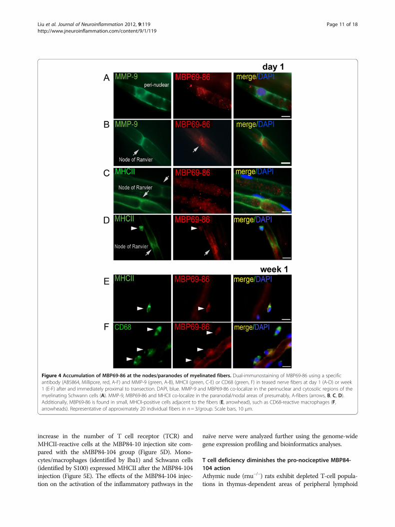
MMP-9 within the domains of myelinated fibers was ana-lyzed in the teased rat sciatic nerve preparations at day 1and week 1 after transection (Figure 4). Due to Walleriandegeneration altering the integrity of A-fibers in the distalsegment, we analyzed the fibers from the segment immedi-ately proximal to transection, the site where MMP-9 ex-pression and activity were induced [44,56]. MMP-9 co-localized with MBP69-86 in the Schwann cell cytoplasmand perinuclear areas of the teased nerve fiber preparations(Figure 4A). Intriguingly, at day 1 post-injury, MBP69-86localized in the paranodal/nodal regions, in close proximityto MMP-9 (Figure 4B) and MHCII (Figure 4C) on theSchwann cell plasma and/or basement membranes.MHCII-positive round small cells adjacent to the myelin-ated fibers were reactive for MBP69-86 at day 1 (Figure 4D)and, especially week 1 (Figure 4E) post-injury. A number ofMBP69-86-reactive macrophages (identified by CD68) wereadjacent to the fibers (Figure 4F). The later finding mayrepresent phagocytosed myelin in hematogenous or resi-dent macrophages, or the degraded Golli-MBP, expressedin immune cells [26].
MBP peptides induce allodyniaOur data indicate that as a result of MMP-9 proteolysis ofMBP in vitro, cryptic epitopes are released at the N-
terminal and central portions of the MBP sequence, in-cluding MBP68-86, MBP84-104 (summarized inFigure 5A). In agreement, the immunodominant MBP69-86 peptide sequence was produced in the injured nerve.Next, we analyzed the effect of the synthetic MBP pep-tides on pain-like behaviors. Mechanical and heat hyper-sensitivity and spontaneous pain-like behavior parameterswere assessed in rats after a single intraneural injection ofMBP84-104, MBP68-86, MBP2-18 and scrambledMBP84-104 (sMBP84-104) peptides (>97-99% pure,50 μg each) into a naïve sciatic nerve (Figure 5B). A dropin the mechanical withdrawal threshold after the injectionof the MB-P84-104 and MBP68-86 peptides correspondedto robust allodynia lasting for up to 9 days. In contrast, in-jection of MBP2-18 and sMBP84-104 was without effectand resulted in a withdrawal threshold comparable to PBSinjection. Robust decline in the mechanical withdrawalthreshold was also readily observed after a single injectionof 10 μg of MBP68-86 or MBP84-104 (data not shown).Further investigation was done using MBP-84-104 (the
most potent modulator of allodynia) and its scrambled pep-tide control. The neuropathology of the respective nervesat the injection sites was analyzed at day 3 after theMBP84-104 or sMBP84-104 injection, when the differencein pain-like behavior was highly significant. In contrast withsMBP84-104, MBP84-104 produced focal myelin splitting,endoneurial edema and infiltration of phagocytic immunecells (Figure 5C). These findings were accompanied by an
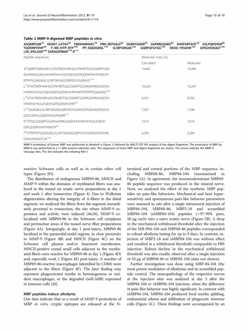
Table 2 MMP-9-digested MBP peptides in vitro
ASQKRPSQR10 HGSKY#LATAS20 TMDHARHGFL30 PRH#RDTGILD40 SIGRFFGGDR50 GAPKRGSGKD60 SHHPARTAHY70 GSLPQKSHGR80
TQDENPVVHF90 F#KN#IVTP#RTP100 PP#SQGKGRGL110 SLSRFSWGAE120 GQRPGFGYGG130 RASD#YKSAHK140 GFKGVDAQGT150
LSK#IFKLGGR160 DSRSGSPMAR170 R171
Peptide sequences Molecular mass, Da
Calculated Measured
A2SQKRPSQRHGSKYLATASTMDHARHGFLPRHRDTGILDSIGRFFGGD 14,482 14,498
RGAPKRGSGKDSHHPARTAHYGSLPQKSHGRTQDENPVVHFFKNIVTP
RTPPPSQGKGRGLSLSRFSWGAEGQRPGFGYGGRASD134
L16ATASTMDHARHGFLPRHRDTGILDSIGRFFGGDRGAPKRGSGKDSH 10,229 10,247
HPARTAHYGSLPQKSHGRTQDENPVVHFFKNIVTPRTPPPSQGKGRG109
L16ATASTMDHARHGFLPRHRDTGILDSIGRFFGGDRGAPKRGSGKDSH 8,357 8,356
HPARTAHYGSLPQKSHGRTQDENPVVHFF91
S103QGKGRGLSLSRFSWGAEGQRPGFGYGGRASDYKSAHKGFKGVDA 7,307 7,296
QGTLSKIFKLGGRDSRSGSPMARR171
R34DTGILDSIGRFFGGDRGAPKRGSGKDSHHPARTAHYGSLPQKSH 7,010 7,018
GRTQDENPVVHFFKNIVTP97
I94VTPRTPPPSQGKGRGLSLSRFSWGAEGQRPGFGYGGRASDYKSAHK 6,293 6,284
GFKGVDAQGTLSK153
MMP-9 proteolysis of human MBP was performed as detailed in Figure 3, followed by MALTI-TOF MS analysis of the digest fragments. The proteolysis of MBP byMMP-9 was performed at a 1:1,000 enzyme-substrate ratio. The sequences of intact MBP and digest fragments are shown. The arrows indicate the MMP-9cleavage sites. The dot indicates the initiating Met-1.
Liu et al. Journal of Neuroinflammation 2012, 9:119 Page 10 of 18http://www.jneuroinflammation.com/content/9/1/119
increase in the number of T cell receptor (TCR) andMHCII-reactive cells at the MBP84-10 injection site com-pared with the sMBP84-104 group (Figure 5D). Mono-cytes/macrophages (identified by Iba1) and Schwann cells(identified by S100) expressed MHCII after the MBP84-104injection (Figure 5E). The effects of the MBP84-104 injec-tion on the activation of the inflammatory pathways in the
naïve nerve were analyzed further using the genome-widegene expression profiling and bioinformatics analyses.
T cell deficiency diminishes the pro-nociceptive MBP84-104 actionAthymic nude (rnu−/−) rats exhibit depleted T-cell popula-tions in thymus-dependent areas of peripheral lymphoid
Figure 4 Accumulation of MBP69-86 at the nodes/paranodes of myelinated fibers. Dual-immunostaining of MBP69-86 using a specificantibody (AB5864, Millipore, red, A-F) and MMP-9 (green, A-B), MHCII (green, C-E) or CD68 (green, F) in teased nerve fibers at day 1 (A-D) or week1 (E-F) after and immediately proximal to transection. DAPI, blue. MMP-9 and MBP69-86 co-localize in the perinuclear and cytosolic regions of themyelinating Schwann cells (A). MMP-9, MBP69-86 and MHCII co-localize in the paranodal/nodal areas of presumably, A-fibers (arrows, B, C, D).Additionally, MBP69-86 is found in small, MHCII-positive cells adjacent to the fibers (E, arrowhead), such as CD68-reactive macrophages (F,arrowheads). Representative of approximately 20 individual fibers in n= 3/group. Scale bars, 10 μm.
Liu et al. Journal of Neuroinflammation 2012, 9:119 Page 11 of 18http://www.jneuroinflammation.com/content/9/1/119

organs. Because rnu−/− rats are less susceptible to the devel-opment of NP [9], we used this strain to test our hypothesisthat the pro-nociceptive effect of MBP84-104 depended onT cell production, infiltration or homing to the nerve andthe corresponding spinal cord (Figures 6 and 7).The mechanical withdrawal thresholds readily declined
following a single MBP84-104 injection in the wild-type(rnu+/−) rats (Figure 6A). In turn, incomplete attenuation ofallodynia was observed in nude rats. The threshold levelssignificantly increased by day 3 post-injection, althoughremained below the baseline. The injection of MBP84-104
did not cause a change in thermal withdrawal latencies(Figure 6B) or spontaneous pain-like behaviors (Figure 6C)in both rat types. We concluded that immunodominantMBP peptides, specifically MBP84-104, initiated mechan-ical but not thermal hypersensitivity in naïve animals, andthat the pro-nociceptive activity of MBP84-104 was, at leastpartly, T cell-dependent.At the completion of behavioral analyses (1 week after
the MBP84-104 injection), the sciatic nerve (the injectedand the contralateral side) and the dorsal horn spinal cord(the lumbar enlargement and above-the-level thoracic
Figure 5 Pro-nociceptive activity of MBP peptides. (A) The 1–171 sequence of human MBP (GenBank #AAH08749). The immunogenic regionsare shown at the bottom of the panel using human MBP residue numbering. Following MBP cleavage by MMP-9, the mass and, consecutively,the sequence of the digest fragments was determined by MALDI-TOF MS. The italicized numbers indicate the positions of the cleavage sites. (B)von Frey testing after the intraneural injection of MBP peptides (50 μg in 5 μL) or vehicle (PBS) into a naïve rat sciatic nerve. Within 1 day adecline in mechanical withdrawal thresholds was observed after the MBP84-104 and MBP68-86 injection. Control MBP2-18 and MBP84-104scrambled (sMBP84-104) peptide induced no significant change in thresholds compared to PBS. The mean withdrawal thresholds (gram force;g) ± SEM of n= 6/group (**, P< 0.01, *; P< 0.05). (C) Methylene Blue Azure II staining in 1-μm-thick sciatic nerve sections. MBP84-104 producedmyelin splitting and cell infiltration 3 days post-injection into the naïve nerve. Uncompromised axons, surrounded by a compact rim of myelinare observed in the nerves after the sMBP84-104 injection. Representative micrographs of n= 4/group. Scale bars, 20 μm. (D) Immunostaining ofMHCII (green) or T cell receptor (TCR, green) in the nerve at 3 days after the MBP injection into the intact nerve. Scale bars, 25 μm. The graphrepresents morphometry of the mean MHCII-positive cell numbers in the sciatic nerves ± SEM of n= 4/group and three sections per n (*, P< 0.05).(E) Immunostaining of MHCII (green) and Iba1 in the monocytes/macrophages (red) or S100 in the Schwann cells (red) in the nerve after theMBP84-104 injection in the wild-type rats. Macrophages (arrowheads) and Schwann cells (arrows) represent MHCII-reactive cells in the nerve,exposed to the immunodominant MBP84-104 peptide. DAPI, blue. Representative micrographs of n= 4/group. Scale bars, 10 μm.
Liu et al. Journal of Neuroinflammation 2012, 9:119 Page 12 of 18http://www.jneuroinflammation.com/content/9/1/119

segments) from the wild-type and nude rats were collectedfor the further genome-wide transcriptional profiling. Theaim of these experiments was two-fold: (1) correlate the de-cline in MBP84-104-induced allodynia with the decline inT cell response via the unbiased screening; and (2) identifyT cell-independent changes induced by MBP84-104 thatmight persist longer in the T cell-deficient tissues. The topbiological functions and pathways induced by the
intraneural MBP84-104 injection both in the nerve and thecord were categorized as canonical inflammatory response,immune cell trafficking, inflammatory disease and antigenpresentation pathways (Figure 7 and Table 3). In the nerve,1 week after the MBP-84-10 injection the antigen presenta-tion function was elevated approximately 10-fold(Figure 7A), communication signaling between innate andadaptive immune cells signaling was elevated five-fold
Figure 6 MBP-induced allodynia is T cell-dependent. (A) von Frey testing for mechanical allodynia in athymic nude (rnu−/−) and wild-typeheterozygous (rnu+/−) rats after the MBP84-104 injection (50 μg in 5 μL) into the naïve sciatic nerve. The mean withdrawal thresholds (gram force,g) ± SEM of n= 6/group decline rapidly after the MBP84-104 injection into normal rats, corresponding to allodynia (arrow), but remain significantlyhigher in nude rats (**, P< 0.01; *, P< 0.05). (B) Hargreaves testing for thermal sensitivity. The mean paw withdrawal latency ± SEM of n= 6/groupafter thermal stimulation (radiant heat) before (baseline) and at the indicated days after the MBP84-104 injection were not different between thegroups. (C) Spontaneous pain scoring of the MBP84-104 injected paw positioning for 6 min (3 x 120 s) using a 0–5 numerical scale demonstratesthe absence of spontaneous pain-like behaviors in both rat types. The mean score ± SEM of n= 6/group.
Figure 7 Functional analysis of global gene expression in nerves and spinal cords after intraneural MBP84-104 injection. Ingenuitypathway analysis of the gene expression used as the input list for generation of the top functional categories and canonical pathways (X-axis).The bars represent -log10 (P value) for a function or a pathway to be represented with a threshold (dashed line) set at 1.3 (P <0.05) in n= 6/group (right-tailed Fisher's exact test). Biological functions and canonical pathways are listed from most to least significant in the experimentalMBP-injected animals in the injected compared to control contralateral sciatic nerves (A, B) or in the dorsal horns of the corresponding lumbarcompared to control thoracic spinal cords (C, D) of the same animals. LXR, liver X receptor; RXR, retinoid X receptor; VDR, vitamin D receptor.
Liu et al. Journal of Neuroinflammation 2012, 9:119 Page 13 of 18http://www.jneuroinflammation.com/content/9/1/119

(Figure 7B) and the T cell activation pathway was elevated7.8-fold (Table 3), as compared to contralateral nerve inthe same animals. In lumbar dorsal horn spinal cord, theintraneural MBP84-104 injection produced an over three-fold increase in antigen presentation function (Figure 7C),an about 1.8-fold increase in IL-17 signaling (Figure 7D)and 2.6-fold increase in T cell activation pathway (Table 3)relative to the thoracic segment in the same animals. Thesignaling cascades representative of autoimmune demye-lination were activated at the MBP84-104 nerve injectionsite of the wild-type but not the nude rats (Figure 7B, Add-itional file 3: Figure S1). In addition, MBP84-104 activatescalcium, liver X receptor (LXR), retinoid X receptor(RXR), and vitamin D receptor (VDR) signaling in thenerve and the corresponding spinal cord (Figure 7).
DiscussionDefinite progress in elucidating the immunologicalmechanisms of NP has been achieved in recent years[17,57-60]. However, the molecular and cellular processesthat cause myelinated Aβ-afferents to enter nociceptive cir-cuits after nerve damage remain poorly understood as yet[1,61]. There is growing evidence for the direct relationshipbetween axonal demyelination and pain [2-4,6]. We haveimplicated MMP proteolysis of myelin, specifically MBP, inmechanical hypersensitivity [5]. Herein, we provide strongevidence that the MMP-generated MBP digest peptidescomprising the potent immunogenic epitopes are releasedduring Wallerian degeneration. We also demonstrated thatthese immunogenic MBP peptides initiate mechanical allo-dynia in both T cell-dependent and -independent manners.
Multiphasic roles of MMPs in MBP/golli-MBP cleavage andNPMMPs are key degrading proteases of MBP. The peptidesgenerated because of MMP-9 proteolysis of various MBP/Golli-MBP isoforms include MBP84-104, 69–86, as well asother immunodominant regions [27,28,37,62,63]. At least
six distinct peptides in the 6–14.5 kDa range are formed asa result of MMP-9 proteolysis of pure human 18.5 kDaMBP in vitro. Accordingly, an 18.5 kDa and other MBP iso-forms accumulated in the MMP-9-deficient nerves. Further,more in-depth studies are required to assess the in vivo kin-etics of MMP-9 proteolysis of multiple MBP/Golli-MBP se-quence and post-translationally modified isoforms [23] overthe course of nerve injury. The ability of MMP-9 to affectthe MBP expression by regulation of Schwann cell signaling[56] also needs to be taken into consideration.MMP-9 co-localizes with the endogenous MBP69-86 in
the myelinating Schwann cell cytoplasm [5] and perinuclearareas, and in the paranodal/nodal regions of myelinatedfibers. In addition, macrophages deposit MBP69-86 in thenerve. Among the Golli-MBP splice forms produced by im-mune cells, BG21 and J37 contain the cryptic epitopesequences [21,64-66] that we have found to be released byMMPs [27,37]. Because of the cleavage redundancy amongthe MMP family, several MMPs are likely to proteolyzeMBP/Golli-MBP over the course of nerve injury. The dif-ferential ability of MBP84-104, 68–86, and 2–18 peptidesto produce allodynia suggests that proteolysis of MBP influ-ences susceptibility to NP.In addition to protecting myelin from proteolysis, on-
going broad-spectrum MMP inhibition may prevent allody-nia by protecting DRG neurons from apoptosis [5] andsuppressing the expression of certain voltage-gated sodiumchannels [44], although the latter effect may also relate tothe myelin integrity [2,4]. MMP-2/9 also release the pro-nociceptive cytokines (TNF-α and IL-1β) from their trans-membrane precursors, promoting peripheral and spinalglial activation and immune cell-mediated pain [33,34,58].The nerves acutely treated immediately post-CCI with theMMP-9/2 inhibitor, SB-3CT, sustain the low levels of theTNF-α expression (produced by Schwann cells, macro-phages, endothelial, Th1, and other cells) and the IL-17Aexpression (produced by Th17 and other cells) [11,15,67].Because MMP-9 and not MMP-2 is induced immediatelypost-injury and has been implicated in the initiation of NP[5,33], we attribute the effects of acute SB-3CT therapymainly to MMP-9 inhibition. Likewise, therapy with TIMP-1 prevents NP by MMP-9 inhibition [33], as TIMP-1 bindsMMP-9 stoichiometrically and blocks access of its catalyticsite to substrates [29].
T cells in pain and pro-nociceptive action of MBPT cell-deficient animals are less susceptible to NP[9,11,12,14]. Progress has been made in identifying T cellsubset phenotypes involved in NP. For example, a declinein hypersensitivity in CD4 null mice in the spinal nerveligation model was resumed with adoptive transfer of CD4+ Th cells [12]. Specifically, transfer of Th1 cells (that pro-duce pro-inflammatory cytokines) restores allodynia, asTh2 cells (that produce anti-inflammatory cytokines)
Table 3 MBP84-104 activates inflammatory pathways innerve
Functions annotation Nerve, -log(P value)
Spinal cord, -log(P value)
WT Nude WT Nude
Immune response 20.2 - 4.32 -
Inflammatory response 15.8 - 2.79 1.42
Activation of T lymphocytes 7.8 - 2.64 -
Chemotaxis/aggregation of APCs 5.5 2.39 2.54 2.88
Chemotaxis of monocytes 4.2 2.61 1.86 2.36
Ingenuity pathway analysis of the genome 1 week after the MBP84-104 injectioninto the intact sciatic nerve in the wild-type and nude rats. Up-regulated functionsas a result of inflammatory response in the injected nerve compared to thecontralateral sciatic nerve and in the lumbar compared to the thoracic spinal cord,dorsal horn, are shown. Cutoff is set at P< 0.05 (−log10> 1.3). Minus indicates thatthe Ingenuity software did not detect the presence of the pathway in the sample.
Liu et al. Journal of Neuroinflammation 2012, 9:119 Page 14 of 18http://www.jneuroinflammation.com/content/9/1/119

sustain resistance to CCI-induced pain in nude rats,strongly implicating Th1 but not Th2 cells in promotingNP [9]. IL-17A, produced by a unique subset of Th17 cells,is detected at week 1 post-CCI [11], and IL-17 deletion pro-tects from the development of NP [15].Activated T cells patrol the intact PNS during immuno-
surveillance irrespective of their antigen specificity [31].They infiltrate the nerve at 1 week after a physical nervedamage [9-11,43] via the coordinated action of chemokines,cytokines, and MMPs that compromise the blood-nervebarrier and trigger demyelination [31,68,69]. T cell infiltra-tion into the spinal cord also contributes to the develop-ment of peripheral NP [13,14,16]. It is plausible thatrepeated exposure of MBP/Golli-MBP epitopes results inthe formation of MBP-specific T cell clones, which then in-filtrate the corresponding central segments, where antigen-presenting systems are in place [14,16,19]. It is interestingto point out that the classic MBP and Golli-MBP differen-tially regulate T cell signaling [70].The ability of MBP84-104 to initiate allodynia is dimin-
ished in nude rats, indicating the presence of the T cell-dependent mechanism of MBP action in neuropathic pain.In agreement, our data clearly demonstrated that followingthe MBP84-104 injection there was no increase in the in-flammation and antigen presentation signaling in both thenerve and the corresponding spinal cord in nude animals.However, both the ability of MBP84-104 to initiate painshortly after the injection and residual hypersensitivity inthe nude rats imply that there is an additional, T cell-inde-pendent component in the ability of MBP84-104 to pro-mote pain. For example, MBP regulates intracellularcalcium flow [71,72], a key factor in pain facilitation [73]. Itappears that MBP84-104 (in a T cell-independent manner)affects calcium flow in the wild-type and nude rats.
1Schwann cells in myelin clearance and antigenpresentationMBP84-104 injection into the intact nerve induced MHCIIin the macrophages and Schwann cells. The endogenouscryptic MBP69-86 epitope was detected in MHCII-reactivemyelinating Schwann cells within day 1 post-injury. This isnot surprising, since during the first few days post-injury,Schwann cells are actively involved in the degradation andremoval of the myelin debris and in the presenting of mye-lin antigens [69,74-78]. The deposits of the endogenousMBP69-86 in CD68-reactive injured nerve may representphagocytosed myelin debris or the Golli species, expressedby monocytes and other blood cells [26]. Overall, myelindegradation and clearance in the damaged PNS appears toconsist of an early phase mediated by Schwann cells andresident macrophages [31] and an antibody-dependent laterphase mediated by hematogenous macrophages [25,69].Each phase of this event may have distinct function in theNP state.
Neuro-immune interactions at A-fibers: mechanical vs.thermal hypersensitivityAccumulation of MBP69-86 and MHCII around the nodesof Ranvier is intriguing. We speculate that MBP69-86 bothactivates the pro-nociceptive changes in the calcium flow[71,72] and facilitates the T cell homing at these action-gen-erating sites on A-afferents. In agreement, MBP84-104induced mechanical allodynia but not thermal hyperalgesia,at least in the intact nerve environment, and T cell-deficientIL-17 null mice develop resistance to mechanical allodyniabut not thermal hyperalgesia [15]. There is a growing appre-ciation that differential mechanisms underlie these NP states,as A-afferents mediate mechanical allodynia and heat-noci-ceptive C-fibers mediate thermal hyperalgesia [79,80]. TheMBP degradation and T cell homing to the regions whichare immediately proximal to transection and in which ele-vated MMP-9 levels and other features of Schwann cell acti-vation manifest [44,56,81], may explain pain facilitationdespite the fiber loss at the lesion site. Finally, the presentstudy does not distinguish between the myelinated afferentsand efferents, supporting a model that demyelinating motorefferents contribute to nociceptive pain [3].
ConclusionsThe present data implicate proteolyzed MBP in pain. Overthe course of Wallerian degeneration, the repeated expos-ure of the MBP epitopes normally sheltered from immuno-surveillance may lead to the formation of the MBP-specificT cell clones and a self-sustaining immune reaction both ofwhich contribute to the transition of ‘protective auto-immunity’ and acute pain to a chronic NP state. Thus, pre-venting proteolysis of MBP may prove as a viabletherapeutic strategy against neuropathic pain. It is temptingnow to hypothesize that our findings broadly relate to painassociated with autoimmune demyelinating neuropathiesand neurodegenerative disorders where the formation ofcryptic MBP epitopes has also been documented [24,82].
Additional files
Additional file 1: Table S1. Unique proteins in CCI nerve listing, basedon the 2D-LC-MS/MS of the sciatic nerve proteome after CCI (day 7).
Additional file 2: Table S2. Unique proteins in normal nerve listingbased on the 2D-LC-MS/MS of the sciatic nerve proteome after shamoperation (day 7).
Additional file 3: Figure S1. Autoimmune demyelination signalingin nerve after intraneural MBP84-104 injection. Ingenuity PathwayAnalysis of the gene expression used for generation the autoimmunedemyelination signaling cascades at week 1 after intraneural MBP84-104injection in wild-type rats and nude rats. Up-regulated expression ofchemokine receptors and ligands are indicated in red. The intensity ofred color corresponds to fold-change of expression level of respectivegenes. Activated T cells, producing CCR5 and CXCR3 (vertical rectangles)and monocytes, producing CCR5 and CCR1 receptors, are recruited intothe intact nerve after MBP84-104 injection into the wild-type rats but notnude rats. CXCL9, CXCL10, and CXCL11 are ligands for CXCR3. CCL5,CCL3, and CCL4 are ligands for CCR5 and CCR1.
Liu et al. Journal of Neuroinflammation 2012, 9:119 Page 15 of 18http://www.jneuroinflammation.com/content/9/1/119

Competing interestsThe authors declare no competing interests.
AcknowledgementsThe authors gratefully acknowledge Jennifer Dolkas and Calvin Lai fortechnical assistance to HL, YK, IS, and VIS, and Wenhong Zhu and KhaterehMotamedchaboki for help with performing mass spectrometry analyses. Thiswork is supported by NIH/NINDS R21 NS060307-01 and the Department ofVeterans Affairs Merit Review Award to VIS and by NIH/NCI R01CA83017 andR01CA157328 to AYS.
Author details1Department of Anesthesiology, University of California, San Diego, 9500Gilman Dr., Mail Box 0629, La Jolla, CA 92093-0629, USA. 2VA San DiegoHealthcare System, La Jolla, CA, USA. 3Sanford-Burnham Medical ResearchInstitute, La Jolla, CA, USA. 4Agilent Technologies, La Jolla, CA, USA.
Authors’ contributionsHL, YK, and IS performed animal surgeries and microinjections, behavioraltesting, neuropathology, zymography, immunofluorescence and qPCR of theneuronal tissues, statistical analysis, and drafted the manuscript. SAS, AGR,and VSG carried out the MMP-9 purification, the biochemical characterizationof MMP-9 proteolysis of MBP, mass spectrometry and proteomics, anddrafted the manuscript. AVC and SB performed the microarray gene profilingexperiments, the follow-on bioinformatics analyses, and drafted themanuscript. VIS and AYS conceived the study, participated in its design,coordinated the execution of the studies, and wrote the manuscript. Allauthors read and approved the final manuscript.
Received: 6 February 2012 Accepted: 23 April 2012Published: 7 June 2012
References1. Devor M: Ectopic discharge in Abeta afferents as a source of neuropathic
pain. Exp Brain Res 2009, 196:115–128.2. Devor M: Sodium channels and mechanisms of neuropathic pain. J Pain
2006, Suppl 1:S3–S12.3. Wu G, Ringkamp M, Murinson BB, Pogatzki EM, Hartke TV, Weerahandi HM,
Campbell JN, Griffin JW, Meyer RA: Degeneration of myelinated efferentfibers induces spontaneous activity in uninjured C-fiber afferents.J Neurosci 2002, 22:7746–7753.
4. Henry MA, Luo S, Foley BD, Rzasa RS, Johnson LR, Levinson SR: Sodiumchannel expression and localization at demyelinated sites in painfulhuman dental pulp. J Pain 2009, 10:750–758.
5. Kobayashi H, Chattopadhyay S, Kato K, Dolkas J, Kikuchi S, Myers RR, ShubayevVI: MMPs initiate Schwann cell-mediated MBP degradation and mechanicalnociception after nerve damage. Mol Cell Neurosci 2008, 39:619–627.
6. Zhu YL, Xie ZL, Wu YW, Duan WR, Xie YK: Early demyelination of primaryA-fibers induces a rapid-onset of neuropathic pain in rat. Neuroscience2012, 200:186–198.
7. Treede RD, Jensen TS, Campbell JN, Cruccu G, Dostrovsky JO, Griffin JW,Hansson P, Hughes R, Nurmikko T, Serra J: Neuropathic pain: redefinitionand a grading system for clinical and research purposes. Neurology 2008,70:1630–1635.
8. Scholz J, Woolf CJ: The neuropathic pain triad: neurons, immune cellsand glia. Nat Neurosci 2007, 10:1361–1368.
9. Moalem G, Xu K, Yu L: T lymphocytes play a role in neuropathic painfollowing peripheral nerve injury in rats. Neuroscience 2004, 129:767–777.
10. Tsai YC, Won SJ: Effects of tramadol on T lymphocyte proliferation andnatural killer cell activity in rats with sciatic constriction injury. Pain 2001,92:63–69.
11. Kleinschnitz C, Hofstetter HH, Meuth SG, Braeuninger S, Sommer C, Stoll G:T cell infiltration after chronic constriction injury of mouse sciatic nerveis associated with interleukin-17 expression. Exp Neurol 2006, 200:480–485.
12. Cao L, DeLeo JA: CNS-infiltrating CD4+ T lymphocytes contribute tomurine spinal nerve transection-induced neuropathic pain. Eur J Immunol2008, 38:448–458.
13. Sweitzer SM, Hickey WF, Rutkowski MD, Pahl JL, DeLeo JA: Focal peripheralnerve injury induces leukocyte trafficking into the central nervous system:potential relationship to neuropathic pain. Pain 2002, 100:163–170.
14. Costigan M, Moss A, Latremoliere A, Johnston C, Verma-Gandhu M, HerbertTA, Barrett L, Brenner GJ, Vardeh D, Woolf CJ, Fitzgerald M: T-cell infiltrationand signaling in the adult dorsal spinal cord is a major contributor toneuropathic pain-like hypersensitivity. J Neurosci 2009, 29:14415–14422.
15. Kim CF, Moalem-Taylor G: Interleukin-17 contributes toneuroinflammation and neuropathic pain following peripheral nerveinjury in mice. J Pain 2011, 12:370–383.
16. Sweitzer SM, White KA, Dutta C, DeLeo JA: The differential role of spinalMHC class II and cellular adhesion molecules in peripheral inflammatoryversus neuropathic pain in rodents. J Neuroimmunol 2002, 125:82–93.
17. Austin PJ, Moalem-Taylor G: The neuro-immune balance in neuropathicpain: involvement of inflammatory immune cells, immune-like glial cellsand cytokines. J Neuroimmunol 2010, 229:26–50.
18. Hu P, Bembrick AL, Keay KA, McLachlan EM: Immune cell involvement indorsal root ganglia and spinal cord after chronic constriction ortransection of the rat sciatic nerve. Brain Behav Immun 2007, 21:599–616.
19. Alzate O, Hussain SR, Goettl VM, Tewari AK, Madiai F, Stephens RL Jr,Hackshaw KV: Proteomic identification of brainstem cytosolic proteins ina neuropathic pain model. Brain Res Mol Brain Res 2004, 128:193–200.
20. Salzer JL: Polarized domains of myelinated axons. Neuron 2003, 40:297–318.21. Givogri MI, Bongarzone ER, Campagnoni AT: New insights on the biology
of myelin basic protein gene: the neural-immune connection. J NeurosciRes 2000, 59:153–159.
22. Garbay B, Heape AM, Sarqueil F, Cassagne C: Myelin synthesis in theperipheral nervous system. Prog Neurobiol 2000, 61:267–304.
23. Boggs JM: Myelin basic protein: a multifunctional protein. Cell Mol Life Sci2006, 63:1945–1961.
24. Moalem-Taylor G, Allbutt HN, Iordanova MD, Tracey DJ: Painhypersensitivity in rats with experimental autoimmune neuritis, ananimal model of human inflammatory demyelinating neuropathy. BrainBehav Immun 2007, 21:699–710.
25. Vargas ME, Watanabe J, Singh SJ, Robinson WH, Barres BA: Endogenousantibodies promote rapid myelin clearance and effective axonregeneration after nerve injury. Proc Natl Acad Sci U S A 2010, 107:11993–11998.
26. Marty MC, Alliot F, Rutin J, Fritz R, Trisler D, Pessac B: The myelin basicprotein gene is expressed in differentiated blood cell lineages and inhemopoietic progenitors. Proc Natl Acad Sci U S A 2002, 99:8856–8861.
27. Shiryaev SA, Savinov AY, Cieplak P, Ratnikov BI, Motamedchaboki K, SmithJW, Strongin AY: Matrix metalloproteinase proteolysis of the myelin basicprotein isoforms is a source of immunogenic peptides in autoimmunemultiple sclerosis. PLoS One 2009, 4:e4952.
28. Proost P, Van Damme J, Opdenakker G: Leukocyte gelatinase B cleavagereleases encephalitogens from human myelin basic protein. BiochemBiophys Res Commun 1993, 192:1175–1181.
29. Nagase H, Visse R, Murphy G: Structure and function of matrixmetalloproteinases and TIMPs. Cardiovasc Res 2006, 69:562–573.
30. Shubayev VI, Angert M, Dolkas J, Campana WM, Palenscar K, Myers RR:TNFalpha-induced MMP-9 promotes macrophage recruitment intoinjured peripheral nerve. Mol Cell Neurosci 2006, 31:407–415.
31. Kieseier BC, Hartung HP, Wiendl H: Immune circuitry in the peripheralnervous system. Curr Opin Neurol 2006, 19:437–445.
32. Chattopadhyay S, Myers RR, Janes J, Shubayev V: Cytokine regulation ofMMP-9 in peripheral glia: implications for pathological processes andpain in injured nerve. Brain Behav Immun 2007, 21:561–568.
33. Kawasaki Y, Xu Z-Z, Wang X, Park JY, Zhuang Z-Y, Tan P-H, Gao Y-J, Roy K, CorfasG, Lo EH, Ji R-R: Distinct roles of matrix metalloproteases in the early- and late-phase development of neuropathic pain. Nat Med 2008, 14:331–336.
34. Dev R, Srivastava PK, Iyer JP, Dastidar SG, Ray A: Therapeutic potential ofmatrix metalloprotease inhibitors in neuropathic pain. Expert Opin InvestigDrugs 2010, 19:455–468.
35. Bennett GJ, Xie YK: A peripheral mononeuropathy in rat that producesdisorders of pain sensation like those seen in man. Pain 1988, 33:87–107.
36. Livak KJ, Schmittgen TD: Analysis of relative gene expression data usingreal-time quantitative PCR and the 2(−Delta Delta C(T)) Method. Methods2001, 25:402–408.
37. Shiryaev SA, Remacle AG, Savinov AY, Chernov AV, Cieplak P, Radichev IA,Williams R, Shiryaeva TN, Gawlik K, Postnova TI, Ratnikov BI, Eroshkin AM,
Liu et al. Journal of Neuroinflammation 2012, 9:119 Page 16 of 18http://www.jneuroinflammation.com/content/9/1/119

Motamedchaboki K, Smith JW, Strongin AY: Inflammatory proproteinconvertase-matrix metalloproteinase proteolytic pathway in antigen-presenting cells as a step to autoimmune multiple sclerosis. J Biol Chem 2009,284:30615–30626.
38. Chaplan SR, Bach FW, Pogrel JW, Chung JM, Yaksh TL: Quantitativeassessment of tactile allodynia in the rat paw. J Neurosci Methods 1994,53:55–63.
39. Hargreaves K, Dubner R, Brown F, Flores C, Joris J: A new and sensitivemethod for measuring thermal nociception in cutaneous hyperalgesia.Pain 1988, 32:77–88.
40. Attal N, Jazat F, Kayser V, Guilbaud G: Further evidence for ‘pain-related’behaviours in a model of unilateral peripheral mononeuropathy. Pain1990, 41:235–251.
41. Backonja M, Woolf CJ: Future directions in neuropathic paintherapy: closing the translational loop. Oncologist 2010,Suppl 2:24–29.
42. Costigan M: Pain’s peptide signature. Pain 2012, 153:509–510.43. Kim CF, Moalem-Taylor G: Detailed characterization of neuro-
immune responses following neuropathic injury in mice. Brain Res2011, 1405:95–108.
44. Kim Y, Remacle AG, Chernov AV, Liu H, Shubayev I, Lai C, Dolkas J, ShiryaevSA, Golubkov VS, Mizisin AP, Strongin AY, Shubayev VI: The MMP-9/TIMP-1axis controls the status of differentiation and function of myelin-formingSchwann cells in nerve regeneration. PLoS One 2012, 7:e33664.
45. Shubayev VI, Myers RR: Endoneurial remodeling by TNFalpha- andTNFalpha-releasing proteases. A spatial and temporal co-localizationstudy in painful neuropathy. J Peripher Nerv Syst 2002, 7:28–36.
46. Shubayev VI, Myers RR: Upregulation and interaction of TNFalphaand gelatinases A and B in painful peripheral nerve injury. BrainRes 2000, 855:83–89.
47. Brown S, Bernardo MM, Li Z-H, Kotra LP, Tanaka Y, Fridman R, Mobashery S:Potent and selective mechanism-based inhibition of gelatinases. J AmChem Soc 2000, 122:6799–6800.
48. Liu H, Shubayev VI: Matrix metalloproteinase-9 controls proliferation ofNG2+ progenitor cells immediately after spinal cord injury. Exp Neurol2011, 231:236–246.
49. Gu Z, Cui J, Brown S, Fridman R, Mobashery S, Strongin AY, Lipton SA: Ahighly specific inhibitor of matrix metalloproteinase-9 rescues lamininfrom proteolysis and neurons from apoptosis in transient focal cerebralischemia. J Neurosci 2005, 25:6401–6408.
50. Gould KE, Swanborg RH: T and B cell responses to myelin basic proteinand encephalitogenic epitopes. J Neuroimmunol 1993, 46:193–198.
51. Katsara M, Deraos G, Tselios T, Matsoukas J, Apostolopoulos V: Design ofnovel cyclic altered peptide ligands of myelin basic protein MBP83-99that modulate immune responses in SJL/J mice. J Med Chem 2008,51:3971–3978.
52. Matsoukas J, Apostolopoulos V, Kalbacher H, Papini AM, Tselios T,Chatzantoni K, Biagioli T, Lolli F, Deraos S, Papathanassopoulos P,Troganis A, Mantzourani E, Mavromoutsakos T, Mouzaki A: Designand synthesis of a novel potent myelin basic protein epitope 87–99 cyclic analogue: enhanced stability and biological properties ofmimics render them a potentially new class of immunomodulators.J Med Chem 2005, 48:1470–1480.
53. Stepaniak JA, Gould KE, Swanborg RH: Encephalitogenic T cells arepresent in Lewis rats protected from autoimmune encephalomyelitis bycoimmunization with MBP73-84 and its analog. J Neurosci Res 1996,45:447–454.
54. Tselios T, Apostolopoulos V, Daliani I, Deraos S, Grdadolnik S,Mavromoustakos T, Melachrinou M, Thymianou S, Probert L, MouzakiA, Matsoukas J: Antagonistic effects of human cyclic MBP(87–99)altered peptide ligands in experimental allergic encephalomyelitisand human T-cell proliferation. J Med Chem 2002, 45:275–283.
55. Matsuo A, Lee GC, Terai K, Takami K, Hickey WF, McGeer EG, McGeerPL: Unmasking of an unusual myelin basic protein epitope duringthe process of myelin degeneration in humans: a potentialmechanism for the generation of autoantigens. Am J Pathol 1997,150:1253–1266.
56. Chattopadhyay S, Shubayev VI: MMP-9 controls Schwann cellproliferation and phenotypic remodeling via IGF-1 and ErbBreceptor-mediated activation of MEK/ERK pathway. GLIA 2009,57:1316–1325.
57. Thacker MA, Clark AK, Marchand F, McMahon SB: Pathophysiology ofperipheral neuropathic pain: immune cells and molecules. Anesth Analg2007, 105:838–847.
58. Myers RR, Campana WM, Shubayev VI: The role of neuroinflammation inneuropathic pain: mechanisms and therapeutic targets. Drug DiscovToday 2006, 11:8–20.
59. Wieseler-Frank J, Maier SF, Watkins LR: Glial activation and pathologicalpain. Neurochem Int 2004, 45:389–395.
60. Ren K, Dubner R: Interactions between the immune and nervous systemsin pain. Nat Med 2010, 16:1267–1276.
61. Djouhri L, Lawson SN: Abeta-fiber nociceptive primary afferent neurons: areview of incidence and properties in relation to other afferent A-fiberneurons in mammals. Brain Res Brain Res Rev 2004, 46:131–145.
62. D’Souza CA, Moscarello MA: Differences in susceptibility of MBP chargeisomers to digestion by stromelysin-1 (MMP-3) and release of animmunodominant epitope. Neurochem Res 2006, 31:1045–1054.
63. Chandler S, Coates R, Gearing A, Lury J, Well G, Bone E: Matrixmetalloproteinases degrade myelin basic protein. Neurosci Lett 1995,201:223–226.
64. Campagnoni AT, Pribyl TM, Campagnoni CW, Kampf K, Amur-Umarjee S,Landry CF, Handley VW, Newman SL, Garbay B, Kitamura K: Structure anddevelopmental regulation of Golli-mbp, a 105-kilobase gene thatencompasses the myelin basic protein gene and is expressed in cells in theoligodendrocyte lineage in the brain. J Biol Chem 1993,268:4930–4938.
65. Filipovic R, Rakic S, Zecevic N: Expression of Golli proteins in adult humanbrain and multiple sclerosis lesions. J Neuroimmunol 2002,127:1–12.
66. Tienari PJ, Kuokkanen S, Pastinen T, Wikstrom J, Sajantila A, Sandberg-Wolheim M, Palo J, Peltonen L: Golli-MBP gene in multiple sclerosissusceptibility. J Neuroimmunol 1998, 81:158–167.
67. Myers RR, Shubayev VI: The ology of neuropathy: an integrative review ofthe role of neuroinflammation and TNF-alpha axonal transport inneuropathic pain. J Peripher Nerv Syst 2011, 16:277–286.
68. Meyerzu Horste G, Hu W, Hartung HP, Lehmann HC, Kieseier BC: Theimmunocompetence of Schwann cells. Muscle Nerve2008, 37:3–13.
69. Vargas ME, Barres BA: Why is Wallerian degeneration in the CNS so slow?Annu Rev Neurosci 2007, 30:153–179.
70. Feng JM, Fernandes AO, Campagnoni CW, Hu YH, Campagnoni AT: Thegolli-myelin basic protein negatively regulates signal transduction in Tlymphocytes. J Neuroimmunol 2004, 152:57–66.
71. Paez PM, Spreuer V, Handley V, Feng JM, Campagnoni C, Campagnoni AT:Increased expression of golli myelin basic proteins enhances calciuminflux into oligodendroglial cells. J Neurosci 2007, 27:12690–12699.
72. Smith GS, Paez PM, Spreuer V, Campagnoni CW, Boggs JM, Campagnoni AT,Harauz G: Classical 18.5-and 21.5-kDa isoforms of myelin basic proteininhibit calcium influx into oligodendroglial cells, in contrast to golliisoforms. J Neurosci Res 2011, 89:467–480.
73. Yaksh TL: Calcium channels as therapeutic targets in neuropathic pain.J Pain 2006, Suppl 1:S13–30.
74. Fernandez-Valle C, Bunge RP, Bunge MB: Schwann cells degrade myelinand proliferate in the absence of macrophages: evidence from in vitrostudies of Wallerian degeneration. J Neurocytol 1995, 24:667–679.
75. Bruck W: The role of macrophages in Wallerian degeneration. Brain Pathol1997, 7:741–752.
76. Stoll G, Griffin JW, Li CY, Trapp BD: Wallerian degeneration in theperipheral nervous system: participation of both Schwann cells andmacrophages in myelin degradation. J Neurocytol 1989,18:671–683.
77. Wekerle H, Schwab M, Linington C, Meyermann R: Antigen presentation inthe peripheral nervous system: Schwann cells present endogenousmyelin autoantigens to lymphocytes. Eur J Immunol 1986,16:1551–1557.
78. Lilje O: The processing and presentation of endogenous and exogenousantigen by Schwann cells in vitro. Cell Mol Life Sci 2002,59:2191–2198.
79. Shir Y, Seltzer Z: A-fibers mediate mechanical hyperesthesia and allodyniaand C-fibers mediate thermal hyperalgesia in a new model ofcausalgiform pain disorders in rats. Neurosci Lett 1990,115:62–67.
Liu et al. Journal of Neuroinflammation 2012, 9:119 Page 17 of 18http://www.jneuroinflammation.com/content/9/1/119

80. Ossipov MH, Bian D, Malan TP Jr, Lai J, Porreca F: Lack of involvement ofcapsaicin-sensitive primary afferents in nerve-ligation injury inducedtactile allodynia in rats. Pain 1999, 79:127–133.
81. Cheng C, Zochodne DW: In vivo proliferation, migration and phenotypicchanges of Schwann cells in the presence of myelinated fibers.Neuroscience 2002, 115:321–329.
82. Solaro C, Messmer Uccelli M: Pharmacological management of pain inpatients with multiple sclerosis. Drugs 2010, 70:1245–1254.
doi:10.1186/1742-2094-9-119Cite this article as: Liu et al.: Immunodominant fragments of myelinbasic protein initiate T cell-dependent pain. Journal of Neuroinflammation2012 9:119.
Submit your next manuscript to BioMed Centraland take full advantage of:
• Convenient online submission
• Thorough peer review
• No space constraints or color figure charges
• Immediate publication on acceptance
• Inclusion in PubMed, CAS, Scopus and Google Scholar
• Research which is freely available for redistribution
Submit your manuscript at www.biomedcentral.com/submit
Liu et al. Journal of Neuroinflammation 2012, 9:119 Page 18 of 18http://www.jneuroinflammation.com/content/9/1/119