comparative evaluation of the immunodominant proteins of
TRANSCRIPT
Veterinary World, EISSN: 2231-0916 803
Veterinary World, EISSN: 2231-0916Available at www.veterinaryworld.org/Vol.14/March-2021/35.pdf
RESEARCH ARTICLEOpen Access
Comparative evaluation of the immunodominant proteins of Brucella abortus for the diagnosis of cattle brucellosis
Mohandoss Nagalingam1 , Thaslim J. Basheer1 , Vinayagamurthy Balamurugan1 , Rajeswari Shome1 , S. Sowjanya Kumari1 , G. B. Manjunatha Reddy1 , Bibek Ranjan Shome1 , Habibur Rahman2, Parimal Roy1 ,
J. Joseph Kingston3 and R. K. Gandham4
1. ICAR-National Institute of Veterinary Epidemiology and Disease Informatics, Bengaluru, Karnataka, India;2. International Livestock Research Institute, New Delhi, India; 3. Defense Food Research Laboratory, Mysore,
Karnataka, India; 4. National Institute of Animal Biotechnology, Hyderabad, Telangana, India.Corresponding author: Mohandoss Nagalingam: [email protected]
Co-authors: TJB: [email protected], VB: [email protected], RS: [email protected], SSK: [email protected], GBMR: [email protected], BRS: [email protected], HR: [email protected],
PR: [email protected], JJK: [email protected], RKG: [email protected]: 28-05-2020, Accepted: 15-02-2021, Published online: 30-03-2021
doi: www.doi.org/10.14202/vetworld.2021.803-812 How to cite this article: Nagalingam M, Basheer TJ, Balamurugan V, Shome R, Kumari SS, Reddy GBM, Shome BR, Rahman H, Roy P, Kingston JJ, Gandham RK (2021) Comparative evaluation of the immunodominant proteins of Brucella abortus for the diagnosis of cattle brucellosis, Veterinary World, 14(3): 803-812.
AbstractBackground and Aim: The present serodiagnosis of brucellosis in livestock is based on the whole cell or smooth lipopolysaccharide of the Brucella organism in which specificity is hampered by the cross-reactivity, especially with the antibodies against Yersinia enterocolitica O:9 organism. The problem can be addressed by screening for better immunodominant antigens. Hence, the present study was undertaken to screen protein antigens of Brucella abortus for their diagnostic potential in cattle brucellosis.
Materials and Methods: Protein antigens of B. abortus (n=10) non-reactive to antibodies against Y. enterocolitica O:9 were selected, expressed in Escherichia coli, assessed the reactivity of expressed recombinant proteins by Western blot, standardized indirect-enzyme-linked immunosorbent assay (ELISA) for detecting Brucella antibodies in cattle serum, and comparative evaluation was done.
Results: All the selected protein antigens were expressed and in the Western blot with Brucella antibodies positive cattle serum, six recombinant (Brucella protein 26 [BP26], Cu-Zn Superoxide dismutase [SodC], B. abortus I-1885, Serine protease, Bacterioferritin, and Brucella Lumazine Synthase [BLS]) proteins showed reaction whereas none of the proteins showed reactivity with Brucella negative cattle serum. ELISA has been done using known Brucella positive and negative cattle sera samples (n=113 each) in which the performance of recombinant proteins in diagnosing brucellosis was in the order of BP26 > BLS > SodC followed by rest of the proteins. BP26 based ELISA was found to be better with area under the curve as 0.953, and diagnostic sensitivity, diagnostic specificity, and Youden’s index of 90.27%, 95.58%, and 0.8584, respectively, with the excellent agreement (k=0.85).
Conclusion: BP26 could be a potential diagnostic antigen among the immunodominant proteins of B. abortus in ruling out Y. enterocolitica O:9 infection while diagnosing brucellosis in cattle herds.
Keywords: Brucella abortus, Brucella lumazine synthase, Brucella protein26, cattle brucellosis, Cu-Zn superoxide dismutase, Yersinia enterocolitica O:9.
Introduction
Brucellosis is a multifaceted zoonotic disease with significant animal and human health impact, caused by facultative, intracellular bacteria of genus Brucella, which comprises six main species, namely Brucella abortus, Brucella melitensis, Brucella suis, Brucella ovis, Brucella canis, and Brucella neotomae, and other recently identified species include Brucella pinnipedialis, Brucella ceti, Brucella microti, Brucella inopinata, Brucella papionis, and Brucella vulpis [1].
The disease is usually represented by abortion, reduced fertility, and reduced milk production in ruminants. The common species involved in causing brucellosis in cattle is B. abortus.
Since the discovery of the brucellosis, the con-ventional serological tests, namely, Rose Bengal plate test (RBPT), standard tube agglutination test (SAT), enzyme-linked immunosorbent assay (ELISA), and complement fixation tests which utilize the whole Brucella cell and/or its smooth lipopolysaccharide (S-LPS) fractions are being used in diagnosis [2]. Cross-reactivity arises due to the resemblance of Brucella O-polysaccharide, a component of LPS with corresponding epitopes of Yersinia enterocolitica O:9, Salmonella urbana group N, Vibrio cholerae, Francisella tularensis, Escherichia coli O157, and Stenotrophomonas maltophilia [3]. In particular, sero-logical interference induced by Y. enterocolitica O:9
Copyright: Nagalingam, et al. Open Access. This article is distributed under the terms of the Creative Commons Attribution 4.0 International License (http://creativecommons.org/licenses/by/4.0/), which permits unrestricted use, distribution, and reproduction in any medium, provided you give appropriate credit to the original author(s) and the source, provide a link to the Creative Commons license, and indicate if changes were made. The Creative Commons Public Domain Dedication waiver (http://creativecommons.org/publicdomain/zero/1.0/) applies to the data made available in this article, unless otherwise stated.

Veterinary World, EISSN: 2231-0916 804
Available at www.veterinaryworld.org/Vol.14/March-2021/35.pdf
has complicated the eradication of brucellosis in some European countries such as France, Belgium, United Kingdom [4,5] with the prevalence of Y. enterocolitica ranging from 18% to 58% in cattle [6]. In European Union, 15% of the herds in regions free from brucello-sis were infected with Y. enterocolitica O:9 [7]. After 1990, the isolation of Y. enterocolitica O:9 from cat-tle has become a regular phenomenon not only from European Union but also from other parts of the world including New Zealand [8]. As diagnosis plays a key role in disease control/eradication programs, there is a strong need for tests that can avoid cross-reactivity, in particular against Y. enterocolitica O:9. This can be overcome by screening antigens other than surface antigens like S-LPS. Various proteins of Brucella spp. have been investigated for their use as a diagnostic antigen, replacing S-LPS either in the form of the purified native protein(s) or synthesized recombinant protein(s). Different proteins were reported from dif-ferent research groups for their capacity in diagnosis; however, still, there is a scope for further screening.
The present study was aimed to screen better pro-tein antigens for diagnosing cattle brucellosis which is void of cross-reactivity, especially against antibodies of Y. enterocolitica O:9. ELISA was done with these antigens and different cattle sera samples and com-pared for better performance. To the best of our knowl-edge, it is the first study comparing ten recombinant proteins that are non-reactive to Y. enterocolitica O:9.Materials and MethodsEthical approval
The Institute Biosafety committee approval (F. No. 6-52/NIVEDI/Biosafety/2016/07-19 dtd.11.12.2017) was obtained for the work.Study period and location
The work was conducted at ICAR-NIVEDI from January 2016 to March 2018. The cattle sera were col-lected from Karnataka, India.Bacterial strains, vectors, and serum
B. abortus Strain 99 (NCTC 11363) pro-cured from Indian Council of Agricultural Research Institute (ICAR)-Indian Veterinary Research Institute, Izatnagar, India, was used to obtaining deoxyribonu-cleic acid (DNA). HI-Control 10G and HI-Control BL21 (DE3) chemically competent cells with pETite N-His Kan Vector (M/s Lucigen, USA) were used in this study for expression of proteins in E. coli sys-tem. Hyperimmune serum raised against B. abortus S99 and Y. enterocolitica O:9 in rabbit, rabbit serum tested negative for Brucella antibodies, and serum samples collected from different cattle were used in this study. The reference DNA sequence of B. abortus biovar 1 strain 9-941 was used for designing primers, comparison of amplified sequences, etc., The NCBI reference number for chromosome 1 is NC_006932 (2124241 bp) and that of chromosome 2 (1162204 bp) is NC_006933.
Selection of immunodominant proteins/protein antigens for expression
The immunodominant proteins were identified based on a literature survey concerning their reactivity with Brucella positive serum and non-reactivity with Y. enterocolitica O:9 positive serum. They include Cu-Zn superoxide dismutase (SodC), Serine protease, BAB1-1885, Twin arginine translocation pathway signal sequence domain-containing protein (Twin arginine), Brucella protein26 (BP26), Solute-binding family 5 protein, Leu/Ile/Val-binding family protein, Branched-chain amino-acid ABC transporter sub-strate-binding protein, Thiamine transporter binding protein, Invasion protein B (InvB), Brucella lumazine synthase (BLS), bacterioferritin (Bfr), malate dehy-drogenase (Mdh), VirB12, and Aldehyde dehydro-genase. The selected proteins are outer membrane or periplasmic except for BLS, Bfr, Mdh, VirB12, and Aldehyde dehydrogenase which are cytoplasmic pro-teins. These identified proteins were further checked through BLASTP for amino acid sequence identity and those showing ≤60% with other cross-reacting patho-gens were selected for further expression (Table-1).Expression of immunodominant proteins
The primers were designed for the selected gene sequences to suit enzyme-free cloning in the pETite N-His Kan vector (Table-2). The respective gene sequences of selected proteins were amplified by PCR with reaction mixture comprising of 10× PCR buffer-5 µL; forward and reverse primers 10 µM each 1 µL; dNTP (10 mM)-1 µL; B. abortus S99 template DNA 2 µL (94 ng); high fidelity Taq polymerase 0.5 µL (M/s. Thermo Scientific, USA); and nucle-ase-free water were added to make up 50 µL. PCR was performed in the thermal cycler (M/s. Eppendorf, Germany) with the cycling conditions as mentioned in Table-2. The amplified product was run on 1.5% aga-rose gel containing ethidium bromide (0.5 µg/mL), visualized and documented in UV trans-illuminator.
The PCR amplicons were individually trans-formed with pETite N-His Kan vector into HI-Control 10G chemically competent cells, respectively, and recombinant colonies obtained were screened by col-ony PCR. The plasmids were extracted from the pos-itive clones and double pass sequencing of plasmid DNA was carried out commercially (M/s. Eurofins, India) by vector-specific primers. After confirmation by sequencing, pETite N-His Kan vectors contain-ing gene insert were individually transformed into HI-Control BL21 (DE3) and confirmed by colony PCR. The recombinant pETite N-His Kan BL21 clones were induced with IPTG for expression. The expres-sion was optimized for the incubation time (1-5 h after induction) and IPTG concentration (0.5-2.0 mM). The expressed proteins were characterized by SDS-PAGE and purified with Ni-NTA purification system and subsequently dialyzed. The Western blot was carried out with rabbit negative and hyperimmune sera raised against B. abortus S99 antigen, Y. enterocolitica O:9
Veterinary World, EISSN: 2231-0916 805
Available at www.veterinaryworld.org/Vol.14/March-2021/35.pdf
antigen, and cattle sera (positive and negative for Brucella antibodies confirmed by RBPT and Svanovir Indirect ELISA [Svanova, Sweden] for brucellosis).Selection of Brucella antibodies positive and nega-tive cattle sera
A total of 573 cattle sera were collected both from brucellosis infected (abortions were common and B. abortus etiology established earlier) and non-infected farms (No abortion and B. abortus eti-ology absent) and none of the animals were vacci-nated for brucellosis. The samples were screened by RBPT [9], Svanovir Indirect ELISA kit for brucel-losis (Svanova, Sweden), and Svanovir Competitive ELISA kit for brucellosis (Svanova) following manu-facturer instructions, and out of that 114 samples were positive by all the three tests/assays and 322 samples were negative by all the tests for Brucella antibodies. For establishing diagnostic sensitivity (DSe) and diag-nostic specificity (DSp), a sample size of 113 posi-tive and 113 negative sera from the above samples by all tests was selected for estimated DSe and DSp as 92%, with an error margin of 5% at 95% confidence interval [10]. Further, the Brucella antibody titers were determined by the STAT/SAT [9]. The titer of selected positive sera from cattle ranged from 1 in 10 to 1 in 640 and hyperimmune serum from rabbit was having more than 1 in 2560 whereas all the negative sera were not having any titer. The negative samples were selected only from non-infected farms where no single positive reactor was present.Standardization of recombinant protein Indirect ELISA (I-ELISA)
I-ELISA was standardized for antigen concen-tration and serum dilution by checkerboard titration using Brucella positive and negative serum samples. The recombinant proteins which have reacted in Western blot with cattle serum positive for Brucella antibodies were subjected individually for checker-board titration for antigen concentration and serum dilution [11]. ELISA was done according to stan-dard ELISA protocol. The recombinant proteins as antigen(s) were doubly diluted in a 96 well ELISA plate ranging from 16 µg to 7.8 ng along the row of the ELISA plate (Maxisorb, Nunc, Thermo Fisher Scientific, USA) in coating buffer (PBS-7.2 pH and kept overnight at 4°C. The next day, the ELISA plate was incubated at 37°C, 150× g for 1 h. Further washing and blocking were done with wash buffer (Phosphate-Buffered saline-Tween 20) and blocking buffer (5% Skim Milk powder, 3% Lactalbumin hydrolysate, and 0.1% Tween-20), respectively. For each antigen concentration Brucella positive and negative samples were titred along the column of the plate (1 in 100; 1 in 50; 1 in 25; and 1 in 12.5 in vertical directions). Further, blocked with blocking buffer for 1 h at 37°C. After washing, conjugate (50 uL of 1:6000 dilution of horseradish peroxidase-conjugated anti-bovine IgG) was added and incubated at 37°C for 1 h. The reaction Ta
ble
-1:
Imm
unod
omin
ant
prot
eins
of Bru
cella
abo
rtus
sel
ecte
d fo
r ex
pres
sion
in E
sche
rich
ia c
oli.
Pro
tein
/G
ene
Ch
rom
osom
e n
o./
locu
s ta
gA
cces
sion
N
o.S
tart
p
osit
ion
En
d
pos
itio
nB
ase
pai
rs (
bp
)A
min
o ac
idS
ign
al
pro
tein
Am
ino
acid
s w
ith
out
sig
nal
p
rote
in if
an
y
Esti
mat
ed M
W
wit
hou
t si
gn
al
pro
tein
if a
ny
(KD
a)
Twin
arg
inin
e (B
AB1_
0521
)/tw
in a
rgin
ine
1/BRU
AB_R
S02
470
NC_0
0693
251
6063
5167
1665
421
71-
2918
820
.68
Inva
sion
pro
tein
B (
InvB
)/in
vB2/
BRU
AB_R
S13
540
NC_0
0693
366
3014
6636
3161
820
51-
2917
619
.36
Thia
min
e tr
ansp
orte
r su
bstr
ate
bind
ing
subu
nit
(Thi
B)/
thiB
1/BRU
AB_R
S08
405
NC_0
0693
217
0610
517
0710
910
0533
41-
2331
134
.21
BP2
6 (O
MP2
8)/b
p26
1/BRU
AB_R
S07
100
NC_0
0693
214
4772
014
4847
275
325
01-
2822
224
.72
Cu/
Zn
supe
roxi
de d
ism
utas
e (S
odC)/
sodC
2/BRU
AB_R
S12
940
NC_0
0693
353
4081
5346
0252
217
31-
1915
416
.94
BAB1-
1885
/bab
1‑18
851/
BRU
AB_R
S08
980
NC_0
0693
218
3621
918
3759
513
7745
8-
457
50.2
7Ser
ine
prot
ease
(ht
rA)/
serine
pro
teas
e1/
BRU
AB_R
S03
035
NC_0
0693
262
1517
6230
5815
4251
31-
2548
853
.68
Bac
teriof
erritin
/bfr
2/BRU
AB_R
S13
595
NC_0
0693
367
3947
6744
3248
616
1-
160
17.6
0VirB12
(Bru
Ab2
_005
8)/v
irB12
2/BRU
AB_R
S10
670
NC_0
0693
355
9345
5645
251
917
2-
171
18.8
1Bru
cella
Lum
azin
e Sy
ntha
se (
BLS
)/bl
s1/
BRU
AB_R
S03
755
NC_0
0693
277
6191
7766
6447
415
7-
156
17.1
6
Veterinary World, EISSN: 2231-0916 806
Available at www.veterinaryworld.org/Vol.14/March-2021/35.pdf
was visualized by adding substrate (o-phenylene-diamine dihydrochloride + 30% H2O2). Finally, the reaction was arrested using a stopping solution (1M H2SO4). The OD was read at 492 nm by ELISA reader (Teccan, Switzerland).Evaluation of the suitability of recombinant protein I-ELISA
After optimizing the antigen concentration and serum dilution, ELISA was carried out on 113 each Brucella antibodies positive and negative cattle sera.Statistical analysis
Following standard definitions, a 2×2 contin-gency table and receiver operating characteristic (ROC) curve analysis were used to calculate all the diagnostic accuracy measures and confidence intervals based on OD values of the ELISA. The Youden’s index
was employed for the analysis. The accuracy measures were expressed as DSe and DSp. Fleiss kappa (κ) sta-tistics was used to assess the agreement between two tests, where a value of κ>0.75 is considered excellent, κ=0.40-0.75 is considered fair to good, and κ<0.40 is considered marginal to poor. p<0.05 was considered statistically significant for proportional analyses.
For the ELISA which has shown higher DSe, DSp, and area under curve (AUC) with a particular antigen, percent positivity (PP) {(Sample OD/Positive OD)×100} values were calculated. Further for the ELISA with the same antigen, other measures such as positive predictive value (PPV), negative predictive value (NPV), positive likelihood ratio, and negative likelihood ratio were calculated at different assumed prevalence levels (5%, 10%, and 20%).
Table-2: List of designed oligonucleotides for amplification of gene sequences coding for selected immunodominant proteins of B. abortus and their size along with PCR cycling conditions.
Gene Primer* Sequence (5’-3’)# Primer start position in the gene
(bp)
Primer end
position in the gene
Amplicon size
Cycling conditions for PCR amplification
twin arginine
F CAT CAT CAC CAC CAT CAC CAG CAA CAC GCC CCG GAA G
88 651 564 1. Initial denaturation
94ºC for 3 min2. Cycles (n = 35)i. Denaturation:
94ºC for 1 minii. Annealing:
50-60ºC for 1 min. with increment of 1ºC per cycle
iii. Extension: 72ºC for 1 min. for genes less than 1 Kb and 72ºC for 2 min. for genes more than 1 Kb.
3. Final extension72ºC for 10 min
R GTG GCG GCC GCT CTA TTA AAG AGC GCT GTC GAT GAA TCC
invB F CAT CAT CAC CAC CAT CAC CAG CAG CCG CCG CAG GGT T
88 615 528
R GTG GCG GCC GCT CTA TTA TTT GGC AGC GCC TTT TGC CTT
thiB F CAT CAT CAC CAC CAT CAC AAG GAC AAG CTT ACT ATC TAT AC
70 1002 933
R GTG GCG GCC GCT CTA TTA TCT GCT GGT GGC TGC CAG C
bp26 F CAT CAT CAC CAC CAT CAC CAG GAG AAT CAG ATG ACG ACG
85 750 666
R GTG GCG GCC GCT CTA TTA CTT GAT TTC AAA AAC GAC ATT GAC
sodC F CAT CAT CAC CAC CAT CAC GAA AGC ACG ACG GTA AAA ATG TAT G
58 519 462
R GTG GCG GCC GCT CTA TTA TTC GAT CAC GCC GCA GGC AAA AC
BAB‑1885 F CAT CAT CAC CAC CAT CAC GCG AAA TCC GGC ACC CCG
4 1374 1371
R GTG GCG GCC GCT CTA TTA CTG ACC GGA AGA GGC CGG
serine protease
F CAT CAT CAC CAC CAT CAC TTC GTC GTA ACC GGC CCG
76 1539 1464
R GTG GCG GCC GCT CTA TTA TTC CTG ATT GAT CGG CAG CG
R GTG GCG GCC GCT CTA TTA TTT CAG CGA CGG AGC AAT AC
virB12 F CAT CAT CAC CAC CAT CAC CGC ACA TTG GTT ATG GTC GC
4 516 513
R GTG GCG GCC GCT CTA TTA CTT GCG TAA AAT TTC GAT ATC C
bfr F CAT CAT CAC CAC CAT CAC AAA GGC GAA CCA AAG GTC ATC
4 483 480
R GTG GCG GCC GCT CTA TTA CTC AGC TTC GTC GGC GGG
bls F CAT CAT CAC CAC CAT CAC AAC CAA AGC TGT CCG AAC AAG AC
4 474 471
R GTG GCG GCC GCT CTA TTA GAC AAG CGC GGC GAT GCG
*F = Forward, R = Reverse, #Nucleotides in bold bind to the vector for enzyme free cloning
Veterinary World, EISSN: 2231-0916 807
Available at www.veterinaryworld.org/Vol.14/March-2021/35.pdf
All the statistical analyses were carried out using MedCalc, version 17.7.2 (MedCalc Software Ltd, Belgium).ResultsSelection of immunodominant proteins for expression
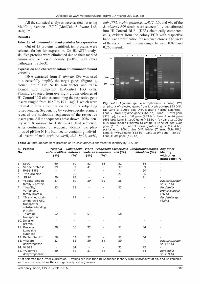
Out of 15 proteins identified, ten proteins were selected further for expression. On BLASTP analy-sis, five proteins were eliminated due to their marked amino acid sequence identity (>60%) with other pathogens (Table-3).Expression and characterization of immunodominant proteins
DNA extracted from B. abortus S99 was used to successfully amplify the target genes (Figure-1), cloned into pETite N-His Kan vector, and trans-formed into competent HI-Control 10G cells. Plasmid extracted from overnight grown colonies of HI-Control 10G clones containing the respective gene inserts ranged from 102.7 to 191.1 ng/µL which were optimal in their concentration for further subjecting to sequencing. Sequencing by vector-specific primers revealed the nucleotide sequences of the respective insert gene. All the sequences have shown 100% iden-tity with B. abortus bv. 1 str. 9-941 DNA sequence. After confirmation of sequence identity, the plas-mids of pETite N-His Kan vector containing individ-ual inserts of twin-arginine, invB, thiB, bp26, sodC,
bab-1885, serine protease, virB12, bfr, and bls, of the B. abortus S99 strain were successfully transformed into HI-Control BL21 (DE3) chemically competent cells, evident from the colony PCR with respective band size amplification for screened clones. The yield of the recombinant proteins ranged between 0.029 and 0.260 mg/mL.
Table-3: Immunodominant proteins of Brucella abortus analyzed for identity by BLASTP.
S. No.
Protein Yersinia enterocolitica
(%)
Salmonella enterica
(%)
Vibrio cholerae
(%)
Francisella tularensis
(%)
Escherichia coli (%)
Stenotrophomonas maltophilia (%)
Any other identity with other pathogens (%)
1. SodC 60 60 53 53 52 34 -2. Serine protease 39 39 41 - 37 39 -3. BAB1-1885 - - - - - 60 -4. Twin-arginine 26 27 26 -5. BP26 29 28 - - - 41 -6. *Solute-binding
family 5 protein37 39 39 32 36 - Haematobacter
sp. (67%)7. *Leu/Ile/
Val-binding family protein
23 23 - - 23 - Bordetella bronchiseptica (76%)
8. *Branched chain amino-acid ABC transporter substrate-binding protein
- 27 - - - - Bordetella sp. (62%)
9. Thiamine transporter
52 58 51 - 58 - -
10. Invasion protein B
- - - - - - -
11. Brucella Lumazine synthase
38 56 32 - 51 34 -
12. Bacterioferritin 50 52 52 - 52 54 -13. *Malate
dehydrogense33 32 30 64 39 - Haematobacter
sp. (77%)14. VirB12 - 34 - - 32 42 -15. *Aldehyde
dehydrogenase30 31 31 54 31 54 Bordetella
sp. (69%)
*Not selected for further expression. E values are less than 0; Sequence identity with Ochrobactrum sp. and Rhizobiales were not considered as they are generally soil organisms
Figure-1: Agarose gel electrophoresis showing PCR amplicons of selected genes from Brucella abortus S99 DNA. (a) Lane 1: 100bp plus DNA ladder (Thermo Scientific); Lane 2: twin arginine gene (564 bp); Lane 3: invB gene (528 bp); Lane 4: thiB gene (933 bp); Lane 5: bp26 gene (666 bp); Lane 6: sodC gene (462 bp). (b) Lane 1: 100bp plus DNA ladder (Thermo Scientific);; Lane 2: bab‑1885 gene (1371 bp); Lane 3: serine protease gene (1464 bp). (c) Lane 1: 100bp plus DNA ladder (Thermo Scientific); Lane 2: virB12 gene (513 bp); Lane 3: bfr gene (480 bp); Lane 4: bls gene (471 bp).
a b c
Veterinary World, EISSN: 2231-0916 808
Available at www.veterinaryworld.org/Vol.14/March-2021/35.pdf
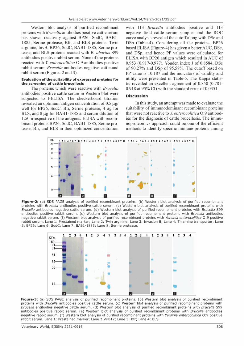
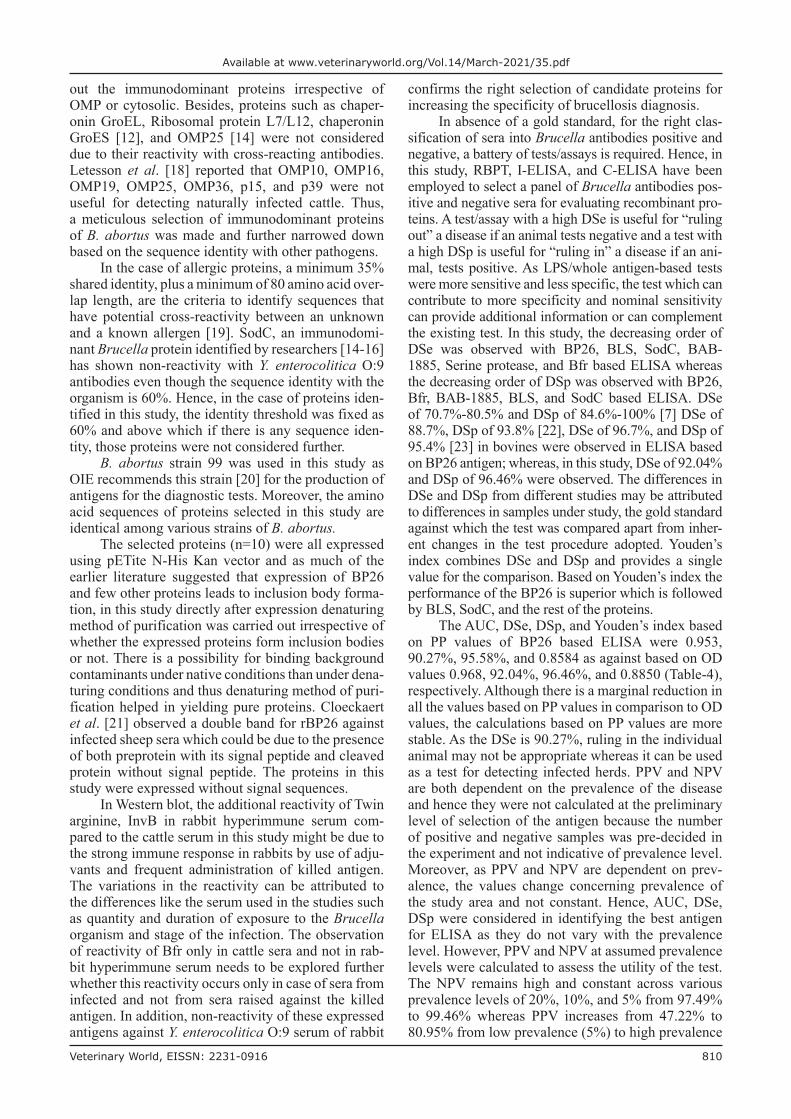
Western blot analysis of purified recombinant proteins with Brucella antibodies positive cattle serum has shown reactivity against BP26, SodC, BAB1-1885, Serine protease, Bfr, and BLS proteins. Twin arginine, InvB, BP26, SodC, BAB1-1885, Serine pro-tease, and BLS proteins reacted with B. abortus S99 antibodies positive rabbit serum. None of the proteins reacted with Y. enterocolitica O:9 antibodies positive rabbit serum, Brucella antibodies negative cattle and rabbit serum (Figures-2 and 3).Evaluation of the suitability of expressed proteins for the screening of cattle brucellosis
The proteins which were reactive with Brucella antibodies positive cattle serum in Western blot were subjected to I-ELISA. The checkerboard titration revealed an optimum antigen concentration of 0.5 µg/well for BP26, SodC, Bfr, Serine protease, 4 µg for BLS, and 8 µg for BAB1-1885 and serum dilution of 1:50 irrespective of the antigens. ELISA with recom-binant proteins BP26, SodC, BAB1-1885, Serine pro-tease, Bfr, and BLS in their optimized concentration
with 113 Brucella antibodies positive and 113 negative field cattle serum samples and the ROC curve analysis revealed the cutoff along with DSe and DSp (Table-4). Considering all the proteins, BP26 based ELISA (Figure-4) has given a better AUC, DSe, and DSp, and hence PP values were calculated for ELISA with BP26 antigen which resulted in AUC of 0.953 (0.917-0.977), Youden index J of 0.8584, DSe of 90.27% and DSp of 95.58%. The cutoff based on PP value is 10.187 and the indicators of validity and utility were presented in Table-5. The Kappa statis-tic revealed an excellent agreement of 0.850 (0.781-0.918 at 95% CI) with the standard error of 0.0351.Discussion
In this study, an attempt was made to evaluate the suitability of immunodominant recombinant proteins that were not reactive to Y. enterocolitica O:9 antibod-ies for the diagnosis of cattle brucellosis. The immu-noproteomics approach could be one of the efficient methods to identify specific immune-proteins among
Figure-2: (a) SDS PAGE analysis of purified recombinant proteins. (b) Western blot analysis of purified recombinant proteins with Brucella antibodies positive cattle serum. (c) Western blot analysis of purified recombinant proteins with Brucella antibodies negative cattle serum. (d) Western blot analysis of purified recombinant proteins with Brucella S99 antibodies positive rabbit serum. (e) Western blot analysis of purified recombinant proteins with Brucella antibodies negative rabbit serum. (f) Western blot analysis of purified recombinant proteins with Yersinia enterocolitica O:9 positive rabbit serum. Lane 1: Prestained marker; Lane 2: Twin arginine; Lane 3: Invasion B; Lane 4: Thiamine transporter; Lane 5: BP26; Lane 6: SodC; Lane 7: BAB1-1885; Lane 8: Serine protease.
a b c d e f
Figure-3: (a) SDS PAGE analysis of purified recombinant proteins. (b) Western blot analysis of purified recombinant proteins with Brucella antibodies positive cattle serum. (c) Western blot analysis of purified recombinant proteins with Brucella antibodies negative cattle serum. (d) Western blot analysis of purified recombinant proteins with Brucella S99 antibodies positive rabbit serum. (e) Western blot analysis of purified recombinant proteins with Brucella antibodies negative rabbit serum. (f) Western blot analysis of purified recombinant proteins with Yersinia enterocolitica O:9 positive rabbit serum. Lane 1: Prestained marker; Lane 2:VirB12; Lane 3: Bfr; Lane 4: BLS.
a b c d e f

Veterinary World, EISSN: 2231-0916 809
Available at www.veterinaryworld.org/Vol.14/March-2021/35.pdf
the greater range of proteins expressed by Brucella spp. [12]. Understanding the humoral immune response during Brucella infections will aid in the identification of immunogenic proteins. Appropriate identification and characterization of several immuno-reactive proteins concerning seroreactivity can result in a satisfactory diagnostic test [13]. Although indi-vidual recombinant proteins have been described by many researchers for the diagnosis of brucellosis, a methodical way of comparing various immunodom-inant proteins and identification of best among them was undertaken in this study.
Using bovine sera, Ko et al. [14] identified immunodominant proteins of B. abortus 1119-3 strain, Kim et al. [15] identified that of B. abortus RB51 and
Lee et al. [16] identified that of B. abortus 544. Lee et al. [17] used mice sera for B. abortus 544. From these studies, 61 Brucella proteins which are reactive only with Brucella antibodies and not with any other cross-reacting antibodies, in particular antibodies of Y. enterocolitica O:9 were identified. Further with the criteria of the outer membrane proteins (OMPs) or periplasmic proteins as more quantity of antibod-ies is expected against OMPs, the list was narrowed down to 11 proteins. However, since Brucella is an intracellular organism, the host immune response is not restricted to surface proteins and cytoplasmic proteins can also induce a higher antibody response [18], other proteins that were documented earlier were also considered with the idea of not missing
Table-4: DSe and DSp of various recombinant proteins based enzyme-linked immune sorbent assay for serodiagnosis of cattle brucellosis.
Antigen DSe% DSp% Area under curve
Standard Error 95% Confidence interval
z statistic Significance level P
Youden index J
rBP26 92.04 96.46 0.968 0.0111 0.936-0.987 42.319 <0.0001 0.8850rSodC 76.11 57.52 0.676 0.0360 0.610-0.736 4.869 <0.0001 0.3363rBAB1-1885 54.87 69.91 0.603 0.0380 0.536-0.667 2.706 0.0068 0.2478rHtrA 54.87 62.83 0.599 0.0375 0.532-0.664 2.641 0.0083 0.1770rBfr 37.17 72.57 0.528 0.0385 0.461-0.595 0.737 0.4608 0.09735rBLS 82.30 65.49 0.750 0.0331 0.688-0.805 7.564 <0.0001 0.4779
DSe: Diagnostic sensitivity, DSp: Diagnostic specificity
Table-5: Parameters of BP26 based ELISA for diagnosis of bovine brucellosis under various assumed prevalence level.
Assumed prevalence (%)
DSe% CI% DSp% CI% AUC CI LR+ CI LR- CI PPV% CI% NPV% CI%
5 90.27 83.25 to
95.04
94.69 88.80 to
98.03
0.92 0.88 to
0.96
17.00 7.79 to
37.12
0.10 0.06 to
0.18
47.22 29.07 to
66.14
99.46 99.06 to
99.6910 90.27 83.25
to 95.04
94.69 88.80 to
98.03
0.92 0.88 to
0.96
17.00 7.79 to
37.12
0.10 0.06 to
0.18
65.38 46.38 to
80.49
98.87 98.03 to
99.3520 90.27 83.25
to 95.04
94.69 88.80 to
98.03
0.92 0.88 to
0.96
17.00 7.79 to
37.12
0.10 0.06 to
0.18
80.95 66.06 to
90.27
97.49 95.68 to
98.56
DSe=Diagnostic Sensitivity, DSp=Diagnostic Specificity, CI=Confidence interval, AUC=Area under curve, LR+=Positive Likelihood ratio, LR- =Negative Likelihood ratio, PPV=Positive predictive value, NPV=Negative predictive value
Figure-4: (a) ROC graph analysis of PP values of BP26 in ELISA with Brucella antibodies positive and negative cattle serum to optimize sensitivity and specificity. (b) Dot plot graph showing PP values of BP26 in ELISA with Brucella antibodies positive and negative cattle serum with cut off obtained from ROC curve analysis.
a b
Veterinary World, EISSN: 2231-0916 810
Available at www.veterinaryworld.org/Vol.14/March-2021/35.pdf
out the immunodominant proteins irrespective of OMP or cytosolic. Besides, proteins such as chaper-onin GroEL, Ribosomal protein L7/L12, chaperonin GroES [12], and OMP25 [14] were not considered due to their reactivity with cross-reacting antibodies. Letesson et al. [18] reported that OMP10, OMP16, OMP19, OMP25, OMP36, p15, and p39 were not useful for detecting naturally infected cattle. Thus, a meticulous selection of immunodominant proteins of B. abortus was made and further narrowed down based on the sequence identity with other pathogens.
In the case of allergic proteins, a minimum 35% shared identity, plus a minimum of 80 amino acid over-lap length, are the criteria to identify sequences that have potential cross-reactivity between an unknown and a known allergen [19]. SodC, an immunodomi-nant Brucella protein identified by researchers [14-16] has shown non-reactivity with Y. enterocolitica O:9 antibodies even though the sequence identity with the organism is 60%. Hence, in the case of proteins iden-tified in this study, the identity threshold was fixed as 60% and above which if there is any sequence iden-tity, those proteins were not considered further.
B. abortus strain 99 was used in this study as OIE recommends this strain [20] for the production of antigens for the diagnostic tests. Moreover, the amino acid sequences of proteins selected in this study are identical among various strains of B. abortus.
The selected proteins (n=10) were all expressed using pETite N-His Kan vector and as much of the earlier literature suggested that expression of BP26 and few other proteins leads to inclusion body forma-tion, in this study directly after expression denaturing method of purification was carried out irrespective of whether the expressed proteins form inclusion bodies or not. There is a possibility for binding background contaminants under native conditions than under dena-turing conditions and thus denaturing method of puri-fication helped in yielding pure proteins. Cloeckaert et al. [21] observed a double band for rBP26 against infected sheep sera which could be due to the presence of both preprotein with its signal peptide and cleaved protein without signal peptide. The proteins in this study were expressed without signal sequences.
In Western blot, the additional reactivity of Twin arginine, InvB in rabbit hyperimmune serum com-pared to the cattle serum in this study might be due to the strong immune response in rabbits by use of adju-vants and frequent administration of killed antigen. The variations in the reactivity can be attributed to the differences like the serum used in the studies such as quantity and duration of exposure to the Brucella organism and stage of the infection. The observation of reactivity of Bfr only in cattle sera and not in rab-bit hyperimmune serum needs to be explored further whether this reactivity occurs only in case of sera from infected and not from sera raised against the killed antigen. In addition, non-reactivity of these expressed antigens against Y. enterocolitica O:9 serum of rabbit
confirms the right selection of candidate proteins for increasing the specificity of brucellosis diagnosis.
In absence of a gold standard, for the right clas-sification of sera into Brucella antibodies positive and negative, a battery of tests/assays is required. Hence, in this study, RBPT, I-ELISA, and C-ELISA have been employed to select a panel of Brucella antibodies pos-itive and negative sera for evaluating recombinant pro-teins. A test/assay with a high DSe is useful for “ruling out” a disease if an animal tests negative and a test with a high DSp is useful for “ruling in” a disease if an ani-mal, tests positive. As LPS/whole antigen-based tests were more sensitive and less specific, the test which can contribute to more specificity and nominal sensitivity can provide additional information or can complement the existing test. In this study, the decreasing order of DSe was observed with BP26, BLS, SodC, BAB-1885, Serine protease, and Bfr based ELISA whereas the decreasing order of DSp was observed with BP26, Bfr, BAB-1885, BLS, and SodC based ELISA. DSe of 70.7%-80.5% and DSp of 84.6%-100% [7] DSe of 88.7%, DSp of 93.8% [22], DSe of 96.7%, and DSp of 95.4% [23] in bovines were observed in ELISA based on BP26 antigen; whereas, in this study, DSe of 92.04% and DSp of 96.46% were observed. The differences in DSe and DSp from different studies may be attributed to differences in samples under study, the gold standard against which the test was compared apart from inher-ent changes in the test procedure adopted. Youden’s index combines DSe and DSp and provides a single value for the comparison. Based on Youden’s index the performance of the BP26 is superior which is followed by BLS, SodC, and the rest of the proteins.
The AUC, DSe, DSp, and Youden’s index based on PP values of BP26 based ELISA were 0.953, 90.27%, 95.58%, and 0.8584 as against based on OD values 0.968, 92.04%, 96.46%, and 0.8850 (Table-4), respectively. Although there is a marginal reduction in all the values based on PP values in comparison to OD values, the calculations based on PP values are more stable. As the DSe is 90.27%, ruling in the individual animal may not be appropriate whereas it can be used as a test for detecting infected herds. PPV and NPV are both dependent on the prevalence of the disease and hence they were not calculated at the preliminary level of selection of the antigen because the number of positive and negative samples was pre-decided in the experiment and not indicative of prevalence level. Moreover, as PPV and NPV are dependent on prev-alence, the values change concerning prevalence of the study area and not constant. Hence, AUC, DSe, DSp were considered in identifying the best antigen for ELISA as they do not vary with the prevalence level. However, PPV and NPV at assumed prevalence levels were calculated to assess the utility of the test. The NPV remains high and constant across various prevalence levels of 20%, 10%, and 5% from 97.49% to 99.46% whereas PPV increases from 47.22% to 80.95% from low prevalence (5%) to high prevalence
Veterinary World, EISSN: 2231-0916 811
Available at www.veterinaryworld.org/Vol.14/March-2021/35.pdf
(20%). Furthermore, the LR provides a suitable sum-mary measure, which is independent of prevalence.
BP26 is highly conserved in the genus Brucella, and the recombinant protein from one Brucella spe-cies might be used for the serological diagnosis of infections caused by all the Brucella species, provided that the animal host can induce an immune response against this antigen [24]. Letesson et al. [18] docu-mented that antibody responses to proteins were found to be limited to animals that developed an active bru-cellosis infection and this can be validated further with BP26 ELISA to identify active shedders of Brucella organisms in a herd. In this study, one of the important cross-reacting organism which has created trouble in few of the countries of the world, namely, Y. entero-colitica O:9 was addressed through the selection of Brucella antibodies reacting recombinant proteins but not reacted to experimentally raised hyperimmune rabbit serum to Y. enterocolitica O:9. Further, the identified recombinant protein should also be checked against other cross-reacting antibodies to ensure max-imum specificity.Conclusion
As there are many claims for various recom-binant proteins as the best recombinant proteins for diagnosing cattle brucellosis, this study has brought out the ranking and identified BP26 as the best suitable recombinant protein among them. BP26 based ELISA can be used as a screening test for detecting infected herds, to rule out cross-reactivity due to Y. enteroco-litica O:9 in infected herds. It can also complement the existing diagnostic tests such as RBPT and sLPS based ELISA.Authors’ Contributions
MN conceived, designed, carried out the exper-iment, and wrote the draft of the manuscript. TJB and SSK assisted in carrying out the experiment. RS, GBMR, and JJK assisted in sample collection and other biological collection. MN and VB analyzed data. RKG, BRS, HR, and PR provided guidance and support to carry out the experiments. All authors read and approved the final manuscript.Acknowledgments
The authors wish to thank the ICAR, New Delhi, India, for providing facilities, encourage-ment, and support. This research work is part of the Ph.D. program of the first author under ICAR-IVRI and ICAR-NIVEDI. The research project work was funded by the Department of Biotechnology (DBT), Government of India (F. No. DBT Network Project on Brucellosis/2012; 102/IFD/SAN/3142/2012-13 dated 27-09-2012).Competing Interests
The authors declare that they have no competing interests.
Publisher’s Note
Veterinary World remains neutral with regard to jurisdictional claims in published institutional affiliation.References1. Whatmore, A.M., Koylass, M.S., Muchowski, J., Edwards-
Smallbone, J., Gopaul, K.K. and Perrett, L.L. (2016) Extended multilocus sequence analysis to describe the global population structure of the genus Brucella: Phylogeography and relationship to biovars. Front. Microbiol, 7 : 2049.
2. Ducrotoy, M.J., Conde-Álvarez, R., Blasco, J.M. and Moriyón, I. (2016) A review of the basis of the immuno-logical diagnosis of ruminant brucellosis. Vet. Immunol. Immunopathol., 171: 81-102.
3. Al Dahouk, S., Sprague, L.D. and Neubauer, H. (2013) New developments in the diagnostic procedures for zoonotic bru-cellosis in humans. Review. Rev. Sci. Tech., 32(1): 177-188.
4. Adone, R. and Pasquali, P. (2013) Epidemiosurveillance of brucellosis. Rev. Sci. Tech., 32(1): 199-205.
5. Hilbink, F., Fenwick, S.G., Thompson, E.J., Kittelberger, R., Penrose, M. and Ross, G.P. (1995) Non-specific seroreac-tions against Brucella abortus in ruminants in New Zealand and the presence of Yersinia enterocolitica 0:9. N. Z. Vet. J., 43(5): 175-178.
6. Nagaraju, N.R., Isloor, S., Rao, S.M. and Rajasekhar, M. (2003) Seroprevalence of bovine yersiniosis in India. Indian J. Anim. Sci. 73(1): 48-50.
7. Munoz, P.M., Marin, C.M., Monreal D., Gonzalez, D., Garin-Bastuji, B., Diaz, R., Mainar-Jaime, R.C., Moriyon, I. and Blasco, J.M. (2005) Efficacy of several serological tests and antigens for diagnosis of bovine brucellosis in the presence of false-positive serological results due to Yersinia enteroco-litica O:9. Clin. Diagn. Lab. Immunol., 12(1): 141-151.
8. Godfroid, J. and Kasbohrer, A. (2002) Brucellosis in the European Union and Norway at the turn of the twenty-first century. Vet. Microbiol., 90(1-4): 135-145.
9. Alton, G.G., Jones, L.M., Angus, R.D. and Verger, J.M. (1988) Techniques for the Brucellosis Laboratory. Institute National De La Recherche Agronomique, Paris, France.
10. World Organization for Animal Health (OIE). (2016) Principles and methods of validation of diagnostic assays for infectious diseases. In: Manual of Diagnostic Tests and Vaccines for Terrestrial Animals. Ch. 1.1.2. OIE, Paris. Available from: http://www.oie.int/fileadmin/home/eng/health_standards/aahm/current/chapitre_validation_diag-nostics_assays.pdf. Retrieved on 25-08-2016.
11. Crowther, J.R. (2009) The ELISA Guidebook. Methods in Molecular Biology. 2nd ed., Vol. 516. Humana Press, United States. p566.
12. Al Dahouk, S., Nockler, K., Scholz, H.C., Tomaso, H., Bogumil, R. and Neubauer, H. (2006) Immunoproteomic characterization of Brucella abortus 1119-3 prepara-tions used for the serodiagnosis of Brucella infections. J. Immunol. Methods, 309(1-2): 34-47.
13. Pajuaba, A.C., Silva, D.A., Almeida, K.C., Cunha-Junior, J.P., Pirovani, C.P., Camillo, L.R. and Mineo, J.R. (2012) Immunoproteomics of Brucella abortus reveals differential antibody profiles between S19-vaccinated and naturally infected cattle. Proteomics, 12(6): 820-831.
14. Ko, K.Y., Kim, J.W., Her, M., Kang, S.I., Jung, S.C., Cho, D.H. and Kim, J.Y. (2012) Immunogenic proteins of Brucella abortus to minimize cross-reactions in brucellosis diagnosis. Vet. Microbiol., 156(3-4): 374-380.
15. Kim, J.Y., Sung, S.R., Lee, K., Lee, H.K., Kang, S.I., Lee, J.J., Jung, S.C., Park, Y.H. and Her, M. (2014) Immunoproteomics of Brucella abortus RB51 as candi-date antigens in serological diagnosis of brucellosis. Vet. Immunol. Immunopathol., 160(3-4): 218-224.
16. Lee, J.J., Simborio, H.L., Reyes, A.W., Kim, D.G.,
Veterinary World, EISSN: 2231-0916 812
Available at www.veterinaryworld.org/Vol.14/March-2021/35.pdf
Hop, H.T., Min, W., Her, M., Jung, S.C., Yoo, H.S. and Kim, S. (2014) Proteomic analyses of the time course responses of mice infected with Brucella abortus 544 reveal immunogenic antigens. FEMS Microbiol. Lett., 357(2): 164-174.
17. Lee, J.J., Simborio, H.L., Reyes, A.W., Kim, D.G., Hop, H.T., Min, W., Her, M., Jung, S.C., Yoo, H.S. and Kim, S. (2015) Immunoproteomic identification of immunodom-inant antigens independent of the time of infection in Brucella abortus 2308-challenged cattle. Vet. Res., 46: 17.
18. Letesson, J.J., Tiber, A., Van-Eynde, G., Wansard, V., Weynants, V., Denoel, P. and Saman, E. (1997) Humoral immune responses of Brucella-infected cattle, sheep and goats to eight purified recombinant Brucella proteins in an indirect enzyme-linked immunosorbent assay. Clin. Diagn. Lab. Immunol., 4(5): 556-564.
19. McClain, S. (2017) Bioinformatic screening and detection of allergen cross-reactive IgE-binding epitopes. Mol. Nutr. Food Res., 61(8): 1600676.
20. World Organization for Animal Health (OIE). (2016a) Brucellosis (Brucella abortus, B. melitensis and B. suis) (infection with B. abortus, B. melitensis and B. suis). In: Manual of Diagnostic Tests and Vaccines for Terrestrial
Animals. Ch. 2.1.4. OIE, Paris. Available from: http://www.oie.int/fileadmin/home/eng/health_standards/tahm/2.01.04_brucellosis.pdf. Retrieved on 27-01-2017.
21. Cloeckaert, A., Debbarh, H.S., Vizcaíno, N., Saman, E., Dubray, G. and Zygmunt, M.S. (1996) Cloning, nucleotide sequence, and expression of the Brucella melitensis bp26 gene coding for a protein immunogenic in infected sheep. FEMS Microbiol. Lett., 140(2-3): 139-144.
22. Chaudhuri, P., Prasad, R., Kumar, V. and Gangaplara, A. (2010) Recombinant OMP 28 antigen-based indirect ELISA for serodiagnosis of bovine brucellosis. Mol. Cell. Probes, 24(3): 142-145.
23. Lim, J.J., Kim, D.H., Lee, J.J., Kim, D.G., Min, W., Lee, H.J., Rhee, M.H., Chang, H.H. and Kim, S. (2012) Evaluation of recombinant 28 kDa outer membrane protein of Brucella abortus for the clinical diagnosis of bovine bru-cellosis in Korea. J. Vet. Med. Sci., 74(6): 687-691.
24. Seco-Mediavilla, P., Verger, J.M., Grayon, M., Cloeckaert, G.A., Marín, C.M., Zygmunt, M.S., Fernández-Lago, L. and Vizcaíno, N. (2003) Epitope mapping of the Brucella melitensis BP26 immunogenic protein: Usefulness for diagnosis of sheep brucellosis. Clin. Diagn. Lab. Immunol., 10(4): 647-651.
********