hodgkin lymphoma - pg cme haematologypgcmehematology.net/articles/016-13-hodgkin-lymphoma.pdf ·...
TRANSCRIPT
C
1
2
34
5
6
2
1h
Critical Reviews in Oncology/Hematology 85 (2013) 216–237
Hodgkin lymphoma
Paolo G. Gobbi a, Andrés J.M. Ferreri b,c,∗, Maurilio Ponzoni b,d, Alessandro Levis e
a Divisione di Medicina Interna e Gastroenterologia, Università di Pavia, Fondazione IRCCS Policlinico S. Matteo, Pavia, Italyb Unit of Lymphoid Malignancies, Department of Onco-Hematology, San Raffaele Scientific Institute, Milan, Italy
c Medical Oncology Unit, Department of Onco-Hematology, San Raffaele Scientific Institute, Milan, Italyd Pathology Unit, San Raffaele Scientific Institute, Milan, Italy
e Department of Hematology, Azienda Ospedaliera ss. Antonio e Biagio e c. Arrigo, Alessandria, Italy
Accepted 3 July 2012
ontents
. General information . . . . . . . . . . . . . . . . . . . . . . . . . . . . . . . . . . . . . . . . . . . . . . . . . . . . . . . . . . . . . . . . . . . . . . . . . . . . . . . . . . . . . . . . . . . . . . . . . . 2171.1. Definition . . . . . . . . . . . . . . . . . . . . . . . . . . . . . . . . . . . . . . . . . . . . . . . . . . . . . . . . . . . . . . . . . . . . . . . . . . . . . . . . . . . . . . . . . . . . . . . . . . . . . 2171.2. Incidence. . . . . . . . . . . . . . . . . . . . . . . . . . . . . . . . . . . . . . . . . . . . . . . . . . . . . . . . . . . . . . . . . . . . . . . . . . . . . . . . . . . . . . . . . . . . . . . . . . . . . . 2171.3. Risk factors . . . . . . . . . . . . . . . . . . . . . . . . . . . . . . . . . . . . . . . . . . . . . . . . . . . . . . . . . . . . . . . . . . . . . . . . . . . . . . . . . . . . . . . . . . . . . . . . . . . 218
. Pathology and biology . . . . . . . . . . . . . . . . . . . . . . . . . . . . . . . . . . . . . . . . . . . . . . . . . . . . . . . . . . . . . . . . . . . . . . . . . . . . . . . . . . . . . . . . . . . . . . . . 2182.1. Morphology . . . . . . . . . . . . . . . . . . . . . . . . . . . . . . . . . . . . . . . . . . . . . . . . . . . . . . . . . . . . . . . . . . . . . . . . . . . . . . . . . . . . . . . . . . . . . . . . . . . 218
2.1.1. Nodular lymphocyte predominance . . . . . . . . . . . . . . . . . . . . . . . . . . . . . . . . . . . . . . . . . . . . . . . . . . . . . . . . . . . . . . . . . . . . . . 2182.1.2. Nodular sclerosis . . . . . . . . . . . . . . . . . . . . . . . . . . . . . . . . . . . . . . . . . . . . . . . . . . . . . . . . . . . . . . . . . . . . . . . . . . . . . . . . . . . . . . 2182.1.3. Mixed cellularity . . . . . . . . . . . . . . . . . . . . . . . . . . . . . . . . . . . . . . . . . . . . . . . . . . . . . . . . . . . . . . . . . . . . . . . . . . . . . . . . . . . . . . . 2192.1.4. Lymphocyte depletion . . . . . . . . . . . . . . . . . . . . . . . . . . . . . . . . . . . . . . . . . . . . . . . . . . . . . . . . . . . . . . . . . . . . . . . . . . . . . . . . . . 2192.1.5. Lymphocyte-rich . . . . . . . . . . . . . . . . . . . . . . . . . . . . . . . . . . . . . . . . . . . . . . . . . . . . . . . . . . . . . . . . . . . . . . . . . . . . . . . . . . . . . . . 219
2.2. Immunophenotype . . . . . . . . . . . . . . . . . . . . . . . . . . . . . . . . . . . . . . . . . . . . . . . . . . . . . . . . . . . . . . . . . . . . . . . . . . . . . . . . . . . . . . . . . . . . . 2192.2.1. Lymphocyte predominance HL . . . . . . . . . . . . . . . . . . . . . . . . . . . . . . . . . . . . . . . . . . . . . . . . . . . . . . . . . . . . . . . . . . . . . . . . . . 2192.2.2. Nodular sclerosis, mixed cellularity and lymphocyte depletion . . . . . . . . . . . . . . . . . . . . . . . . . . . . . . . . . . . . . . . . . . . . . . 219
2.3. Genetic and biological features . . . . . . . . . . . . . . . . . . . . . . . . . . . . . . . . . . . . . . . . . . . . . . . . . . . . . . . . . . . . . . . . . . . . . . . . . . . . . . . . . . 219. Clinical presentations . . . . . . . . . . . . . . . . . . . . . . . . . . . . . . . . . . . . . . . . . . . . . . . . . . . . . . . . . . . . . . . . . . . . . . . . . . . . . . . . . . . . . . . . . . . . . . . . . 221. Staging and restaging . . . . . . . . . . . . . . . . . . . . . . . . . . . . . . . . . . . . . . . . . . . . . . . . . . . . . . . . . . . . . . . . . . . . . . . . . . . . . . . . . . . . . . . . . . . . . . . . . 221
4.1. Staging system and procedures . . . . . . . . . . . . . . . . . . . . . . . . . . . . . . . . . . . . . . . . . . . . . . . . . . . . . . . . . . . . . . . . . . . . . . . . . . . . . . . . . . 2214.2. Molecular analysis of minimal residual disease . . . . . . . . . . . . . . . . . . . . . . . . . . . . . . . . . . . . . . . . . . . . . . . . . . . . . . . . . . . . . . . . . . . . 2214.3. Post-treatment evaluation . . . . . . . . . . . . . . . . . . . . . . . . . . . . . . . . . . . . . . . . . . . . . . . . . . . . . . . . . . . . . . . . . . . . . . . . . . . . . . . . . . . . . . . 2214.4. Early response evaluation . . . . . . . . . . . . . . . . . . . . . . . . . . . . . . . . . . . . . . . . . . . . . . . . . . . . . . . . . . . . . . . . . . . . . . . . . . . . . . . . . . . . . . . 2224.5. Follow up evaluations . . . . . . . . . . . . . . . . . . . . . . . . . . . . . . . . . . . . . . . . . . . . . . . . . . . . . . . . . . . . . . . . . . . . . . . . . . . . . . . . . . . . . . . . . . 222
. Prognosis . . . . . . . . . . . . . . . . . . . . . . . . . . . . . . . . . . . . . . . . . . . . . . . . . . . . . . . . . . . . . . . . . . . . . . . . . . . . . . . . . . . . . . . . . . . . . . . . . . . . . . . . . . . . 222
5.1. Natural history . . . . . . . . . . . . . . . . . . . . . . . . . . . . . . . . . . . . . . . . . . . . . . . . . . . . . . . . . . . . . . . . . . . . . . . . . . . . . . . . . . . . . . . . . . . . . . . . . 2225.2. Prognostic factors . . . . . . . . . . . . . . . . . . . . . . . . . . . . . . . . . . . . . . . . . . . . . . . . . . . . . . . . . . . . . . . . . . . . . . . . . . . . . . . . . . . . . . . . . . . . . . 224. Treatment . . . . . . . . . . . . . . . . . . . . . . . . . . . . . . . . . . . . . . . . . . . . . . . . . . . . . . . . . . . . . . . . . . . . . . . . . . . . . . . . . . . . . . . . . . . . . . . . . . . . . . . . . . . 2256.1. Treatment of early-stage disease (stage IA–IIA ± IIB) . . . . . . . . . . . . . . . . . . . . . . . . . . . . . . . . . . . . . . . . . . . . . . . . . . . . . . . . . . . . . 225
6.1.1. Treatment of favourable early-stage disease . . . . . . . . . . . . . . . . . . . . . . . . . . . . . . . . . . . . . . . . . . . . . . . . . . . . . . . . . . . . . . . 225
6.1.2. Treatment of unfavourable early-stage disease. . . . . . . . . . . . . . . . . . . . . . . . . . . . . . . . . . . . . . . . . . . . . . . . . . . . . . . . . . . . . 225∗ Corresponding author at: Unit of Lymphoid Malignancies, Department of Onco-Hematology, San Raffaele Scientific Institute, Via Olgettina 60,0132 Milan, Italy. Tel.: +39 02 26437649; fax: +39 02 26437625.
E-mail address: [email protected] (A.J.M. Ferreri).
040-8428/$ – see front matter © 2012 Elsevier Ireland Ltd. All rights reserved.ttp://dx.doi.org/10.1016/j.critrevonc.2012.07.002
A
ilataqibd
ffda
twceewpsc©
K
1
1
ctic[dt‘l
P.G. Gobbi et al. / Critical Reviews in Oncology/Hematology 85 (2013) 216–237 217
6.2. Treatment of advanced-stage disease (stage III–IV ± IIB) . . . . . . . . . . . . . . . . . . . . . . . . . . . . . . . . . . . . . . . . . . . . . . . . . . . . . . . . . . 2266.3. Treatment of special categories of patients . . . . . . . . . . . . . . . . . . . . . . . . . . . . . . . . . . . . . . . . . . . . . . . . . . . . . . . . . . . . . . . . . . . . . . . . 227
6.3.1. Pregnant patients with HL . . . . . . . . . . . . . . . . . . . . . . . . . . . . . . . . . . . . . . . . . . . . . . . . . . . . . . . . . . . . . . . . . . . . . . . . . . . . . . 2276.3.2. HIV-positive patients . . . . . . . . . . . . . . . . . . . . . . . . . . . . . . . . . . . . . . . . . . . . . . . . . . . . . . . . . . . . . . . . . . . . . . . . . . . . . . . . . . . 2276.3.3. Elderly patients . . . . . . . . . . . . . . . . . . . . . . . . . . . . . . . . . . . . . . . . . . . . . . . . . . . . . . . . . . . . . . . . . . . . . . . . . . . . . . . . . . . . . . . . 227
6.4. Treatment of relapsed or refractory HL . . . . . . . . . . . . . . . . . . . . . . . . . . . . . . . . . . . . . . . . . . . . . . . . . . . . . . . . . . . . . . . . . . . . . . . . . . . 2276.5. Treatment-related late complications and second tumours . . . . . . . . . . . . . . . . . . . . . . . . . . . . . . . . . . . . . . . . . . . . . . . . . . . . . . . . . . 2286.6. New active drugs and ongoing trials . . . . . . . . . . . . . . . . . . . . . . . . . . . . . . . . . . . . . . . . . . . . . . . . . . . . . . . . . . . . . . . . . . . . . . . . . . . . . . 229
Conflict of interest . . . . . . . . . . . . . . . . . . . . . . . . . . . . . . . . . . . . . . . . . . . . . . . . . . . . . . . . . . . . . . . . . . . . . . . . . . . . . . . . . . . . . . . . . . . . . 230Reviewers . . . . . . . . . . . . . . . . . . . . . . . . . . . . . . . . . . . . . . . . . . . . . . . . . . . . . . . . . . . . . . . . . . . . . . . . . . . . . . . . . . . . . . . . . . . . . . . . . . . . . 230References . . . . . . . . . . . . . . . . . . . . . . . . . . . . . . . . . . . . . . . . . . . . . . . . . . . . . . . . . . . . . . . . . . . . . . . . . . . . . . . . . . . . . . . . . . . . . . . . . . . . 230Biographies . . . . . . . . . . . . . . . . . . . . . . . . . . . . . . . . . . . . . . . . . . . . . . . . . . . . . . . . . . . . . . . . . . . . . . . . . . . . . . . . . . . . . . . . . . . . . . . . . . . 236
bstract
Hodgkin lymphoma (HL) is a curable malignancy which shows a bimodal curve in incidence in economically developed countries; theres a putative association with Epstein–Barr virus. The WHO 2008 classification schema recognises two histological types of HL: the nodularymphocyte predominant and the “classic” HL. The latter encompasses four entities: nodular sclerosis, mixed cellularity, lymphocyte depletion,nd lymphocyte-rich. Most patients with HL present with asymptomatic superficial lymphadenopathy. The commonest sites of disease arehe cervical, supraclavicular and mediastinal lymph nodes, while sub-diaphragmatic presentations and bone marrow and hepatic involvementre less common. Splenic involvement is usually concomitant with hepatic disease and systemic symptoms; extranodal presentations areuite rare. Systemic symptoms are present in ∼35% of cases. The stage of disease is defined according to the Ann Arbor staging system orts Cotswolds variant, and staging work-up includes physical examination, chest X-rays, chest and abdominal CT scan, and bone marrowiopsy. 18FDG-PET (18fluordeoxyglucose positron emission tomography) plays a central role in staging, response assessment and prognosisefinition.
Classic HL usually spreads by contiguity within the lymphatic tissue network, with a late extension to adjacent and distant viscera. Mortalityrom HL has been progressively decreasing, as confirmed by the most recent 5-year survival figure of 81%. The list of putative prognosticactors in HL has been increasing, but most factors still require prospective validation. Some of these variables are used to stratify early-stageisease into “favourable” and “unfavourable” categories, with “unfavourable early-stage” being intermediate between “favourable early-stage”nd “advanced-stage”.
ABVD (adriamycin(doxorubicin), bleomycin, vinblastine, dacarbazine) combination chemotherapy followed by involved-field irradiation ishe standard treatment for patients with early-stage HL, with a 5-year OS >95%. Several trials assessing less intensive approaches for patientsith favourable early-stage HL are ongoing. More intensified combinations, such as the BEACOPP (bleomycin, etoposide, adriamycin,
yclophosphamide, vincristine (Oncovin), procarbazine, prednisone) regimen, are being investigated, usually in patients with unfavourablearly-stage HL and interim PET+. ABVD is the standard chemotherapy treatment also for patients with advanced disease. Although somevidence suggests that more intensive combinations provide better disease control, the inevitable increased risk of relevant late toxicityorries investigators. Consequently, there has been a shift towards investigating the innovative strategy of a more aggressive schedule foratients with 18FDG-PET positive results after the first 2 courses of ABVD. High-dose chemotherapy supported by ASCT (autologoustem cell transplantation) is considered the standard of care in patients with HL which has relapsed after, or is refractory to conventionalhemoradiotherapy, while allogeneic transplant is a suitable tool for patients with chemorefractory disease and patients failed after ASCT.
2012 Elsevier Ireland Ltd. All rights reserved.
ET
1
cMmicoo
eywords: Hodgkin lymphoma; ABVD; BEACOPP; Epstein–Barr virus; P
. General information
.1. Definition
Hodgkin lymphoma (HL) is one of the few adult malignan-ies that can be cured in most instances. The salient feature ofhis lymphoma is the rarity (about 1%) of neoplastic elementsn the cell population, whereas the overwhelming majority ofells are non-neoplastic, mostly consisting of T-lymphocytes1]. Although the clonal B-cell origin of both lymphocyte pre-
ominant and “classic” HL was recently demonstrated [2],hus enabling the term ‘Hodgkin disease’ to be changed toHodgkin lymphoma’ [3], the pathogenic mechanisms of thisymphoma are still largely unknown.macs
.2. Incidence
HL is an uncommon malignancy, with 7000–7500 newases diagnosed annually in the United States of America.ost of these patients present with early stage disease. Thisalignancy displays a bimodal curve in incidence in econom-
cally developed countries. In economically underdevelopedountries, the overall incidence of HL is lower than in devel-ped countries, with the exception of children under the agef 15, where a higher incidence is seen. There is only a
ild increase in incidence throughout adolescence and youngdulthood [4]. A difference in the distribution of histologi-al subgroups occurs as well, since the incidence of nodularclerosis is lower in underdeveloped countries.
2 Oncol
1
sooamohrwa([raertroedbtlscaodfff
NriwhhSohpNpamivpao[
2
2
gdaina
2
mTwrngcitiahai
iafoTatalrwnpo
2
iebttt
18 P.G. Gobbi et al. / Critical Reviews in
.3. Risk factors
The dual-peak incidence of HL supports the hypothe-is that this malignancy may actually be a common resultf two distinct pathogenic processes: an infectious agentf low infectivity may be related to the disease in youngdults, while a mechanism shared with other lymphomasay account for the pathogenesis of HL occurring in the
lder age group [5]. The Epstein–Barr virus (EBV) genomeas been detected in one third to one half of HLs occur-ing in patients without known immunodeficiencies [5–7],ith the expression of latent membrane protein (LMP-1
nd -2), EBERs (Epstein–Barr encoded RNAs), and EBNAEpstein–Barr nuclear antigen) 1 in 30–50% of tumours8]. Patients with infectious mononucleosis are at higherisk of developing EBV-associated HL [9] and this risk islso enhanced in subjects carrying HLA-A*01 [10]. How-ver, the larger group of EBV-negative HL could still beegarded as an infectious-driven neoplasm in which the puta-ive causative agent has not yet been detected [11]. Perhapselevant to this controversy is the relatively obscure findingf an apparent elevated risk of HL in persons with systemicxposure to blood or blood products, such as intravenousrug users and haemophiliacs, who may be presumed toe HIV-negative. This could reflect the presence of unde-ected HIV infection, which has been well-established toead to a substantially elevated risk of HL in all expo-ure groups [12–14]. In this context, much weaker putativeandidate mechanisms include the deregulated immunitygainst foreign antigen(s). An altered B-cell response sec-ndary to a virus infection, or some factors known toecrease or delay early exposure to infections, such asewer siblings, single-family houses, early birth-order, andewer playmates have been proposed as risk-increasingactors.
The role of HL as inherited disease remains to be defined.early 40% of patients with HL seen at a tertiary care refer-
al centre reported a first-degree relative with cancer; thencidence was significantly lower with respect to patientsith NHL or CLL [15]. Six percent of these HL patientsad a relative with a lymphoproliferative malignancy, withigh rates of both HL and NHL in first-degree relatives.ubstantial evidence suggesting that familial aggregationf lymphoproliferative disorders such as CLL and NHLas a significant genetic component is less clear in HLatients. However, the differences observed between CLL,HL and HL indicate that the underlying biological predis-osition may vary among the diseases, and is not merelyn artefact of different age bands. Patterns of inheritanceay provide some clues to pathogenesis. However, stud-
es on familial HL are strongly limited by the unreliablealidity of self-reported positive family histories of lym-
homa. In fact, while familial HL reported by HL patientsnd controls is likely to be lymphoma, even in membersf the extended family, it is unlikely to be HL per se16].uolo
ogy/Hematology 85 (2013) 216–237
. Pathology and biology
.1. Morphology
The updated 2008 WHO classification recognises tworoups of histological types of HL: nodular lymphocyte pre-ominant (LP), which includes about 5% of all HL cases,nd the “classic” HL (cHL), which accounts for the remain-ng cases. In cHL, the following subgroups can be identified:odular sclerosis, mixed cellularity, lymphocyte depleted,nd lymphocyte-rich.
.1.1. Nodular lymphocyte predominanceThe lymph node architecture is usually effaced and, in
ost instances, without residual reactive germinal centres.his malignancy usually displays a nodular growth pattern,hich may or may not be accompanied by diffuse areas; more
arely, a purely diffuse pattern may occur. At low power,odular lesions are basically B-cell areas with nearby pro-ressive transformation of germinal centres; these featuresan be further elucidated by means of CD20 and anti-CD21nvestigation (a follicular dendritic cell marker). The neoplas-ic cells are large and characterised by vesicular, somewhatrregular and polylobated nuclei with small nucleoli andbundant cytoplasm. These cells, named lymphocytic andistiocytic (L&H cells) or “popcorn” cells, may occur in vari-ble amounts, but they are never prominent and are oftensolated, without any tendency to form dense aggregates.
The non-neoplastic background in lymphocyte predom-nant HL is mostly represented by small lymphocytesnd a variable amount of histiocytes, which may focallyorm non-necrotising granulomas. When epithelioid histi-cytes become numerous, the differential diagnosis with-cell rich large B-cell lymphoma is difficult and requiresdditional investigations. Plasma cells, eosinophils and neu-rophils are rarely seen [17]. This subtype may occur at anyge, but it is more common in adult males. It is usuallyocalised at diagnosis, and stage-IV disease with bone mar-ow involvement is rare. The mediastinum is usually spared,hile peripheral lymph nodes, mainly cervical or inguinalodes, are frequently involved. Importantly, deep-seated lym-hadenopathies may occur as well (M. Ponzoni, personalbservation).
.1.2. Nodular sclerosisThis is the most common subtype of cHL, account-
ng for 75–80% of cHL cases. The salient feature of thisntity is the occurrence of nodules of variable size separatedy dense collagenous fibrous bands. These bands displayypical green birefringence in polarised light, a cardinal fea-ure which enables the distinction of nodular sclerosis fromhe lymphocyte depletion subtype; variable areas of coag-
lative necrosis are common. The pathognomonic elementf cHL is the Reed–Sternberg (R–S) cell; this element isarge, polynucleated and with prominent, basophilic nucle-li. Mononuclear variants of R–S cells include lacunar cells
Oncol
aacaerwica
2
nstnti
2
pbtwiaovnlpc
cniwng
2
aalaid(a
iao
aoi
2
2
gEpiru[sBie
naa(pft
2l
ClsabtItilmtMcptlBtt
P.G. Gobbi et al. / Critical Reviews in
nd Hodgkin cells. In the nodular sclerosis variant, R–S cellsre easily visible, but never prominent. Diagnosis of cHLan only be made when R–S cells occur within the appropri-te background including small lymphocytes, plasma cells,osinophils, neutrophils and histiocytes [18]. Nodular scle-osis more commonly affects adolescents and young adults,ith a slightly higher prevalence in females. Mediastinal
nvolvement is frequent and patients with nodular sclerosisHL preferentially display upper thoracic disease that gener-lly remains localised in lymph nodes and adjacent structures.
.1.3. Mixed cellularityIn mixed cellularity HL, the infiltrate is diffuse or vaguely
odular, without band-forming sclerosis, although fine inter-titial fibrosis may be present. R–S cells are more representedhan nodular sclerosis. Patients are usually adults; males out-umber females, and the stage is frequently more advancedhan in nodular sclerosis or lymphocyte predominant, involv-ng lymph nodes, spleen, liver, or marrow.
.1.4. Lymphocyte depletionThe lymph node architecture is completely effaced and
redominantly represented by diffuse and dense fibrosis (notirefringent collagen). Necrosis may occur as well. Most ofhe residual cells are exclusively represented by R–S cells,hile non neoplastic elements in the background are rare,
f present. Overall, these features confer a “sarcomatoid”ppearance to the lymph node architecture. Confluent sheetsf R–S cells may occur and therefore constitute the “reticular”ariant (or Hodgkin’s sarcoma) [18]. The differential diag-osis between the “reticular” variant and anaplastic large cellymphoma may be difficult, cHL requiring absence of ALKrotein and absence of rearrangement of the genes whichodify for T-cell receptor [19].
Lymphocyte depletion is the least common variant ofHL and occurs preferentially in elderly patients, and inon-industrialised countries. The most frequent presentationsnvolve abdominal lymphadenopathy, or extranodal diseaseith spleen, liver and marrow involvement. The stage at diag-osis is usually advanced and the response to treatment isenerally worse than in other subtypes [20].
.1.5. Lymphocyte-richThis subtype involves about 6% of HL cases [21]. There is
diffuse or focal, sometimes interfollicular involvement withreactive cellular milieu, essentially represented by small
ymphocytes, with very few, if any, neutrophils, eosinophilsnd plasma cells. Importantly, R–S cells are present, albeitnfrequently, as well as some lacunar cells; both of themisplay the immunophenotypic/molecular properties of cHLsee below). Often, nodules show germinal centres and focalreas of fibrosis.
A lymphocyte-rich subtype, which was only recentlyntroduced into the cHL classification [21], also demonstrates
characteristic clinical profile. It is characterised by lateccurrence (i.e., patients older than 50 years of age), low
2
i
ogy/Hematology 85 (2013) 216–237 219
ggressiveness, early stage at presentation, and involvementf subdiaphragmatic sites, whereas mediastinal or extranodalnvolvement, systemic symptoms and bulky masses are rare.
.2. Immunophenotype
.2.1. Lymphocyte predominance HLL&H cells are typically CD45+ and B-cell-associated anti-
ens (CD19, CD20, CD22, and CD79a)-positive, CDw75+,MA+/− and CD15−. Bcl-6 is very often expressed in neo-lastic cells. CD30 is usually absent, although it may occurn some instances, and with somewhat less intensity withespect to cHL. In paraffin-embedded material, immunoglob-lin light chain restriction can sometimes be demonstrated22]. J-chain has been shown in many cases [23,24]. Tran-cription factors PAX-5, Oct-2, PU.1 and the coactivatorOB.1 are almost constantly expressed [21]. Other recently
ntroduced markers, such as HGAL, AID and centerin arexpressed by L&H cells [25].
Small lymphocytes, present within the background of theodules, are predominantly B cells. T-cells, which occur tolesser degree, tend to form rosettes surrounding L&H cellsnd there is a relative prevalence of CD57+, MUM-1, PD-1+ref) small-sized elements. The microenvironment is com-leted by CD68+ histiocytes and a prominent meshwork ofollicular dendritic cells, which is particularly evident withinhe nodules [21].
.2.2. Nodular sclerosis, mixed cellularity andymphocyte depletion
In paraffin-embedded material, R–S cells are intensivelyD30+ (with its characteristic crispy membrane and/or ‘dot-
ike’ staining pattern), PAX-5+ (with a less intensive nuclearignal when compared to bystander small B lymphocytes),nd CD45- (Table 1). CD15 is usually positive, but it shoulde taken into account that the rates of immunoreactivity ofhis marker may vary according to the employed clone [26].mportantly, CD20 could be detected in about 30–40% ofhe cases of cHL; in most instances, this marker shows a lessntensive signal with respect to T-cell rich diffuse large B-cellymphoma and, importantly, a variable amount of R–S ele-ents present within the same tissue are not reactive against
his molecule. Other common markers of R–S cells includeUM-1 and, in about one fourth of cases, BLIMP1 [27]. T-
ell antigens are reported in a small minority of cases. Therevalence is slightly higher in Japanese patients [28]. Impor-antly, many markers expressed in lymphocyte predominantymphoma are absent in cHL; these markers include Oct-2,OB.1, and CD45. The diagnosis is made on routine sec-
ions; however, immunophenotyping studies are essential forhe diagnosis and are highly recommended.
.3. Genetic and biological features
The putative normal counterpart of HL cells differ, fallingnto two main groups. In fact, L&H cells of lymphocyte
220 P.G. Gobbi et al. / Critical Reviews in Oncol
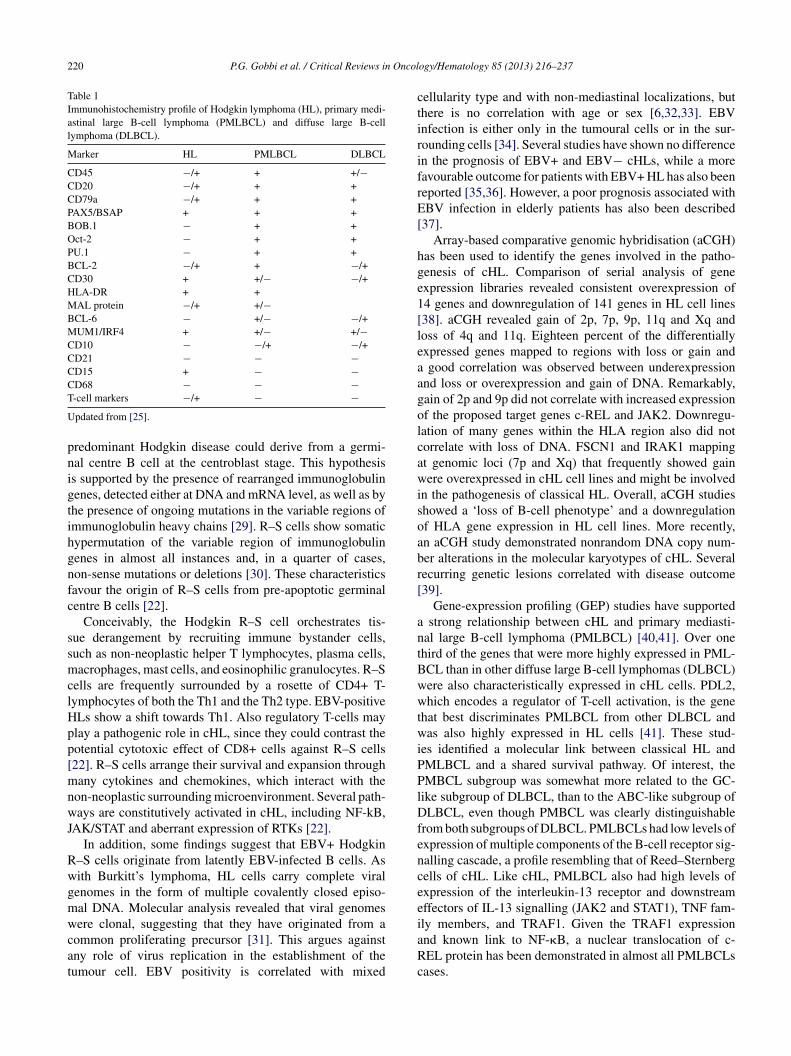
Table 1Immunohistochemistry profile of Hodgkin lymphoma (HL), primary medi-astinal large B-cell lymphoma (PMLBCL) and diffuse large B-celllymphoma (DLBCL).
Marker HL PMLBCL DLBCL
CD45 −/+ + +/−CD20 −/+ + +CD79a −/+ + +PAX5/BSAP + + +BOB.1 − + +Oct-2 − + +PU.1 − + +BCL-2 −/+ + −/+CD30 + +/− −/+HLA-DR + +MAL protein −/+ +/−BCL-6 − +/− −/+MUM1/IRF4 + +/− +/−CD10 − −/+ −/+CD21 − − −CD15 + − −CD68 − − −T
U
pnigtihgnfc
ssmclHpp[mnwJ
Rwgmwcat
ctirifrE[
hge1[leaagolcawisoabr[
antBwwtwiPPlDfencee
-cell markers −/+ − −pdated from [25].
redominant Hodgkin disease could derive from a germi-al centre B cell at the centroblast stage. This hypothesiss supported by the presence of rearranged immunoglobulinenes, detected either at DNA and mRNA level, as well as byhe presence of ongoing mutations in the variable regions ofmmunoglobulin heavy chains [29]. R–S cells show somaticypermutation of the variable region of immunoglobulinenes in almost all instances and, in a quarter of cases,on-sense mutations or deletions [30]. These characteristicsavour the origin of R–S cells from pre-apoptotic germinalentre B cells [22].
Conceivably, the Hodgkin R–S cell orchestrates tis-ue derangement by recruiting immune bystander cells,uch as non-neoplastic helper T lymphocytes, plasma cells,acrophages, mast cells, and eosinophilic granulocytes. R–S
ells are frequently surrounded by a rosette of CD4+ T-ymphocytes of both the Th1 and the Th2 type. EBV-positiveLs show a shift towards Th1. Also regulatory T-cells maylay a pathogenic role in cHL, since they could contrast theotential cytotoxic effect of CD8+ cells against R–S cells22]. R–S cells arrange their survival and expansion throughany cytokines and chemokines, which interact with the
on-neoplastic surrounding microenvironment. Several path-ays are constitutively activated in cHL, including NF-kB,
AK/STAT and aberrant expression of RTKs [22].In addition, some findings suggest that EBV+ Hodgkin
–S cells originate from latently EBV-infected B cells. Asith Burkitt’s lymphoma, HL cells carry complete viralenomes in the form of multiple covalently closed episo-al DNA. Molecular analysis revealed that viral genomes
ere clonal, suggesting that they have originated from aommon proliferating precursor [31]. This argues againstny role of virus replication in the establishment of theumour cell. EBV positivity is correlated with mixed
iaRc
ogy/Hematology 85 (2013) 216–237
ellularity type and with non-mediastinal localizations, buthere is no correlation with age or sex [6,32,33]. EBVnfection is either only in the tumoural cells or in the sur-ounding cells [34]. Several studies have shown no differencen the prognosis of EBV+ and EBV− cHLs, while a moreavourable outcome for patients with EBV+ HL has also beeneported [35,36]. However, a poor prognosis associated withBV infection in elderly patients has also been described
37].Array-based comparative genomic hybridisation (aCGH)
as been used to identify the genes involved in the patho-enesis of cHL. Comparison of serial analysis of genexpression libraries revealed consistent overexpression of4 genes and downregulation of 141 genes in HL cell lines38]. aCGH revealed gain of 2p, 7p, 9p, 11q and Xq andoss of 4q and 11q. Eighteen percent of the differentiallyxpressed genes mapped to regions with loss or gain andgood correlation was observed between underexpression
nd loss or overexpression and gain of DNA. Remarkably,ain of 2p and 9p did not correlate with increased expressionf the proposed target genes c-REL and JAK2. Downregu-ation of many genes within the HLA region also did notorrelate with loss of DNA. FSCN1 and IRAK1 mappingt genomic loci (7p and Xq) that frequently showed gainere overexpressed in cHL cell lines and might be involved
n the pathogenesis of classical HL. Overall, aCGH studieshowed a ‘loss of B-cell phenotype’ and a downregulationf HLA gene expression in HL cell lines. More recently,n aCGH study demonstrated nonrandom DNA copy num-er alterations in the molecular karyotypes of cHL. Severalecurring genetic lesions correlated with disease outcome39].
Gene-expression profiling (GEP) studies have supportedstrong relationship between cHL and primary mediasti-
al large B-cell lymphoma (PMLBCL) [40,41]. Over onehird of the genes that were more highly expressed in PML-CL than in other diffuse large B-cell lymphomas (DLBCL)ere also characteristically expressed in cHL cells. PDL2,hich encodes a regulator of T-cell activation, is the gene
hat best discriminates PMLBCL from other DLBCL andas also highly expressed in HL cells [41]. These stud-
es identified a molecular link between classical HL andMLBCL and a shared survival pathway. Of interest, theMBCL subgroup was somewhat more related to the GC-
ike subgroup of DLBCL, than to the ABC-like subgroup ofLBCL, even though PMBCL was clearly distinguishable
rom both subgroups of DLBCL. PMLBCLs had low levels ofxpression of multiple components of the B-cell receptor sig-alling cascade, a profile resembling that of Reed–Sternbergells of cHL. Like cHL, PMLBCL also had high levels ofxpression of the interleukin-13 receptor and downstreamffectors of IL-13 signalling (JAK2 and STAT1), TNF fam-
ly members, and TRAF1. Given the TRAF1 expressionnd known link to NF-�B, a nuclear translocation of c-EL protein has been demonstrated in almost all PMLBCLsases.
Oncol
3
tucsrmd5atiwhutotaagtaaa
cato
4
4
amiaa
hbc(prs
Hoo
csdbpb
tn1
e[aiPsaosto
4
iasttIytmecia
4
wtpRbraft
P.G. Gobbi et al. / Critical Reviews in
. Clinical presentations
Most patients with HL present with superficial adenopa-hy and are asymptomatic. The lymph node enlargement issually painless, rubbery, matted, or discrete, and is mostommonly located in the neck and supraclavicular areas. It isometimes detected during a physical examination for othereasons. A presentation of mediastinal enlargement is com-on during routine chest X-rays. The commonest sites of
isease are cervical, supraclavicular and mediastinal (over0% of cases) nodes, while sub-diaphragmatic presentationsre less common, and epitrochlear nodes, Waldeyer’s ring,esticular, and gastrointestinal sites are uncommon. Abdom-nal nodal involvement is more common in older patients orhen fever or night sweats are present. Bone marrow andepatic involvement are uncommon. Spleen involvement issually concomitant with hepatic disease and systemic symp-oms. Systemic symptoms are present at diagnosis in aboutne third of cases, and among them fever is more commonhan night sweats and weight loss, whereas pruritus is rare andlcohol-induced pain is very rare. Pruritus does not constitutesystemic symptom, but it should be recorded, especially ifeneralised, if it is the cause of scratch lesions and if resistanto steroids [42]. Alcohol-induced pain is nearly diagnosticnd consists of pain triggered by the ingestion of moder-te amount of alcoholic drinks and localised in one of thenatomical deep sites involved by the disease.
Other rare clinical presentations, more commonly asso-iated with advanced HL, are superior vena cava syndrome,cute spinal cord compression, central nervous system soli-ary lesion, Waldeyer’s ring involvement, testicular masses,r intestinal occlusion.
. Staging and restaging
.1. Staging system and procedures
The standard staging system used for HL was proposedt the Ann Arbor Conference in 1971 [43], and partiallyodified at the Cotswolds Meeting in 1988 [43]. The stag-
ng system reflects both the number of sites of involvementnd the presence of disease above or below the diaphragm,ccording to four stages of disease (Table 2).
Complete staging work-up for HL includes a detailedistory, which records both presence and duration of possi-le systemic symptoms, an accurate physical examination,omplete haematological and biochemical examinationsincluding erythrocyte sedimentation rate, serum alkalinehosphatase, renal function and liver function tests), chest X-ays, chest and abdominal computed tomography (CT) scans,keletal X-rays when necessary, and bone marrow biopsy.
Bone marrow core biopsy, not aspiration, is useful.owever, patients in clinical supradiaphragmatic stage Ir II without B symptoms show a minimal probabilityf marrow involvement. Bone marrow biopsy is therefore
pmLp
ogy/Hematology 85 (2013) 216–237 221
onsidered particularly important in patients with Bymptoms and/or clinical advanced stage and/or infra-iaphragmatic presentation and in those with bone lesions,one pain, hypercalcaemia, or an elevated serum alkalinehosphatase [44,45]. Whether it can be replaced in the futurey 18FDG-PET is still a matter of debate [46–48].
The sensitivity of 18FDG-PET and PET/CT is higherhan that of CT in order to identify both nodal and extra-odal disease in primary staging [49–52]. The superiority of8FDG-PET or PET-CT over conventional CT staging is morevident for evaluating extra-nodal than nodal involvement53]. 18FDG-PET false-positives at diagnosis are around 2%,nd some doubts arise about the risk of 18FDG-PET upstag-ng, rather than downstaging, patients [53]. Although 18FDG-ET may be superior to CT it is not yet considered the newtandard staging imaging technique, however, prospective tri-ls are useful in documenting its favourable impact on patientutcome [54,48]. The inclusion of 18FDG-PET among initialtaging procedures is in any case useful in order to comparehe subsequent 18FDG-PET results to allow better evaluationf both early and final responses to chemotherapy [55].
.2. Molecular analysis of minimal residual disease
Application of tumour cell-specific rearrangedmmunoglobulin DNA sequences by single cell PCRllows for the identification of R–S cells in different tumouramples, including peripheral blood or bone marrow. Usinghese techniques, R–S cells were identified genetically inhe peripheral blood of a patient with relapsed HL [56].nterestingly, the tumour cells identified at relapse afterears of clinical remission had identical genetic markerso those at first presentation [57]. This formal proof of
inimal residual disease in HL could, in theory, be used tovaluate persistent molecular disease in patients in clinicalomplete remission as well as to detect contaminating cellsn autographs, but its role remains a matter of investigation,nd it is not useful for a routine clinical application.
.3. Post-treatment evaluation
Restaging should include all the diagnostic procedureshich were positive at time of initial staging. The evalua-
ion of the response is complicated in HL by the frequentersistence of residual masses, mainly at mediastinum level.esidual masses can be due to fibrosis and they do noty themselves indicate active disease and increased risk ofelapse. Until now, CT scans were the cornerstone for evalu-ting remission, but they cannot discriminate active diseaserom fibrosis. 18FDG-PET is a more reliable instrument forhe assessment of persistent active disease.
In 1999, an International Working Group (IWG) of experts
ublished guidelines for response assessment and outcomeeasures of patients affected by both HL or non-Hodgkinymphoma [58]. These recommendations considered theossibility of unconfirmed or uncertain complete remission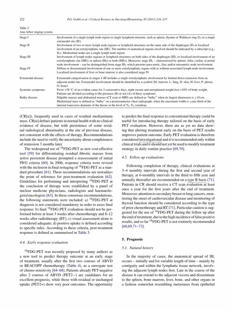
222 P.G. Gobbi et al. / Critical Reviews in Oncology/Hematology 85 (2013) 216–237
Table 2Ann Arbor staging system.
Stage I Involvement of a single lymph node region or single lymphoid structure, such as spleen, thymus or Waldeyer ring (I), or a singleextranodal site (IE).
Stage II Involvement of two or more lymph node regions or lymphoid structures on the same side of the diaphragm (II) or localisedinvolvement of an extralymphatic site (IIE). The number of anatomical regions involved should be indicated by a subscript (e.g.,II3). Mediastinal nodes are a single lymph node region.
Stage III Involvement of lymph nodes regions or lymphoid structures on both sides of the diaphragm (III), or localised involvement of anextralymphatic site (IIIE), or spleen (IIIs) or both (IIIEs). Moreover, stage III1 – characterised by splenic, hilar, coeliac or portalnode involvement – can be distinguished from stage III2 which presents para-aortic, iliac and/or mesenteric node involvement.
Stage IV Diffuse or disseminated involvement of one or more extralymphatic organs with or without associated lymph node involvement.Localised involvement of liver or bone marrow is also considered stage IV.
Extranodal disease: Extranodal categorisation in stages I–III includes a single extralymphatic involvement by limited direct extension from anadjacent nodal site. Extranodal involvement should be identified by a symbol (M: marrow, L: lung, D: skin, H: liver, P: pleura,O: bone).
Systemic symptoms: Fever >38 ◦C of no evident cause for 3 consecutive days, night sweats and unexplained weight loss >10% of body weight.Patients are divided according to the presence (B) or not (A) of these symptoms.
Bulky disease: Palpable masses and abdominal masses (CT scan or MRI) are defined as “bulky” when its largest dimension is ≥10 cm.osteroae level
(meunio
taIwdtGtngtdrfwctr
4
aoooaeu
tuPiiccs
4
3taPoMitogtr[
5
5
oc
Mediastinal mass is defined as “bulky” on a pinternal transverse diameter of the thorax at th
CR[u]), frequently used in cases of residual mediastinumass. CR[u] defines patients in normal health with no clinical
vidence of disease, but with persistence of some resid-al radiological abnormality at the site of previous disease,ot consistent with the effects of therapy. Recommendationsnclude the need to verify the uncertainty about completenessf remission 3 months later.
The widespread use of 18FDG-PET as new cost-effectiveool [59] for differentiating residual fibrotic masses fromctive persistent disease prompted a reassessment of initialWG criteria [60]. In 2006, response criteria were revisedith the inclusion in final restaging of 18FDG-PET as a stan-ard procedure [61]. These recommendations are nowadayshe point of reference for post-treatment evaluation [62].uidelines for performing and interpreting 18FDG-PET at
he conclusion of therapy were established by a panel ofuclear medicine physicians, radiologists and haematolo-ists/oncologists [63]. In these consensus recommendations,he following statements were included: a) 18FDG-PET atiagnosis is not considered mandatory in order to asses finalesponse; b) final 18FDG-PET evaluation should not be per-ormed before at least 3 weeks after chemotherapy and 8–12eeks after radiotherapy (RT); c) visual assessment alone is
onsidered adequate; d) positive uptake is defined accordingo specific rules. According to these criteria, post-treatmentesponse is defined as summarised in Table 3.
.4. Early response evaluation
18FDG-PET was recently proposed by many authors asnew tool to predict therapy outcome at an early stage
f treatment, usually after the first two courses of ABVDr BEACOPP chemotherapy (Table 4), as a surrogate test
f chemo-sensitivity [64–68]. Patients already PET-negativefter 2 courses of ABVD (PET2−) are candidates for anxcellent prognosis, while those with residual or unchangedptake (PET2+) show very poor outcomes. The opportunityidta
nterior chest radiograph, when the maximum width is ≥one-third of theof T5–T6 vertebrae.
o predict the final response to conventional therapy could beseful for introducing therapy tailored on the basis of earlyET evaluation. However, there are as yet no data show-
ng that altering treatment early on the basis of PET resultsmproves patient outcome. Early PET evaluation is thereforeonsidered investigational and it is recommended only withinlinical trials and it should not yet be used to modify treatmenttrategy in daily routine practice [69,70].
.5. Follow up evaluations
Following completion of therapy, clinical evaluations at–4 monthly intervals during the first and second year ofherapy, at 6-monthly intervals in the third to fifth year andnnually thereafter are recommended on a type R basis [71].atients in CR should receive a CT scan evaluation at leastnce a year for the first years after the end of treatment.oreover, attention to secondary breast or lung cancers, mon-
toring the onset of cardiovascular disease and monitoring ofhyroid function should be considered according to the typef prior chemotherapy and RT [71]. Particular caution is sug-ested for the use of 18FDG-PET during the follow up afterhe end of treatment, due to the high incidence of false positiveesults, and so far 18FDG-PET is not routinely recommended60,69,71–73].
. Prognosis
.1. Natural history
In the majority of cases, the anatomical spread of HLccurs – initially and for variable length of time – mainly byontiguity and within the lymphatic tissue network, involv-
ng the adjacent lymph nodes first. Late in the course of theisease it can extend to the adjacent viscera and disseminateo the spleen, bone marrow, liver, bone, and other organs infashion somewhat resembling metastases from epithelial
P.G. Gobbi et al. / Critical Reviews in Oncology/Hematology 85 (2013) 216–237 223
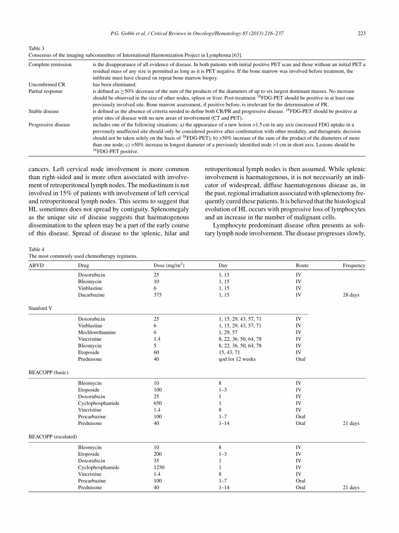
Table 3Consensus of the imaging subcommittee of International Harmonization Project in Lymphoma [63].
Complete remission is the disappearance of all evidence of disease. In both patients with initial positive PET scan and those without an initial PET aresidual mass of any size is permitted as long as it is PET negative. If the bone marrow was involved before treatment, theinfiltrate must have cleared on repeat bone marrow biopsy.
Unconfirmed CR has been eliminated.Partial response is defined as ≥50% decrease of the sum of the products of the diameters of up to six largest dominant masses. No increase
should be observed in the size of other nodes, spleen or liver. Post-treatment 18FDG-PET should be positive in at least onepreviously involved site. Bone marrow assessment, if positive before, is irrelevant for the determination of PR.
Stable disease is defined as the absence of criteria needed to define both CR/PR and progressive disease. 18FDG-PET should be positive atprior sites of disease with no new areas of involvement (CT and PET).
Progressive disease includes one of the following situations: a) the appearance of a new lesion >1.5 cm in any axis (increased FDG uptake in apreviously unaffected site should only be considered positive after confirmation with other modality, and therapeutic decisionshould not be taken solely on the basis of 18FDG-PET); b) >50% increase of the sum of the product of the diameters of more
ameter
ctmiaHado
rictqe
TT
A
S
B
B
than one node; c) >50% increase in longest di18FDG-PET positive.
ancers. Left cervical node involvement is more commonhan right-sided and is more often associated with involve-
ent of retroperitoneal lymph nodes. The mediastinum is notnvolved in 15% of patients with involvement of left cervicalnd retroperitoneal lymph nodes. This seems to suggest thatL sometimes does not spread by contiguity. Splenomegaly
s the unique site of disease suggests that haematogenousissemination to the spleen may be a part of the early coursef this disease. Spread of disease to the splenic, hilar and
a
t
able 4he most commonly used chemotherapy regimens.
BVD Drug Dose (mg/m2)
Doxorubicin 25Bleomycin 10Vinblastine 6Dacarbazine 375
tanford V
Doxorubicin 25Vinblastine 6Mechlorethamine 6Vincristine 1.4Bleomycin 5Etoposide 60Prednisone 40
EACOPP (basic)
Bleomycin 10Etoposide 100Doxorubicin 25Cyclophosphamide 650Vincristine 1.4Procarbazine 100Prednisone 40
EACOPP (escalated)
Bleomycin 10Etoposide 200Doxorubicin 35Cyclophosphamide 1250Vincristine 1.4Procarbazine 100Prednisone 40
of a previously identified node >1 cm in short axis. Lesions should be
etroperitoneal lymph nodes is then assumed. While splenicnvolvement is haematogenous, it is not necessarily an indi-ator of widespread, diffuse haematogenous disease as, inhe past, regional irradiation associated with splenectomy fre-uently cured these patients. It is believed that the histologicalvolution of HL occurs with progressive loss of lymphocytes
nd an increase in the number of malignant cells.Lymphocyte predominant disease often presents as soli-ary lymph node involvement. The disease progresses slowly,
Day Route Frequency
1, 15 IV1, 15 IV1, 15 IV1, 15 IV 28 days
1, 15, 29, 43, 57, 71 IV1, 15, 29, 43, 57, 71 IV1, 29, 57 IV8, 22, 36, 50, 64, 78 IV8, 22, 36, 50, 64, 78 IV15, 43, 71 IVqod for 12 weeks Oral
8 IV1–3 IV1 IV1 IV8 IV1–7 Oral1–14 Oral 21 days
8 IV1–3 IV1 IV1 IV8 IV1–7 Oral1–14 Oral 21 days
2 Oncol
wrtlnqaf
lw1WdHtcp
5
alsmpbpnia
toeiteTHtgoptt“usE
tfe
-
-
Iw<tHoofum
rit(fswwsAvt[
awicfaPitiaiBStfd“wt
24 P.G. Gobbi et al. / Critical Reviews in
ith fairly frequent relapses, which are rarely fatal. Lateelapses have been reported to be more common than in otherypes of HL [74]. This may be associated with or progress toarge B-cell lymphoma [75,76], while secondary low-gradeon-Hodgkin’s lymphomas can also occur. This is more fre-uently observed after lymphocyte predominant HL thanfter classical HL. Survival is long, with or without treatment,or localised cases.
The mortality of HL has progressively reduced over theast 30 years. In the 1950s, the mortality in the United Statesas 1.8 per 100,000, while in the early 90s it was 0.47 per00,000. The most recent 5-year survival figure is 81% [77].hile this malignancy accounted for 30% of total lymphoma
eaths in 1950, it accounted for only 6% in 1994. UntreatedL is rare today because both chemotherapy and RT are effec-
ive curative treatments. If the disease is left untreated, theourse is brief, spanning 1–2 years with fewer than 5% ofatients alive at 5 years.
.2. Prognostic factors
The definition of prognostic factors and risk groups is stillmatter for debate. A very long list of clinical, histopatho-
ogical and laboratory parameters have individually beenuggested to be of prognostic value [78–83]. Nearly all areore or less directly correlated with the amount of tumour
resent at diagnosis [84]. The measurement of the tumoururden on the serial slices of total body CT is a very powerfulrognostic tool [85,86]. However, the increasing effective-ess of therapies requires periodic reevaluation of the actuallymportant prognostic factors, with a remodelling of their hier-rchical order [87].
The criteria of the Ann Arbor Conference [43] are interna-ionally accepted as defining the two major prognostic groupsf limited (early-stages) and advanced disease. Early-stagesncompass stage I and II, while stages III and IV are includedn the advanced disease group. Stage II with systemic symp-oms (IIB) can be included in the group of either unfavourablearly-stage disease (European Organization for Research andreatment of Cancer – EORTC) or advanced disease (Germanodgkin’s Lymphoma Study Group – GHSG), according to
he policy of individual cooperative groups. The early-stageroup is usually further subdivided into the two categoriesf “favourable” and “unfavourable” disease according to theresence or absence of other clinical and laboratory prognos-ic variables. The “unfavourable early-stage” group defines,herefore, a group whose prognosis is intermediate betweenfavourable early-stage” and “advanced-stage”. Variablessed to differentiate “favourable” from “unfavourable” earlytages are not universally codified, but they are similar inORTC and GHSG classification systems [88].
One or more of the following unfavourable prognostic fea-
ures is needed to shift an individual patient in stage I or IIrom the category of “favourable” to that of “unfavourable”arly stage in the EORTC and GHSG classifications:ftiS
ogy/Hematology 85 (2013) 216–237
EORTC classification: bulky mediastinal mass; age ≥50years; ESR ≥50 without B symptoms or ≥30 with B symp-toms; ≥4 nodal areas.GSHG classification: bulky mediastinal mass; extranodalsite; ESR ≥50; ≥3 nodal areas.
Seven prognostic factors (age ≥45 years, male sex, stageV, serum albumin <4 mg/dL, haemoglobin <10.5 mg/dL,hite blood cell count ≥15,000 x/L, and lymphocytes count600 x/L) associated with a reduction of 7–8% in tumour con-
rol at 5 years have been identified in patients with advancedL [89]. These variables are the basis for the constructionf the International Prognostic Score (IPS). The numberf factors present has been related to 5-year progression-ree survival (PFS), which was 74% for patients with 0–2nfavourable prognostic factors and 55% for those with 3 orore.The identification of prognostic factors in patients with
elapsed or refractory HL is confounded by the use of variednclusion criteria in clinical trials. However, it would appearhat relapses within the primary irradiated area, early relapsesfirst year) and chemorefractory diseases as well as poor per-ormance status, mediastinal bulky disease, female sex, Bymptoms, and extranodal disease at relapse are associatedith a worse outcome [90–99]. Results are significantly betterhen the disease is still chemosensitive, and a second remis-
ion or at least a minimal disease status is reached beforeSCT. Recent data show that 18FDG-PET positivity after sal-age debulking chemotherapy and before ASCT is probablyhe factor indicating the poorest prognosis in failed patients100,101].
In patients with advanced-stage HL, the early responsefter 2 courses of ABVD chemotherapy, when evaluatedith 18FDG-PET scan, shows important prognostic signif-
cance [64–67]. It is considered to be similar to a test ofhemosensitivity and it overrides all conventional prognosticactors, including IPS score. PET2-advanced-stage patientsfter 2 courses of ABVD are projected to achieve a 2-yearFS of >90%, while a 2-year PFS of <10% is expected
n PET2+ patients. However, some concerns arise abouthe routine application of this technique in order to mod-fy the treatment strategy for individual patients. First ofll, the prognostic value of early 18FDG-PET may be lowern patients treated with new regimens, such as escalatedEACOPP, which are more aggressive than ABVD [102].econdly, conventional criteria for 18FDG-PET interpreta-
ion were not intended for interim analysis evaluation, butor end-treatment response assessment, and an importantebate has arisen about how to evaluate the grey zone ofminimal residual uptake” [103]. International Workshopsere organised and prospective studies are ongoing in order
o reach a consensus on simple and reproducible criteria18
or FDG-PET interpretation and to validate internationallyhe role of PET2 scans [104]. Several trials investigat-ng 18FDG-PET response-adapted therapy are ongoing (seeection 6.6), but until their results become available,
Oncol
tr
6
6I
ltwpEwaih
mrfirdtswfw[euRayt
iies
aefAimrwsfasp
iiIm
6
ffo3warHacRfaorslaCmipRdpb
6
fItbbca[Ifietr4w
P.G. Gobbi et al. / Critical Reviews in
his prognostic factor is considered investigational and it isecommended only within clinical trials [69,70].
. Treatment
.1. Treatment of early-stage disease (stageA–IIA ± IIB)
Primary ABVD combination chemotherapy (Table 4), fol-owed by involved-field irradiation (IF-RT) is the standardreatment for patients with early-stage HL (type 1 evidence),ith an overall survival (OS) >95% [105–108]. Even amongatients with very favourable disease, according to theORTC criteria [109], the use of RT alone was associatedith an unacceptable relapse rate [110] and is no longer advis-
ble. The superiority of the combined chemo-radiotherapyn comparison with extended-field irradiation (EF-RT) aloneas been confirmed in diverse randomised trials [111–116].
The dose and the extension of irradiation field after pri-ary chemotherapy is an important issue considering the
isk of late morbidity related to combined treatment. Smallerelds and lower doses were progressively introduced toeduce the risk of death from second cancers [117,118], heartisease [119] and other complications. Several randomisedrials using diverse chemotherapy regimens have all demon-trated that the extent of radiation can be safely reducedhen chemotherapy is added [119–122]. No significant dif-
erence in survival has been observed among patients treatedith IF-RT or EF-RT after ABVD [121] or COPP-ABVD
123,124]. This suggests that brief chemotherapy is able toradicate all un-irradiated disease even when only IF-RT issed. Recent publications suggest a further reduction of IF-T to involved-nodal irradiation (IN-RT) [125]. However,randomised comparison between these techniques is not
et available and IN-RT is still considered an investigationalechnique.
The optimal radiation dose remains to be defined. Accord-ng to the paediatric experience 20–25 Gy may be adequaten patients in CR after primary chemotherapy [126,127]; oth-rwise the classical doses of 30 Gy are still considered thetandard treatment.
Whether early-stage HL can be treated with chemotherapylone without any radiation is still an open question. Straust al. randomised a group of patients to 6 courses of ABVDollowed by IF-RT with 6 courses of ABVD alone [128].fter a median follow up of 5 years, there was no difference
n PFS between the two groups, but patients with a bulkyediastinal mass were excluded from this study. In another
andomised trial involving unfavourable early-stage patientsithout bulky disease, the combined modality treatment was
uperior to ABVD alone in terms of PFS, even though no dif-
erences were seen in OS [129]. Although a strategy of ABVDlone is considered a rational option in favourable early-tage patients [130], a recent meta-analysis shows that inatients with early-stage disease adding RT to chemotherapyCdaI
ogy/Hematology 85 (2013) 216–237 225
mproves both tumour control and OS [131]. Thus, a lim-ted number of courses of ABVD chemotherapy followed byF-RT is considered the best strategy on type 1 basis for theanagement of stage IA and IIA HL [71,108,132].
.1.1. Treatment of favourable early-stage diseaseThe recently reported results of the HD10 trial are use-
ul for informing the therapeutic approach for patients withavourable early-stage HL [133]. In this trial, four coursesf ABVD were compared with two courses of ABVD and0-Gy IF-RT was compared to 20-Gy IF-RT. No differencesere seen in terms of freedom-from-treatment failure (FFTF)
nd OS according to the number of courses of ABVD oradiation doses, with actuarial rates of ∼90% in each arm.owever, four courses of ABVD were more toxic than two,
nd 30 Gy were more toxic than 20 Gy. The authors haveoncluded that two courses of ABVD followed by 20 Gy IF-T is an adequate and less toxic strategy for patients with
avourable early-stage HL. This strategy can be consideredt present the standard of care on type 1 basis for this groupf patient, against which different combinations of chemo-adiotherapy can be compared. The impact on outcome ofome less intensive strategies aimed at reducing acute andate toxicity is under study. A chemotherapy regimen lessggressive than ABVD is being assessed in the HD13 trial.onversely, the use of the VBM (vinblastine, bleomycin,ethotrexate) regimen [120,134] or its VbMp variant [135]
s no longer recommended since it increased the risk of severeulmonary toxicity and the need to deliver full doses of EF-T. Omitting RT in selected patients with very favourableisease is another strategy under investigation, particularly, inatients with negative interim PET. Prolonged follow-up wille necessary to reach firm conclusions from these studies.
.1.2. Treatment of unfavourable early-stage diseaseABVD chemotherapy followed by IF-RT is the most use-
ul strategy in patients with unfavourable early-stage HL.n these patients, several chemotherapy regimens – poten-ially less toxic than ABVD – such as EBVM (epirubicin,leomycin, vinblastine, methotrexate) EBVP (epirubicin,leomycin, vinblastine, prednisone) or EVE (epirubicin, vin-ristine, etoposide), were assessed in large randomised trialsnd none were superior to ABVD in terms of FFTF or OS136–138]. At present, four courses of ABVD followed byF-RT 30 Gy is considered the standard of care on type 1 basisor this group of patients [112]. Whether more aggressive reg-mens (Table 4), such as BEACOPP baseline or BEACOPPscalated, in association with IF-RT can improve disease con-rol in comparison with ABVD still is an open question. Finalesults of the HD11 trial have showed no difference between
courses of ABVD and 4 courses of BEACOPP baselinehen they are followed by 30 Gy IF-RT. However, BEA-
OPP improves FFTF in comparison to ABVD if radiationoses are reduced to 20 Gy [139]. When comparing the bestnd least toxic arms of this four arm study, BEACOPP plusF-RT 20 Gy might have the disadvantage of higher acute
2 Oncol
tsmotcebsw8O(oiIt
6I
wMdtdOtbopbcbitdrol6cmp[ttohAwfoTi
p[aafenrotosPtsc
adbswta[crcacocwbcsOooattdschHande1
r
26 P.G. Gobbi et al. / Critical Reviews in
oxicity, and a higher incidence of second myelodysplasticyndromes and infertility, while ABVD plus IF-RT 30 Gyight have the disadvantage of a potentially increased risk
f secondary breast or lung cancer. Final results of the HD14rial were recently presented [140]. This randomised trialompared 4 cycles of ABVD (arm A) or 2 cycles BEACOPPscalated followed by 2 cycles ABVD (arm B), both followedy 30 Gy IF-RT, in 1,655 patients with unfavourable early-tage HL. With a median follow-up of 42.4 months, thereas a significantly better FFTF in the intensified arm (4-year:9.3% vs. 94.7%; p = 0.0001), but without any difference inS. Acute grade III–IV toxicity was more common in arm B
87% vs. 51%), but no differences in treatment-related deathsr secondary neoplasia rates were observed. German authorsmplemented BEACOPP escalated followed by ABVD andFRT as new standard of care for early unfavourable HL inhe follow-up study: the HD17 study.
.2. Treatment of advanced-stage disease (stageII–IV ± IIB)
ABVD is also the standard chemotherapy for patientsith advanced-stage HL, with a 10-year OS >50% [141,142].ore intensive combinations have been developed in the last
ecade to improve outcome. A weekly, seven-drug regimen,he Stanford V regimen (Table 4), with reduced cumulativeoses of doxorubicin and bleomycin has produced a 5-yearS of 94% [143]. This regimen was originally designed
o be used with a combined extensive IF-RT, which cannote reduced without impairment of outcome [144]. The riskf late radiation-related adverse events is therefore a majorroblem with Stanford V. Two intensified combinations ofleomycin, etoposide, doxorubicin, cyclophosphamide, vin-ristine, prednisone, and procarbazine regimens (BEACOPPaseline and BEACOPP escalated) have been developed tomprove the outcome by escalating the dose intensity ofhe more effective drugs. The 10-year results of the ran-omised trial comparing the two modalities of BEACOPPegimens with alternating COPP/ABVD showed survivalf 75% with COPP/ABVD, 80% with BEACOPP base-ine and 86% with BEACOPP escalated, with a FFTF of4%, 70% and 82%, respectively [145,107,146,147]. Someoncerns are still present about BEACOPP-related toxicity,ainly acute haematological toxicity, secondary myelodys-
lastic syndromes and/or acute leukaemias, and infertility148,149,147]. By contrast, infertility is not a problem withhe ABVD regimen [150,151]. Moreover, the BEACOPPoxicity is particularly high and unacceptable in patientslder than 65 years [152]. Two subsequent randomised trialsave confirmed the superiority of BEACOPP escalated overBVD in terms of disease control, but a difference in OSas not evident as a consequence of the salvage of ABVD
ailures with high-dose chemotherapy supported by autol-
gous stem cell transplantation (HDC/ASCT) [153,154].he superiority of BEACOPP over ABVD in terms of PFSs mainly evident in patients with a highly unfavourable
gmm
ogy/Hematology 85 (2013) 216–237
resentation, such as those with IPS prognostic score ≥3153]. Considering that about 60–70% of patients withdvanced disease can actually be cured with front-line ABVDlone with minimal toxicity, and that the rescue of ABVDailures is possible, the ideal would be to reserve BEACOPPscalated for patients with a very poor prognosis. Unfortu-ately, the IPS score is not a perfect predictor of ABVDesponse. However, the early response to the first two coursesf ABVD evaluated with 18FDG-PET seems to be a very goodest for ABVD chemosensitivity. Many trials are thereforengoing to evaluate the investigational strategy of an earlyhift to a more aggressive schedule for patients with 18FDG-ET positive results after the first 2 courses of ABVD. Until
he results of these trials are available, both the more aggres-ive and toxic BEACOPP and the less toxic ABVD can beonsidered rational options.
The consolidative role of IF-RT after full-dose chemother-py in patients with advanced disease has been matter ofebate for several years. The rationale for this strategy isased on the fact that relapse occurs in previously involvedites and often exclusively in lymph nodes, even in patientsith advanced disease. In a first randomised trial, consolida-
ion with IF-RT did not improve outcome in patients withdvanced disease in CR after MOPP-ABV chemotherapy155]. Conversely, a retrospective analysis of the impact ofonsolidation RT in patients registered in the UKLG LY09andomised trial showed that the addition of RT was asso-iated with significantly better PFS (5-year: 86% vs. 71%)nd OS, suggesting that RT contributes significantly to theure rate for advanced HL [156]. In a meta-analysis basedn 1740 patients from 14 controlled adjuvant irradiationlinical trials [119], the addition of RT to chemotherapyas associated with an 11% improvement in tumour controlut OS remained unchanged. The meta-analysis of studiesomparing consolidation chemotherapy vs. consolidation RThowed that tumour control is similar in both groups whileS is 8% better in the chemotherapy-only patients becausef fewer late treatment-related deaths. As a standard strategyn type 2 basis, consolidation IF-RT should be avoided indvanced HL, due to the potential for late morbid effects andhe lack of a survival benefit demonstrated by randomisedrials [157,158,155]. RT limited to the region of initial bulkyisease or residual masses after effective chemotherapy istill an open question. Mediastinum irradiation after primaryhemotherapy in patients with initial bulky mediastinal massas been considered so far a required consolidation treatment.owever, preliminary results of the HD12 trial show that radi-
tion of initial bulk and residual masses after BEACOPP doesot seem to be useful [159]. A useful new tool to tailor irra-iation of residual masses is 18FDG-PET evaluation at thend of chemotherapy. In the HD15 trial, patients with final8FDG-PET-negative residual masses were not irradiated andesults showed 96% PFS [160]. However, some studies sug-
est that irradiation of initial bulky and residual PET negativeasses are useful in patients treated with chemotherapy regi-ens less aggressive than BEACOPP [161,162]. Thus, RT of
Oncol
isa
afiidic
6
6
d∼sbudHnpbtotst(dvtnrpld
manbtwpotB
6
oHa
TdTre
6
HeoIihiirtchsneysEttcistafr
6
tterttRbtoio
P.G. Gobbi et al. / Critical Reviews in
nitial bulk and residual masses is therefore still considered atandard treatment on type 3 basis when first-line chemother-py is ABVD.
Consolidation with HDC/ASCT is an investigationalpproach in patients with HL in first CR after conventionalrst-line chemoradiotherapy. In spite of some encourag-
ng early results [163], two subsequent randomised trialsemonstrated that HDC/ASCT does not improve outcomen high-risk patients responding to front-line conventionalhemotherapy [164,165].
.3. Treatment of special categories of patients
.3.1. Pregnant patients with HLHL is one of the more frequent malignant conditions
iscovered during pregnancy, with concurrent pregnancy in3% of all patients with HL [166]. Efforts to determine the
tage of disease in pregnant patients are somewhat restrictedy the need to avoid CT scans and PET, but abdominalltrasonography can be used to detect subdiaphragmaticisease. Overall, the clinical behaviour and prognosis ofL diagnosed in pregnant women are similar to those ofonpregnant women [167]. In general terms, treatment ofregnant patients with asymptomatic, early-stage HL shoulde deferred until after the second trimester. In fact, morehan 50% of patients can continue pregnancy to term with-ut treatment. If treatment is required, it is usually possibleo control the lymphoma with single-agent chemotherapy,uch as vinblastine or anthracycline, allowing the pregnancyo go to term [168,167]. The use of single agent vinblastine6 mg/m2) is associated with 75% ORR and normal infantelivery in most cases [169]. Patients who progress despiteinblastine can be treated with ABVD during the second orhird trimester. Although RT should be avoided during preg-ancy, recent advances in RT techniques have resulted inemarkably reduced risk of fetal complications [170], and,resently, it is relatively safe to irradiate patients with iso-ated supradiaphragmatic disease, with a whole body fetalose ≤0.1 Gy.
If advanced HL is diagnosed during the first trimester, ter-ination of the pregnancy should be considered followed by
ppropriate staging and polychemotherapy. Treatment shouldot be delayed during pregnancy if patient presents with B,ulky, subdiaphragmatic, or progressive HL after the firstrimester. If treatment is required and the patient does notant a therapeutic abortion, the successful completion ofregnancy without fetal malformation is possible with ABVDr similar regimens [171]. There are no available data onhe use of more intensive regimens such as Stanford V orEACOPP in pregnancy.
.3.2. HIV-positive patients
HL is one of the defining illnesses of the acquired immun-deficiency syndrome. Usually, HIV-positive patients withL have mixed-cellularity or lymphocyte-depletion subtype,
dvanced and extranodal disease, and systemic symptoms.
blsA
ogy/Hematology 85 (2013) 216–237 227
he availability of highly active antiretroviral therapy hasramatically improved outcome among these patients [172].oday, HIV-infected patients with early-stage HL shouldeceive the same treatment as HIV-negative patients witharly-stage disease.
.3.3. Elderly patientsElderly patients, defined by chronological age, with
L represent a heterogeneous population in terms of lifexpectancy, morbidities, and functional status. Nearly 20%f HL patients are >65 years in some developed countries.n general, less than 10% of patients included in broad clin-cal trials are >60 years. The proportion of mixed cellularityistopathology and EBV-genome-positive tumours is highern older adults. Five-year OS of patients 66–80 and >80 yearss 58% and 26%, respectively. Older patients have loweremission rates, but PFS is less impaired. One reason forhe relatively poor outcome in elderly patients is their sus-eptibility to the toxic effects of intensive therapy, and manyave coexisting conditions that affect their ability to toleratetandard treatments. For example, elderly patients did sig-ificantly less well with EF-RT than with IF-RT; no suchffect was observed in younger patients. Elderly fit patientsounger than 65–70 years should be treated following theame modern therapeutic guidelines used in young patients.ven if elderly patients seem to benefit proportionally more
han younger patients from the inclusion of doxorubicin inhe treatment regimen [173], contraindications to use anthra-ycline should follow the well-known recommendations usedn other lymphoma patients. In these cases, some regimensuch as VBM appear advisable [174]. Thorough estima-ion of the individual patient’s frailness/comorbidities tollow proper adjustment of treatment, thus saving patientsrom over/undertreatment, remains an important, but a rarelyespected, recommendation.
.4. Treatment of relapsed or refractory HL
The choice of salvage treatment in relapsed or refrac-ory HL is strongly influenced by previous treatments andhe duration of the previous response. HDC/ASCT is consid-red the standard of care on type 2 basis in patients with HLelapsed or refractory at conventional first-line chemoradio-herapy, while patients relapsed after RT as exclusive first-linereatment can be treated with ABVD (followed or not by IF-T) on a type 3 basis. Patients with late relapse disease cane treated with a conventional dose combination, includinghe same previous regimen, with complementary RT to previ-usly un-irradiated bulky sites [175]. Salvage treatment withntense chemotherapy regimens such as BEACOPP [176]r MOPPEBVCAD (mechlorethamine, vincristine, procar-
azine, prednisone, epidoxirubicin, bleomycin, vinblastine,omustine, doxorubicin, and vindesine) [177] can be useduccessfully in patients with late relapse after front-lineBVD on a type R basis.
2 Oncol
p[rlrlcoariOtt
6abIiHHpny8sllosc
rprsI(geoi
d1
Asbo
srAo
rfimoatapfshpeo
iitAhTtat
aso1hmAc
pfitfsts
6t
iotAt
28 P.G. Gobbi et al. / Critical Reviews in
The advantage of HDC/ASCT in relapsed or refractoryatients has been demonstrated in small randomised trials163,178,179] and has also been suggested by many non-andomised studies [99,180–182]. HDC/ASCT can produceong-term DFS in 30–65% of selected patients with advancedelapsed disease, with a greater benefit among patients with aess favourable prognosis. In these studies, HDC/ASCT effi-acy was related to the amount of primary therapy, presencef B symptoms, extent of disease at time of transplantationnd the responsiveness to prior chemotherapy. Overall, theseandomised trials are old studies that showed a significantmpact in PFS, but no differences were shown in terms ofS, which seems to reflect the small number of patients and
he possible bias due to the subsequent high-dose salvagereatment for non-transplantation arms.
Recently reported studies demonstrated that more than0% of patients who have had a relapse after ABVD andbout 30% of patients with initially refractory lymphoma cane reliably cured with HDC/ASCT [183]. A recently reportedtalian randomised trial comparing BEACOPP vs. ABVDn patients with advanced disease HL showed that salvageDC/ASCT could even cure patients with chemorefractoryL, offering a second chance at a cure to otherwise doomedatients [154]. In that trial, the use of salvage HDC/ASCTegated the initial PFS advantage in favour of BEACOPP (7-ear: 85% vs. 73%; p = 0.004) resulting in similar OS (7-year:9% vs. 84%; p = 0.39). Accordingly, patients with advanced-tage HL can be cured, with a lower risk of side effects,eukemogenesis and infertility, by using first-line ABVD fol-owed by salvage HDC/ASCT in failed patients [184]. Thesebservations underscore the full impact of reliably curativeecondary treatment following the best choice of primaryhemotherapy [184].
The best induction debulking regimen before HDC/ASCTemains to be defined since no randomised trials have beenublished comparing the efficacy and toxicity of availableegimens. Several salvage regimens of different intensity,uch as DHAP (dexamethasone, cytarabine, cisplatin) [98],CE (ifosfamide, carboplatin, etoposide) [185], and IGEVifosfamide, gemcitabine, vinorelbine) [186] have showedood activity in reducing bulk disease, mobilising periph-ral blood stem cells and evaluating chemosensitivity. Eachf these regimens can be independently utilised as anndividualised treatment based on a type 3 evidence.
Some authors suggest that residual positivity afterebulkying chemotherapy and before ASCT detected by8FDG-PET is associated with poor outcome after a singleSCT [100,101]. In this set, a double ASCT procedure is fea-
ible with acceptable toxicity and preliminary good results,ut it should be considered as an investigational treatmentption [187,188].
HDC supported by allogeneic stem cell transplantation
eems to be a suitable clinical tool for patients with earlyelapsed or refractory HL, or for patients relapsing afterSCT. Until some years ago, transplant-related mortalityf allogeneic transplantation represented a main limitation,lBpt
ogy/Hematology 85 (2013) 216–237
eaching an overall rate of 40%. The recent advances in theeld of transplantation, along with the introduction of non-yeloablative regimens and with an accurate management
f the septic complications, increased the interest toward thispproach [189,190]. A broad spectrum of evidence supportshe existence of graft vs. HL (GVHL). Some studies showedlower rate of relapse after allogeneic transplantation com-ared with ASCT and data from the EBMT (European Groupor Blood and Marrow Transplantation) registry demon-trate a direct correlation between grade 2 and 4 graft vs.ost disease (GVHD) and probability of recovery. Moreover,reliminary data suggest a direct anti-lymphoma activityxerted by donor’s lymphocytes after allotransplant in casesf relapse [191,192].
Limited conditioning regimens before transplantation –ntroduced with the purpose of increasing GVHL and reduc-ng myeloablation – have decreased the peritransplant mor-ality and allowed a better evaluation of the beneficial effects.
wide spectrum of reduced-intensity conditioning regimensas been proposed, the majority being based on fludarabine.he same goes for immunosuppressants following transplan-
ation. Also, more homogeneous protocols on the use of DLIre advisable, according to the clinical features of disease, tohe grade of GVHD and to immunosuppressant drugs.
At present, the estimated transplant-related mortality isbout 20% and destined to decrease further [193]. A revi-ion of the EBMT registry demonstrated the clinical efficacyf this procedure in HL, with 2-year OS of 47%, and a5% peritransplant mortality [194]. Further EBMT studiesave showed good results with reduced conditioning regi-ens [195], and a survival benefit in patients relapsing afterSCT, who had a donor and were allografted with a reduced
onditioning regimen [196].Response to debulking chemotherapy before allotrans-
lant is an important prognostic factor [197], and ASCTollowing induction therapy might be useful in significantlyncreasing the proportion of patients in remission before allo-ransplant [198]. Accordingly, both double ASCT and ASCTollowed by non-myeloablative allotransplant are promisingtrategies for very high-risk patients with relapsed or refrac-ory HL, and they should be considered as investigationaltrategies in patients chemo-resistant before the first ASCT.
.5. Treatment-related late complications and secondumours
The toxicity of chemotherapy is the major drawback tots widespread use. Early toxicity is usually manageable andf short duration. Conversely, late toxicity is often relatedo irreversible and sometimes life-threatening abnormalities.lkylating agents may induce male and female sterility, but
his is much less frequent in patients treated with ABVD-
ike regimens than alkylating-containing regimens, mainlyEACOPP [148–151]. Semen cryopreservation should berogrammed into the treatment schedule for all men youngerhan 50 years. Over 50 years old, it is up to the patient and
Oncol
hwPbdostfi
dnoMlcaaaobaipbap
dbadiisRtyrasvTls
6
trsaaai
aiumt
famanTlsrfra(
maciCt5ac(ritgEepCti2awvtiwhtti
P.G. Gobbi et al. / Critical Reviews in
is preference. A major problem concerns pre-puberal boys,ho should be treated with non-gonadotoxic chemotherapy.ulmonary fibrosis depends mainly, but not exclusively, onleomycin use (1–6% of patients treated with ABVD); it isose-dependent with a cumulative limited dose of bleomycinf 170 mg/m2. However, this toxicity may appear even after aingle dose of bleomycin. The addition of thoracic irradiationo bleomycin increases the risk of paramediastinal pulmonarybrosis and cutaneous sclerosis.
RT is associated with unaesthetic modifications due to theisappearance of the fatty subepidermal tissues, mainly in theeck, and with skin sclerosis, xerostomia, hypothyroidism,edema of the arm or thigh, amenorrhoea, and female sterility.ost severe non-tumoural late effects of RT are cardiovascu-
ar complications due to vascular obstructions (infarction andardiac failure) and pericardiac sclerosis. Lung complicationsfter mantle-field irradiation are often limited to paramedi-stinal fibrosis. In paediatric cases, where there is concernbout the late effects on statural growth and thorax morphol-gy, a dose reduction of radiation therapy to 20–25 Gy haseen adopted in complete responders to primary chemother-py. Fertility may be compromised in young women withnfradiaphragmatic disease requiring pelvic irradiation. If theatient has no children, surgical ovarian displacement shoulde suggested. In this case, one ovary is moved to a medianrea just under the uterus, and the other is shifted out of therogrammed RT field.
Secondary acute leukaemia and myelodysplastic syn-romes are almost always fatal complications, often inducedy alkylating agents [199]. Non-Hodgkin lymphomas iscomplication of HL treatment [200–202] that should be
istinguished from relapses by using excisional biopsy. Thencidence of lung and breast cancer is higher than expectedn patients irradiated with mantle field, and the risk ofecond cancers is increasing in proportion with intensity ofT and time after treatment [203,204,116,117]. In women
reated with mediastinal RT, breast cancer develops at aounger age than the average, and regular breast screening isecommended. Other solid tumours arising within irradiatedreas have been described [205–208]. The most appropriatecreening methods, time of initiation and intervals betweenisits are currently uncertain and guidelines vary widely.here is a need to evaluate methods, benefits and costs of
ong-term follow-up, including the impact of breast cancercreening on mortality.
.6. New active drugs and ongoing trials
Several investigational approaches are presently beingested in relapsed HL. Among others, Yttrium-labelled fer-itin combined with HDC/ASCT [209], T cells againstome antigens of EBV infused after HDC/ASCT [210],
nti-CD30 antibodies-ricin A-chain immunotoxin [211],nti-CD25 antibodies-ricin A-chain immunotoxin [212],nti-CD16 × anti-CD30 bispecific monoclonal antibod-es [213], and interleukin-2-diphtheria-toxin fusion toxinriom
ogy/Hematology 85 (2013) 216–237 229
re being investigated [214]. Some histone-deacetylasenhibitors, such as panobinostat [215], and immunomod-lators such as lenalidomide [216] are other interestingolecules under study alone or in association with conven-
ional or HDC/ASCT [217].Among new therapeutic options, attention is presently
ocused particularly on the potential role of rituximab, annti-CD20 monoclonal antibody largely used in the treat-ent of B-cell non-Hodgkin lymphomas. It seems to be
ctive, not only in CD20+ nodular lymphocyte predomi-ant [218,219], but also in classical CD20 negative forms.he central hypothesis for using rituximab is that benign B
ymphocytes in the lymph-node microenvironment promoteurvival of R-S cells. Consequently, targeting them by usingituximab may deprive neoplastic cells of survival signals andacilitate the action of chemotherapy [216,220]. Preliminaryesults of a phase II study combining rituximab with ABVDre encouraging [221], and randomised studies are ongoingNCT00992030).
The development of novel therapies targeting CD30 is aajor advance in the treatment of HL. Normal CD30 hasrelatively restricted distribution on activated B-cells, T-
ells and eosinophils thus making it a very attractive targetn this disease. After an initial phase where several anti-D30 antibodies showed considerable in vitro activity (i.e.,
he human Ig G1k antibody MDX-060, the human antibodyF11, the humanised antibody XmAb 2513, the chimericntibody SGN-30, the immunotoxin ki-4dgA), but modestlinical activity in patients with CD30-positive lymphomasi.e., HL and anaplastic large cell lymphoma), more recentlyeported studies showed relevant clinical activity with somenteresting molecules. Brentuximab vedotin (SGN-35) seemso be the more promising candidate. This agent is a conju-ate comprising the antitubulin agent monomethyl auristatinand a CD30-specific monoclonal antibody that has shown
xcellent activity both in cHL and anaplastic lymphoma. Thehase I study evaluating this agent in relapsed or refractoryD30+ lymphomas included mostly patients with HL. The
reatment was well tolerated with mostly grade 1 and 2 tox-city, mainly peripheral neuropathy [222]. A pivotal, phase
multicentre trial – recently reported in abstract form –ddressed the efficacy and safety of SGN-35 in 102 patientsith HL relapsed or refractory to ASCT [223]. Brentuximabedotin 1.8 mg/kg was administered every 3 weeks for upo 16 cycles of treatment, resulting in tumour size reductionn 95% of patients and resolution of B symptoms in 83%,ith manageable adverse events. This encouraging activity ineavily pretreated patients deserves to be confirmed in furtherrials. An ongoing randomised, placebo-controlled, multicen-re phase 3 trial will assess the efficacy and safety of SGN-35n patients with residual HL after ASCT (NCT01100502).
The chance to tailor subsequent treatment according to the
esults of PET scan after the first 2 courses of ABVD is annvestigational strategy and many international trials focusedn this topic are ongoing. Mostly, these trials try to deter-ine if PET/CT imaging can be reproducibly and effectively
2 Oncol
aaPwrsaltfisuaotanotwCI(Isc
C
R
pH
R
30 P.G. Gobbi et al. / Critical Reviews in
pplied in the early assessment of response to chemotherapynd whether patients with a negative or a positive FDG-ET/CT scan after 2 courses of ABVD can be safely treatedith a reduced or intensified therapy. Therapeutic intensity
eduction in ongoing trials consists of excluding RT in early-tages (H10, HD0801), deleting some drugs from ABVD indvanced stage disease (CRUK-2007-006064-30), deesca-ating from BEACOPP to ABVD (AHL 2011) or reducinghe number of courses (HD18). On the other hand, intensi-ed treatment consists of including more aggressive regimensuch as BEACOPP instead of ABVD (HD0607), adding rit-ximab (HD18) and early crossing to HDC/ASCT (HD0801)re also being investigated. In this context, and with the aimf reducing iatrogenic toxicity, the HD16 and the RAPIDrials are assessing – with different designs – whether IF-RTvoidance in patients with favourable early-stage HL and PETegative after 2–3 courses of ABVD will result in non-inferiorutcome with respect to conventional chemoradiationherapy. The HD17 will assess the role of PET in patientsith intermediate risk HL, comparing 2 courses of BEA-OPP escalated plus 2 courses ABVD followed by 30 Gy
F-RT irrespective of FDG-PET results after chemotherapycontrol arm) vs. the same chemotherapy followed by 30 GyN-RT if FDG-PET is positive after chemotherapy or theame chemotherapy without RT if FDG-PET is negative afterhemotherapy (experimental arm).
onflict of interest
Authors have no conflict of interest to be disclosed.
eviewers
Frederick B. Hagemeister, M.D., Professor, Lym-homa/Myeloma, MD Anderson Cancer Center, 1515olcombe Blvd, Houston, TX 77030, USA.
eferences
[1] Poppema S, Kaleta J, Hepperle B, Visser L. Biology of Hodgkinlymphoma. Annals of Oncology 1992;3(Suppl. 4):5–8.
[2] Küppers R. Clonotypic B cells in classic Hodgkin lymphoma. Blood2009;114(18):3970–1.
[3] Harris NL, Stein H, Coupland SE, et al. New approaches to lymphomadiagnosis. Hematology American Society of Hematology EducationalProgram 2001:194–220.
[4] Correa P, O’Conor GT. Geographic pathology of lymphoreticulartumors: summary of survey from the geographic pathology commit-tee of the International Union Against Cancer. Journal of the NationalCancer Institute 1973;50:1609–17.
[5] Hjalgrim H, Engels EA. Infectious aetiology of Hodgkin and non-Hodgkin lymphomas: a review of the epidemiological evidence.
Journal of Internal Medicine 2008;264(6):537–48.[6] Armstrong AA, Alexander FE, Paes RP, et al. Association ofEpstein–Barr virus with pediatric Hodgkin Lymphoma. AmericanJournal of Pathology 1993;142:1683–8.
ogy/Hematology 85 (2013) 216–237
[7] Herbst H, Pallesen G, Weiss LM, et al. Hodgkin lymphomaand Epstein–Barr virus. Annals of Oncology 1992;3(Suppl. 4):27–30.
[8] Levine PH, Pallesen G, Ebbesen P, et al. Evaluation of Epstein–Barrvirus antibody patterns and detection of viral markers in the biopsiesof patients with Hodgkin’s disease. International Journal of Cancer1994;59(1):48–50.
[9] Hjalgrim H, Askling J, Rostgaard K, et al. Characteristics ofHodgkin’s lymphoma after infectious mononucleosis. New EnglandJournal of Medicine 2003;349(14):1324–32.
[10] Niens M, Jarrett RF, Hepkema B, et al. HLA-A*02 is associated witha reduced risk and HLA-A*01 with an increased risk of developingEBV+ Hodgkin lymphoma. Blood 2007;110(9):3310–5.
[11] Jarrett RF. Viruses and lymphoma/leukaemia. Journal of Pathology2006;208(2):176–86.
[12] Frisch M, Biggar RJ, Engels EA, et al. Association of cancer withAIDS-related immunosuppression in adults. Journal of the AmericanMedical Association 2001;285(April (13)):1736–45.
[13] Clarke CA, Glaser SL. Epidemiologic trends in HIV-associated lym-phomas. Current Opinion in Clinical Oncology 2001;13:354–9.
[14] Dolcetti R, Boiocchi M, Gloghini A, Carbone A. Pathogenetic andhistogenetic features of HIV-associated Hodgkin’s disease. EuropeanJournal of Cancer 2001;37(July (10)):1276–87.
[15] Brown JR, Neuberg D, Phillips K, et al. Prevalence of famil-ial malignancy in a prospectively screened cohort of patientswith lymphoproliferative disorders. British Journal of Haematology2008 Nov;143(3):361–8. Erratum in Br J Haematol 2009;145(May(4)):551.
[16] Glaser SL, Chang ET, Horning SJ, Clarke CA. Understanding thevalidity of self-reported positive family history of lymphoma inextended families to facilitate genetic epidemiology and clinical prac-tice. Leukemia and Lymphoma 2007;48(June (6)):1110–8.
[17] Burns BF, Colby TV, Dorfman RF. Differential diagnostic featuresof nodular L & H Hodgkin’s disease, including progressive transfor-mation of germinal centers. American Journal of Surgical Pathology1984;8(April (4)):253–61.
[18] Lukes RJ, Butler JJ. The pathology and nomenclature of HodgkinLymphoma. Cancer Research 1966;26:1063–83.
[19] Harris NL, Jaffe ES, Diebold J, et al. The World Health Orga-nization classification of neoplastic diseases of the hematopoieticand lymphoid tissues. Report of the Clinical Advisory CommitteeMeeting, Airlie House, Virginia, November 1997. Histopathology2000;236:69–86.
[20] Kant JA, Hubbard SM, Longo DL, Simon RM, DeVita VT, JaffeES. The pathologic and clinical heterogeneity of lymphocyte-depleted Hodgkin Lymphoma. Journal of Clinical Oncology 1986;4:284–94.
[21] Gaulard P, Jaffe E, Krenacs L, Macon WR. Hepatosplenic T-celllymphoma. In: Swerdlow SH, et al., editors. WHO Classification ofTumours of hematopoietic and lymphoid tissues. Lyon: IARC; 2008.p. 292–3.
[22] Schmid C, Pan L, Diss T, Isaacson PG. Expression of B-cell anti-gens by Hodgkin’s and Reed–Sternberg cells. American Journal ofPathology 1991;139:701–7.
[23] Poppema S. The diversity of the immunohistological staining patternof Sternberg–Reed cells. Journal of Histochemistry and Cytochem-istry 1980;28:788–91.
[24] Schmitz R, Stanelle J, Hansmann ML. Pathogenesis of classicaland lymphocyte-predominant Hodgkin lymphoma. Annual Reviewof Pathology 2009;4:151–74.
[25] Martelli M, Ferreri AJ, Johnson P. Primary mediastinal large B-cell lymphoma. Critical Reviews in Oncology/Hematology 2008Dec;68(3):256–63.
[26] Pellegrini W, Bresciani G, De Zorzi A, et al. MMA mono-clonal antibody is a superior anti-CD15 reagent for the diagnosisof classical Hodgkin’s lymphoma? Haematologica 2007;92(5):708–9.
Oncol
P.G. Gobbi et al. / Critical Reviews in[27] Buettner M, Greiner A, Avramidou A, et al. Evidence ofabortive plasma cell differentiation in Hodgkin and Reed–Sternbergcells of classical Hodgkin lymphoma. Hematological Oncology2005;23(3–4):127–32.
[28] Asano N, Oshiro A, Matsuo K, et al. Prognostic significance ofT-cell or cytotoxic molecules phenotype in classical Hodgkin’s lym-phoma: a clinicopathologic study. Journal of Clinical Oncology2006;24:4626–33.
[29] Marafioti T, Hummel M, Anagnostopoulos I, et al. Origin of nodularlymphocyte-predominant Hodgkin’s disease from a clonal expansionof highly mutated germinal-center B cells. New England Journal ofMedicine 1997;337(7):453–8.
[30] Küppers R. Molecular biology of Hodgkin’s lymphoma. Advances inCancer Research 2002;84:277–312.
[31] Langerak AW, Moreau E, van Gastel-Mol EJ. Detection of clonalEBV episomes in lymphoproliferations as a diagnostic tool. Leukemia2002;16(8):1572–3.
[32] Boiocchi M, De RV, Dolcetti R, Carbone A, Scarpa A, MenestrinaF. Association of Epstein–Barr virus genome with mixed cellularityand cellular phase nodular sclerosis Hodgkin Lymphoma subtypes.Annals of Oncology 1992;3:307–10.
[33] Khan G, Norton AJ, Slavin G. Epstein–Barr virus in Hodgkin disease.Relation to age and subtype. Cancer 1993;71:3124–9.
[34] Hummel M, Anagnostopoulos I, Dallenbach F, Korbjuhn P, DimmlerC, Stein H. EBV infection patterns in Hodgkin Lymphoma and normallymphoid tissue: expression and cellular localization of EBV geneproducts. British Journal of Haematology 1992;82:689–94.
[35] Morente MM, Piris MA, Abraira V. Adverse clinical outcome inHodgkin’s disease is associated with loss of retinoblastoma pro-tein expression, high Ki67 proliferation index, and absence ofEpstein–Barr virus-latent membrane protein 1 expression. Blood1997;90(6):2429–36.
[36] Murray PG, Constandinou CM, Crocker J, et al. Analysis of major his-tocompatibility complex class I, TAP expression, and LMP2 epitopesequence in Epstein–Barr virus-positive Hodgkin’s disease. Blood1998;92(7):2477–83.
[37] Stark GL, Wood KM, Jack F, Angus B, Proctor SJ, Taylor PL, onbehalf of the Northern Region Lymphoma Group. Hodgkin Lym-phoma in the elderly: a population-based study. British Journal ofHaematology 2002;119:432–40.
[38] Kluiver J, Kok K, Pfeil I, et al. Global correlation of genome and tran-scriptome changes in classical Hodgkin lymphoma. HematologicalOncology 2007;25(March (1)):21–9.
[39] Slovak ML, Bedell V, Hsu YH, et al. Molecular karyotypes of Hodgkinand Reed-Sternberg cells at disease onset reveal distinct copy numberalterations in chemosensitive versus refractory Hodgkin lymphoma.Clinical Cancer Research 2011;17(May (10)):3443–54.
[40] Savage KJ, Monti S, Kutok JL, et al. The molecular signature ofmediastinal large B-cell lymphoma differs from that of other diffuselarge B-cell lymphomas and shares features with classical Hodgkinlymphoma. Blood 2003;102(December (12)):3871–9.
[41] Rosenwald A, Wright G, Leroy K, et al. Molecular diagnosis ofprimary mediastinal B cell lymphoma identifies a clinically favor-able subgroup of diffuse large B cell lymphoma related to Hodgkinlymphoma. Journal of Experimental Medicine 2003;198(September(6)):851–62.
[42] Gobbi PG, Cavalli C, Gendarini A, et al. Re-evaluation of prog-nostic significance of symptoms in Hodgkin Lymphoma. Cancer1985;56:2874–80.
[43] Carbone PP, Kaplan HS, Musshoff K, Smithers DW, Tubiana M.Report of the Committee on Hodgkin Lymphoma Staging Classifi-cation. Cancer Research 1971;31:1860–1.
[44] Levis A, Pietrasanta D, Godio L, et al. A large scale study of bone
marrow involvement in Hodgkin Lymphoma. Clinical Lymphoma2004;5:50–5.[45] Vassilakopoulos TP, Angelopoulou MK, Constantinou N, et al.Development and validation of a clinical prediction rule for bone
ogy/Hematology 85 (2013) 216–237 231
marrow involvement in patients with Hodgkin Lymphoma. Blood2005;105:1875–80.
[46] Moulin-Romsee G, Hindié E, Cuenca X, et al. (18)F FDG PET/CTbone/bone marrow findings in Hodgkin’s lymphoma may circum-vent the use of bone marrow trephine biopsy at diagnosis staging.European Journal of Nuclear Medicine and Molecular Imaging2010;37:1095–105.
[47] Pakos EE, Fotopoulos AD, Ioannidis JP. 18F-FDG PET for evaluationof bone marrow infiltration in staging of lymphoma: a meta-analysis.Journal of Nuclear Medicine 2005;46(6):958–63.
[48] Cheson B. The case against heavy PETing. Journal of Clinical Oncol-ogy 2009;27(11):1742–3.
[49] Friedberg JW, Fischman A, Neuberg D, et al. FDG PET is superiorto gallium scintigraphy in staging and more sensitive in the follow upof patients with de novo Hodgkin Lymphoma: a blinded comparison.Leukemia and Lymphoma 2004;45:85–92.
[50] Naumann R, Beuthien Baumann B, Reiss A, et al. Substantialimpact of FDG PET imaging on the therapy decision in patientswith early stage Hodgkin’s lymphoma. British Journal of Cancer2004;90:620–5.
[51] Hutchings M, Loft A, Hansen M, et al. Position emission tomogra-phy with or without computed tomography in the primary staging ofHodgkin’s lymphoma. Haematologica 2006;91:482–9.
[52] Rigacci L, Vitolo U, Nassi L, et al. Positron emission tomographyin the staging of patients with Hodgkin’s lymphoma. A prospectivemulticentric study by the Intergruppo Italiano Linfomi. Annals ofHematology 2007;86:897–903.
[53] Hutchings M, Loft A, Hansen M. Clinical impact of FDG-PET/CTin the planning of radiotherapy for early-stage Hodgkin lymphoma.European Journal of Haematology 2007;78(3):206–12.
[54] Kwee TC, Kwee RM, Nievelestein RA. Imaging in staging of malig-nant lymphoma: a systematic review. Blood 2008;111:504–16.
[55] Jhanwar YS, Straus DJ. The role of PET in lymphoma. Journal ofNuclear Medicine 2006;47:1326–34.
[56] Wolf J, Kapp U, Bohlen H, et al. Peripheral blood mononuclear cells ofa patient with advanced Hodgkin’s lymphoma give rise to permanentlygrowing Hodgkin-Reed Sternberg cells. Blood 1996;87:3418–28.
[57] Jox A, Zander T, Diehl V, Wolf J. Clonal relapse in Hodgkin Lym-phoma. New England Journal of Medicine 1997;337:499.
[58] Cheson BD, Horning SJ, Coiffier B, et al. Report of an Interna-tional Workshop to standardize response criteria for non Hodgkin’slymphomas. Journal of Clinical Oncology 1999;17:1244–54.
[59] Cerci JJ, Trindade E, Pracchia LF, et al. Cost effectiveness of positronemission tomography in patients with Hodgkin’s lymphoma inunconfirmed complete remission or partial remission after first-linetherapy. Journal of Clinical Oncology 2010;28:1415–21.
[60] Hutchings M, Barrington SF. PET/CT for therapy response assess-ment in lymphoma. Journal of Nuclear Medicine 2009;50:21S–30S.
[61] Cheson BD, Pfistner B, Juweid ME, et al. Revised response criteria formalignant lymphoma. Journal of Clinical Oncology 2007;25:579–86.
[62] Cheson B. The case against heavy PETing. Journal of Clinical Oncol-ogy 2009;27(April (11)):1742–3.
[63] Juweid ME, Stroobants S, Hoekstra OS, et al. Use of positron emis-sion tomography for response assessment of lymphoma: consensusof the imaging subcommittee of International Harmonization Projectin Lymphoma. Journal of Clinical Oncology 2007;25:571–8.
[64] Hutchings M, Loft A, Hansen M, et al. FDG-PET after two cycles ofchemotherapy predicts treatment failure and progression free survivalin Hodgkin Lymphoma. Blood 2006;107:52–9.
[65] Zinzani PL, Tani M, Fanti S, et al. Early positron emission tomography(PET) restaging: a predictive final response in Hodgkin Lymphomapatients. Annals of Oncology 2006;17:1296–300.
[66] Gallamini A, Hutchings M, Rigacci L, et al. Early interim 2-
[18F]-fluoro-2-deoxy-d-glucose positron emission tomography isprognostically superior to International Prognostic Score in advanced-stage Hodgkin’s lymphoma: a report from joint Italian-Danish study.Journal of Clinical Oncology 2007;25:3746–52.
2 Oncol
32 P.G. Gobbi et al. / Critical Reviews in[67] Terasawa T, Lau J, Bardet S, et al. Fluorine-18-fluorodeoxyglucosepositron emission tomography for interim response assessmentof advanced-stage Hodgkin’s lymphoma and diffuse large B-celllymphoma: a systematic review. Journal of Clinical Oncology2009;27:1906–14.
[68] Avigdor A, Bulvik S, Levi I, et al. Two cycles of escalated BEACOPPfollowed by four cycles of ABVD utilizing early-interim PET/CT scanis an effective regimen for advanced high-risk Hodgkin’s lymphoma.Annals of Oncology 2010;21(1):126–32.
[69] Cheson BD. New staging and response criteria for non-Hodgkinand Hodgkin Lymphoma. Radiologic Clinics of North America2008;46:213–23.
[70] Schöder H, Moskowitz C. PET imaging for response assessment inlymphoma: potential and limitations. Radiologic Clinics of NorthAmerica 2008;46:225–41.
[71] Brusamolino E, Bacigalupo A, Barosi G, et al. Classical Hodgkin’slymphoma in adults: guidelines of the Italian Society of Hematology,the Italian Society of Experimental Hematology, and the Italian Groupfor Bone Marrow Transplantation on initial work up, management andfollow up. Haematologica 2009;94:550–65.
[72] Zinzani PL, Tani M, Trisolini R, et al. Histological verification ofpositive positron emission tomography findings in the follow-up ofpatients with mediastinal lymphoma. Haematologica 2007;92:771–7.
[73] Mocikova H, Obtlikova P, Vackova B, Trneny M. Positron emissiontomography at the end of first-line therapy and during follow-up inpatients with HODGKIN LYMPHOMA: a retrospective study. Annalsof Oncology 2010;21:1222–7.
[74] Regula DPJ, Hoppe RT, Weiss LM. Nodular and diffuse types of lym-phocyte predominance Hodgkin Lymphoma. New England Journal ofMedicine 1988;318:214–9.
[75] Hansmann ML, Stein H, Fellbaum C, Hui PK, Parwaresch MR,Lennert K. Nodular paragranuloma can transform into high-grademalignant lymphoma of B type. Human Pathology 1989;20:1169–75.
[76] Sundeen JT, Cossman J, Jaffe ES. Lymphocyte predominant HodgkinLymphoma nodular subtype with coexistent “large cell lymphoma”.Histological progression or composite malignancy? American Journalof Surgical Pathology 1988;12:599–606.
[77] Landis SH, Murray T, Bolden S, Wingo PA. Cancer statistics, 1998.CA: A Cancer Journal for Clinicians 1998;48:6–29.
[78] Zander T, Wiedenmann S, Wolf J. Prognostic factors in Hodgkin’slymphoma. Annals of Oncology 2002;13(Suppl. 1):67–74.
[79] Ferry JA, Linggood RM, Convery KM, Efird JT, Eliseo R, Harris NL.Hodgkin disease, nodular sclerosis type. Implications of histologicsubclassification. Cancer 1993;71:457–63.
[80] MacLennan KA, Bennett MH, Tu A, et al. Relationship of histopatho-logic features to survival and relapse in nodular sclerosing HodgkinLymphoma. A study of 1659 patients. Cancer 1989;64:1686–93.
[81] Strickler JG, Michie SA, Warnke RA, Dorfman RF. The “syncytialvariant” of nodular sclerosing Hodgkin Lymphoma. American Journalof Surgical Pathology 1986;10:470–7.
[82] Von Wasielewsky S, Franklin J, Fisher R, et al. Nodular sclerosingHodgkin Lymphoma: new grading predicts prognosis in intermediateand advanced stages. Blood 2003;101:4063–9.
[83] Steidl C, Lee T, Shah SP. Tumor-associated macrophages and survivalin classic Hodgkin’s lymphoma. New England Journal of Medicine2010;362(10):875–85.
[84] Specht L. Prognostic factor studies in Hodgkin’s disease: problemsand pitfalls. Leukemia 1993;7(11):1915–6.
[85] Gobbi PG, Ghirardelli ML, Solcia M, et al. Image-aided estimate oftumor burden in Hodgkin Lymphoma: evidence of its primary prog-nostic importance. Journal of Clinical Oncology 2001;19:1388–94.
[86] Gobbi PG, Broglia C, Di Giulio G, et al. The clinical valueof tumor burden at diagnosis in Hodgkin’s lymphoma. Cancer
2004;101:1824–34.[87] Hasenclever D. The disappearance of prognostic factors in HodgkinLymphoma. Annals of Oncology 2002;13(Suppl. 1):75–8.
ogy/Hematology 85 (2013) 216–237
[88] Gisselbrecht C, Mounier N, André M, et al. How to define intermedi-ate stage in Hodgkin Lymphoma? European Journal of Haematology2005;75:111s–4s.
[89] Hasenclever D, Loeffler M, Diehl V. Rationale for dose escala-tion of first line conventional chemotherapy in advanced HodgkinLymphoma. German Hodgkin’s Lymphoma Study Group. Annals ofOncology 1996;7(Suppl. 4):95–8.
[90] Bierman PJ, Bagin RG, Jagannath S, et al. High dose chemotherapyfollowed by autologous hematopoietic rescue in Hodgkin Lym-phoma: long-term follow-up in 128 patients. Annals of Oncology1993;4:767–73.
[91] Chopra R, McMillan AK, Linch DC, et al. The place of high-doseBEAM therapy and autologous bone marrow transplantation in poor-risk Hodgkin Lymphoma. A single-center eight-year study of 155patients. Blood 1993;81:1137–45.
[92] Reece DE, Connors JM, Spinelli JJ, et al. Intensive therapy withcyclophosphamide, carmustine, etoposide +/− cisplatin, and autol-ogous bone marrow transplantation for Hodgkin Lymphoma in firstrelapse after combination chemotherapy. Blood 1994;83:1193–9.
[93] Bodis S, Henry-Amar M, Bosq J, et al. Late relapse in early-stageHodgkin’s disease patients enrolled on European Organization forResearch and Treatment of Cancer protocols. Journal of ClinicalOncology 1993;11(2):225–32.
[94] Brice P, Bastion Y, Divine M, et al. Analysis of prognostic factorsafter the first relapse of Hodgkin Lymphoma in 187 patients. Cancer1996;78:1293–9.
[95] Herman TS, Hoppe RT, Donaldson SS, Cox RS, Rosenberg SA,Kaplan HS. Late relapse among patients treated for Hodgkin Lym-phoma. Annals of Internal Medicine 1985;102:292–7.
[96] Roach M, Brophy N, Cox R, Varghese A, Hoppe RT. Prognostic fac-tors for patients relapsing after radiotherapy for early-stage HodgkinLymphoma. Journal of Clinical Oncology 1990;8:623–9.
[97] Fermé C, Mounier M, Diviné M, et al. Intensive salvage therapywith high-dose chemotherapy for patients with advanced HodgkinLymphoma in relapse or failure after initial chemotherapy; results ofthe Group d’Etude des Lymphomes de l’Adulte H89 trial. Journal ofClinical Oncology 2002;20:467–75.
[98] Josting A, Rudolph C, Reiser M, et al. Time intensified dexam-ethasone/cisplatin/cytarabine: an effective salvage therapy with lowtoxicity in patients with relapsed and refractory Hodgkin Lymphoma.Annals of Oncology 2002;13:1628–35.
[99] Tarella C, Cuttica A, Vitolo U, et al. High-dose sequential chemother-apy and peripheral blood progenitor cells autografting in patientswith refractory and/or recurrent Hodgkin Lymphoma. A multicen-ter study of the Inergruppo Italiano Linfomi showing prolongeddisease free survival in patients treated at first recurrence. Cancer2003;97:2748–59.
[100] Crocchiolo R, Canevari C, Assanelli A, et al. Pre-transplant 18FDG-PET predicts outcome in lymphoma patients treated with high-dosesequential chemotherapy followed by autologous stem cell transplan-tation. Leukemia and Lymphoma 2008;49:727–33.
[101] Castagna L, Bramanti S, Balzarotti M, et al. Predictive value of early18F-fluorodeoxyglucose positron emission tomography (FDG-PET)during chemotherapy in relapsing/refractory Hodgkin Lymphoma(HL) treated with high-dose chemotherapy. British Journal of Haema-tology 2009;145:369–72.
[102] Gallamini A, Viviani S, Bonfante V, et al. Early interim FDG-PET during intensified BEACOPP therapy shows a lower predictivevalue than during conventional ABVD chemotherapy. Haematologica2007;92:66.
[103] Gallamini A, Hutchinson M, Avigdor A, Polliack A. Early interimPET scan in Hodgkin Lymphoma: where do we stand? Leukemia andLymphoma 2008;49:659–62.
[104] Meignan M, Gallamini A, Haioun C. Report on the first Interna-tional Workshop on interim-PET scan in lymphoma. Leukemia andLymphoma 2009;50:1257–60.
Oncol
P.G. Gobbi et al. / Critical Reviews in[105] Klasa RJ, Connors JM, Fairey R, et al. Treatment of early stageHodgkin Lymphoma: improved outcome with brief chemotherapyand radiotherapy without staging laparotomy. Annals of Oncology1996;7(Suppl. 3):21.
[106] Santoro A, Bonfante V, Viviani S, et al. Subtotal nodal (STNI) vs.involved field (IFRT) irradiation after 4 cycles of ABVD in earlystage Hodgkin Lymphoma (HD). Proceedings of the Annual Meetingof the American Society of Clinical Oncology 1996;15:415.
[107] Tesch H, Diehl V, Lathan B, et al. Moderate dose escalation foradvanced stage Hodgkin Lymphoma using the bleomycin, etoposide,adriamycin, cyclophosphamide, vincristine, procarbazine, and pred-nisone scheme and adjuvant radiotherapy: a study of the GermanHodgkin’s Lymphoma Study Group. Blood 1998;92:4560–7.
[108] Armitage JO. Early stage Hodgkin’s lymophoma. New England Jour-nal of Medicine 2010;363:653–62.
[109] Tubiana M, Henry-Amar M, Carde P, et al. Toward comprehen-sive management tailored to prognostic factors of patients withclinical stages I and II in Hodgkin Lymphoma. The EORTC Lym-phoma Group controlled clinical trials: 1964–1987. Blood 1989;73:47–56.
[110] Hagenbeek A, Carde P, Noordijk E, et al. Prognostic factors tailoredtreatment of early stage Hodgkin Lymphoma. Results from a prospec-tive randomized phase III clinical trial in 762 patients (H7 study).Blood 1997;90(Suppl. 1):585a.
[111] Carde P, Noordijk EM, Hagenbeek A, et al. Superiority ofEBVP chemotherapy in combination with involved field irradiation(EBVP/IF) over subtotal nodal irradiation (SBNI) in favorable clin-ical stage (CS) I-II Hodgkin Lymphoma: the EORTC-GPMC H7Frandomized trial. Proceedings of the Annual Meeting of the AmericanSociety of Clinical Oncology 1997;16:13a.
[112] Radford JA, Cowan RA, Ryder WDJ, et al. Four weeks of neo-adjuvant chemotherapy significantly reduces the progression rate inpatients treated with limited field radiotherapy for clinical stage (CS)IA/IIA Hodgkin Lymphoma. Results of a randomised pilot study.Annals of Oncology 1996;7(Suppl. 3):21.
[113] Press OW, LeBlanc M, Lichter AS, et al. Phase III randomizedintergroup trial of subtotal lymphoid irradiation versus doxorubicin,vinblastine and subtotal lymphoid irradiation for stage IA to IIAHodgkin Lymphoma. Journal of Clinical Oncology 2001;19:4238–44.
[114] Noordijk EM, Carde P, Dupouy N, et al. Combined-modality ther-apy for clinical stage I or II Hodgkin’s lymphoma: long-term resultsof the European Organization for Research and Treatment of Can-cer H7 randomized controlled trial. Journal of Clinical Oncology2006;24:3128–35.
[115] Fermé C, Eghbali H, Meerwaldt JH, et al. Chemotherapy plus involvedfield radiation in early-stage Hodgkin Lymphoma. New England Jour-nal of Medicine 2007;357:1916–27.
[116] Engert A, Franklin J, Eich HT, et al. Two cycles of doxorubicin,bleomycin, vinblastine, and dacarbazine plus extended-field radio-therapy is superior to radiotherapy alone in early favorable HodgkinLymphoma: final results of the GHSG HD7 trial. Journal of ClinicalOncology 2007;25:3495–502.
[117] Henry-Amar M, Hayat M, Meerwaldt JH, et al. Causes of death aftertherapy for early stage Hodgkin Lymphoma entered on EORTC pro-tocols. EORTC Lymphoma Cooperative Group. International Journalof Radiation Oncology, Biology, Physics 1990;19:1155–7.
[118] Hodgson DC, Gilbert ES, Dores GM, et al. Long-term solid cancerrisk among 5-years survivors of Hodgkin’s lymphoma. Journal ofClinical Oncology 2007;25:1489–97.
[119] Hancock SL, Tucker MA, Hoppe RT. Factors affecting late mortalityfrom heart disease after treatment of Hodgkin Lymphoma. JAMA1993;270:1949–55.
[120] Horning SJ, Hoppe RT, Hancock SL, Rosenberg SA. Vinblastine,
bleomycin, and methotrexate: an effective adjuvant in favorableHodgkin Lymphoma. Journal of Clinical Oncology 1988;6:1822–31.[121] Rosenberg SA, Kaplan HS. The evolution and summary results of theStanford randomized clinical trials of the management of Hodgkin
ogy/Hematology 85 (2013) 216–237 233
Lymphoma: 1962–1984. International Journal of Radiation Oncology,Biology, Physics 1985;11:5–22.
[122] Zittoun R, Audebert A, Hoerni B, et al. Extended versus involvedfields irradiation combined with MOPP chemotherapy in early clin-ical stages of Hodgkin Lymphoma. Journal of Clinical Oncology1985;3:207–14.
[123] Loeffler M, Brosteanu O, Hasenclever D, et al. Meta-analysisof chemotherapy versus combined modality treatment trials inHodgkin Lymphoma. International Database on Hodgkin Lym-phoma Overview Study Group. Journal of Clinical Oncology1998;16:818–29.
[124] Engert A, Schiller P, Josting A, et al. Involved-field radiotherapy isequally effective and less toxic compared with extended-field radio-therapy after four cycle of chemotherapy in patients with early-stageunfavorable Hodgkin’s lymphoma: results of the HD8 trial of theGerman Hodgkin’s Lymphoma Study Group. Journal of ClinicalOncology 2003;21:3601–8.
[125] Campbell BA, Voss N, Pickles T, et al. Involved-nodal radiation ther-apy as a component of radiation therapy for limited-stage Hodgkin’slymphoma: a question of field size. Journal of Clinical Oncology2008;26:5170–4.
[126] Hudson MM, Greenwald C, Thompson E, et al. Efficacy and toxicityof multiagent chemotherapy and low-dose involved-field radiother-apy in children and adolescents with Hodgkin Lymphoma. Journal ofClinical Oncology 1993;11:100–8.
[127] Hunger SP, Link MP, Donaldson SS. ABVD/MOPP and low-doseinvolved-field radiotherapy in pediatric Hodgkin’s disease: the Stan-ford experience. Journal of Clinical Oncology 1994;12(10):2160–6.
[128] Straus DJ, Portlock CS, Qin J, et al. Results of a prospective ran-domized clinical trial of doxorubicin, bleomycin, vinblastine, anddacarbazine (ABVD) followed by radiation therapy (RT) versusABVD alone for stages I, II, and IIIA nonbulky Hodgkin disease.Blood 2004;104(December (12)):3483–9 [Epub 2004, August 17].
[129] Meyer RM, Gospodarowicz MK, Connors JM, et al. randomizedcomparison of ABVD chemotherapy with a strategy that includesradiation therapy in patients with limited-stage Hodgkin’s lymphoma:National Cancer Institute of Canada Clinical Trials Group and theEastern Cooperative Oncology Group. Journal of Clinical Oncology2005;23:4634–42.
[130] Canellos GP, Abramson JS, Fisher DC, LaCasce AS. Treatmentof favorable limited-stage Hodgkin’s lymphoma with chemotherapywithout consolidation by radiation therapy. Journal of Clinical Oncol-ogy 2010;28:1611–5.
[131] Herbst C, Rehan FA, Brillant C, et al. Combined modality treat-ment improves tumor control and overall survival in patients withearly stage Hodgkin’s lymphoma: a systematic review. Haematologica2010;95:494–500.
[132] Bar V, Paltiel O, Glatstein E. Radiotherapy for early-stage Hodgkin’slymphoma: a 21st century perspective and review of multiple ran-domized clinical trials. International Journal of Radiation Oncology,Biology, Physics 2008;72:1472–9.
[133] Eich HT, Engenhart-Cabillic R, Hansemann K, et al. Quality controlof involved field radiotherapy in patients with early-favorable (HD10)and early-unfavorable (HD11) Hodgkin’s lymphoma: an analysis ofthe German Hodgkin Study Group. International Journal of RadiationOncology, Biology, Physics 2008;71(August (5)):1419–24.
[134] Gobbi PG, Pieresca C, Frassoldati A, et al. Vinblastine, bleomycin,and methotrexate chemotherapy plus extended-field radiotherapy inearly, favorably presenting, clinically staged Hodgkin’s patients: theGruppo Italiano per lo Studio dei Linfomi Experience. Journal ofClinical Oncology 1996 Feb;14(2):527–33.
[135] Gobbi PG, Broglia C, Merli F, et al. Vinblastine, bleomycin, andmethotrexate chemotherapy plus irradiation for patients with early-
stage, favorable Hodgkin lymphoma: the experience of the GruppoItaliano Studio Linfomi. Cancer 2003;98(December (11)):2393–401.[136] Le Maignan C, Desablens B, Delwail V, et al. Three cyclesof adriamycin, bleomycin, vinblastine, and dacarbazine (ABVD)
2 Oncol
34 P.G. Gobbi et al. / Critical Reviews inor epirubicin, bleomycin, vinblastine, and methotrexate (EBVM)plus extended field radiation therapy in early and intermediateHodgkin disease: 10-year results of a randomized trial. Blood2004;103(January (1)):58–66.
[137] Noordijk EM, Carde P, Dupouy N, et al. Combined-modality ther-apy for clinical stage I or II Hodgkin’s lymphoma: long-term resultsof the European Organization for Research and treatment of Can-cer H7 randomized controlled trial. Journal of Clinical Oncology2006;24:3128–35.
[138] Pavone V, Ricardi U, Luminari S, et al. ABVD plus radiother-apy versus EVE plus radiotherapy in unfavorable stage IA and IIAHodgkin’s lymphoma: results from an Intergruppo Italiano Linfomirandomized study. Intergruppo Italiano Linfomi (IIL). Annals ofOncology 2008;19(April (4)):763–8.
[139] Eich HT, Engenhart-Cabillic R, Hansemann K, et al. Quality controlof involved field radiotherapy in patients with early-favorable (HD10)and early-unfavorable (HD11) Hodgkin’s lymphoma: an analysis ofthe German Hodgkin Study Group. International Journal of RadiationOncology, Biology, Physics 2008;71(August (5)):1419–24.
[140] Eich HT, Diehl V, Görgen H, et al. Intensified chemotherapy and dose-reduced involved-field radiotherapy in patients with early unfavorableHodgkin’s lymphoma: final analysis of the German Hodgkin StudyGroup HD11 trial. Journal of Clinical Oncology 2010;28(September(27)):4199–206.
[141] Bonfante V, Santoro A, Viviani S, Valagussa P, Bonadonna G. ABVDin the treatment of Hodgkin Lymphoma. Seminars in Oncology1992;19:38–44.
[142] Canellos GP, Anderson JR, Propert KJ, et al. Chemotherapy ofadvanced Hodgkin Lymphoma with MOPP, ABVD, or MOPPalternating with ABVD. New England Journal of Medicine1992;327:1478–84.
[143] Bartlett NL, Rosenberg SA, Hoppe RT, Hancock SL, Horning SJ.Brief chemotherapy, Stanford V, and adjuvant radiotherapy for bulkyor advanced-stage Hodgkin Lymphoma: a preliminary report. Journalof Clinical Oncology 1995;13:1080–8.
[144] Gobbi PG, Levis A, Chisesi T, et al. ABVD vs. modified StanfordV vs. MOPPEBVCAD with optional and limited radiotherapy inintermediate- and advance-stage Hodgkin’s lymphoma: final resultsof a multicenter randomized trial by the Intergruppo Italiano Linfomi.Journal of Clinical Oncology 2005;23:9198–207.
[145] Diehl V, Sieber M, Rüffer U. BEACOPP: an intensified chemother-apy regimen in advanced Hodgkin’s disease. The German Hodgkin’sLymphoma Study Group. Annals of Oncology 1997;8(2):143–8.
[146] Engert A, Diehl V, Franklin J, et al. Escalated-dose BEACOPP inthe treatment of patients with advanced-stage Hodgkin’s lymphoma:10 years of follow-up of the GHSG HD9 study. Journal of ClinicalOncology 2009;27:4548–54.
[147] Diehl V, Franklin J, Pfreundshuh M, et al. Standard and increaseddose BEACOPP chemotherapy compared with COPP-ABVD foradvanced Hodgkin Lymphoma. New England Journal of Medicine2003;348:2386–95.
[148] Behringer K, Breuer K, Reineke T, et al. Secondary amenorrhea afterHodgkin’s lymphoma is influenced by age at treatment, stage of dis-ease, chemotherapy regimen, and the use of oral contraceptives duringtherapy: a report from the German Hodgkin’s lymphoma Study Group.Journal of Clinical Oncology 2005;23:7555–64.
[149] Sieniawski M, Reineke T, Josting A, et al. Assessment of male fer-tility in patients with Hodgkin’s lymphoma treated in the GermanHodgkin Study Group (GHSG) clinical trials. Annals of Oncology2008;19:1795–801.
[150] Kulkarni SS, Sastry PS, Saikia TK, Parikh PM, Gopal R, Advani SM.Gonadal function following ABVD therapy for Hodgkin Lymphoma.American Journal of Clinical Oncology 1997;20:354–7.
[151] Hodgson DC, Pintilie M, Gitterman L, et al. Fertility amongfemale Hodgkin Lymphoma survivors attempting pregnancy fol-lowing ABVD chemotherapy. Hematological Oncology 2007;25:11–5.
ogy/Hematology 85 (2013) 216–237
[152] Ballova V, Rüffer JU, Haverkamp H, et al. A prospectively random-ized trial carried out by the German Hodgkin Study Group (GHSG) forelderly patients with advanced Hodgkin’s disease comparing BEA-COPP baseline and COPP-ABVD (study HD9elderly). Annals ofOncology 2005;16(1):124–31.
[153] Federico M, Luminari S, Iannitto E, et al. ABVD compared with BEA-COPP compared with CEC for the initial treatment of patients withadvanced Hodgkin’s lymphoma: results from the HD2000 GruppoItaliano per lo Studio dei Linfomi trial. Journal of Clinical Oncology2009;27:805–11.
[154] Viviani S, Zinzani PL, Rambaldi A, et al. ABVD versus BEACOPPfor Hodgkin’s lymphoma when high-dose salvage is planned. NewEngland Journal of Medicine 2011;365(3):203–12.
[155] Aleman BMP, Raemaekers JMM, Tirelli U, et al. Involved-field radio-therapy for advanced Hodgkin’s lymphoma. New England Journal ofMedicine 2003;348:2396–406.
[156] Johnson PW, Sydes MR, Hancock BW, et al. Consolidationradiotherapy in patients with advanced Hodgkin’s lymphoma: sur-vival data from the UKLG LY09 randomized controlled trial(ISRCTN97144519). Journal of Clinical Oncology 2010;28(July(20)):3352–9.
[157] Diehl V, Pfreundschuh M, Löffler M. Cooperative trials of Hodgkin’slymphoma in the Federal Republic of Germany. Journal of CancerResearch and Clinical Oncology 1990;116(1):106–8.
[158] Fabian CJ, Mansfield CM, Dahlberg S, et al. Low-doseinvolved field radiation after chemotherapy in advancedHodgkin disease. A Southwest Oncology Group random-ized study. Annals of Internal Medicine 1994;120(June (11)):903–12.
[159] Eich HT, Gossmann A, Engert A, et al. A contribution to solve theproblem of the need for consolidative radiotherapy after intensivechemotherapy in advanced stages of Hodgkin’s lymphoma – analysisof a quality control program initiated by the Radiotherapy ReferenceCenter of the German Hodgkin Study Group (GHSG). Interna-tional Journal of Radiation Oncology, Biology, Physics 2007;69:1187–92.
[160] Kobe C, Dietlein M, Franklin J, et al. Positron emission tomogra-phy has a high negative predictive value for progression of earlyrelapse for patients with residual disease after first-line chemother-apy in advanced-stage Hodgkin lymphoma. Blood 2008;112:3989–94.
[161] Picardi M, De Renzo A, Pane F, et al. Randomized comparison of con-solidation radiation versus observation in bulky Hodkin’s lymphomawith post-chemotherapy negative positron emission tomography scan.Leukemia and Lymphoma 2007;48:1721–7.
[162] Yahalom J. Omitting radiotherapy after attaining FDG PET-negativestatus following chemotherapy alone for Hodgkin Lymphoma: a ran-domized study caveat. Leukemia and Lymphoma 2007;48:1667–9.
[163] Carella AM, Prencipe E, Pungolino E, et al. Twelve yearsexperience with high-dose therapy and autologous stem cell trans-plantation for high-risk Hodgkin Lymphoma patients in firstremission after MOPP/ABVD chemotherapy. Leukemia and Lym-phoma 1996;21:63–70.
[164] Federico M, Bellei M, Brice P, et al. High-dose therapy and stem-cell transplantation versus conventional therapy for patients withadvanced Hodgkin’s lymphoma responding to front-line therapy.Journal of Clinical Oncology 2003;21:2320–5.
[165] Proctor SJ, Jackson GH, Lennard A, et al. Strategic approachto the management of Hodgkin Lymphoma incorporating salvagetherapy with high-dose ifosfamide, etoposide and epirubicin: a North-ern Region Lymphoma Group study (UK). Annals of Oncology2003;14(Suppl. 1):47–50.
[166] Woo SY, Fuller LM, Cundiff JH, et al. Radiotherapy during pregnancy
for clinical stages IA–IIA Hodgkin’s disease. International Journal ofRadiation Oncology, Biology, Physics 1992;23(2):407–12.[167] Yahalom J. Treatment options for Hodgkin’s disease during preg-nancy. Leukemia and Lymphoma 1990;2:151.
Oncol
P.G. Gobbi et al. / Critical Reviews in[168] Pereg D, Koren G, Lishner M. The treatment of Hodgkin’sand non-Hodgkin’s lymphoma in pregnancy. Haematologica2007;92(September (9)):1230–7.
[169] Connors JM. Challenging problems: coincident pregnancy, HIVinfection, and older age. Hematology American Society of Hema-tology Educational 2008:334–9.
[170] Kal HB, Struikmans H. Radiotherapy during pregnancy: fact andfiction. Lancet Oncology 2005;6(May (5)):328–33.
[171] Rizack T, Mega A, Legare R, Castillo J. Management of hematologi-cal malignancies during pregnancy. American Journal of Hematology2009;84(December (12)):830–41.
[172] Hentrich M, Maretta L, Chow KU, et al. Highly active antiretroviraltherapy (HAART) improves survival in HIV-associated Hodgkin’sdisease: results of a multicenter study. Annals of Oncology2006;17(June (6)):914–9.
[173] Evens AM, Sweetenham JW, Horning SJ. Hodgkin lymphoma in olderpatients: an uncommon disease in need of study. Oncology (WillistonPark) 2008;22(November (12)):1369–79 [Review].
[174] Gobbi PG, Federico M. What has happened to VBM (vinblastine,bleomycin, and methotrexate) chemotherapy for early-stage Hodgkinlymphoma? Critical Reviews in Oncology/Hematology 2011;82(May(16)):18–24.
[175] Canellos GP. Treatment of relapsed Hodgkin Lymphoma: strategiesand prognostic factors. Annals of Oncology 1998;9(Suppl. 5):S91–6.
[176] Cavalieri E, Matturro A, Annechini G, et al. Efficacy of BEACOPPregimen in refractory and relapsed Hodgkin Lymphoma. Leukemiaand Lymphoma 2009;50:1803–8.
[177] Gobbi PG, Broglia C, Levis A, et al. MOPPEBVCAD chemotherapywith limited and conditioned radiotherapy in advanced Hodgkin’slymphoma: 10-year results, late toxicity and second tumors. ClinicalCancer Research 2006;12(2):529–35.
[178] Linch DC, Winfield D, Goldstone AH, et al. Dose intensificationwith autologous bone-marrow transplantation in relapsed and resis-tant Hodgkin Lymphoma: results of a BNLI randomised trial. Lancet1993;341:1051–4.
[179] Schmitz N, Pfistner B, Sextro M, et al. Aggressive conven-tional chemotherapy compared with high-dose chemotherapy withautologous haemopoietic stem-cell transplantation for relapsedchemosensitive Hodgkin’s disease: a randomised trial. Lancet2002;359(9323):2065–71.
[180] Lavoie JC, Connors JM, Phillips GL, et al. High-dose chemotherapyand autologous stem cell transplantation for primary refrac-tory or relapsed Hodgkin Lymphoma: long-term outcome inthe first 100 patients treated in Vancouver. Blood 2005;106:1473–8.
[181] Sirohi B, Cunningham B, Powles R, et al. Long-term outcome of autol-ogous stem cell transplantation in relapsed or refractory Hodgkin’slymphoma. Annals of Oncology 2008;19:1312–9.
[182] Yuen AR, Rosenberg SA, Hoppe RT, Halpern JD, Horning SJ. Com-parison between conventional salvage therapy and high-dose therapywith autografting for recurrent or refractory Hodgkin Lymphoma.Blood 1997;89:814–22.
[183] Lavoie JC, Connors JM, Phillips GL, et al. High-dose chemother-apy and autologous stem cell transplantation for primary refractoryor relapsed Hodgkin lymphoma: long-term outcome in the first 100patients treated in Vancouver. Blood 2005;106(August (4)):1473–8.
[184] Connors JM. Hodgkin’s lymphoma—the great teacher. New EnglandJournal of Medicine 2011;365(July (3)):264–5.
[185] Moskowitz CH, Nimer SD, Zelenetz AD, et al. A 2-step comprehen-sive high-dose chemo-radiotherapy second-line program for relapsedand refractory Hodgkin Lymphoma: analysis by intent to treat anddevelopment of a prognostic model. Blood 2001;97:616–23.
[186] Santoro A, Magagnoli M, Spina M, et al. Ifosfamide, gemcitabine,
and vinorelbine (IGEV): a new induction regimen for refractory andrelapsed Hodgkin’s lymphoma. Haematologica 2007;92:35–41.[187] Ritchie DS. The role of second autografts in the treatment of HodgkinLymphoma. Leukemia and Lymphoma 2007;48:847–8.
ogy/Hematology 85 (2013) 216–237 235
[188] Morchhauser F, Brice P, Fermé C, et al. Risk-adapted salvage treat-ment with single or tandem autologous stem-cell transplantation forfirst relapse/refractory Hodgkin’s lymphoma: results of the prospec-tive multicenter H96 trial by the GELA/SFGM Study Group. Journalof Clinical Oncology 2008;26:5980–7.
[189] Anderson JE, Litzow MR, Appelbaum FR, et al. Allogeneic,syngeneic, and autologous marrow transplantation for Hodgkin Lym-phoma: the 21-year Seattle experience. Journal of Clinical Oncology1993;11:2342–50.
[190] Dann EJ, Daugherty CK, Larson RA. Allogeneic bone mar-row transplantation for relapsed and refractory Hodgkin’s diseaseand non-Hodgkin’s lymphoma. Bone Marrow Transplantation1997;20(September (5)):369–74.
[191] Nagler A, Ackerstein A, Or R, Naparstek E. Adoptive immunother-apy with haploidentical allogeneic peripheral blood lymphocytesfollowing autologous bone marrow transplantation. ExperimentalHematology 2000;28(11):1225–31.
[192] Peggs KS, Hunter A, Chopra R. Clinical evidence of a graft-versus-Hodgkin’s lymphoma affect after reduced-intensity allogeneictransplantation. Lancet 2005;365:1906–8.
[193] Anderlini P, Giralt S, Anderson B, et al. Reduced-intensityallogeneic stem cell transplantation in relapsed and refractoryHodgkin Lymphoma: low transplant-related mortality and impactof intensity of conditioning regimen. Bone Marrow Transplantation2005;35:943–51.
[194] Sureda A, Schmitz N, Canals C, et al. Allogeneic peripheral bloodstem cell transplantation after a reduced conditioning regimen inrefractory or relapsed Hodgkin Lymphoma. Leukemia and Lym-phoma 2002;2(Suppl. 2):75–8.
[195] Sureda A, Robinson S, Canals C, et al. Reduced-intensity condition-ing compared with conventional allogenic stem cell transplantationin relapsed or refractory Hodgkin’s lymphoma: an analysis fromthe Lymphoma Working Party of the European Group for BoneMarrow Transplantation. Journal of Clinical Oncology 2008;26:455–62.
[196] Corradini P, Castagna L, Farina L, et al. Allogenic transplanta-tion improves the overall and progression-free survival of HodgkinLymphoma patients relapsing after autologous transplantation: aretrospective study based on the time of HLA typing and donoravailability. Blood 2010;115:3671–7.
[197] Vandenberghe E, Pearce R, Taghipour G, Fouillard L, Goldstone AH.Role of a second transplant in the management of poor-prognosislymphomas: a report from the European Blood and Bone MarrowRegistry. Journal of Clinical Oncology 1997;15:1595–600.
[198] Carella AM, Cavaliere M, Lerma E, et al. Autografting followed bynonmyeloablative immunosuppressive chemotherapy and allogeneicperipheral-blood hematopoietic stem cell transplantation as treat-ment of resistant Hodgkin’s lymphoma. Journal of Clinical Oncology2000;18:3918–20.
[199] Pedersen-Bjergaard J, Specht L, Larsen SO, et al. Risk of therapy-related leukaemia and preleukaemia after Hodgkin Lymphoma.Relation to age, cumulative dose of alkylating agents, and time fromchemotherapy. Lancet 1987;2:83–8.
[200] Krikorian JG, Burke JS, Rosenberg SA, Kaplan HS. Occurrenceof non-Hodgkin’s lymphoma after therapy for Hodgkin Lymphoma.New England Journal of Medicine 1979;300:452–8.
[201] Valagussa P. Second neoplasms following treatment of Hodgkin Lym-phoma. Current Opinion in Oncology 1993;5:805–11.
[202] van Leeuwen FE, Chorus AM, van den Belt-Dusebout AW, et al.Leukemia risk following Hodgkin’s disease: relation to cumulativedose of alkylating agents, treatment with teniposide combinations,number of episodes of chemotherapy, and bone marrow damage.Journal of Clinical Oncology 1994;12(5):1063–73.
[203] Mauch P, Tarbell N, Weinstein H, et al. Stage IA and IIA supra-diaphragmatic HODGKIN LYMPHOMA: prognostic factors insurgically staged patients treated with mantle and paraaortic irradia-tion. Journal of Clinical Oncology 1988;6:1576–83.
2 Oncol
B
MF
pOI
DS
36 P.G. Gobbi et al. / Critical Reviews in
[204] Clemons M, Loijens L, Goss P. Breast cancer risk followingirradiation for Hodgkin Lymphoma. Cancer Treatment Reviews2000;26:291–302.
[205] Janjan NA, Zellmer DL. J Calculated risk of breast cancer followingmantle irradiation determined by measured dose. Cancer Detectionand Prevention 1992;16(5–6):273–82.
[206] Shapiro CL, Mauch PM. Radiation-associated breast cancer afterHodgkin Lymphoma: risks and screening in perspective. Journal ofClinical Oncology 1992;10:1662–5.
[207] Swerdlow AJ, Douglas AJ, Hudson GV, Hudson BV, Bennett MH,MacLennan KA. Risk of second primary cancers after HodgkinLymphoma by type of treatment: analysis of 2846 patients in theBritish National Lymphoma Investigation. British Medical Journal1992;304:1137–43.
[208] Swerdlow AJ, Douglas AJ, Vaughan HG, Vaughan HB, MacLen-nan KA. Risk of second primary cancer after Hodgkin Lymphomain patients in the British National Lymphoma Investigation:relationships to host factors, histology and stage of HodgkinLymphoma, and splenectomy. British Journal of Cancer 1993;68:1006–11.
[209] Bierman PJ, Vose JM, Leichner PK. Yttrium 90-labeled antiferritinfollowed by high-dose chemotherapy and autologous bone marrowtransplantation for poor-prognosis Hodgkin’s disease. Journal of Clin-ical Oncology 1993;11(4):698–703.
[210] Rooney CM, Smith CA, Ng CY, et al. Use of gene-modifiedvirus-specific T lymphocytes to control Epstein–Barr-virus-relatedlymphoproliferation. Lancet 1995;345:9–13.
[211] Engert A, Burrows F, Jung W, et al. Evaluation of ricin A chain-containing immunotoxins directed against the CD30 antigen aspotential reagents for the treatment of Hodgkin Lymphoma. CancerResearch 1990;50:84–8.
[212] Engert A, Diehl V, Schnell R, et al. A phase-I study ofan anti-CD25 ricin A-chain immunotoxin (RFT5-SMPT-dgA) inpatients with refractory Hodgkin’s lymphoma. Blood 1997;89:403–10.
[213] Hartmann F, Renner C, Jung W, et al. Treatment of refractoryHodgkin Lymphoma with an anti-CD16/CD30 bispecific antibody.Blood 1997;89:2042–7.
[214] Tepler I, Schwartz G, Parker K, et al. Phase I trial of an interleukin-2fusion toxin (DAB486IL-2) in hematologic malignancies: com-plete response in a patient with Hodgkin Lymphoma refractory tochemotherapy. Cancer 1994;73:1276–85.
[215] Dickinson M, Ritchie D, DeAngelo DJ, et al. preliminary evidenceof disease response to the pan deacetylase inhibitor panobinos-tat (LBH589) in refractory Hodgkin Lymphoma. British Journal of
Haematology 2009;147:97–101.[216] Larson S, Trinkaus K, Siegel MJ, et al. A phase II multicenter study oflenalidomide in relapsed or refractory classical Hodgkin Lymphoma.Blood 2009 [abstract 3693].
Hg
ogy/Hematology 85 (2013) 216–237
[217] Younes A, Romaguera J, Hagemeister F, et al. A pilot study of rit-uximab in patients with recurrent, classical Hodgkin disease. Cancer2003;98:310–4.
[218] Schulz H, Rehawald U, Morschhauser F, et al. Rituximab in relapsedlymphocyte-predominant Hodgkin Lymphoma: long-term results ofa phase 2 trial by the German Hodgkin Lymphoma Study Group(GHSG). Blood 2008;111:109–11.
[219] Jackson C, Sirhoi B, Cunningham D, Horwich A, Thomas K, Wolther-spoon A. Lymphocyte-predominant Hodgkin Lymphoma – clinicalfeatures and treatment outcomes from a 30-year experience. Annals ofOncology 2010, pre-pub http://dx.doi.org/10.1093/annonc/mdq063.
[220] Oki Y, Younes A. Does rituximab have a place in treating classi-cal Hodgkin Lymphoma? Current Hematologic Malignancy Report2010;5:135–9.
[221] Copeland A, Yumei C, Fanale M, et al. Final report of a phase-IIstudy of rituximab plus ABVD for patients with newly diagnosedadvanced stage classical Hodgkin Lymphoma: results of long fol-low up and comparison to institutional historical data. Blood 2009[abstract 1680].
[222] Younes A, Bartlett NL, Leonard JP, et al. Brentuximab vedotin (SGN-35) for relapsed CD30-positive lymphomas. New England Journal ofMedicine 2010;363(November (19)):1812–21.
[223] Chen R, Gopal AK, Smith SE, et al. Results of a pivotalphase 2 study of brentuximab vedotin (SGN-35) in patients withrelapsed or refractory Hodgkin lymphoma. Blood 2010 [abstract#283].
iographies
Paolo G. Gobbi is a member of the Division of Internaledicine and Gastroenterology at the University of Pavia,
ondazione IRCCS Policlinico San Matteo, Pavia, Italy.
Andrés J.M. Ferreri is the Coordinator of the Unit of Lym-hoid Malignancies and Vice Director of the Division ofnco-Hematological Medicine at the San Raffaele Scientific
nstitute, Milan, Italy.
Maurilio Ponzoni is the Coordinator of Hematopathology,iagnostic Area in the Pathology Unit of the San Raffaelecientific Institute, Milan, Italy.
Alessandro Levis is the Director of the Department ofaematology of the Azienda Ospedaliera ss. Antonio e Bia-io e c. Arrigo, Alessandria, Italy.
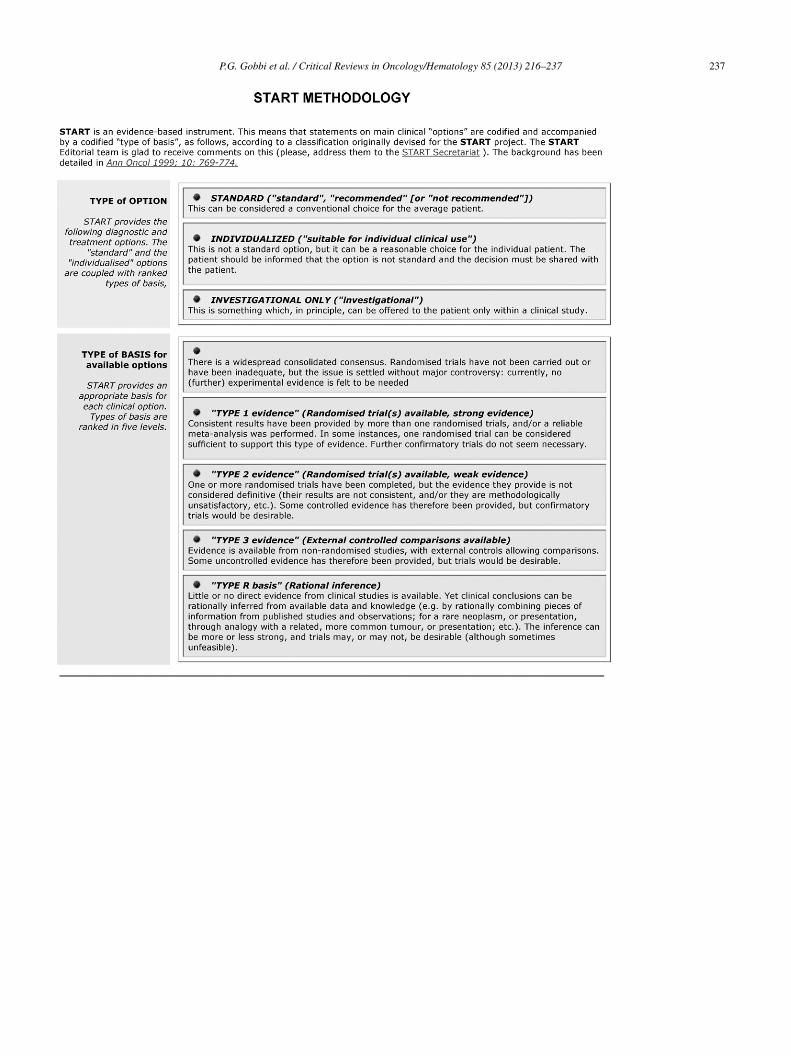
Oncology/Hematology 85 (2013) 216–237 237
P.G. Gobbi et al. / Critical Reviews in