histology of the endocrine system - universitas...
TRANSCRIPT

Jeanne A Pawitan 1
Histology of the endocrine system
• Jeanne Adiwinata Pawitan• Dept. of Histology FMUI

Jeanne A Pawitan 2
Endocrine system
• Regulate metabolic activities – coordinate – integrate° Organs° Tissues whole body - communication° Cells
∀ ≈ nervous system – electrochemical signal• Chemical messengers – hormones – blood
→ target cells
Jeanne A Pawitan 3
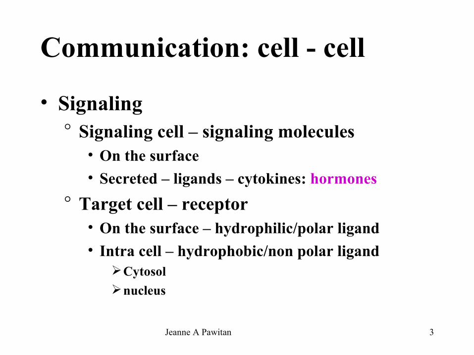
Communication: cell - cell
• Signaling° Signaling cell – signaling molecules
• On the surface• Secreted – ligands – cytokines: hormones
° Target cell – receptor• On the surface – hydrophilic/polar ligand• Intra cell – hydrophobic/non polar ligand
Cytosolnucleus
Jeanne A Pawitan 4
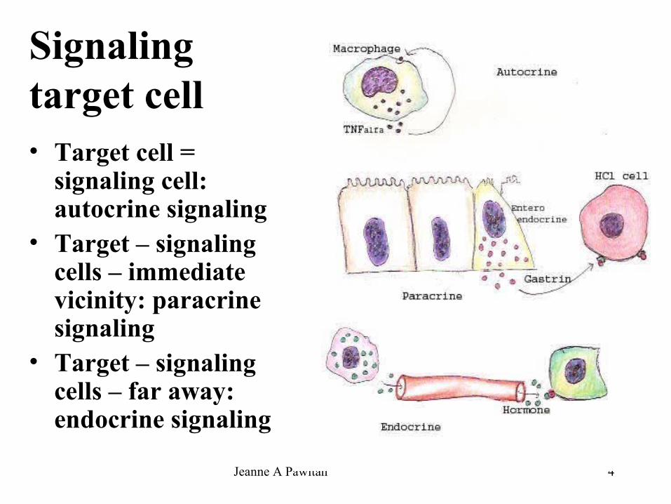
Signaling target cell• Target cell =
signaling cell: autocrine signaling
• Target – signaling cells – immediate vicinity: paracrine signaling
• Target – signaling cells – far away: endocrine signaling

Jeanne A Pawitan 5
Endocrine system
• Ductless gland – organ • Cluster of cells – certain organ/tissue –
pancreas, endothelium, adipose tissue, renin-angiotensin system, etc
• Endocrine cells – epithelium – certain organs° Digestive system° Respiratory system
Jeanne A Pawitan 6
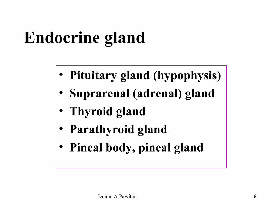
Endocrine gland
• Pituitary gland (hypophysis)• Suprarenal (adrenal) gland• Thyroid gland• Parathyroid gland• Pineal body, pineal gland

Jeanne A Pawitan 7
Hormones ~ composition• Proteins& polypeptides
° Mostly water soluble/hydrophilic° Insulin, glucagon, FSH
• Amino acid derivatives° Mostly water soluble/hydrophilic° Thyroxine, epinephrine
• Steroids& fatty acid derivatives° Mostly lipid soluble/hydrophobic ° Progesterone, estradiol, testosterone
Jeanne A Pawitan 8
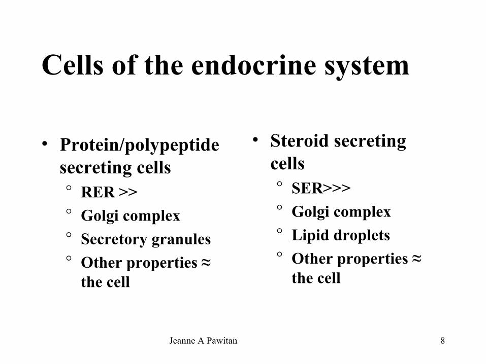
Cells of the endocrine system
• Protein/polypeptide secreting cells° RER >>° Golgi complex° Secretory granules° Other properties ≈
the cell
• Steroid secreting cells° SER>>>° Golgi complex° Lipid droplets° Other properties ≈
the cell

Jeanne A Pawitan 9
Pituitary gland (hypophysis)
• Hormones: growth, repro, metab• Location:
° Below – connected to hypothalamus – diencephalon° In hypophyseal fossa – in sella turcica – sphenoid
bone • Capsule
° Duramater – under° Diaphragma sellae – upper (incomplete)

Jeanne A Pawitan 10
Pituitary gland (hypophysis)• Adenohypophisis (anterior pituitary)
° Pars distalis, pars anterior, anterior lobe° Pars intermedia (between pars distalis –
nervosa)° Pars tuberalis (sleeves – around
infundibular stem/ stalk) – together → hypophyseal stalk/ infundibular stalk
• Neurohypophysis (posterior pituitary) – pars nervosa - hypothalamo-hypophyseal tract° Median eminence of tuber cinereum° Infundibulum –infundibular stem/stalk ° Pars nervosa (infundibular process,
posterior lobe)
Jeanne A Pawitan 11
Pituitary gland – embryonic origin
• Adenohypophysis – Rathke’s pouch = evagination – oral ectoderm = lining of stomodeum (primitive oral cavity)
• Neurohypophysis – neural ectoderm = down growth of diencephalon → connected to hypothalamus – brain° Neural pathways – hypothalamohypophyseal tract° Rich vascular supply from the brain
→ Encapsulated – single organ
Jeanne A Pawitan 12
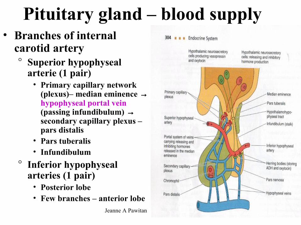
Pituitary gland – blood supply• Branches of internal
carotid artery° Superior hypophyseal
arterie (1 pair)• Primary capillary network
(plexus)– median eminence → hypophyseal portal vein (passing infundibulum) → secondary capillary plexus – pars distalis
• Pars tuberalis• Infundibulum
° Inferior hypophyseal arteries (1 pair)
• Posterior lobe• Few branches – anterior lobe

Jeanne A Pawitan 13
Pituitary gland –control of secretion
• Hypophyseal portal system – connect° Primary capillary plexus – fenestrated° Secondary capillary plexus – fenestrated
• Neurosecretory hormones: hypothalamus → median eminence (axons – hypothalamic neurons) → anterior lobe (pars distalis) – hormonal regulation by hypothalamus° Releasing hormones (factors)° Inhibiting hormones (factors)

Jeanne A Pawitan 14
Pituitary gland –control of secretion• TRH = thyroid stimulating hormone (TSH) –
releasing hormone• CRH = (Adreno)corticotropin – releasing
hormone• SRH = Somatotropin (growth hormone, GH)
– releasing hormone• GnRH = Gonadotropin – releasing hormone -
stimulate release of FSH, LH• PRH = Prolactin – releasing hormone• PIF = Prolactin inhibitory factor

Jeanne A Pawitan 15

Jeanne A Pawitan 16
Pars distalis, pars anterior, anterior lobe• Fibrous capsule• Composed of
° reticular fibers-surrounding° Cords of cells
• Folliculostellate cells - nonsecretory• Parenchymal cells
Chromophils – affinity for dyes° Acidophils (secretory granules – acid dyes)° Basophils (secretory granules – basic dyes)
Chromophobes – no affinity for dyes
° Large sinusoidal capillaries - fenestrated

Jeanne A Pawitan 17
Pars intermedia
• Colloid containing (Rathke’) cysts° Lined by cuboidal cells° Remnants of the ectoderm of Rathke’s pouch° Sometimes contains basophils
• In cords along the network of capillaries• Produce prohormone (proopiomelanocortin) –
posttranlational cleavage → α and β MSH (melanocyte stimulating hormone) – stimulate melanin production in lower animalHuman ?

Jeanne A Pawitan 18
Pars tuberalis• Frequently absent on the posterior aspect of
hypophyseal stalk• Separated from infundibulum by thin layers of
pia arachnoid-like connective tissue• Highly vascular – longitudinally orriented
° Arteries° Hypophyseal portal system
• Longitudinal cords of cells: cuboidal-low columnar – basophilic – small dense granules (FSH?, LH?), lipid droplets, glycogen, occasional colloid droplets

Jeanne A Pawitan 19
Hypothalamohyphophyseal tract
• Unmyelinated axons of neurosecretory cells ° Cell bodies in hypothalamus
• Supraoptic nuclei• Paraventricular nuclei
° Synthesize• hormones
Vasopressin (antidiuretic hormone,ADH)Oxytocin
• Neurophysin (carrier protein – binds the hormones)

Jeanne A Pawitan 20
Pars nervosa (posterior lobe)• Distal terminals of axons ← hypothalamo-
hypophyseal tract ° LM: chrome-alumhematoxylin –Herring bodies – blue
black – also along axon – contains neurosecretory granules → perivascular space
• Capillary plexus – fenestrated • Pituicytes–glial-like supporting cells
° Ensheating axons & their dilations° Cytoplasmic processes – gap junctions° Contains lipid droplets, some pigments & intermediate
filaments

Jeanne A Pawitan 21
Suprarenal (adrenal gland)• Location: superior poles of kidney –
adipose tissue – retroperitoneal• Capsule: connective tissue –many
adipose tissue → septa – blood vessel, nerves
• Parenchyma° Cortex – yellowish – 80-90% - mesoderm° Medulla – dark – ectodermal neural crest
cells

Jeanne A Pawitan 22
Suprarenal – blood supply• One of the richest blood supply• Source
° Inferior phrenic arteries – superior suprarenal arteries° Aorta – middle suprarenal arteries° Renal arteries – inferior suprarenal arteries
→ Penetrate capsule – subcapsular plexus → ° short cortical arteries → network-sinusoidal fenestrated
capillaries → venous plexus → small venules (medula) → suprarenal vein (hilus) →
• Inferior vena cava (right)• Left renal vein
° Long cortical arteries →medulla – capillary network

Jeanne A Pawitan 23
Cells -suprarenal gl• Cortex – steroid
secreting cells° Zona glomerulosa° Zona fasciculata
-spongiocytes° Zona reticularis
• Medulla – medullary vein° Chromaffin cells° Ganglion cells

Jeanne A Pawitan 24
Thyroid gland
• Location: anterior –neck, inferior to larynx, ant – junction thyroid –cricoid cartilage
• Right lobe-isthmus-left lobe – (pyramidal lobe)
• Capsule: slender, dense, irregular collagenous connective tissue → septa – blood, lymph vessels, nerves → lobules
• Follicles – thin basal lamina, reticular fibers, capillary plexus

Jeanne A Pawitan 25
Follicles – cyst-like
• Follicular cells (principal cells) ° May contact with
follicular cells of other follicles w/o basal lamina
• Parafollicular cells (clear cells, C cells)° At the periphery of
follicles° Individual/small clumps

Jeanne A Pawitan 26
Thyroid- follicular cell-function
Jeanne A Pawitan 27
Parathyroid gland• Location: posterior surface of thyroid
° Each pole – superior – inferior° Left – right lobe of thyroid° Can be else where along the pathway of descent
• Small, number 4 - /more (supernumerary)• Capsule: thin collagenous connective tissue→ septa
– blood vessels, lymphatic, nerves• Embryonic origin – pharyngeal pouches of
pharyngeal/branchial arch° 3rd pharyngeal pouches ≈ thymus → inferior° 4th pharyngeal pouches → superior
Jeanne A Pawitan 28
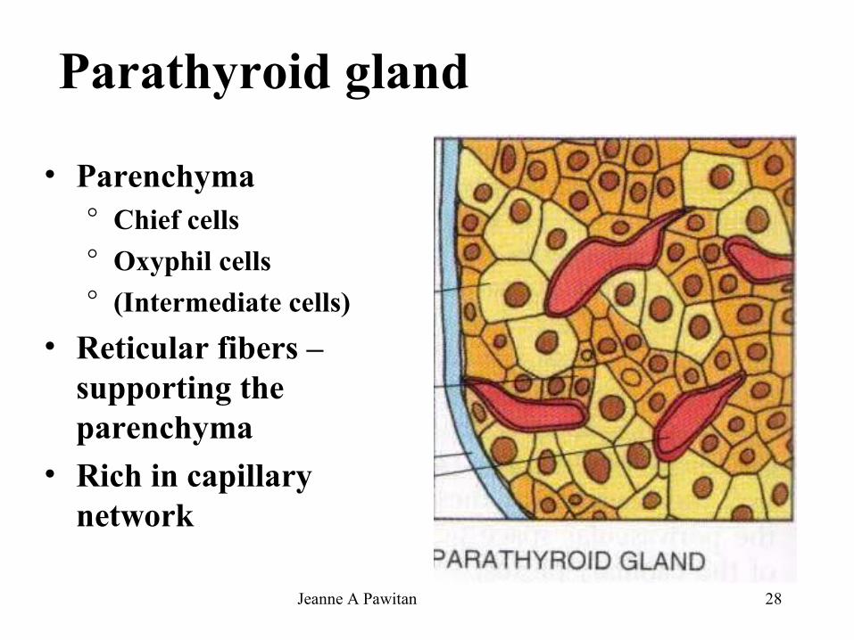
Parathyroid gland
• Parenchyma° Chief cells° Oxyphil cells° (Intermediate cells)
• Reticular fibers – supporting the parenchyma
• Rich in capillary network

Jeanne A Pawitan 29
Pineal gland, pineal body, epiphysis cerebri• Conical, grey body• Location: midline of brain
° From the roof of diencephalon° Shallow recess of the 3rd ventricle-extent → stalk
• Piamater → capsule → septa – blood vessels → incomplete lobules
• Parenchyma ° Pinealocytes° Interstitial cells° Corpora arenacea (brain sand) – concretions of Ca
P/carbonate –concentric rings –around organic matrix

Jeanne A Pawitan 30
The end Pinealocyte