highlights - karl storz se · tp 7 2.1 04/2018-us. 2. ... may 2014 flex-x. c. image1 s™ video...
TRANSCRIPT

mORe than a camera IMAGE1 S™ — An Adaptable Video Architecture For Multi-disciplinary Use
Highlights TP 7 2.1 04/2018-US

2
Highlights IMAGE1 S™ Adaptable Video Architecture | 2/2018
© K
AR
L S
TOR
Z 97
2010
35 T
P 7
2.1
04/
2018
/PH
L-U
S

3
mORe than a camera – the IMAGE1 S™ Adaptable Video ArchitectureAchieving optimal surgical results is the main goal of every operating surgeon. In order to meet this objective, the visualization and display of important and crucial structures during the surgical procedure is of paramount importance.
The rapid development of camera technology in recent years has resulted in a better view of the surgical field and a much wider treatment spectrum. This ultimately leads to better outcomes for patients.
New standards in resolution as well as technologies form the basis of this trend.
Highlights IMAGE1 S™ Adaptable Video Architecture | 2/2018©
KA
RL
STO
RZ
9720
1035
TP
7 2
.1 0
4/20
18/P
HL-
US
4
Highlights IMAGE1 S™ Adaptable Video Architecture | 2/2018
1
23
45
A
B C
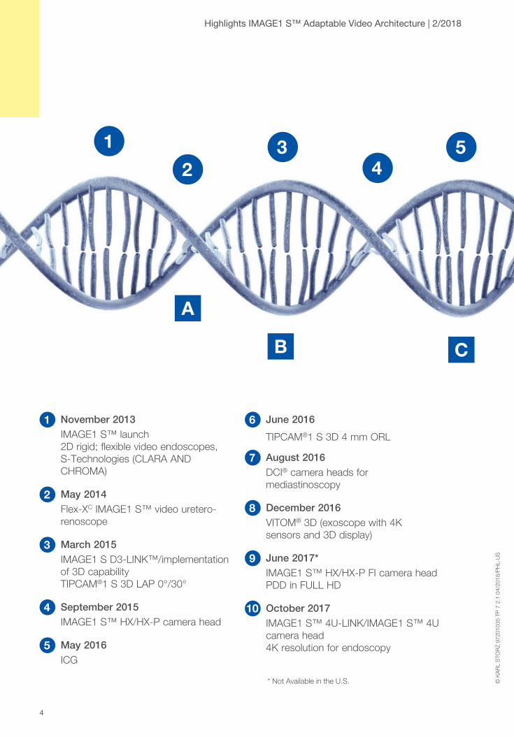
1 November 2013 IMAGE1 S™ launch
2D rigid; flexible video endoscopes, S-Technologies (CLARA AND CHROMA)
2 May 2014 Flex-XC IMAGE1 S™ video uretero-
renoscope
3 March 2015 IMAGE1 S D3-LINK™/implementation
of 3D capability TIPCAM®1 S 3D LAP 0°/30°
4 September 2015 IMAGE1 S™ HX/HX-P camera head
5 May 2016 ICG
6 June 2016
TIPCAM®1 S 3D 4 mm ORL
7 August 2016 DCI® camera heads for
mediastinoscopy
8 December 2016 VITOM® 3D (exoscope with 4K
sensors and 3D display)
9 June 2017* IMAGE1 S™ HX/HX-P FI camera head
PDD in FULL HD
10 October 2017 IMAGE1 S™ 4U-LINK/IMAGE1 S™ 4U
camera head 4K resolution for endoscopy
© K
AR
L S
TOR
Z 97
2010
35 T
P 7
2.1
04/
2018
/PH
L-U
S
* Not Available in the U.S.

5
A May 2014 + Expanded preset functionality
+ New languages + Enhanced user guidance
B March 2015 + Stand-by function for light sources
+ Temporary display of camera head/video endoscope data + Increased amount of patient data
C August 2015 + Image rotation in 3D
+ 2D output for 3D signals + SCB communication expanded
D June 2016 + S-Technologies in 3D
+ Zoom in 3D + SCB control for ENDOFLATOR® 40/50
E June 2017 + Patient data in live menu
+ Enhanced privacy settings
Continuous improvement of hardware
Continuous improvement of software
Highlights IMAGE1 S™ Adaptable Video Architecture | 2/2018
67
89
10
D
E
© K
AR
L S
TOR
Z 97
2010
35 T
P 7
2.1
04/
2018
/PH
L-U
S

6
IMAGE1 S™ – a system that meets all needsTo meet the greater demands placed on visualization in minimally invasive surgery and to keep step with the growing complexity of such procedures, KARL STORZ launched a unique, modular camera system on the market in 2013: IMAGE1 S™.
This system offers the user maximum flexibility to meet present and future needs.
Benefits of modularity:
1. Needs-based procurement – A specific technology that is required at a particular point in time can be selected from the large number of available technologies.
2. Synergistic effects – all modules have the same control module so that computing power does not need constant and unnecessary adjustment. This eliminates the need to procure peripherals such as monitors, documentation units etc. several times.
3. Gains in efficiency thanks to standardization – all OR rooms have the same technological basis, independent of the surgical discipline and specialty.
4. Future-readiness – whereas stand-alone systems are confined to the technology relevant at a specific point in time, the modularity concept ensures that the adaptable video architecture can be upgraded with new technologies at any time. This leaves room for divergent trends so that the user is never confronted with an either/or option.
Highlights IMAGE1 S™ Adaptable Video Architecture | 2/2018
© K
AR
L S
TOR
Z 97
2010
35 T
P 7
2.1
04/
2018
/PH
L-U
S

7
Highlights IMAGE1 S™ Adaptable Video Architecture | 2/2018
Open forfuture
technologies
2Dendoscopy
IMAGE1 S™H3-LINK
2D rigid / flexibleIMAGE1 S™
X-LINK
VITOM® 3D
4K camera head10 mm 3Dvideo endoscope
4 mm 3Dvideo endoscope
Flexible video endoscopes
1-chipcamera heads Microscopy
camera head
Near Infrared(NIR/ICG) 3-chipcamera head FI
3-chip camera heads
3DendoscopyIMAGE1 SD3-LINK™
Combines alltechnologies
IMAGE1 SCONNECT™
4K endoscopy IMAGE1 S™
4U-LINK
© K
AR
L S
TOR
Z 97
2010
35 T
P 7
2.1
04/
2018
/PH
L-U
S

8
4K ResolutionResolution is one of the key factors enabling the identification of fine details in endoscopic images. In this context, the terms 4K or UHD are now commonly used in the consumer sector as well as the medical field.
This development offers 4 times the resolution of existing FULL HD standard systems.
With IMAGE1 S™ 4U-LINK and the IMAGE1 S™ 4U camera head, the IMAGE1 S™ Adaptable Video Architecture integrates two components that benefit from this system.
The adaptable video architecture eliminates the need to choose between individual technologies and helps select the most appropriate technological solution for a specific procedure.
Highlights IMAGE1 S™ Adaptable Video Architecture | 2/2018
© K
AR
L S
TOR
Z 97
2010
35 T
P 7
2.1
04/
2018
/PH
L-U
S

9
3D VisualizationOne of the greatest challenges in MIS is undoubtedly the difficulty of performing complex maneuvering in three-dimensional space (e.g., suturing, dissecting) as the endoscopic image often lacks the third dimension – i.e. depth.
Studies show that 3D vision increases accuracy and reduces operating times1,2,3. These effects could be demonstrated for both novices and experienced surgeons by up to 15% for mini-gastric bypass surgery3.
The IMAGE1 S D3-LINK™ enables 3D endoscopes with diameters of 10 mm and 4 mm to be easily integrated into the IMAGE1 S™ Adaptable Video Architecture and thus utilize the benefits offered by 3D visualization4.
Furthermore, VITOM® 3D offers the possibility of three-dimensional visualization in minimally invasive open surgical procedures. In contrast to an operating microscope, the VITOM® 3D features a high depth of focus, small dimensions and an ergonomic working position. Moreover, the system offers enormous flexibility and allows more observers to view the OR field. Ultra-high resolution sensors also ensure a loss-free zoom.
Highlights IMAGE1 S™ Adaptable Video Architecture | 2/2018©
KA
RL
STO
RZ
9720
1035
TP
7 2
.1 0
4/20
18/P
HL-
US

10
Highlights IMAGE1 S™ Adaptable Video Architecture | 2/2018
OPAL1® NIR/ICGRelevant information and structures are often concealed deep under a layer of tissue or are invisible under white light (e.g., perfusion). Even the highest possible resolution is not able to display these effects.
With the help of illumination with near infrared light, the Near Infrared (NIR/ICG) system from KARL STORZ allows visualization under the surface of the tissue in laparoscopy and open surgery. The use of indocyanine green (ICG) enables the visualization of anatomical structures up to one centimeter deep. Important information such as, for example, perfusion, or the bile duct anatomy can thus be displayed much more quickly; in many cases, fluorescence imaging may be the only possibility to display information and is thus suitable for multidisciplinary use.
© K
AR
L S
TOR
Z 97
2010
35 T
P 7
2.1
04/
2018
/PH
L-U
S

11
Visualization of the biliary anatomy during laparoscopic cholecystectomy. (Prof. L. Boni, Milano, Italy)
NIR/ICG fluorescence imaging might also present a clear advantage for perfusion assessment, e.g., in the case of anastomoses.
Perfusion control of the bowel for identification of the resection zone (Dr. Skrovina, Nový Jičín, Czech Republic)
NIR/ICG fluorescence imaging has the potential to drastically lower the rate of anastomotic leakage and thus prevent anastomotic insufficiency 5, 6, 7, 8. By reducing associated morbidity and mortality and subsequent cost savings, NIR/ICG fluorescence imaging could offer enormous benefits for the patient and the healthcare system.
Highlights IMAGE1 S™ Adaptable Video Architecture | 2/2018©
KA
RL
STO
RZ
9720
1035
TP
7 2
.1 0
4/20
18/P
HL-
US

12
PiP – Multiple Views, One TowerIn modern surgery, the surgical methods used are becoming increasingly complex. These PiP support surgeons in their work. The simultaneous use of rigid and flexible endoscopes offers great benefits, particularly for operations such as choledocholithiasis as well as gastric, bariatric and colorectal surgery. The acquisition of additional information of the organ to be treated by means of laparoscopic and endoscopic visualization, for example, offers the possibility of pre- and postoperative diagnosis or improved intraoperative management of potential complications (e.g., defining resection boundaries, checking for leakages, correct visualization and identification of the OR site). Conventional systems have major problems in terms of ergonomics, costs and documentation. Consequently, these types of interventions often require the use of two towers. This not only restricts the freedom of movement of all participants in the already confined OR environment, it also contributes to higher costs due to the need to procure both towers. Documentation is also critical as a timely overlap of endoscopic and laparoscopic images with two separate video sources may be important but is difficult to achieve. The modularity of IMAGE1 S™ drastically reduces or eliminates these problems completely.
Highlights IMAGE1 S™ Adaptable Video Architecture | 2/2018
© K
AR
L S
TOR
Z 97
2010
35 T
P 7
2.1
04/
2018
/PH
L-U
S

13
Highlights IMAGE1 S™ Adaptable Video Architecture | 2/2018
S-TechnologiesIn addition to the quantity (amount) of pixels, the quality of the individual pixels is important for the quality of the entire endoscopic image. In this context, KARL STORZ attaches great importance to the correct color display and the best possible sensors. Furthermore, S-Technologies help to overcome the current limitations of endoscopic images.
Structures that are located farther away often appear much darker because light intensity is inversely proportional to the square of the distance from the source. The entire image information for this area is missing. Even increasing the light intensity does not help in this case as this is limited, on the one hand, by the size of the telescope and, on the other hand, it causes overexposed areas in the image. Based on a sophisticated software algorithm, CLARA provides a perfectly illuminated image as it dynamically brightens up dark areas in the background.
Standard image CLARA
Contrast is also an extremely important factor in endoscopic imaging. To offer the user optimal visualization, electronic contrast enhancement has been a standard feature in all standard camera systems for decades. This contrast enhancement, however, is very non-specific and results in enhanced contrast for structures that are already easily identifiable by shifting brightness values. CHROMA, therefore, is focused on enhancing contrast in areas that are difficult to see and therefore ensures better visibility.
Standard image CHROMA
© K
AR
L S
TOR
Z 97
2010
35 T
P 7
2.1
04/
2018
/PH
L-U
S

14
Highlights IMAGE1 S™ Adaptable Video Architecture | 2/2018
As the wish for greater contrast and a more homogenous illumination often coincide, CLARA + CHROMA offers a combination of both these technologies.
Standard image CLARA + CHROMA
© K
AR
L S
TOR
Z 97
2010
35 T
P 7
2.1
04/
2018
/PH
L-U
S

15
Highlights IMAGE1 S™ Adaptable Video Architecture | 2/2018
Future ReadinessThe modular design of the IMAGE1 S™ Adaptable Video Architecture offers the customer the possibility of a needs-based procurement. The customer is able to assemble individual components according to the building block principle. If requirements change, further technologies can be modularly integrated with little additional costs and minimal effort. This offers the best possible protection for the original investment. New technologies can be integrated according to the same principle and with the same benefits.
© K
AR
L S
TOR
Z 97
2010
35 T
P 7
2.1
04/
2018
/PH
L-U
S
Competitors IMAGE1 S™
Cost c
urve
Total costs k
Investment period t
Firstexchange
Procurement
2 k
3 k
Secondexchange
Thirdexchange
Costs for individual module a
Total costs k
Cost curve
Investment period t
Procurement
2 k
3 k
Firstexchange
Secondexchange
Thirdexchange

16
PeripheralsA famous proverb says that a chain is only as strong as its weakest link. In imaging, many components are responsible for a good image. In addition to the more obvious components that influence the imaging chain (telescope, camera head, image processor, monitor), peripheral units also make a significant contribution to the viewing and user experience. In order to achieve the goal of the best possible visualization, technologies and solutions must also be considered here.
Peripherals from KARL STORZ can communicate with the IMAGE1 S™ adaptable video architecture which also enables automated control of these units.
Highlights IMAGE1 S™ Adaptable Video Architecture | 2/2018
© K
AR
L S
TOR
Z 97
2010
35 T
P 7
2.1
04/
2018
/PH
L-U
S
17
Highlights IMAGE1 S™ Adaptable Video Architecture | 2/2018
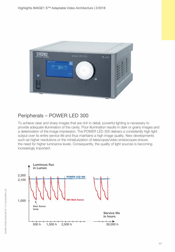
Peripherals – POWER LED 300To achieve clear and sharp images that are rich in detail, powerful lighting is necessary to provide adequate illumination of the cavity. Poor illumination results in dark or grainy images and a deterioration of the image impression. The POWER LED 300 delivers a consistently high light output over its entire service life and thus maintains a high image quality. New developments such as higher resolutions or the miniaturization of telescopes/video endoscopes ensure the need for higher luminance levels. Consequently, the quality of light sources is becoming increasingly important.
. . .
Luminous flux in Lumen
New Xenon lamp
POWER LED 300
300 Watt Xenon
30,000 h 1,500 h 2,500 h
2,1002,300
500 h
1,000
Service life in hours
© K
AR
L S
TOR
Z 97
2010
35 T
P 7
2.1
04/
2018
/PH
L-U
S

18
Peripherals – S-Pilot®
An ideal imaging chain and sufficient light ensure the best possible display of in-situ conditions. What happens if the in-situ conditions themselves are poor? The use of HF appliances in particular often generate a lot of smoke in the body and thus greatly obscures the image impression. To counteract this, S-PILOT® from KARL STORZ offers an active smoke evacuation system which quickly and efficiently removes suspended particles from the site and thus ensures clear vision. In addition to improving image quality, this system also reduces unpleasant odors in the OR.
Highlights IMAGE1 S™ Adaptable Video Architecture | 2/2018
© K
AR
L S
TOR
Z 97
2010
35 T
P 7
2.1
04/
2018
/PH
L-U
S
19
Highlights IMAGE1 S™ Adaptable Video Architecture | 2/2018
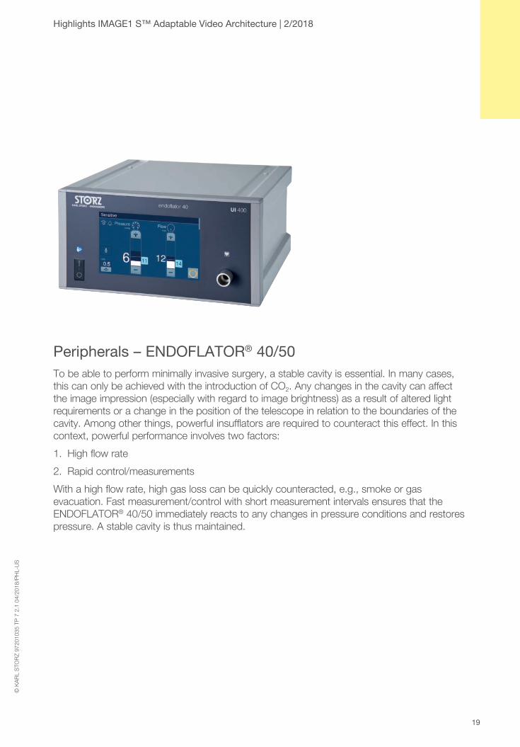
Peripherals – ENDOFLATOR® 40/50To be able to perform minimally invasive surgery, a stable cavity is essential. In many cases, this can only be achieved with the introduction of CO2. Any changes in the cavity can affect the image impression (especially with regard to image brightness) as a result of altered light requirements or a change in the position of the telescope in relation to the boundaries of the cavity. Among other things, powerful insufflators are required to counteract this effect. In this context, powerful performance involves two factors:
1. High flow rate
2. Rapid control/measurements
With a high flow rate, high gas loss can be quickly counteracted, e.g., smoke or gas evacuation. Fast measurement/control with short measurement intervals ensures that the ENDOFLATOR® 40/50 immediately reacts to any changes in pressure conditions and restores pressure. A stable cavity is thus maintained.
© K
AR
L S
TOR
Z 97
2010
35 T
P 7
2.1
04/
2018
/PH
L-U
S
20
1 Tanagho, Y.S. et al., Journal of Laparoendoscopic & Advanced Surgical Techniques. November 2012, 22(9): 865-870. doi:10.1089/lap.2012.0220. “2D Versus 3D Visualization: Impact on Laparoscopic Proficiency Using the Fundamentals of Laparoscopic Surgery Skill Set”
2 Alaraimi, B., et al. World J Surg (2014) 38: 2746. doi:10.1007/s00268-014-2674-0 “A Randomized Prospective Study Comparing Acquisition of Laparoscopic Skills in Three-Dimensional (3D) vs. Two-Dimensional (2D) Laparoscopy”
3 Feng, X. et al., Surgical Endoscopy. May 2015, Volume 29, Issue 5, pp 1231-1239 “3-Dimensional (3D) laparoscopy improves operating time in small spaces without impact on hemodynamics and psychomental stress parameters of the surgeon”
4 A compatible 3D monitor is required to facilitate display.
5 Koh, F.H. & Tan, KK. Ann Surg Oncol (2016) 23(Suppl 5): 692. doi:10.1245/s10434-016-5581-9 “Fluorescent Angiography Used to Evaluate the Perfusion Status of Anastomosis in Laparoscopic Anterior Resection”
6 Boni, L., David, G., Mangano, A. et al. Surg Endosc (2015) 29: 2046. doi:10.1007/s00464-014-3895-x. “Clinical applications of indocyanine green (ICG) enhanced fluorescence in laparoscopic surgery”
7 Carus T, Lienhard H (2009), Meeting Abstracts, 126. Kongress der Deutschen Gesellschaft für Chirurgie, 28.04.–01.05.2009. „Die laparoskopische Fluoreszenzangiografie mit Indocaningrün zur intraoperativen Beurteilung der Perfusion bei kolorektarlen Anastomosen.“
8 Karliczek, A., Harlaar, N.J., Zeebregts, C.J. et al. Int J Colorectal Dis (2009) 24: 569. doi:10.1007/s00384-009-0658-6. “Surgeons lack predictive accuracy for anastomotic leakage in gastrointestinal surgery”
Highlights IMAGE1 S™ Adaptable Video Architecture | 2/2018
© K
AR
L S
TOR
Z 97
2010
35 T
P 7
2.1
04/
2018
/PH
L-U
S

21
Highlights IMAGE1 S™ Adaptable Video Architecture | 2/2018©
KA
RL
STO
RZ
9720
1035
TP
7 2
.1 0
4/20
18/P
HL-
US

22
Highlights IMAGE1 S™ Adaptable Video Architecture | 2/2018
© K
AR
L S
TOR
Z 97
2010
35 T
P 7
2.1
04/
2018
/PH
L-U
S

23
It is recommended to check the suitability of the product for the intended procedure prior to use.
Highlights IMAGE1 S™ Adaptable Video Architecture | 2/2018©
KA
RL
STO
RZ
9720
1035
TP
7 2
.1 0
4/20
18/P
HL-
US

KARL STORZ SE & Co. KG Dr.-Karl-Storz-Straße 34, 78532 Tuttlingen/Germany Postbox 230, 78503 Tuttlingen/Germany Phone: +49 (0)7461 708-0 Fax: +49 (0)7461 708-105 E-Mail: [email protected]
www.karlstorz.com 9720
1035
TP
7 2
.1 0
4/20
18/P
HL-
US
KARL STORZ Endoscopy-America, Inc. 2151 East Grand Avenue El Segundo, CA 90245-5017, USA Phone: +1 424 218-8100 Phone toll free: 800 421-0837 (US only) Fax: +1 424 218-8525 Fax toll free: 800 321-1304 (US only) E-Mail: [email protected]