hidden biochemical action of ethanol on colon carcinoma
TRANSCRIPT

PhD Course in Biomedicine and Neuroscience
Department of Experimental Biomedicine and Clinical neurosciences
(BioNeC)
SSD BIO/10
Hidden biochemical action of Ethanol on colon
carcinoma cell models
IL CANDIDATO LA COORDINATRICE
Dott. Cesare Cernigliaro Prof.ssa Felicia Farina
TUTOR
Prof.ssa Marianna Lauricella
Co-tutor
Prof.ssa Antonella D’Anneo
XXXI CYCLE
ACHIEVEMENT YEAR TITLE 2018/2019
2
Hidden biochemical action of Ethanol on colon carcinoma cell models
by
Dott. Cesare Cernigliaro
Dissertation
Presented for the requirements
toward the completion
for the Degree of
Doctor of Philosophy

3
Sommario Abstract ..................................................................................................... 5
Introduction .................................................................................................. 6
Ethanol ...................................................................................................... 7
Ethanol metabolism .................................................................................. 9
Colorectal cancer .................................................................................... 13
Genetic factors and forms of hereditary CRC ........................................................................ 18
Nutrition and onset of CRC ................................................................................................... 19
Ethanol and Colorectal Cancer.............................................................................................. 21
Endoplasmic Reticulum Stress .............................................................. 25
Authophagy............................................................................................. 28
Project aim .................................................................................................. 30
Materials and Methods .............................................................................. 32
Cell culture condition ............................................................................. 33
MTT assay .............................................................................................. 33
Clonogenic assay ..................................................................................... 33
Measurement of ROS production .......................................................... 34
Western blotting analysis ....................................................................... 34
Cytosol and nuclear extraction .............................................................. 35
Monodansylcadaverine test .................................................................... 35
Transient down-regulation of Nrf2 by short interfering RNA (siRNA)
................................................................................................................. 36
Gelatin zymography ............................................................................... 36
Immunofluorescence .............................................................................. 37
Statistical analysis .................................................................................. 37
Results ......................................................................................................... 38
Effects of EtOH in colon cancer cell viability ....................................... 39
EtOH treatment induced oxidative stress in HCT116 colon cancer cells
................................................................................................................. 41
Treatment with EtOH induces ER stress in colon cancer cells ............ 43
EtOH stimulate autophagic process in colon cancer cells .................... 45
EtOH increases antioxidant enzymes levels .......................................... 47

4
EtOH promotes HO-1 nuclear translocation ........................................ 51
EtOH increased MMPs and VEGF in colon cancer cells ..................... 53
Role of Nrf2/HO-1 axis in colon cancer survival and invasiveness ...... 55
Discussion ................................................................................................... 58
Bibliography ............................................................................................... 63
5
Abstract
Colorectal cancer (CRC) is one of the most widespread cancers in the world (Haggar FA et
al 2009). Numerous risk factors have been correlated with the development of CRC,
including genetic factors, inflammation, intestinal microflora composition, as well as
lifestyle factors, such as smoking, high consumption of red meats and alcohol intake (Baan
R et al 2007).
Epidemiological studies support the conclusion that chronic and heavy alcohol
consumption increases the risk to develop CRC as well as favors the progression of this
form of cancer. However, the biochemical mechanisms responsible for these events have
not yet been fully clarified.
The aim of my doctoral project was to study the effects of ethanol in human colorectal
carcinoma cells in culture and to evaluate its molecular action mechanism. In particular,
my research focused on the identification of one or more molecules involved both in the
survival of the tumor cells and, especially, in tumor progression and invasiveness. To this
end, I investigated the effect of high doses of ethanol on survival and progression of three
different colon cancer cells (HCT116 , HT29 and Caco2 cells). The results demonstrated
that ethanol promotes oxidative and ER stress in colon cancer cells as demonstrated by
ROS increase and upregulation of ER markers Grp78 and CHOP. Despite the activation of
stress, colon cancer cells did not present sign of toxicity because they are able to activate
an autophagic survival mechanism. Moreover, in response to oxidative stress, ethanol
promoted nuclear translocation of Nrf2 and upregulated the level of the antioxidant
enzymes SOD, catalase and heme-oxygenase (HO-1). Silencing Nrf2 in HCT116 cells
abrogated the effect of ethanol on upregulation of SOD and HO-1, thereby suggesting that
the induction of antioxidant enzymes is dependent on Nrf2 activation. Interestingly,
ethanol also promoted HO-1 nuclear translocation. Preventing HO-1 nuclear translocation
by addition of E64d, the activation of antioxidant response by Nrf2 was reduced. Finally,
the results demonstrated that the activation of Nrf2/HO-1 axis induced by ethanol is also
responsible for the induction of MMP-2 and VEGF, two well known factors favoring
cellular invasiveness.

6
Introduction
7
Ethanol
Ethanol (CH3CH2OH, EtOH) commonly called alcohol or ethyl alcohol is a liquid,
colorless substance that is formed by fermentation of some simple sugars or by distillation
of fermented must. The etymology of the word is debated, some argue that it derives from
the Arabic term "alkukhul", which literally translated means "the spirit", others as "spirit"
in reference to the first distillation processes carried out in the Middle East in the 2nd
millennium Before Christ. EtOH present in drinks is obtained by alcoholic fermentation of
plant products such as grapes, cereals and potatoes; in particular, EtOH is the product of
various microorganisms starting from the degradation of sugars with six carbon atoms such
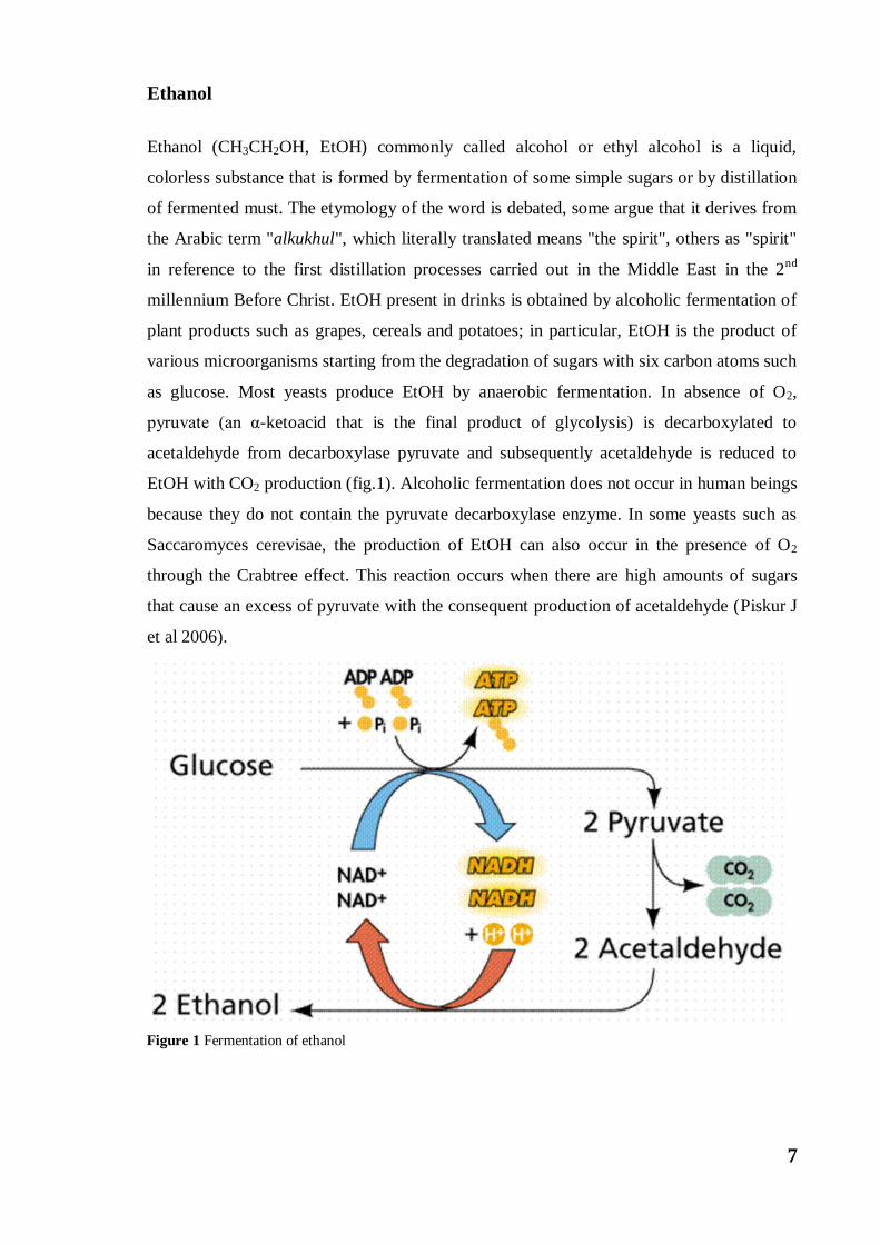
as glucose. Most yeasts produce EtOH by anaerobic fermentation. In absence of O2,
pyruvate (an α-ketoacid that is the final product of glycolysis) is decarboxylated to
acetaldehyde from decarboxylase pyruvate and subsequently acetaldehyde is reduced to
EtOH with CO2 production (fig.1). Alcoholic fermentation does not occur in human beings
because they do not contain the pyruvate decarboxylase enzyme. In some yeasts such as
Saccaromyces cerevisae, the production of EtOH can also occur in the presence of O2
through the Crabtree effect. This reaction occurs when there are high amounts of sugars
that cause an excess of pyruvate with the consequent production of acetaldehyde (Piskur J
et al 2006).
Figure 1 Fermentation of ethanol
8
EtOH is the main psychoactive component of alcoholic beverages. It is in fact able to act
on the central nervous system and to alter states of consciousness.
In the course of human history, EtOH has been considered food, medical remedy, object of
initiation rites, symbol of transgression and finally intoxicating drink. The consumption of
EtOH took on different aspects in the history of humankind, the ancient Greeks worshiped
the God Dionysus, the Americans forbade it in the early 1900s. While today in Western
countries it is easily consumed even by very young people. Before the development of
modern drugs, EtOH was used as a drug, suggested as a medicine for depression and
anesthetic. It is also used as an antibacterial hand sanitizer (at a volume / weight
concentration of about 70%), and as an antiseptic.
EtOH denatures proteins and dissolves lipids, so it is effective against most bacteria and
fungi and many viruses, but it is ineffective against bacterial spores. The variety of these
meanings probably depends on his own effects: alcohol can simultaneously make people
feel strong and weak.
There are different drinks containing EtOH, some with low alcohol content, such as wine
and beer, and others with a high alcohol content, such as hard liquors. The quantity of
EtOH contained in the drinks is expressed in % vol. To calculate the alcohol content of a
beverage, the total volume of the consumed drink must be multiplied with the ethanol
conversion factor. The ethanol conversion factor differs slightly in the various countries
but it usually falls within the range of 4-5%vol for beer, 12%vol for wine and about
40%vol for hard liquors. Therefore, in a glass of wine we will find about 12mL of EtOH
(100mL x 0.12 = 12mL).
The World Health Organization (WHO) estimated that around 2 billion people consume
EtOH every day with an average annual consumption of about 6.2 l for adult (Rehm J et al
2009). 73.6 million people have EtOH-related illnesses such as: alcoholic polyneuropathy,
alcoholic cardiomyopathy, alcoholic gastritis, depression and other mental disorders; and
still with hemorrhagic stroke, acute and chronic pancreatitis and liver cirrhosis (Thakker
KD 1998). Furthermore, consumption of EtOH during pregnancy is associated with
abortion, intrauterine growth retardation and fetal alcohol syndrome (Connor PD et al
1996). It has also been shown that EtOH consumption is correlated with more than 60
diseases in young adults, 1.8 million deaths a year, and only in Europe, a death toll is
estimated at 55,000 cases per year (WHO, Department of Mental Health and Substance
Abuse. Global status report on alcohol 2004).
Furthermore, it has been shown that there is a high correlation between EtOH consumption
and the development of tumors. The incidence of EtOH-related cancers is higher in men

9
than in women, accounting for 5.2% in men and 1.7% in women respectively. In particular,
ingestion of alcoholic drinks is involved in the development of cancer of the larynx,
pharynx, esophagus, liver, breast cancer and colon (Baan R et al 2007). Recent studies
identify a greater incidence of developing proximal colon cancer than distal colon,
following consumption of EtOH (Thygesen LC et al 2008). Although several
epidemiological studies show that regular consumption of EtOH is correlated with the
development of different types of cancers, such as colorectal cancer, (in particular 45% for
colon cancer and 49% for rectal cancer) it is still unclear and strongly debated the amount
of EtOH associated with the development of colon cancer. There are different results based
on the reference population. In a study conducted in North America and in Europe, there is
a risk of developing colon cancer with average consumption of> 45g / day (Cho E et al
2004). Instead, a study conducted in Japan found a correlation consumption of EtOH and
colon cancer with average daily consumption of about 23g / day (Munira A et al 2007).
Other studies show that the carcinogenic effects associated with the consumption of EtOH
seem to be attributable to an average daily consumption of more than 76g / day, especially
if EtOH is consumed in the form of wine rich in polyphenols (Corrao et al 2000).
To date, the threshold value to be attributed to a greater probability of developing colon
cancer remains unresolved. Evidence suggests that the
risk to develop colon cancer correlated with EtOH consumption is modulated by genetic
factor like different variants in genes for EtOH metabolism.
Ethanol metabolism
EtOH is a water-soluble and fat-soluble molecule; this dual nature allows the EtOH to be
rapidly absorbed by the intestine by passive diffusion. Only a small part of ingested EtOH
is absorbed by the cells of the gastric mucosa, most of the EtOH is released into the
bloodstream to reach the liver (organ involved in the metabolism of EtOH). A small
amount of ingested EtOH (2%) is eliminated through the lungs and kidneys.
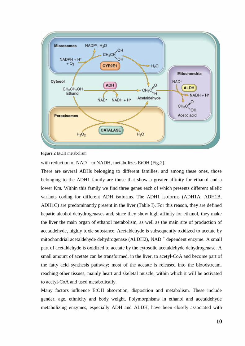
EtOH is considered a pseudo food because through the action of two enzymes it is first
converted into acetic acid and then into acetyl-CoA, an important metabolite. In the liver, a
cytosolic enzyme, Alcohol Dehydrogenase (ADH), which oxidizes EtOH to acetaldehyde
10
with reduction of NAD + to NADH, metabolizes EtOH (Fig.2).
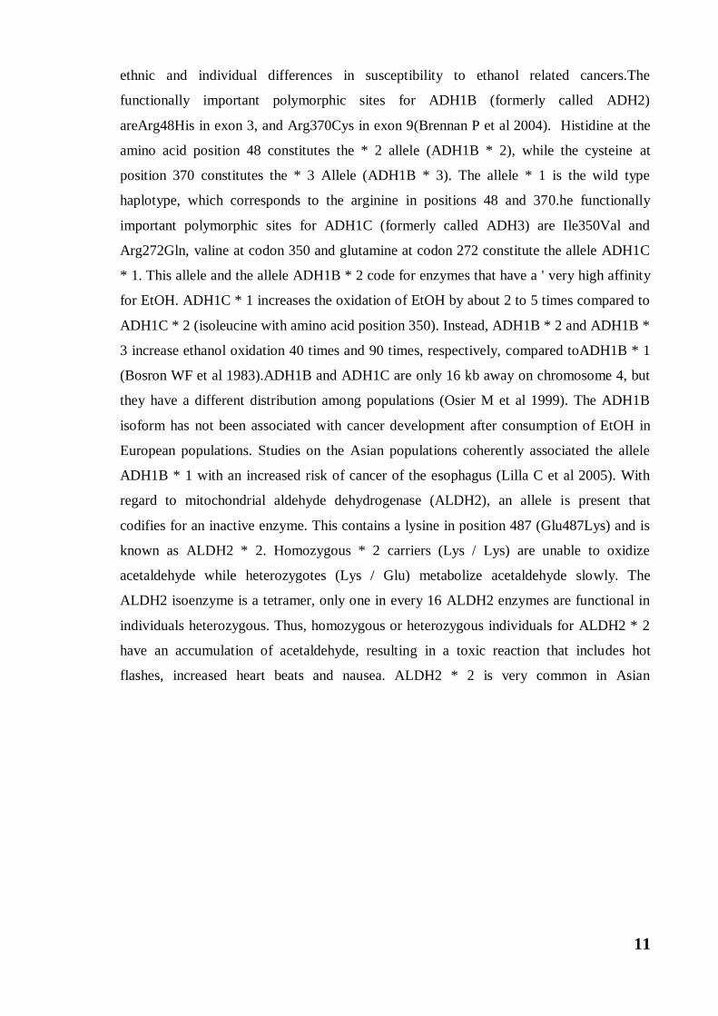
There are several ADHs belonging to different families, and among these ones, those
belonging to the ADH1 family are those that show a greater affinity for ethanol and a
lower Km. Within this family we find three genes each of which presents different allelic
variants coding for different ADH isoforms. The ADH1 isoforms (ADH1A, ADH1B,
ADH1C) are predominantly present in the liver (Table I). For this reason, they are defined
hepatic alcohol dehydrogenases and, since they show high affinity for ethanol, they make
the liver the main organ of ethanol metabolism, as well as the main site of production of
acetaldehyde, highly toxic substance. Acetaldehyde is subsequently oxidized to acetate by
mitochondrial acetaldehyde dehydrogenase (ALDH2), NAD + dependent enzyme. A small
part of acetaldehyde is oxidized to acetate by the cytosolic acetaldehyde dehydrogenase. A
small amount of acetate can be transformed, in the liver, to acetyl-CoA and become part of
the fatty acid synthesis pathway; most of the acetate is released into the bloodstream,
reaching other tissues, mainly heart and skeletal muscle, within which it will be activated
to acetyl-CoA and used metabolically.
Many factors influence EtOH absorption, disposition and metabolism. These include
gender, age, ethnicity and body weight. Polymorphisms in ethanol and acetaldehyde
metabolizing enzymes, especially ADH and ALDH, have been closely associated with
Figure 2 EtOH metabolism
11
ethnic and individual differences in susceptibility to ethanol related cancers.The
functionally important polymorphic sites for ADH1B (formerly called ADH2)
areArg48His in exon 3, and Arg370Cys in exon 9(Brennan P et al 2004). Histidine at the
amino acid position 48 constitutes the * 2 allele (ADH1B * 2), while the cysteine at
position 370 constitutes the * 3 Allele (ADH1B * 3). The allele * 1 is the wild type
haplotype, which corresponds to the arginine in positions 48 and 370.he functionally
important polymorphic sites for ADH1C (formerly called ADH3) are Ile350Val and
Arg272Gln, valine at codon 350 and glutamine at codon 272 constitute the allele ADH1C
* 1. This allele and the allele ADH1B * 2 code for enzymes that have a ' very high affinity
for EtOH. ADH1C * 1 increases the oxidation of EtOH by about 2 to 5 times compared to
ADH1C * 2 (isoleucine with amino acid position 350). Instead, ADH1B * 2 and ADH1B *
3 increase ethanol oxidation 40 times and 90 times, respectively, compared toADH1B * 1
(Bosron WF et al 1983).ADH1B and ADH1C are only 16 kb away on chromosome 4, but
they have a different distribution among populations (Osier M et al 1999). The ADH1B
isoform has not been associated with cancer development after consumption of EtOH in
European populations. Studies on the Asian populations coherently associated the allele
ADH1B * 1 with an increased risk of cancer of the esophagus (Lilla C et al 2005). With
regard to mitochondrial aldehyde dehydrogenase (ALDH2), an allele is present that
codifies for an inactive enzyme. This contains a lysine in position 487 (Glu487Lys) and is
known as ALDH2 * 2. Homozygous * 2 carriers (Lys / Lys) are unable to oxidize
acetaldehyde while heterozygotes (Lys / Glu) metabolize acetaldehyde slowly. The
ALDH2 isoenzyme is a tetramer, only one in every 16 ALDH2 enzymes are functional in
individuals heterozygous. Thus, homozygous or heterozygous individuals for ALDH2 * 2
have an accumulation of acetaldehyde, resulting in a toxic reaction that includes hot
flashes, increased heart beats and nausea. ALDH2 * 2 is very common in Asian
12
populations, while almost all Europeans are homozygous for the ALDH2 * 1 (Gln / Gln)
allele. Studies conducted in Japan have consistently reported an increased risk of oral,
pharyngeal, laryngeal and esophageal cancer related to the ALDH2 * 2 allele (Yokoyama
A et al 2003).
At hepatic level, EtOH can be oxidized to acetaldehyde also by the "microsomal ethanol
oxidation system” (MEOS). The enzymes belonging to this complex are isoenzymes of the
cytochrome P450 family, in particular CYP2E1. Like all cytochromes P450, CYP2E1 are
monooxygenases that use NADPH as an electron donor and O2 as an electron acceptor.
The CYP2E1, compared to the ADH, has a low affinity for EtOH and a much higher Km.
This means that the system intervenes only when the EtOH is present in the body in large
quantities, then after high assumptions. Moreover, one of the most important
characteristics of these enzymes is the inducibility, which is the ability to be induced by
their own substrate, hence from EtOH. In fact, EtOH is able to bind a cytosolic receptor,
induce its migration into the nucleus, where it promotes transcription of the gene coding
for CYP2E1. In fact, the chronic consumption of EtOH does increase the hepatic levels of
CYP2E1 by about 5-10 times. This causes an increase in endoplasmic reticulum size with a
consequent increase in microsomal enzymes, including those not involved in the EtOH
metabolism. The purpose of CYP2E1 induction is to stabilize and protect the same protein
from degradation, as well as to increase the clearance of blood EtOH. There are several
polymorphisms for CYP2E1 such as: RsaI, DraI and TaqI. For RsaI there are 2 different
alleles with different enzymatic activity, the allele c2 has a lower enzymatic activity than
the allele c1. The functionality of the enzymes encoded by DraI and TaqI is not very clear.
A meta-analysis of 5 studies on the various polymorphisms of CYP2E1 found no
correlation between EtOH consumption and colon cancer development (Yang C et al
2005).
Table I: Mammalian Alcohol Dehydrogenases (ADH) and aldehyde dehydrogenases (ALDH) involved in
alcohol metabolism (Bbosa G et al 2014).
13
Colorectal cancer
Colorectal cancer (CRC) is one of the most widespread cancers in the world. Despite
progress in the surgical and therapeutic field, CRC still occupies the third place for cancer
mortality, after that of breast and prostate cancer. Although it may occur at any age, it
affects predominantly over six-year-old subjects of both sexes. In Italy, there are about 20
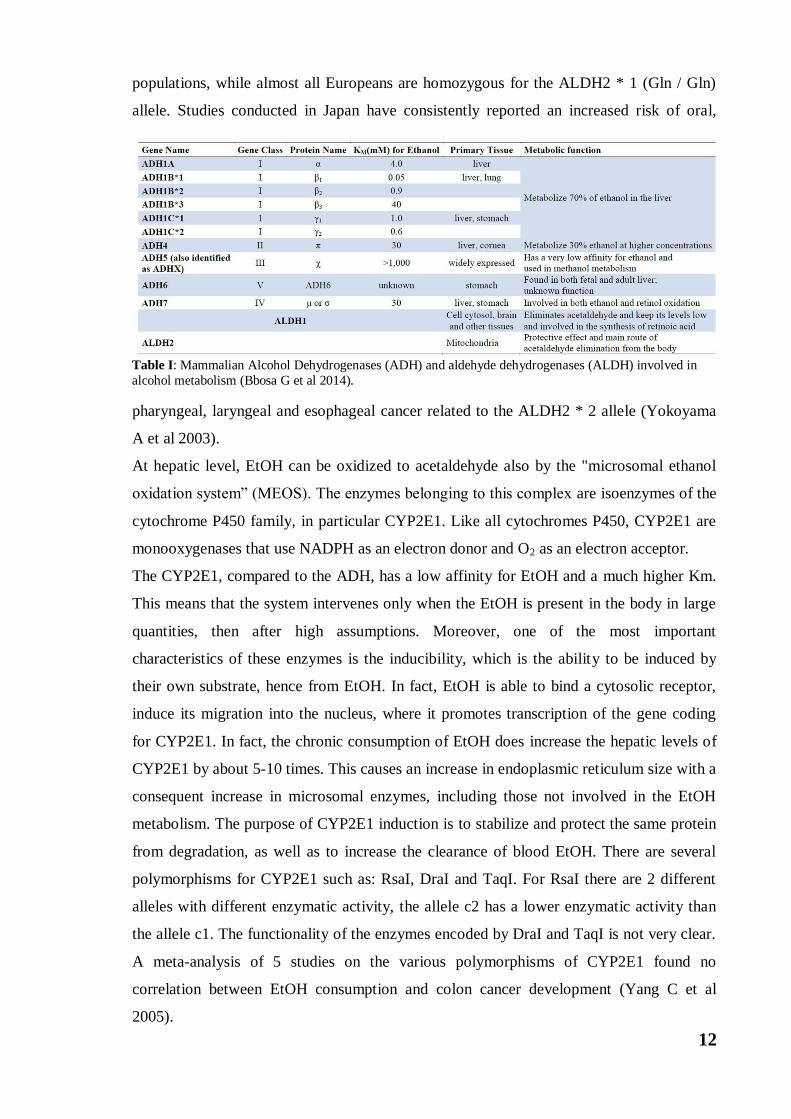
thousand new cases a year. CRC is a malignant neoplasm that affects the large intestine
and is characterized by an uncontrolled proliferation of cells from the mucosa lining the
intestinal walls(Fig 3). At the level of the large intestine, it is possible to distinguish colon
tumors from those of the rectum. Although the two tumor forms seem to have related
causes, they are clearly different from each other both histologically and
epidemiologically. The evolution of this neoplasm is closely related to the stage of the
disease at the time of diagnosis. If the tumor is still limited to the basement membrane,
there will be high chances of healing, if it exceeds the boundaries of the wall and
metastasizes to the lymph nodes, the lethality will be high.
Although the mechanisms underlying carcinogenesis are not yet clear, CRC is certainly to
be considered a multifactorial pathology involving genetic, epigenetic and environmental
factors.
Figure 3 Colorectal Cancer
14
Hereditary genetic alterations can be determinants of genetic predisposition to the onset of
CRC. The most frequent forms of hereditary CRC are Familial Adenomatous Polyposis
(FAP) and Hereditary Non-Polyposis Colon Cancer (HNPCC).
In addition to hereditary genetic alterations, an important contribution to the onset of CRC
can be attributed to environmental factors, such as a high-calorie diet rich in saturated fatty
acids, obesity, ethanol abuse and smoking. Furthermore, it has also been observed that
patients with other inflammatory colon diseases are more at risk of developing CRC.
The colorectal carcinoma is a multistep pathology in which there are well distinct
morphological stages characterized by the presence of different mutations. In most cases of
colorectal carcinoma, the initial stage is characterized by the formation of small polyps
which, when enlarged, transform into benign tumors and which then, further increasing in
size and acquiring various morphological characteristics, pass from the stage of early
adenoma to that of carcinoma. Not all polyps, however, turn into a carcinoma; the
transformation process, in fact, depends on the presence of some morphological
characteristics, such as the presence of villi, and especially the size of the polyps. The
acquisition of different mutations is associated at different stages of carcinogenesis and it
have been seen to be a common event both in the form of sporadic and hereditary CRC
(Ionov Y. et al 1993).
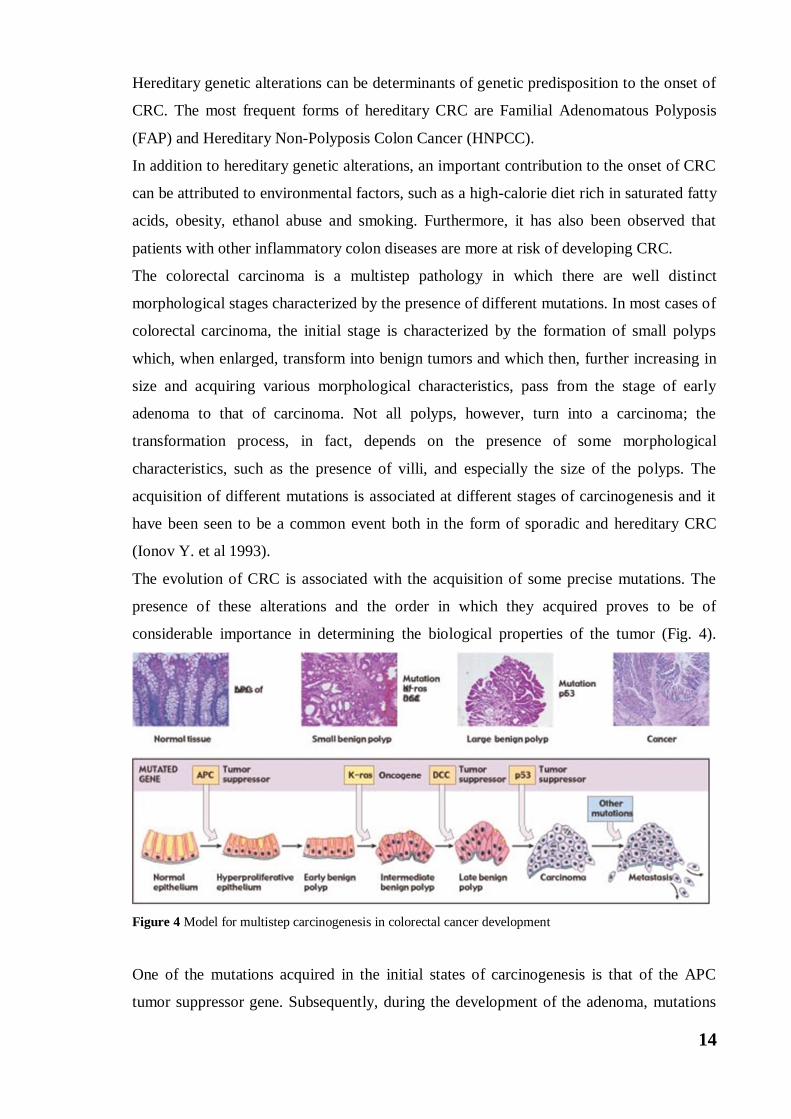
The evolution of CRC is associated with the acquisition of some precise mutations. The
presence of these alterations and the order in which they acquired proves to be of
considerable importance in determining the biological properties of the tumor (Fig. 4).
Figure 4 Model for multistep carcinogenesis in colorectal cancer development
One of the mutations acquired in the initial states of carcinogenesis is that of the APC
tumor suppressor gene. Subsequently, during the development of the adenoma, mutations
15
occur on the K-ras oncogene, and finally, during the phase of transition from adenoma to
carcinoma, further mutations are acquired in the DCC genes (Deleted in Colorectal
Cancer), TP53, MET and deletions involving chromosome 18.
The succession of this series of events is representative of the so-called "CIS pathway", the
traditional adenoma-carcinoma pathway first described in 1990 (Fearon ER et al 1990).
This pathway is the basis of CRC onset in 85% of cases, and it is characterized by an
accumulation of mutations in the key genes that control the cell cycle, intercellular
communication and apoptosis, which are APC, K-ras and p53 genes (Armaghany T et al
2012).
The formation of adenomatous polyps is the initial step in the development of CRC and
begins following the occurrence of mutations on the APC gene, mutation found in most
patients with this disease.
The APC gene encodes a cytoplasmic protein, the APC protein, the product of a tumor-
suppressor that intervenes in the inhibition of cell proliferation, in the control of cell
adhesion and migration, and in the maintenance of chromosomal stability and cytoskeletal
organization. The main function of APC is to control the cytoplasmic levels of beta-catenin
and to prevent its translocation into the nucleus where it mediates proliferative effects
supporting the Wnt pathway. In the absence of the Wnt ligand, beta-catenin interacts with a
protein complex consisting of APC, AXIN1, AXIN2 and GSK3β. The GSK3β
phosphorylates the terminal residues of serine and threonine of beta-catenin and promotes
ubiquitination with the consequent proteasome dependent degradation. In presence of the
Wnt ligand that interacts with the Frizzled membrane receptor, however, the protein
complex is inhibited and beta-catenin is not degraded. This promotes the accumulation of
beta-catenin and its translocation into nuclear sites where it interacts with many
transcription factors including those of the TCF (T-cell factor) / LEF (lymphoid enhancing
factor) family. The stimulation of TCF / LEF determines the activation of some genes,
such as c-MYC and cyclin D1, with consequent stimulation of proliferation and inhibition
of apoptosis. Mutations of APC are very frequent in CRC and they are associated with the
production of a truncated protein that is unable to bind beta-catenin. Consequently, the
levels of beta-catenin in the cytoplasm and the translocation in the nucleus increase with
consequent stimulation of Wnt proliferative pathway (Choi SH et al 2013).
The adenoma-carcinoma progression is closely linked to the appearance of mutations
concerning the K-ras gene, a proto-oncogene that encodes a membrane G protein (K-ras)
with GTPase activity activated by extracellular growth factors.

16
The K-ras gene is constituted by a short gene sequence subject to point mutations, and
within this, a single substitution of a base in a nucleotide, can generate an activating
mutation found in 30-50% of CRC cases. This mutation provides the colonocyte of a
growth advantage as the mutated K-ras protein loses its GTPase activity and is
constitutively active. In this manner, it stimulates cell proliferation in a continuous way. K-
ras is also responsible for transduction of mitogenic signals mediated by the EGF receptor
(EGFR) (Dobre M et al 2013). The activation of the signal transduction cascade starting
from the EGFR can also be favored by the extracellular binding of ligands other than EGF.
Riboforin 2 (RPN2), for example, is one of these ligands that, by binding this receptor,
enhances its proliferative activity by promoting the progression of CRC.
EGFR is a highly N-glycosylated surface glycoprotein which plays a crucial role in most
human cancers as it is correlated with increased growth, proliferation and differentiation of
cells (Ellina MI et al 2014). An increasing number of studies have indicated the importance
of N-glycosylation of EGFR in the regulation of receptor functional properties. This
includes the expression on the cell surface (Coskun Üet al 2011), ligand binding,
conformational stability (Taylor ES et al 2016), dimerization (Wang XQ et al 2001),
interaction with membranes (Prakash A et al 2010) and endocytosis (Lopez PH et al 2009).
There are 11 potential N-glycosylation sites in the extracellular domain of the EGFR. It has
been seen that in the presence of an N-glycosylation inhibitor, an immature EGFR protein
that apparently does not reach the cell surface is synthesized and does not acquire the
ability to bind EGF. One of the glycosylases of EGFR is riboforin 2 (RPN2). In some
studies, it has been found that in CRC cases, the expression levels of RPN2 in tissues are
significantly elevated and RPN2 is predominantly located in the cytoplasm of cells. These
data therefore suggest that RPN2 has a potential role in CRC progression because, by
activating EGFR glycosylation, it promotes cell growth and inhibits its differentiation.
The K-ras mutation, therefore, favors the adenoma-carcinoma transformation, however the
sequence through which this mutation occurs, in relation to the APC mutation is
particularly relevant. If this mutation occurs after an alteration of APC, dysplastic lesion
will progress to cancer (Grady WM et al 2008). This is because K-ras mutated seems to
promote greater aggressiveness of cancer cells.
However, the K-ras mutation alone is not sufficient to guide the complete adenoma-
carcinoma transformation; in fact, the main event responsible for malignant transformation
is represented by the acquisition of mutations concerning the TP53 gene. This gene
encodes p53, a 53 kDa protein present in all cells, which has the function of inhibiting the
cell cycle and preserving the genomic stability (Suzuki Ket al 2011). When replication
17
errors or mutations occur, p53 stops or slows the cell cycle progression in the G1 / S phase
and promotes DNA repair. If the damage to be repaired is too large, p53 activates caspase-
dependent apoptosis (Amaral JDet al 2010). This protein, in addition to being activated by
DNA damage, is also activated by other numerous factors such as ultraviolet radiation,
oxidative stress, chemicals and viruses.
In non-tumor cells, p53 protein is maintained at low concentrations by its continuous
degradation mediated by the MDM2 protein. Mutations in the TP53 gene that result in
functional protein inactivation were found in 75% of CRC cases (Bahnassy AAet al 2014).
The transition from intermediate to late adenoma is determined by mutations in the DCC
gene. DCC (Deleted in Colorectal Cancer) is a tumor suppressor gene implicated in the
development of colorectal cancer. DCC coding for the netrin-1 receptor (Patrick Mehlen, et
al., 1998). In the presence of the ligand, netrin-1 activates pathway leading to cellular
proliferation and migration. In the absence of the netrin-1 ligand, it promotes apoptosis, in
fact an intracellular domain of netrin-1 is cut from caspase-3 and this seems to favor the
activation of caspase-9-dependent apoptosis. Mutations in DCC that occur in the CRC
result in reduced receptor expression with consequence failure to induce apoptosis. The
DCC mutation is not considered a key genetic change in tumor formation, but one of the
alterations that can promote the growth of existing cancer. The division of these cells takes
place at the base of the villi and the old cells are pushed towards the luminal side from the
successive cell divisions until they are eliminated by the activation of apoptosis. The
netrin-1 is produced at the base of the villi, so there is a gradient of netrin that is lower in
the luminal side. Physiologically, the presence of netrin-1 inhibits DCC-mediated cell
death until the epithelial cell reaches the luminal side, where DCC not associated with the
ligand promotes cell death. Mutations of DCC in the colonocytes associated with the
absence of the receptor make it more likely that cells will continue to survive.
Other genes, which are interested in CRC, are the SMAD2 and SMAD4 genes, tumor-
suppressor genes, which codes for proteins act as signal transducers involved in tumor
suppression. The transition from invasive cancer to metastatic cancer is instead under the
control of the MET gene. This is a protoncogene coding for a tyrosine kinase receptor
called c-MET (mesenchymal epithelial transition factor) which binds to HGF factor
(hepatocyte growth factor) activating different signaling pathways, including those
involved in proliferation, in motility, migration and cellular invasion. In physiological
conditions c-MET is important in the control of tissue homeostasis. The amplification of c-
MET expression, found in most cases of human tumors (Comoglio PM et al 2008),
18
including CRC, is associated with tumor invasion and formation of lymph node metastases
(Takeuchi Het al 2003).
Genetic factors and forms of hereditary CRC
Recently it has been found that at the base of Familial Adenomatous Polyposis (FAP) and
of Hereditary Non-Polyposis Colorectal Cancer (HNPCC), both types of hereditary CRC,
there are alterations of a single gene transmitted through germ line cells that increase the
predisposition familiar to the CRC.
In both types of hereditary cancer, the tumor occurs at a much lower age than the average
age of onset of sporadic CRC. Moreover, while in FAP the gene alteration determines the
onset of the tumor; in the HNPCC it favors the progression of carcinogenesis. In the FAP,
the first mutation that triggers tumor genesis concerns the APC tumor suppressor gene.
Since the APC gene is responsible for controlling cell growth in the colonocytes, one
mutation promotes the formation of a series of benign tumors within which, over time,
other mutations accumulate that promote the adenoma-carcinoma transition.
At the base of HNPCC onset, however, mutations of genes responsible for DNA repair are
found. In this type of tumors, in fact, the repair system of DNA damage repair Mismatch
(MMR) is altered. The mutations that are most frequently found in this type of cancer
affect the MLH1 genes, MSH2 and MSH6, each of which encodes a different protein
belonging to the repair system that has the task of repairing at DNA level errors deriving
from a wrong base matching that may occur during a normal replication cycle. Mutations
involving only one of these genes favor the accumulation of replication errors, especially at
the level of microsatellites that become unstable, which is why this form of colorectal
cancer is classified as CRC with microsatellite instability.
Recently, it has been shown that the MMR repair system, in addition to repairing post-
replication mismatches, also performs several other functions that are quite important in
the process of carcinogenesis, one of which, for example, is the protection of DNA from
damage induced by oxidizing agents. Usually in the initial phase of HNPCC cancer there is
a mutation at the level of one of the alleles belonging to one of the MMR genes, which is
then followed by the inactivation of the other allelic copy. This causes, as mentioned
before, an altered function of the repair complex and, at the somatic level, leads to a
consequent accumulation of mutations at the level of other genes, especially those involved
in cell cycle control (Calabrese Pet al 2004). Here the cells of the adenomas that are
19
formed and that have mutations regarding the MMR, tend to accumulate mutations much
more quickly than a normal cell, reason why the tumor progression turns out to be rather
rapid. As previously reported, alterations in the MMR repair system lead to the inability to
repair any replication errors and therefore, to an accumulation of these mutations
especially at the level of microsatellites (Pedroni Met al 2001), or small DNA sequences
containing repeats of tandem mono-, di-, or tri nucleotides that make this sequence
particularly susceptible to replication errors. In most cases, microsatellites are placed
within non-coding DNA sequences, but we can also find them inside coding regions of
DNA belonging to genes mainly involved in the cell growth mechanism. Usually in cases
of HNPCC with microsatellite instability, mutations regarding the APC and p53 genes are
much less frequent and less incident. What is very important is that these types of cancer
are very aggressive but at the same time show high chances of recovery, given by the
instability of microsatellites that apparently appears to be associated with the possibility of
survival.
Nutrition and onset of CRC
Incorrect food habits play an important role in the onset of CRC (Lynch HT et al 2003).
Although cells are rapidly adapting to diet changes, during the adaptation process,
colonocytes tend to accumulate genetic and epigenetic variations that often lead to
genomic instability, favoring carcinogenesis.
The malignant transformation of the colonocytes may also be due to constant and
prolonged exposure to carcinogens. Some data show that consumption of meat, tobacco
and alcohol, considered carcinogenic substances, increase the risk of CRC, and how,
instead, the consumption of dietary fibers reduces it. A diet rich in meat protein and low in
fiber is considered a risky diet for CRC as it increases luminal pH facilitating the
development of neoplastic cells. It has been shown that for every 30 gr of meat consumed
per day there is an increase in the risk of CRC of about 10%. In a protein-rich diet,
undigested protein residues and other nitrogenous compounds are degraded and fermented
by the intestinal bacterial flora. These bacteria produce ammonia (NH3), phenols and
hydrogen sulphides. The presence of these compounds in the colon causes inflammation
and damage to the mucous membranes. For example, free ammonia is most toxic; it is
easily absorbed by the colonocytes and induces inflammation, increases the proliferative
rate and the intraluminal pH (Rao YK et al 2009). The presence of nitrogen, coming from
20
ammonia, facilitates the formation of nitrous compounds (NOC). The cooked meat
provokes the formation of various carcinogens among which the NOC, closely related to
inflammation and mucosal damage (Ben Qet al 2014). These compounds are mainly
formed in the colon through nitrosylation of nitrosamines and starches by bacterial
decarboxylation on amino acids in the presence of nitrosilating agents. Once the NOCs are
formed, they can form DNA adducts and determine the onset of mutations in the main
oncogenes and tumor suppressor genes (Bastide NM et al 2011).
The high consumption of animal fats, consequently to a high consumption of meat, causes
an increased release of primary bile salts in the colon. These are metabolized by anaerobic
intestinal bacteria and transformed into secondary bile salts through mechanisms of
enzymatic deconjugation and dehydroxylation. Secondary bile salts, such as deoxycholic
acid and lithocolic acid, are considered carcinogenic molecules as they alter the
proliferative activity of mucosal cells by increasing the number of cells that synthesize
DNA (Bernstein H et al 2009). The increase in cell proliferation predisposes to the risk of
the onset of mutations and consequent malignant transformations. Furthermore, exposure
of the colonocytes to high concentrations of secondary bile salts induces the production of
ROS responsible for oxidative damage and mitotic aberrations that could induce changes in
DNA and consequent gene instability. Therefore, greater is fats introduced into the body
through food, greater is the production of secondary biliary salts and the predisposition to
the onset of CRC. On the other hand, numerous studies show that a high consumption of
dietary fibers, especially cereals fibers and whole grains, reduces the risk of CRC (Aune D
et al 2011).When the fibers reach the colon they are partially or completely fermented with
consequent production of short-chain fatty acids and gases that influence the
gastrointestinal functions. Short-chain fatty acids reduce intraluminal pH by providing
optimal conditions for the colonocytes and decreasing the conversion of bile acids into
secondary bile acids. The dietary fibers also increase the volume by stimulating the growth
of normal intestinal flora (Eswaran Set al 2013) and reduce the time and concentration of
carcinogens in contact with the intestinal wall (Anderson JW et al 2009).There is also a
well-established link between the high percentage of fats in the diet, inflammation and
tumorigenesis. The high percentage of animal fats influences the microbiome favoring the
expansion of pro-inflammatory microorganisms that lead to the development of intestinal
inflammation (Candela M et al 2014). To support the relationship between CRC and
inflammation there is the observation that the development of CRC is more frequent in
patients with inflammatory bowel disease (Medzhitov R. 2008). In addition, the intestinal
microbiome changes with age, as it tends to increase the opportunistic facultative

21
anaerobes and to decrease the anti-inflammatory species. Therefore, aging also contributes
to a state of chronic inflammation that characterizes the whole organism. Intestinal
inflammation could therefore elicit greater stimulation of the inflammatory response,
allowing opportunistic pathogens to thrive at the expense of symbionts (Sansonetti PJ et al
2011).
Ethanol and Colorectal Cancer
While moderate EtOH consumption appears to have beneficial effects on cardiovascular
disease, it is a common knowledge that a chronic and heavy consumption of EtOH exerts
toxic effects and increases the risk of the occurrence of different types of cancer, including
colorectal cancer (CRC) (Baan Ret al 2007). Most of the harmful effects due to the high
consumption of EtOH can be attributed to the production of both acetaldehyde and oxygen
reactive species (ROS) (Fig.5).
Figura 5 EtOH metabolism determines the production of ROS and acetaldehyde
22
Infact, high consumption of EtOH results in a NADH / NAD+ ratio increase at the cellular
level, due to the high activity of both alcohol dehydrogenase (ADH) and aldehyde
dehydrogenase (ALDH). When the consumption of EtOH is moderate, the oxidation rate of
NADH by the ADH is also moderate, and this is useful to maintain, within the cell, the
right levels of NAD+ that serve the ALDH for transform acetaldehyde into acetate. Instead,
an increase intake of EtOH correlates with an increase in the NADH / NAD+ ratio. In some
cases the NADH will tend to accumulate because the rate at which this is oxidized within
the cell is now far outweighed by the rate at which ethanol, present in large quantities, is
metabolized. All this involves a malfunction of the ALDH with consequent accumulation
of acetaldehyde inside the cell. Moreover, when high doses of ethanol are ingested, this is
metabolized not only by hepatic ADH1, but also by ADH belonging to other families.
These ADH normally are not very active given their high Km. Instead, when EtOH is
present at high concentration in the stretch proximal gastrointestinal, it is also metabolized
by gastric ADH (ADH4) with consequent production of large amounts of acetaldehyde on
site. The high production of acetaldehyde in the intestine has been correlated with the
increased risk of the occurrence of colorectal cancer. Under conditions of high EtOH
assumptions, the amount of acetaldehyde in the bloodstream increases, also because the
EtOH is metabolized by the microsomal ethanol oxidation system (MEOS). The latter,
which has high Km, intervenes few at low doses of EtOH; a high consumption of EtOH
determines the involvement of the MEOS system in the disposal of alcohol, also because
EtOH itself induces an increase in the levels of the enzyme. The aim of the induction of the
MEOS system, besides increasing the half-life of the CYP2E1 microsomal enzymes
belonging to this oxidation system of EtOH, is to increase the clearance of the circulating
EtOH. The increased clearance, despite representing a positive response of the organism in
these cases, can also have negative implications. In these circumstances, in fact,
acetaldehyde is produced faster than it is metabolised by acetaldehyde dehydrogenase. This
involves its accumulation inside the liver cells but also a greater discharge of the same in
the bloodstream where, through the blood flow, it will reach other organs, such as the
digestive tract. The explicit toxic action of acetaldehyde at the cellular level is linked to the
fact that it is a highly reactive molecule that tends to covalently bind to amino and
sulfhydryl groups of proteins, nucleotides and phospholipids, forming adducts, leading to
structural and functional changes of the molecules. The formation of adducts by
acetaldehyde, especially at the level of certain proteins, inhibits the cellular defense
mechanisms against oxidative stress. In particular, the interaction of acetaldehyde with
GSH cysteine residues determines a drop in reduced glutathione and therefore deprives the
23
cell of an important system of defense against ROS. In a study of 24 heavy drinkers of
EtOH it was shown that in their lymphocytes there was a quantity of DNA adducts 7 times
higher than non-drinkers (Fang JL et al 1997). In addition to the metabolic activity of ADH
in the colonic mucosa, the ADH of the bacteria present in the colon to produce
acetaldehyde can also oxidize EtOH. To this end, it has been shown that the bacteria of the
intestinal microflora incubated at 37 °C with different concentrations of EtOH produced
high amounts of acetaldehyde (Jokelainen K et al 1994). This high production of
acetaldehyde, both from colonocytes and intestinal bacteria, associated with a low
enzymatic activity of ALDH of the colon mucosa, determines a substantial amount of
acetaldehyde that accumulates in the colon. This would increase the probability of
developing polyps of the colon and CRC that have been associated with a high
consumption of EtOH (Salaspuro M, 1996). In fact, there is a statistically significant
correlation between microbial ADH activity and production of acetaldehyde from EtOH in
the colon (Jokelainen K et al 1996). More than 500 bacterial strains isolated from the
faeces of Japanese alcoholics showed a high production of acetaldehyde following
exposure of EtOH. In particular, among these bacteria some obligate anaerobes were large
producers of acetaldehyde. The oxidation of ethanol by obligate intestinal anaerobes under
aerobic conditions in the colon and rectum probably plays an important role in the
pathogenesis of alcohol-associated colorectal cancer (Tsuruya A et al 2016). Alcohol
administration gave rise to intracolonic levels of very high acetaldehyde in the rats, these
levels were markedly reduced following the concomitant treatment with ciprofloxacin, an
antibiotic (Homann N et al 2000). Furthermore, rats without bacteria had a significantly
lower accumulation of acetaldehyde in the rectum and in the cecum than conventional
animals, and this was directly proportional to the number of faecal bacteria present in the
gut. Furthermore, individual variations in microflora of the human colon can influence the
relative risk of alcohol-related colorectal cancer (Nosova T et al 1997).
An important function of the epithelial cells lining the gastrointestinal tract via the tight
junctions is to provide a barrier against the hostile environment of the gastrointestinal
lumen. Dysregulation of interactions between the intestinal epithelium and intestinal
bacteria leads to loss of host immune tolerance, and thus promotes the development of
colon cancer. Excessive intake of EtOH changes the composition of enteric microflora
induces excessive growth of gram-negative bacteria and destroys the intestinal epithelial
barrier. These results increase intestinal permeability and increase accumulation of
proinflammatory cytokines, such as tumor necrosis factor (TNF) and interleukin (IL) -6
(Amin P.B et al 2009). EtOH facilitates the absorption of environmental carcinogens by
24
modifying the permeability and molecular composition of the gastrointestinal tract (Seitz
H.K et al 2007). EtOH also acts as a solvent that improves the penetration of carcinogenic
compounds into the mucosa.
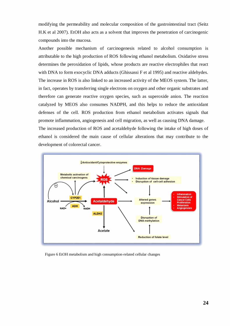
Another possible mechanism of carcinogenesis related to alcohol consumption is
attributable to the high production of ROS following ethanol metabolism. Oxidative stress
determines the peroxidation of lipids, whose products are reactive electrophiles that react
with DNA to form exocyclic DNA adducts (Ghissassi F et al 1995) and reactive aldehydes.
The increase in ROS is also linked to an increased activity of the MEOS system. The latter,
in fact, operates by transferring single electrons on oxygen and other organic substrates and
therefore can generate reactive oxygen species, such as superoxide anion. The reaction
catalyzed by MEOS also consumes NADPH, and this helps to reduce the antioxidant
defenses of the cell. ROS production from ethanol metabolism activates signals that
promote inflammation, angiogenesis and cell migration, as well as causing DNA damage.
The increased production of ROS and acetaldehyde following the intake of high doses of
ethanol is considered the main cause of cellular alterations that may contribute to the
development of colorectal cancer.
Figure 6 EtOH metabolism and high consumption-related cellular changes
25
The use of heavy alcohol could also lead to nutritional deficiencies by reducing the intake
of foods rich in micronutrients, and altering intestinal absorption. The most relevant effect
appears to be on folate metabolism (Lieber CS, 2003).
In fact the ethanol intake determines a reduction in the absorption of folic acid in the
intestine (Kono S et al 2005). This has been attributed to the production of acetaldehyde in
the colon by intestinal microflora. Indeed, it has been demonstrated in vitro that
acetaldehyde determines a degradation of folic acid (Fig. 6) (Homann N et al 2000). A
reduced availability of folic acid in cells compromises methionine metabolism and is
associated with accumulation of homocysteine and S-adenosyl-homocysteine. The latter
has been shown to inhibit DNA methyltransferase with the consequent presence in the cells
of a hypomethylated state of DNA (Zakhari S. 2013).The methylation status of promoters
of some tumor suppressor genes such as APC and p53 is more reduced in CRC associated
with low folate / high levels of ethanol (Van Engeland M et al 2003).
Endoplasmic Reticulum Stress
The endoplasmic reticulum (ER) is an organelle consisting of a continuous compartment
that extends from the nucleus to the cytosol and occupies about 10% of the cell volume.
The ER carries out many important functions including
the synthesis and distribution of phospholipids and sterols, reserve and release of Ca2 +
ions
within the cytosol (Scheuner D et al 2008). Furthermore, ER is involved in the synthesis,
folding and post translational modifications of membrane and secreted proteins (Ma Y et al
2004). Proteins synthesis inside the ER is monitored by different disulfide isomerases,
which catalyze the formation of disulfide bridges. Protein synthesis is also regulated by
molecular chaperones (GRP78, calreticulina, GRP94) which are associated with nascent
peptides avoiding their aggregation and helping them to achieve correct folding in both
physiological and pathological conditions (Kaufman RJ, 1999). Inside the ER, there is a
real quality control, which ensures that only properly folded and functioning proteins leave
ER. On the other hand, the non-folded or malfunctioning proteins are retained and finally
sent to the 26S proteasome to be degraded, a process mediated by activation of the ER
Associated Degradation (ERAD) system, or through the lysosomal mechanism, defined as
macroautophagy, also commonly known as autophagy. Protein folding within the ER is
highly regulated and susceptible to changes in intracellular energy levels, redox status and
Ca2 +
ion concentration. Under conditions of sudden or constant variations of one of these
26
three conditions, there is accumulation and aggregation of poorly folded proteins in the
lumen of ER, a condition known as ER stress. ER stress is observed as a consequence of
nutrients deprivation or differentiation of type B lymphocytes in plasma cells, as well as in
pathological conditions, such as viral infections, ischemia, neurodegenerative diseases,
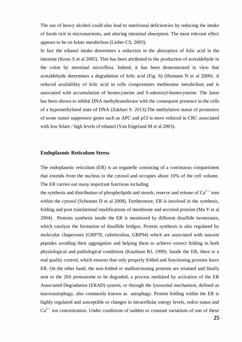
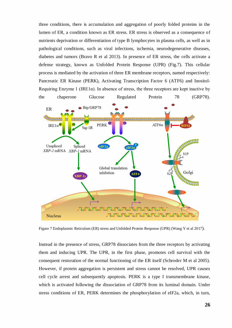
diabetes and tumors (Bravo R et al 2013). In presence of ER stress, the cells activate a
defense strategy, known as Unfolded Protein Response (UPR) (Fig.7). This cellular
process is mediated by the activation of three ER membrane receptors, named respectively:
Pancreatic ER Kinase (PERK), Activating Transcription Factor 6 (ATF6) and Inositol-
Requiring Enzyme 1 (IRE1α). In absence of stress, the three receptors are kept inactive by
the chaperone Glucose Regulated Protein 78 (GRP78).
Figure 7 Endoplasmic Reticulum (ER) stress and Unfolded Protein Response (UPR) (Wang Y et al 2017).
Instead in the presence of stress, GRP78 dissociates from the three receptors by activating
them and inducing UPR. The UPR, in the first phase, promotes cell survival with the
consequent restoration of the normal functioning of the ER itself (Schroder M et al 2005).
However, if protein aggregation is persistent and stress cannot be resolved, UPR causes
cell cycle arrest and subsequently apoptosis. PERK is a type I transmembrane kinase,
which is activated following the dissociation of GRP78 from its luminal domain. Under
stress conditions of ER, PERK determines the phosphorylation of eIF2a, which, in turn,
27
determines the inhibition of protein translation (Wek RC et al2006). This event favors cell
survival by inhibiting the accumulation of nascent proteins that arrive in the endoplasmic
reticulum. However, eIF2a also activates proteins that are not involved in the blocking of
protein translation. Among these there is the ATF4 protein encoding a cAMP respsonse
element-binding transcription factor (C-EBP) (Schroder M et al 2005). ATF4 promotes cell
survival through the activation of genes involved in the metabolism of amino acids, in
redox reactions, and in protein secretion (Harding HP et al 2003). However, ATF4 can also
activate C / EPB transcription factor homologous protein (CHOP), which promotes cell
death by apoptosis (Wang XZ et al 1998). The second receptor involved in the UPR is
ATF6, a type II transmembrane glycoprotein, whose luminal domain is responsible for the
detection of misfolded proteins. The cytoplasmic portion of ATF6 is able to act as a
transcription factor as it contains a DNA binding domain. Following its dissociation from
the chaperone protein GRP78, ATF6 translocates into the Golgi apparatus, where it
undergoes a proteolytic cut generating an active transcription factor (Chen X et al 2002).
Among the ATF6 target genes there are those coding for some chaperone proteins, such as
GRP78, GRP94, disulfide isomerase proteins (PDI), for the transcription factor GADD153
/ CHOP and for X box-binding protein 1 (XBP1). Since the activation of ATF6 determines
the induction of the expression of genes coding for the chaperones, its activation is
responsible for the increase in the ability of the reticulum to replicate proteins, contributing
to the restoration of initial homeostasis. Usually ATF6 mediates pro-survival signals in
order to counteract the effect of ER stress (Wu J et al 2007). The last receptor / sensor of
the activated UPR is IRE1α.It has a dual function, activates survival mechanisms by
activating the transcription of chaperones as GRP78, but also activates pro apoptotic
proteins under prolonged stress conditions. IRE1α is a sensor with double enzymatic
activity, as it is equipped with both a serin-threonine kinase domain and an
endoribonuclease domain (Tirasophon W et al 1998). Under ER stress conditions, the
detachment from GRP78 determines the activation of IRE1α, through dimerization and
autophosphorylation. Once activated IRE1α determines alternative splicing of the XBP1
mRNA. The generated splicing variant encodes a sXBP1 transcriptional factor that will
activate ER chaperone transcripts such as P58INK
. This chaperone ends the block of
translation activated by PERK and activates the transcription of pro apoptotic proteins
(Ladiges WC et al 2005). Therefore, in presence of ER stress the activation of the IRE1α
sensor plays a critical role in the initiation of pro-apoptotic signals, while the activation of
the PERK and ATF6 sensors would seem to precede the activation of the IRE1α in the
attempt to resolve the stress in the presence of a pro-survival action. If ER stress persists,
28
the pathways of PERK and IRE1α can converge, mediating the induction of the apoptotic
process through mutual reinforcement. However, it has also been shown that in the
presence of ER stress both IRE1α and CHOP can activate autophagy processes in human
colon carcinoma cells (Shimodaira Y et al 2014).
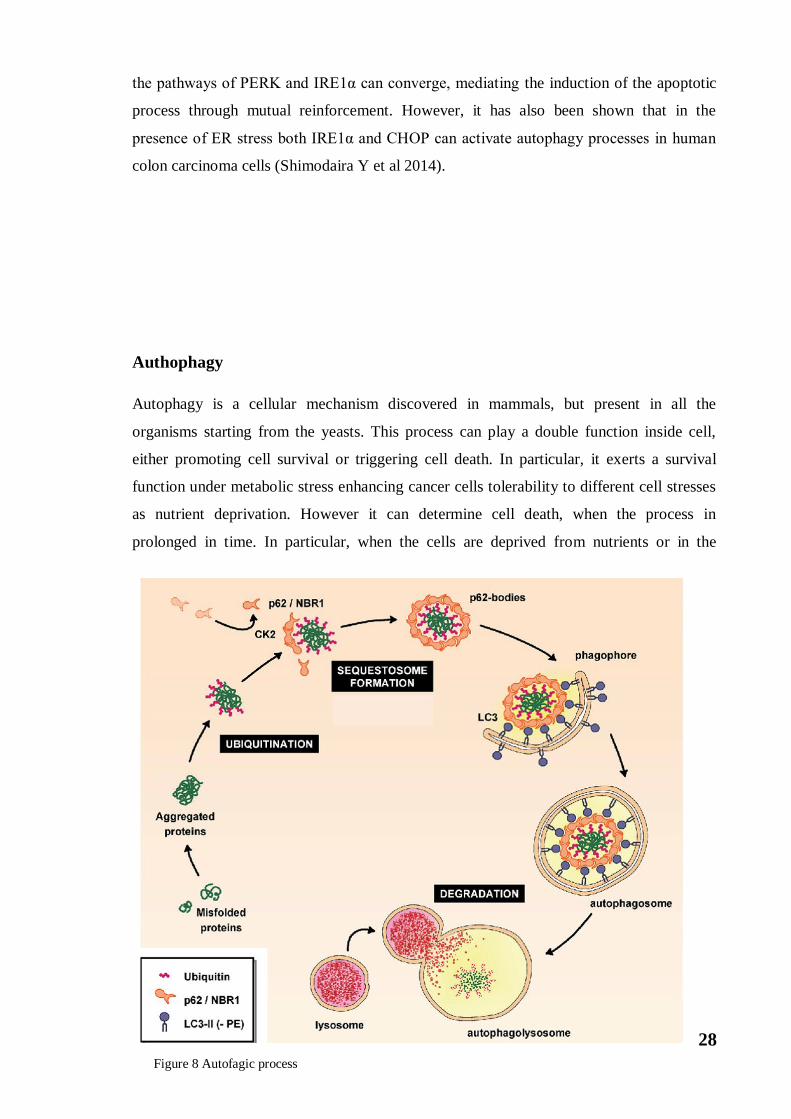
Authophagy
Autophagy is a cellular mechanism discovered in mammals, but present in all the
organisms starting from the yeasts. This process can play a double function inside cell,
either promoting cell survival or triggering cell death. In particular, it exerts a survival
function under metabolic stress enhancing cancer cells tolerability to different cell stresses
as nutrient deprivation. However it can determine cell death, when the process in
prolonged in time. In particular, when the cells are deprived from nutrients or in the
Figure 8 Autofagic process

29
presence of the autophagic signal, it has been observed the formation of double membrane
vesicles, called autophagosomes, which incorporate portions of the cytoplasm (Levine B et
al 2005). The membrane of these autophagosomes originates from intracellular organelles
(eg mitochondrion) or from pre-existing intracellular molecules. Later these
autophagosomes merge with the lysosomes, thus favouring the degradation of
macromolecules and releasing monomers and ATP, available for a new synthesis of
macromolecules. This process is called macroautophagy. However, there are two other
types of autophagy such as microautophagy and chaperone-mediated autophagy.
Autophagy also has a protective role because it allows the degradation of damaged
organelles or of protein aggregates (misfolded proteins) that could damage cellular
wellness. The main regulator of autophagy is the target kinase of rapamycin, mTOR. This
kinase acts as a nutritional sensor, when there is abundantly nutrients is active and prevents
the formation of autophagosome. In cases of stress or decreased nutrient availability (due
to prolonged fasting) mTOR is inhibited and allows the formation of autophagosome.
Generally, mTOR inhibits the kinase ULK1 (Gammoh N et al 2012). Akt following
phosphorylation in Ser774 inhibits ULK1. In the same way, it can be activated by AMPK
that phosphorylates it in different amino acid residues such as: Ser555, Ser467, Thr574 and
Ser637. In the absence of nutrients, ULK1 undergoes a conformational change by an active
state following autophosphorylation in the Thr180 residue. The ULK1 protein in the active
form phosphorylates the proteins ATG13 and FIP200, with which it forms a fundamental
complex in the maturation of autophagosomes, as it is able to recruit another protein
complex consisting of Vps15, PI-3K of class III and the Beclin-1. This mega protein
complex, as it is composed, recruits proteins encoded by ATG genes. ATG proteins are
involved in the different phases of autophagy, performing functions both in the nucleation
of the vesicles and in the maturation of autophagosomes (Kourtis N et al 2009). These
ATG proteins also act on the cytosolic LC3-I protein converting it, following a proteolytic
cut (by ATG4) and conjugation with phosphatidylethanolamine, in the LC3-II form (Liu Y
et al 2010). Consequently, LC3-II produced is anchored to the autophagosome membrane
where it remains even after fusion with lysosomes (Fig.8). The LC3-II protein is a marker
of autophagy (Pyo JO et al 2012). In the last phase of autophagy, when the fusion of
autophagosome with lysosomes occurs, the generation of autofagolisosomes, favors the
degradation of the molecules. There is an interesting relationship between autophagy and
ER stress since both mechanisms are aimed at restoring normal cellular functions
following stress. In particular, CHOP activates autophagy mechanisms in colon carcinoma
cells (Shimodaira et al 2014).

30
Project aim
31
In the western countries the colorectal cancer (CRC) is the third tumor for aggressiveness
and incidence after the lung and breast/prostate cancer. The frequency of this cancer is
higher in older age. There are different causes related to this disease, like genetics and
lifestyle. However, the exact cause of colon cancer is not clear yet. Although the genetic
alterations play a significant role in colon cancer development and progression, different
studies support that lifestyle and environmental factors can increase the risk of developing
this tumor. To this purpose epidemiologic studies correlating alcohol consumption with
risk of developing colon cancer demonstrate that the incidence of the disease is 5-fold
higher among drinkers compared to nondrinkers. Alcohol consumption is also associated
with Colorectal Cancer patients’ progression and poorer prognosis. Epidemiological
investigations also indicate that chronic and heavy alcohol consumption is associated not
only with the increased risk of developing CRC, but also with metastasis. Alcohol
consumption is an independent risk factor for liver metastasis in colorectal carcinoma
patients. The presence of tumor metastasis determines the failure of the chemotherapeutic
treatment and a lower postoperative survival rate. The 5-year overall survival rates are
around 68% for colorectal cancer. In the presence of concomitant metastases, the
maximum survival rates after surgery do not exceed 20%. However, the relationship
between alcohol consumption and tumor metastasis needs to be deepened further.
Although it is now clear that the consumption of high doses of ethanol is an important risk
factor for colorectal cancer and its tumor progression, the biochemical mechanisms
responsible for these events have not yet been fully clarified. The aim of my doctoral
project is to study the effects of ethanol in human colorectal carcinoma cells and to
evaluate the biochemical mechanisms underlying survival and invasiveness in CRC.

32
Materials and Methods
33
Cell culture condition
The human colon cancer HCT116, HT29 and Caco-2 cells (Interlab Cell Line Collection,
ICLC, Genova, Italy) were grown in monolayer in flasks of 75cm2 in RPMI 1640 medium,
supplemented with 10% (v/v) heat-inactivated fetal bovine serum (FBS), 2 mM L-
glutamine, 100 U/ml penicillin and 50 µg/ml streptomycin in a humidified atmosphere of
5% CO2 in air at 37°C. To study the effects of ethanol, cells were detached using trypsin-
EDTA (2.5mg/ml trypsin and 1mg/ml EDTA) and plated in accordance to the experimental
conditions, as described in the paragraphs below. Cells were allowed to adhere for 24h and
then treated with different concentration of ethanol at different times.
All the reagents used for cell culture were purchased from Euroclone (Pero, Italy). Ethanol,
E64d and all chemicals, except when stated otherwise, were supplied by Sigma-Aldrich
(Milan, Italy).
MTT assay
To evaluate the effect of ethanol on cell viability the MTT (3-(4,5-dimethylthiazol-2-yl)-
2,5-diphenyl tetrazolium bromide) colorimetric assay was used. HCT116, HT29 and Caco2
cells (7x10³/200µl/well) were plated in 96-wells and treated with various concentrations of
ethanol (30-300mM) for different times. Then, 20 μl MTT (11mg/1ml) was added and cells
were incubated at 37°C for 4 h. Finally, the medium was removed and 100µl of lysis buffer
(20% sodium dodecyl sulfatein 50% N,N-dimethylformamide) was added. At the end, the
absorbance of the formazan was measured directly at 490 nm with 630 nm as a reference
wavelength using an automatic ELISA plate reader (OPSYS MR, Dynex Technologies,
Chantilly, VA). Cell viability was expressed as the percentage of the OD value of ethanol-
treated cells compared with untreated samples used as control. Each experiment was
performed in triplicate.
Clonogenic assay
HCT116 cells were seeded in 6-well plates at a density of 200 cells/well with 3 ml culture
medium and incubated for 10 days with or without 100 and 300 mM ethanol. The medium
was changed every 3 days. There after medium was removed and cells were washed in
cold PBS and then incubated on ice in cold methanol for 15 min. Then, the cells were
washed in PBS, incubated for 1h in the presence of 0.01% crystal violet (Sigma-Aldrich,
St. Louis, MO, USA). Representative views were photographed.
34
Measurement of ROS production
The production of reactive oxygen species (ROS) was measured using 2', 7' dichloro-
dihydro-fluorescein diacetate (H2DCFDA, Molecular Probes Eugene, OR). This
compound contains two acetyl groups and is able to cross the cell membrane by its
lipophilic nature. After cell entry, cellular esterases remove acetyl groups by transforming
H2DCFDA into the DCFH molecule that is oxidized by ROS and converted into
dichlorofluorescein (DCF), a compound that emits a fluorescent green color.
HCT116 cells (7x10³/200µl) were seeded in 96-well plates and incubated with 100 or 300
mM ethanol for different times. After treatment, cells were washed with PBS and
incubated with 10 μM H2DCFDA for 15 minutes at 37° C in the dark. Finally, cells were
resuspended in PBS and analysed by fluorescence microscopy using a Leica DMR (Leica
Microsystems S.r.l., Wetzlar, Germany) inverted microscope equipped with a FITC filter
system (excitation wavelength of 485 nm and emission wavelength of 530 nm). Images
were acquired by computer-imaging system (Leica DC300F camera).
Western blotting analysis
Cells were seeded in 6-well plates (1.5x10⁵/2 ml culture medium) and treated with ethanol
for the indicated times. Then, cells were washed in PBS and harvested with trypsin-EDTA
(2.5mg/ml trypsin and 1mg/ml EDTA), and centrifuged at 800 rpm for 8minutes. The
pellets were lysed in RIPA buffer (1% NP-40, 0.1% SDS and 0.5% sodium deoxycholate
in PBS), containing protease inhibitor cocktail (25μg/ml aprotinin, 1mM PMSF, 25 μg/ml
leupeptin and 0.2 mM sodium pyrophosphate). After sonication (3 cycles of 10 seconds, at
an intensity of 10 REV in Soniprep 150), protein content of cell extracts was determined
by the Bradford method. Equivalent amounts of proteins (30-50 µg) were separated by
SDS–polyacrylamide gel electrophoresis and transferred to a nitrocellulose membrane
(Bio-RadHercules, CA, USA). Equal proteinloading was ascertained by Ponceau-S
staining of blotted membranes. Then, the filter was incubated with "blocking" solution (1%
or 5% milk in TBST (20mM Tris-HCl, 150mM NaCl, 0.005% Tween-20, pH 7.5) for 1h.
So, the filter was incubated overnight in a solution containing specific primary antibody
(1μg/ml TBST). Primary antibodies used for catalase, lamin B, MnSOD, Nrf2, iNOS,
Cox2, Grp78, CHOP, MMP-2 and MMP-9 were purchased from Santa Cruz
Biotechnology (St. Cruz, CA); β-actin from Sigma Aldrich; Hsp60, Hsp90 and HO-1 from
Enzo Life Sciences, (Milan, Italy). After incubation, the filter was incubated for 1h with
secondary antibody (1μg/3ml of TBST; Pierce, Thermo Fisher Scientific) conjugated with
35
horseradish peroxidase (HRP). Immunoreactive signals were detected using enhanced
chemiluminescence (ECL) reagents (Cyanagen, Bologna, Italy). The correct protein
loading was confirmed by stripping the immunoblot and reprobing with primary antibody
for β-actin. The signal obtained through ECL development was detected through
CHEMIDOC and processed using the "Quantity One" software (BioRad).
Cytosol and nuclear extraction
HCT116 cells were seeded in 100-mm tissue culture dishes (1x10⁶ cells/5 ml culture
medium) and, after treatment with ethanol, washed in PBS and harvested with lysis buffer
(250 mM Sucrose, 20 mM HEPES, 10 mM KCl, 1.5 mM MgCl2,1 mM EDTA, 1 mM
EGTA, 1 mM DTT, and protease inhibitors,pH7.4). Next, cells were passed 10 times
through a needle of 25 G on ice for 20 minutes. The homogenates were centrifuged at
1,000 g for 10 minutes at 4°C. The pellets (nuclear fraction) were resuspended in lysis
buffer and passed 10 times through a needle of 25 G and centrifuged at 1,000 g for 10
minutes at 4°C. The pellets were lysed with RIPA buffer (1% NP-40, 0.5% sodium
deoxycholate, 0.1% SDS, inhibitors of proteases: 25μg/ml aprotinin, 1mM PMSF, 25
μg/ml leupeptin and 0.2 mM sodium pyrophosphate) and sonicated. The supernatants
obtained from the two centrifugations were united, centrifuged at 10,000 g for 30 minutes
at 4°C. The supernatants obtained were considered as cytosolic fraction.
Nuclear and cytosolic fractions were used to evaluate Nrf2 and HO-1; β-actin and lamin-B
were used as cytoplasmic and nuclear markers, respectively.
Monodansylcadaverine test
To evaluate the formation of autophagic vacuoles Monodansylcadaverine (MDC) test was
employed. HCT116 cells (7x10³/200µl culture medium) were plated in 96-wells and
treated with ethanol. After the treatment, cells were incubated with 50 µM MDC for 10
min at 37°C in the dark. Then, cells were washed with PBS and analysed by fluorescence
microscopy using a Leica DMR (Leica Microsystems) microscope equipped with a DAPI
filter system (excitation wavelength of 372 nm and emission wavelength of 456 nm).
Images were acquired by computer-imaging system (Leica DC300F camera) using SMX
software.
36
Transient down-regulation of Nrf2 by short interfering RNA (siRNA)
HCT116 cells were seeded (2×10⁵ cells/well) in 6-well plates and cultured in RPMI 1640
medium, supplemented with 10% FBS, without antibiotic, for 24 h to reach approximately
60–80% confluence before transfection. Specific siRNAs directed against Nrf2, obtained
by Qiagen, Hilden, Germanyas a pool of double-stranded RNA oligonucleotides,
(SI03246950, SI03246614) were transfected for 5 h into the cells. Each siRNA was
transfected into cells at 25 nM. A non silencing siRNA (SI03650318, Qiagen) was used as
a negative control (50nM). For transfection, specific siRNAs and negative control were
transfected in the presence of 5μl Lipofectamine 2000 (Invitrogen, Carlsbad, US) in a final
volume of 1 ml serum-antibiotic free RPMI 1640 medium. At the end the reaction was
stopped replacing the culture medium with complete RPMI 1640 medium plus FBS and
antibiotic. After 24h of transfection, cells were treated with ethanol for other 24h. Then,
the cells were examined for Nrf2 down-regulation and other proteins by western blotting
analysis.
Gelatin zymography
HCT116 cells were seeded in 100-mm tissue culture dishes (5x10⁵ cells/ 5 ml culture
medium). After 48h of ethanol treatment, cells were washed in PBS and harvested with
lysis buffer (25 mM Tris-HCl pH 7.5, 100 mM NaCl, 1% (vol/vol) IGEPAL CA-630,
Protease inhibitors: 10 μg/mL aprotinin, 2 μg/mL leupeptin, and 4 mM benzamidine). The
lysates were centrifuged at 800 g for 10 minutes. The samples (50μg of proteinsprepared in
sample Buffer: 50 mM Tris-HCl, 2% SDS, 0.1% Bromophenol Blue, 40% Glycerol, pH
6.8) were loaded on polyacrylamide gels (10%) with 10X gelatin and subjected to
electrophoresis. Then, the gel was washed for 1 hour with enzyme renaturing buffer
(200mM NaCl, 5mM CaCl2, 5μM ZnCl2, 2,5% (v/v) Triton X-100 and 50 mM Tris HCl,
pH 7.5) and incubated overnight at 37°C with developing buffer (50mM Tris base, 200mM
NaCl, 5mM CaCl2, pH 7.5). Then, the gel was incubated for 30 min at RT with Staining
solution (0.125% Coomassie brilliant blue R-250, 50% methanol, 20% acetic acid) and
washed with Destaining solution (30% methanol, 0,01% formic acid) until clear bands of
MMP activity are visible in the blue background.
37
Immunofluorescence
HCT116 cells were plated on coverslips (8x10³) and treated with 300 mM EtOH for
different times. After washing two times with PBS, cells were fixed in methanol for 30
minutes at room temperature. After fixation, cells were washed three times in PBS for 5
minutes and treated with a blocking solution (3% BSA in PBS) for 30minutes.
Subsequently, the cells were washed two times with PBS and incubated with primary
antibody against HO-1 (anti-rabbit, Enzo Life Sciences) or against Nrf2 (anti-rabbit, Santa
Cruz Biotecnology) at dilution 1:100, overnight at 4°C. Then, cells were washed three
times with PBS for 5 minutes and incubated for 1h with a conjugated secondary antibodies:
anti-rabbit IgG-FITC produced in goat (Sigma-Aldrich) at dilution 1:200. Nuclei were
stained with Hoechst Staining Solution (1:1,000, Hoechst 33258, Sigma-Aldrich). The
images were captured using a Leica Confocal Microscope TCS SP8 (Leica Microsystems).
Statistical analysis
Data, represented as mean ± S.D., were analyzed using the Student t test for densitometric
analyses. The Graph Pad Software (GraphPad Software 7825 Fay Avenue, Suite 230 La
Jolla, CA 92037 USA) was used for statistical calculations. Differences were considered
significant when p< 0.05 or p< 0.01.

38
Results

39
Effects of EtOH in colon cancer cell viability
Although epidemiological studies have shown that heavy and chronic alcohol
consumption increases the risk to develop colon cancer and favors tumor progression
(Baan R et al 2007), the underlying molecular mechanisms are not still clear.
To shed light on this aspect, I studied the effects of Ethanol (EtOH) on HCT116, HT29 and
Caco-2 cells, three human colon cancer cell lines. HCT116 cells are derived from a colon
carcinoma line characterized by a mutation in codon 13 of the K-ras gene (Schroy PC et al
Fig. 1. Effect of Ethanol on colon cancer cell viability and colony generation ability (A) HCT116, HT29 and Caco-2 cells (7x10³) were incubated in the presence of various doses of EtOH for different periods. Cell
viability was assessed by MTT assay as described in Materials and Method section. Values are the means of
three independent experiments ±S.E. (*) p<0.05 compared to the untreated sample. (B) Effect of EtOH on
colony generation ability of HCT116 cells. Clonogenic assay was performed seeding a single cell suspension
(200 cells/well) in 6-well plates and after 48h was treating with different doses of EtOH. The ability of cells
to generate colonies was evaluated after 10 days as reported in Methods. Photographic images of cells after
staining with 0,1% crystal violet were reported. Three different visual fields were examined for each
condition. Results are representative of three independent experiments.
40
1995). The HT29 cells were isolated from a primary tumor in 1964 by J. Fogh and are
characterized by mutations in the oncogenes myc +, ras +, myb +, fos +, sis +, and p53 +
(Trainer DL et al 1988). Caco-2 cells form moderately well differentiated adenocarcinoma
consistent with colonic primary (grade II) in nude mice. This cell line expresses heat stable
enterotoxin (Sta, E. coli) and epidermal growth factor (EGF) as reported by the American
Type Culture Collection (ATCC) (Cohen MB et al 1993).
To perform my studies, circulating levels of EtOH in the bloodstream were taken into
account after alcohol consumption. Considering that the concentrations ranging from 10 to
100 mM correspond to the circulating levels of EtOH in the blood following moderate to
heavy consumption (Singletary et al., 2001), the HCT116, HT29 and Caco-2 cells were
exposed to a range of concentrations of EtOH from 10 to 300 mM for different times. As
shown in figure 1A, compared with untreated cells the viability of HCT116, Caco-2 and
HT29 cells was not significantly affected by EtOH treatment for 72 h, also with higher
doses of EtOH (300mM).
To explore the ability of EtOH to form colonies, HCT116 cells were plated with and
without the addition of increasing doses of EtOH. Control and treated cells were
maintained in culture for an additional 10 days to allow formation of colonies. The results
demonstrated that there are no difference between control and EtOH-treated cells (figure
1B), leading to the conclusion that high doses of EtOH favor survival of colon cancer cells,
although not increase cell proliferation. To evaluate the biochemical mechanisms linking
high doses of alcohol consumption and colon carcinogenesis, subsequent experiments were
performed in colon cancer cells using two different high concentrations of EtOH, 100 mM
and 300 mM.
41
EtOH treatment induced oxidative stress in HCT116 colon cancer cells
I further evaluated whether in my experimental conditions EtOH induced oxidative events.
It is well known that EtOH metabolism by CYP2E1 produces acetaldehyde and ROS,
which have been correlated with the toxic effects of the compound in several cell types and
tissue (Yang C et al 2005). To ascertain ROS generation in my experimental system, I
performed immunofluorescence analysis by employing the fluorochrome H2-DCFDA, a
general indicator of the level of the reactive oxygen species (ROS). This compound crosses
the cell membrane thanks to its lipophilic nature; inside the cell it is metabolized by
cellular esterases and, in the presence of radical species, it is oxidized by emitting green
fluorescence. As shown in figure 2A, in HCT116 cells EtOH treatment rapidly increased
intracellular ROS level. The effect appeared at 10 minutes of exposure with 100 and 300
mM EtOH and then rapidly decreased with the time of treatment.
Further analysis were performed to assess the levels of other stress markers. iNOS
(inducible Nitric Oxide Synthase) and COX2 (cyclooxygenase-2) are two important
enzymes that mediate inflammatory processes and favor tumor progression. Western
blotting analysis showed that EtOH upregulated iNOS and COX2 levels in HCT116 cells.
In particular, in comparison with control, treatment for 3h with 300 mM EtOH increased
the levels of iNOS and COX2 by 2.2 and 2.7 fold respectively. I also evaluated the effect
of EtOH on the levels of HSP90 and HSP60, two heat shock proteins, whose levels
increase after cellular stress. As showed in figure 2B, 300 mM EtOH at 3h of treatment
increased the level of HSP90 (1.7 fold) and HSP60 (2.4 fold) compared to control.

42
Fig. 2. Ethanol induces oxidative stress in colon cancer cells. (A) HCT116 cells (7x10³) were treated with
100 or 300 mM EtOH for different times. ROS production was assayed by H2DCFDA staining under a Leica DC300F microscope (100X original magnification) using a FITC filter. Three different visual fields were
examined for each condition. Results are representative of three independent experiments. (B) Western
blotting of iNOS, COX2, HSP60 and HSP90 in EtOH-treated HCT116 cells. All analyses were performed
after 3h of treatment with 100 or 300 mM EtOH. The correct protein loading was ascertained by
immunoblotting for β-actin. Representative blots of three independent experiments and densitometric
analysis are shown. (*) p<0.05 compared to the untreated sample.

43
Treatment with EtOH induces ER stress in colon cancer cells
The endoplasmic reticulum (ER) is an organelle that plays multiple roles but in
particular, it is specialized in the folding of proteins destined to the cellular membrane or to
other organelles. ER is very sensitive to changes in intracellular homeostasis and
extracellular stimuli. Alterations in the protein-folding environment cause accumulation of
misfolded proteins in the ER and this profoundly affects a variety of cellular signaling
processes, including reduction–oxidation (redox) homeostasis, energy production,
inflammation, differentiation, and apoptosis. Under ER stress, ER actives a response
known as Unfolded Protein Response (UPR) in order to resolve protein misfolding and
reestablish an efficient protein-folding environment (Bravo R et al 2013). Current advances
assert that production of reactive oxygen species (ROS) has been related to ER stress and
the UPR. ROS show a critical role in many cellular processes and can be produced in the
cytosol and in several organelles, including ER and mitochondria. Studies suggest that
altered redox homeostasis in the ER is sufficient to cause ER stress, which could, in turn,
induce the production of ROS in the ER and mitochondria (Siyan Cao S et al 2014).To
evaluate the possible induction of endoplasmic reticulum stress the levels of ER stress
markers, as Grp78 (BiP) and CHOP, were analyzed by western blotting. Grp78 is one of
the best-described ER chaperones, which is also indicated as BiP. Grp78 is involved in
many cellular processes, as translocation of new synthesized polypeptides across the ER
membrane to facilitate protein folding and assembly, targeting of misfolded proteins for
ER-associated degradation (ERAD), regulation of calcium homeostasis; moreover, it
serves as an ER stress sensor. Grp8 is a master regulator of ER stress; it plays an
antiapoptotic role and control the activation of UPR signaling (Wang M et al 2009).The
transcription factor CHOP (CCAAT / enhancer binding protein (C / EBP) homologous
protein) is considered one of the main sensors of endoplasmic reticulum stress. In
particular, this factor is known to mediate cell death in response to stress; in fact, over-
expression of CHOP has been reported to lead to cell cycle arrest and/or apoptosis.
Furthermore, recent findings support that CHOP can promote autophagy by activating
IRE1α (Shimodaira Y et al 2014). The densitometric analysis of the bands obtained in
western blotting experiments showed that in HCT116 cells, EtOH up-regulated Grp78
levels already after 3h and the levels increased after 24h of treatment. In particular,
compared to the control, in cells treated for 24h with 300 mM EtOH, the Grp78 level
increased by 1.7 times (figure 3A). Data also showed that treatment with EtOH up

44
regulated CHOP already after 3h. After 24h its level remained higher (2.2-fold) than
control in cells treated with 100 mM EtOH, while it lowered (0.8 fold) in cells treated with
300 mM EtOH. In the same way, EtOH increased the levels of ER markers also in HT29
cells, although the effects were observed after 24h of treatment (figure 3B).
Fig. 3. Ethanol induces ER stress in colon cancer cells. HCT116 and HT29 cells (1.5x10⁵) were
treated for different times with 100 or 300 mM EtOH. Western blotting analyses of CHOP and Grp78 in HCT116 cells (A) or HT29 cells (B). The correct protein loading was ascertained by
immunoblotting for β-actin. Representative blots of three independent experiments and densitometric
analysis are shown. (*) p<0.05, (**) p<0.01 compared to the untreated sample.
45
Ethanol stimulates a prosurvival effect sustained by an autophagic flux in
colon cancer cells
Under ER stress conditions, autophagy can be activated to degrade unfolded/aggregated
proteins and maintain cell survival (Shimodaira et al 2014). To evaluate the possible
induction of the autophagic process, the expression levels of the main markers, Beclin,
LC3 and p62 were analyzed. The expression of the autophagic microtubule-associated
protein light chain (LC3) was determined by western blotting. LC3 may be present in two
forms: a cytosolic form (LC3-I) and a lipidated form (LC3-II). During the autophagic
process, LC3-I is converted into LC3-II, which is the autophagosome membrane-bound
form. As shown in figure 4A, in HCT116 cells both 100 and 300 mM EtOH induced the
conversion of LC3-I into LC3-II.
In addition to LC3, the level of Beclin, a protein that plays an essential role in the
formation of autophagosome, has been evaluated. The results indicated that the relative
Beclin/β-actin ratio increased already after 3h of treatment with 100 or 300 mM EtOH and
remained high after 24h.
The p62 protein is an ubiquitin-binding scaffold protein that is colocalized with
ubiquitinated protein aggregates. The protein is able to polymerize through an N-terminal
PB1 domain and can interact with ubiquitinated proteins through the UBA C-terminal
domain. Furthermore, p62 is directly bound to the proteins of the LC3 family and it is
degraded by autophagy. p62 has the role of connecting ubiquitinated proteins to autophagic
machinery to allow their degradation in the lysosome. p62 is a marker of autophagic flow
as it accumulates when autophagy is inhibited, while it is degraded when autophagy is
induced. In my experimental conditions the level of p62 increased after 3h in EtOH-treated
cells and then lowered after 24h, thereby indicating that the autophagic process is
completed. The induction of the autophagic process was confirmed by fluorescence
microscopic observations following staining the cells with monodansylcadaverin (MDC), a
fluorescent molecule that highlights the presence of acidic vesicular organelles. As figure
4B shows, compared with control cells, in EtOH-treated HCT116 cells a large number of
autophagic vacuoles appeared in the cytoplasm. MDC-positive fluorescent cells were
already observed after 24h of treatment with either 100 or 300 mM EtOH. Furthermore the
addition of the antioxidant N-acetylcysteine (NAC) to EtOH-treated HCT116 cells
markedly reduced the presence of autophagic vacuoles (figure 4B) as well as cell viability
(figure 4C). This result suggests that autophagy is activated in response to oxidative stress
as a survival mechanism.

46
Fig. 4. Ethanol induces authophagy in colon cancer cells. (A) Western blotting analysis of Beclin, LC3
and p62 in HCT116 cells treated for different times with 100 or 300 mM EtOH.The correct protein loading
was ascertained by immunoblotting for β-actin. Representative blots of three independent experiments and
densitometric analysis are shown. (*) p<0.05 compared to the untreated sample. (B) HCT116 cells (7x10³)
were treated with 100 or 300 mM EtOH for 24 h in the presence or absence of 2 mM NAC . Autophagic
vacuoles production was assayed by MDC staining under a Leica DC300F microscope (400X original magnification) using a DAPI filter. Three different visual fields were examined for each condition. Results
are representative of three independent experiments. (C) HCT116 (7x10³) were incubated in the presence of
various doses of EtOH for different periods in the presence or absence of 2 mM NAC. Cell viability was
assessed by MTT assay as described in Materials and Method section. Values are the means of three
independent experiments.
47
EtOH increases antioxidant enzymes levels
Although EtOH induces ER and oxidative stress in colon cancer cells, no cytotoxic effects
were observed, thus suggesting the activation of an antioxidant response. The nuclear
factor erythroid 2–related factor 2 (Nrf2) is a member of the cap ‘n’ collar (CNC)
subfamily of basic region leucine zipper (bZip) transcription factors, which is activated in
response to oxidative stress. Nrf2 is a short-lived medium protein subject to proteolytic
degradation catalyzed by the ubiquitin-proteasome proteolytic pathway (Kobayashi A et al
2005). Nrf2 is present in the cytosol in an inactive state linked to the Keap1 protein
(Kelch-like erythroid cell-derived protein with CNC homology-associated protein 1), a
component of the ubiquitin ligase complex. Two molecules of Keap1, once bound to Nrf2,
form a trimeric complex whose structure accelerates the ubiquitination of lysine residues of
Nrf2 and its subsequent proteasomal degradation (Zhang DD. et al. 2004). This proteolytic
mechanism, under physiological conditions, serves to keep the concentrations of Nrf2 low
in the cell. When oxidative stress increases in the cells, this result in a stabilization and
activation of Nrf2. ROS, in fact, induce the oxidation of some cysteine residues of Keap1
and its consequent degradation. As consequence, Nrf2 dissociates from Keap1 to
accumulate in the cytosol and subsequently migrate to the nucleus. At the nuclear level,
Nrf2 forms transcriptionally active complexes with other proteins, such as the Mafs
(musculoaponeurotic fibrosarcoma). The result is an increase in the transcription of
cytoprotective and antioxidant genes, such as mitochondrial superoxide dismutase
(MnSOD), catalase and hemeoxygenase-1 (HO-1). All these genes contain in their
promoters the sequence ARE (antioxidant response element) recognized by the
transcription factor Nrf2. As shown in figure 5A, EtOH increased Nrf2 levels in HCT116
cells compared to control cells. The effect is observed in cells treated with both doses of
EtOH (100 and 300mM). In fact, in comparison with control cells the level of Nrf2 protein
increased with 300 mM approximately by 1.5-fold after 3h and by 1.2 fold after 24h of
treatment. Concomitantly, the same figure shows a decrease in the level of Keap1 in EtOH-
treated HCT116 cells. I further evaluated whether EtOH promoted the Nrf2 nuclear
localization in HCT116 cells. As shown in figure 5B, Western blot analysis revealed that
EtOH treatment increased the expression of Nrf2 in nuclear fraction and concomitantly
decreased the expression of Nrf2 in cytosolic fraction. Nuclear translocation of Nrf2 was
further evaluated by confocal microscopy in which an increase of nuclear Nrf2 is clearly
seen in cells exposed to EtOH (figure 5C). In light of these data, I evaluated if EtOH

48
influences the levels of MnSOD, catalase and HO-1, antioxidant enzymes, which are target
of Nrf2. MnSOD, is an oxidoreductase that catalyzes the following reaction: 2O2-
+2H+⇌O2+H2O2, removing the superoxide anion O2
-, a highly toxic radical species.
Fig. 5. Ethanol induces activation of Nrf2 in HCT116 cells (A) Western blotting analysis of Nrf2 and
Keap1 in HCT116 cells treated for different time with 100 or 300 mM EtOH. The correct protein loading was
ascertained by immunoblotting for β-actin. Representative blots of three independent experiments and densitometric analysis are shown. (*) p<0.05 compared to the untreated sample. (B) Western blotting
analysis of Nrf2 in nuclear and cytosolic fractions after 3h and 24h of 300 mM EtOH treatment. The correct
protein loading was ascertained by immunoblotting for Lamin B or β-actin. Representative blots of three
independent experiments are shown. (*) p<0.05 compared to the untreated sample. (C) EtOH-induced
nuclear translocation was observed under confocal microscope. Nrf2 detection was performed by incubating
the cells with Nrf2 specific antibody followed by incubation with a TRITC-conjugated secondary antibody
(red). The cells were also stained with DAPI (blue fluorescence) to visualize nuclear morphology. Each
image shown is representative of 10 random fields observed. Original magnification 400X. The results are
representative of three independent experiments.
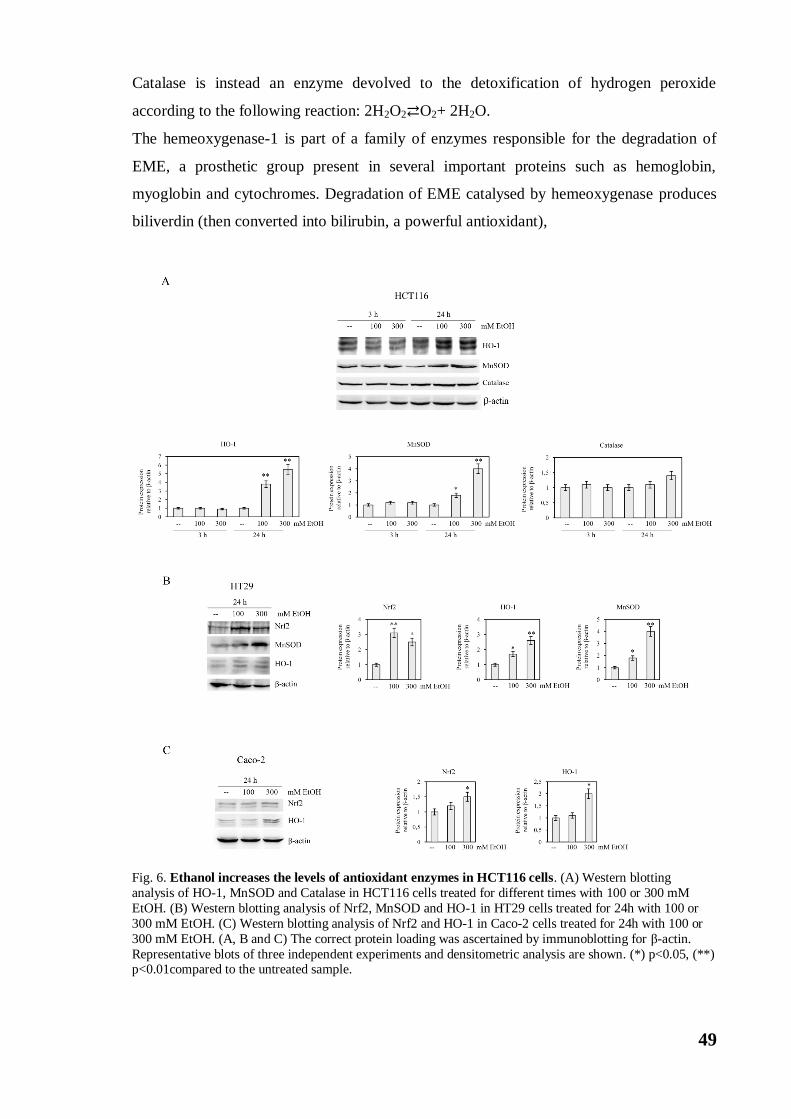
49
Catalase is instead an enzyme devolved to the detoxification of hydrogen peroxide
according to the following reaction: 2H2O2⇄O2+ 2H2O.
The hemeoxygenase-1 is part of a family of enzymes responsible for the degradation of
EME, a prosthetic group present in several important proteins such as hemoglobin,
myoglobin and cytochromes. Degradation of EME catalysed by hemeoxygenase produces
biliverdin (then converted into bilirubin, a powerful antioxidant),
Fig. 6. Ethanol increases the levels of antioxidant enzymes in HCT116 cells. (A) Western blotting
analysis of HO-1, MnSOD and Catalase in HCT116 cells treated for different times with 100 or 300 mM
EtOH. (B) Western blotting analysis of Nrf2, MnSOD and HO-1 in HT29 cells treated for 24h with 100 or
300 mM EtOH. (C) Western blotting analysis of Nrf2 and HO-1 in Caco-2 cells treated for 24h with 100 or
300 mM EtOH. (A, B and C) The correct protein loading was ascertained by immunoblotting for β-actin.
Representative blots of three independent experiments and densitometric analysis are shown. (*) p<0.05, (**) p<0.01compared to the untreated sample.
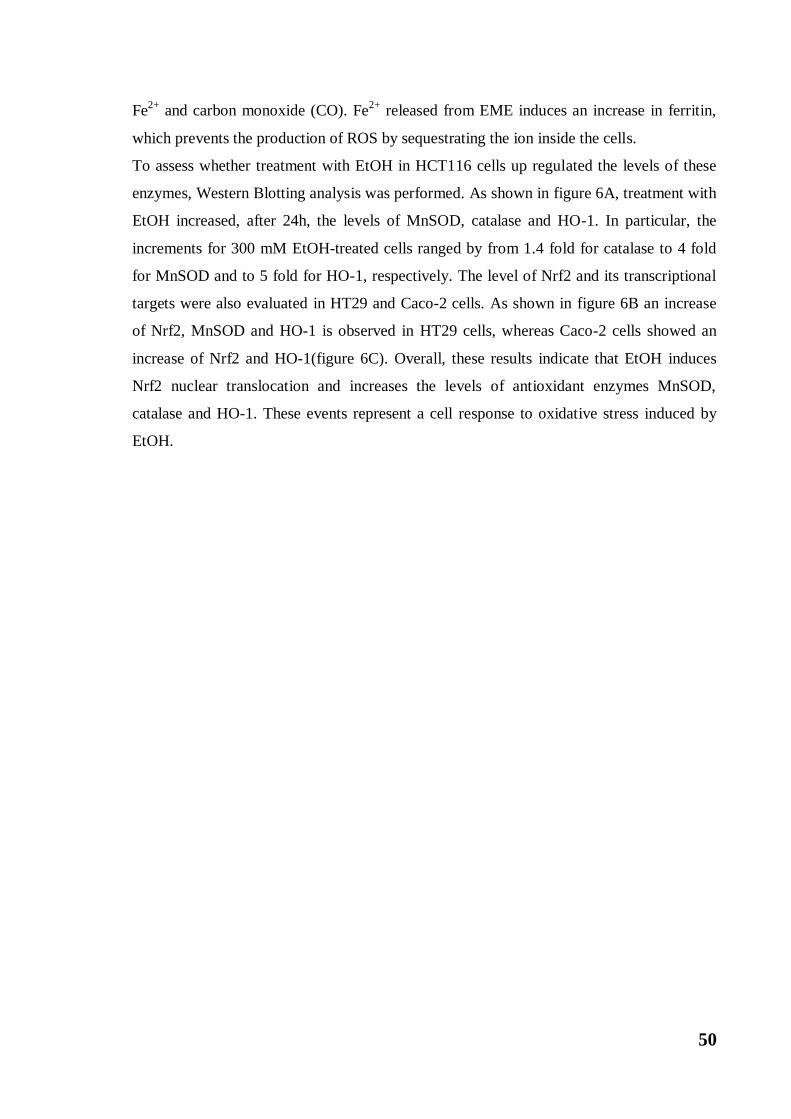
50
Fe2+
and carbon monoxide (CO). Fe2+
released from EME induces an increase in ferritin,
which prevents the production of ROS by sequestrating the ion inside the cells.
To assess whether treatment with EtOH in HCT116 cells up regulated the levels of these
enzymes, Western Blotting analysis was performed. As shown in figure 6A, treatment with
EtOH increased, after 24h, the levels of MnSOD, catalase and HO-1. In particular, the
increments for 300 mM EtOH-treated cells ranged by from 1.4 fold for catalase to 4 fold
for MnSOD and to 5 fold for HO-1, respectively. The level of Nrf2 and its transcriptional
targets were also evaluated in HT29 and Caco-2 cells. As shown in figure 6B an increase
of Nrf2, MnSOD and HO-1 is observed in HT29 cells, whereas Caco-2 cells showed an
increase of Nrf2 and HO-1(figure 6C). Overall, these results indicate that EtOH induces
Nrf2 nuclear translocation and increases the levels of antioxidant enzymes MnSOD,
catalase and HO-1. These events represent a cell response to oxidative stress induced by
EtOH.
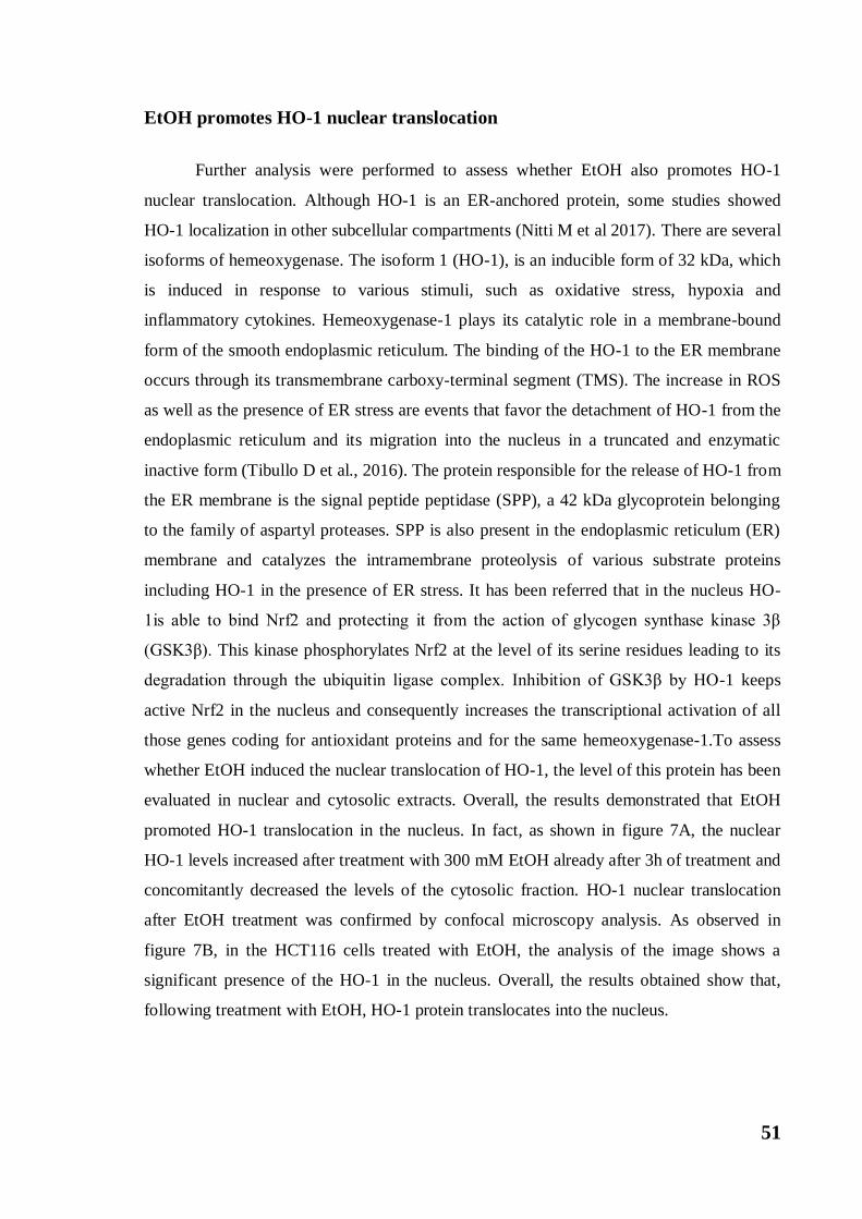
51
EtOH promotes HO-1 nuclear translocation
Further analysis were performed to assess whether EtOH also promotes HO-1
nuclear translocation. Although HO-1 is an ER-anchored protein, some studies showed
HO-1 localization in other subcellular compartments (Nitti M et al 2017). There are several
isoforms of hemeoxygenase. The isoform 1 (HO-1), is an inducible form of 32 kDa, which
is induced in response to various stimuli, such as oxidative stress, hypoxia and
inflammatory cytokines. Hemeoxygenase-1 plays its catalytic role in a membrane-bound
form of the smooth endoplasmic reticulum. The binding of the HO-1 to the ER membrane
occurs through its transmembrane carboxy-terminal segment (TMS). The increase in ROS
as well as the presence of ER stress are events that favor the detachment of HO-1 from the
endoplasmic reticulum and its migration into the nucleus in a truncated and enzymatic
inactive form (Tibullo D et al., 2016). The protein responsible for the release of HO-1 from
the ER membrane is the signal peptide peptidase (SPP), a 42 kDa glycoprotein belonging
to the family of aspartyl proteases. SPP is also present in the endoplasmic reticulum (ER)
membrane and catalyzes the intramembrane proteolysis of various substrate proteins
including HO-1 in the presence of ER stress. It has been referred that in the nucleus HO-
1is able to bind Nrf2 and protecting it from the action of glycogen synthase kinase 3β
(GSK3β). This kinase phosphorylates Nrf2 at the level of its serine residues leading to its
degradation through the ubiquitin ligase complex. Inhibition of GSK3β by HO-1 keeps
active Nrf2 in the nucleus and consequently increases the transcriptional activation of all
those genes coding for antioxidant proteins and for the same hemeoxygenase-1.To assess
whether EtOH induced the nuclear translocation of HO-1, the level of this protein has been
evaluated in nuclear and cytosolic extracts. Overall, the results demonstrated that EtOH
promoted HO-1 translocation in the nucleus. In fact, as shown in figure 7A, the nuclear
HO-1 levels increased after treatment with 300 mM EtOH already after 3h of treatment and
concomitantly decreased the levels of the cytosolic fraction. HO-1 nuclear translocation
after EtOH treatment was confirmed by confocal microscopy analysis. As observed in
figure 7B, in the HCT116 cells treated with EtOH, the analysis of the image shows a
significant presence of the HO-1 in the nucleus. Overall, the results obtained show that,
following treatment with EtOH, HO-1 protein translocates into the nucleus.
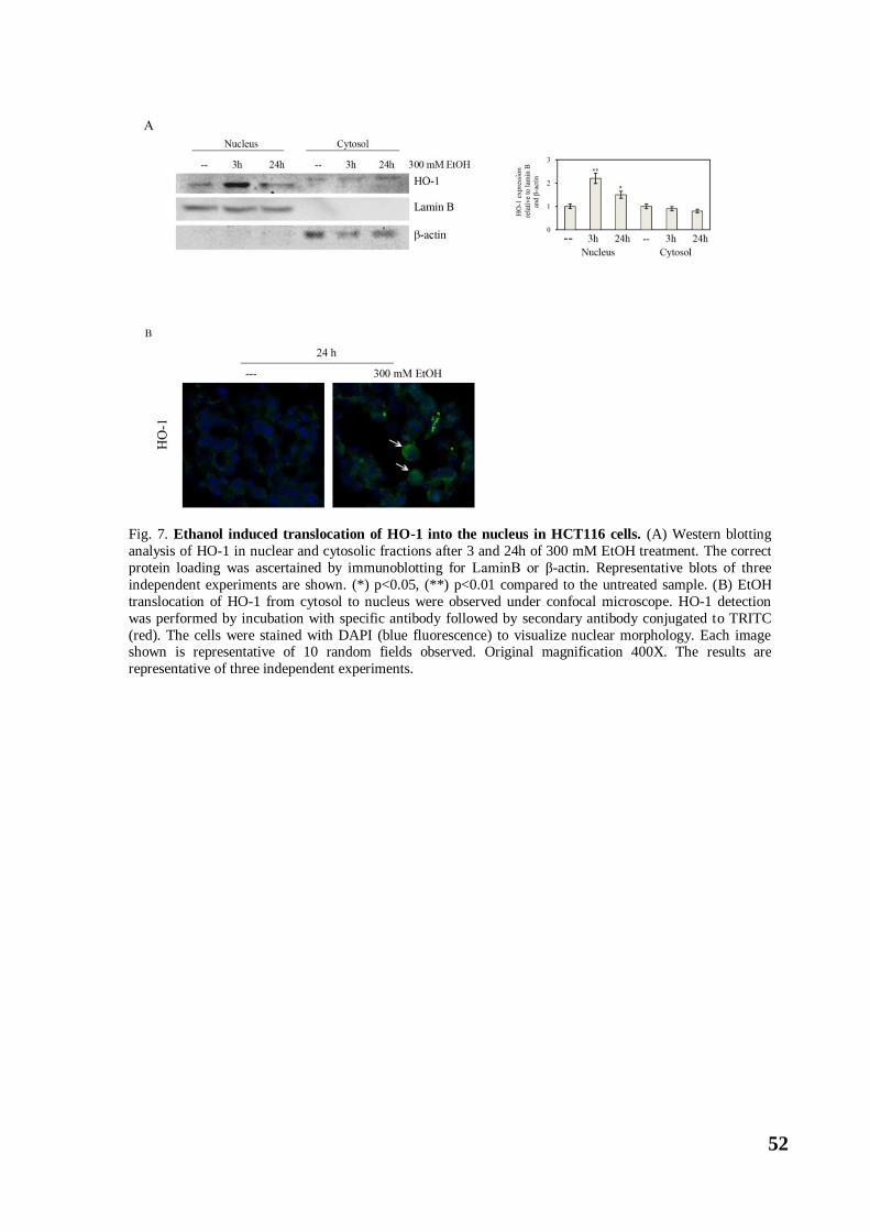
52
Fig. 7. Ethanol induced translocation of HO-1 into the nucleus in HCT116 cells. (A) Western blotting
analysis of HO-1 in nuclear and cytosolic fractions after 3 and 24h of 300 mM EtOH treatment. The correct
protein loading was ascertained by immunoblotting for LaminB or β-actin. Representative blots of three
independent experiments are shown. (*) p<0.05, (**) p<0.01 compared to the untreated sample. (B) EtOH
translocation of HO-1 from cytosol to nucleus were observed under confocal microscope. HO-1 detection
was performed by incubation with specific antibody followed by secondary antibody conjugated to TRITC
(red). The cells were stained with DAPI (blue fluorescence) to visualize nuclear morphology. Each image shown is representative of 10 random fields observed. Original magnification 400X. The results are
representative of three independent experiments.
53
EtOH increased MMPs and VEGF in colon cancer cells
It has been shown that activation ofNrf2/HO-1 axis at the nuclear level is associated with
tumor progression and invasiveness (Hsu FF et al 2014). In fact, activation of Nrf2 has
been associated with up regulation of MMP9 in hepatocellular carcinoma (Zhang M et al,
2015) and hemeoxygenase-1 (HO-1) has been reported to promote angiogenesis (Dulak Jet
al 2004). In light of the results obtained, the effect of EtOH on the levels of matrix
metalloproteases 2 and 9 (MMP-2 and MMP-9) was assessed. The matrix metalloproteases
are zinc dependent endopeptidases that degrade many components of the extracellular
matrix, in particular the collagen IV and the lamin, allowing the cells to penetrate inside
Fig. 8.Ethanol increases the levels of VEGF and activates metalloproteases. (A) Western blotting
analysis of MMP-2, MMP-9 and VEGF in HCT116 cells treated for different times with 100 or 300 mM
EtOH. The correct protein loading was ascertained by immunoblotting for β-actin. Representative blots of three independent experiments and densitometric analysis are shown. (**) p<0.01, (***) p<0.001compared to
the untreated sample. (B) The activity of MMP-2 and MMP-9 in HCT116 cells treated with 300 mM EtOH
for 48h was examined by gelatin zymography assay, as described in Materials and Method. The results are
representative of three independent experiments.
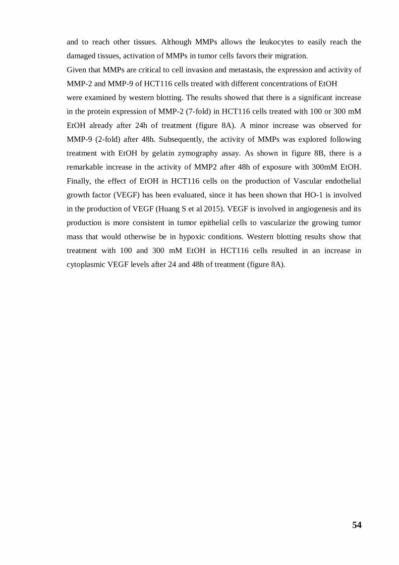
54
and to reach other tissues. Although MMPs allows the leukocytes to easily reach the
damaged tissues, activation of MMPs in tumor cells favors their migration.
Given that MMPs are critical to cell invasion and metastasis, the expression and activity of
MMP-2 and MMP-9 of HCT116 cells treated with different concentrations of EtOH
were examined by western blotting. The results showed that there is a significant increase
in the protein expression of MMP-2 (7-fold) in HCT116 cells treated with 100 or 300 mM
EtOH already after 24h of treatment (figure 8A). A minor increase was observed for
MMP-9 (2-fold) after 48h. Subsequently, the activity of MMPs was explored following
treatment with EtOH by gelatin zymography assay. As shown in figure 8B, there is a
remarkable increase in the activity of MMP2 after 48h of exposure with 300mM EtOH.
Finally, the effect of EtOH in HCT116 cells on the production of Vascular endothelial
growth factor (VEGF) has been evaluated, since it has been shown that HO-1 is involved
in the production of VEGF (Huang S et al 2015). VEGF is involved in angiogenesis and its
production is more consistent in tumor epithelial cells to vascularize the growing tumor
mass that would otherwise be in hypoxic conditions. Western blotting results show that
treatment with 100 and 300 mM EtOH in HCT116 cells resulted in an increase in
cytoplasmic VEGF levels after 24 and 48h of treatment (figure 8A).
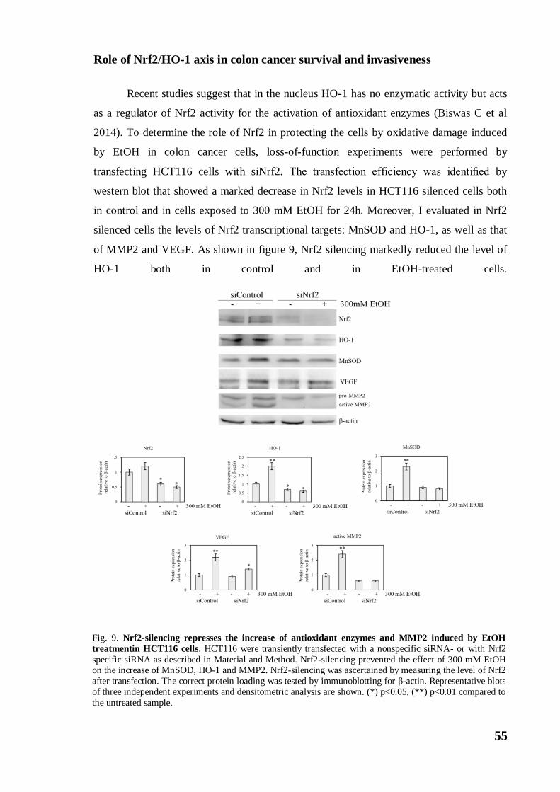
55
Role of Nrf2/HO-1 axis in colon cancer survival and invasiveness
Recent studies suggest that in the nucleus HO-1 has no enzymatic activity but acts
as a regulator of Nrf2 activity for the activation of antioxidant enzymes (Biswas C et al
2014). To determine the role of Nrf2 in protecting the cells by oxidative damage induced
by EtOH in colon cancer cells, loss-of-function experiments were performed by
transfecting HCT116 cells with siNrf2. The transfection efficiency was identified by
western blot that showed a marked decrease in Nrf2 levels in HCT116 silenced cells both
in control and in cells exposed to 300 mM EtOH for 24h. Moreover, I evaluated in Nrf2
silenced cells the levels of Nrf2 transcriptional targets: MnSOD and HO-1, as well as that
of MMP2 and VEGF. As shown in figure 9, Nrf2 silencing markedly reduced the level of
HO-1 both in control and in EtOH-treated cells.
Fig. 9. Nrf2-silencing represses the increase of antioxidant enzymes and MMP2 induced by EtOH
treatmentin HCT116 cells. HCT116 were transiently transfected with a nonspecific siRNA- or with Nrf2
specific siRNA as described in Material and Method. Nrf2-silencing prevented the effect of 300 mM EtOH on the increase of MnSOD, HO-1 and MMP2. Nrf2-silencing was ascertained by measuring the level of Nrf2
after transfection. The correct protein loading was tested by immunoblotting for β-actin. Representative blots
of three independent experiments and densitometric analysis are shown. (*) p<0.05, (**) p<0.01 compared to
the untreated sample.
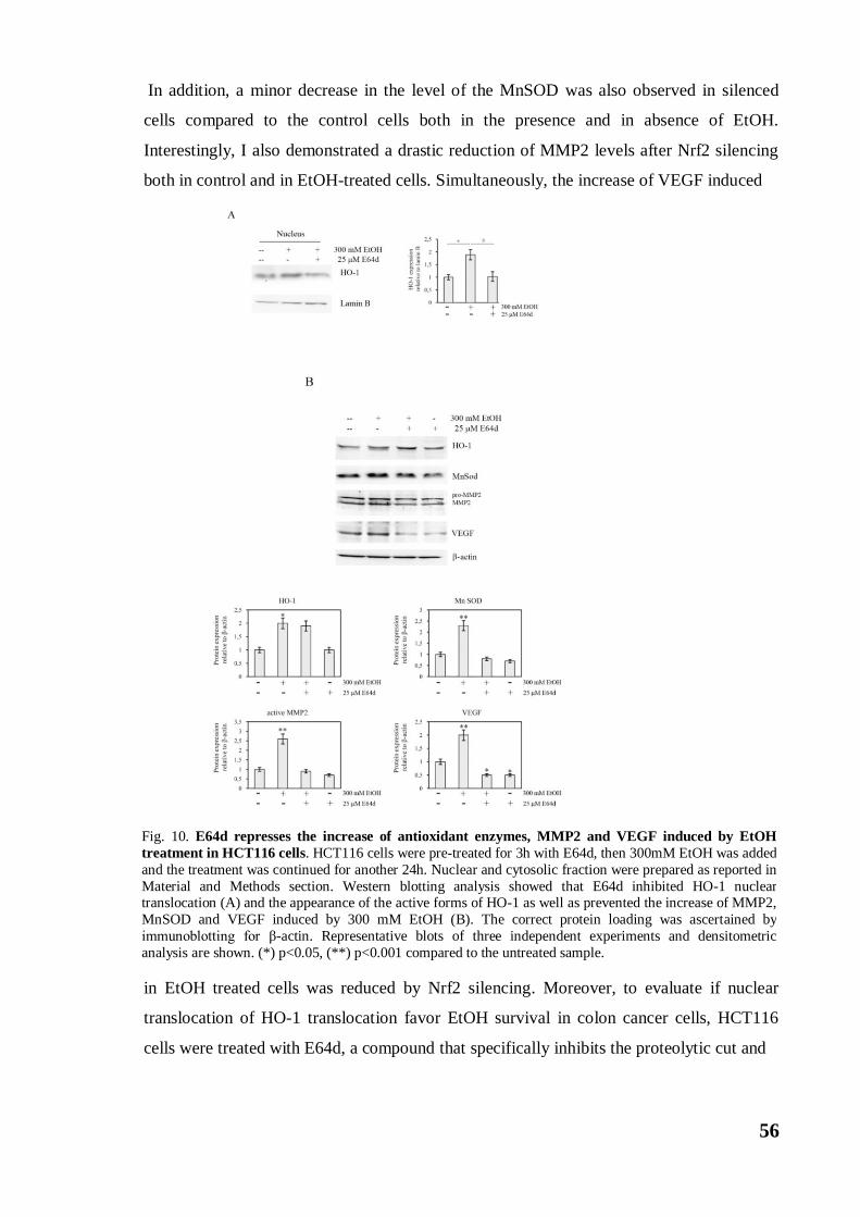
56
In addition, a minor decrease in the level of the MnSOD was also observed in silenced
cells compared to the control cells both in the presence and in absence of EtOH.
Interestingly, I also demonstrated a drastic reduction of MMP2 levels after Nrf2 silencing
both in control and in EtOH-treated cells. Simultaneously, the increase of VEGF induced
in EtOH treated cells was reduced by Nrf2 silencing. Moreover, to evaluate if nuclear
translocation of HO-1 translocation favor EtOH survival in colon cancer cells, HCT116
cells were treated with E64d, a compound that specifically inhibits the proteolytic cut and
Fig. 10. E64d represses the increase of antioxidant enzymes, MMP2 and VEGF induced by EtOH
treatment in HCT116 cells. HCT116 cells were pre-treated for 3h with E64d, then 300mM EtOH was added
and the treatment was continued for another 24h. Nuclear and cytosolic fraction were prepared as reported in
Material and Methods section. Western blotting analysis showed that E64d inhibited HO-1 nuclear translocation (A) and the appearance of the active forms of HO-1 as well as prevented the increase of MMP2,
MnSOD and VEGF induced by 300 mM EtOH (B). The correct protein loading was ascertained by
immunoblotting for β-actin. Representative blots of three independent experiments and densitometric
analysis are shown. (*) p<0.05, (**) p<0.001 compared to the untreated sample.

57
nuclear translocation of HO-1. Combination of E64d with 300 mM EtOH was
accompanied by a reduction in nuclear localization of HO-1 (figure 10A). In addition,
E64d significantly reduced the cleaved form of HO-1 as well as suppressed the increase of
MnSOD protein level induced by 300 mM EtOH treatment (figure 10B), thereby
suggesting that nuclear HO-1 could regulate MnSOD expression. Interesting co-treatment
with E64d and EtOH also causes a considerable reduction in MMP-2 and VEGF levels
(figure 10B), thus suggesting that nuclear HO-1 could cooperate with Nrf2 to promote
invasiveness of colon cancer cells.

58
Discussion
59
Many studies support the conclusion that chronic and heavy alcohol consumption
not only increases the risk to develop CRC, but also promotes tumor progression (Zhao H
et al 2018). My results provided evidence that in colon cancer cells activation of Nrf2/HO-
1 axis in response to high doses of ethanol promotes cell survival and induces the
acquisition of markers of aggressiveness.
Some evidence suggest that EtOH is able to increase ROS in different cell lines,
including colon cancer cells, through the activity of ADH and CYP2E2 (Yang C et al
2005). In accordance with these observations, I showed that EtOH rapidly increased ROS
production in different colon cancer cells (Caco-2, HCT116 and HT29 cells). In addition, I
demonstrated an increase in the level of both iNOS and COX2, two enzymatic markers of
inflammation, as well as the stress chaperones HSP90 and HSP60, thus suggesting that
EtOH stimulates the production of oxidative stress.
Accumulation of unfolded/misfolded proteins in the ER stimulates a response
known as Unfolded Protein Response (UPR) that is mediated by three types of ER
transmembrane receptors: protein kinase RNA-like ER kinase (PERK), activating
transcription factor 6 (ATF6) and inositol-requiring enzyme 1 (IRE1α). ER stress is known
to contribute to alcoholic damage of major organs such as the liver, pancreas, and brain.
The mechanisms responsible for alcoholic ER stress response are either directly or
indirectly related to alcohol metabolism, including the production of acetaldehyde protein
adducts and oxidative modification of proteins (Ji C 2012). My results provided evidence
that high doses of EtOH up-regulate both Grp78 and CHOP, two ER stress markers. Grp78
is an ER chaperone protein that binds to ATF6, PERK, and IRE1α in healthy cells. When
unfolded proteins accumulate, Grp78 dissociates from these proteins that activate down-
stream signaling genes to initiate UPR. CHOP is a transcription factor whose expression
markedly increases in response to ER stress and which mediates induction of apoptosis
under prolonged ER stress. The observation that the level of CHOP lowered after 24h of
treatment with 300 mM EtOH suggest that in my conditions ER stress has not been
correlated with the induction of apoptotic cell death. Despite the induction of oxidative and
ER stress in my system, MTT and clonogenic assay showed that colon cancer cells survive
also after prolonged treatment with high doses of EtOH, thereby suggesting the activation
of survival mechanisms. Accumulating evidence suggest that activation of autophagy,
which degrades proteins in organelles such as the mitochondria and the ER, can play a
60
protective role against the toxic effects of ER stress (Shimodaira Y et al 2014).
Interestingly, recent studies showed that alcohol consumption activates in CRC patients’
autophagy and that this event increased risk of tumor metastasis (Zhao H et al 2018).
It is now known that ER stress can activate autophagy and there is considerable
crosstalk between the ER and autophagy. It has been reported that ER stress activates
PERK, a kinase which in turn promotes the phosphorylation of eIF2α to induce the
activation of LC3, an autophagosome marker. Moreover, Shimodaira et al. (Shimodaira Y
et al 2014) demonstrated that activation of CHOP, an ER marker, CHOP promotes
autophagy in colon cancer cells by activating inositol-requiring enzyme 1α (IRE1α). My
results provided evidence that in HCT116 colon cancer cells EtOH activated LC3 by
promoting the cleavage of its cytosolic form LC3-I to LC3-II, as well as increased the level
of Beclin, a molecule involved in the initiation of autophagosome formation. The
involvement of autophagy was also confirmed by the augmented accumulation of acidic
intracellular compartments. Moreover, the observation that the level of p62 protein, a
marker of autophagic degradation, decreased after 24h of EtOH treatment, suggested that
the compound triggered in my conditions a complete autophagic response. This effect is
prevented by the addition of the antioxidant N-acetylcysteine, thus suggesting that
autophagy is activated as a survival mechanism in response to oxidative stress.
Concomitantly to oxidative and ER stress production, I showed a significant
activation of Nrf2, a transcription factor which acts as a key regulator of antioxidant-
responsive genes (Kim J et al 2016). Under my experimental conditions, EtOH promoted
translocation of Nrf2 into the nucleus, as suggested by the increase of nuclear Nrf2 content
already at 3h following EtOH treatment. This event was associated with a reduction in the
level of Keap1 (Kelch-like ECH-associated protein1), a protein which in unstressed
condition sequester Nrf2 in a cytoplasmic complex leading to its ubiquitination and
consequent proteasomal degradation (Dodson M et al 2015). Nuclear activation of Nrf2 by
EtOH could be promoted by oxidative and ER stress. In line with this conclusion, it has
been reported that induction of oxidative stress triggers Nrf2 nuclear import through the
oxidation of redox-sensitive cysteines within Keap1. Moreover, phosphorylation of Nrf2
by PERK, a kinase activated following the accumulation of unfolded proteins in the
endoplasmic reticulum (ER), promotes dissolution of Nrf2/Keap1 complexes and Nrf2
nuclear import. Finally, a relationship between autophagy and Nrf2 activation has been
also demonstrated. In fact, recent evidence revealed that the autophagic protein p62
interacts with Keap1 favoring its dissociation from Nrf2 (Katsuragi Y et al 2016).
61
Nrf2 is the main antioxidant system in the cells. After nuclear translocation, Nrf2
form transcriptionally active complexes with other proteins, such as Mafs, thereby
transactivating the expression of a battery of genes encoding antioxidant enzymes such as
MnSOD, catalase and HO-1. My data indicated that the level of both SOD and HO-1 was
significantly up-regulated after EtOH treatment in colon cancer cells, suggesting a
protective role of these enzymes against EtOH-induced oxidative stress. The observation
that by silencing Nrf2, EtOH-induced HO-1 and SOD increased expression was
significantly reduced, suggested that this effect was mediated by activation of the
transcription factor Nrf2.
Interestingly, my data also provided evidence that EtOH promotes HO-1 nuclear
translocation already after 3h of treatment. HO-1 is a stress-inducible enzyme localized in
the endoplasmic reticulum which exerts anti-oxidant and anti-inflammatory effects through
the production of carbon monoxide (CO) and biliverdin. Recent evidences suggest that
HO-1 can translocate in a truncated form into the nucleus where it binds to transcription
factors favoring gene transcription (Biswas C et al 2014). Nuclear expression of HO-1 has
been detected in several tumors and it has been correlated with tumor growth and invasion
(Nitti M et al 2017). To demonstrate whether nuclear translocation of HO-1 is involved in
cell survival of EtOH-treated colon cancer cells, I treated HCT116 cells with EtOH in
combination with E64d, a cysteine protease inhibitor. Inhibition of the proteolytic cleavage
necessary for HO-1 nuclear translocation significantly reduced the EtOH-induced HO-1
nuclear translocation increase as well as prevented EtOH-induced MnSOD increase,
showing that HO-1 nuclear cooperates with Nrf2 to stimulate antioxidant response and
colon cancer cell survival. This suggestion is in accordance with some recent evidence
which demonstrated that nuclear HO-1 modulates the activation of Nrf2, leading to
activation of antioxidant genes. Interestingly, it has been shown that nuclear HO-1 prevents
GSK3β induced phosphorylation of Nrf2 leading to its stabilization and activation (Biswas
C et al 2014).
Activation of the Nrf2/HO-1 axis is a double-edged sword in cancer. The decrease
of oxidative stress by activation of antioxidant response by both Nrf2 and HO-1 can
prevent the development of tumors by counteracting the genotoxic damage induced by
ROS. In line with this consideration, several dietary phytochemicals exert cancer
preventive effect by activating Nrf2/HO-1 axis. Moreover, activation of Nrf2 reduced
chronic inflammation which has been correlated with CRC induction (Ahmed SM et al
2017). On the other hand, the activation of Nrf2 antioxidant response in tumor cells can
promote the survival of cancer cells by creating an optimal environment for cell growth.
62
Overexpression of Nrf2 has been detected in primary CRC and metastatic tissue relative to
normal colon and contributes to chemo-resistance in CRC cell lines (Sadeghi MR et al
2017). In addition, it has been reported that Nrf2 increased CRC risk by promoting colonic
inflammation or promoting angiogenesis and uncontrolled proliferation. Moreover, HO-1
overexpression has been associated with a more aggressive behavior of tumors and poor
prognosis in various cancers.
Western blotting and zymography analyses demonstrated that EtOH increased both the
levels and the activity of MMP-2 and 9, two enzymes involved in tumor progression.
Moreover, EtOH up-regulated VEGF, the main angiogenetic factor. To clarify whether the
activation of MMPs and VEGF was regulated by Nrf2/HO-1 axis, I evaluated the effect of
EtOH after Nrf2 silencing or in the presence of E64d, the cysteine protease inhibitor able
to inhibit either the proteolytic cut or the nuclear translocation of HO-1. Interestingly, Nrf2
silencing prevented EtOH-induced MMP-2 increase and reduced VEGF increase.
Moreover, inhibition of HO-1 nuclear translocation by E64d addition counteracted the
effect of EtOH on both MMP-2 and VEGF, thus suggesting a role of Nrf2/HO-1 axis in
colon cancer progression.
Collectively, my findings demonstrate that high doses of EtOH enhanced in colon
cancer cells autophagy and activation Nrf-2/HO-1 axis. These events sustain the survival
of cancer cells by protecting the cells from oxidative and ER stress induced by EtOH.
Moreover, the activation of Nrf2/HO-1 axis seems to be also responsible for colon cancer
progression through the acquisition of markers of invasiveness, like MMP-2 and VEGF.

63
Bibliography
64
Ahmed SM, Luo L, Namani A, Wang XJ, Tang X. Nrf2 signaling pathway: Pivotal roles
in inflammation.Biochimicaetbiophysicaacta- molecular basis of disease, 585-597, 2017.
Amaral JD, Xavier JM, Steer CJ, Rodrigues CM. The role of p53 in apoptosis. Discovery
Medicine, 145–152, 2010.
Amin P.B, Diebel L.N, Liberati D.M. Dose-dependent effects of ethanol and E. coli on
gut permeability and cytokine production. Journal Surgery Research, 187–192, 2009.
Anderson JW, Baird P, Davis RH, Ferreri S, Knudtson M, Koraym A, Waters V, Williams
CL. Health benefits of dietary fiber. Nutrition Reviews, 188–205, 2009.
Armaghany T, Wilson JD, Chu Q, Mills G. Genetic alterations in colorectal cancer.
Gastrointestinal Cancer Research, 19–27, 2012.
Aune D, Chan DS, Lau R, Vieira R, Greenwood DC, Kampman E, Norat T. Dietary fibre,
whole grains, and risk of colorectal cancer: systematic review and dose-response
meta-analysis of prospective studies. Biology Molecular Journal, 343-363, 2011.
Baan R, Straif K, Grosse Y, Secretan B, El Ghissassi F, Bouvard V, Altieri A, Cogliano V.
Carcinogenicity of alcoholic beverages.Lancet Oncology, 292-293, 2007.
Bahnassy AA, Zekri AR, Salem SE, Abou-Bakr AA, Sakr MA, Abdel-Samiaa AG, Al-
Bradei M. Differential expression of p53 family proteins in colorectal adenomas and
carcinomas: Prognostic and predictive values. Histology and Histopathology, 207–216,
2014.
Bastide NM, Pierre FH, Corpet DE. Heme iron from meat and risk of colorectal cancer:
a meta-analysis and a review of the mechanisms involved. Cancer Prevention Research,
177–184, 2011.
Bbosa G, Kyegombe D, Anokbonggo W, Lubega A, Mugisha A, Ogwal-Okeng J. Chronic
Alcohol Consumption Affects Serum Enzymes Levels in the HIV-Infected Patients on
Stavudine (d4T)/Lamivudine (3TC)/Nevirapine (NVP) Treatment Regimen.
Pharmacology & Pharmacy, 430-444, 2014.
Ben Q, Sun Y, Chai R, Qian A, Xu B, Yuan Y. Dietary fiber intake reduces risk for
colorectal adenoma: a meta-analysis. Gastroenterology, 689–699, 2014.
Bernstein H, Bernstein C, Payne CM, Dvorak K. Bile acids as endogenous etiologic
agents in gastrointestinal cancer. World Journal Gastroenterology, 3329–3340, 2009.
Biswas C, Shah N, Muthu M, La P, Fernando AP, Sengupta S, Yang G, Dennery PA.
Nuclear Heme Oxygenase-1 (HO-1) Modulates Subcellular Distribution and
Activation of Nrf2, Impacting Metabolic and Anti-oxidant Defenses.Journal of
biological chemistry, 26882–26894, 2014.
65
Bosron WF, Crabb DW, Li TK. Relationship between kinetics of liver alcohol
dehydrogenase and alcohol metabolism.Pharmacology Biochemistry and Behavior, 223-
227, 1983.
Bravo R, Parra V, Gatica D, Rodiguez A E, Torrealba N, Paredes F, Wang Z V, Zorzano
A, Hill J A, Jaimovich E, Quest A F G, Lavandero S. Endoplasmic Reticulum and the
Unfolding Protein Response: dynamics and metabolic integration. International
Review of Cell and Molecular Biology, 301- 315, 2013.
Brennan P, Lewis S, Hashibe M, Bell DA, Boffetta P, Bouchardy C, Caporaso N, Chen C,
Coutelle C, Diehl SR, Hayes RB, Olshan AF, Schwartz SM, Sturgis EM, Wei Q, Zavras
AI, Benhamou S Pooled analysis of alcohol dehydrogenase genotypes and head and
neck cancer: a HuGE review. American Journal of Epidemiology, 1-16, 2004.
Calabrese P, Tsao JL, et al. Colorectal pretumor progression before and after loss of
DNA mismatch repair. The American Journal of Pathology, 1447-1453, 2004.
Candela M, Turroni S, Biagi E, Carbonero F, Rampelli S, Fiorentini C, Brigidi P.
Inflammation and colorectal cancer, when microbiota-host mutualism breaks. World
Journal of Gastroenterology, 908–922, 2014.
Chen X, Shen J, Prywes R. The luminal domain of ATF6 senses endoplasmic reticulum
(ER) stress and causes translocation of ATF6 from the ER to the Golgi. Journal of
Biological Chemistry, 13045-13052, 2002.
Cho E, Smith-Warner SA, Ritz J, van den Brandt PA, Colditz GA, Folsom AR,
Freudenheim JL, Giovannucci E, Goldbohm RA, Graham S, Holmberg L, Kim DH, Malila
N, Miller AB, Pietinen P, Rohan TE, Sellers TA, Speizer FE, Willett WC, Wolk A, Hunter
DJ. Alcohol intake and colorectal cancer: a pooled analysis of 8 cohort studies.Annals
of Internal Medicine,603-613, 2004.
Choi SH, Estarás C, Moresco JJ, Yates JR, Jones KA. α-Catenin interacts with APC to
regulate β-catenin proteolysis and transcriptional repression of Wnt target genes.
Genes and Development, 2473–2488, 2013.
Cohen MB, Jensen NJ, Hawkins JA, Mann EA, Thompson MR, Lentze MJ, Giannella RA.
Receptors for Escherichia coli heat stable enterotoxin in human intestine and in a
human intestinal cell line (Caco-2). Journal of Cellular Physiology. 138-144. 1993.
Comoglio P.M, Trusolino L. Invasive growth: from development to metastasis. The
Journal of Clinical Investigation, 857–862, 2002.
Connor PD, Streissguth AP. Effect of prenatal exposure to alcohol across the life span.
Alcohol Health Research World, 170-174, 1996.
Corrao G, Rubbiati L, Bagnardi V, Zambon A, Poikolainen K: Alcohol and coronary
heart disease: a meta-analysis. Addiction, 1505-1523, 2000.
Coskun Ü, Grzybek M, Drechsel D, Simons K. Regulation of human egf receptor by
lipids. Proceeding of the National Academy of Sciences of the United States of
America,9044–9048, 2011.
66
Dobre M, Comănescu M, Arsene D, Iosif C, Bussolati G. K-ras gene mutation status in
colorectal cancer: comparative analysis of pyrosequencing and PCR-RFLP. Romanian
Journal of Morphology and Embryology, 567–574, 2013.
Dodson M, Redmann M, Rajasekaran NS, Darley-Usmar V, Zhang J.KEAP1-NRF2
signalling and autophagy in protection against oxidative and reductive
proteotoxicity.Biochemistry Journal, 347-355. 2015.
Dulak J, Łoboda A, Zagórska A, Józkowicz A.Complex role of heme oxygenase-1 in
angiogenesis. Antioxidant Redox Signal,858-66, 2004
el Ghissassi F, Barbin A, Nair J, Bartsch H. Formation of 1,N6-ethenoadenine and 3,N4-
ethenocytosine by lipid peroxidation products and nucleic acid bases.Chemical
Research Toxicology, 278-283, 1995.
Ellina MI, Bouris P, Aletras AJ, Theocharis AD, Kletsas D, Karamanos NK. Egfr and
her2 exert distinct roles on colon cancer cell functional properties and expression of
matrix macromolecules. BiochimicaetBiophysicaActa, 2651–2661, 2014.
Eswaran S, Muir J, Chey WD. Fiber and functional gastrointestinal disorders. The
American Journal of Gastroenterology, 718–727, 2013.
Fang JL, Vaca CE. Detection of DNA adducts of acetaldehyde in peripheral white
blood cells of alcohol abusers.Carcinogenesis, 627-32, 1997.
Fearon ER, Vogelstein B. A genetic model for colorectal tumorigenesis. Cell, 759-
767,1990.
Gammoh N, Marks P and Jiang X.Curbing autophagy and histone deacetylases to kill
cancer cells. Autophagy, 1521-1522, 2012.
Grady WM, Carethers JM. Genomic and epigenetic instability in colorectal cancer
pathogenesis. Gastroenterology, 1079–1099, 2008.
Haggar FA, Boushey RP. Colorectal cancer epidemiology: incidence, mortality,
survival, and risk factors. Clinics in Colon and Rectal Surgery. 191-7,2009.
Harding HP , Zhang Y, Zeng H, Novoa I, Lu PD, Calfon M, Sandri N, Yun C, Popko B,
Paules R, Stojdi DF, Bell JC, Hettmann T, Leiden JM, Ron D. An integrated stress
response regulates amino acid metabolism and resistance to oxidative stress.
MolecularCell.619-633,2003.
Homann N, Tillonen J, Salaspuro M. Microbially produced acetaldehyde from ethanol
may increase the risk of colon cancer via folate deficiency. International Journal Cancer,
169–173, 2000.
Hsu FF, Yeh CT, Sun YJ, Chiang MT, Lan WM, Li FA. Signal peptide peptidase-
mediated nuclear localization of heme oxygenase-1 promotes cancer cell proliferation
and invasion independent of its enzymatic activity.Oncogene, 2360-2370, 2014.
Huang S, Lin C, Lin H, Chiu C, Fang C,Liao K, Chen D and Yeh W. Brain-derived
neurotrophic factorregulates cell motility in human colon cancer. Endocrine-Related
Cancer,455–464, 2015.
67
Ionov Y, Peinado MA, Malkhosyan S, Shibata D, Perucho M.Ubiquitous somatic
mutations in simple repeated sequences reveal a new mechanism for colonic
carcinogenesis. Nature, 558–561, 1993.
Ji C. Mechanisms of alcohol-induced endoplasmic reticulum stress and
organinjuries.Biochemistry research international, 550-562, 2012.
Jokelainen K, Roine R.P, Vaananen H, Farkkila M, Salaspuro M. In Vitro acetaldehyde
formation by human colonic bacteria. Gut, 1271–1274, 1994.
Jokelainen K, Siitonen A, Jousimies-Somer H, Nosova T, Heine R, Salaspuro M. In Vitro
alcohol dehydrogenase-mediated acetaldehyde production by aerobic bacteria
representing the normal colonic flora in man. Alcoholism Clinical and Experimental
Research, 967–972, 1996.
Katsuragi Y, Ichimura Y and Komatsu M. p62/SQSTM1 functions as a signaling hub
and an autophagy adaptor. Current Opinion in Toxicology, 54-61, 2016.
Kaufman RJ. Stress signaling from the lumen of the endoplasmic reticulum
coordination of gene transcriptional and translational controls. Genes& Development,
1211-1233,1999.
Kim J, Keum YS. NRF2, a Key Regulator of Antioxidants with Two Faces
towardsCancer.Oxidative Medicine and Cellular longevity. 380-387, 2016
Kobayashi M, Yamamoto M.Molecular mechanisms activating the Nrf2-Keap1
pathway of antioxidant gene regulation. Antioxid Redox Signal.385-394, 2005.
Kono S, Chen K. Genetic polymorphisms of methylenetetrahydrofolate reductase and
colorectal cancer and adenoma. Cancer Science, 535–542, 2005.
Kourtis N, Tavernarakis N, Autophagy and cell death in model organisms. Cell Death
eDifferentiation, 21-30, 2009.
Ladiges WC, Knoblaugh SE, Morton JF, Korth MJ, Sopher BL, Baskin CR, MacAuley A,
Goodman AG, LeBoeuf RC, Katze MG. Pancreatic beta-cell failure and diabetes in
mice with a deletion mutation of the endoplasmic reticulum molecular chaperone
gene P58IPK. Diabetes, 1074-81, 2005.
Levine B, Yuan J.Autophagy in cell death: an innocent convict?The Journal of Clinical
Investigation, 2679-2688, 2005.
Lieber CS. Relationships between nutrition, alcohol use, and liver disease. Alcohol
Research Health, 220-31, 2003.
Lilla C, Koehler T, Kropp S, Wang-Gohrke S, Chang-Claude J. Alcohol dehydrogenase
1B (ADH1B) genotype, alcohol consumption and breast cancer risk by age 50 years in
a German case-control study. British Journal of Cancer 2039-41, 2005.
Liu Y, Yang P, Shun C, Wu M, Weng J and Chen C.Autophagy potentiates the
anticancereffects of the histone deacetylase inhibitors in hepatocellular
carcinoma.Autophagy, 1057-1065, 2010.
Lopez PH, Schnaar RL. Gangliosides in cell recognition and membrane protein
regulation. Current Opinion in Structural Biology, 549–557, 2009.
68
Lynch HT, de la Chapelle A. Hereditary colorectal cancer. The New England Journal of
Medicine, 919–932, 2003.
Ma Y, Hendershot LM. ER chaperone functions during normal and stress conditions.
Journal of Chemicalneuroanatomy, 51-65, 2004.
Medzhitov R. Origin and physiological roles of inflammation. Nature, 428–435, 2008.
Mehlen P, Rabizadeh S, Snipas S, Assa-Munt N, Salvesen G &Bredesen D. The DCC
gene product induces apoptosis by a mechanism requiring receptor proteolysis.
Nature, 801–804, 1998.
Munira Akhter, Shinichi Kuriyama, Naoki Nakaya, TaichiShimazu, Kaori Ohmori,
Yoshikazu Nishino, Yoshitaka Tsubono, Akira Fukao, Ichiro Tsuji.Alcohol consumption
is associated with an increased risk of distal colon and rectal cancer in Japanese men:
The Miyagi Cohort Study. European Journal of cancer,383-390, 2007.
Nitti M, Piras S, Marinari U, Moretta L, Pronzato M and Furfaro A. HO-1 Induction in
Cancer Progression: A Matter of Cell Adaptation. Antioxidants,29-35, 2017
Nosova T, Jousimies-Somer H, Kaihovaara P, Jokelainen K, Heine R, Salaspuro M.
Characteristics of alcohol dehydrogenases of certain aerobic bacteria representing
human colonic flora. Alcoholism Clinical&Experimental Research, 489–494, 1997.
Osier M, Pakstis AJ, Kidd JR, Lee JF, Yin SJ, Ko HC, Edenberg HJ, Lu RB, Kidd KK.
Linkage disequilibrium at the ADH2 and ADH3 loci and risk of alcoholism. American
Journal of Human Genetics, 1147-57. 1999.
Pedroni M, Sala E, et al. Microsatellite instability and mismatch-repair protein
expression in hereditary and sporadic colorectal carcinogenesis. CancerResearch, 896-
899, 2001.
Piskur J, Rozpedowska E, Polakova S, Merico A, Compagno C. How did
Saccharomyces evolve to become a good brewer?Trends Genetic, 183-186, 2006.
Prakash A, Janosi L, Doxastakis M. Self-association of models of transmembrane
domains of erbb receptors in a lipid bilayer. Biophysical Journal, 3657–3665, 2010.
Pyo JO, Nah J, Jung YK.Molecules and their functions in autophagy. Experimental
eMolecularMedicine, 73-80, 2012.
Rao YK, Fang SH, Tzeng YM. Synthesis and biological evaluation of 3’,4’,5’-
trimethoxychalcone analogues as inhibitors of nitric oxide production and tumor cell
proliferation. Bioorganic and Medicinal Chemistry, 7909–7914, 2009.
Rehm J, Mathers C, Popova S, Thavorncharoensap M, Teerawattananon Y, Patra J. Global
burden of disease and injury and economic cost attributable to alcohol use and
alcohol-use disorders. Lancet, 2223-33, 2009.
Sadeghi MR, Jeddi F, Soozangar N, Somi MH, Samadi N.The role of Nrf2-Keap1 axis in
colorectal cancer, progression, and chemoresistance.Tumor Biology, 516-523, 2017.
Salaspuro M. Bacteriocolonic pathway for ethanol oxidation: Characteristics and
implications. Annals of Medicine, 195–200, 1996.
69
Sansonetti PJ. To be or not to be a pathogen: that is the mucosally relevant question.
Mucosal Immunology, 8–14, 2011.
Scheuner D, Kaufman R J. The unfolded protein response: a pathway thatlinks insulin
demand with b-cell failure and diabetes. Endocrinology Review,317–333, 2008.
Schroder M, Kaufman RJ. The mammalian unfolded protein response. Annual Review
Biochemistry, 739-789, 2005.
Schroy PC, Brown-Shimer S, Kim K, Johnson KA, Murnane MJ, Yang S, O'Brien MJ,
Carney WP, Kupchik HZ. Detection of p21ras mutations in colorectal adenomas and
carcinomas by enzyme-linked immunosorbent assay. Cancer, 201-209, 1995.
Seitz H.K, Stickel F. Molecular mechanisms of alcohol-mediated carcinogenesis.
Nature Review Cancer, 599–612, 2007.
Shimodaira Y, Takahashi S, KinouchiY, Endo K, Shiga H,Kakuta Y, Kuroha M,
Shimosegawa T. Modulation of endoplasmic reticulum (ER) stress-induced
autophagyby C/EBP homologous protein (CHOP) and inositol-requiring enzyme1a
(IRE1a) in human colon cancer cells. Biochemical and Biophysical Research
Communications, 524–533, 2014.
Singletary KW, Frey RS, Yan W. Effect of ethanol on proliferation and
estrogenreceptor-alpha expression in human breast cancer cells. Cancer Letter, 131-
137, 2001.
Siyan Cao S. and Kaufman R.J.Endoplasmic Reticulum Stress and Oxidative Stress in
Cell Fate Decision and Human Disease.Antioxidant Redox Signal, 396–413, 2014.
Suzuki K, Matsubara H. Recent advances in p53 research and cancer treatment.
Journal of Biomedicine and Biotechnology, 978-985, 2011.
Takeuchi H, Bilchik A, Saha S, Turner R, Wiese D, Tanaka M, Kuo C, Wang HJ, Hoon
DS. c-MET expression level in primary colon cancer: a predictor of tumor invasion
and lymph node metastases. Clinical Cancer Research, 1480–1488, 2003.
Taylor ES, Pol-Fachin L, Lins RD, Lower SK. Conformational stability of the
epidermal growth factor (egf) receptor as influenced by glycosylation, dimerization
and egf hormone binding. Proteins, 561–570, 2016.
Thakker KD. An overview of health risks and benefits of alcohol consumption.
Alcoholism, Clinical and Experimental Research, 285-298, 1998.
Thygesen LC, Wu K, Grønbaek M, Fuchs CS, Willett WC, GiovannucciE.Alcohol intake
and colorectal cancer: a comparison of approaches for including repeated measures
of alcohol consumption.Epidemiology, 258-64, 2008.
Tibullo D, BarbagalloI, Giallongo C, Vanella L, Conticello C, Romano A, Saccone S,
Godos J, Di Raimondo F, Li Volti G. Heme oxygenase-1 nuclear translocation regulates
bortezomibinduced cytotoxicity and mediates genomic instability in myeloma cells.
Oncotarget, 28868-28881, 2016.
Tirasophon W, Welihinda AA, Kaufman RJ. A stress response pathway from the
endoplasmic reticulum to the nucleus requires a novel bifunctional protein

70
kinase/endoribonuclease (Ire1p) in mammalian cells. Genes& Development, 1812-1824,
1998.
Trainer DL, Kline T, McCabe FL, Faucette LF, Feild J, Chaikin M, Anzano M, Rieman D,
Hoffstein S, Li DJ. Biological characterization and oncogene expression in human
colorectal carcinoma cell lines. International Journal of Cancer. 287-96. 1988.
Tsuruya A, Kuwahara A, Saito Y, Yamaguchi H, Tenma N, Inai M, Takahashi S, Tsutsumi
E, Suwa Y, Totsuka Y. Major Anaerobic Bacteria Responsible for the Production of
Carcinogenic Acetaldehyde from Ethanol in the Colon and Rectum. Alcohol Alcohol,
395–401, 2016.
Van Engeland M, Weijenberg M.P, Roemen G.M, Brink M, de Bruine A.P, Goldbohm
R.A, van den Brandt P.A, Baylin S.B, de Goeij A.F, Herman J.G. Effects of dietary folate
and alcohol intake on promoter methylation in sporadic colorectal cancer: The
Netherlands cohort study on diet and cancer. Cancer Research, 3133–3137, 2003.
Wang M., Wey S., Zhang Y., Ye R., and Lee A.Role of the Unfolded Protein Response
Regulator GRP78/BiP in Development, Cancer, and Neurological Disorders.
Antioxidant Redox Signal, 2307–2316, 2009.
Wang Y, Liu X and Xu Z. Endoplasmic Reticulum Stress in Hearing Loss. Journal of
Otorhinolaryngology, Hearing and Balance Medicine, 250-267, 2017.
Wang XQ, Sun P, O'Gorman M, Tai T, Paller AS. Epidermal growth factor receptor
glycosylation is required for ganglioside gm3 binding and gm3-mediated suppression
of activation. Glycobiology,515–522, 2001.
Wang XZ, Kuroda M, Sok J, Batchvarova N, Kimmel R, Chung P, Zinszner H, Ron D.
Identification of novel stress-induced genes downstream of chop. TheEMBO Journal,
3619-3630,1998.
Wek RC, Jiang HY, Anthony TG. Coping with stress: eIF2 kinases and translational
control. Biochemical Society Transactions, 7-11, 2006.
World Health Organization, Department of Mental Health and Substance Abuse.
Global status report on alcohol 2004. Geneva: World Health Organization, Department
of Mental Health and Substance Abuse 2004.
Wu J, Rutkowski DT, Dubois M, Swathirajan J, Saunders T, Wang J, Song B, Grace D,
Yau Y, Kaufman RJ. ATF6α optimizes long-term endoplasmic reticulum function to
protect cells from chronic stress.Developmental Cell, 351-364, 2007.
Yang C, Matsuo K, Wang Z, and Tajima K. Phase I/II enzyme gene polymorphisms and
esophageal cancer risk: A meta-analysis of the literature. World Journal of
Gastroenterology, 2531–2538, 2005.
Yokoyama A, Omori T. Genetic polymorphisms of alcohol and aldehyde
dehydrogenases and risk for esophageal and head and neck cancers.Japanese Journal
of Clinical Oncology, 111-21,2003.
Zakhari S. Alcohol metabolism and epigenetics changes. Alcohol Research, 6–16, 2013.

71
Zhang DD, Lo SC, Cross JV, Templeton DJ, Hannink M. Keap1 is a redox-regulated
substrate adaptor protein for a Cul3-dependent ubiquitin ligase complex. Molecular
and Cellular Biology, 10941–10953, 2004.
Zhang M, Zhang C, Zhang L, Yang Q, Zhou S, Wen Q, and Wang J. Nrf2 is a potential
prognostic marker and promotes proliferation and invasion in human hepatocellular
carcinoma. BiomedicalCentral Cancer. 531-41, 2015.
Zhao H, Chen D, Cao R, Wang S, Yu D, Liu Y, Jiang Y, Xu M, Luo J, Wang S. Alcohol
consumption promotes colorectal carcinoma metastasis via a CCL5-inducedand
AMPK-pathway-mediated activation of autophagy.Scientific Reports. 518-525, 2018.