heterochirality restricts the self- assembly of
TRANSCRIPT
1
Heterochirality Restricts the Self-
Assembly of Phenylalanine Dipeptides
Capped with Highly Aromatic Groups
Ana M. Gil,1Jordi Casanovas,
2 Enric Mayans,
3 Ana I. Jiménez,
1,* Jordi
Puiggalí,1,3,4,*
and Carlos Alemán1,3,4,*
1 Departamento de Quimica Organica, Instituto de Sintesis Quimica y Catalisis Homogenea
(ISQCH), CSIC–Universidad de Zaragoza, 50009 Zaragoza, Spain
2 Departament de Química, Universitat de Lleida, Escola Politècnica Superior, C/ Jaume II
nº 69, Lleida E-25001, Spain
3 Departament d’Enginyeria Química and Barcelona Research Center for Multiscale
Science and Engineering, Universitat Politecnica de Catalunya, EEBE, C/ Eduard
Maristany, 10-14, Ed. I2, 08019 Barcelona, Spain
4 Institute for Bioengineering of Catalonia (IBEC), The Barcelona Institute of Science and
Technology, Baldiri Reixac 10-12, 08028 Barcelona, Spain
* Corresponding authors: [email protected] , [email protected] and
2
Abstract
The influence of the stereochemistry in the self-assembly of phenylalanine (Phe)
dipeptides bearing aromatic fluorenyl groups at both the N- and C-termini (Fmoc, OFm)
has been investigated. For this purpose, Fmoc-D-Phe-L-Phe-OFm and Fmoc-L-Phe-L-Phe-
OFm have been examined considering a wide variety of solvents, which differ in the
dielectric constant and the volatility. Results reveal that replacement of L-Phe by D-Phe has
a major impact on the self-assembly propensities, restricting drastically the structural
diversity and polymorphism shown by the homochiral dipeptide. Thus, the analogous
heterochiral dipeptide shows a great propensity to form micro-/nanofibers, independently of
the environment properties. Theoretical calculations revealed that the stability of
antiparallel disposition is much higher (a factor of ca. 15) for the Fmoc-D-Phe-L-Phe-OFm
than for Fmoc-L-Phe-L-Phe-OFm, which has been attributed to the hydrophobic core
formed in the former. Overall, results suggest that control on the backbone chirality is a
potent and versatile strategy to drive and finely tune the self-assembly propensities of
highly aromatic peptides.
3
INTRODUCTION
Much effort has been devoted to characterize the effect of increasing aromatic
interactions on peptide self-assembly processes.1-4
As has been proposed that aromatic
interactions play a central role in early self-assembly recognition events, recent studies
have been focused on small peptides with aromatic residues, such as L-phenylalanine (L-
Phe).2-11
In fact intermolecular - stacking involving aromatic side chains plays a key role
in the assembly of the very simple L-phenylalanine dipeptide (L-Phe-L-Phe), which was
found to form nanotubes.12
The contribution of aromatic interactions increases when
peptides are blocked at the ends with aromatic groups, such as the
fluorenylmethoxycarbonyl (Fmoc) group.13-15
A well-known example is the Fmoc-L-Phe-L-
Phe dipeptide which is a potent hydrogelator and, together with L-Phe-L-Phe, the most
widely studied motif in this field.5,16,17
Intermolecular hydrogen bonds and - stacking interactions between aromatic rings
have been found to be crucial for uncapped di-, tri- and tetra-L-Phe, which spontaneously
organize into nanotubes, nanofibers and many other supramolecular structures (e.g.
nanospheres, microrods, helical ribbons and plate-like crystals).7-9,12
Based on the premise
that large aromatic trycyclic fluorenyl moiety provides additional - stacking interactions
for peptide self-assembly, we hypothesized that introduction of a fluorenyl group at the C-
terminus should also have a strong impact on the self-assembly properties of such short L-
Phe homopeptides. This possibility had not been explored, in high contrast to the well-
known Fmoc-L-Phe-L-Phe (note that Fmoc is a typical protecting group in peptide
synthesis). Accordingly, we undertook a systematic study aimed at evaluating the influence
of fluorenyl-based groups blocking one or both peptide termini of short L-Phe
4
homopeptides.13,14
Our results evidenced that a single C-terminal fluorenyl group
(incorporated as a fluorenylmethyl ester that we abbreviate as OFm) has a great impact on
self-assembly13,14
when compared to the uncapped dipeptide L-Phe-L-Phe or Fmoc-L-Phe-L-
Phe. The same holds true for the dipeptide bearing fluorenyl functionalities at both
termini,15
Fmoc-L-Phe-L-Phe-OFm, and the effect increases with peptide length.15
Non
unexpectedly, a C-terminal fluorenyl moiety proved to have a more pronounced effect than
the smaller benzyl ester13,14
(phenyl vs fluorenyl). The effect of aromatic capping groups on
the self-assembly of peptides has been recently reviewed and discussed by Martin and
Thordarson.18
An interesting topic to explore within this context is the influence of stereochemistry
(i.e. partial replacement of L-Phe by D-Phe) on the self-assembly of highly aromatic Phe
homopeptides. Although some D-Phe containing peptides have been investigated,19-21
such
investigations were mainly aimed to study their biological activity. Besides, a few recent
works reported the self-assembly of L-/D-Phe-containing homopeptides.22,23
However, to
the best of our knowledge, no study has addressed the effect produced by a change in
backbone chirality in small Phe-containing peptides capped at both termini with highly
aromatic groups.
Herein, we report the self-assembly of a dipeptide blocked with two fluorenyl moieties
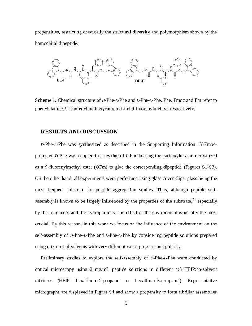
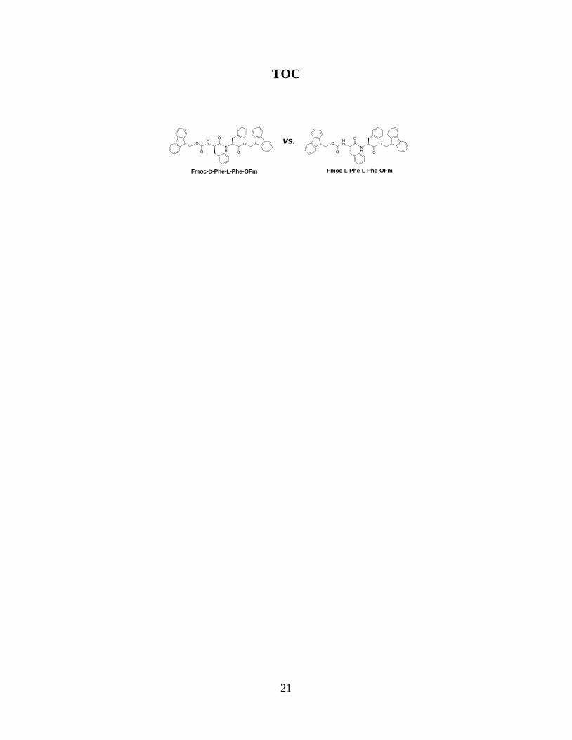
that contains D-Phe and L-Phe, namely Fmoc-D-Phe-L-Phe-OFm (Scheme 1; abbreviated as
D-Phe-L-Phe in the following), and compare it to the analogous homochiral dipeptide with
two L-Phe residues, Fmoc-L-Phe-L-Phe-OFm (Scheme 1; L-Phe-L-Phe in the following).
Although the latter peptide was preliminary studied,15
investigations have extended it in
this work for comparison with D-Phe-L-Phe under identical experimental conditions.
Results reveal that replacement of L-Phe by D-Phe has a major impact on the self-assembly
5
DL-FLL-F
propensities, restricting drastically the structural diversity and polymorphism shown by the
homochiral dipeptide.
Scheme 1. Chemical structure of D-Phe-L-Phe and L-Phe-L-Phe. Phe, Fmoc and Fm refer to
phenylalanine, 9-fluorenylmethoxycarbonyl and 9-fluorenylmethyl, respectively.
RESULTS AND DISCUSSION
D-Phe-L-Phe was synthesized as described in the Supporting Information. N-Fmoc-
protected D-Phe was coupled to a residue of L-Phe bearing the carboxylic acid derivatized
as a 9-fluorenylmethyl ester (OFm) to give the corresponding dipeptide (Figures S1-S3).
On the other hand, all experiments were performed using glass cover slips, glass being the
most frequent substrate for peptide aggregation studies. Thus, although peptide self-
assembly is known to be largely influenced by the properties of the substrate,24
especially
by the roughness and the hydrophilicity, the effect of the environment is usually the most
crucial. By this reason, in this work we focus on the influence of the environment on the
self-assembly of D-Phe-L-Phe and L-Phe-L-Phe by considering peptide solutions prepared
using mixtures of solvents with very different vapor pressure and polarity.
Preliminary studies to explore the self-assembly of D-Phe-L-Phe were conducted by
optical microscopy using 2 mg/mL peptide solutions in different 4:6 HFIP:co-solvent
mixtures (HFIP: hexafluoro-2-propanol or hexafluoroisopropanol). Representative
micrographs are displayed in Figure S4 and show a propensity to form fibrillar assemblies
6
in all the solvents tested. On this basis, deeper studies were performed for 5.0, 4.0, 2.0, 0.5,
0.25, 0.1, 0.05 and 0.01 mg/mL D-Phe-L-Phe concentrations in a variety of HFIP:co-solvent
mixtures. The co-solvents used (water, methanol, isopropanol, acetone) differed in polarity,
volatility and hydrogen bonding capacity. Identical experiments were carried out with the
homochiral L-Phe-L-Phe dipeptide for comparative purposes (for details on sample
preparation, see ESI).
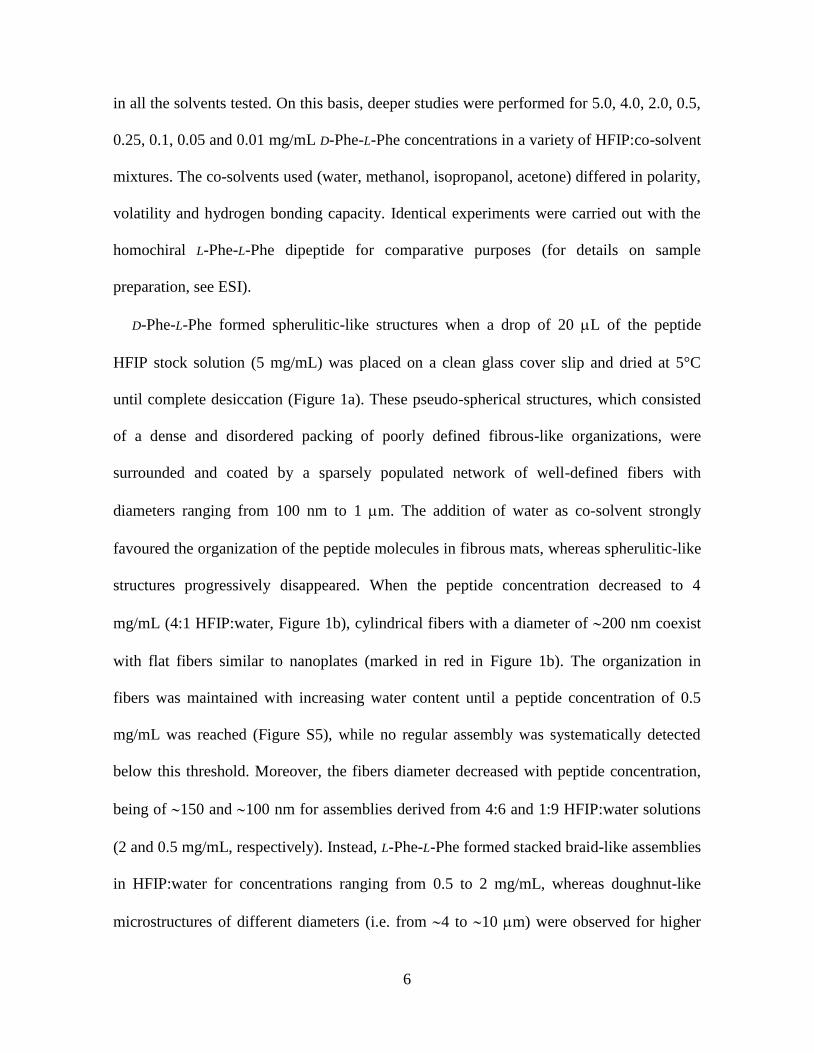
D-Phe-L-Phe formed spherulitic-like structures when a drop of 20 L of the peptide
HFIP stock solution (5 mg/mL) was placed on a clean glass cover slip and dried at 5°C
until complete desiccation (Figure 1a). These pseudo-spherical structures, which consisted
of a dense and disordered packing of poorly defined fibrous-like organizations, were
surrounded and coated by a sparsely populated network of well-defined fibers with
diameters ranging from 100 nm to 1 m. The addition of water as co-solvent strongly
favoured the organization of the peptide molecules in fibrous mats, whereas spherulitic-like
structures progressively disappeared. When the peptide concentration decreased to 4
mg/mL (4:1 HFIP:water, Figure 1b), cylindrical fibers with a diameter of 200 nm coexist
with flat fibers similar to nanoplates (marked in red in Figure 1b). The organization in
fibers was maintained with increasing water content until a peptide concentration of 0.5
mg/mL was reached (Figure S5), while no regular assembly was systematically detected
below this threshold. Moreover, the fibers diameter decreased with peptide concentration,
being of 150 and 100 nm for assemblies derived from 4:6 and 1:9 HFIP:water solutions
(2 and 0.5 mg/mL, respectively). Instead, L-Phe-L-Phe formed stacked braid-like assemblies
in HFIP:water for concentrations ranging from 0.5 to 2 mg/mL, whereas doughnut-like
microstructures of different diameters (i.e. from 4 to 10 m) were observed for higher
7
concentrations (Figure S6). In no case were fibrous organizations detected from L-Phe-L-
Phe HFIP:water solutions.
Figure 1. Self-assembly structures obtained at 5°C from D-Phe-L-Phe solutions in the
solvents and concentrations indicated. (a) SEM images of the spherulitic-like structures
formed in HFIP (5 mg/mL, stock solution). (b) Optical microscopy (top), SEM (bottom
left) and AFM (bottom right) images of the nanofibers formed in 4:1 HFIP:water (4
mg/mL); red dashed circles indicate the coexisting nanoplates.
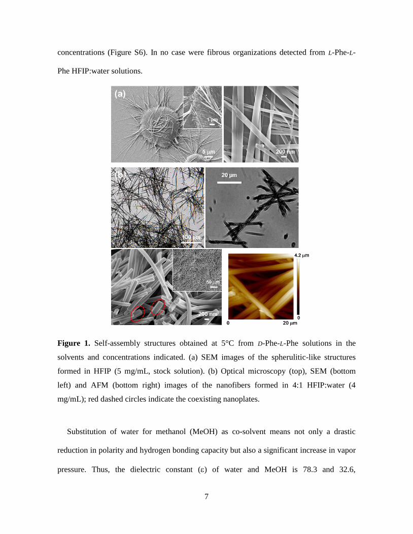
Substitution of water for methanol (MeOH) as co-solvent means not only a drastic
reduction in polarity and hydrogen bonding capacity but also a significant increase in vapor
pressure. Thus, the dielectric constant () of water and MeOH is 78.3 and 32.6,
8
respectively, while their respective vapor pressures at 5°C (Pv,5) are 0.87 and 5.50 kPa. Due
to these distinctive properties that affect the drying process, when MeOH dominates in the
HFIP:MeOH mixtures (i.e. peptide concentration 2 mg/mL), D-Phe-L-Phe self-assembles
into ultrathin nanofibers that align and fix together to form fibres of micrometric diameter
(Figure 2a).
Figure 2. Self-assembly structures obtained at 5°C from D-Phe-L-Phe solutions in the
solvents and concentrations indicated. (a) SEM and optical microscopy (inset) images of
the nanofibers (grouped into microfibers) formed in 4:6 HFIP:MeOH (2 mg/mL). (b) SEM,
optical microscopy (inset) and AFM images of the microfibers formed in 4:6 HFIP:iPrOH
(2 mg/mL).
The use of isopropanol (iPrOH) as co-solvent, which is less polar and less volatile than
both water and MeOH at 5°C (= 18 and Pv,5= 0.69 kPa), triggers the assembly of D-Phe-L-
Phe into smooth microfibers with an average diameter of 1.5 m (Figure 2b). This
9
assembly is preserved with increased iPrOH content and lower peptide concentration, but
with reduced diameter (Figure S7).
The influence of alcohols on the self-assembly properties of L-Phe-L-Phe differ
substantially from that described above for the heterochiral peptide. Stacked braid-like
assemblies similar to those displayed in Figure S6 were reported15
for L-Phe-L-Phe in 1:49
and 1:99 HFIP:MeOH mixtures (0.1 and 0.05 mg/mL, respectively), no other regular and
reproducible structure being detected for higher peptide concentrations. Interestingly, in
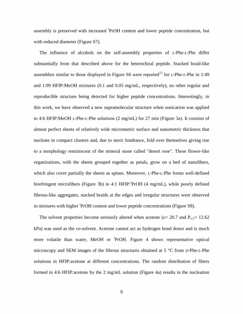
this work, we have observed a new supramolecular structure when sonication was applied
to 4:6 HFIP:MeOH L-Phe-L-Phe solutions (2 mg/mL) for 27 min (Figure 3a). It consists of
almost perfect sheets of relatively wide micrometric surface and nanometric thickness that
nucleate in compact clusters and, due to steric hindrance, fold over themselves giving rise
to a morphology reminiscent of the mineral stone called "desert rose". These flower-like
organizations, with the sheets grouped together as petals, grow on a bed of nanofibers,
which also cover partially the sheets as spines. Moreover, L-Phe-L-Phe forms well-defined
birefringent microfibers (Figure 3b) in 4:1 HFIP:iPrOH (4 mg/mL), while poorly defined
fibrous-like aggregates, stacked braids at the edges and irregular structures were observed
in mixtures with higher iPrOH content and lower peptide concentrations (Figure S8).
The solvent properties become seriously altered when acetone (= 20.7 and Pv,5= 12.62
kPa) was used as the co-solvent. Acetone cannot act as hydrogen bond donor and is much
more volatile than water, MeOH or iPrOH. Figure 4 shows representative optical
microscopy and SEM images of the fibrous structures obtained at 5 °C from D-Phe-L-Phe
solutions in HFIP:acetone at different concentrations. The random distribution of fibers
formed in 4:6 HFIP:acetone by the 2 mg/mL solution (Figure 4a) results in the nucleation
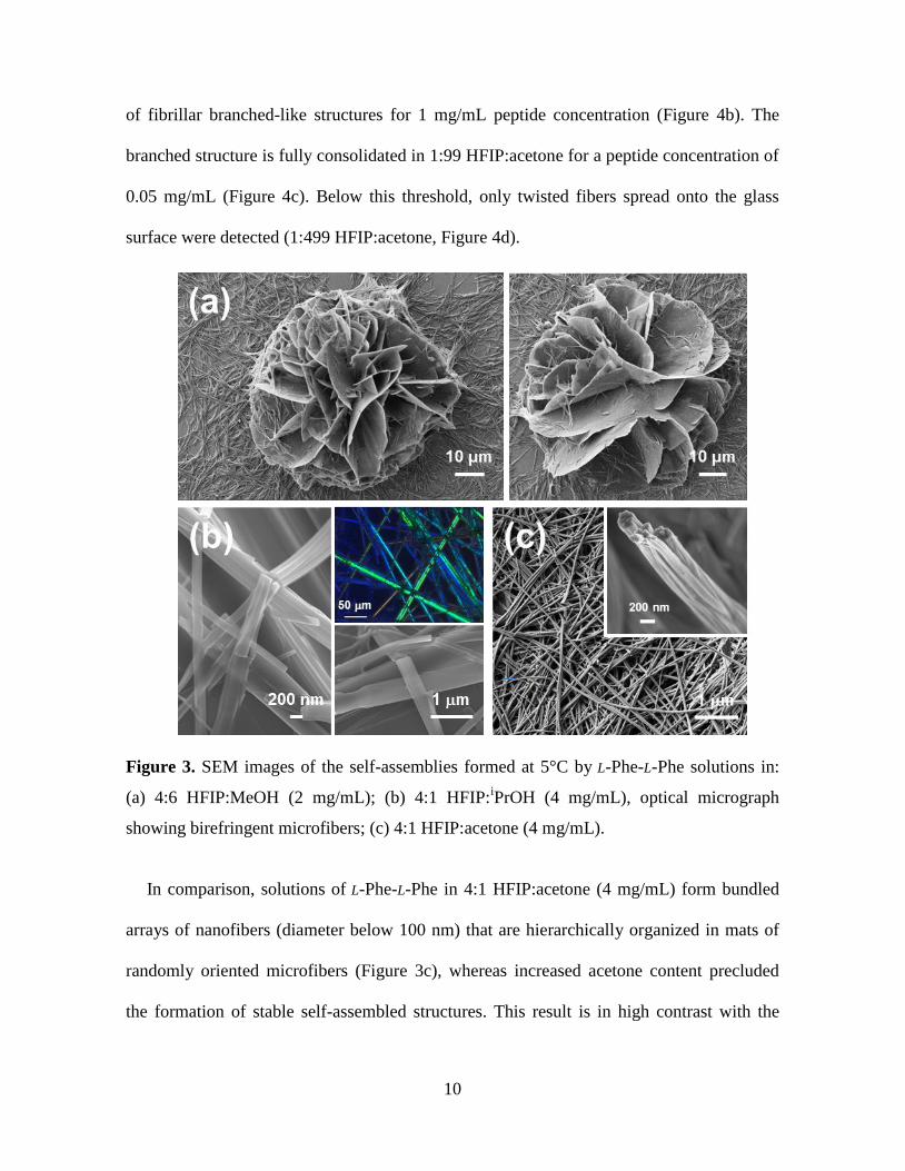
10
of fibrillar branched-like structures for 1 mg/mL peptide concentration (Figure 4b). The
branched structure is fully consolidated in 1:99 HFIP:acetone for a peptide concentration of
0.05 mg/mL (Figure 4c). Below this threshold, only twisted fibers spread onto the glass
surface were detected (1:499 HFIP:acetone, Figure 4d).
Figure 3. SEM images of the self-assemblies formed at 5°C by L-Phe-L-Phe solutions in:
(a) 4:6 HFIP:MeOH (2 mg/mL); (b) 4:1 HFIP:iPrOH (4 mg/mL), optical micrograph
showing birefringent microfibers; (c) 4:1 HFIP:acetone (4 mg/mL).
In comparison, solutions of L-Phe-L-Phe in 4:1 HFIP:acetone (4 mg/mL) form bundled
arrays of nanofibers (diameter below 100 nm) that are hierarchically organized in mats of
randomly oriented microfibers (Figure 3c), whereas increased acetone content precluded
the formation of stable self-assembled structures. This result is in high contrast with the
11
great tendency shown by D-Phe-L-Phe to adopt fibrillar assemblies from acetone-based
solutions even at very low concentrations.
Figure 4. Microstructures obtained by self-assembly from D-Phe-L-Phe solutions in
HFIP:acetone: (a) 4:6 (2 mg/mL); (b) 1:4 (1 mg/mL); (c) 1:99 (0.05 mg/mL); and 1:499
(0.01 mg/mL).
12
The energy landscape of D-Phe-L-Phe was examined using density functional theory
(DFT) calculations with the M06L functional,25
which has broad accuracy for non-covalent
interactions26,27
including - stacking, and the 6-31G(d,p) basis set. Calculations were
performed in vacuum to compare the intrinsic stability of the different pre-formed
structures with the experimental results on the dry state and also with our previous
theoretical study of L-Phe-L-Phe.15
The most stable conformation of D-Phe-L-Phe (Figure
5a) corresponds to the fully-extended arrangement, which is stabilized by two
intramolecular hydrogen bonds closing 5-membered cycles (C5) and two - interactions
involving the Fmoc···D-Phe and L-Phe···OFm aromatic rings. The second minimum also
shows an extended conformation, even though in this case the Fmoc···D-Phe - stacking
is replaced by an N–H··· interaction between the L-Phe amide and the phenyl ring of D-
Phe (Figure S9). This structure is 1.7 kcal/mol less stable than the global minimum,
indicating that the formation of two - stacking interactions is preferred.
The stability of the parallel and antiparallel -sheet alignments was calculated
considering three D-Phe-L-Phe molecules (Figure 5b), and the two fully-extended
conformations mentioned above. The antiparallel disposition was significantly more
favoured than the parallel -sheet in both cases (E > 27 kcal/mol), which is consistent
with the FTIR band observed at 1694 cm-1
(Figure S3). The former is stabilized by multiple
- stacking interactions, in addition to the intermolecular N–H···O H-bonds typical of -
sheets. These aromatic interactions involve all six terminal fluorenyl groups as well as most
of the side chains of D-Phe and L-Phe, which mainly interact with fluorenyl moieties
(Figure 5c). Our study of L-Phe-L-Phe15
showed that the antiparallel sheet is preferred over
the parallel one by only 1.8 kcal/mol. Thus, the disposition of aromatic rings, which is
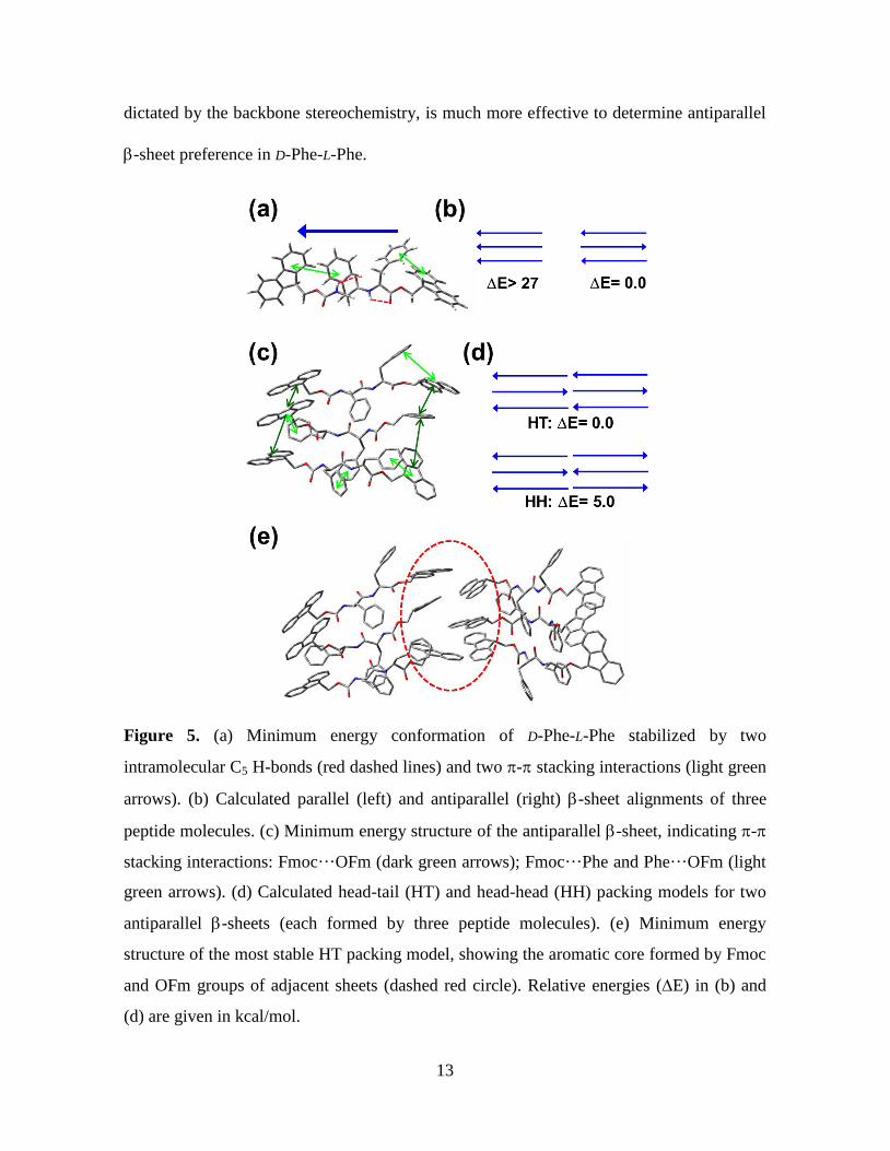
13
dictated by the backbone stereochemistry, is much more effective to determine antiparallel
-sheet preference in D-Phe-L-Phe.
Figure 5. (a) Minimum energy conformation of D-Phe-L-Phe stabilized by two
intramolecular C5 H-bonds (red dashed lines) and two - stacking interactions (light green
arrows). (b) Calculated parallel (left) and antiparallel (right) -sheet alignments of three
peptide molecules. (c) Minimum energy structure of the antiparallel -sheet, indicating -
stacking interactions: Fmoc···OFm (dark green arrows); Fmoc···Phe and Phe···OFm (light
green arrows). (d) Calculated head-tail (HT) and head-head (HH) packing models for two
antiparallel -sheets (each formed by three peptide molecules). (e) Minimum energy
structure of the most stable HT packing model, showing the aromatic core formed by Fmoc
and OFm groups of adjacent sheets (dashed red circle). Relative energies (E) in (b) and
(d) are given in kcal/mol.
14
To investigate the packing between neighbouring sheets, pre-formed antiparallel -
sheets made of three D-Phe-L-Phe strands were constructed considering the head-tail (HT)
and head-head (HH) models (Figure 5d). The HT assembly is favoured by 5.0 kcal/mol. In
addition to the intra-sheet stabilizing forces described for Figure 5c, this HT packing
exhibits a hydrophobic core with Fmoc···OFm - stacking interactions involving
molecules of adjacent sheets (red circle in Figure 5e). The nucleation of such - stacked
cores is consistent with the remarkable ability shown by D-Phe-L-Phe to form fibrillar
assemblies even in the absence of water or other solvents able to act as H-bond donors, and
at very low concentrations. Thus, the formation of aromatic cores due to the inability to
interact with solvent molecules is the driving force of the aggregation process, leading to a
decrease in the entropy of the system as it occurs in the hydrophobic collapse effect in
protein folding processes.
Actually, the large energy gap between the antiparallel and parallel -sheets calculated
in this work (E > 27 kcal/mol, Figure 5b) deserves special attention. In our previous
work15
on L-Phe-homopeptides blocked with Fmoc and OFm groups, such energy
difference increased with peptide length, being of 1.8, 3.3 and 9.8 kcal/mol for the di-, tri-
and tetrapeptide, respectively. These stability differences correlated with the distinct self-
assembly propensities observed experimentally for these Fmoc-(L-Phe)n-OFm peptides,
allowing us to conclude that additional - stacking interactions associated to increased
peptide length favoured antiparallel -sheet formation with concomitant stabilization of the
nanostructures formed and reduction of polymorphism.15
Such a conclusion was also in
perfect agreement with previous works by us and other authors on L-Phe homopeptides
15
uncapped or containing an N-Fmoc group only.28
On the other hand, the preference towards
the antiparallel -sheets found for D-Phe-L-Phe and L-Phe-L-Phe is fully consistent with that
reported for other highly aromatic Phe-based peptides. For example, Accardo and co-
workers observed similar features for the different hexaphenylalanine, (L-Phe)6, variants
using molecular dynamics simulations.29
Therefore, the strong preference for antiparallel -sheet formation calculated for the
heterochiral dipeptide studied in the present work (Fmoc-D-Phe-L-Phe-OFm) may be at the
basis of the singular behavior observed for such a small peptide in comparison to its
homochiral analogue, both from quantitative and qualitative viewpoints: higher tendency to
self-assembly and much more restricted structural diversity.
CONCLUSIONS
In conclusion, the self-assembly of Phe-dipeptides blocked with two fluorenyl
functionalities is governed by the backbone stereochemistry. The heterochiral D-Phe-L-Phe
dipeptide exhibits a high propensity to form fibrillar structures independently of the solvent
composition, whereas the morphology of the L-Phe-L-Phe assemblies changes with the
environment. Moreover, the heterochiral compound retains the ability to self-assembly
under conditions in which its homochiral counterpart does not adopt any kind of stable
supramolecular organization. The outstanding capacity of Fmoc-D-Phe-L-Phe-OFm to adopt
fibrillar assemblies even in non-aqueous media is essential for the potential development of
nanotechnological applications. The results in this work, together with our previous reports
on L-Phe-dipeptides capped with one C-terminal13,14
or two15
fluorenyl groups and the
numerous studies on Fmoc-L-Phe-L-Phe,5,16,17
evidence that the adequate combination of
16
terminal fluorenyl moieties with controlled peptide backbone chirality (which dictates the
orientation of all aromatic groups in the molecule) is a potent and versatile strategy to drive
and finely tune the self-assembly propensities of Phe-based peptides.
ACKNOWLEDGEMENTS
This work was supported by MINECO (RTI2018-098951-B-I00, RTI2018-101827-B-
I00, and CTQ2013-40855-R), the AGAUR (2017SGR359 and 2017SGR373), and
Gobierno de Aragon (research group Aminoacidos y Peptidos E19_20R). Support for the
research of C.A. was received through the prize “ICREA Academia” for excellence in
research funded by the Generalitat de Catalunya.
REFERENCES
1. Adler-Abramovich, L.; Gazit, E. The Physical Properties of Supramolecular Peptide
Assemblies: From Building Block Association to Technological Applications. Chem.
Soc. Rev. 2014, 43, 6881-6893.
2. Xing, Q. G.; Zhang, J. X.; Xie, Y. Y.; Wang, Y. F.; Qi, W.; Rao, H. J.; Su, R. X.; He,
Z. M. Aromatic Motifs Dictate Nanohelix Handedness of Tripeptides. ACS Nano
2018, 12, 12305-12314.
3. Li, J.; Du, X. W.; Hashim, S.; Shy, A.; Xu, B. Aromatic-Aromatic Interactions
Enable α-Helix to β-Sheet Transition of Peptides to Form Supramolecular Hydrogels.
J. Am. Chem. Soc. 2017, 139, 71-74.
4. Amit, M.; Yuran, S.; Gazit, E.; Reches, M.; Ashkenahy, N. Tailor-Made Functional
Peptide Self-Assembling Nanostructures. Adv. Mater 2018, 30, 1707083.
17
5. Tao, K.; Levin, A.; Adler-Abramovich, L.; Gazit, E. Fmoc-Modified Amino Acids
and Short Peptides: Simple Bio-inspired Building Blocks for the Fabrication of
Functional Materials. Chem. Soc. Rev. 2016, 45, 3935-3953.
6. Xie, Y. Y.; Wang, X. C.; Huang, R. L.; Qi, W.; Wang, Y. F.; Su, R. X.; He, Z. M.
Electrostatic and Aromatic Interaction-Directed Supramolecular Self-Assembly of a
Designed Fmoc-Tripeptide into Helical Nanoribbons. Langmuir 2015, 31, 2885-2894.
7. Arnon, Z. A.; Vitalis, A.; Levin, A.; Michaels, T. C. T.; Caflisch, A.; Knowles, T. P.
J.; Adler-Abramovich, L.; Gazit, E. Dynamic Microfluidic Control of Supramolecular
Peptide Self-Assembly. Nat. Commun. 2016, 7, 13190.
8. Mayans, E.; Casanovas, J.; Gil, A.; Jimenez, A. I.; Cativiela, C.; Puiggalí, J.; Aleman,
C. Diversity and Hierarchy in Supramolecular Assemblies of Triphenylalanine: From
Laminated Helical Ribbons to Toroids. Langmuir 2017, 33, 4036-4048.
9. Mayans, E.; Ballano, G.; Casanovas, J.; Díaz, A.; Pérez Madrigal, M. M.; Estrany, F.;
Puiggalí, J.; Cativiela, C.; Alemán, C. Self‐Assembly of Tetraphenylalanine Peptides.
Chem. Eur. J. 2015, 21, 16895-16905.
10. Sasselli, I. R.; Pappas, C. G.; Matthews, E.; Wang, T.; Hunt, N. T.; Uljin, R. V.;
Tuttle, T. Using Experimental and Computational Energy Equilibration to Understand
Hierarchical Self-Assembly of Fmoc-Dipeptide Amphiphiles. Soft Matter 2016, 12,
8307-8315.
11. Raeburn, J.; Mendoza-Cuenca, C.; Cattoz, B. N.; Little, M. A.; Terry, A. E.; Cardoso,
A. Z.; Griffiths, P. C.; Adams, D. J. The Effect of Solvent Choice on the Gelation and
Final Hydrogel Properties of Fmoc-Diphenylalanine. Soft Matter 2015, 11, 927-935.
12. Reches, M.; Gazit, E. Casting Metal Nanowires Within Discrete Self-Assembled
Peptide Nanotubes. Science 2003, 300, 625-627.
18
13. Casanovas, J.; Mayans, E.; Díaz, A.; Gil, A. M.; Jiménez, A. I.; Cativiela, C.;
Puiggalí, J.; Aleman, C. Amyloid Fibrils from Organic Solutions of An Amphiphilic
Dipeptide. Chem. Commun. 2019, 55, 8556-8559.
14. Martí, D.; Mayans, E.; Gil, A. M.; Díaz, A.; Jiménez, A. I.; Yousef, I.; Keridou, I.;
Cativiela, C.; Puiggalí, J.; Aleman, C. Amyloid-like Fibrils from a Diphenylalanine
Capped with an Aromatic Fluorenyl. Langmuir 2018, 34, 15551-15559.
15. Mayans, E.; Ballano, G.; Casanovas, J.; del Valle, L. J.; Pérez-Madrigal, M. M.;
Estrany, F.; Jiménez, A. I.; Puiggalí, J.; Cativiela, C.; Alemán, C. Hierarchical Self-
Assembly of Di-, Tri- and Tetraphenylalanine Peptides Capped with Two Fluorenyl
Functionalities: From Polymorphs to Dendrites. Soft Matter 2016, 12, 5475-5488.
16. Makam, P.; Gazit, E. Minimalistic Peptide Supramolecular Co-assembly: Expanding
the Conformational Space for Nanotechnology. Chem. Soc. Rev. 2018, 47, 3406-
3420.
17. Fleming, S.; Ulijn, R. V. Design of Nanostructures Based on Aromatic Peptide
Amphiphiles. Chem. Soc. Rev. 2014, 43, 8150-8177.
18. Martin, A. D.; Thordarson, P. Beyond Fmoc: A Review of Aromatic Peptide Capping
Groups. J. Mater. Chem. B 2020, 8, 863-877.
19. Bagheri, M.; Amininasab, M.; Dather, M. Arginine/Tryptophan‐Rich Cyclic α/β‐
Antimicrobial Peptides: The Roles of Hydrogen Bonding and
Hydrophobic/Hydrophilic Solvent‐Accessible Surface Areas upon Activity and
Membrane Selectivity. Chem. Eur. J. 2018, 24, 12242-12253.
20. Giri, S.; Mandal, B. Formation of Supramolecular Single and Double Helix-Like
Structures from Designed Tripeptides. Cryst. Eng. Comm. 2019, 21, 5618-5625.
19
21. Yordanova, Y.; Vanderlinden, W.; Stoll, R.; Rüdiger, D.; Tosstorff, A.; Zaremba, W.;
Winter, G.; Zahler, S.; Friess, W. Zn2+
-Triggered Self-Assembly of Gonadorelin [6-
D-Phe] to Produce Nanostructures and Fibrils. Sci. Rep. 2018, 8, 11280
22. Li, J.; Bullara, D.; Du, X.; He, H.; Sofou, S.; Kevrekidis, I. G.; Epstein, I. R.; Xu, B.
Kinetic Analysis of Nanostructures Formed by Enzyme-Instructed Intracellular
Assemblies against Cancer Cells. ACS Nano 2018, 12, 3804-3815.
23. Ozawa, Y.; Sato, H.; Kayano, Y.; Yamaki, N.; Izato, Y.-i.; Miyake, A.; Naito, A.;
Kawamura, I. Self-Assembly of Tripeptides into γ-Turn Nanostructures. Phys. Chem.
Chem. Phys. 2019, 21, 10879-10883.
24. Mayans, E.; Fabregat, G.; Juárez, R.; Cativiela, C.; Puiggalí, J. Surface Mediated
Hierarchical Assemblies of Highly Hydrophobic Phenylalanine‐Based Peptides.
ChemistrySelect 2017, 2, 1133-1139.
25. Zhao, Y.; Truhlar, D. G. A New Local Density Functional for Main-Group
Thermochemistry, Transition Metal Bonding, Thermochemical Kinetics, and
Noncovalent Interactions. J. Chem. Phys. 2006, 125, 194101.
26. Mardirossian, N.; Head-Gordon, M. How Accurate Are the Minnesota Density
Functionals for Noncovalent Interactions, Isomerization Energies, Thermochemistry,
and Barrier Heights Involving Molecules Composed of Main-Group Elements?. J.
Chem. Theory Comput. 2016, 12, 4303-4325.
27. Li, Z.; Couzijn, E. P. A.; Zhang, X. A Quantitative Study of Intrinsic Non-Covalent
Interactions Within Complexes of α-Cyclodextrin and Benzoate Derivatives. Chem.
Commun. 2012, 48, 9864-9866.
20
28. Polymorphism has been associated, among other factors, to the lack of preference for
parallel and antiparallel -sheet alignments. See discussion in reference 15 and
references therein.
29. Diaferia, C.; Balasco, N.; Altamura, D.; Sibillano, T.; Gallo, E.; Roviello, V.;
Giannini, C.; Morelli, G.; Vitagliano, L.; Accardo, A. Assembly Modes of
Hexaphenylalanine Variants as Function of the Charge States of Their Terminal
Ends. Soft Matter 2018, 14, 8219--8230
21
TOC
Fmoc-L-Phe-L-Phe-OFmFmoc-D-Phe-L-Phe-OFm
vs.