head injuries meningitis - jnnp.bmj.com · developed pneumococcal meningitis for which he received...
TRANSCRIPT

HEAD INJURIES AND MENINGITISBY
E. A. LINELL AND W. L. ROBINSON
Fr-om Divisions of Surgical Pathology and Neuropathology, University of Toronto(RECEIVED 11TH NOVEMBER, 1940)
IntroductionTHE following cases have been selected from the files of the Division of Neuro-pathology in the University of Toronto to illustrate the types of head injurywhich may cause death from meningitis. The fatal infection of the meningesmay occur at the time of the injury, killing the patient within a few days orweeks of the accident, but its onset may be delayed for years after the headinjury. Three of the seven cases described below fall into the latter group, thepatients having apparently recovered from their head injuries shortly after theiraccidents and having shown no symptoms until the sudden onset of a rapidlyfatal meningitis.
The pneumococcus was the organism responsible for the meningitis in allthese cases and, except for some variations mentioned in the text, the gross andmicroscopical pathological picture was typical of pneumococcal meningitis.
Case ReportsCase 1: M. W. (NP: 315-34), a man of 30 years, was probably kicked in the
face by a horse on the 22nd October, 1934, although he could remember no details ofthe accident. He remained conscious but vomited several times during the next twodays. He became unconscious on the 24th October and rapidly developed the signsof meningitis of which he died on the 26th October, four days after his accident.
Post mortem.-There was severe laceration of the face, with fractured nasal bones.Removal of the brain showed severe hkmorrhage covering the floor of the anteriorcranial fosse. The bone of these fosse was shattered around the crista galli, whichwas loose, freely movable and displaced to the left. Scissors could be passed easilyfrom the cranial cavity into the nasal fosse. A fracture 3 cm. long radiated outwardsinto the left orbital plate of the frontal bone.
There was greenish-yellow purulent exudate in the subarachnoid space. Pneu-mococci were cultured from this exudate and from the cerebrospinal fluid.
Comment.-This, then, is a case of rapidly fatal meningitis following aninjury to the face of which the main external sign was simple fracture of the nasalbones.
Case 2: L. W. (NP: 170-36), a man of 50, was riding a bicycle when he was hitby a truck on the 21st May, 1936. On admission to hospital he was conscious butirritable. There was considerable bleeding from the back of the nose, with hema-temesis. Leakage of cerebrospinal fluid from his nose was noted on 25th May.On the following day spinal puncture withdrew turbid fluid containing large numbers
23
on 21 April 2019 by guest. P
rotected by copyright.http://jnnp.bm
j.com/
J Neurol P
sychiatry: first published as 10.1136/jnnp.4.1.23 on 1 January 1941. Dow
nloaded from

E. A. LINELL AND W. L. ROBINSON
of polymorphonuclear leucocytes. His temperature rose till his death on the 27thMay, six days after the accident.
Post mortem.-There was a large area of subcutaneous hemorrhage on the leftside of the scalp extending into the temporal muscle. Extradural haemorrhageswere found in the left anterior and middle cranial fosse. A complicated fracture ofthe base of the skull began at the outer end of the left supra-orbital ridge. Thisextended backwards and inwards, crossing the midline through the body of thesphenoid bone and running along the antero-inferior border of the petrous portion ofthe right temporal bone to end in the right middle-ear cavity which was full of blood.A branch of this fracture extended inwards completely encircling the cribriform plateof the ethmoid and from here ran into the right frontal sinus which was pneumatizingthe right orbital plate of the frontal bone. A second branch of the main fractureextended backwards and outwards across the left middle cranial fosse to end in thebase of the petrous portion of the left temporal bone. The sphenoidal and ethmoidalsinuses contained purulent exudate mixed with hkmorrhage.
Culture taken from the base of the brain yielded pneumococcus type 4 and therewas well-marked purulent exeudate in the subarachnoid space.
Comment.-This case is reported because it illustrates a basal skull fracturewhich had opened practically all the accessory nasal sinuses as well as bothmiddle ear cavities. Purulent exudate in the frontal and ethmoidal sinuses wasresponsible for the meningitis in this case.
Case 3: R. T. (NP: 371-39), a boy of 3 years, was hit by a motor car shortlybefore his admission to hospital on the 17th September, 1939. Four days later hedeveloped pneumococcal meningitis for which he received antipneumococcal serumcombined with sulphapyridine. He gradually went down hill with persistence of hissymptoms till his death on the 15th October, about a month after the trauma. Therewas no clinical evidence of fracture of the skull.
Post mortem.-There was an area of extradural hkmorrhage in the right posteriorcranial fosse. This overlay a fracture which began slightly below and to the right ofthe external occipital protuberance. The fracture ran downwards and outwards toreach the petrous portion of the right temporal bone. It then ran inwards to end inthe apex of this bone. Both middle-ear cavities showed evidence of chronic inflam-mation. There was also a mottled grey and dark-red thrombus in the lumen of theright lateral sinus in relation to the line of fracture. There was massive purulentexudate in the basal subarachnoid space.
Comment.-This case is of interest because the relationship of the head-injury to the meningitis was obscure until it was revealed at post mortem.It also illustrates the danger of chronic middle-ear disease in cases of fracture ofthe base of the skull. Finally the life of the patient was prolonged for nearly amonth after the onset of meningitis, probably by the serum and sulphapyridinetherapy.
Case 4: J. B. (NP: 164-37), a man of 49 years, was unconscious for 20 minutesfollowing a fall downstairs on to a cement floor in the evening of the 12th February,1937. On his recovery of consciousness he complained of severe frontal headacheand he vomited. On admission to hospital the following day there was tendernessand cedema over the left parieto-occipital region of the scalp. His right knee-jerkwas brisk and there was a doubtfully positive right Babinski. His cerebrospinal fluidpressure was high. There was no X-ray evidence of fracture of the skull.
Two days later, on the 15th February, he began to develop evidence of left otitismedia, which required paracentesis of the drum on the 1st March. By the 11th Marchhe showed a complete right hemiplegia with aphasia. On the following day an opera-
24
on 21 April 2019 by guest. P
rotected by copyright.http://jnnp.bm
j.com/
J Neurol P
sychiatry: first published as 10.1136/jnnp.4.1.23 on 1 January 1941. Dow
nloaded from
HEAD INJURIES AND MENINGITIS 25
tive exploration revealed an infected left subdural hrmatoma from the contents ofwhich pneumococcus, Type 3, was isolated. He improved temporarily but thedevelopment of a cerebral hernia obstructed efficient drainage of the infected subduralspace and he gradually became more comatose, dying on the 24th April, 24 monthsafter his accident.
Post mortem.-A linear fracture, 10 cm. long, ran horizontally through the lateralportion of the base of the occipital bone on the left side into the petrous portion of theleft temporal bone. This bone was softened and contained purulent material.
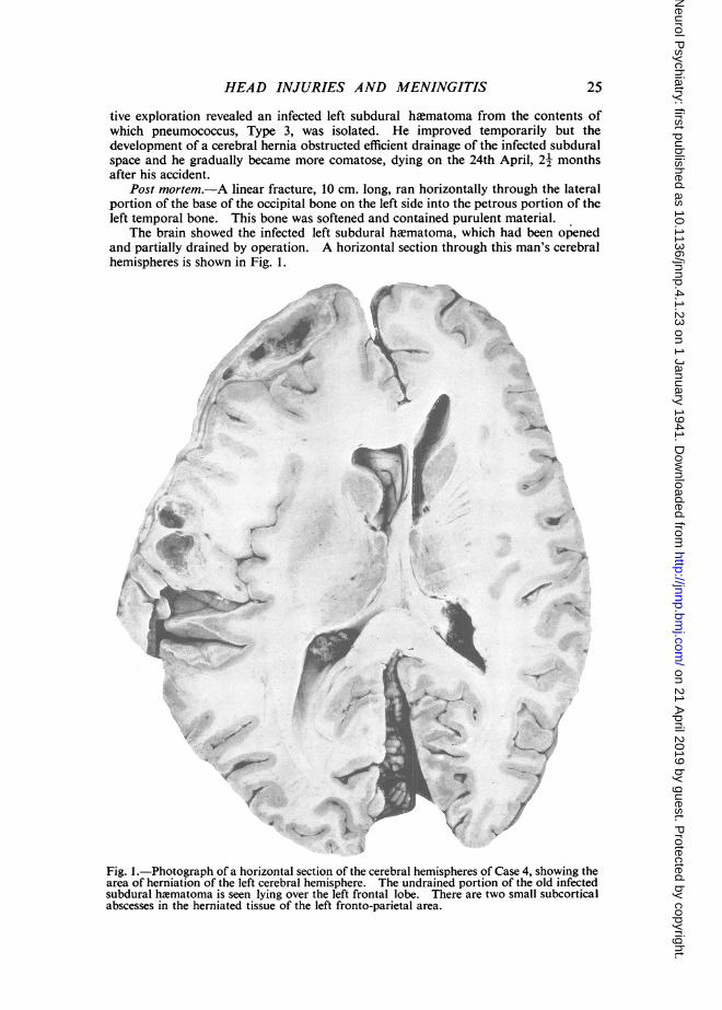
The brain showed the infected left subdural hematoma, which had been openedand partially drained by operation. A horizontal section through this man's cerebralhemispheres is shown in Fig. 1.
-4~~~~~~~V
A~~~~~~~~~~~~~~~~~AO
*~~~~ .-" £r
^, ,,' T#' o'~~~I,
Fig. 1.-Photograph of a horizontal section of the cerebral hemispheres of Case 4, showing thearea of herniation of the left cerebral hemisphere. The undrained portion of the old infectedsubdural h2ematoma is seen lying over the left frontal lobe. There are two small subcorticalabscesses in the herniated tissue of the left fronto-parietal area.
on 21 April 2019 by guest. P
rotected by copyright.http://jnnp.bm
j.com/
J Neurol P
sychiatry: first published as 10.1136/jnnp.4.1.23 on 1 January 1941. Dow
nloaded from
E. A. LINELL AND W. L. ROBINSON
Comment.-This is a case in which infection from the patient's left middle-earcavity and petrous temporal bone had been able to spread upwards through anunsuspected fracture line to infect a traumatic subdural hematoma, causing hisdeath two and a half months after his fall.
Case 5: D. B. (NP: 176-39), was a professional jockey 22 years of age when hedied on the 16th May, 1939. In 1934 he had had his nose broken by a horse. The skinlaceration required sutures and he was in hospital for 11 days at that time. Therewere no significant after-effects.
On the 12th May, 1939, 5 years later, he had a slight cold in the head. Two dayslater he had a headache in the morning, by 3 p.m. he was vomiting and having con-vulsions. He was admitted to hospital comatose at 6 p.m. with all the signs of menin-gitis. The right maxillary and frontal sinuses were opaque to transillumination.Pneumococci were seen in a smear of his cerebrospinal fluid. In spite of vigoroustreatment by soludagenan he died on the 16th May, four days after his first complaintof cold in the head.
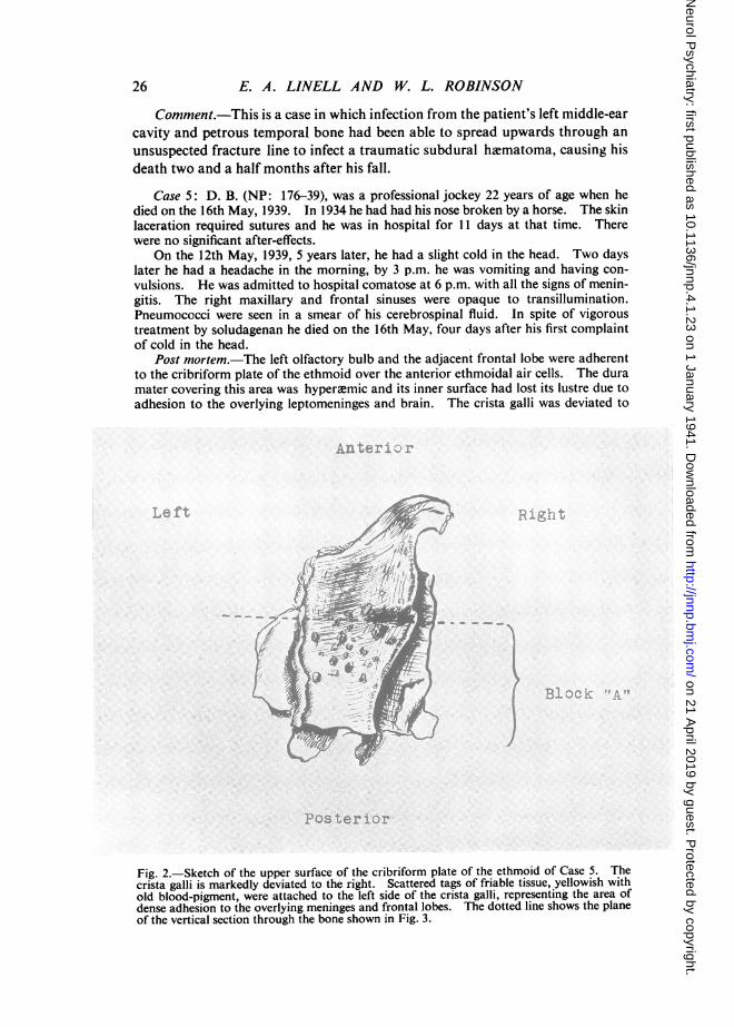
Post mortem.-The left olfactory bulb and the adjacent frontal lobe were adherentto the cribriform plate of the ethmoid over the anterior ethmoidal air cells. The duramater covering this area was hyperaemic and its inner surface had lost its lustre due toadhesion to the overlying leptomeninges and brain. The crista galli was deviated to
Fig. 2.-Sketch of the upper surface of the cribriform plate of the ethmoid of Case 5. Thecrista galli is markedly deviated to the right. Scattered tags of friable tissue, yellowish withold blood-pigment, were attached to the left side of the crista galli, representing the area ofdense adhesion to the overlying meninges and frontal lobes. The dotted line shows the planeof the vertical section through the bone shown in Fig. 3.
26
on 21 April 2019 by guest. P
rotected by copyright.http://jnnp.bm
j.com/
J Neurol P
sychiatry: first published as 10.1136/jnnp.4.1.23 on 1 January 1941. Dow
nloaded from

HEAD INJURIES AND MENINGITIS
the right (Fig. 2), and there was old blood pigment covering the left side of the cribri-form plate. There was pus in the right and left ethmoidal sinuses. Microscopicalexamination of a vertical section cut through the area of old trauma to the cribriformplate of the ethmoid in the plane of the dotted line shown in Fig. 2 is shown in Fig. 3.This demonstrates, under low magnification, the pathway of the patient's acute terminalpneumococcal infection from the roof of the right nasal foss2 to the overlying adherentleptomeninges.
There was no gross purulent exudate in the subarachnoid space, but widespreadmeningitis was seen microscopically.
4,~~~~~~~~~%- , ..
Fig. 3.-A low-power photomicrograph (x 14) of a vertical section through the cribriformplate of the ethmoid of Case 5. This shows the roof of the right nasal fosse, to the left of thephotograph, with the nasal septum between the two nasal cavities. It demonstrates the path-way of infection from the right nasal fosse to the overlying, adherent leptomeninges.
Comment.-An illustration of an important type of case, further illustratedby Case 6, in which an apparently trivial facial injury did sufficient damage tothe cribriform plate of the ethmoid to provide a pathway for the passage of apneumococcal nasal infection to the adjacent leptomeninges, causing deathfrom a fulminating meningitis, five years after the original injury.
Case 6: W. P. (NP: 198-37), a man of 36 years at the time of his death, had aninjury 14 years before, when he was hit in the midline of the forehead by the limb of afalling tree. He was unconscious for some hours and was off work for about twomonths. After this time he had complained of intermittent headaches often sufficientlysevere to prevent his working.
In 1931 an X-ray showed a fracture near the midline of the vertical plate of thefrontal bone extending obliquely downwards and outwards through the left frontalsinus and into the left ethmoidal cells. Late in May, 1937, he went to his doctorcomplaining of a foul-smelling nasal discharge. Increasing severity of headachenecessitated his stopping work on the 4th June. He became confused and drowsy
27
on 21 April 2019 by guest. P
rotected by copyright.http://jnnp.bm
j.com/
J Neurol P
sychiatry: first published as 10.1136/jnnp.4.1.23 on 1 January 1941. Dow
nloaded from
E. A. LINELL AND W. L. ROBINSON
by the 6th June and was admitted to hospital in extremis on the following day. Hiscerebrospinal fluid on his admission was found to be opalescent and under greatlyincreased pressure. He died 17 hours later.
Post mortem.-The gross pathological findings were very similar to those recordedin Case 5. The orbital surface of the left frontal lobe was densely adherent in theregion of the cribriform plate. An old fracture of the anterior cranial fossoe was foundunder this adherent area. It extended from the left optic foramen forwards and in-wards to the cribriform plate and then turned backwards towards the right opticforamen. This fracture of the anterior cranial fosse was considered to be a continua-tion of the frontal bone fracture seen by X-ray examination in 1931.
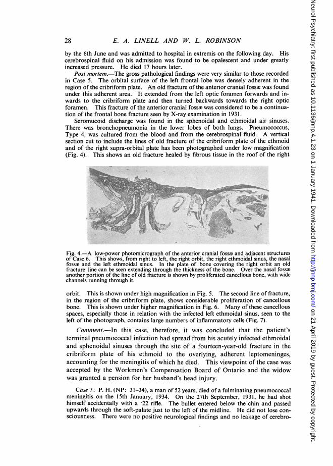
Seromucoid discharge was found in the sphenoidal and ethmoidal air sinuses.There was bronchopneumonia in the lower lobes of both lungs. Pneumococcus,Type 4, was cultured from the blood and from the cerebrospinal fluid. A verticalsection cut to include the lines of old fracture of the cribriform plate of the ethmoidand of the right supra-orbital plate has been photographed under low magnification(Fig. 4). This shows an old fracture healed by fibrous tissue in the roof of the right
-~~~~~~~~
Fig. 4.-A low-power photomicrograph of the anterior cranial fossa and adjacent structuresof Case 6. This shows, from right to left, the right orbit, the right ethmoidal sinus, the nasalfossme and the left ethmoidal sinus. In the plate of bone covering the right orbit an oldfracture line can be seen extending through the thickness of the bone. Over the nasal fosseanother portion of the line of old fracture is shown by proliferated cancellous bone, with widechannels running through it.
orbit. This is shown under high magnification in Fig. 5. The second line of fracture,in the region of the cribriform plate, shows considerable proliferation of cancellousbone. This is shown under higher magnification in Fig. 6. Many of these cancellousspaces, especially those in relation with the infected left ethmoidal sinus, seen to theleft of the photograph, contains large numbers of inflammatory cells (Fig. 7).
Comment.-In this case, therefore, it was concluded that the patient'sterminal pneumococcal infection had spread from his acutely infected ethmoidaland sphenoidal sinuses through the site of a fourteen-year-old fracture in thecribriform plate of his ethmoid to the overlying, adherent leptomeninges,accounting for the meningitis of which he died. This viewpoint of the case wasaccepted by the Workmen's Compensation Board of Ontario and the widowwas granted a pension for her husband's head injury.
Case 7: P. H. (NP: 31-34), a man of 52 years, died of a fulminating pneumococcalmeningitis on the 15th January, 1934. On the 27th September, 1931, he had shothimself accidentally with a *22 rifle. The bullet entered below the chin and passedupwards through the soft-palate just to the left of the midline. He did not lose con-sciousness. There were no positive neurological findings and no leakage of cerebro-
28
on 21 April 2019 by guest. P
rotected by copyright.http://jnnp.bm
j.com/
J Neurol P
sychiatry: first published as 10.1136/jnnp.4.1.23 on 1 January 1941. Dow
nloaded from
HEAD INJURIES AND MENINGITIS 29
_fa~~~~~~~~~~~~~~~~~~~~~~I-;:,- So -.Jj*
6
kz.~~~~~~~~~~~~~~~~~~
-} x ~* '-
VV~ ~ ~
Awt?,~~~~
IN I
'-A I sy
'i, V .< Q A,o
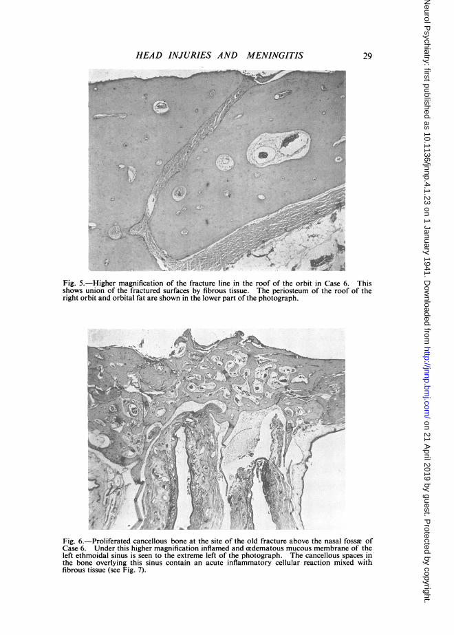
Fig. 5. Higher magnification of the fracture line in the roof of the orbit in Case 6. Thisshows union of the fractured surfaces by fibrous tissue. The periosteum of the roof of theright orbit and orbital fat are shown in the lower part of the photograph.
a~~~~~~
Fig. 6.-Proliferated cancellous bone at the site of the old fracture above the nasal fossw ofCase 6. Under this higher magnification inflamed and cedematous mucous membrane of theleft ethmoidal sinus is seen to the extreme left of the photograph. The cancellous spaces in'the bone overlying this sinus contain an acute inflammatory cellular reaction mixed withfibrous tissue (see Fig. 7).
on 21 April 2019 by guest. P
rotected by copyright.http://jnnp.bm
j.com/
J Neurol P
sychiatry: first published as 10.1136/jnnp.4.1.23 on 1 January 1941. Dow
nloaded from
30 E. A. LINELL AND W. L. ROBINSON
.1vi
;rt.-.-
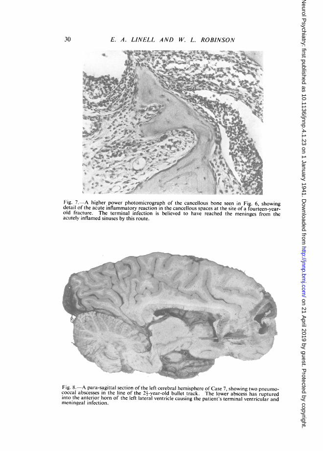
Fig. 7.-A higher power photomicrograph of the cancellous bone seen in Fig. 6, showingdetail of the acute inflammatory reaction in the cancellous spaces at the site of a fourteen-year-old fracture. The terminal infection is believed to have reached the meninges from theacutely inflamed sinuses by this route.
coca absess sinteln fte 2 -ya-l bletrc. Thf oe bcsshsrpue
inotA ;o
meningealinfection
meningealinfection
on 21 April 2019 by guest. P
rotected by copyright.http://jnnp.bm
j.com/
J Neurol P
sychiatry: first published as 10.1136/jnnp.4.1.23 on 1 January 1941. Dow
nloaded from

HEAD INJURIES AND MENINGITIS
spinal fluid. The bullet was seen by X-ray to be lying inside the vertex of the skullover the left frontal lobe just to the left of the superior sagittal sinus. On the followingday the bullet was easily removed through a small left frontal bone-flap. The bullettrack was explored with a soft rubber catheter which was passed from above throughthe left frontal lobe and through the bullet entry wound. After the operation thelower end of the catheter was seen by X-ray to be lying on the upper surface of thesoft palate. It was gradually removed from below and the patient made an uninter-rupted recovery.
He continued to work steadily and with no disability until a few days before hisdeath, 2- years later, when he developed a slight cold followed by a fulminatingpneumococcal meningitis.
Post mortem.-The old bullet entry wound in the posterior part of the left orbitalplate was covered by fibrous tissue. Its margin was continuous with a posteriorethmoidal air-cell. The bullet had passed upwards between the left optic nervelaterally and the left anterior cerebral artery mesially. The exit wound of the bulletfrom the brain was seen at the supero-mesial border of the left frontal lobe, 2 5 cm.behind its pole. A para-sagittal section cut to the left of the midline in the line of thebullet track (Fig. 8), shows two abscesses in the left frontal lobe in the line of the bullettrack. The lower abscess has broken into the cavity of the left lateral ventricle toproduce the patient's terminal ventricular and meningeal infection.
Comment.-This case is of interest in suggesting that pneumococci hadprobably remained alive in the bullet-track for a period of two and a half years,during which time they gradually produced two asymptomatic brain abscessesin the patient's left frontal lobe. It is also intriguing to wonder whether thecause of the rupture of the abscess into the ventricle was the sudden increasesof intracranial pressure, which probably occurred when he coughed andsneezed with his cold in the head. In this case there was no suggestion of arecent invasion of the cranial cavity by pneumococci from the nasal fosse.
Summary(1) Seven cases of meningitis are described in which the fatal infection
followed a fracture of the base of the skull.(2) In three of these cases the meningitis occurred 14, 5 and 24 years,
respectively, after injury.
31
on 21 April 2019 by guest. P
rotected by copyright.http://jnnp.bm
j.com/
J Neurol P
sychiatry: first published as 10.1136/jnnp.4.1.23 on 1 January 1941. Dow
nloaded from