development of the ethmoid sinus and extramural migration: the
TRANSCRIPT

Development of the Ethmoid Sinus andExtramural Migration: The Anatomical
Basis of this Paranasal SinusSAMUEL MARQUEZ,1* BELACHEW TESSEMA,2 PETER AR CLEMENT,3
AND STEVEN D SCHAEFER2
1Departments of Anatomy and Cell Biology; Otolaryngology,SUNY Downstate Medical Center, Brooklyn, New York
2Department of Otolaryngology, New York Eye & Ear Infirmary,New York Medical College, New York, New York
3Department of Otorhinolaryngology, University Hospital-FreeUniversity Brussel, Belgium
ABSTRACTFrontal and/or maxillary sinusitis frequently originates with patho-
logic processes of the ethmoid sinuses. This clinical association isexplained by the close anatomical relationship between the frontal andmaxillary sinuses and the ethmoid sinus, since developmental trajectoriesplace the ethmoid in a strategic central position within the nasal com-plex. The advent of optical endoscopes has permitted improved visualiza-tion of these spaces, leading to a renaissance in intranasal sinus surgery.Advancing patient care has consequently driven the need for the properand accurate anatomical description of the paranasal sinuses, regrettablythe continuing subject of persistent confusion and ambiguity in nomencla-ture and terminology. Developmental tracking of the pneumatization ofthe ethmoid and adjacent bones, and particularly of the extramural cellsof the ethmoid, helps to explain the highly variable adult morphology ofthe ethmoid air sinus system. To fully understand the nature and under-lying biology of this sinus system, multiple approaches were employedhere. These include CT imaging of living humans (n 5 100), examinationof dry cranial material (n 5 220), fresh tissue and cadaveric anatomicaldissections (n 5 168), and three-dimensional volume rendering methodsthat allow digitizing of the spaces of the ethmoid sinus for graphical ex-amination. Results show the ethmoid sinus to be highly variable in formand structure as well as in the quantity of air cells. The endochondralbony origin of the ethmoid sinuses leads to remarkably thin bony con-tours of their irregular and morphologically unique borders, making themsubstantially different from the other paranasal sinuses. These investiga-tions allow development of a detailed anatomical template of this regionbased on observed patterns of morphological diversity, which can initiallymask the underlying anatomy. For example, the frontal recess, ethmoidinfundibulum, and hiatus semilunaris are key anatomical components ofthe ethmoid structural complex that are fully documented and explainedhere on the basis of the template we have developed, as well as beingcomprehensively illustrated. In addition, an exhaustive 2000-year litera-ture search identified original sources of nomenclature, in order to helpclarify the persistent confusions found in the literature. Modified anatom-ical terms are suggested to permit proper description of the ethmoid
*Correspondence to: Samuel Marquez, Ph.D., SUNY Down-state Medical Center, Department of Anatomy & Cell Biology,450 Clarkson Ave, Box 5, Brooklyn, NY 11203.E-mail: [email protected]
Received 22 April 2008; Accepted 23 April 2008
DOI 10.1002/ar.20775Published online in Wiley InterScience (www.interscience.wiley.com).
� 2008 WILEY-LISS, INC.
THE ANATOMICAL RECORD 291:1535–1553 (2008)
region. This clarification of nomenclature will permit better communica-tion in addition to eliminating redundant terminology. The combinationof anatomical, evolutionary, and clinical perspectives provides an impor-tant strategy for gaining insight into the complexity of these sinuses.Anat Rec, 291:1535–1553, 2008. � 2008 Wiley-Liss, Inc.
Key words: paranasal sinus; ethmoid sinus; anatomy
The paranasal sinuses are among the most poorlydescribed anatomic sites in the human body. In largepart this is because of the great morphological variationsseen among individuals; but it is also due to the incon-sistency of the terminology used to describe these struc-tures. Much of the terminological confusion arose fromuncertainty about the origin of individual sinuses versustheir function, and from an arbitrary system of nomen-clature in which a sinus might be named for where itdrained, or in which bone it laid, or after an individual.Most of the terms now used were generated during theactive debate among surgeons in the first half of the20th century over the treatment of acute and chronic si-nusitis. Among the paranasal sinuses, the ethmoid sinusis often considered as the ‘‘keystone of the sinus system,’’because each paranasal sinus drainage pathway isthrough, or adjacent to its lateral wall (Terrier, 1991).This investigation examines the adult condition of
the ethmoid sinus in order to better understand its de-velopment. Multiple approaches are used, including CTimaging, anatomical dissection, three-dimensional recon-structions, and examination of dry cranial material.Tracking the migration of extramural cells and under-standing their variation helps to clarify the anatomicalbasis of this paranasal sinus.
BACKGROUND
Phylogenetically the ethmoid sinus appears to haveonly an olfactory function, and it is not generally consid-ered a ‘‘true’’ paranasal sinus because it lacks pneumati-zation. The recognition of ‘‘true’’ paranasal sinuses isthus based purely on the ontogenetic pattern given byCave (1967). In this view a ‘‘true’’ paranasal sinus musthave its respiratory diverticulum originate from, andremain in communication with, the nasal cavity. Itsgrowth must be from a given meatus of the nasal cavity,and it must retain a patent communication via anostium that remains permanently associated with thatmeatus. The sole guide to the morphological identity of asinus is provided not by the bone or bones it may ulti-mately pneumatize, but by the bone or bones that cir-cumscribe its ostium, or point of origin. Cave (1967)makes the point that neglect of this basic considerationhas resulted all too frequently in the confusing misinter-pretation of analogous cavities as homologues. An exam-ple of such misclassification can be drawn from a CTexamination of an orangutan (Fig. 1), which has beentraditionally reported as exhibiting only one paranasalsinus—the maxillary sinus.In most of the mammals, the ethmoid bone and its
associated turbinal extensions, along with the nasoturbi-nal and maxilloturbinal, are placed posterior to the par-
anasal sinuses. The lamina cribrosa is in a frontal posi-tion, for optimal olfactory function. Only in humans,chimpanzees and perhaps in orangutans (Marquezet al., 1999), does the ethmoid become pneumatized andcontain a sinus. This is probably related to the uprightposture of humans, in association with which a retrac-tion of the snout occurs and the orbit migrates anteri-orly, resulting in a deepening of the face. The anteriormigration of the ethmoid bone forces its sinus betweenthe nasoturbinal and maxilloturbinal, displacing thefrontal sinus upwards, and disconnecting the latter fromthe maxillary sinus. This bony ethmoidal migration inhumans made possible an upper aerodigestive tractcrossing so that, in conjunction with the descent of thelarynx, the unique modern human speech apparatuscould eventually develop (see Laitman and Reidenberg,1993). As olfaction became less important for humansurvival compared with more macrosmatic mammals,the position of the lamina cribrosa becomes more hori-zontal, the olfaction function shifts toward the micro-smatic, and the ethmoid bone becomes pneumatized toattain paranasal sinus status. This apparent adaptiveevolutionary shift has resulted in very narrow drainagechannels (i.e., tight spots) of the ethmoid; in contrast tomost other mammals in which rhinosinusitis is a raredisease, this configuration favors infection. As a result,Stackpole et al. (1996) described the ostiomeatal complexas the ‘‘eye of the needle" of the paranasal sinuses.Thus, for adventitious reasons, the ethmoid bone, phylo-genetically never destined to be a sinus, has evolvedinto a drainage and ventilation pathway of the maxillaryand frontal paranasal sinuses (i.e., ‘‘ostiomeatal com-plex’’), forming not an anatomical but a ‘‘functional’’unit.Embryologically the ethmoid is different from all the
other sinuses. The ethmoid bone originates from the car-tilaginous nasal capsule or paleosinus (endochondralbone), whereas the other paranasal sinuses are exten-sions from the ethmoid (extracapsular) into membranousbone (neosinus) via epithelial diverticula extensions.Specific stages of development are necessary for para-nasal sinus development to occur. These include primaryand secondary pneumatization patterns that involve dif-ferential growth of the cartilagionous nasal capsule,which initially produces diverticular pouches thatexpand within the confines of the capsule creating elabo-rate intracapsular airway spaces (Smith et al., 2005).Secondary pneumatization patterns involve the para-nasal recess, the region of the nasal capsule that is des-tined to become a sinus; as its diverticula leave the con-fines of the nasal capsule, this becomes extracapsularand is left occupying space in adjacent structures. Thus,the paranasal recess becomes a paranasal sinus througha specific sequence of developmental events and fulfils
1536 MARQUEZ ET AL.

the sole criteria for proper sinus identification (Rossie,2006). Although the extent of the paleosinus, or ethmoid,is inherently fixed and constrained, the sizes of the neo-sinus (frontal, maxillary and sphenoidal) are highly vari-able, because paranasal recesses are not predictable.There is no apparent uniformity in the rate of develop-ment or the degree of differentiation in the migration ofthese extracapsular diverticula, which when extramural,expanding beyond the confines of the ethmoid bone, canvariably pneumatize adjacent structures leaving diversedrainage pathways.Historically, investigators have mixed extramural and
intramural structures in their anatomical descriptions ofthis region. To avoid confusion, Layton (1934) advisedthat the following principles should be followed whendescribing anatomical structures: ‘‘a name must (1)
denote one thing only; (2) be used by all to denote thisthing; and, if it connotes any attribute, then (3) this at-tribute should always be present.’’ According to Layton(1934), two terms that violate these principles are ‘‘in-fundibulum’’ and ‘‘hiatus semilunaris.’’ Both are appliedto regions or spaces, much as the pharynx is a spacebounded by muscles, rather than a structure. This con-cept, that spaces and structures are of equal importance,is critical for a full understanding of the nasal com-plex—defined as the nose plus the paranasal sinuses(Marquez, 2002).Probably, no other term has been more confused in
the anatomical literature of the nose and paranasalsinuses than the term infundibulum (meaning‘‘funnel’’). Layton contends that the term infundibulumentered the anatomical language as a name passedfrom Latin to the other romance languages. The appa-rent first appearance of this term in the English ana-tomical literature was in the 1797 Edinburgh textbookof anatomy known as ‘‘Monro’s’’ (Layton, 1934). Thefirst part contains the anatomy of the Human Bones byAlexander Monro the First (the senior Monro), and thefirst volume says:
‘‘A cellular and spongy bone substance dependsfrom the cribriform plate. The number and figureof the cells, in this irregular process of each side,are very uncertain, and not to be represented inwords; only the cells open into each other, and intothe cavity of the nose: The uppermost, which arebelow the aperture of the frontal sinuses, are foundlike funnels.’’
However, in volume II it states:
‘‘The frontal, maxillary, and sphenoidal sinuses openinto the internal nares, but in different manners.The frontal sinuses open from above downwards,answering to the infundibula of the os ethmoidalesdescribed in the history of the skeleton.’’
Zuckerkandl (1892), on the other hand, credits BaronAlexis de Boyer with being the originator of the terminfundibulum, a term that emerges in his publishedFrench work of 1815 (vol. 1, p 125):
‘‘Entre la face externe de ce cornet (i.e., middle tur-binate) et les masses laterales se trouve un enfonce-ment qui fait partie du meat moyen des fossesnasales. A la partie anterieure de cet enfoncementon remarque une espece de gouttiere qui monte dederriere en devant dans les cellules anterieures del’os; la cellule dans laquelle cette gouttiere aboutitest large superieurement et etroite inferieurement,ce qui lui a fait donner le nom l’infundibulum; elles’abouche avec l’ouverture du sinus frontal.’’
Comparing these accounts, it appears that the Edin-burgh School of Anatomists can claim credit for originat-ing the usage of the term familiar today, since they rec-ognized the frontal sinuses as being of the same natureas the ethmoidal cells, and applied the term infundibu-lum specifically to the channel that often erroneouslyknown today as the fronto-nasal duct (see Discussionlater).
Fig. 1. A: An axial scan of a subadult orangutan (Pongo pygmaeus)showing what appears to be a sphenoid sinus but is actually the max-illary sinus invading the sphenoid bone (red arrow). B: In an adultorangutan, the communication (yellow arrows) between the left maxil-lary sinus and the evacuated sphenoid bone is clearly shown; the rightmaxillary sinus (red arrows) is seen to encroach upon the sphenoidbone.
1537ETHMOID SINUS AND EXTRAMURAL MIGRATION

Layton (1934) alerts the reader to the fact that Boyerbelieved that the frontal sinus drained through an eth-moidal cell into the nose, that and it is to this cell,rather than the groove, that Boyer applied the term in-fundibulum. Layton’s contention is justified on the basisof Boyer’s description of the nasal fossa under splanch-nology, where we read (Tome IV, p 176), under ‘‘Le Meatmoyen,’’ that:
‘‘On voit a la partie moyenne et anterieure de cemeat une gouttiere etroite qui monte de deriere endevant, et va communiquer dans les cellules anteri-eures de l’ethmoide, et par le moyen de celles-cidans le sinus frontal.’’
It is precisely to this erroneous notion that the frontalsinus opens through an ethmoidal cell that Layton(1934) attributes the ongoing nomenclatural confusion.It may be that Monro had the same idea when he gavehis many lectures on the topic. But even as anatomists’understanding of this region improved, some continuedto use infundibulum to describe the less common drain-age of the frontal sinus via an intermediate cell,whereas some used the term ‘‘nasofrontal duct,’’ someretained the term for the groove into which the sinusopened; and others used it only for the upper part ofthis groove where an ethmoidal cell would lie if the fron-tal sinus opened through it. What is certain is thatZuckerkandl set the standard for the use of the term in-fundibulum for the groove, and also added the modifierthe ethmoidale, in an attempt to distinguish this featureclearly from the hiatus semilunaris. The latter is prop-erly a two-dimensional space, the gap through whichthis groove opens on the middle meatus. We note thatHis (1895) in his pamphlet Die Anatomische Nomencla-tur, which became the basis of the British Nomina Ana-tomica, distinguishes the two terms by an indentation ofthe margin:
‘‘Infundibulum EthmoidaleHiatus Semilunaris.’’
However, a number of workers still used the term in-fundibulum (i.e., Tillaux, 1878; Bosworth, 1888; Turner,1901), whereas others used infundibulum ethmoidalis(i.e., Shambaugh, 1907).Currently, there are three structures in the nose
described as infundibula: (1) the frontal infundibulum;(2) the maxillary infundibulum; and (3) the ethmoid in-fundibulum. The frontal and maxillary infundibula lietotally within their respective sinuses, and their open-ings are their respective ostia. In contrast, the ethmoidinfundibulum is not contained within a given sinus, butrather is a three-dimensional space whose funnel ismuch wider at its lateral base, permitting it to receivethe common drainage of the frontal, maxillary, and ante-rior ethmoid sinuses. The apex of this space orients to-ward the hiatus semilunaris, before emptying into themiddle meatus.The ontogeny of the ethmoid sinus permits the catego-
rization of the ethmoid sinus into anterior and posteriorethmoid components. However, a survey of anatomytextbooks used in first-year medical gross anatomycourses (i.e., Rose and Gaddum-Rose, 1997, p 832; Snell,2000, p 747; Drake et al., 2005, p 971; Moore and Dalley,
2006, p 1019) as well as laboratory dissecting manuals(Tank, 2005, p 197), continue to divide the ethmoidsinuses into anterior, middle, and posterior groups. Asearly as 1901, Turner (1901) divided the ethmoid sinusesinto two groups, anterior and posterior groups, based ondevelopment and their position of their ostia. Douglas(1906) believed that division of the ethmoid sinus intoan anterior and posterior group is the correct classifica-tion because it is based on sound embryological groundsbecause it is to be understood that the criteria of group-ing divisions is on its drainage destiny. Any ethmoidalcell, which normally drains into the middle meatus ofthe nose belong to the anterior ethmoidal cell group; andthat all ethmoidal cells that normally drain into thesuperior meatus of the nose belong to the posterior eth-moidal cell group. The addition of a middle ethmoidgroup serves only to confuse students in the area of rhi-nology (Douglas, 1906, p 75).The most recent edition of British Gray’s Anatomy rec-
ognized the confusion in the literature with this state-ment: ‘‘the ethmoidal sinuses are now commonly consid-ered by clinicians as consisting of anterior and posteriorgroups on each side with the middle ethmoidal air cellsbeing incorporated into the anterior group’’ (Standring,2005, 39th edition, p 576). This last comment appears topit anatomists and clinicians as two separate entitiesthat are in conflict when it comes to describing thisregion. Let us remember that the early pioneers whohelped established the discipline of rhinology such asHartmann, Zuckerkandl, Bosworth, His, Hajek, Onodi,Killian, Turner, Mosher, Schaeffer, Van Alyea, and Da-vies were not only all great physicians but also helpedlay the foundation of present-day anatomic descriptionof the region.
MATERIALS AND METHODS
A sample of 488 specimens was examined for thisstudy, and included dry skulls from the osteological col-lections in the Division of Anthropology, American Mu-seum of Natural History, and at SUNY Downstate Medi-cal Center. Additional specimens were examined andphotographed from University Hospital-Free University,Brussels. Cadaveric dissections (n 5 168) from grossanatomy courses over a 4-year period were utilized anddigitally photographed for examination. Fresh tissue dis-section (unembalmed) material was also included in thisstudy (n 5 8), involving the removal of heads in the pa-thology autopsy room at SUNY Downstate Medical Cen-ter and the University of Texas Southwestern MedicalCenter. These specimens were frozen to a temperatureof 2708C. The Head was then fixed to a wood block andanchored with screws. Two-centimeter sagittal sectionswere prepared, utilizing a butcher’s band saw. Speci-mens were digitally photographed using a Nikon D100camera. CT imaging of patients with and without sinusdisease was performed in order to document the vari-ability of this sinus, using a mixed-sex sample size of200 adult individuals. Selective images were reprocessedusing a three-dimensional CT program (Anatomage1).We also drew on the clinical perspective possessed bytwo of the senior authors (SDS and PARC), who togetherhave over 70 years of clinical experience. Together theyhave served on anatomical nomenclature committees(i.e., see Stammberger et al., 1995), taught and trained
1538 MARQUEZ ET AL.

more than 10,000 physicians worldwide, and have oper-ated in this region in more than 9,000 cases combined.
RESULTS
The ethmoid sinus system is comprised by a numberof air cells that ranged from 6 to 16 on one side from theosteological collections of the American Museum of Nat-ural History and at SUNY Downstate, 7–14 from the an-atomical embalmed material (see Table 1), and 8–11 onthe fresh anatomical material Figs. 2–4 (see Discussionregarding the term cell). Observation from hard and softtissue material shows the ethmoid sinus to have a con-sistent pattern of aeration of the bony labyrinth, but thequantity of air cells is highly variable. The hard tissueanatomical material from the osteological collections andunembalmed cadavers illustrate how variable and irreg-ular these air cells are in situ (see Figs. 2–4). The
observed air cells present with ‘‘paper-thin’’ bony lamel-lae1 walls that are morphologically distinct from anyother bony substrate found in our sample (for discussionon bony lamellae see Bohatirchuk et al., 1972).
TABLE 1. Identification of ethmoid air cells inembalmed material
Identification no. Right Left ID Right Left
2003-303 7 9 2004-097 8 102003-304 10 8 2004-149 a 112003-305 9 9 2004-181 a 92003-306 7 11 2004-189 9 82003-307 11 12 2004-193 12 112003-308 12 10 2004-403 11 102003-309 10 10 2004-404 12 72003-310 8 9 2004-405 8 102003-311 12 10 2004-406 a 112003-312 a 9 2004-407 a 92003-313 11 8 2004-408 11 122003-314 7 12 2004-409 8 72003-315 8 10 2004-410 10 102003-316 a 11 2004-411 8 92003-317 a 9 2004-412 12 102003-318 9 12 2004-413 10 92003-319 8 7 2004-414 9 82003-320 12 11 2004-415 12 112003-321 10 7 2004-416 10 102003-322 7 8 2004-417 12 72003-323 9 11 2004-418 10 82003-324 12 7 2004-419 9 132003-325 10 8 2004-420 13 142003-401 7 9 2004-421 12 112003-402 8 14 2004-422 10 72003-403 5 8 2004-423 7 82003-404 12 10 2004-424 9 112003-405 10 10 2004-425 6 72003-406 8 9 2004-426 10 82003-408 6 7 2004-427 7 92003-409 a 9 2004-429 8 a
2003-410 a 8 2004-430 8 102003-411 7 12 2004-431 7 92003-413 9 12 2004-432 10 82003-414 13 a 2004-433 9 92003-415 12 11 2004-434 7 112003-417 10 7 2004-435 11 122003-420 7 8 2004-436 5 72003-421 8 11 2004-437 10 102003-441 12 7 2004-438 8 92003-442 10 8 2005-021 12 102003-443 7 9 2005-029 a 92003-444 8 14 2005-043 11 8
TABLE 1. Identification of ethmoid air cells inembalmed material (continued)
Identification no. Right Left ID Right Left
2005-046 9 12 2006-405 9 82005-050 8 a 2006-406 8 112005-056 6 9 2006-408 a 102005-066 9 7 2006-409 9 112005-067 7 8 2006-410 10 72005-068 a 11 2006-414 7 82005-074 12 7 2006-415 9 112005-078 10 8 2006-418 12 72005-083 7 9 2006-419 10 82005-084 8 14 2006-420 7 92005-086 12 11 2006-421 8 a
2005-092 13 12 2006-422 7 112005-093 6 6 2006-423 13 a
2005-099 10 7 2006-427 12 112005-108 7 8 2006-430 10 72005-168 9 11 2006-431 7 82005-169 8 7 2007-101 8 112005-190 5 9 2007-102 6 72005-193 10 7 2007-103 10 82005-402 7 8 2007-104 7 92005-406 13 11 2007-105 8 172005-407 7 9 2007-106 12 112005-408 10 8 2007-107 10 72005-412 9 9 2007-108 7 82005-414 7 11 2007-109 11 112005-415 11 12 2007-110 12 72006-104 8 10 2007-111 7 92006-105 a 11 2007-112 10 82006-106 14 9 2007-071 9 92006-107 9 8 2007-099 7 112006-108 9 12 2007-114 11 122006-109 13 14 2007-115 a 102006-110 12 11 2007-118 10 102006-111 10 7 2007-128 8 92006-112 7 8 2007-130 12 102006-113 13 11 2007-132 a 92006-114 12 7 2007-136 13 82006-117 10 8 2007-180 8 102006-120 7 9 2007-181 14 92006-122 8 a 2007-183 9 82006-123 8 10 2007-197 10 102006-132 10 11 2007-401 9 82006-402 a 9 2007-403 11 a
aUnable to identify air cell quantity due to preservation ofmaterial.
1The term lamella has been used by Vitruvius Pollio (DeArchitectura 7, cap 3, paragraph 9, 25 BC). ‘‘Quemadmodumenim speculum argenteum tenui lamella ductum incertas etsine viribus habet remissiones splendoris. . . ‘‘. ‘‘Lamella’’ is hereused as a diminutive of lamina and means to describe a thinmetal plate. Nathaniel Highmore was one of the original anato-mists to use the thickness of paper to describe a thin wall struc-ture by the following: ‘‘It is covered by thin bone or bony scale;. . ..barely exceeds the thickness of packing paper’’ (Highmore,1651, p 226).
1539ETHMOID SINUS AND EXTRAMURAL MIGRATION

Fig. 2. A: Sagittal paramedian section through dried skull. Labels:ES, ethmoid sinus; FS, frontal sinus; SS, sphenoid sinus; MS, maxil-lary sinus. B: Magnified view showing thin enchondral bone of eth-moid sinus. C: Sagittal paramedian section through unembalmed
cadaver illustrating paranasal sinus anatomy. Labels: SS, sphenoidsinus; FS, frontal sinus; D: 1882 illustration by Emil Zuckerkandl show-ing the paranasal sinuses in a coronal plane.
1540 MARQUEZ ET AL.
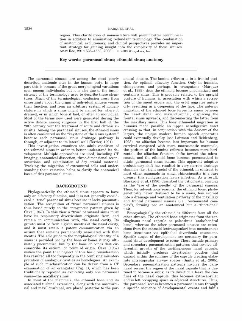
An analysis of the medial wall of the bony orbit showsthe contour of lamina orbitalis ossis ethmoidalis, or laminapapyracea, of the recessus frontalis and of os lacrimale (Fig.5). A number of sutures can be clearly identified thatinclude the frontoethmoid, frontolacrimal, frontomaxil-lary, and frontonasal sutures (Fig. 5). A number of ocu-lar muscles attach on the medial wall of the orbit (Fig.6). The lateral wall of the frontal recess, the area ofattachment of the lamella bullae ethmoidalis, and theattachment of the basal lamella of the middle turbinatecan also be observed in the skull (Fig. 6). It was docu-mented on this specimen that a small part of the lateralwall of the frontal recess is made up by lamina papyra-
cea. The attachment of the middle turbinate can be out-lined on the ethmoid bone (Fig. 7) and a small aggernasi cell can be detected on the lateral wall of the eth-moid (Fig. 8). Considerable pneumatization of the aggernasi can take place and can be easily observed in CTscans (see Fig. 9). The topographical location of theagger nasi is considered as the anterior limb of the unci-nate process. Further pneumatization of the aggerincludes the migration of ethmoid cells into the lacrimalbone, increasing the prominence of this eminence of thelateral nasal wall. Identification of the lacrimal bone isan important landmark because it defines the borderbetween the frontal bone and the frontal process of the
Fig. 3. A: Sagittal paramedian section through dried skull indifferent specimen than Fig. 2A–B illustrating variations in ethmoidanatomy. Inset of whole skull provides for orientation. Labels: SS,sphenoid sinus; MS, maxillary sinus; FS, frontal sinus. B: Sagittal par-amedian section through additional unembalmed cadaver head show-
ing variation in sinus anatomy. C: Paramedian three-dimensionalreconstruction of multiple CT images showing complexity of sinusanatomy. Labels: SS, sphenoid sinus; MS, maxillary sinus; FS, frontalsinus; pe, posterior ethmoid sinus; ae, anterior ethmoid sinus.
1541ETHMOID SINUS AND EXTRAMURAL MIGRATION

maxilla (see Fig. 10). When the ethmoid bulla is promi-nent by CT imaging, the lamina papyracea is observedas its lateral border (Fig. 10). The lamina cranialis ofthe frontal bone is the roof of the supraorbital recess,which is shown in Fig. 10.Frontal sinus variation exhibited a number of differ-
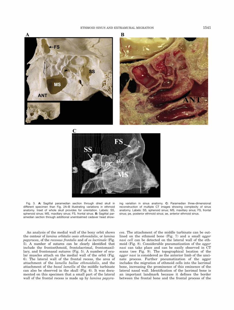
ent morphological patterns. For example, utilizing theBent classification (Bent, 1994), Type-III cells are foundin both frontal sinuses of one individual on CT images
(Fig. 11). However, parasagittal CT slices of the sameperson reveal that these cells communicate with thefrontal recess where the anterior part of the supraorbitalrecess becomes part of the frontal recess (Fig. 12). In aCT scan of an individual with frontal sinus diseaseType-A-II and Type-A-III cells (see Discussion later fordefinition of Type-A versus Type-B cells) are identifiedwith a frontal recess that extends into the nasal bonesand septum (the so-called nasal recess) (Fig. 13). Figures16 and 17 show the variation of Type-A-II and Type-A-III cells in the frontal sinus region. Divisions of the fron-tal sinus can occur with septated bony divisions that canaffect the frontal sinus outflow tract. These bony divi-sions referred to as interfrontal septum or septum sin-uum frontalium can present partial (incomplete) or com-plete division of the sinus (Fig. 14). Figure 15 shows thepotential effect of a deviated nasal septum on frontalsinus drainage. Examples of migrating extramural cellsby the ethmoid into adjacent structures are consistentlyvariable. A concha media bullosa is a pneumatization ofthe anterior middle turbinate bone via a posterior eth-moid air cell. This is referred to as an interlaminar cell
Fig. 4. A: Sagittal paramedian section through dried skull in a thirdspecimen, again illustrating the variation in development of the eth-moid sinus. Inset of whole skull provides for orientation. Labels: SS,sphenoid sinus; MS, maxillary sinus; B: Sagittal paramedian sectionthrough unembalmed cadaver other than seen in prior figures showinganatomy of ethmoids, frontal recess and frontal sinus. Labels: FS,frontal sinus; fo, frontal ostium or outflow tract; fr, frontal recess; pe,posterior ethmoid cell.
Fig. 5. A and B: view on medial wall of right orbit. Thick blacklines: 1, contour of lamina orbitalis ossis ethmoidalis (lamina papyra-cea); 2, contour of lamina recessus frontalis; 3, contour of lamina of oslacrimale. Green arrows: superior border of lamina papyracea (suturafrontoethmoidalis). Red arrows: superior border of os lacrimale (suturafrontolacrimalis). Black arrows: antero-superior border of frontal recess(in A) and superior border only (in B). Yellow arrows: sutura frontomax-illaris. Blue arrow: sutura frontonasalis. Note the difference in heightbetween the superior border of the lacrimal and ethmoidal bone! Thisdifference results in a bend (in A) or even a notch (in B) of the suturafrontoethmoidalis (incisura lamina orbitalis, ossis ethmoidalis, or laminapapyracea—purple line).
1542 MARQUEZ ET AL.

of Grunwald (Fig. 18). Other extramural cell migrationoccurs when these cells migrate to the floor of the orbitor infraorbital cells (Fig. 19). These cells are called Hal-ler cells in honor of August von Haller who originallydescribed them in the early 19th century (Haller andCullen, 1803). Other findings include extensive pneuma-tization that envelops the nasal bones, nasal septum,and frontal recess (Fig. 20).
DISCUSSION
The hard tissue anatomy of the ethmoid is of endochon-dral origin forming part of the cranial base, which is con-sidered a highly phylogenetically conserved region amongthe bony elements of the skull (De Beer, 1937). From ananatomical perspective, the ethmoid is significantly dif-ferent than all the other sinuses in that it is the only par-anasal sinus that presents with very thinned bony walllamellae. The other paranasal sinuses can form septa-tions, but these are much more rigid and robust. The con-sequence is that these very thin bony lamellae and cellscan migrate easily (extracapsular) into adjacent bones orother paranasal sinuses. These extramural extensionscan migrate into frontal recess cells, frontal cells, supra-orbital cells, and/or infraorbital cells, or any combinationsthereof. This crucial developmental concept explains thesource of ethmoid sinus cell variation in that extramuralmigration can take different paths thus presenting differ-ent morphologies. Classic anatomic studies of the eth-moid have failed to document the entire spectrum of eth-moid sinus construction either due to low sample sizes orrandom sampling error. For the surgeon, however, thevaried manifestations of extramural migration of the eth-moid is critically important for it decides the proper sur-gical procedure to be implemented to resolve the patho-logic condition of the patient. The intramural cells andlamellae do not show any uniformity, resulting in the‘‘ethmoidal labyrinth.’’ One cell may outgrow its neighborand force the latter to progress in a direction other thantoward it was primarily directed (Anon et al., 1996).The architectural construction of the ethmoid sinus can
be understood by examining the early ontogeny of theregion (Shambaugh, 1907). During prenatal developmentthe attachments of bony structures originating from theethmoid to the lateral wall such as the uncinate process,
Fig. 7. A and B medial view on sagittal part of middle turbinate intwo different cases. Thick blue line: attachment of sagittal part of mid-dle turbinate. B has a well developed ‘‘agger nasi cell,’’ whereas in Athe agger nasi is not very pneumatized resulting in a small ‘‘agar nasicell.’’ Shaded areas indicate the site of the agger nasi. Blue doubleheaded arrows: site of insertion of the middle turbinate on frontal pro-cess of the maxilla (crista ethmoidalis ossis maxillaris). Red double
headed arrows: site of insertion of middle turbinate to floor of frontalsinus when frontal recess when the latter is extensively pneumatizedmedially. Green double headed arrows: part of the middle turbinatethat attaches to the skull base. Thick green arrow points to a dashedgrey line (free border of the middle turbinate). Note that B has a welldeveloped ‘‘agger nasi cell.’’
Fig. 6. View on medial wall of orbit. Numbers (see Fig. 5). Redlines: attachment of ocular muscles. Red arrows point to shadedplane (5 lateral wall of frontal recess). Thick green line: attachment oflamella bullae ethmoidalis. Thick blue line: attachment of basal lamella(lamella basalis, frontal part of middle turbinate). Note that only a smallpart of the lateral wall of the frontal recess is made up by laminapapyracea.
1543ETHMOID SINUS AND EXTRAMURAL MIGRATION

middle and superior concha, are formed by ground plates,or basal lamellae. Although the lateral attachments ofthese lamellae end abruptly, their medial aspects projectbeyond the labyrinth to form prominences that extendsinto the nasal cavity. The most anterior of these lamellaeis the lateral extension of the uncinate process. The sec-
ond lamella is referred to as the plate of the bulla becauseits extension into the nasal cavity forms the bulla ethmoi-dalis.2 The third lamella serves as the attachment of thefrontal part of the middle turbinate. The middle turbinatehas three parts: the parasagittal, the frontal, and thetransversal. The parasagital and the transversal do notplay a role in the division of anterior and posterior eth-moid. This is an important anatomic structure, demarcat-ing the division between the anterior ethmoid cells fromthe posterior cells and directing the drainage patterns ofthese air cells into the middle and superior meatus,respectively. It is also clinically significant as it is consid-ered as a natural boundary to the spread of infection intothe posterior ethmoid and serves as the posterior land-mark in anterior ethmoidectomy (Schaefer et al., 1998).The ethmoid air cells during development tend to expandto occupy all available space, a phenomenon, which Seydel(1891) has called ‘‘the struggle for space of the ethmoid.’’This results in air cells impinging upon and perhapsdistorting the osseous barrier, but the lamella alwaysremains intact and prevents intermingling of cells (VanAlyea, 1946; Stammberger, 1991).Another important variation in ethmoid anatomy is
the lateral sinus. This space is bounded laterally by thelamina papyracea, posteriorly by the lamella of the fron-tal part of the middle turbinate, anteriorly by the poste-rior aspect of the bulla ethmoidalis, and superiorly bythe fovea ethmoidalis. When the second lamella does not
Fig. 9. A and B: coronal CT scans. A: a large agger nasi cell (notethe intimate relation between agger nasi cell and uncinate process). B:transition of the agger nasi cell into a lacrimal cell (note again that thefloor of this cell is shaped by the uncinate process that is now more visi-ble). Yellow arrows: frontal process of the maxilla. Blue arrows: orbitalplate of the frontal bone. Green arrows: lacrimal bone. Note in A the freeborder of the middle turbinate is not visible or adjacent to the agger nasicell, and the lateral wall of that cell is made up by the frontal recess ofthe maxilla. This is a true ‘‘agger nasi cell’’ because it is located withinthe surface feature of the lateral nose known as the agger nasi. In B thelateral wall of this cell is made up by the lacrimal bone and free borderof the middle turbinate begins to become visible. As this cell is posteriorto the agger nasi, it would be considered a ‘‘lacrimal cell.’’
Fig. 8. Internal view of the lateral wall of the ethmoid (parasagittalsection). Dashed line represents the agger nasi. In this case, there existsa small agger nasi cell in close contact with the uncinate pocess (yellowline). fr, Frontal recess. Black double headed arrow: posterior border ofagger nasi. Black arrows: anterior and posterior ethmoidal artery. Greenline: attachment of the bulla and lamina bullae to lamina papyracea.Blue line: attachment of ground, or basal lamella (lamina basalis) to lam-ina papyracea. Red line: anterior border of lamina papyracea. Note smallpart of lateral wall of the frontal recess that is made up by lamina papy-racea (less than 10% of area is between red and green line).
Fig. 10. A and B: coronal CT scans. A: Purple arrows point to thelamina orbitalis of the frontal bone that is always superior to the lacri-mal bone (red arrows). The lacrimal bone is an easy landmark todefine the difference between frontal bone and frontal process of themaxilla. In this CT scan one can see the frontal recess underneath thefloor of the frontal sinus. B: Green arrows: lamina papyracea (lateralwall of the bulla ethmoidalis). 1: Frontal sinus; 2: Frontal recess; 3:Suprabullar recess. Red line: Lamina orbitalis of frontal bone. Yellowline: Lamina cranialis of frontal bone (roof of the supraorbital recess).Purple line: roof of the suprabullar recess (or fovea/frontal bone).
2Other frequently made errors are inconsistent with Latingrammar. ‘‘Bulla lamella’’ (Stammberger et al. 1995, Owen andKuhn 1997, Wormald 2003) is grammatically incorrect. InLatin, the correct term is lamella bullae ethmoidalis or in Eng-lish a bullar lamella, as in suprabullar recess. Furthermore theterm ‘‘lamella’’ does not exist in the ‘‘Terminologia Anatomica’’nor in Cassel’s Latin Dictionary (see Simpson, 2000 and foot-note 1). A more appropriate Latin name for the basal, or groundlamella which divides the ethmoid into an anterior and poste-rior sinus) is ‘‘lamella basalis.’’
1544 MARQUEZ ET AL.

extend to the fovea ethmoidalis-frontal bone (thishyphenated term is used to remind the reader of thecomplex nature of the roof of the ethmoid sinus), thesinus lateralis continues anteriorly to the frontal recess.Superiorly, this ‘‘sinus’’ is formed by a space known asthe suprabullar furrow, or suprabullar recess, whichcommunicates with the middle meatus (Grunwald, 1925;Messerklinger, 1978; Stammberger et al., 1995). Some-times the sinus lateralis can extend very laterally whenthere exists a well-developed supra-orbital recess. A truesinus lateralis is rare, perhaps only occurring 7% ofspecimens (Bolger and Mawn, 2001) as a bony lamellamore often separated the superior spaces from inferiorand posterior retrobullar and infrabullar spaces. The
superior aspect of the hiatus semilunaris communicateswith this space, and this crescent-shaped two-dimen-sional structure forms the final common pathway forsecretion to drain into the nose. The fourth lamella is atthe attachment of the superior concha, and when asupreme concha3 is also present, a fifth lamella ariseslateral to this turbinate.The frontal recess is an important space for it is part
of the outflow drainage pathway of the frontal sinus(Fig. 11). A description of the pneumatization patternsof adjacent structures by the extramural migration ofthe ethmoid is essential for describing the anatomy ofthe frontal recess and its contiguous structures that bor-ders the recess. Describing supraorbital and ethmoidbullar cells requires clarifying what is meant when re-ferring to an ethmoid air cell. We suggest with sometrepidation that the term ‘‘cell’’ should only be used forevery sphere-like structure, which are composed of thinbone of endochondral origin. For this reason, the frontaland maxillary sinus cannot be considered as extramuralethmoidal cells. Consistent with the above, Zuckerkandl(1892) stated that the frontal sinus is not an invasion bythe ethmoid into the frontal bone, but a bulging of thenasal mucosa of the frontal recess into the frontal bone,
Fig. 11. A and B are coronal CT scans whereas C, D, and E areparasagittal CT scans. Type-A-III cells in both frontal sinuses. Note theadhesions between the walls of the cells and the frontal sinus walls(according to the Bent et al. classification, these cells would be Type-
IV cells but on the parasagittal sections it is obvious that these cellscommunicate with the frontal recess. Therefore, these cells would beclassified as Type-III by Bent et al.). 1: Frontal sinus, 2: Frontal recess,Black arrow: frontal sinus aperture (apertura sinus frontalis).
3Giovanni Santorini, described this conchae in 1724, and inhonor of him, this conchae bears his name today as the ‘‘Conchaeof Santorini’’ (Santorini, 1724). In the hollow walls of Padua,where there is so much tradition with the many famed anato-mists that trained and taught there that the highest honour wasto have the skull of an anatomy master bequeath to the medicalschool. Santorini’s skull is still displayed in the library of theanatomy department at the University of Padua Medical School(Fig. 21).
1545ETHMOID SINUS AND EXTRAMURAL MIGRATION
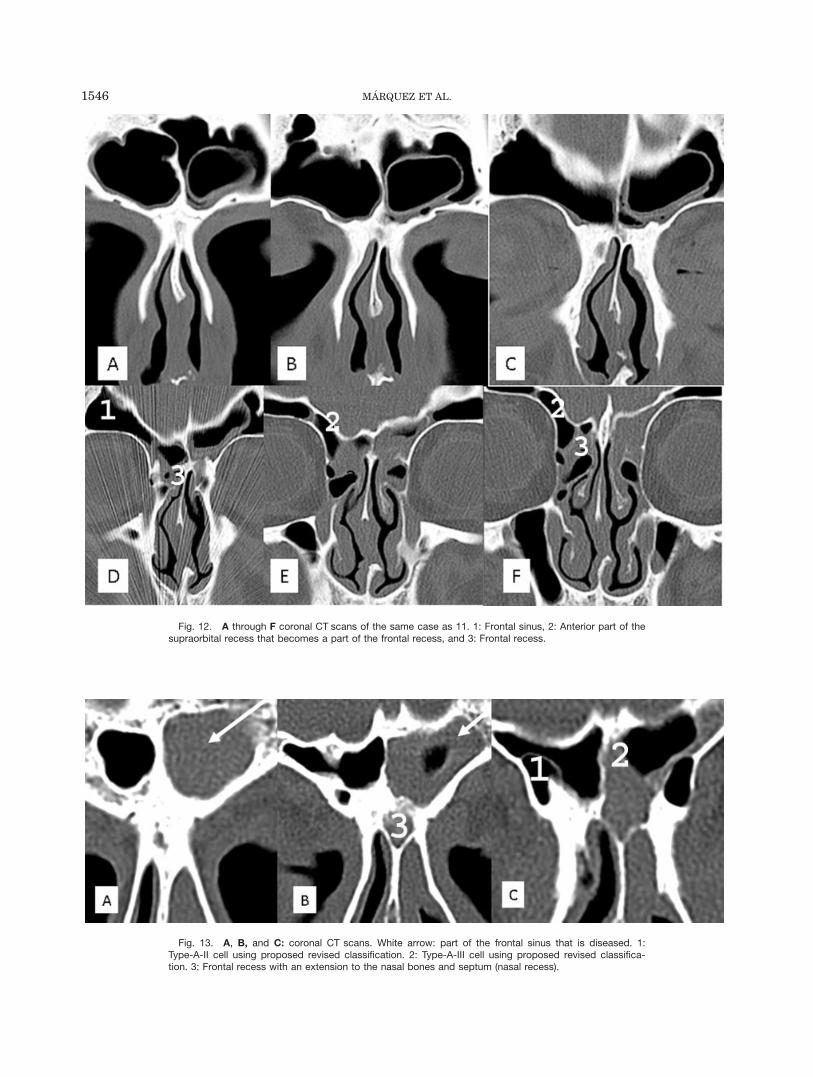
Fig. 12. A through F coronal CT scans of the same case as 11. 1: Frontal sinus, 2: Anterior part of thesupraorbital recess that becomes a part of the frontal recess, and 3: Frontal recess.
Fig. 13. A, B, and C: coronal CT scans. White arrow: part of the frontal sinus that is diseased. 1:Type-A-II cell using proposed revised classification. 2: Type-A-III cell using proposed revised classifica-tion. 3: Frontal recess with an extension to the nasal bones and septum (nasal recess).
1546 MARQUEZ ET AL.
and has since been corroborated by other investigators(Kasper, 1936). In fact, the frontal sinus consists of apneumatization of the frontal bone (membranous bone).Sometimes it is difficult to differentiate between the pos-sible origins of a structure. When the frontal recesspneumatizes the floor of the frontal sinus, the nasalbones and the region of the crista galli, these structuresand spaces may appear as if an ethmoidal cell hasinvaded the frontal sinus or frontal recess. In thosecases, the bony wall of such a recess may look like acell, but the walls are mostly much thicker (membra-nous bone) than the thin bony walls of cells from eth-moidal origin (endochondral bone). Sometimes theserecesses are often misinterpreted as the presence of adouble frontal sinus (Shambaugh, 1907). Despite theearlier discussion of a ‘‘cell,’’ our definition becomes clini-cally more problematic when we observe the migrationof posterior ethmoid cells into the sphenoid bone. Theseextramural, endochondrial-derived cells are extending
into the membraneous sphenoid bone. Although thesecells begin as thin-walled structures they pneumatize inthe adult into the much thicker membraneous bonealong their superior and lateral surfaces, hence notappearing on these surfaces as endochondrial derivedcells.The term ‘‘frontal recess’’ originated from developmen-
tal embryology, whereas already discussed by Killian(1895) employed the term to describe a frontal curveor bend in the superoanterior middle meatus(‘‘Stirnbucht’’). Later, Van Alyea (1946) used frontalrecess to describe the supero-anterior prolongation ofthe infundibulum ethmoidale. The processus frontalis, arecognized term in the ‘‘Terminologia Anatomica, p 14,’’has sometimes been incorrectly used to describe thefrontal recess. One can define the frontal recess as themost anterior-superior part of the middle meatus(Marquez et al., 2002). The lateral part of it is also the
Fig. 16. Coronal CT scans: frontal Type-A-II cell (inside of cell isdisease free) surrounded by opacified frontal sinus.
Fig. 14. Coronal CT scans. A: Preoperative image (white arrowsshow the site of the interfrontal septum (septum sinuum frontalium). B:Postoperative image shows the defect in the interfrontal septum. It isthis defect that aid in forming the convexity in both frontal aperturesduring the surgery (or the Type-B cell).
Fig. 15. A and B: coronal CT scans frontal sinusitis. White arrow:Septal deviation may be theoretically a cause of ethmoid sinusitis vialateral displacement of the middle turbinate and narrowing of the out-flow tract of the middle meatus. 1: Agger nasi cell (free of disease). 2:Lacrimal cell (diseased).
Fig. 17. A and B: coronal CT scans: Bilateral Type-A-III cells (dis-ease free inside cells).
1547ETHMOID SINUS AND EXTRAMURAL MIGRATION

most anterior extension of the supraorbital recess reach-ing the posterior wall of the frontal sinus (Figs. 10 and12). McLaughin et al. (2001) defined it as the cleft in theanterior ethmoid complex, just inferior to the frontalsinus ostium. The frontal recess (when a supraorbitalrecess is present) invades the frontal bone just inferio-posterior to the aperture of the frontal sinus. Thismakes the medial orbital plate (pars orbitalis ossis fron-talis) of the frontal bone pneumatized resulting in a‘‘lamina orbitalis ossis frontalis’’ and a ‘‘lamina cranialisossis ethmoidalis’’ (Fig. 10B red and yellow line). Thelamina orbitalis of the frontal bone should be differenti-ated from the lamina orbitalis of the ethmoidal bone.Therefore, it would be better to keep the older nomencla-ture ‘‘lamina papyracea’’ in order to prevent confusionwith the lamina orbitalis of the frontal bone. The lam-ina cranialis of the frontal bone forms the roof of thefrontal recess (fovea/frontal bone) and supraorbitalrecess. The roof of the ethmoid sinus is frequentlyreferred to as the fovea ethmoidalis, when in fact thefovea or pit-like migration of the ethmoids cells into thefrontal bone forms the roof of this sinus (Figs. 10 and22). When looking at the facies orbitalis of the frontalbone in the orbits (Fig. 6) one can observe that the cra-nial border of the lacrimal bone is always lower than thecranial border of the lamina papyracea of the ethmoid (arange difference of 3–6 mm). This results in a bent andsometimes even a notch of the margo ethmoidalis of thefrontal bone (Figs. 5 and 6). This curve or notch can becalled ‘‘incisura laminae papyracea’’ or ‘‘incisura laminaeorbitalis ossis ethmoidalis.’’ It is not possible to call thisincisura ‘‘incisura ethmoidalis’’ because this term existsalready and is reserved for the defects in the frontalbone, caused by the cribriform plate (intraorbital gap).This incisura has never been described before, but it isimportant because it explains why the lateral wall of thefrontal recess is not made up by the lamina papyraceabut mainly by the orbital plate of the frontal bone(Lang, 1988).Another frequent mistake is to use the same name for
very different anatomical structures. A typical examplefor this is ‘‘agger nasi cell.’’ According to Berkovitz andMoxham (2002), agger nasi is ‘‘. . . a curved ridge called
agger nasi may be found close to the atrium. This ridgeoverlies the anterior part of the ethmoidal crest of themaxilla.’’ As the agger is an eminence or mound anteriorto the middle turbinate or concha media, a coronal CTprojection through this structure must show laterallythe frontal process of the maxilla (site of the ethmoidalcrest). On the same CT projection, the middle turbinateshould be absent because the agger nasi is anterior tothe free border of the concha media. In the literature,however, many anatomical structures of the frontal recessare misnamed ‘‘agger nasi cell.’’ The term agger nasi cellhas been given to lacrimal cells, to the recessus terminalis(Wormald, 2003, 2005a,b) or even frontal sinus cells(Brunner et al., 1996; Orlandi et al., 2001). According toa recent study of Chaiyasate et al. (2007) in twins, anagger nasi cell can be seen on CT of normal test subjectsin 65% of the cases. Mostly, these cells have a very smallsize. We recommend that the term agger nasi be assignedto the surface feature anterior to the insertion of themiddle turbinate on the lateral nasal wall which is con-sistent with the definition of an agger as an eminenceand agger nasi cell be reserved for the ethmoid cellswhich constitute its pneumatization (Figs. 7–9).Some comments need to be given on the nomenclature
of the anterior extramural ethmoidal cells. These eth-moidal cells should not be called a ‘‘bulla’’ (Navarroet al., 1997). The term bulla should only be used for the
Fig. 18. A and B: coronal CT scans. A: White arrow: concha media bullosa (this is a true Grunwaldcell: there is a pneumatization of the middle turbinate by the posterior ethmoid). Green arrow: conchamedia bullosa. B: Blue arrow: concha suprema bullosa.
Fig. 19. Coronal CT scans of bilateral infraorbital cells (‘‘Haller’’cells). A and B show an extensive extension of ethmoidal cells intothe maxillary sinus (Haller cells).
1548 MARQUEZ ET AL.

ethmoidal bulla (bulla ethmoidalis), which is normallythe most prominent cell in the anterior ethmoid. It isalso very confusing to link the name of the cell to theregion where it comes from or where it drains into(infundibular cell for the lacrimal and anterior frontalrecess cell). Cells should be named according to the sitewhere they actually are (i.e. frontal sinus cells, frontalrecess cells, lacrimal cell). Furthermore, for clinical pur-poses, the classifications of cells must have a clinicalrelevance. Therefore, the classification of Bent et al.(1994) should be adapted for the following reasons: (1)the difference between Type-I (single frontal recess cellabove agger nasi) and Type II (tier of cells in the frontalrecess above agger nasi) is not very relevant. Both typescan result in a blockage of the drainage of the frontalsinus into the frontal recess and if this is caused by two,three, or even more cells in the frontal recess, then thisis not relevant and (2) the most important fact is thatthe frontal recess needs to be cleared from obstructingcells. The same authors define Type-III cells as thosearising above the agger nasi and extending into thefrontal sinus. These are examples of extramural endo-chondral ethmoid cells, which have migrated into frontalsinus (Fig. 7). Type-IV cells are described as a single iso-lated cell within the frontal sinus which is pneumatiza-tion of the membranous frontal bone and not an isolated,migrated endochondral ethmoid cell. Sometimes aftertrauma a compartmentalization of the frontal sinus canoccur, or an excessive septation can arise resulting in apartial opacification of the frontal sinus, but these arecompletely different structures from the frontal cells.Wormald (2005a,b) suggested modifying the Bent-classi-fication, a Type-IV cell should arise from the frontalrecess and extend over 50% of the vertical height of thefrontal sinus. In this revised classification, Type-III cells
would be extramural ethmoid cells, which occupy lessthan 50% of the vertical height of the frontal sinus.Such a differentiation is useful because Type-IV cellsmay potentially adhere to the frontal sinus walls and bedifficult to remove. So, a better classification of cellsblocking the drainage of the frontal recess and sinusshould be:A. Concave structures (Figs. 7–9, 12, and 13)
� Type I: excessive number, or size of frontal cellsblocking the frontal recess.
� Type II: frontal cells occupying less than 50% ofthe height of the frontal sinus.
� Type III: frontal sinus cells occupying more than50% of the height of the frontal sinus. Cell TypesII and III occur in about 10% of the adult popula-tion (Chaiyasate et al., 2007).
B. Convex structures (Fig. 10): The intersinus septum ofthe frontal sinus (5septum sinuum frontalium) canunder pathologic conditions involve the aperture ofthe frontal sinus forming a convexity. This very rarecondition can confuse the surgeon as one is alwaysexpecting a concave structure obstructing the drain-age of the frontal sinus and instead finds a convexity.The concave structures (Type A) are extramural eth-moidal cells, whereas the convex structures (Type B)are not from ethmoidal origin.
Sometimes peculiar nomenclature is introduced suchas ‘‘internal frontal ostium’’ (Bent et al., 1994; Kuhn,1996), which means that there should also exist an‘‘external frontal ostium.’’ The term ‘‘ostium’’ alsodeserves some comments. In basic anatomy textbooksthe term ‘‘ostium’’ is not typically used to refer to open-ing of a paranasal sinus. The term ‘‘ostium’’ is mostlyused for a dynamic structure that opens and closes, that
Fig. 20. A–C coronal CT scans. Extensively pneumatization of frontal sinus extending into nasal bones(A), frontal recess (B), and nasal septum (C).
1549ETHMOID SINUS AND EXTRAMURAL MIGRATION

is, ‘‘ostium pharyngeum tubae auditivae,’’ or ‘‘ ostiumtympanicum tubae auditiva.’’ Ostium in Latin means a‘‘door,’’ whereas ‘‘apertura’’ is a rigid opening in bonesuch as ‘‘apertura piriformis,’’ ‘‘apertura sinus frontalis,’’‘‘apertura canaliculi vestibuli,’’ or ‘‘apertura canaliculicochlea,’’ which are all accepted terminologia anatomicaterms (Terminologia Anatomica, 1998).As sinus ostia are never dynamic structures, they
should be called ‘‘aperturae,’’ but the term sinus ostiumhas been used extensively by ENT surgeons and manyanatomists.4 Therefore, we suggest ostium is best re-stricted to a rounded sinus opening, through which thesinus drains and ventilates, and that can be found in ev-ery individual. Although the ostia of the maxillary and
the sphenoid sinuses are such structures, the drainageof the frontal sinus is completely different. Also, thedrainage openings of the ethmoidal cells are mostlyirregular in shape and cannot be called ostia.Presently, there is a gathering if not general consen-
sus that the nasofrontal duct is a myth because this‘‘duct’’ is a highly variable space, or outflow tract, ratherthan the homologue of the nasolacrimal duct. Vogt andSchrade (1979) using contrast radiography showed thatthe drainage of the frontal sinus into the ethmoid isquite complex. Also Lee et al. (1997), Friedman et al.(2004), and Daniels et al. (2003) describe different drain-age patterns from the frontal sinus into the frontalrecess, ethmoidal infundibulum, or directly into the mid-dle meatus (Duque and Casiano, 2005). Since the intro-duction of endoscopes, the frontal sinus is now more of-ten approached endonasally. In adults one very seldomfinds a structure that looks like a nasofrontal duct. Fol-lowing the surgical removal of the frontal recess cells,most often a triangular-shaped opening leading to thefrontal sinus is found. This triangular opening is formedlateroinferiorly by the orbital plate of the frontal bone,
Fig. 21. Skulls of famed anatomists who taught at the predecessorof the current University of Padua Medical School can be seen todayin that institution’s Anatomy Department library in Padua, Italy. Gio-vanni Domenico Santorini’s skull can be seen fourth from the right.
Photograph kindly provided by Drs. Paulette Bernd and Steven Erdefrom SUNY Downstate Medical Center and New York PresbyterianHospital, respectively.
4To memorize the meaning of apertura and ostium one shouldremember this sentence of Cicero (M. Tullius Cicero pro S RoscioAmerino,’’ paragraph 25, 2, 100 BC). ‘‘Tamen, cum planum iudi-cibus esset factum aperto ostio dormientes eos repertos esse,iudicio absoluti adulescentes et suspescione omni liberati sunt.’’‘‘Aperta ostio dormientes’’ means sleeping with an open door.
1550 MARQUEZ ET AL.

superiorly by the cranial plate of the frontal bone, andmedially by a bony process from the floor of the frontalsinus up to the posterior wall of the frontal sinus. Thefrontal aperture is in a plane perpendicular to the poste-rior wall of the frontal sinus (Daniels et al., 2003) goingfrom laterosuperior to inferomedial, and therefore, thisstructure is very difficult to visualize on conventionalCT scanning using only coronal, axial, or sagittal projec-tions. This opening of the frontal sinus into the frontalrecess is called ‘‘apertura sinus frontalis.’’ Given the cur-rent practice of utilizing an hour–glass analogy todescribe the frontal sinus outflow tract, the aperturasinus frontalis is the neck of the glass, the upper body ofthe hour glass in the frontal sinus, and the lower bodyis the frontal recess (Figs. 7, 11, and 22).With these new definitions and recalling the migration
of the ethmoid cells, one can redefine the boundaries ofthe frontal recess, although still remembering that thisterm has evolved from its original application for devel-opmental embryology. When viewed from within, thewalls of the frontal recess can be defined as follows(Fig. 5).The anterior wall is formed by the upper-posterior
face of the frontal process of the maxilla and partly bythe posterior part of the sulcus lacrimalis of the ethmoidbone (Fig. 5 between arrow a and b). If the frontal recessextends posterior to the frontal sinus the upper anteriorpart of the wall is made up by the posterior wall of thisfrontal sinus.The posterior wall is formed by the anterior face of
the bulla ethmoidalis and of the lamella bullae.The superior wall or the roof is formed by the skull
base or the roof of the supraorbital recess and frontalbone (Figs. 6B, 22, and 23).The lateral wall is made up mainly (45%) by the or-
bital plate of the frontal bone and for another 45% by
the upper part of the lacrimal bone (Owen and Kuhn,1997). The lamina papyracea makes up never more than10% of the lateral wall of the frontal recess Figs. 6 and8). The inclusion of the all three bones in the lateralboundary of the recess is a departure from other authors(Stammberger et al., 1995; Daniels et al., 2003; Wor-mald, 2003; Friedman et al., 2004), and reflects the diffi-culty in defining bony borders from CT or anatomicalspecimens. Only skull preparation will show the exactrelationship between the sutures of the bones of theskull.The medial wall is formed by the sagittal attachment
of the middle turbinate to the skull base and the floor ofthe frontal sinus. Sometimes the anterosuperior part ofthe frontal recess can extend toward the posterior wallof the frontal sinus anteriorly of the sagittal attachmentof the middle turbinate and the crista galli (sometimespneumatizing the latter) and rarely reaching as far asthe nasal bones and the nasal septum. In this configura-tion, both frontal recesses meet each other at the mid-line and form a small triangular septum in line with thenasal septum and the septum sinuum frontalium. A pos-sible name for this septum can be ‘‘septum recessumfrontalium.’’One way on coronal CT projections to differentiate
between lamina orbitalis of the frontal bone and laminapapyracea is to identify the lacrimal bone. As long as ona coronal CT-scan the lacrimal bone can be located, thebony structures superior to it must be frontal bone(Figs. 9A and 10A).The anterosuperior attachment of the uncinate process
is very complex and most likely influences the popula-
Fig. 22. Unembalmed cadaver section used in Fig. 4B illustratesthe outflow tract of the frontal sinus. The frontal sinus and its outflowtract are currently compared to an hourglass (yellow broken line). Thesuperior aspect of the hourglass is the frontal sinus (FS), the frontalaperture (fa) is the neck, and frontal recess (fr) is the bottom.
Fig. 23. Coronal CT scan of patient with fibrous dysplasia of theleft frontal bone. As this process involves the entire frontal bone theimage also illustrates that the roof of the ethmoid sinus is the frontalbone [(yellow arrow), courtesy of William Lawson, MD]. The term foveaethmoidalis refers to the pit-like evagination formed by the migrationof the ethmoid cells into the frontal bone. Contrary to common usage,the fovea ethmoidalis is not the roof of the ethmoid sinus.
1551ETHMOID SINUS AND EXTRAMURAL MIGRATION
tion of the frontal recess by migrating ethmoid cells. Inmost textbooks one of the most common variants of thisattachment is described laterally onto the lamina papy-racea. As was already demonstrated before, this is notpossible because the lateral wall of the frontal recessconsists mainly of the orbital plate of the frontal bone.Mostly, the anterosuperior attachment of the uncinateprocess is like a palm tree with branches going in everydirection, mainly orbital plate, skull base, floor of thefrontal sinus, and middle turbinate. A very masterly dis-section of the superior attachment of the uncinate pro-cess in the frontal recess can be seen in Bolger andMawn (2001). Sometimes the latter attachment of theuncinate process can make the ethmoid infundibulumend blindly in a dome-like structure obstructing a goodview of the frontal recess and apertura of the frontalsinus. Such a blind ending recess is called terminalrecess or recessus terminalis (Stammberger et al., 1995).
CONCLUSION
The advent of optical telescopes to permit minimallyinvasive endoscopic sinus surgery is probably the eventthat has most driven the need for proper and accurateanatomical description of the region. In this article,most structures of the lateral nasal wall have beendefined and described extensively. The tracking of extra-mural cells offers a greater understanding of the varia-tion of contiguous structures observed in the adult. Anattempt is made to identify and correct anatomicalerrors and new terms are suggested in order to properlydescribe the region. The reexamination of the historicalfindings from past anatomy masters served only to reaf-firm present day findings of modern applied surgicalanatomists employing the latest in technological advan-ces in endoscopic sinus assessment and the use of CTimaging for three-dimensional reconstructions of theregion. In conclusion, one can state that the ethmoid(sinus ethmoidale) is phylogenetically, embryologically,anatomically, and functionally completely different fromall the other paranasal sinuses, and that only in humansit has taken a key position between all the other para-nasal sinuses, controlling their ventilation and drainage.
ACKNOWLEDGMENTS
The authors thank the following personnel fromSUNY Downstate Medical Center: Ms. Cira Peragineand Mr. Noel Caceres from the department of Anatomyand Cell Biology for their tireless support in transport-ing bony material, assistance in recording observationsand providing the cadaveric material for our study. Forphotography and graphic design assistance we thankMr. Vincent Garofalo. Special thanks to Drs. ChristenRusso and Brent Rogers for identifying and obtainingsome of the sources found in the older literature. Dr. An-thony Nicastri, Jaik Koo, Neville Saunders, and HenryRodriquez of the department of pathology for use of theirfreezer and autopsy room for our dissection. We also liketo thank Timothy Smith of Slippery Rock University,William Lawson of Mount Sinai School of Medicine, IanTattersall of the American Museum of Natural History,Timothy Bromage of New York University College ofDentistry, Frank Lucente of Long Island College Hospi-tal and one anonymous reviewer for their critical analy-
sis and insightful suggestions. The authors would like toexpress their gratitude to Prof. Rudolf De Smet of theFaculty of Arts and Philosophy (Free University Brus-sels: V.U.B.) and department of Linguistics and Litera-ture for his much appreciated assistance in using thecorrect Latin nomenclature and in identifying the refer-ences for several terms found in the Latin literature.
LITERATURE CITED
Anon JB, Rontal M, Zinreich SJ. 1996. Anatomy of the paranasalsinuses. New York: Thieme.
Bent JP, Cuilty-Siller C, Kuhn FA. 1994. The frontal cell as a causeof frontal sinus obstruction. Am J Rhinology 8:185–191.
Berkovitz BKB, Moxham BJ. 2002. Head and neck anatomy: a clini-cal reference. London: Martin Dunitz.
Bohatirchuk FP, Campbell JS, Jeletzky TF. 1972. Bone lamellae.Acta Anat 83:321–332.
Bolger WE, Mawn CB. 2001. Analysis of the suprabullar and retro-bullar recess for endoscopic sinus surgery. Ann Otol LaryngolSuppl 186:3–14.
Bosworth FH. 1888. The physiology of the nose. medical news53:117–124.
Brunner E, Jacobs JB, Lebowitz RA, Shpizner BA, Holliday RA.1996. Role of the agger nasi cell in chronic frontal sinusitis. AnnOtol Rhinol Laryngol 105:694–700.
Cave AJE. 1967. Observations on the platyrrhine nasal fossa. Am JPhys Anthropol 26:277–288.
Chaiyasate S, Baron I, Clement P. 2007. Analysis of paranasal sinusdevelopment and anatomical variations: a genetic study in twins.Clin Otolaryngol 32:93–97.
Cicero MT. c. 100 BCE. Pro S Roscio Amerino, paragraph 65, 2.Daniels DL, Mafee MF, Smith MM, Smith TL, Naidich TP, Brown
WD, Bolger WE, Mark LP, Ulmer JL, Hacein-Bey L, StrottmannJM. 2003. Am J Neuroradiol 24:1618–1626.
De Beer GR. 1937. Development of the vertebrate skull. London:Oxford University Press.
Douglass B. 1906. Nasal sinus surgery with operations on nose andthroat. Philadelphia: F. A. Davis Company.
Drake RL, Vogl W, Mitchell AWM. 2005. Gray’s Anatomy for stu-dents. Philadelphia: Elsevier Churchill Livingstone.
Duque CS, Casiano RR. 2005. Surgical anatomy and embryology ofthe frontal sinus. In: Kountakis S, Brent A, Sr, Draf W, editors.The frontal sinus. Berlin: Springer Berlin-Heidelberg. p 21–31.
Friedman M, Bliznikas D, Vidyasagar R, Landsberg R. 2004. Fron-tal sinus surgery 2004: update of clinical anatomy and surgicaltechniques. Oper Tech Otolaryngol Head Neck Surg 15:23–31.
Grunwald E. 1925. Deskriptive und topographische Anatomie derNase und Ihrer Nebenhohlen. In: Denker A, Kahler O, editors.Handbuch der Hals-Nasen-Ohrenheilkunde Capitel I. Berlin:Spriger-Bergmann. p 1–94.
Haller A, Cullen W. 1803. First lines of physiology. 1st American ed.Edinburgh: Obraban, Penninman. p 224.
Highmore N. 1651. Corporis humani disquisitio anatomica in qua san-guinis circulationem in quavis corporis particular plurimis typisnovis, ac anygmatum medicorum succincta dilucidatione ornatumprosequutus est. Hagae-Comitis, S. Brown.
His W. 1895. Die Anatomische Nomenclatur. Leipzig: Verlag VonVeit.
Kasper KA. 1936. Nasofrontal connections. A study based on onehundred consecutive dissections. Arch Otolaryngol 23:322–343.
Killian G. 1895. Zur Anatomie der Nase menschlicher Embryonen.Archiv fur Laryngologie III:2–47.
Kuhn FA. 1996. Chronic frontal sinusistis: the endoscopic frontalrecess approach. Oper Techn Otolaryngol Head Neck Surg 7:222–229.
Laitman JT, Reidenberg JS. 1993. Specializations of the humanupper respiratory and upper digestive systems as seen throughcomparative and developmental anatomy. Dysphagia 8:318–325.
1552 MARQUEZ ET AL.
Lang J. 1988. Klinische Anatomie der Nase, Nasenhohle undNebenhohlen. In: Aktuelle Oto-Rhino-Laryngologie Bd 11, SinusParanasales. Stutgart: Thieme Verlag. p 56–95.
Layton TB. 1934. Catalogue of the onodi collection (In the Museumof the Royal College of Surgeons of England). London: ReadleyBrothers.
Lee JB, Brody R, Har-el G. 1997. Frontal sinus outflow anatomy.Am J Rhinology 11:283–285.
Marquez S. 2002. The human nasal complex: a study of its anatomy,function and evolution by CT, comparative and morphometricmethods. Ph.D. Dissertation, City University of New York,New York.
Marquez S, Delson E, Silvers A, Lawson W, Laitman JT. 1999.The meaning of emptiness: pneumatization patterns of great apeparanasal sinuses via CT imaging. Am Ass Phys Anthropol28: 191.
Marquez S, Lawson W, Schaefer S, Laitman JT. 2002. Anatomy ofthe nasal accessory sinuses. In: Wackym PA, Rice DH, editors.Minimally invasive surgery of the head, neck, and cranial base.Philadelphia: Lippincott Williams & Wilkins. p 153–193.
McLaughin RB Jr, Rehl RM, Lanza DC. 2001. Clinically relevantfrontal sinus anatomy and physiology. Otolaryngol Clin North Am34, 1:1–22.
Messerklinger W. 1978. Endoscopy of the nose. Baltimore, Munich:Urban and Schwarzenberger. p 6–18.
Moore KL, Dalley AF. 2006. Clinically oriented anatomy. 5th ed.Philadelphia: Lippincott Williams and Wilkins.
Navarro JAC. 1997. The Nasal Cavity and Paranasal Sinuses: Sur-gical Anatomy. New York: Springer Verlag Berlin Heidelberg. p27–30.
Orlandi RR, Kennedy DW. 2001. Revision endoscopic frontal sinussurgery. Otolaryngol Clin North Am 34,1:77–90.
Owen RG, Kuhn FA. 1997. Supraorbital ethmoidal cell. OtolaryngolHead Neck Surg 116:254–261.
Rose C, Gaddum-Rose P. 1997. Hollinshead’s textbook of anatomy.5th ed. Philadelphia: Lippincott-Raven Press.
Rossie JB. 2006. Ontogeny and homology of the paranasal sinusesin Platyrrhini (Mammalia: Primates). J Morph 267:1–40.
Santorini GD. 1724. Observationes anatomicae. Venetiis: apud J. B.Recurti.
Schaefer SD. 1998. An anatomic approach to endoscopic intranasalethmoidectomy. Laryngoscope 108(11 Pt1):1628–1634.
Seydel O. 1891. Uber die Nasenholen der hoheren Sðugethiere unddes Menschen. Gegenbaurs Morphol Jahrb 17:44–99.
Shambaugh GE. 1907. The construction of the ethmoid labyrinth.Ann Otol Rhinol Laryngol 16:771–792.
Simpson DP. 2000. Cassell’s latin dictionary. London: Continuum.
Smith TD, Rossie JB, Cooper GM, Mooney MP, Siegel MI. 2005.Secondary pneumatization of the maxillary sinus in callitrichidprimates: insights from immunohistochemistry and bone cell dis-tribution. Anat Rec 285:677–689.
Snell RS. 2000. Clinical anatomy for medical students. 6th ed. Phil-adelphia: Lippincott Williams & Wilkins.
Stackpole SA, Edelstein DR. 1996. Anatomic variants of the para-nasal sinuses and their implications for sinusitis. Curr Opin Oto-laryngol Head Neck Surg 4:1–6.
Stammberger H. 1991. Functional endoscopic sinus surgery. In M.Hawke (adapted and edited) Philadelphia: B.C. Decker.
Stammberger HR, Bolger WE, Clement PA, Hosemann W, KuhnFA, Lanza DC, Leopold DA, Onishi T, Passali D, Shaeffer SD,Wayoff MR, Zinreich J. 1995. Paranasal sinuses: anatomic termi-nology and nomenclature. Ann Otol Rhinol Larygol Suppl 167:7–16.
Standring S. 2005. Gray’s anatomy: The anatomical basis of clinicalpractice. 39th ed. New York: Elsevier Churchill Livingstone.
Tank PW. 2005. Grant’s dissector. 13th ed. Philadelphia: LippincottWilliams & Wilkins.
Terminologia Anatomica. 1998. International Anatomical Terminol-ogy. Federative Committee on Anatomical Terminology (FCAT).Stuttgart, New York: Thieme.
Terrier G. 1991. Rhinosinusal endoscopy: diagnosis and surgery.Milano: Zambon.
Tillaux P. 1878. Traite D’Anatomie Topographique avec Applicationsa la Chirurgie. Paris: P. Asselin, Libraire de la faculte de mede-cine.
Turner LA. 1901. The accessory sinuses of the nose. Their surgicalanatomy and the diagnosis and treatment of their inflammatoryaffections. Edinburgh: William Green & Sons.
Van Alyea OE. 1946. Frontal sinus drainage. Ann Otol (St. Louis)55:267–277.
Vitruvius P. c. 25 BCE. De Architectura 7, cap 3, paragraph 9.VogtKl, Schrade F. 1979. Anatomische varianten des ausfuhrungsgang-
systems der stirnhohle. LaryngRhinolOtol (Stuttg) 58:783–794.Wormald P-J. 2003. The agger nasi cell: the key to understanding
the anatomy of the frontal recess. Otolaryngol Head Neck Surg129:497–507.
Wormald P-J. 2005a. Surgery of the frontal recess and frontal sinus.Rhinology 43:82–85.
Wormald P-J. 2005b. The anatomy of the frontal recess and frontalsinus with three dimensional reconstruction. New York: ThiemeStuttgart. p 35–51.
Zuckerkandl E. 1892. Normale und pathologische anatomie dernasenhohlen und ihrer pneumatischen anhange. Wien und Leip-zig: Wilhelm Braumuller.
1553ETHMOID SINUS AND EXTRAMURAL MIGRATION