growth formation inculturesof sensory neurons - pnas · chicine, in keeping with the effects these...
TRANSCRIPT

Proc. Nati. Acad. Sci. USAVol. 75, No. 10, pp. 5226-5229, October 1978Neurobiology
Growth cone formation in cultures of sensory neurons(filopodia/microtubules/collateral sprouting)
D. BRAY, C. THOMAS, AND G. SHAWMedical Research Council Cell Biophysics Unit, King's College, 26-29 Drury Lane, London WC2B 5RL, England
Communicated by S. Brenner, August 7, 1978
ABSTRACT Three experimental situations have been foundin which cultured sensory neurons from embryonic chicken willform growth cones from positions along the length of the neu-rite. If the neurons are dissected with a remaining short axonalstump and plated into serum-free medium, they can form amorphologically normal growth cone from the stump within 15min, even in the presence of cycloheximide or puromycin. Whenneurites growing in culture media with low levels of serum arecut at any point with microneedles, growth cones are producedquickly from the amputated stump, usually within 20 min.Treatment of growing neurons with low concentrations of col-chicine, Colcemid, or podophyllotoxin results in the progressiveappearance of lateral filopodia and regions of flattened cyto-plasm that closely resemble growth cones except for theirpreterminal positions. These observations show that the po-tential to form growth cones is distributed throughout the neu-ron and suggest that this is normally repressed in some way bythe neuronal microtubules.
Wherever the axons or dendrites of vertebrate nerve cells havebeen observed to grow in culture, they have been found topossess a terminal appendage known as the growth cone. Thisis distinguishable from the rest of the neuron by its numerousfilopodia and flattened veil-like regions and by its continualmovement (1, 2). Its special functions may include the primaryattachment of the cell to the substratum (3), an active part inmicropinocytosis (4), guidance of the neurite (5), and the as-sembly of surface membrane during growth (6).
In the most extensively studied kind of nerve culture-thatof embryonic sensory or sympathetic neurons growing in thepresence of nerve growth factor (7)-neurite extension does notoccur immediately after explantation (8). Furthermore, onceinitiation has occurred, growth cones are formed from existingones rather than from other parts of the cell (3) and under theusual conditions they do not regenerate quickly upon experi-mental amputation (9).
These observations raise the possibility that the growth coneis a complex structure whose formation is the rate-limiting stepin the initiation of neurite growth. We now describe experi-ments that bear upon this possibility and examine the formationof growth cones from cultured sensory neurons under a varietyof conditions.
MATERIALS AND METHODSCultures were prepared from the dorsal root ganglia of 11- to13-day chicken embryos by procedures that have been de-scribed (9). Cells were incubated at 370 in medium C, whichis composed of Leibovitz L15 (GIBCo BioCult, Glasgow,Scotland) with 0.6% glucose, 2 mM L-glutamine, 100 units ofpenicillin per ml, 100 Aig of streptomycin per ml, and 0.1 jig ofmouse nerve growth factor per ml purified to the DEAE-cel-lulose fraction described by Varon et al. (10). With a few ex-ceptions, which are noted, this medium was supplemented with
0.6% methylcellulose and 10% fetal calf serum. All cells weregrown on glass coverslips which in some experiments were builtinto the culture dish to give good optical properties. Amputa-tions were performed as described (9), with a glass microelec-trode broken to a tip of about 10 ,um diameter.The following drugs were used in various experiments: cy-
cloheximide, cytochalasin B, demecolcine (Colcemid), col-chicine, puromycin (all from Sigma Chemical Co.) and podo-phyllotoxin (Aldrich Chemical Co.). Lumicolchicine was pre-pared from colchicine by the method of Wilson and Friedkin(11).Growth Cones from Freshly Dissected Neurons. Ganglia
from 11- to 13-day chicken embryos were dissected togetherwith a short length of postganglionic nerve, and cleaned of as-sociated tissue. They were incubated in 0.25% trypsin in acalcium- and magnesium-free balanced salt solution at 370 forprecisely 25 min. They were quickly washed in balanced saltsolution (GIBCo BioCult, Glasgow, Scotland) and put into 0.1%soybean trypsin inhibitor (Sigma Chemical Co.) in balancedsalt solution. After incubation for 5 min at room temperature,they were again washed into balanced salt solution and disso-ciated by gentle trituration- with a pasteur pipette of terminalbore 1.0-1.5 mm. The cells were then plated into mediumlacking serum.
For the assays described in Table 1, the cells were pipettedonto plain glass coverslips in warm serum-free medium con-taining, where appropriate, a drug to be tested. They were in-cubated at 370 and then fixed with warm Vaughn-Peters al-dehyde (12). After, usually, 16 hr in the aldehyde at roomtemperature the cultures were washed several times in distilledwater, drained, and mounted onto glass slides with Aquamount(Gurr, High Wycombe, England). They were examined byphase-contrast microscopy and the following types of cell werecounted: rounded neuronal cell bodies with no major extension,neuronal cell bodies with an axonal stump at least one somadiameter long (Fig. la), and neuronal cell bodies with an axonalstump bearing a terminal growth cone showing filopodia andflattened veil-like regions (Fig. 1 b and c). Because of the sub-jective nature of the assay, all counts were made blind, on slidesthat had been covertly numbered in a random sequence by asecond person.
Protein Synthesis. Protein synthesis was measured in culturesthat had been freshly plated in the above manner. The cellswere put into dishes containing 3 ml of medium C lackingmethionine and supplemented with 10,uCi of L-['5S]methionine(780 Ci/mmol; Radiochemical Centre, Amersham, England).Where appropriate, cycloheximide, or puromycin was added.The cultures were incubated for 1 hr at 370, rinsed in balancedsalt solution, and then dissoved in 1 ml of 1% sodium dodecylsulfate/1% 2-mercaptoethanol/1% bovine serum albumin andheated for 2 min at 1000. The samples were extensively di-alyzed against 10 mM NaCl/0.1% sodium dodecyl sulfate andthe nondialyzable radioactivity was measured in a scintillationcounter. Protein synthesis in established cultures was measuredin the same way except that the incubation in L-[35S]methioninewas for 3 hr.
5226
The publication costs of this article were defrayed in part by pagecharge payment. This article must therefore be hereby marked "ad-vertisement" in accordance with 18 U. S. C. §1734 solely to indicatethis fact.

Proc. Natl. Acad. Sci. USA 75(1978) 5227
RESULTSWhen chicken sensory ganglia are dispersed with trypsin,neurons are seen that retain a short stump which is, presumably,derived from the original axon (8). Under the usual conditionsof culture these quickly retract into the cell body (8), but wefound that in serum-free medium many remain extendedthrough adhesion of the culture substratum. These were foundto have formed terminal plaque-like specializations at their cutend which developed filopodia and broad flattened regions andbegan to move in the distinctive way of normal developinggrowth cones (Fig. 1).
If the conditions of dissociation and trypsinization were
carefully controlled, up to 70-80% of the neurons recoveredfrom the ganglion-had stumps. If these were plated under de-fined conditions, the formation of growth cones could be as-
sessed quantitatively (Table 1). The number of cells with stumpsfell to a lower value soon after plating, presumably because
fib . :J
K::~~~~~ ~ ~ ~ ~ ~ ~ ~ ~ ~ ~ ~ ~ ~ :
vjs
FIG. 1. Formation of growth cones from the axonal stumps offreshly dissected neurons. (a) Sensory neurons 5 min after they wereplated onto a plastic surface. (X220.) (b and c) Growth cones formedfrom axonal stumps 20-30 min after they were plated. (X550.)
Table 1. Effect of drugs on growth cone formation from axonalstumps of freshly dissected sensory neurons
% stumpsAssay Cells with Cells with withmedium stumps growth cones,growth cones
Medium C 46 12 26Balanced salt solution 75 18 24C + puromycin (10 gg/ml) 75 17 23C + cytochalasin B (1 tg/ml) 181 1 1C + colchicine (5 ,g/ml) 27 1 4
Assays were carried out as described in Materials and Methods;400 neuronal cell bodies were counted under each experimentalcondition. All incubations were for 60 min at 370, and the drugs werepresent at the indicated levels throughout the incubation. In a sepa-rate experiment (not shown), we found that cycloheximide at 10 Pg/mldid not reduce the fraction of cells bearing growth cones under theseconditions. Incorporation of radioactive methionine into nondialyz-able material under these conditions was reduced to 1% of controlvalues by cycloheximide and to 30% by puromycin.
some retraction into the soma still occurred. This reduction innumber was inhibited by cytochalasin but accelerated by col-chicine, in keeping with the effects these drugs had on thecollapse of isolated segments of neurites (9). Growth cones wereseen as early as 10 min after plating into serum-free mediumand increased in number from that time. In one assay, carriedout under the conditions described in Table 1, 1% of the axonalstumps had growth cones after 10 min, 9% at 20 min, 17% at50 min, and 53% at 80 min. In the absence of nerve growthfactor fewer growth cones were present-between 30 and 50%of those formed in control conditions.
Growth cones were found in normal numbers in cells that hadbeen plated into simple buffers without amino acids or intomedium that contained cycloheximide or puromycin at 10,gg/ml (Table 1). A number of individual cells were observedfor 2-3 hr after changing into normal medium C with serum.Although these showed normal ruffling activity at their cutends, their neurites did not commence growth during thistime.
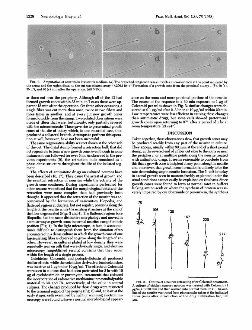
These observations seemed to contradict a result obtainedearlier in which it was found that when neurites were cut, theexisting growth cones could migrate and branch but that otherregions of the fiber did not readily form new growth cones (9).A possible reason for this difference was thought to be thelimited adhesion that the neurites have to the substratum underthese conditions which might not provide an opportunity forthe cut segment to initiate a growth cone. The initiation ofneurite extension is promoted by an increase in the adhesivityof the culture substratum (13, 14), and so we repeated ourearlier experiments on cultures that were in low serum medium(15). (It was also found to be an advantage to use the largerneurones from 13- to 14-day embryos.) Such cells formed amore secure attachment to the surface without showing a majorchange in their form or behavior. When they were cut, thestumps quickly formed "beads" or retraction bulbs. This wasfollowed in the proximal bulb by the appearance of filopodiaand, usually within 35 min but sometimes as soon as 10 minafter the amputation, by the development of morphologicallynormal growth cones. Once again, the newly formed growthcones showed the same movements as established growth conesand, in contrast to the previous situation, the neurite that borethem recommenced growth from the time of their formation(Fig. 2).
In a series of 19 amputations, 13 resulted in the formation ofa growth cone from the proximal stump within 35 min. Thisincluded fibers cut less than 50 ,.m from the cell soma as well
Neurobiology: Bray et al.
.r..j,:04
Proc. Natl. Acad. Sci. USA 75 (1978)
a, .. :. R ..
.} ........... t
.. ...t
,# A
.;/ ::,#<;'S #f 1:xj /'00::f. Xs...
*t \:.
8, -
j.j
C.
e.I>a
FIG. 2. Amputation of neurites in low serum medium. (a) The branched outgrowth was cut with a microelectrode at the point indicated bythe arrow and the region distal to the cut was cleared away. (X200.) (b -e) Formation of a growth cone from the proximal stump 1 (b), 20 (c),35 (d), and 40 (e) min after the operation. (All X350.)
as those cut near the periphery. Although all of the 13 hadformed growth cones within 35 min, in 7 cases these were ap-parent 15 min after the operation. On three other occasions, asingle fiber was cut more than once, twice in two fibers andthree times in another, and at every cut new growth conesformed quickly from the stump. Two isolated observations weremade of fibers that were, fortuitously, only partially severedwith the microelectrode. These gave rise to preterminal growthcones at the site of injury which, in one recorded case, thenproduced a collateral branch. Attempts to perform this opera-tion at will, however, have not been successful.The same regenerative ability was not shown at the other side
of the cut. The distal stump formed a retraction bulb that didnot regenerate to form a new growth cone, even though in someinstances it was followed for over 5 hr. As observed in the pre-vious experiments (9), the retraction bulb remained as aphase-dense structure throughout the life of the isolated seg-ment.The effects of antimitotic drugs on cultured neurons have
been described (16, 17). They cause the arrest of growth andthe eventual retraction of neurites while the activity of thegrowth cone continues. During experiments performed forother reasons we noticed that the morphological details of theretraction were more complex than had previously beenthought. It appeared that the retraction of the neurite was ac-companied by the formation of varicosities, filopodia, andflattened regions at discrete, but not regular, positions along thelength of the neurite while the existing structure at the end ofthe fiber degenerated (Figs. 3 and 4). The flattened regions borefilopodia, had the same distinctive morphology and moved ina similar way as growth cones in normal neurites except for theirposition (Fig. 4). In the light microscope, in fact, it was some-times difficult to distinguish them from the situation oftenencountered in a dense culture in which the growth cone of onefasciculating fiber is observed to grow along the length of an-other. However, in cultures plated at low density they wererepeatedly seen on cells that were obviously single, and electronmicroscopy (unpublished results) confirms that they occurwithin the length of a single process.
Colchicine, Colcemid, and podophyllotoxin all producedsimilar effects, while the colchicine derivative, lumicolchicine,was inactive at 1 Atg/ml or 10 ,tg/ml. The effects of Colcemidwere seen in cultures that had been pretreated for 3 hr with 10,gg of cycloheximide or puromycin, treatments that reducedthe incorporation of radioactive methionine into nondialyzablematerial to 3% and 7%, respectively, of the value in controlcultures. The changes produced by those drugs were restrictedto the terminal region of the neurite (Fig. 3) and, at least at theearly stages, cells examined by light or scanning electron mi-croscopy were found to have a normal morphological appear-
ance on the soma and more proximal portions of the neurite.The course of the response to a 30-min exposure to 1 t'g ofColcemid per ml is shown in Fig. 3; similar changes were ob-served at 0.1Itg/ml after 2-3 hr or at 10 Aug/ml within 20 min.Low temperatures were less efficient in causing these changesthan antimitotic drugs, but some cells showed preterminalgrowth cones upon returning to 370 after a period of 1 hr atroom temperature (21-24°).
DISCUSSIONTaken together, these observations show that growth cones maybe produced readily from any part of the neurite in culture.They appear, usually within 30 min, at the end of a short axonalstump, at the severed end of a fiber cut close to the soma or nearthe periphery, or at multiple points along the neurite treatedwith antimitotic drugs. It seems reasonable to conclude fromthis that a growth cone is incipient at any point along the neuriteand, moreover, that growth cone formation is unlikely to be therate-determining step in neurite formation. The 5- to 8-hr delayin axonal growth seen in neurons freshly explanted under theusual conditions cannot easily be explained on this basis. Sincegrowth cones were found to form at normal rates in bufferslacking amino acids or where the synthesis of protein was se-verely impaired by cycloheximide or puromycin, the synthesis
70
106 130
_
16 7220
277
FIG. 3. Outline of a neurite retracting after Colcemid treatment.A culture of chicken sensory neurons was treated with Colcemid (1jg/ml) for 30 min and then washed into normal medium C. The out-line of the neurite was traced from photographs taken at the indicatedtimes (min) after introduction of the drug. Calibration bar, 100Am.
5228 Neurobiology: Bray et al.
::.: :"".:..-
.1114rl* --A.

Proc. Nati. Acad. Sci. USA 75 (1978) 5229
l I I I
ace..\}.~~~~~~~~~~~~.
1l* M fIi~~~~~~~~~~I
TIt .t IIf
FIG. 4. Neurite after Colcemid treatment. Photograph of the same fiber shown in Fig. 3 taken 260 min after exposure to Colcemid (1 sg/ml).Positions of preterminal growth cones, lateral filopodia, and phase-dense dilations are indicated by arrows. Calibration bar, 50,gm.
of new proteins is probably not an essential prerequisite. Ifspecial macromolecules are required for the functions of thegrowth cones, they are presumably present in adequateamounts throughout the neuron.The simplest interpretation of our results, however, is that
the growth cone is an outwelling of ordinary neuronal cyto-plasm. The distinctive properties of the growth cone would thenbe, largely, those of undifferentiated cytoplasm confronted witha culture substratum. Indeed, many similar features are shownby fibroblasts and other cells in culture as they settle onto asurface (18). Filopodia (or microspikes) and flattened areasextend outwards from such cells, and those that do not establishcontact are withdrawn (19). A growth cone could be like sucha cell that is trying to settle onto a surface but never achievesa stable flattened state.
If a growth cone is potentially able to form at any point alongthe neurite, why is it normally restricted in position to thegrowing end? Presumably, a form of repression or containmentis at work, and our observations with cultures treated with an-timitotic drugs suggest that microtubules are involved in this.A related observation has been made of the effects of Colcemidon cultured fibroblasts, in which the flattened ruffling areasnormally restricted to the poles of the cells are seen to extendaround the periphery (20). We do not know how these effectsare caused but, for the neurite, there are two distinct possibil-ities. One is that it is the absence of microtubules, produced bydepolymerization, that allows the growth cones to form; andit is relevant to note that in normally growing neurites the mi-crotubules terminate at or near the base of the growth cone (21,22). The other is that the primary target is the fast componentof axonal flow (see, for example, ref. 23), and that it is the re-striction of this transport system that causes filopodia andflattened regions to form closer to the cell body.
It is always dangerous to extrapolate from observations intissue culture to the animal. However, many examples areknown in which branches form from the preterminal regionsof dendrites, for example, in the normal differentiation of ce-rebral dendrites (24) or in the terminal regions of motoneuronsin response to the axotomy of neighboring nerves (25), and itcould be that the present study is of relevance to those. If our
results are applied to such phenomena, two predictions may bemade. The formation of preterminal branches would be ex-pected to involve local disruption, or inactivation, of microtu-bules; and branches would be expected to be initiated withoutthe immediate need for protein synthesis.We thank Michael Brookes for help with the assay of growth cone
formation.1. Harrison, R. G. (1910) J. Exp. Zool. 9,787-848.2. Nakai, J. (1956) Am. J. Anat. 99,81-130.3. Bray, D. (1973) J. Cell Biol. 56, 702-712.4. Bunge, M. B. (1977) J. Neurocytol. 6,407-439.5. Letourneau, P. C. (1975) Dev. Biol. 44,92-101.6. Bray, D. (1970) Proc. Nati. Acad. Sci. USA 65,905-910.7. Levi-Montalcini, R. & Angeletti, P. U. (1963) Dev. Biol. 7,
653-659.8. Luduefia, M. A. (1973) Dev. Biol. 33,268-284.9. Shaw, G. & Bray, D. (1977) Exp. Cell Res. 104,55-62.
10. Varon, S., Nomura, J. & Shooter, E. M. (1967) Proc. Natl. Acad.Sc:. USA 57, 1782-1789.
11. Wilson, L. & Friedkin, M. (1966) Biochemistry 5,2463-2468.12. Vaughn, J. & Peters, A. (1967) Am. J. Anat. 121, 131-152.13. Letourneau, P. C. (1975) Dev. Biol. 44,77-91.14. Patrick, J., Heinemann, S. & Schubert, D. (1978) Annu. Rev.
Neurosci. 1, 417-443.15. Luduefia, M. A. (1973) Dev. Biol. 33,470-476.16. Yamada, K. M., Spooner, B. S. & Wessells, N. K. (1970) Proc. Natl.
Acad. Sci. USA 66,1206-1212.17. Daniels, M. P. (1972) J. Cell Biol. 53, 164-176.18. Rajaraman, R., Rounds, D. E., Yen, S. P. S. & Rembaum, A. (1974)
Exp. Cell Res. 88, 327-339.19. Albrecht-Buehler, G. & Goldman, R. D. (1976) Exp. Cell Res.
97,329-339.20. Vasiliev, J. M., Gelfand, I. M., Domnina, L. V., Ivanova, 0. Y.,
Komm, S. G. & Olshevkaja, L. V. (1970) J. Embryol. Exp. Mor-phol. 24, 625-640.
21. Yamada, K. M., Spooner, B. S. & Wessells, N. K. (1971) J. CellBiol. 49, 614-635.
22. Bunge, M. B. (1973) J. Cell Biol. 56, 713-735.23. Paulson, J. C. & McClure, W. 0. (1975) J. Cell Biol. 67, 461-
467.24. Morest, D. K. (1969) Anat. Entwlckl.-Gesch. 128,271-289.25. Edds, M. V. (1953) Quart. Rev. Biol. 28,260-276.
M.
Neurobiology: Bray et al.
*