slide 1 stimulus (input) receptors (sensory neurons) integrators (interneurons) motor neurons...
TRANSCRIPT
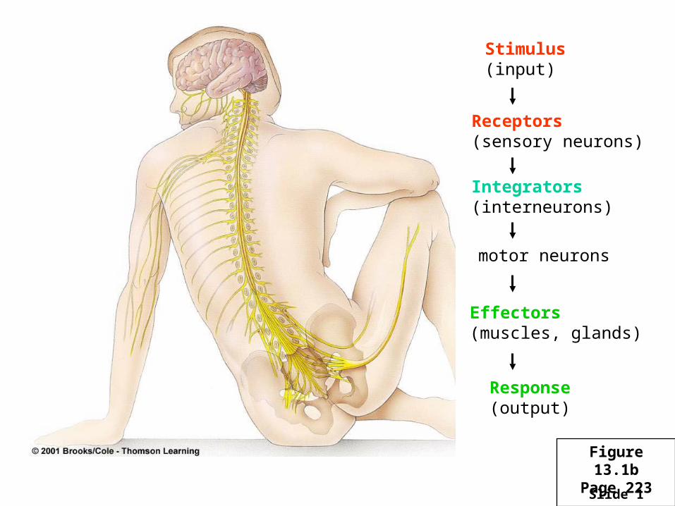
Slide 1
Stimulus(input)
Receptors(sensory neurons)
Integrators(interneurons)
motor neurons
Effectors(muscles, glands)
Response(output)
Figure 13.1bPage 223
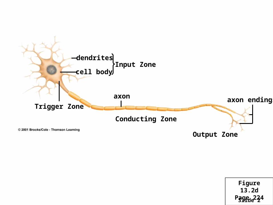
Slide 2
dendrites
cell body
Trigger Zone
Input Zone
Conducting Zone
Output Zone
axon axon endings
Figure 13.2dPage 224

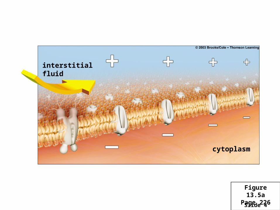
Slide 3
lipid bilayer of neuron’s plasma membrane
Interstitial Fluid
Cytoplasm
K+/Na+ pumpchannel proteins, continually open
channel proteins withvoltage-sensitive gates
Figure 13.3Page 225
Slide 4
interstitial fluid
cytoplasm
Figure 13.5aPage 226
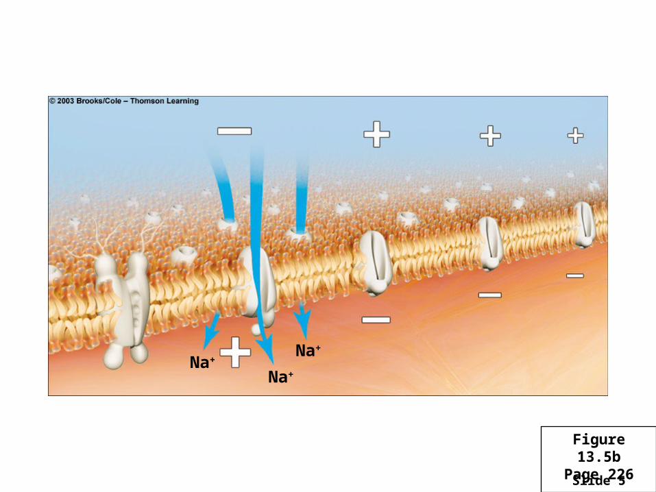
Slide 5
Na+
Na+
Na+
Figure 13.5bPage 226
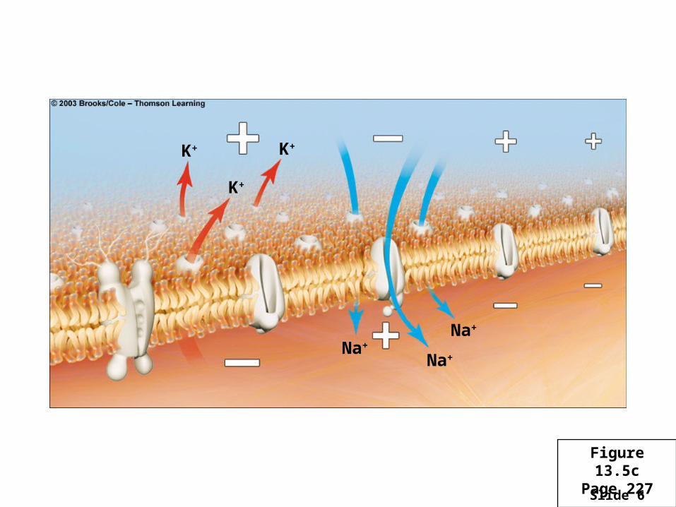
Slide 6
Na+
Na+
Na+
K+
K+
K+
Figure 13.5cPage 227

Slide 7
Na+Na+
K+ K+K+
K+
Na+
Figure 13.5dPage 227
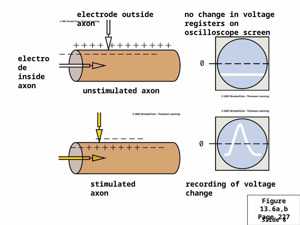
Slide 8
electrode inside axon
electrode outside axon no change in voltage registers on oscilloscope screen
unstimulated axon
recording of voltage change
0
0
stimulated axon
Figure 13.6a,bPage 227
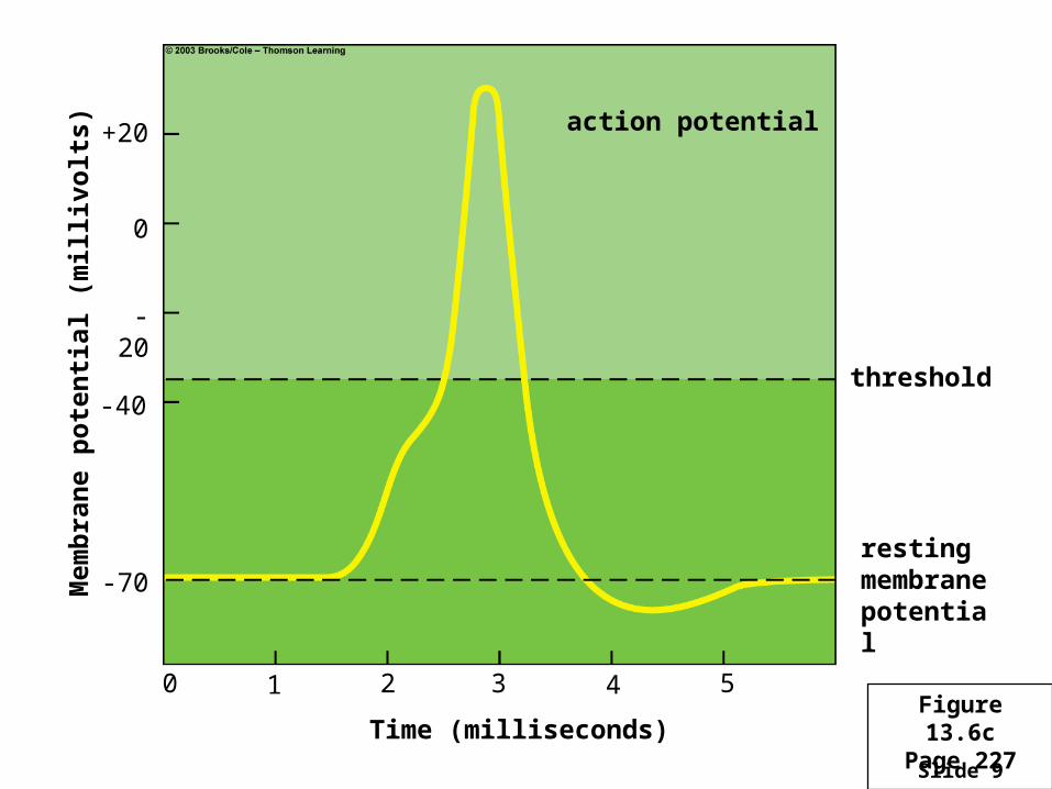
Slide 9
Figure 13.6cPage 227
action potential
threshold
resting membrane potential
Time (milliseconds)
Mem
bra
ne
po
ten
tial
(m
illi
volt
s)
-40
-70
-20
0
+20
0 1 2 3 4 5
Slide 10
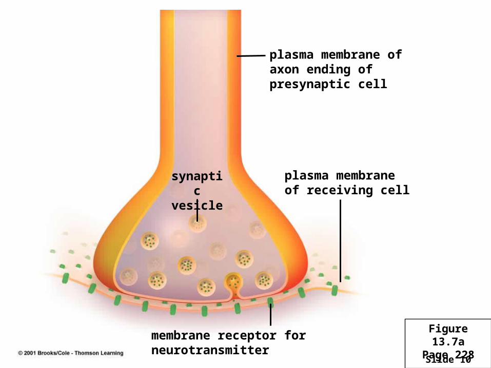
plasma membrane of axon ending of presynaptic cell
synaptic vesicle
membrane receptor for neurotransmitter
plasma membrane of receiving cell
Figure 13.7aPage 228
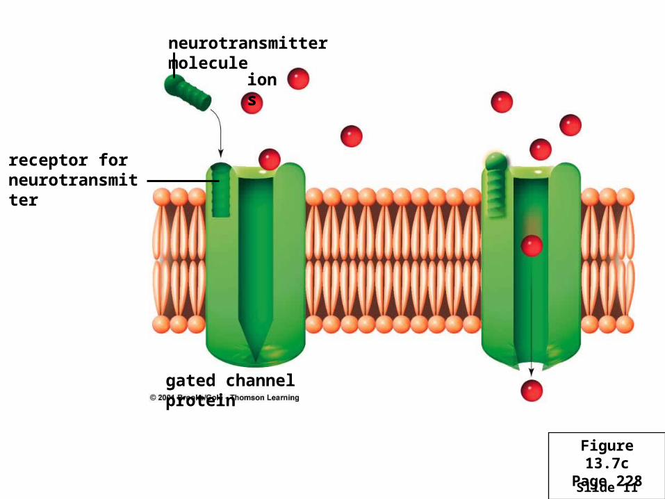
Slide 11
ions
neurotransmitter molecule
receptor for neurotransmitter
gated channel protein
Figure 13.7cPage 228
Slide 12
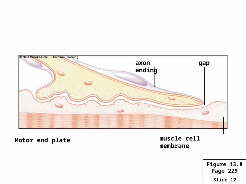
Motor end plate
gapaxon ending
muscle cell membrane
Figure 13.8Page 229
Slide 13
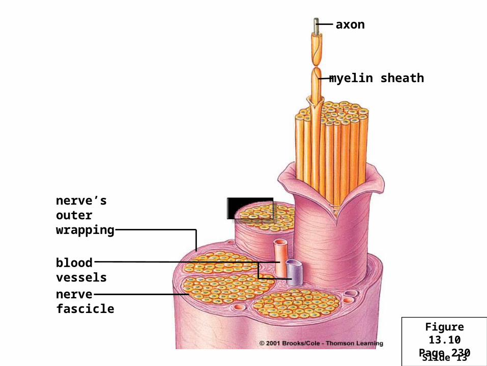
blood vessels
nerve’s outer wrapping
nerve fascicle
axon
myelin sheath
Figure 13.10Page 230
Slide 14
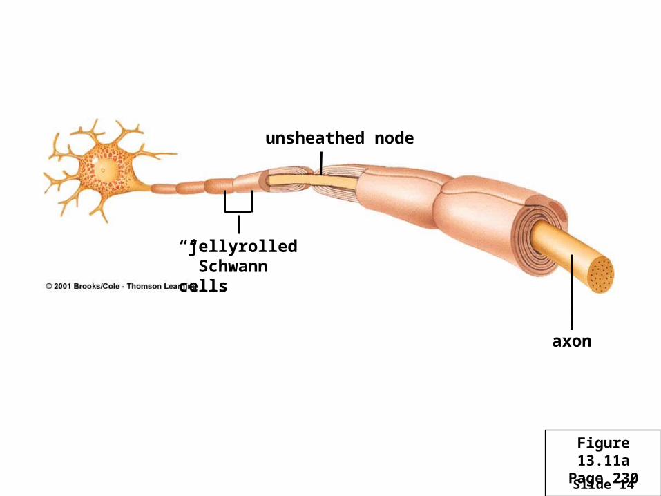
axon
unsheathed node
“jellyrolled” Schwann cells
Figure 13.11aPage 230
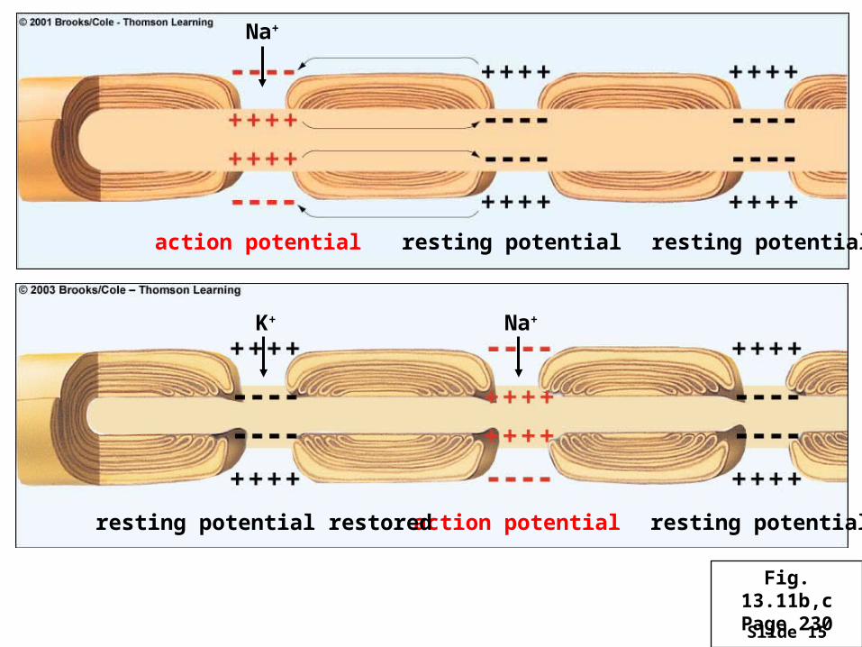
Slide 15
action potential resting potential resting potential
Na+
Na+K+
action potential resting potentialresting potential restored
Fig. 13.11b,cPage 230
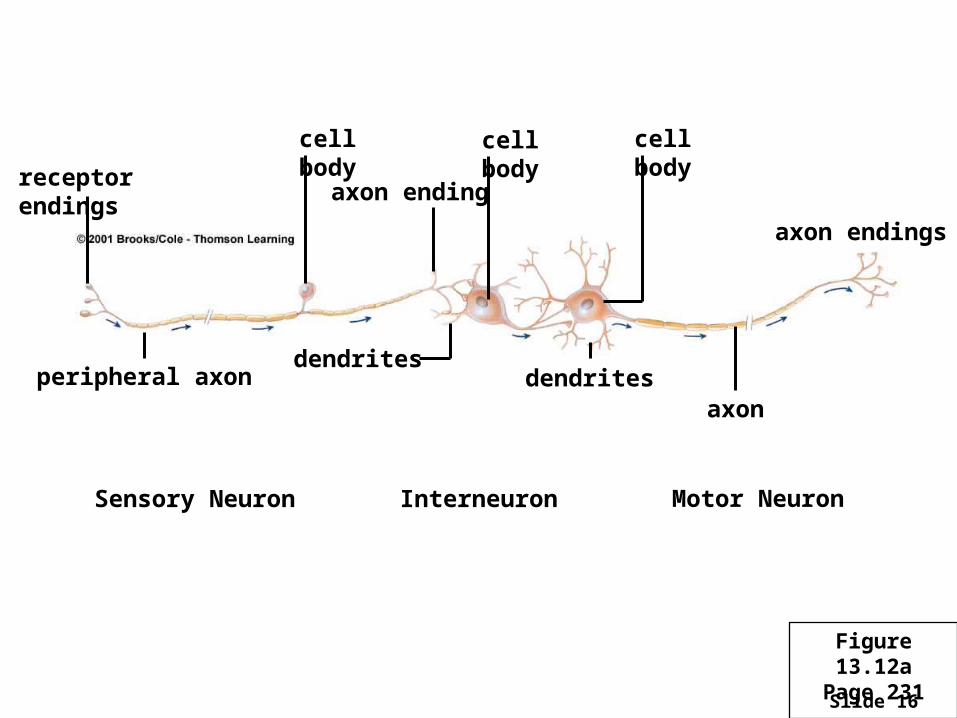
Slide 16
axon endingreceptor endings
peripheral axon
cell body cell body cell body
axon
axon endings
dendritesdendrites
Interneuron Motor NeuronSensory Neuron
Figure 13.12aPage 231
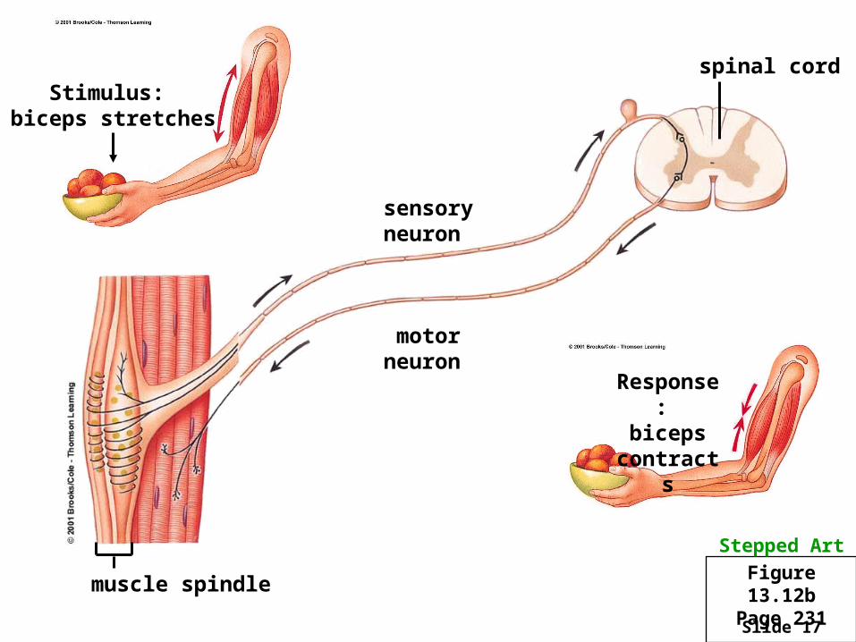
Slide 17
Response: biceps
contracts
spinal cord
muscle spindle
sensoryneuron
motorneuron
Stimulus: biceps stretches
Stepped Art
Figure 13.12bPage 231
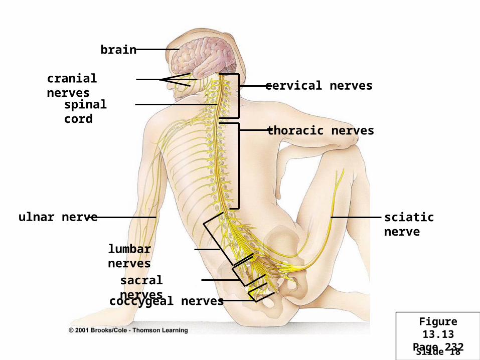
Slide 18
brain
cranial nerves
spinal cord
ulnar nerve
lumbar nerves
sacral nerves
coccygeal nerves
cervical nerves
thoracic nerves
sciatic nerve
Figure 13.13Page 232
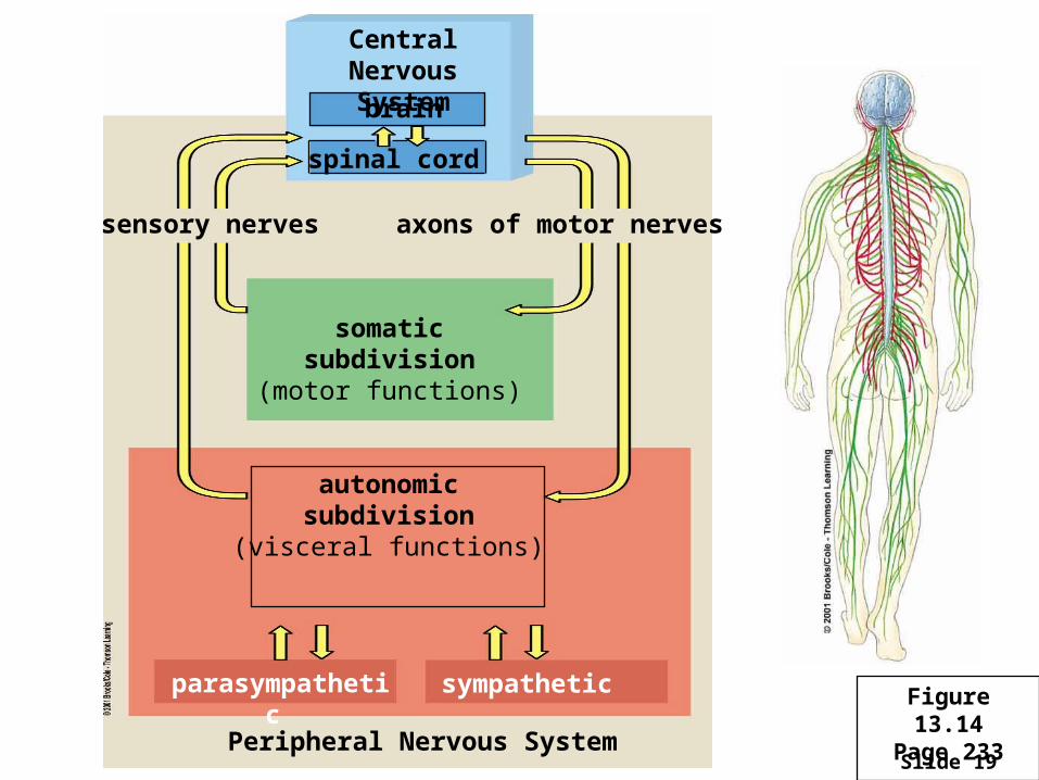
Slide 19
Central NervousSystem
brain
spinal cord
sensory nerves axons of motor nerves
somaticsubdivision
(motor functions)
autonomicsubdivision
(visceral functions)
sympathetic
Peripheral Nervous System
parasympathetic Figure 13.14Page 233
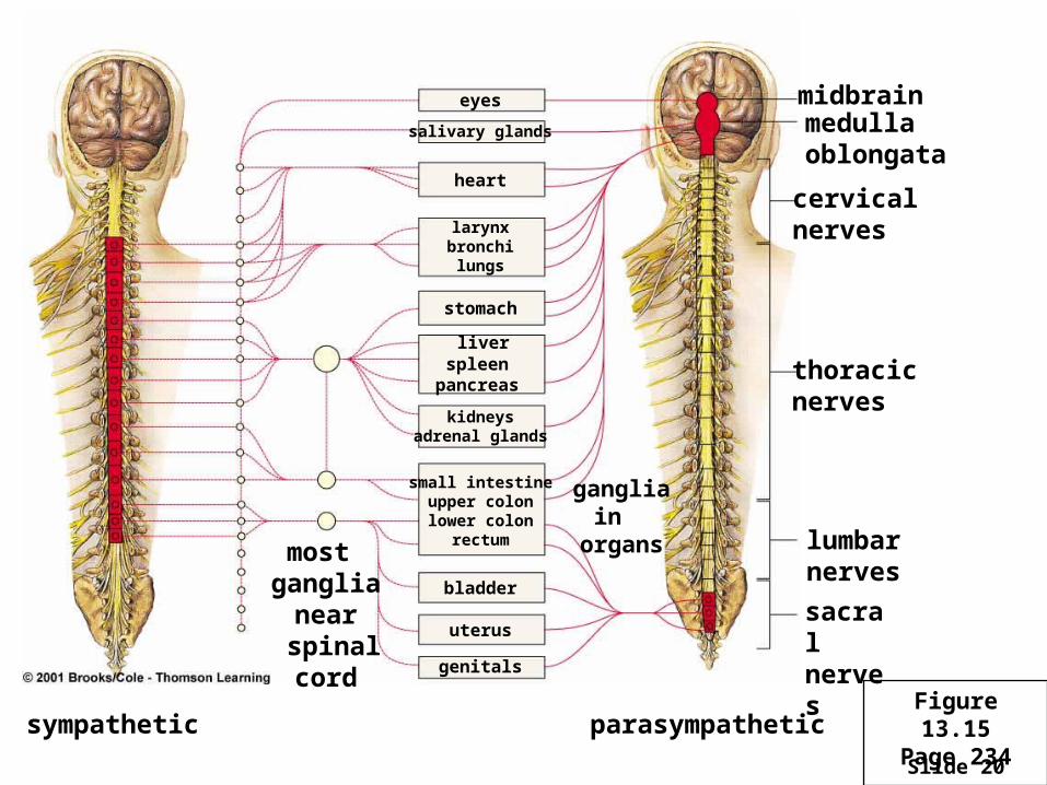
Slide 20
eyes
salivary glands
heart
larynxbronchilungs
stomach
liverspleen
pancreas
kidneysadrenal glands
small intestineupper colonlower colon
rectum
bladder
uterus
genitals
most ganglia
near spinalcord
gangliain
organs
midbrainmedulla oblongata
cervicalnerves
thoracicnerves
lumbarnerves sacralnerves
sympathetic parasympatheticFigure 13.15
Page 234
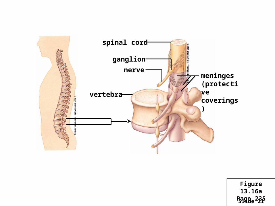
Slide 21
spinal cord
ganglion
nerve
vertebra
meninges(protectivecoverings)
Figure 13.16aPage 235
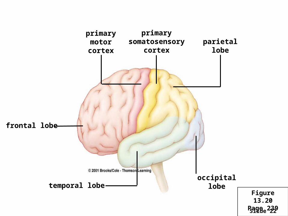
Slide 22
frontal lobe
primarymotorcortex
primarysomatosensory
cortexparietal
lobe
temporal lobe occipital lobe
Figure 13.20
Page 239
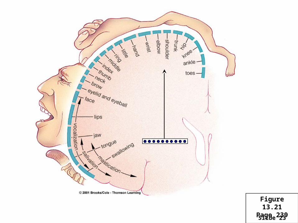
Slide 23
Figure 13.21Page 239
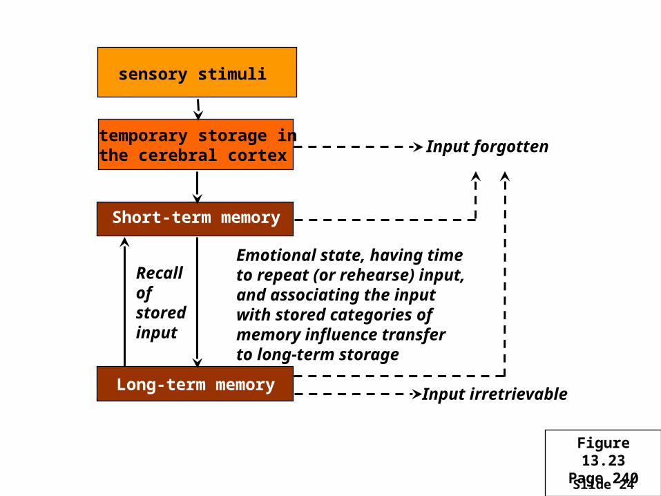
Slide 24
sensory stimuli
temporary storage inthe cerebral cortex
Short-term memory
Long-term memory
Emotional state, having timeto repeat (or rehearse) input,and associating the input with stored categories ofmemory influence transferto long-term storage
Recallof storedinput
Input irretrievable
Input forgotten
Figure 13.23Page 240
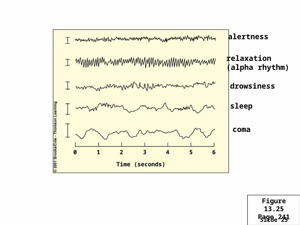
Slide 25
0 1 2 3 4 5 6
Time (seconds)
alertness
relaxation(alpha rhythm)
sleep
coma
drowsiness
Figure 13.25Page 241
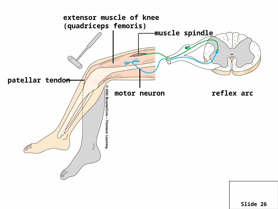
Slide 26
muscle spindle
extensor muscle of knee (quadriceps femoris)
reflex arcmotor neuron
patellar tendon
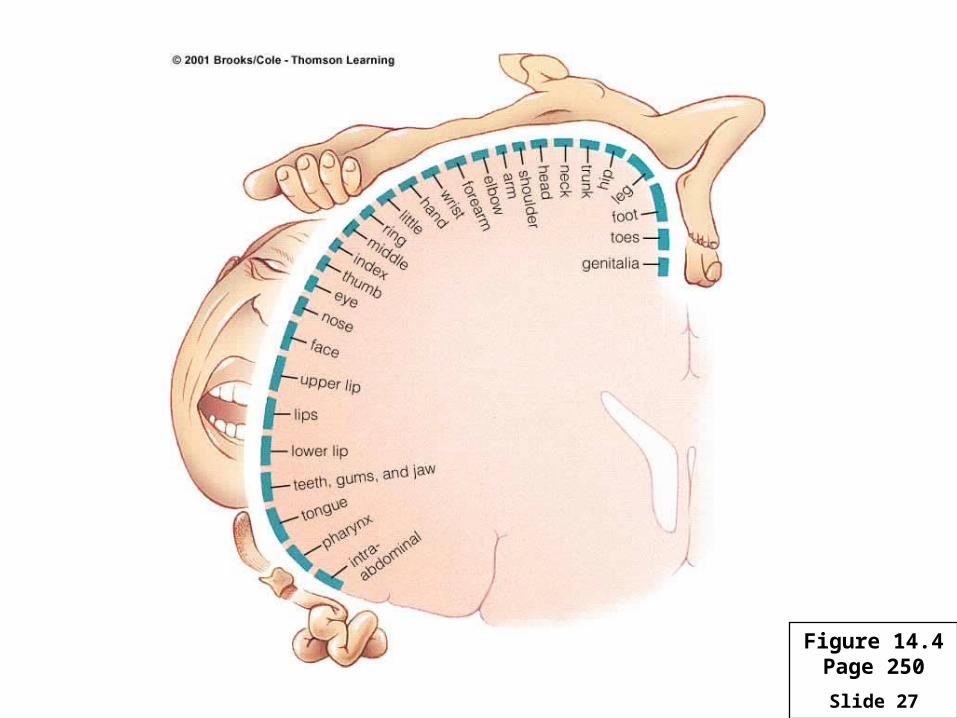
Slide 27
Figure 14.4Page 250
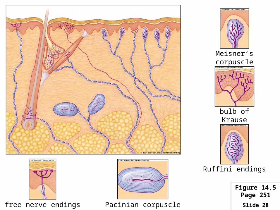
Slide 28free nerve endings Pacinian corpuscle
Ruffini endings
bulb of Krause
Meisner’s corpuscle
Figure 14.5Page 251
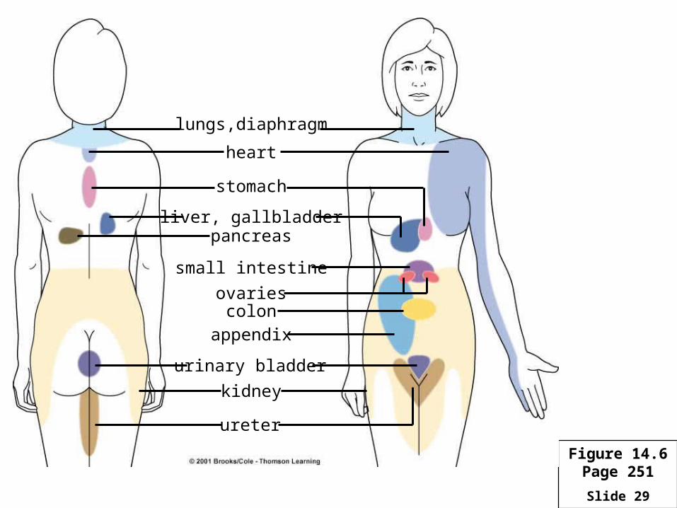
Slide 29
lungs,diaphragm
heart
liver, gallbladderpancreas
small intestine
ovariescolon
appendix
urinary bladder
kidney
ureter
stomach
Figure 14.6Page 251
Slide 30
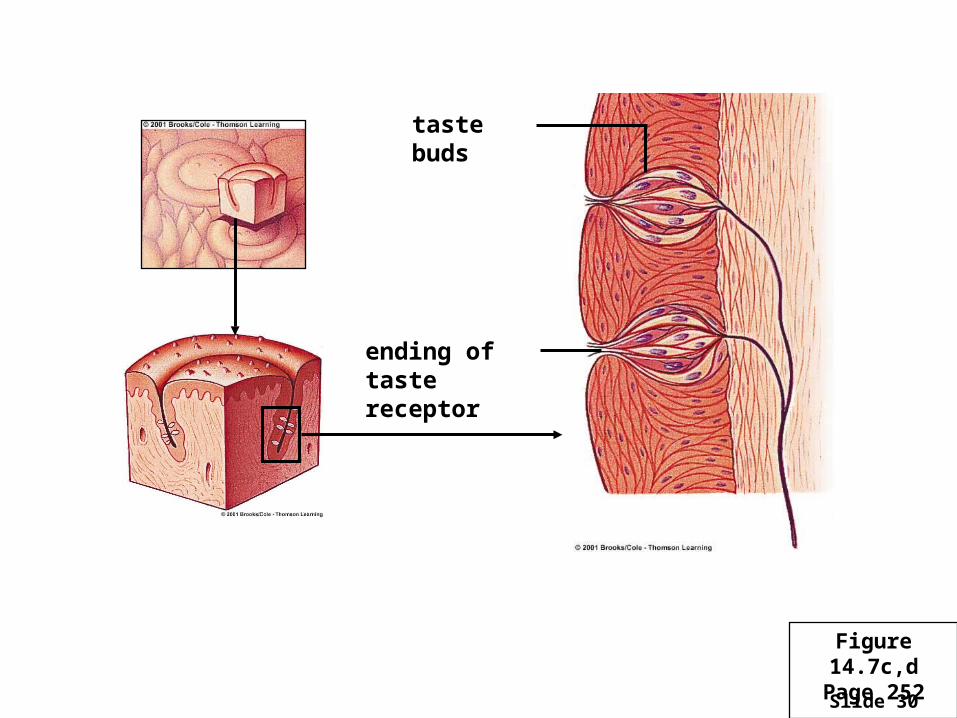
taste buds
ending of taste receptor
Figure 14.7c,dPage 252
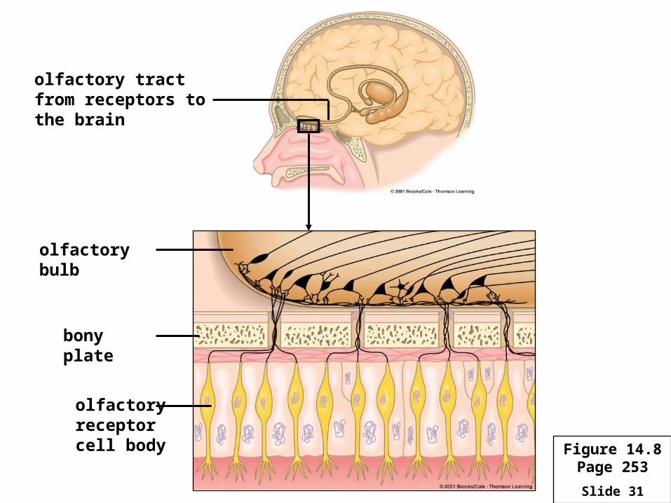
Slide 31
olfactory tract from receptors to the brain
olfactory bulb
bony plate
olfactoryreceptorcell body Figure 14.8
Page 253
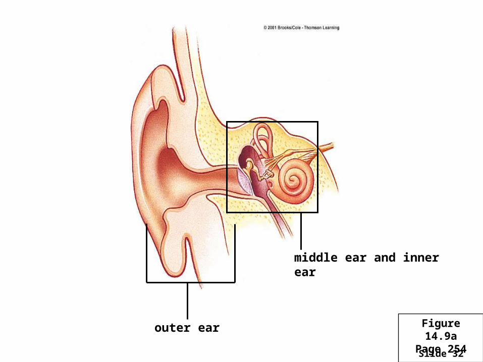
Slide 32
middle ear and inner ear
outer ear Figure 14.9aPage 254
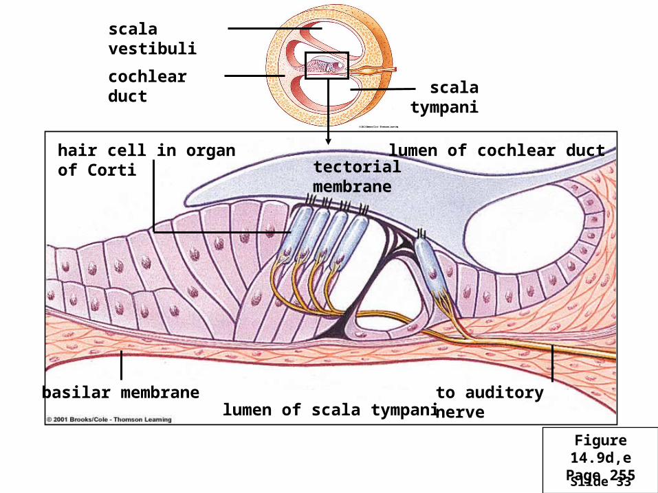
Slide 33
hair cell in organ of Cortitectorial membrane
lumen of cochlear duct
basilar membranelumen of scala tympani
to auditory nerve
scala vestibuli
cochlear duct scala tympani
Figure 14.9d,ePage 255
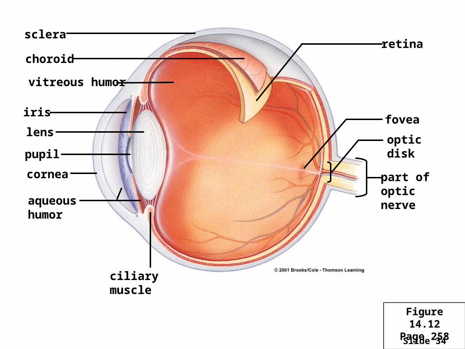
Slide 34
sclera
choroid
iris
lens
pupil
cornea
aqueoushumor
ciliary muscle
vitreous humor
retina
fovea
optic disk
part ofopticnerve
Figure 14.12Page 258
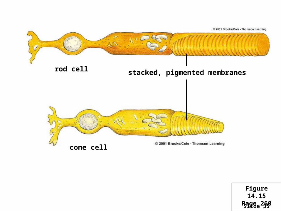
Slide 35
rod cell stacked, pigmented membranes
cone cell
Figure 14.15Page 260
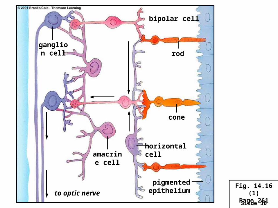
Slide 36
ganglion cell
bipolar cell
rod
cone
amacrine cell
horizontal cell
pigmented epitheliumto optic nerve
Fig. 14.16 (1)Page 261
Slide 37