diagnosis and management of sensory polyneuropathy · reflexes, and sensory ataxia. in sensory...
TRANSCRIPT
For personal use only 1 of 23
terms “small fiber neuropathy”, “sensory ataxia”, “sensory neuronopathy”, “dorsal root ganglionopathy”, “dorsal root ganglion”, “skin biopsy”, “quantitative sensory testing”, “corneal confocal microscopy”, “quantitative sudomotor axon reflex testing”, “thermoregulatory sweat testing”, “elec-trochemical skin conductance”, “sarcoidosis”, “Sjögren’s syndrome”, “fibromyalgia”, “sodium channelopathies”, “transthyretin”, “sensory Guillain-Barré syndrome”, “ataxic Guillain-Barré syndrome”, “acute sensory ataxic neuropa-thy”, “Miller Fisher syndrome”, “disialosyl antibodies”, “ganglioside antibodies”, “CANOMAD”, “CANDA”, “sensory chronic inflammatory demyelinating polyneuropathy”, “dis-tal acquired demyelinating symmetric neuropathy”, “anti-MAG”, “anti-Hu”, and “tabes dorsalis”. We included a few articles of historical importance that were published in the 1980s and 1990s. These sentinel articles set the concep-tual framework for these disorders and their inclusion was necessary. We searched reference lists of articles selected through title, abstract, and full text review. We selected randomized controlled trials, observational, and basic sci-ence studies, systematic reviews, and meta-analyses from these sources. Articles were prioritized by study quality and topic. Given that many of the sensory neuropathies discussed are extremely rare, case studies and case series were also reviewed and included if deemed important.
S TAT E O F T H E A R T R E V I E W
IntroductionPeripheral sensory nerves vary in size and function, rang-ing from the smallest unmyelinated C fibers and thinly myelinated Aδ fibers that conduct noxious and thermal information1 2 to the larger Aβ fibers that transmit proprio-ceptive and vibratory information.3 As a result, disorders of sensory nerve function are diverse and depend on the type of nerve fiber that is affected; patients present with a wide range of symptoms, from pain predominant (small fiber) to ataxia predominant (large fiber) problems. This article will not attempt to review all peripheral sensory neuropathies that manifest the classic length dependent or “stocking glove” pattern, but will focus on those that have a clearly pain predominant or ataxia predominant presentation. It will also include other disorders that pre-sent with sensory ataxia but affect the dorsal root gan-glia (DRG), sensory fibers of the nerve roots, and dorsal columns. We will also cover the differential diagnosis of sensory polyneuropathies, the diagnostic approach to patients with sensory problems, and disease specific and symptomatic treatments.
Sources and selection criteriaWe searched PubMed for English language articles pub-lished from 1 January 2000 to 1 October 2018 using the
ABSTRACT
Sensory polyneuropathies, which are caused by dysfunction of peripheral sensory nerve fibers, are a heterogeneous group of disorders that range from the common diabetic neuropathy to the rare sensory neuronopathies. The presenting symptoms, acuity, time course, severity, and subsequent morbidity vary and depend on the type of fiber that is affected and the underlying cause. Damage to small thinly myelinated and unmyelinated nerve fibers results in neuropathic pain, whereas damage to large myelinated sensory afferents results in proprioceptive deficits and ataxia. The causes of these disorders are diverse and include metabolic, toxic, infectious, inflammatory, autoimmune, and genetic conditions. Idiopathic sensory polyneuropathies are common although they should be considered a diagnosis of exclusion. The diagnostic evaluation involves electrophysiologic testing including nerve conduction studies, histopathologic analysis of nerve tissue, serum studies, and sometimes autonomic testing and cerebrospinal fluid analysis. The treatment of these diseases depends on the underlying cause and may include immunotherapy, mitigation of risk factors, symptomatic treatment, and gene therapy, such as the recently developed RNA interference and antisense oligonucleotide therapies for transthyretin familial amyloid polyneuropathy. Many of these disorders have no directed treatment, in which case management remains symptomatic and supportive. More research is needed into the underlying pathophysiology of nerve damage in these polyneuropathies to guide advances in treatment.
Diagnosis and management of sensory polyneuropathyKelly Graham Gwathmey, Kathleen T Pearson
Virginia Commonwealth University, Department of Neurology, 1101 E. Marshall Street, PO Box 980599, Richmond, VA 23298, USACorrespondence to: K G Gwathmey [email protected] this as: BMJ 2019;365:l1108doi: 10.1136/bmj.l1108
Series explanation: State of the Art Reviews are commissioned on the basis of their relevance to academics and specialists in the US and internationally. For this reason they are written predominantly by US authors.
on 5 March 2020 by guest. P
rotected by copyright.http://w
ww
.bmj.com
/B
MJ: first published as 10.1136/bm
j.l1108 on 8 May 2019. D
ownloaded from
S TAT E O F T H E A R T R E V I E W
For personal use only 2 of 23
Incidence and prevalenceThe sensory polyneuropathy category includes extremely common conditions such as diabetic neuropathies (the most common cause of neuropathy worldwide) and very rare conditions, such as specific acute ataxic neuropathies (described only in case series). Table 1 lists the incidence and prevalence of these specific polyneuropathies and their underlying causes, if known.
Clinical presentationThe clinical presentation and findings on physical exam-ination depend on the type of affected nerve fiber and the distribution of nerve damage. Patients may report a combination of positive (paresthesia, burning pain) and negative (loss of sensation) sensory disturbances, as well as gait imbalance. Important considerations regarding the clinical presentation include acuity of onset, time course of progression, and the distribution and quality of sensory symptoms.
Small fiber neuropathiesIn small fiber neuropathies (SFNs) the thinly myeli-nated (Aδ) and unmyelinated (C) fibers responsible for the transmission of thermal and noxious sensory input are affected.1 2 Clinically, this nerve damage translates to symptoms of sharp, painful, or burning paresthesia; sensory loss or numbness; and the inability to discrimi-nate between hot and cold sensations. Symptoms may be vague, described as a tight feeling or abnormal sensation in the soles of the feet, intolerance of tactile stimuli (ina-bility to wear socks or touch bedsheets), or a sensation of restless legs. The distribution of symptoms may have a length dependent or non-length dependent pattern that affects the limbs, trunk, face, or it may have a com-bination of patterns.1 2 40-42 Depending on the underlying cause, the onset of symptoms may be gradual, with slowly progressive worsening, or subacute with more rapid pro-gression. Pain may be prominent and disabling, and a recent large Italian cohort study of patients with painful diabetic neuropathy suggests that pain may be more com-mon in women.43
Dysautonomia is often a feature of SFN owing to impairment of the sympathetic and parasympathetic function of Aδ fibers and the postganglionic autonomic function of C fibers. It is essential to ask patients about potential autonomic involvement including orthostasis; palpitations; abnormal sweating; dry mouth, eyes, or skin; gastrointestinal symptoms including cramping, diarrhea, or constipation; flushing or other changes of skin color; and erectile dysfunction.2
A patient with SFN may have decreased temperature and pinprick sensation on examination, and potentially allodynia, dysesthesia, or hyperesthesia on sensory testing. Motor strength, proprioception, and muscle stretch reflexes should be preserved in patients with pure SFN. Skin may have a dry, atrophic, or discolored appearance.1 2 40
Sensory ataxiaDisorders affecting the large myelinated Aβ fibers, 1a fib-ers, sensory nerve roots, or DRG will result in impaired vibratory sensation and proprioception. Clinically this results in a combination of symptoms of sensory loss, paresthesia, and gait imbalance. The ataxic sensory polyneuropathies will present acutely or have an insidi-ous onset and gradually progressive course as a result of dysfunction of the peripheral sensory nerves. Physical examination may show absent or reduced vibratory sen-sations, abnormal proprioception, depressed or absent reflexes, and sensory ataxia.
In sensory neuronopathies (dorsal root ganglionopa-thies), sensory neurons of the dorsal root and trigeminal ganglia are affected. The clinical presentation is charac-terized by pronounced ataxia and sensory loss, which may have a non-length dependent or multifocal pattern. In addition, pain and positive sensory symptoms often occur because of the involvement of small and medium sized nerve fibers.44 The face and trunk may also be affected.44-46 The results of a physical examination will resemble that seen in patients with ataxic sensory poly-neuropathies, although the sensory deficits are more often patchy, non-length dependent, or generalized. The
LIST OF ACRONYMSAAN: American Academy of NeurologyANA: Antinuclear antibodiesASAN: Acute sensory ataxic neuropathyBPI-MSF: Brief Pain Inventory Modified Short FormCANDA: Chronic ataxic neuropathy with disialosyl antibodiesCANOMAD: Chronic ataxic neuropathy, ophthalmoplegia, IgM paraprotein, cold agglutinins, and disialosyl antibodiesCIDP: Chronic inflammatory demyelinating polyneuropathyCISP: Chronic immune sensory polyradiculopathyDADS: Distal acquired demyelinating symmetric neuropathyDRG: Dorsal root ganglia EFNS: European Federation of Neurological SocietiesEMLA: Eutectic mixture of local anestheticESR: Erythrocyte sedimentation rateGBS: Guillain-Barré syndromeIENFD: Intraepidermal nerve fiber densityIFG: Impaired fasting glucoseIGT: Impaired glucose tolerance LEP: Laser evoked potentialMAG: Myelin associated glycoproteinMFS: Miller-Fisher syndromeMRI: Magnetic resonance imagingmNIS+7: Modified Neuropathy Impairment Score +7Norfolk QOL-DN: Norfolk Quality of Life-Diabetic NeuropathyNPS: Neuropathic Pain ScaleQSART: Quantitative sudomotor axon reflex testSFN: Small fiber neuropathySGPG: Sulphated glucuronyl paraglobosideSNAP: Sensory nerve action potentialSNRI: Serotonin-norepinephrine reuptake inhibitorSSEP: Somatosensory evoked potentialTCA: Tricyclic antidepressantTTR-FAP: Transthyretin familial amyloidosis with polyneuropathy
on 5 March 2020 by guest. P
rotected by copyright.http://w
ww
.bmj.com
/B
MJ: first published as 10.1136/bm
j.l1108 on 8 May 2019. D
ownloaded from

S TAT E O F T H E A R T R E V I E W
For personal use only 3 of 23
finding of pseudoathetosis, as a result of impaired affer-ent proprioceptive input, is a hallmark of DRG dysfunc-tion.44 46 47 Although motor strength is preserved in pure sensory neuronopathies, it may seem to be impaired on examination owing to the lack of proprioceptive input during confrontational strength testing. The clinical course may be gradual and insidious in idiopathic forms of the disease, but it will typically have a subacute course in patients with paraneoplastic, immune mediated, and toxic causes.44
Patients with dorsal column dysfunction may also pre-sent with sensory ataxia. Often these patients also have evidence of upper motor neuron signs on examination, which suggests corticospinal tract involvement and will guide the examiner away from localization in the periph-eral nervous system. When the dorsal columns and corti-cospinal tracts are affected, patients will have spasticity, weakness, and reduced vibratory and proprioceptive sen-sations: the so called posterolateral column syndrome.48
Differential diagnosis of small fiber neuropathiesThe causes of SFN fall into six broad categories: meta-bolic, inflammatory, genetic, toxic, infectious, and
idiopathic (cryptogenic) (table 2). Many of the known common causes will not be discussed in detail but are included in table 2. Fibromyalgia, which has been associ-ated with pathologic evidence of SFN, does not easily fall into one of the six categories. Alternatively, classification based on clinical phenotype has also been proposed.49 Despite extensive evaluation, 20-50% of cases of SFN are ultimately classified as idiopathic.50-53 The most common causes include diabetes, immunologic condi-tions, sodium channel mutations, and vitamin B12 defi-ciency.29 Although immunologic conditions were found in 19% of a cohort of 921 patients with SFN, which exceeds the prevalence in the general population, the exact pathogenic role of isolated autoantibodies remains unclear.29 54 In one series, the highest yield blood tests in SFN that appeared to be “initially idiopathic” were erythrocyte sedimentation rate (ESR), antinuclear anti-bodies (ANA), C3 complement values, and autoantibod-ies that are associated with Sjögren’s syndrome and celiac disease.55 It has been recommended that patients are screened for glucose intolerance, vitamin B12 defi-ciency, and sodium channel mutations even if there is a known underlying cause.29 54
Table 1 | Incidence and prevalence of sensory neuropathies highlighted in this review*
Diagnosis Incidence of underlying cause Prevalence of underlying cause Incidence and prevalence of the neuropathy (if known)Small fiber neuropathiesDiabetic neuropathy (including small and large fiber neuropathy)
In the US 1.5 million people are diagnosed as having diabetes every year (6.7/1000)4
In 2015, 9.4% of the US population were estimated to have diabetes4
Lifetime incidence of neuropathy is 37-45% in type 2 diabetes and 54-59% in type 1 diabetes5 Prevalence of diabetic neuropathy is 5-54% depending on the criteria and methods used to define neuropathy and age of included patients6-9
Prediabetic small fiber neuropathy In 2015, 33.9% of the US population over the age of 18 years had prediabetes4
Prevalence IGT and neuropathic pain: 8.7-14.8% IFG and neuropathic pain: 4.2-5.7%6 10 11
Metabolic syndrome In 2007-2012, 34.2% of the US population had metabolic syndrome12
Sarcoidosis Globally, 1.0-35.5/100 00013
This figure is probably higher in black people (40-70/100 000; US data)14 15
Globally, 4.7-64/100 00013 Pain and signs of SFN present in up to 28-60% of patients depending on the criteria and methods used to define neuropathy16-20
Sjogren’s syndrome associated sensory neuropathies
Globally, 6.92/100 00021 Globally, 60.82/100 00021 Prevalence of “pure sensory neuropathy”: 9.2% Prevalence of neuronopathy 0.6% (in a French population) 22
Fibromyalgia Annual incidence in UK: 33.3/100 00023 Globally, 1.78%24 Small fiber pathology is seen in about half of patients with fibromyalgia25 26
Transthyretin familial amyloidosis with polyneuropathy
In Portugal: 0.87/100 00027 In Portugal: 22.93/100 00027 Worldwide: 50 000 cases28
Sodium channelopathies (SCN9A, SCN10A, SCN11A)
In a large series of patients with SFN, 9.6% had genetic variants in SCN9A, 4.5% in SCN10A, and 3.4% in SCN11A29
In a smaller series, 28.6% of patients with idiopathic SFN had the Nav1.7 mutation30
Sensory ataxic neuropathiesSensory GBS Overall incidence (included studies from North
America and Europe) of GBS: 0.8-1.9/100 00031
Sensory GBS unknown
Lifetime risk of developing GBS is less than 1/100032
Miller Fisher syndrome 0.1/100 000 in UK; 15-20% of all GBS in Asia and 1-7% in the West33
Acute sensory ataxic neuropathy Unknown (case reports/case series) Unknown (case reports/case series)Ataxic GBS Unknown (case reports/case series) Unknown (case reports/case series)Chronic ataxic neuropathies associated with anti-disialosyl antibodies
Unknown (case reports/case series) Unknown (case reports/case series)
Sensory CIDP Overall incidence of 07-1.6/100 00034 35
Sensory CIDP unknownOverall prevalence of CIDP: 4.8-8.9/100 00034 35
Prevalence of sensory CIDP: 24-35% of all patients with CIDP34 36 37
DADS Unknown; IgM MGUS is associated with a polyneuropathy in 50% of patients38
Paraneoplastic sensory neuronopathy >500 cases reported39 *CIDP=chronic inflammatory demyelinating polyradiculoneuropathy; DADS=distal acquired demyelinating symmetric neuropathy; GBS=Guillain-Barré syndrome; IFG=impaired fasting glucose; IGT=impaired glucose tolerance; MGUS=monoclonal gammopathy of undetermined significance; SFN=small fiber neuropathy; US=United States.
on 5 March 2020 by guest. P
rotected by copyright.http://w
ww
.bmj.com
/B
MJ: first published as 10.1136/bm
j.l1108 on 8 May 2019. D
ownloaded from
S TAT E O F T H E A R T R E V I E W
For personal use only 4 of 23
Metabolic causes: diabetes and prediabetesDiabetes is the most common cause of polyneuropathy worldwide and the most common cause of SFN specifi-cally.56 The association between prediabetes (impaired glucose tolerance (IGT) and impaired fasting glucose (IFG)) and polyneuropathy is still being delineated. IGT is defined by a raised two hour glucose level on an oral glucose tolerance test of 7.8-11.1 mmol/L (140-199 mg/dL). IFG is defined by a fasting glucose of 5.6-6.9 mmol/L (100-125 mg/dL). It is likely that the risk of neuropathy is higher for IGT than for IFG.57 When considering the diagnostic investigations in these patients, it is important to note that glycosylated hemoglobin may be normal in patients with IGT.58
Some studies support an association between IGT and polyneuropathies,10 59-62 whereas others have failed to show such a correlation.63-65 It is thought that IGT asso-ciated neuropathy mainly affects the small nerve fibers,
perhaps explaining why some researchers have found no correlation between IGT and large fiber polyneuropathy60 66 68 and others have questioned the association between IGT and SFN.64 69 Such incongruent findings across studies are probably the result of differences in definitions of polyneu-ropathy (including the use of symptoms or intraepidermal nerve fiber density (IENFD)), degrees of surveillance, and polyneuropathy endpoints.69 Nonetheless, the identifica-tion of prediabetes is of utmost importance because 50% of patients with prediabetes ultimately develop type 2 diabetes,70 and reducing the risk of conversion to diabetes decreases the risk of developing polyneuropathy.
The Impaired Glucose Tolerance Neuropathy study investigated 32 patients with IGT and neuropathy. It found that 65% of patients had low amplitude or absent sural responses, 83% had decreased IENFD, and 61% had abnormal quantitative sudomotor autonomic reflex test results.71 Skin biopsy was found to be the most sen-
Table 2 | Causes of small fiber neuropathy and ancillary investigations*
Causes Ancillary investigationsImmune mediated Sarcoidosis ACE, chest radiography, histopathologySjögren’s syndrome Anti-SSA/anti-SSB antibodies, Schirmer test, Rose Bengal test, lip and salivary gland biopsySystemic lupus erythematosus ANA, antiphospholipid antibodies, complement levels, ESR, CRP, anti-dsDNA and anti-Smith
antibodiesCeliac disease Antigliadin antibodies (serum IgA endomysial and tissue transglutaminase antibody), IgG
deamidated gliadin peptide, small bowel biopsyInflammatory bowel disease (Crohn’s disease and ulcerative colitis)
Inflammatory markers, endoscopy, barium studies
Paraneoplastic (ganglionic acetylcholine receptor antibody mediated)
Voltage gated potassium channel antibodies, CASPR-2, and anti-Hu antibodies, ganglionic acetylcholine receptor antibodies
“Apparently autoimmune” small fiber neuropathy† Presence of systemic autoimmune disease, abnormal blood markers of autoimmunity (ANA, ESR, SSA/SSB antibodies, or low complement levels)
Metabolic Impaired glucose tolerance and impaired fasting glucose Two hour glucose tolerance test, fasting blood sugar, glycosylated hemoglobinDiabetes Glycosylated hemoglobin, two hour glucose tolerance test, fasting blood sugarTreatment induced neuropathy in diabetes (insulin neuritis) Clinical diagnosis in the setting of rapid correction of hyperglycemiaHyperlipidemia (mostly hypertriglyceridemia) Lipid profile including fasting triglyceride levelHypothyroidism TSH, free T4 and T3Infectious HIV HIV viral load, and CD4 cell countHepatitis C virus Hepatitis C virus antibody, hepatitis C PCRCryoglobulinemia (often associated with hepatitis C) CryoglobulinsLeprosy Serum antibodies to phenolic glycolipid-I, skin or nerve biopsy for acid fast bacilliToxic Numerous implicated drugs (anti-retrovirals, metronidazole, nitrofurantoin, linezolid, flecainide, statins)
History of drug exposure
Alcohol History of excessive alcohol use for a long durationHereditary Sodium channel mutations SCN9A, SCN10A, and SCN11A mutationsFabry disease Alpha-galactosidase enzyme assay, GAL DNA sequencing (especially in women, in whom
the enzyme assay may be normal)Familial amyloidosis Genetic testing for transthyretin (TTR), apolipoprotein A1 (APOA1), and gelsolin (GSN)
mutationsHemochromatosis High serum ferritinEhlers-Danlos syndrome Clinical diagnosisOther Sporadic amyloidosis Serum protein electrophoresis, immunofixation, serum free light chains, abdominal fat pad
biopsy, rectal mucosa biopsyFibromyalgia American College of Rheumatology diagnostic criteria (2010)Idiopathic (cryptogenic) Diagnosis of exclusion*Abbreviations: ACE=angiotensin converting enzyme; ANA= antinuclear antibodie ;CRP=C reactive protein; dsDNA=double stranded DNA; ESR=erythrocyte sedimentation rate; HIV, human immunodeficiency virus; PCR=polymerase chain reaction; SSA=Sjögren’s syndrome A; SSB=Sjögren’s syndrome B; T3=triiodothyronine; T4=thyroxine; TSH=thyroid stimulating hormone.†This category of small fiber neuropathies has been recently described and its classification is evolving and not widely accepted at present.
on 5 March 2020 by guest. P
rotected by copyright.http://w
ww
.bmj.com
/B
MJ: first published as 10.1136/bm
j.l1108 on 8 May 2019. D
ownloaded from
S TAT E O F T H E A R T R E V I E W
For personal use only 5 of 23
sitive measure of the severity of IGT related neuropathy, and partial cutaneous reinnervation was seen after the introduction of a suitable diet and exercise. Other fea-tures of the metabolic syndrome, including hypertriglyc-eridemia and central obesity, are also independent risk factors for SFN.72
Autoimmune causesThe known autoimmune causes of SFN are diverse and include sarcoidosis and Sjögren’s syndrome in addition to systemic lupus erythematosus, celiac disease, and others.
SarcoidosisSFN is the most common peripheral nervous system manifestation in sarcoidosis, and its pathophysiology is probably related to a systemic release of inflammatory mediators rather than granulomatous involvement of the small nerve fibers.16 17 73 74 Unlike pulmonary sarcoido-sis, which preferentially affects African-Americans, SFN seems to affects mainly white people.75 Most patients will have a non-length dependent pattern of numbness, pain, and paresthesia. Half will develop dysautonomia, with orthostasis being the most common manifestation.75
Sjögren’s syndromeSFN is probably the most common neuropathic manifes-tation of Sjögren’s syndrome.76 77 The onset of symptoms is subacute to chronic (weeks to months) although hyper-acute presentations have been reported.77 78 Serologic testing is often unhelpful—the estimated sensitivities of anti-SSA (anti-Ro) and anti-SSB (anti-La) antibodies are 39% of 17%, respectively.79
Other autoimmune small fiber neuropathiesSome experts have proposed an additional category of “apparently autoimmune” SFN that could account for some forms of otherwise idiopathic SFN.80 Patients in this category, who have evidence of systemic autoim-mune disorders and blood markers of autoimmunity, have been described as having an atypical, painful SFN that responds to corticosteroids and intravenous immu-noglobulins.55 81-83 This classification is not universally accepted and these findings need to be reproduced in large prospective clinical trials. Acute onset of painful SFN, which might fall into the Guillain-Barré syndrome (GBS) spectrum, has also recently been described.84
Genetic causesTwo familial causes of SFN—sodium channel mutations and transthyretin familial amyloidosis with polyneuropa-thy (TTR-FAP)—stand out given recent developments in the understanding of their underlying pathophysiology and the emergence of new treatment modalities.
Sodium channelopathiesThe SCN9A, SCN10A, and SCN11A genes encode the Nav1.7, Nav1.8, and Nav1.9 sodium channels, respec-tively. Mutations in these genes have been described in painful, predominantly SFNs.85-87 These mutations pro-duce a gain of function change that results in hyperactive pain signaling in the DRG neurons.88
Transthyretin familial amyloidosis polyneuropathy (TTR-FAP)TTR-FAP is endemic in Japan, Sweden, Portugal, and Bra-zil. In Europe and Latin America, the ATTR-Val30Met muta-tion predominates, whereas the ATTR-Val122Ile mutation is most common in the United States.89 More than 120 TTR gene mutations have been reported to cause amyloi-dosis.90 These mutations induce transthyretin misfolding and systemic deposition of amyloid, resulting in autosomal dominantly inherited transthyretin amyloidosis. As amyloid progressively accumulates, it leads to multiorgan dysfunc-tion and ultimately death. The first stage of TTR-FAP is a length dependent, small fiber predominant sensory poly-neuropathy with autonomic dysfunction. Patients develop progressive difficulty with walking and ultimately cardiomy-opathy. The diagnosis is confirmed by DNA testing and the demonstration of amyloid deposits on biopsy.91 In addition, diagnostic tools such as magnetic resonance neurography and radionucleotide cardiac scintigraphy are emerging.89
Other small fiber neuropathiesFibromyalgiaThe association between fibromyalgia syndrome—charac-terized by chronic widespread pain, fatigue, exercise intol-erance, and cognitive problems—and small fiber pathology was first described in 2013.25 26 92 Nearly half of patients with fibromyalgia have evidence of reduced IENFD on skin biopsy, and emerging evidence indicates that nearly a third of patients have a distal large fiber neuropathy as indicated by low medial plantar responses.93 It is unclear whether patients who have fibromyalgia with and without small fiber pathology are clinically distinguishable,94 although some researchers report that paresthesia and autonomic involvement may predict the presence of small fiber dys-function.95 One prospective study compared 30 patients with fibromyalgia with 34 age and sex matched healthy controls in terms of clinical examination, quantitative sen-sory testing, skin biopsy, blood and cutaneous miRNA iso-lation. It found that 51 miRNAs were aberrantly expressed in the white blood cells and miR-let-7d correlated with reduced IEFND in the patients with fibromyalgia. In addi-tion, in one group of patients with fibromyalgia, aberrantly expressed miR-let-7d microRNA in white blood cells cor-related with reduced IENFD. In the skin of these patients, miR-let-7d and the downstream target of the insulin-like growth factor-1 receptor were also aberrantly expressed in those with small fiber dysfunction.96
Although the association between small fiber disease and fibromyalgia sheds light on the underlying patho-mechanisms of fibromyalgia, most patients with fibro-myalgia do not have the typical symptoms of SFN.25 That said, the identification of the presence of small fiber dysfunction in fibromyalgia enables screening for other causes of SFN, such as diabetes.93 95 97
Differential diagnosis of sensory ataxiaThe ataxic sensory disorders can be classified on the basis of localization (nerve, nerve root, DRG, dorsal column) and further differentiated by time course (acute, subacute, chronic) (table 3). Although dorsal column disorders are not a peripheral nervous system process, they can mimic ataxic neuropathies and will be briefly discussed. The sensory
on 5 March 2020 by guest. P
rotected by copyright.http://w
ww
.bmj.com
/B
MJ: first published as 10.1136/bm
j.l1108 on 8 May 2019. D
ownloaded from
S TAT E O F T H E A R T R E V I E W
For personal use only 6 of 23
ataxic disorders will be organized on the basis of localiza-tion, cause, and time course.
Acute inflammatory sensory neuropathiesSensory Guillain-Barré syndromeThe acute sensory polyneuropathies consist of overlap-ping clinical phenotypes, and the lines are often blurred between sensory GBS, ataxic GBS, acute sensory ataxic
neuropathy (ASAN), and Miller-Fisher syndrome (MFS). In 1981, Asbury proposed diagnostic criteria for sensory GBS that included a monophasic episode of acute onset, dif-fuse, symmetric sensory symptoms; demyelinating elec-trodiagnostic features (often apparent on motor studies); and albuminocytologic dissociation.98 Given the scarcity of such reports in the literature, the existence of sensory GBS has been called into question.99
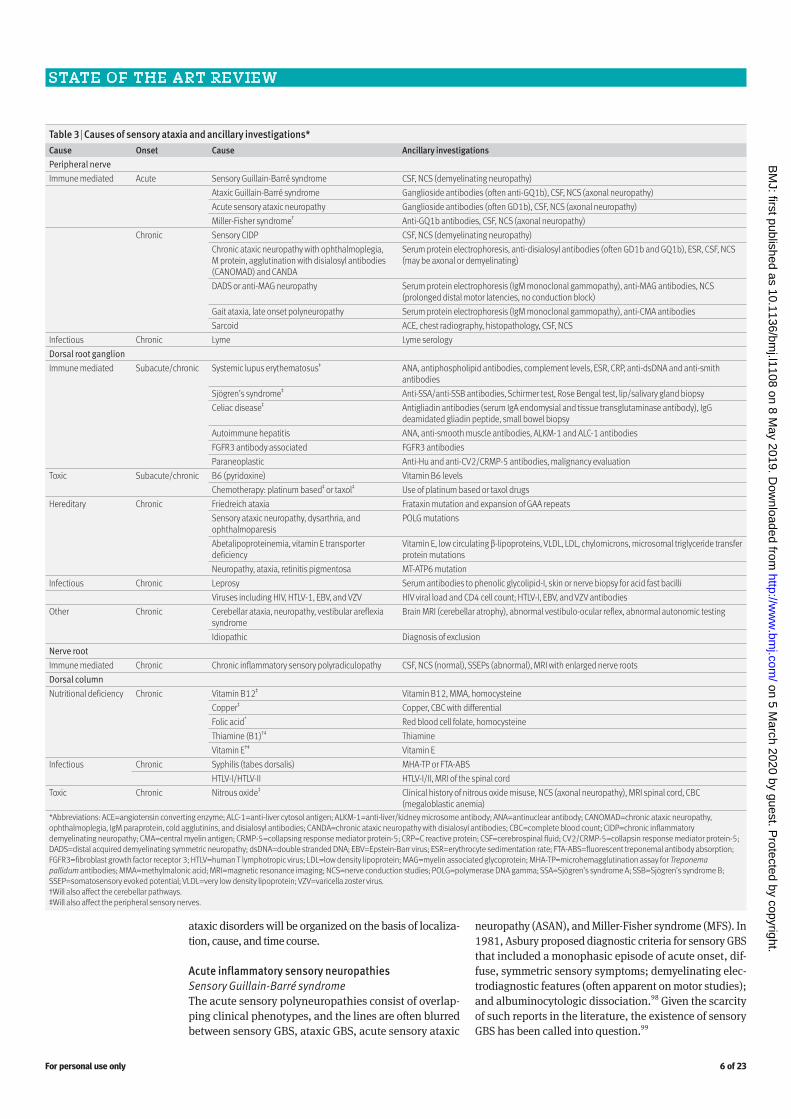
Table 3 | Causes of sensory ataxia and ancillary investigations*Cause Onset Cause Ancillary investigationsPeripheral nerveImmune mediated Acute Sensory Guillain-Barré syndrome CSF, NCS (demyelinating neuropathy)
Ataxic Guillain-Barré syndrome Ganglioside antibodies (often anti-GQ1b), CSF, NCS (axonal neuropathy)Acute sensory ataxic neuropathy Ganglioside antibodies (often GD1b), CSF, NCS (axonal neuropathy)Miller-Fisher syndrome† Anti-GQ1b antibodies, CSF, NCS (axonal neuropathy)
Chronic Sensory CIDP CSF, NCS (demyelinating neuropathy)Chronic ataxic neuropathy with ophthalmoplegia, M protein, agglutination with disialosyl antibodies (CANOMAD) and CANDA
Serum protein electrophoresis, anti-disialosyl antibodies (often GD1b and GQ1b), ESR, CSF, NCS (may be axonal or demyelinating)
DADS or anti-MAG neuropathy Serum protein electrophoresis (IgM monoclonal gammopathy), anti-MAG antibodies, NCS (prolonged distal motor latencies, no conduction block)
Gait ataxia, late onset polyneuropathy Serum protein electrophoresis (IgM monoclonal gammopathy), anti-CMA antibodiesSarcoid ACE, chest radiography, histopathology, CSF, NCS
Infectious Chronic Lyme Lyme serologyDorsal root ganglion Immune mediated Subacute/chronic Systemic lupus erythematosus‡ ANA, antiphospholipid antibodies, complement levels, ESR, CRP, anti-dsDNA and anti-smith
antibodiesSjögren’s syndrome‡ Anti-SSA/anti-SSB antibodies, Schirmer test, Rose Bengal test, lip/salivary gland biopsyCeliac disease‡ Antigliadin antibodies (serum IgA endomysial and tissue transglutaminase antibody), IgG
deamidated gliadin peptide, small bowel biopsyAutoimmune hepatitis ANA, anti-smooth muscle antibodies, ALKM-1 and ALC-1 antibodiesFGFR3 antibody associated FGFR3 antibodiesParaneoplastic Anti-Hu and anti-CV2/CRMP-5 antibodies, malignancy evaluation
Toxic Subacute/chronic B6 (pyridoxine) Vitamin B6 levelsChemotherapy: platinum based‡ or taxol‡ Use of platinum based or taxol drugs
Hereditary Chronic Friedreich ataxia Frataxin mutation and expansion of GAA repeatsSensory ataxic neuropathy, dysarthria, and ophthalmoparesis
POLG mutations
Abetalipoproteinemia, vitamin E transporter deficiency
Vitamin E, low circulating β-lipoproteins, VLDL, LDL, chylomicrons, microsomal triglyceride transfer protein mutations
Neuropathy, ataxia, retinitis pigmentosa MT-ATP6 mutationInfectious Chronic Leprosy Serum antibodies to phenolic glycolipid-I, skin or nerve biopsy for acid fast bacilli Viruses including HIV, HTLV-1, EBV, and VZV HIV viral load and CD4 cell count; HTLV-I, EBV, and VZV antibodiesOther Chronic Cerebellar ataxia, neuropathy, vestibular areflexia
syndromeBrain MRI (cerebellar atrophy), abnormal vestibulo-ocular reflex, abnormal autonomic testing
Idiopathic Diagnosis of exclusionNerve root Immune mediated Chronic Chronic inflammatory sensory polyradiculopathy CSF, NCS (normal), SSEPs (abnormal), MRI with enlarged nerve rootsDorsal column Nutritional deficiency Chronic Vitamin B12‡ Vitamin B12, MMA, homocysteine
Copper‡ Copper, CBC with differentialFolic acid* Red blood cell folate, homocysteineThiamine (B1)†‡ ThiamineVitamin E†‡ Vitamin E
Infectious Chronic Syphilis (tabes dorsalis) MHA-TP or FTA-ABS HTLV-I/HTLV-II HTLV-I/II, MRI of the spinal cord
Toxic Chronic Nitrous oxide‡ Clinical history of nitrous oxide misuse, NCS (axonal neuropathy), MRI spinal cord, CBC (megaloblastic anemia)
*Abbreviations: ACE=angiotensin converting enzyme; ALC-1=anti-liver cytosol antigen; ALKM-1=anti-liver/kidney microsome antibody; ANA=antinuclear antibody; CANOMAD=chronic ataxic neuropathy, ophthalmoplegia, IgM paraprotein, cold agglutinins, and disialosyl antibodies; CANDA=chronic ataxic neuropathy with disialosyl antibodies; CBC=complete blood count; CIDP=chronic inflammatory demyelinating neuropathy; CMA=central myelin antigen; CRMP-5=collapsing response mediator protein-5; CRP=C reactive protein; CSF=cerebrospinal fluid; CV2/CRMP-5=collapsin response mediator protein-5; DADS=distal acquired demyelinating symmetric neuropathy; dsDNA=double stranded DNA; EBV=Epstein-Barr virus; ESR=erythrocyte sedimentation rate; FTA-ABS=fluorescent treponemal antibody absorption; FGFR3=fibroblast growth factor receptor 3; HTLV=human T lymphotropic virus; LDL=low density lipoprotein; MAG=myelin associated glycoprotein; MHA-TP=microhemagglutination assay for Treponema pallidum antibodies; MMA=methylmalonic acid; MRI=magnetic resonance imaging; NCS=nerve conduction studies; POLG=polymerase DNA gamma; SSA=Sjögren’s syndrome A; SSB=Sjögren’s syndrome B; SSEP=somatosensory evoked potential; VLDL=very low density lipoprotein; VZV=varicella zoster virus.†Will also affect the cerebellar pathways.‡Will also affect the peripheral sensory nerves.
on 5 March 2020 by guest. P
rotected by copyright.http://w
ww
.bmj.com
/B
MJ: first published as 10.1136/bm
j.l1108 on 8 May 2019. D
ownloaded from
S TAT E O F T H E A R T R E V I E W
For personal use only 7 of 23
A case series in 2001 reported eight additional patients who met the clinical criteria for sensory GBS.100 Serum autoantibodies (MAG (myelin associated glycoprotein)), GM1, GQ1b, GD1b, anti-Hu, and sulphated glucuronyl par-agloboside (SGPG) were normal in the four patients tested. Sensory GBS, owing to its demyelinating features and the absence of ganglioside antibodies, remains separate from the following disorders which share many clinical, elec-trophysiologic, and laboratory features. These diseases, also classified as GBS variants, are best subdivided into complete MFS and incomplete MFS, which includes the acute ataxic neuropathies (ASAN and ataxic GBS).
Miller-Fisher syndromeMFS is characterized by a classic clinical triad of ophthal-moplegia, ataxia, and areflexia.101 102 Less common clinical features include other cranial neuropathies, blepharop-tosis, limb dysesthesia, and micturition problems. The ataxia of MFS is thought to be caused by both impaired proprioception (reversible conduction failure in 1a affer-ents) and cerebellar dysfunction.103 104 As in other forms of GBS, neurologic symptoms often follow an antecedent illness such as infection with Campylobacter jejuni or Hae-mophilus influenzae.105 The distinctive anti-GQ1b ganglio-side antibodies crossreact with surface epitopes of C jejuni, supporting the theory of molecular mimicry between nerve and bacteria.106 107 These antibodies also crossreact heav-ily with ganglioside GT1a.108 Electrodiagnostic studies, in contrast to sensory GBS, show a sensory predominant axonopathy.109 Recovery is gradual but often complete.
Acute ataxic neuropathiesThe remaining acute ataxic neuropathies, including both ASAN and ataxic GBS, have recently been classi-fied as incomplete forms of MFS by some experts.110 111 In the past, ASAN was not considered to be a GBS vari-ant because affected patients do not meet the diagnostic criteria for sensory GBS and lack demyelinating features on electrodiagnostic studies. Both ASAN and ataxic GBS, however, share many features with MFS includ-ing acute ataxia, areflexia, antecedent infection, and antiganglioside antibodies but lack the typical ophthal-moplegia.110 112 The presence of a Romberg sign helps differentiate ASAN from ataxic GBS. Patients with ASAN may harbor anti-disialosyl antibodies to GD1b alone or in combination with antibodies to CD3, GQ1b, or GT1a. Autoantibodies against gangliosides without disialosyl epitopes (GD1a and GM3) may also be present.112 Given that patients with ASAN typically have an antecedent infection, monophasic course, and excellent recovery, they should be considered under the rubric of GBS, in the subcategory of acute ataxic neuropathy.112 Ataxic GBS is distinguished by cerebellar-like ataxia and absence of a Romberg sign.113 Similar to MFS, these patients also harbor anti-GQ1b IgG antibodies.114 A retrospective chart review identified 54 patients with acute ataxic neuropathy without ophthalmoplegia. The Romberg sign was absent in 37 patients, who were considered to have ataxic GBS. In the other 17 patients, the Romberg sign was present, consistent with a diagnosis of ASAN. In the 37 patients with ataxic GBS, 24 were GQ1b posi-
tive compared with three of the 17 patients with ASAN (P=0.0034). IgG antibodies against GD1b but not GQ1b were more common in patients with ASAN (6/17) than in those with ataxic GBS (5/37), but this did not meet statis-tical significance (P=0.72).115 However, the opposite was true a minority of the time, suggesting that these diseases lie on a spectrum.
Chronic inflammatory sensory neuropathiesChronic ataxic neuropathy with disialosyl antibodies (CANDA)These very rare, acute, and chronic ataxic neuropathies with anti-disialosyl antibodies probably share a com-mon pathogenic mechanism, which is disruption at the node of Ranvier on sensory fibers. Like the acute ataxic neuropathies and MFS, the chronic ataxic neuropathies are also associated with anti-disialosyl antibodies (such as GD1b and GQ1b.) These disialosyl antibody mediated neuropathies can be separately categorized as nodo-paranodopathies.107 111 116 When the full spectrum of clinical features is present in these disialosyl antibody mediated chronic ataxic neuropathies, the disorder goes by the acronym CANOMAD (chronic ataxic neuropathy, ophthalmoplegia, IgM paraprotein, cold agglutinins, and disialosyl antibodies). CANDA (chronic ataxic neuropathy with disialosyl antibodies) is a more general term and allows for the inclusion of patients without ophthalmo-plegia and those in whom the cold agglutinins are IgM antibodies.111 CANDA can relapse, remit, and have cra-nial neuropathies that result in bulbar dysfunction.117 The disease process in CANDA may be the result of antibody mediated attack of the nerve root, DRG, and nerves.111 In electrophysiologic studies, patients with CANDA have absent or reduced sensory responses and diminished motor responses, including demyelinating features.118 119
Sensory chronic inflammatory demyelinating polyneuropathy (CIDP)Patients with sensory CIDP present with a pure sensory neuropathy with intact strength despite often having evi-dence of acquired demyelination on motor nerve conduc-tion studies.120-123 A minority of patients with sensory CIDP probably have electrophysiologic abnormalities in the sen-sory nerves only.124 Features that differentiate patients with sensory CIDP from those with chronic idiopathic axonal polyneuropathies include early gait ataxia, cranial neu-ropathy, diffuse hyporeflexia, onset before 55 years of age, and early involvement of the upper extremities.123
A small subset of patients with sensory CIDP have chronic immune sensory polyradiculopathy (CISP) in which the disease is localized to the nerve roots. These patients will have normal routine nerve conduction studies, abnormal somatosensory evoked potentials, raised concentrations of cerebral spinal fluid protein, and enlarged nerve roots on magnetic resonance imag-ing (MRI), which demonstrate inflammation on biopsy.125
Distal acquired demyelinating symmetric neuropathy (DADS)Distal acquired demyelinating symmetric neuropathy (DADS), a variant of CIDP, is characterized by distal,
on 5 March 2020 by guest. P
rotected by copyright.http://w
ww
.bmj.com
/B
MJ: first published as 10.1136/bm
j.l1108 on 8 May 2019. D
ownloaded from
S TAT E O F T H E A R T R E V I E W
For personal use only 8 of 23
symmetric, sensory, or sensorimotor polyneuropathy occurring in the presence of an IgM monoclonal gam-mopathy and myelin associated glycoprotein (MAG) antibodies.126 Patients who have an identical clinical and electrophysiologic phenotype but lack MAG anti-bodies can be classified as having DADS-CIDP36 127; such patients may carry a better prognosis and respond more favorably to intravenous immunoglobulins, corticoster-oids, and plasma exchange.127 The clinical hallmark of DADS neuropathy is the gradual onset of sensory ataxia resulting from impaired proprioception.128 Weakness is less prominent and, when present, affects the distal lower extremities.129 Action tremor can be a prominent feature.130 131 The electrophysiologic features include extremely prolonged distal motor and sensory latencies representing distal demyelination.132 133 Pathologically, there is segmental demyelination with IgM and comple-ment deposits in the myelin sheaths and widened outer myelin lamellae.134 More than half of patients with DADS have IgM paraproteins that recognize MAG or SGPG (which is present in most patients with anti-MAG anti-bodies). Three quarters of patients with non-anti-MAG DADS have anti-ganglioside antibodies (GD1b, GQ1b, GT1b, and others).128
Sensory neuronopathiesThe sensory neuronopathies, or dorsal root ganglionopa-thies, are a small subset of sensory polyneuropathies that result from damage to the trigeminal ganglion sensory neurons and DRG. These uncommon disorders can be broadly classified as inherited, autoimmune, or acquired. Because a comprehensive discussion of these disorders is beyond the scope of this article, emphasis will be placed on two of the more common, potentially treatable, auto-immune causes of sensory neuronopathy: Sjögren’s syn-drome and anti-Hu paraneoplastic syndrome. Table 3 shows additional causes of sensory neuronopathy.
Paraneoplastic disorders probably affect less than 1% of all patients with cancer making them extremely rare.135 Although other antibodies and other cancers have been reported with paraneoplastic sensory neuronopa-thy, anti-Hu antibodies and their high association with small cell lung cancer are the quintessential clinical sce-nario.136-144 In addition to sensory ataxia, patients may develop concomitant autonomic dysfunction, cerebel-lar and brainstem involvement, motor neuropathy, and
limbic encephalitis.145 146 The anti-Hu antibodies, which attack Hu-expressing tumor cells, are thought to trigger a CD8 cytotoxic T cell response.147-149
The sensory neuronopathy sometimes seen in Sjögren’s syndrome is also associated with autonomic dysfunction and at times brainstem dysfunction.78 150 151 The under-lying pathophysiology of Sjögren’s associated sensory neuronopathy is unknown, although T cell mediated infiltration in the DRG has been demonstrated.152
Posterolateral syndromeNot all sensory ataxic presentations localize to the peripheral nervous system, and disorders affecting the dorsal columns of the spinal cord must also be considered. In contrast to the disorders discussed above, which are mainly autoim-mune, the myelopathic disorders that present with sensory ataxia (in addition to spasticity and weakness) often have nutritional or infectious causes (see table 4). Tabes dorsalis, a presentation of parenchymatous neurosyphilis, may selec-tively affect the dorsal columns and spare the corticospinal tracts.153
Diagnostic approachIn addition to the clinical examination, the diagnostic evalu-ation of the sensory polyneuropathies may include a com-bination of electrodiagnostic studies, testing of autonomic function, laboratory testing, and histopathologic analysis of nerve tissue. Figures 1-3 provide algorithms to guide the diagnostic evaluation of sensory polyneuropathies.
Electrodiagnostic studiesNerve conduction studiesNerve conduction studies are a sensitive and specific method of assessing disease in the large myelinated nerve fibers and can provide useful diagnostic information regarding the underlying pathophysiology of the neuropathy (fig 4).42 154 Most neuromuscular experts advocate for the use of elec-trodiagnostic studies in distal symmetric polyneuropathy if the diagnosis is known or unknown.155 Several studies have shown that electrodiagnostic studies in this population can often change the diagnosis and management.156-158 Others, however, advocate for its use only in patients with atypical presentations.159 Regardless of this, many clinicians will forgo electrodiagnostic testing in patients who have straight-forward distal symmetric polyneuropathy if the underlying cause is known (such as diabetes).
Table 4 | Myelopathies that present with sensory ataxia*
Etiology Causes Associated features Useful tests TreatmentFolic acid deficiency
GI disease, folate antagonists, alcoholism Peripheral neuropathy, optic atrophy, cognitive problems
Serum folate, red blood cell folate, plasma total homocysteine
Folate 1 mg orally twice a day for several days then 1 mg/day
Vitamin E deficiency
Cholestasis, pancreatic insufficiency, hypobetalipoproteinemia, abetalipoproteinemia, chylomicron retention disease
Spinocerebellar syndrome, peripheral neuropathy, pigmented retinopathy, myopathy, movement disorders, gaze palsies
Serum vitamin E Vitamin E 200-1000 IU/day
Copper deficiency
Gastric surgery, malabsorption, zinc toxicity Peripheral neuropathy, megaloblastic anemia, pancytopenia
Serum and urinary copper, serum ceruloplasmin, zinc levels
Copper 8 mg/day orally for 1 week, then 6 mg/day orally for 1 week, then 2 mg/day
HIV Urinary urgency, erectile dysfunction HIV viral load, CD4 cell count Antiretroviral drugsSyphilis “tabes dorsalis”
Tertiary neurosyphilisLatent period 15-30 years Argyll Robertson pupil, erectile dysfunction, urinary incontinence, optic atrophy, cranial neuropathy
Rapid plasma regain, CSF with lymphocytic pleocytosis, raised protein, and VDRL
Parenteral penicillin G for 10-14 days
HTLV-1/2 HTLV- 1/2 in blood Possibly steroids*Abbreviations: CSF=cerebrospinal fluid; GI=gastrointestinal; HIV=human immunodeficiency virus; HTLV=human T lymphotropic virus; IU=international units; VDRL=venereal disease research laboratory.
on 5 March 2020 by guest. P
rotected by copyright.http://w
ww
.bmj.com
/B
MJ: first published as 10.1136/bm
j.l1108 on 8 May 2019. D
ownloaded from
S TAT E O F T H E A R T R E V I E W
For personal use only 9 of 23
Because electrodiagnostic studies will be normal in disorders that mainly affect small unmyelinated fibers, a normal nerve conduction study does not exclude the presence of small fiber dysfunction. In addition, many disorders with a SFN phenotype may subclinically have involvement of the large myelinated fibers and display abnormalities on electrodiagnostic testing; thus, the pres-ence of large fiber involvement does not exclude small fiber dysfunction.1
In sensory neuronopathies, sensory nerve action potentials (SNAPs) may be absent or display reduced amplitudes with relative preservation of conduction velocities. Abnormalities often do not follow a length dependent pattern and may be widespread. In contrast to most polyneuropathies, the upper extremities may be more prominently affected. Motor studies will classically be normal but subtle abnormalities are often encoun-tered.44 45 160
The diagnostic criteria for sensory neuronopathies (fig 5), which are based on a large retrospective analysis pub-lished in 2009, include at least one absent SNAP or three SNAPs less than 30% of the lower limit of normal in the upper extremities and less than two abnormal motor nerve responses in the lower extremities.46 These criteria were further validated after another large multicenter study was published in 2014.161 A recent case-control study suggests that greater than a 50% difference in amplitude in a side-to-side comparison of two or more pairs of sensory nerves
could be used as a rapid screening tool, with sensitivity and specificity greater than 90%.162 Small case series show that blink reflexes may be abnormal in sensory neuronopathies secondary to Sjögren’s syndrome, paraneoplastic disease, and idiopathic sensory neuronopathy, suggesting involve-ment of the trigeminal ganglion.163 164
Evoked potentialsSomatosensory evoked potentials—Somatosensory evoked potentials (SSEPs) evaluate the sensory pathways in both the peripheral and central nervous systems. They are particularly valuable when the proximal portions of the peripheral nerves, which are not studied with rou-tine nerve conduction studies, are affected.165 Bipolar transcutaneous electrical stimulation applied to the skin overlying a selected nerve (often median or tibial) evokes the SSEPs, which are then recorded with standard elec-troencephalograph scalp disk electrodes. They have an important diagnostic role in CISP, which preferentially affects the nerve roots and proximal nerves and spares the distal sensory nerves.166 Evidence of proximal demyelina-tion is also often apparent in sensory CIDP.123
Laser evoked potentials—Laser evoked potentials (LEPs), which assess the nociceptive pathways both peripherally (Aδ and C fibers) and at the spinothalamic tract centrally, have been called the “most widely agreed upon tool for investigating small fiber damage.”167 A car-bon dioxide laser stimulus is applied to the foot and calf.
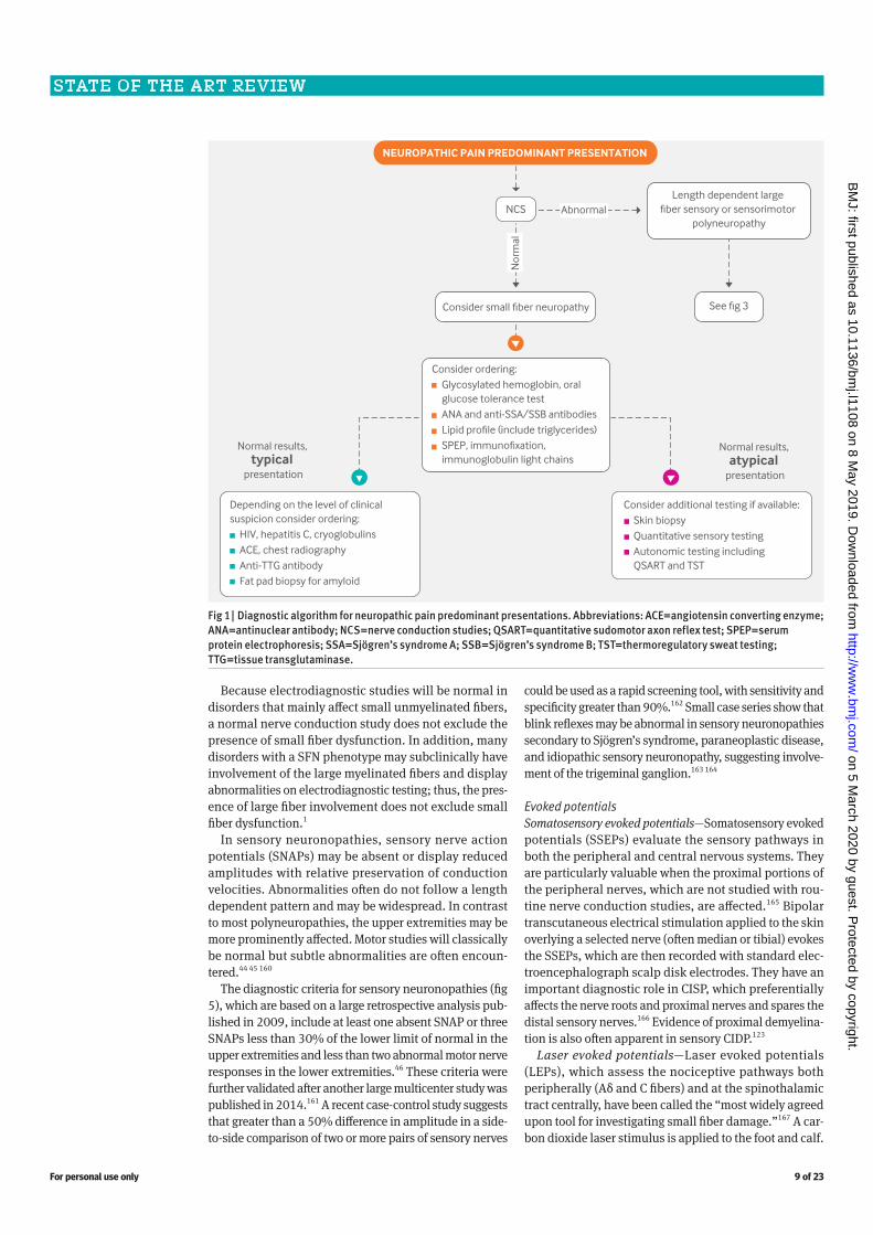
Fig 1 | Diagnostic algorithm for neuropathic pain predominant presentations. Abbreviations: ACE=angiotensin converting enzyme; ANA=antinuclear antibody; NCS=nerve conduction studies; QSART=quantitative sudomotor axon reflex test; SPEP=serum protein electrophoresis; SSA=Sjögren’s syndrome A; SSB=Sjögren’s syndrome B; TST=thermoregulatory sweat testing; TTG=tissue transglutaminase.
on 5 March 2020 by guest. P
rotected by copyright.http://w
ww
.bmj.com
/B
MJ: first published as 10.1136/bm
j.l1108 on 8 May 2019. D
ownloaded from
S TAT E O F T H E A R T R E V I E W
For personal use only 10 of 23
The latency and amplitude of LEPs are measured with scalp electrodes. The pain is perceived as first a prick-ling sensation (Aδ activation) followed by a dull, burning sensation (C fiber activation). Although LEPs have a high sensitivity (in the 70-80% range) for SFN,167 168 there are few laser testing facilities worldwide.168 Given their ease of use, LEPs have been proposed as an alternative to skin biopsy in diabetes associated SFN.167
Quantitative sensory testingQuantitative sensory testing (QST) can provide evidence of small nerve fiber damage on the basis of the meas-urement of abnormal sensory thresholds, and because abnormal QST results correlate with abnormalities of IENFD.169 170 QST has several limitations, such as its ina-
bility to discriminate between central nervous system and peripheral nervous system disease, the need for par-ticipant cooperation and attention, and the fact that it may be easily influenced by other factors. Therefore, it should not be used in isolation and needs to be inter-preted in the clinical context and in conjunction with other st udies.54 171-174
Corneal confocal microscopyCorneal confocal microscopy is an additional diagnostic tool that enables visualization of the peripheral nerves of the cornea and correlates with IENFD (fig 2). This non-invasive technique uses a combination of corneal nerve fiber length, nerve branch density, and nerve fiber den-sity to evaluate the corneal nerve plexus.175 176 It has been
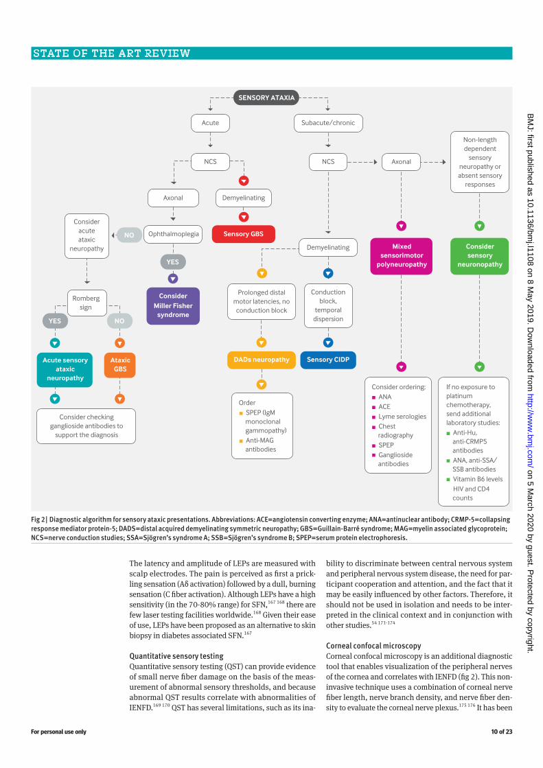
Fig 2 | Diagnostic algorithm for sensory ataxic presentations. Abbreviations: ACE=angiotensin converting enzyme; ANA=antinuclear antibody; CRMP-5=collapsing response mediator protein-5; DADS=distal acquired demyelinating symmetric neuropathy; GBS=Guillain-Barré syndrome; MAG=myelin associated glycoprotein; NCS=nerve conduction studies; SSA=Sjögren’s syndrome A; SSB=Sjögren’s syndrome B; SPEP=serum protein electrophoresis.
on 5 March 2020 by guest. P
rotected by copyright.http://w
ww
.bmj.com
/B
MJ: first published as 10.1136/bm
j.l1108 on 8 May 2019. D
ownloaded from
S TAT E O F T H E A R T R E V I E W
For personal use only 11 of 23
shown to detect early small nerve fiber damage in many disorders.175 177-182 This technique has advantages over skin biopsy as it is rapid and non-invasive, but it is not yet widely available. There is only a modest correlation with disease stage in any patient and the correlation is of limited utility in clinical practice.183-185 A recent study of nearly 1000 patients with type 1 and type 2 diabetes demonstrated the diagnostic validity of corneal confocal microscopy using a 12.5 mm/mm2 optimal threshold for automated corneal nerve fiber length in type 1 diabetes (73% sensitivity, 69% specificity) and a 12.3 mm/mm2 optimal threshold in type 2 diabetes (69% sensitivity, 63% specificity).176 When considering the entire cohort, a lower threshold for automated corneal nerve fiber length of 8.6 mm/mm2 could rule in diabetic polyneuropathy and an upper threshold of 15.3 mm/mm2 could rule it out (88% specificity, 88% sensitivity). How these st udies will be incorporated into clinical practice and their role as a clinical trial outcome measure remain to be d et ermined.176
Autonomic testingAutonomic testing can help in the diagnosis of SFN, espe-cially when dysautonomia is present.186 Sudomotor func-tion testing as a measure of autonomic function may be assessed through thermoregulatory sweat testing, quan-titative sudomotor axon reflex test (QSART), or newer techniques such as electrochemical skin conductance.187 Studies suggest that these autonomic testing modalities provide limited additional diagnostic information when a skin biopsy is abnormal.188
Quantitative sudomotor axon reflex testingQuantitative sudomotor axon reflex testing is a method of assessing postganglionic sudomotor function through the measurement of local sweat production in predeter-mined sites (forearm, distal and proximal leg, and foot) in response to iontophoresis of 10% acetylcholine. Abnor-mal QSART test results have been shown to correlate with decreased IENFD.189 However, a recent moderately sized prospective study found that the addition of QSART to the
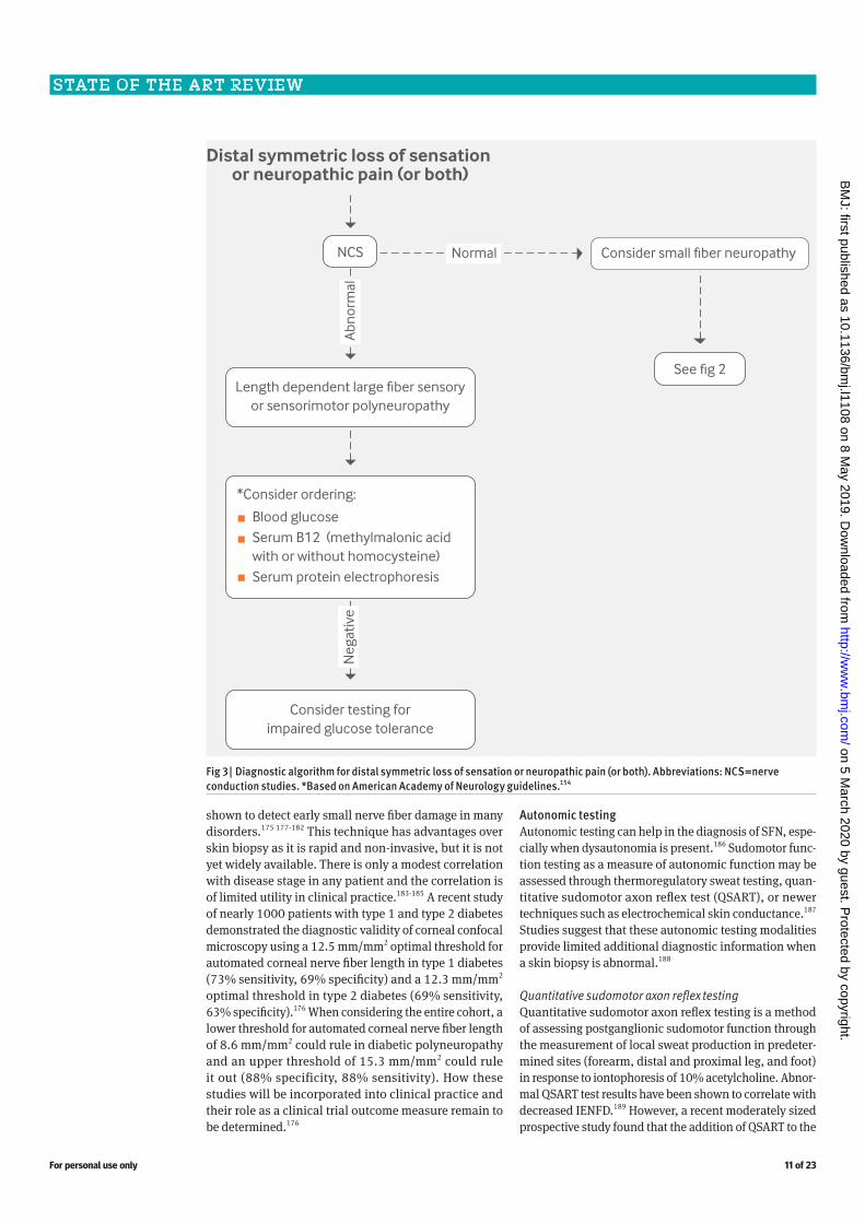
Fig 3 | Diagnostic algorithm for distal symmetric loss of sensation or neuropathic pain (or both). Abbreviations: NCS=nerve conduction studies. *Based on American Academy of Neurology guidelines.154
on 5 March 2020 by guest. P
rotected by copyright.http://w
ww
.bmj.com
/B
MJ: first published as 10.1136/bm
j.l1108 on 8 May 2019. D
ownloaded from
S TAT E O F T H E A R T R E V I E W
For personal use only 12 of 23
measurement of IENFD adds little diagnostic value for SFN.188 The limitations of QSART include the technical dif-ficulty of testing, the cost of equipment, and availability.187
Thermoregulatory sweat testingThermoregulatory sweat testing measures sweating pat-terns of the body with the use of an indicator dye in a humidity controlled, heated setting for typically 70 min-utes. This technique activates peripheral sudomotor func-tion through central autonomic pathways. Advantages of this test include the topographic analysis of sweat pattern abnormalities and the assessment of both pre-ganglionic and post-ganglionic sudomotor function (when other modalities will be normal in pre-ganglionic lesions). However, this test is technically demanding, requires time commitment on the part of the patient, and is not widely available.187 A recent retrospective study suggests that a novel technique of thermal imaging of forced evaporative cooling corresponds with the results from the standard technique using indicator powder and is more efficient.190
Electrochemical skin conductanceElectrochemical skin conductance has been reported in several small studies as a non-invasive, reliable marker of sweat function and SFN.191-193 Electrical stimulation with low direct voltage current is applied to sudomotor fibers of the palms and soles, which in turn activates sweat glands. However, a recent large systematic review determined that evidence on the use of this technique is limited and of overall poor quality; in addition, it is potentially confounded by technical factors, inconsistent normative values, and funding bias.194
Stimulated skin wrinklingStimulated skin wrinkling is the reversible undulation of surface skin that is mediated by post-ganglionic sym-pathetic fibers. It is tested by immersing glabrous skin (smooth skin without hair, as on the palms or soles of the feet) in water or exposing it to EMLA (eutectic mixture of local anesthetic).195 196 It has been shown to correlate with IENFD in patients with sensory polyneuropathy,195 197 and it has shown comparable sensitivity to other testing meth-ods for diabetic neuropathy.196
ImagingMagnetic resonance imagingMost patients who present with sensory neuropathy will not benefit from neuroimaging, but in select situations MRI may provide some additional diagnostic benefit. Small case series have demonstrated non-enhancing, longitudinally extensive dorsal column lesions in patients with sensory neuronopathies, indicative of the degenera-tion of central afferent connections between the DRG and dorsal columns.198 A small case series of patients with CISP suggested that MRI abnormalities such as nerve root enlargement or enhancement may be useful diagnostically in patients with normal nerve conduction study results.125 In patients with posterolateral cord syndrome and sensory dysfunction as a result of dorsal column dysfunction, MRI will often show increased T2 and FLAIR (fluid attenuated inversion recovery) signals at the dorsal columns.
Neuromuscular ultrasoundNeuromuscular ultrasound is an emerging tool that is particularly valuable in immune mediated mixed sen-sory and motor demyelinating polyneuropathies and in entrapment neuropathies, in which focal nerve enlarge-ment can be detected. In a population of patients with SFN, the sural nerve was found to have a greater cross sec-tional area compared with healthy controls.199 Currently, most experts do not recommend using neuromuscular ultrasound in patients with pure sensory polyneuropathy, although this field remains ripe for future study.200
Tissue biopsySkin biopsyThe European Federation of Neurological Societies/Periph-eral Nerve Society Guideline and numerous studies support the use of skin biopsy to assess IEFND and as the gold stand-ard for pathologic diagnosis of SFN (fig 6).201 It is a repro-ducible and reliable technique with a specificity greater than 90%, sensitivity approaching 80%, and favorable positive and negative predictive values.1 2 202-204 Multiple large cohort studies have been conducted to establish normative values for IENFD at the distal leg because age, ethnicity, and sex are known to produce variations.202 203 205 A recent longitudi-nal case-control study showed that rates of IENFD decrease are similar at proximal and distal biopsy sites, regardless of cause, supporting a non-length dependent process.41 Diag-nostic criteria for SFN have been proposed to enable patients to be included in clinical trials. Box 1 provides a comparison of the 2008 Devigili criteria and the 2017 Blackmore and Siddiqui criteria (which do not require a skin biopsy).206 207 In straightforward cases of SFN, supported by a typical history and examination findings, a skin biopsy is often unneces-sary, and further research is needed to elucidate the precise role of skin biopsy in clinical practice.
Nerve biopsyIn general, nerve biopsy is not needed to diagnose patients as having a sensory polyneuropathy, although many of the disorders discussed in this review will have charac-teristic histopathologic features. In sensory CIDP, nerve biopsy may detect demyelinating features, including hypomyelinated fibers on light microscopy and onion bulb
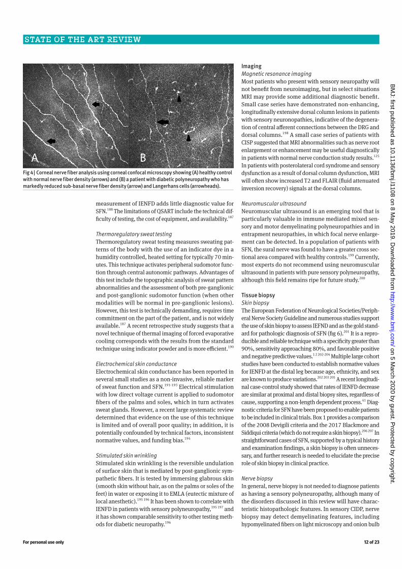
Fig 4 | Corneal nerve fiber analysis using corneal confocal microscopy showing (A) healthy control with normal nerve fiber density (arrows) and (B) a patient with diabetic polyneuropathy who has markedly reduced sub-basal nerve fiber density (arrow) and Langerhans cells (arrowheads).
on 5 March 2020 by guest. P
rotected by copyright.http://w
ww
.bmj.com
/B
MJ: first published as 10.1136/bm
j.l1108 on 8 May 2019. D
ownloaded from
S TAT E O F T H E A R T R E V I E W
For personal use only 13 of 23
formation, as well as mononuclear cell infiltrates in the interstitial tissue.123 Patients with anti-MAG neuropathies show evidence of demyelination and monoclonal IgM and C3d deposits on myelin sheaths.208 Ultrastructural stud-ies show widening of the myelin lamella due to M-protein and activated complement proteins, which colocalize with MAG in these areas.208-212 Although a diagnosis of sen-sory neuronopathy is considered “definite” only if there is pathologic evidence of DRG degeneration, DRG biopsy is discouraged because of the associated morbidity.45 161
Current disease specific treatmentsApart from SFN associated with diabetes and prediabe-tes, the sensory polyneuropathies discussed are relatively rare, and no universally accepted disease specific treat-ments exist. Many of the disease specific treatments dis-
cussed below are based on expert opinion, retrospective studies, and small prospective studies, rather than large randomized placebo controlled trials. Some treatments discussed are emerging and in various stages of study.
Small fiber neuropathiesSarcoidosisEvidence to support the optimal treatment regimen for SFN associated with sarcoidosis is limited. In a retrospec-tive review of 115 patients, the SFN treatment response rates were 76%, 67%, and 71% for treatment with intra-venous immunoglobulins, anti-TNF-α, and combination therapy with both, respectively.75 By contrast, in the same trial patients treated with methotrexate or corticosteroids showed no improvement or even worsening of symptoms.
Transthyretin familial amyloidosis polyneuropathyThe US Food and Drug Administration and the European Commission have recently approved patisiran and inot-ersen as treatments for TTR-FAP. Several other drugs, such as diflunisal and tafadimis, have shown promising results in large randomized placebo controlled clinical trials. Liver transplantation has traditionally been the standard treatment despite continued deposition of wild-type transthyretin.213 Patisiran is an RNA interfer-ence therapeutic agent that inhibits hepatic synthesis of transthyretin.214 In a double blind placebo controlled phase III trial, 225 patients were randomized to either intravenous patisiran (0.3 mg/kg/body weight) or pla-cebo every three weeks. Patients receiving patisiran had a significant improvement in the Modified Neuropathy Impairment Score +7 (mNIS+7) (P<0.001), on the Nor-folk Quality of Life-Diabetic Neuropathy (Norfolk QOL-DN) questionnaire (P<0.001), and gait speed (P<0.001). In addition, a large phase III randomized double blind placebo controlled trial of inotersen, an antisense oligo-nucleotide that inhibits the hepatic production of tran-sthyretin, has recently been published.215 One hundred and seventy two patients(112 in the inotersen group and 60 in the placebo group) were given weekly subcutane-ous injections for 66 weeks. As in the patisiran trial, the treatment arm also significantly improved on the mNIS+7 and the Norfolk QOL-DN scores (both P<0.001). However, inotersen was associated with thrombocytopenia and glo-merulonephritis in some patients.
The transthyretin tetramer stabilizers include diflunisal and tafadimis. Diflunisal, a non-steroidal anti-inflamma-tory drug, strongly inhibits TTR amyloid fibril formation. A large international double blind placebo controlled trial of 130 patients found that diflunisal slowed the progres-sion of patients with and without the TTR-Val30Met and non-Val30Met mutations.216 This orphan drug is widely available and inexpensive. Another randomized dou-ble blind placebo controlled trial studied tafamidis in patients with early stage TTR.217 Although the coprimary endpoints of slowed progression on the Neuropathy Impairment Score-Lower Limbs (NIS-LL) (as determined by NIS-LL response, “responders” had an increase in NIS-LL at 18 months of <2 points) and Norfolk QOL-DN scores were not reached, a statistically significant 52% r eduction in the worsening of neurologic function (as
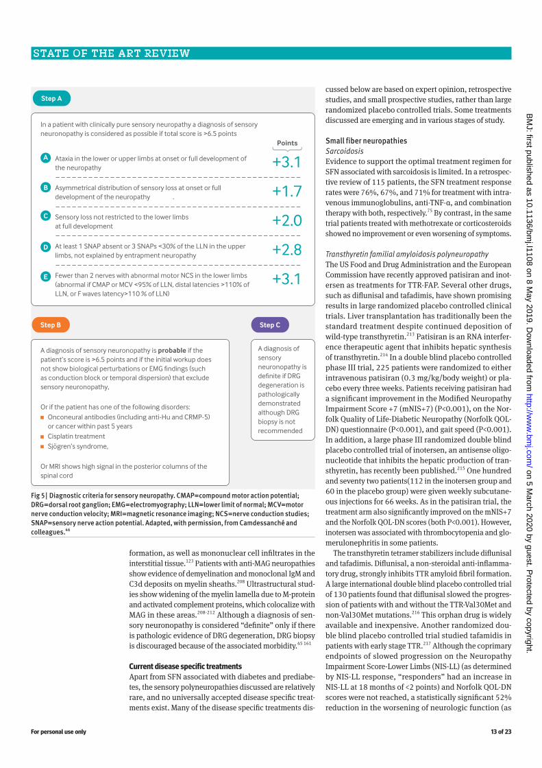
Fig 5 | Diagnostic criteria for sensory neuropathy. CMAP=compound motor action potential; DRG=dorsal root ganglion; EMG=electromyography; LLN=lower limit of normal; MCV=motor nerve conduction velocity; MRI=magnetic resonance imaging; NCS=nerve conduction studies; SNAP=sensory nerve action potential. Adapted, with permission, from Camdessanché and colleagues.46
on 5 March 2020 by guest. P
rotected by copyright.http://w
ww
.bmj.com
/B
MJ: first published as 10.1136/bm
j.l1108 on 8 May 2019. D
ownloaded from
S TAT E O F T H E A R T R E V I E W
For personal use only 14 of 23
determined by change in NIS-LL from baseline to 18 months) was seen in this intention to treat population (P=0.027). This drug is approved in Europe, South Amer-ica, and Japan, but not in the US.89
Sensory ataxic neuropathiesMiller-Fisher syndromeA retrospective study and expert opinion indicate that intravenous immunoglobulin probably reduces the time to recovery and prevents the progression of symp-toms.218 219 However, the use of such an expensive treatment in a condition with a favorable prognosis is controversial.33 218 An evidence based guideline report of the therapeutics and technology assessment subcommit-tee of the American Academy of Neurology stated that there was insufficient evidence to support or refute the use of intravenous immunoglobulin in this condition,220
although patients with considerable overlap with GBS should be offered treatment.
Chronic ataxic neuropathy with disialosyl antibodies (CANDA)Data to guide treatment in these patients are limited.221-224 In case series, intravenous immunoglobulins have been used with some success,119 whereas rituximab was the most effective treatment in one small cohort of patients, halting disease in eight of nine patients.117
Sensory chronic inflammatory demyelinating polyneuropathy (CIDP)It is extremely important to recognize this disease because 90% of patients responded to immunotherapy in one series.123 No prospective randomized placebo controlled trials have studied immunosuppressant or immunomodu-latory therapy in the sensory variant of CIDP specifically. In one retrospective series of 15 patients with CISP, all patients responded to intravenous immunoglobulins or intravenous methylprednisolone.125
Distal acquired demyelinating sensory neuropathy (DADS)Many treatments have been tried and abandoned in MAG neuropathies including corticosteroids, intravenous immunoglobulins, and plasma exchange.212 Although cytotoxic agents such as fludarabine, cyclophosphamide, and chlorambucil may be beneficial, their toxicities limit longterm use.212 Rituximab, a monoclonal antibody that targets CD20 (a B cell surface antigen) and depletes cir-culating B cells, has been used with success in 30-50% of patients in uncontrolled trials.128 225 The primary end-points in two placebo controlled randomized trials of rituximab failed to reach statistical significance, although secondary endpoints such as time-to-walk scales sig-nificantly improved.225-227 Patients with motor deficits and subacute progression may respond more favorably
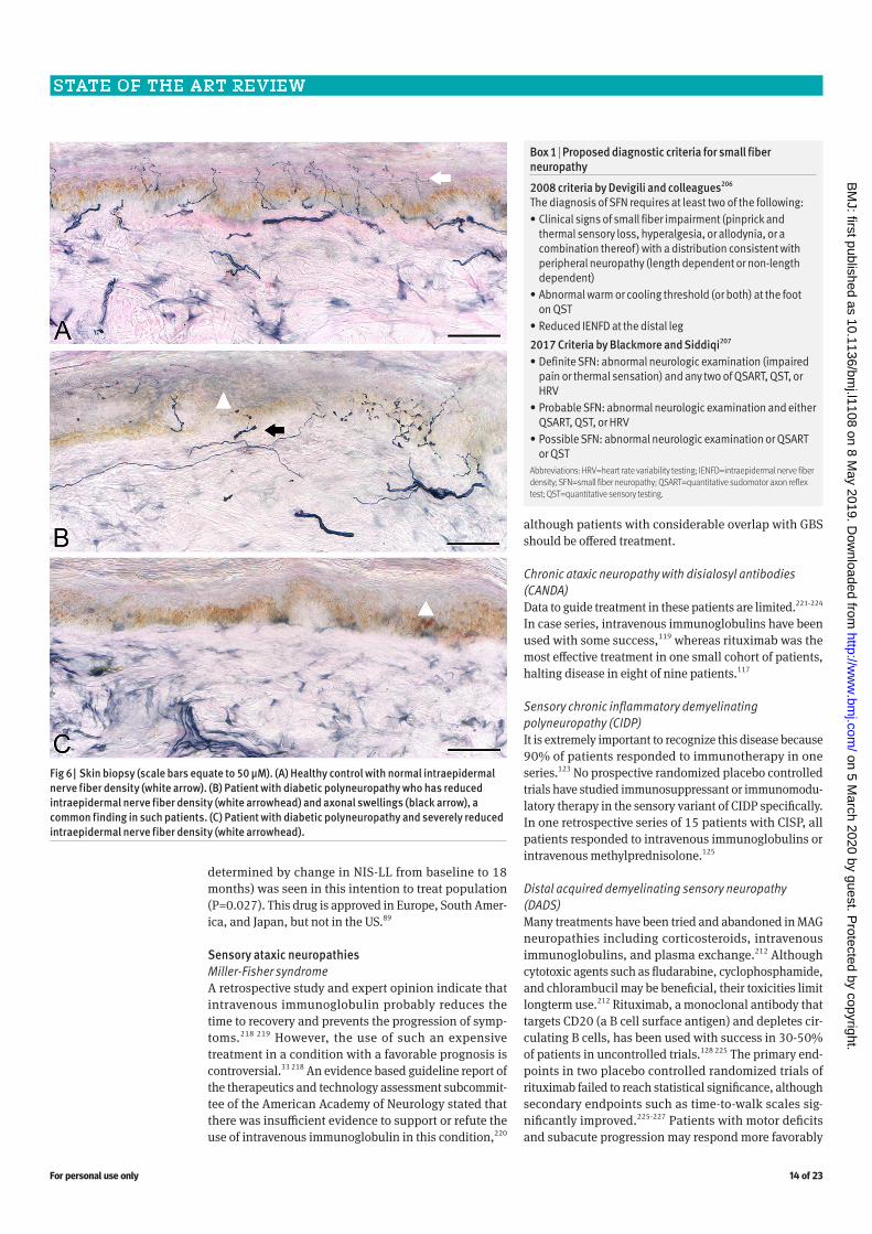
Fig 6 | Skin biopsy (scale bars equate to 50 μM). (A) Healthy control with normal intraepidermal nerve fiber density (white arrow). (B) Patient with diabetic polyneuropathy who has reduced intraepidermal nerve fiber density (white arrowhead) and axonal swellings (black arrow), a common finding in such patients. (C) Patient with diabetic polyneuropathy and severely reduced intraepidermal nerve fiber density (white arrowhead).
Box 1 | Proposed diagnostic criteria for small fiber neuropathy2008 criteria by Devigili and colleagues206
The diagnosis of SFN requires at least two of the following:• Clinical signs of small fiber impairment (pinprick and
thermal sensory loss, hyperalgesia, or allodynia, or a combination thereof) with a distribution consistent with peripheral neuropathy (length dependent or non-length dependent)
• Abnormal warm or cooling threshold (or both) at the foot on QST
• Reduced IENFD at the distal leg2017 Criteria by Blackmore and Siddiqi207
• Definite SFN: abnormal neurologic examination (impaired pain or thermal sensation) and any two of QSART, QST, or HRV
• Probable SFN: abnormal neurologic examination and either QSART, QST, or HRV
• Possible SFN: abnormal neurologic examination or QSART or QST
Abbreviations: HRV=heart rate variability testing; IENFD=intraepidermal nerve fiber density; SFN=small fiber neuropathy; QSART=quantitative sudomotor axon reflex test; QST=quantitative sensory testing.
on 5 March 2020 by guest. P
rotected by copyright.http://w
ww
.bmj.com
/B
MJ: first published as 10.1136/bm
j.l1108 on 8 May 2019. D
ownloaded from
S TAT E O F T H E A R T R E V I E W
For personal use only 15 of 23
to rituximab if the drug is started soon after the onset of symptoms.228 229 In 2010, the European Federation of Neurological Societies and Peripheral Nerve Society published a guideline on the management of parapro-teinemic demyelinating neuropathies. This guideline highlighted the lack of proven efficacy for any therapy in anti-MAG neuropathy but emphasized that some patients may respond to treatment.230 Two patients have been treated with obinutuzumab, a first generation glycoen-gineered type-I, anti-CD20 mediated, B cell depleting monoclonal antibody.231 No improvement or worsening in the patients’ neuropathic symptoms was seen after 12 months of treatment.
Sensory neuronopathiesGiven the rarity of these diseases, little is known about the best approach to treatment, although a treatment window probably exists. A case series of serial nerve con-duction studies in patients with sensory neuronopathy suggests that sensory abnormalities plateau after 7-10 months from symptom onset. On the basis of the rate of decline of sensory response amplitudes, treatment should be started within the first eight months if possi-ble.232 Beyond this window, the inflammatory reaction probably dampens and treatment becomes unsuccessful. For the patients with paraneoplastic disease, detection and treatment of the underlying cancer is obligatory. For both paraneoplastic and Sjögren’s associated sensory neuronopathies, immunosuppressant and immunomodu-latory treatment should be provided. Intravenous immu-noglobulin, plasma exchange, corticosteroids, rituximab, cyclophosphamide, infliximab, and azathioprine have all been used in uncontrolled studies of Sjögren’s associ-ated sensory neuronopathy.233-238 Corticosteroids,239 240 intravenous immunoglobulins,241 242 plasma exchange,243 rituximab,244 and sirolimus245 have been used in patients with paraneoplastic sensory neuronopathy.
Emerging disease specific treatmentsSmall fiber neuropathiesDiabetes and prediabetesIn early clinical trials, physical exercise has shown prom-ise as a treatment of SFN associated with glucose dysregu-lation. In prospective randomized trials, exercise results in increased IENFD in patients with diabetes but no neu-ropathy.246 247 A small prospective pilot study in diabetic neuropathy also found that pain responded to exercise.248 A large prospective randomized study of patients with type 2 diabetes associated peripheral neuropathy (the Activity for Diabetic Polyneuropathy or “ADAPT” study), which is investigating the effect of supervised exercise versus standard care counseling on polyneuropathy, as measured by IENFD and change in quality of life, is cur-rently underway (Clinical trials identifier NCT02341261).
SarcoiditisARA 290 (Cibinetide), an erythropoietin derivative that activates the innate repair receptor and initiates anti-inflammation, cytoprotection, and healing, has been well tolerated and showed benefit in treating sarcoidosis asso-ciated neuropathic pain in two phase II clinical trials.18 249
Sjögren’s syndromeLike all polyneuropathies that are associated with Sjögren’s syndrome, studies of the disease specific treat-ment of Sjogren’s associated SFN are sparse. A small uncontrolled trial of intravenous immunoglobulins in Sjögren’s syndrome found a reduction in neuropathic pain in SFN,250 but in other series the response to corti-costeroids has been poor.150 251 252 A small prospective, phase III clinical trial testing the benefit of intravenous immunoglobulins in patients with painful large fiber sensory polyneuropathy will soon be enrolling and could potentially inform treatment choices in patients with SFN that is associated with Sjögren’s syndrome (Clinical trials identifier NCT03700138).
Sodium channelopathiesThe discovery of the SCN9A, SCN10A, and SCN11A genetic mutations has opened the door to potential therapeutic options for painful SFN.88 253 254 Lacosamide, a blocker of Nav1.3, Nav1.7, and Nav1.8 that stabilizes channels in the slow inactivation state, has been studied in SCN9A associated SFN, although the results have not yet been published255 (Clinical trials identifier NCT01911975).
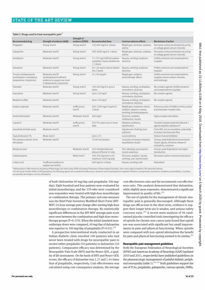
Current management of neuropathic painNeuropathic pain and positive sensory disturbances contribute greatly to the morbidity associated with sen-sory polyneuropathy. Most studies have focused on the treatment of painful neuropathy secondary to diabetes or chemotherapy induced painful neuropathy. A large meta-analysis published in 2015 updated recommendations on the pharmacologic treatment of neuropathic pain.256 This review found moderate to high quality of evidence for the use of serotonin-norepinephrine reuptake inhibitors (SNRIs), pregabalin and gabapentin, tricyclic antidepres-sants (TCAs), opioids, botulinum toxin, and capsaicin. SNRIs, TCAs, gabapentin, and pregabalin were given a strong recommendation and proposed as first line agents, whereas topical capsaicin or lidocaine and tramadol were given a weaker recommendation and proposed as second line. Strong opioids and botulinum toxin A were recom-mended as third line.
A recent large retrospective systematic review of 106 randomized controlled trials examined the effect of vari-ous drugs for diabetic neuropathy on pain and quality of life. Anticonvulsants including pregabalin and oxcarbaz-epine; SNRIs including duloxetine and venlafaxine; TCAs; atypical opioids including tramadol and tapentadol; and botulinum toxin A were determined to be more effective than placebo. The strength of evidence was considered moderate for SNRIs and low for the other listed agents. The review concluded that other commonly used agents including gabapentin, topical capsaicin, typical opioids, dextromethorphan, and mexiletine were no more effective than placebo.257
A large multicenter double blind parallel group study of diabetic neuropathic pain studied whether patients who did not respond to standard dose monotherapy with duloxetine (60 mg/day) or pregabalin (300 mg/day) would respond to high dose duloxetine (120 mg/day), high dose pregabalin (600 mg/day), or a combination
on 5 March 2020 by guest. P
rotected by copyright.http://w
ww
.bmj.com
/B
MJ: first published as 10.1136/bm
j.l1108 on 8 May 2019. D
ownloaded from
S TAT E O F T H E A R T R E V I E W
For personal use only 16 of 23
of both (duloxetine 60 mg/day and pregabalin 300 mg/day). Eight hundred and four patients were evaluated for initial monotherapy, and the 339 who were considered non-responders were treated with high dose monotherapy or combination therapy. The primary outcome measure was the Brief Pain Inventory Modified Short Form (BPI-MSF) 24 hour average pain change after starting high dose monotherapy or combination therapy. No statistically significant differences in the BPI-MSF average pain score were seen between the combination and high dose mono-therapy groups (P=0.370). When the initial standard mon-ontherapy doses were compared, 60 mg/day of duloxetine was superior to 300 mg/day of pregabalin (P<0.01).258
A prospective interventional study conducted in an Indian diabetic clinic enrolled 100 patients who had never been treated with drugs for neuropathic pain to receive either pregabalin (50 patients) or duloxetine (50 patients). Comparative efficacy was determined by the Neuropathic Pain Scale (NPS) and the Neuro-QOL, a qual-ity of life instrument. On the basis of NPS and Neuro-QOL scores, the efficacy of duloxetine was 1.27 and 1.44 times that of pregabalin, respectively. Cost effectiveness was calculated using cost consequence analysis, the average
cost effectiveness ratio and the incremental cost effective-ness ratio. This analysis demonstrated that duloxetine, while slightly more expensive, demonstrated a significant improvement in quality of life.259
The use of opioids for the management of chronic neu-ropathic pain is generally discouraged. Although these drugs are efficacious in the short term, evidence to sup-port their longer term use is weaker, and serious safety concerns exist.260 A recent meta-analysis of 96 rand-omized placebo controlled trials investigating the efficacy of opioids for chronic non-cancer pain found that opioid use was associated with significant but small improve-ments in pain and physical functioning. When opioids were compared with non-opioid alternatives the benefit for pain and physical functioning seemed to be similar.261
Neuropathic pain management guidelinesBoth the European Federation of Neurological Societies (EFNS) and American Academy of Neurology (AAN) (updated 2010 and 2011, respectively) have published guidelines on the pharmacologic management of painful diabetic periph-eral neuropathy (table 5).262 263 Both guidelines support the use of TCAs, pregabalin, gabapentin, various opioids, SNRIs,
Table 5 | Drugs used to treat neuropathic pain*†
Recommended drug Strength of evidence (AAN)Strength of evidence (EFNS) Recommended dose Common adverse effects Mechanism of action
Pregabalin Strong: level A Strong: level A 150-600 mg/d in 3 doses Weight gain, dizziness, sedation, edema
Decreases central sensitization by acting on voltage gated calcium channels
Gabapentin Moderate: level B Strong: level A 300-3600 mg/d in 3 doses Weight gain, dizziness, sedation, edema
Decreases central sensitization by acting on voltage gated calcium channels
Venlafaxine Moderate: level B Strong: level A 75-225 mg/d (XR formulation available); may be divided into 2-3 doses
Nausea, vomiting, headache, dizziness
Inhibits serotonin and norepinephrine reuptake
Duloxetine Moderate: level B Strong: level A 60-120 mg/d; may be divided into 2 doses
Nausea, vomiting, headache, dizziness
Inhibits serotonin and norepinephrine reuptake
Tricyclic antidepressants (amitriptyline, nortriptyline, desipramine, imipramine)
Moderate: level B (amitriptyline);Insufficient evidence to support use: level U (desipramine, imipramine
Strong: level A 25-150 mg qhs Weight gain, sedation, anticholinergic effects
Inhibits serotonin and norepinephrine reuptake, blocks sodium channels, anticholinergic
Tramadol Moderate: level B Strong: level A 100-400 mg/d in up to 4 doses
Nausea, vomiting, constipation, somnolence, dizziness
Mu-receptor agonist, Inhibits serotonin and norepinephrine reuptake
Oxycodone Moderate: level B Strong: level A Up to 120 mg/d Nausea, vomiting, constipation, somnolence, dizziness
Mu-receptor agonist
Morphine sulfate Moderate: level B Up to 120 mg/d Nausea, vomiting, constipation, somnolence, dizziness
Mu-receptor agonist
Sodium valproate Moderate: level B Inefficacious: level B
500-1200 mg/d; may be in 3 doses
Weight gain, headache, tremor, sedation, alopecia, nausea, vomiting, thrombocytopenia
Enhances action of GABA or mimics action at postsynaptic receptor sites
Dextromethorphan Moderate: level B Moderate: level B 400 mg/d Dizziness, sedation, restlessness, nausea
Sigma receptor stimulation
Capsaicin Moderate: level B Inefficacious: level B
0.075% cream up to 4 times daily
Allodynia, erythema, hypertension
Transient receptor potential villanoid-1 agonist depletes substance P
Isosorbide dinitrate spray Moderate: level B Hypotension, flushing, local erythema
Forms NO, acts as vasodilator, potentially increases microvascular flow
Topical lidocaine 5% Weak: level C Up to 12 h Local erythema Sodium channel inhibitionPercutaneous electric nerve stimulation
Moderate: level B 15-60 min/session Local pain, bruising, temporary exacerbation of pain
Unclear; potentially blocks transmission of pain signals, enhances release of endorphins, serotonin
Botulinum toxin Moderate: level B 50 U intradermally over dorsum of foot at 12 sites
Pain, bleeding, local reaction, muscle weakness
Unclear; potentially blocks nociceptor transduction
Carbamazepine Weak: level C 100-200 mg every 4-6 h; maximum 1200 mg daily
Dizziness, sedation, nausea, vomiting, rash, blurred vision
Decreases sodium channel conductance
α-lipoic acid Insufficient evidence to support use: level U
600 mg/d in 2 doses Nausea, vomiting, rash Antioxidant
*Abbreviations: AAN=American Academy of Neurology; bid=twice daily; EFNS=European Federation of Neurological Societies; qhs=every night at bedtime; XR=extended release.†On the basis of either AAN or EFNS guidelines, the following agents are considered inefficacious: serotonin and norepinephrine reuptake inhibitors, zonisaminde, menantine, mexilitine, pentoxifylline, clonidine, lacosamide, lamotrigine, and oxcarbazepine. on 5 M
arch 2020 by guest. Protected by copyright.
http://ww
w.bm
j.com/
BM
J: first published as 10.1136/bmj.l1108 on 8 M
ay 2019. Dow
nloaded from
S TAT E O F T H E A R T R E V I E W
For personal use only 17 of 23
and topical lidocaine for the treatment of neuropathic pain. The AAN guidelines also recommend the use of topical capsaicin and valproate with level B evidence and only pre-gabalin was supported by level A evidence. Each of these guidelines recommend against the use of oxcarbamazepine, lamotrigine, lacosamide, clonidine, and mexiletine in the symptomatic management of painful neuropathy.
Emerging treatments for neuropathic painChemodenervation with botulinum toxin A is thought to inhibit the release of peripheral neurotransmitters such as acetylcholine and nociceptive peptides (substance P, glutamate, calcitonin gene related peptide) from sensory nerves.264 In addition, botulinum toxin inhibits vanilloid receptor TRPV1 expression on the surface of peripheral nociceptors. It has been supported by multiple studies as described in a recent review for the treatment of neu-ropathic pain.265
A 2015 meta-analysis evaluating the use of several treatments for neuropathic pain gave botulinum toxin A a weak recommendation.256 In the same year, a meta-anal-ysis looking at chemodenervation in diabetic peripheral polyneuropathy supported its use as a result of finding clinically significant improvements in pain scores.264 Most recently, in 2017, a large systematic review found that botulinum toxin was more effective than placebo, although the strength of evidence was low.257
There is some evidence from small studies to support the use of acupuncture as a non-pharmacologic treatment for neuropathic pain. A randomized placebo controlled partially blinded trial in Germany is currently looking at the effect of needle acupuncture, laser acupuncture, and placebo laser acupuncture on electrophysiologic param-eters, neurologic deficits, and symptoms.266
ConclusionsThe sensory polyneuropathies are heterogeneous conditions with distinct clinical phenotypes defined by the type of nerve fibers involved and the time course. Although many of the small fiber, pain predominant and large fiber, ataxia pre-dominant neuropathies discussed are relatively uncommon, those reviewed have the shared feature of being potentially
treatable and even reversible. The recognition of these dis-tinctive presentations is of utmost importance to enable treat-ment to be started before permanent nerve damage occurs.Thanks to A Gordon Smith, Peter Hauer, and Stormy Foster-Palmer for contributing the figures.Contributors: KGG and KP performed the literature review and prepared the initial draft of the manuscript. Both authors were involved in the conception, drafting, and editing of the manuscript. KGG is guarantor.Competing interests: The authors have read and understood the BMJ policy on declaration of interests and have no competing interests.Provenance and peer review: Commissioned; externally peer reviewed.1 Terkelsen AJ, Karlsson P, Lauria G, Freeman R, Finnerup NB, Jensen TS. The
diagnostic challenge of small fibre neuropathy: clinical presentations, evaluations, and causes. Lancet Neurol 2017;16:934-44. 10.1016/S1474-4422(17)30329-0. pmid:29029847.
2 Chan ACY, Wilder-Smith EP. Small fiber neuropathy: Getting bigger!Muscle Nerve 2016;53:671-82. 10.1002/mus.25082. pmid:26872938.
3 Grant G. The 1932 and 1944 Nobel Prizes in physiology or medicine: rewards for ground-breaking studies in neurophysiology. J Hist Neurosci 2006;15:341-57. 10.1080/09647040600638981. pmid:16997762.
4 Centers for Disease Control and Prevention USD of H and HS. Centers for Disease Control and Prevention. National diabetes statistics report. 2017. https://www.cdc.gov/diabetes/pdfs/data/statistics/national-diabetes-statistics-report.pdf
5 Zilliox L, Russell JW. Treatment of diabetic sensory polyneuropathy. Curr Treat Options Neurol 2011;13:143-59. 10.1007/s11940-011-0113-1 pmid:21274758.
6 Vinik AI, Nevoret M-L, Casellini C, Parson H. Diabetic neuropathy. Endocrinol Metab Clin North Am 2013;42:747-87. 10.1016/j.ecl.2013.06.001 pmid:24286949.
7 Herman WH, Kennedy L. Underdiagnosis of peripheral neuropathy in type 2 diabetes. Diabetes Care 2005;28:1480-1. 10.2337/diacare.28.6.1480 pmid:15920071.
8 Young MJ, Boulton AJ, MacLeod AF, Williams DR, Sonksen PH. A multicentre study of the prevalence of diabetic peripheral neuropathy in the United Kingdom hospital clinic population. Diabetologia 1993;36:150-4. 10.1007/BF00400697 pmid:8458529.
9 Dyck PJ, Kratz KM, Karnes JL, et al. The prevalence by staged severity of various types of diabetic neuropathy, retinopathy, and nephropathy in a population-based cohort: the Rochester Diabetic Neuropathy Study. Neurology 1993;43:817-24. 10.1212/WNL.43.4.817 pmid:8469345.
10 Ziegler D, Rathmann W, Meisinger C, Dickhaus T, Mielck A. KORA Study Group. Prevalence and risk factors of neuropathic pain in survivors of myocardial infarction with pre-diabetes and diabetes. The KORA Myocardial Infarction Registry. Eur J Pain 2009;13:582-7. 10.1016/j.ejpain.2008.07.007. pmid:18782673.
11 Ziegler D, Rathmann W, Dickhaus T, Meisinger C, Mielck A. KORA Study Group. Neuropathic pain in diabetes, prediabetes and normal glucose tolerance: the MONICA/KORA Augsburg Surveys S2 and S3. Pain Med 2009;10:393-400. 10.1111/j.1526-4637.2008.00555.x pmid:19207236.
12 Moore JX, Chaudhary N, Akinyemiju T. Metabolic Syndrome Prevalence by Race/Ethnicity and Sex in the United States, National Health and Nutrition Examination Survey, 1988-2012. Prev Chronic Dis 2017;14:E24. 10.5888/pcd14.160287 pmid:28301314.
13 Valeyre D, Prasse A, Nunes H, Uzunhan Y, Brillet P-Y, Müller-Quernheim J. Sarcoidosis. Lancet 2014;383:1155-67. 10.1016/S0140-6736(13)60680-7 pmid:24090799.
14 Rybicki BA, Major M, Popovich J Jr, , Maliarik MJ, Iannuzzi MC. Racial differences in sarcoidosis incidence: a 5-year study in a health maintenance organization. Am J Epidemiol 1997;145:234-41. 10.1093/oxfordjournals.aje.a009096 pmid:9012596.
15 Cozier YC, Berman JS, Palmer JR, Boggs DA, Serlin DM, Rosenberg L. Sarcoidosis in black women in the United States: data from the Black Women’s Health Study. Chest 2011;139:144-50. 10.1378/chest.10-0413 pmid:20595459.
16 Hoitsma E, Marziniak M, Faber CG, et al. Small fibre neuropathy in sarcoidosis. Lancet 2002;359:2085-6. 10.1016/S0140-6736(02)08912-2 pmid:12086764.
17 Tavee J, Culver D. Sarcoidosis and small-fiber neuropathy. Curr Pain Headache Rep 2011;15:201-6. 10.1007/s11916-011-0180-8. pmid:21298560.
18 Heij L, Niesters M, Swartjes M, et al. Safety and efficacy of ARA 290 in sarcoidosis patients with symptoms of small fiber neuropathy: a randomized, double-blind pilot study. Mol Med 2012;18:1430-6. 10.2119/molmed.2012.00332. pmid:23168581.
QUESTIONS FOR FUTURE RESEARCH• Because impaired glucose tolerance and type 2 diabetes
are highly associated with neuropathy, further study into the best way to treat hyperglycemia (and other risk factors such as central obesity and hypertriglyceridemia) is needed. This is extremely important given the morbidity and disability associated with diabetic neuropathy, the most common cause of neuropathy worldwide.
• In transthyretin familial amyloidosis with polyneuropathy further studies are needed to determine the effect of inotersen and patisiran on the cardiomyopathy. In addition, we need to find a way to identify this rare subset of patients early in the disease course to avoid delays in start of treatment.
• Advances in understanding the role of antiganglioside antibodies in the sensory neuropathies have recently been made. How will an even greater understanding of the underlying pathomechanisms of these ganglioside antibodies translate to more targeted therapies?
HOW PATIENTS WERE INVOLVED IN THE CREATION OF THIS ARTICLETwo patients provided their experience of having sensory neuropathy in their own words. They were given the option to review the manuscript but declined.
on 5 March 2020 by guest. P
rotected by copyright.http://w
ww
.bmj.com
/B
MJ: first published as 10.1136/bm
j.l1108 on 8 May 2019. D
ownloaded from
S TAT E O F T H E A R T R E V I E W
For personal use only 18 of 23
19 Oudejans LC, Niesters M, Brines M, Dahan A, van Velzen M. Quantification of small fiber pathology in patients with sarcoidosis and chronic pain using cornea confocal microscopy and skin biopsies. J Pain Res 2017;10:2057-65. 10.2147/JPR.S142683 pmid:28894389.
20 Drent M, Strookappe B, Hoitsma E, De Vries J. Consequences of Sarcoidosis. Clin Chest Med 2015;36:727-37. 10.1016/j.ccm.2015.08.013 pmid:26593145.
21 Qin B, Wang J, Yang Z, et al. Epidemiology of primary Sjögren’s syndrome: a systematic review and meta-analysis. Ann Rheum Dis 2015;74:1983-9. 10.1136/annrheumdis-2014-205375 pmid:24938285.
22 Carvajal Alegria G, Guellec D, Mariette X, et al. Epidemiology of neurological manifestations in Sjögren’s syndrome: data from the French ASSESS Cohort. RMD Open 2016;2:e000179. 10.1136/rmdopen-2015-000179 pmid:27110384.
23 Collin SM, Bakken IJ, Nazareth I, Crawley E, White PD. Trends in the incidence of chronic fatigue syndrome and fibromyalgia in the UK, 2001-2013: a Clinical Practice Research Datalink study. J R Soc Med 2017;110:231-44. 10.1177/0141076817702530 pmid:28358988.
24 Heidari F, Afshari M, Moosazadeh M. Prevalence of fibromyalgia in general population and patients, a systematic review and meta-analysis. Rheumatol Int 2017;37:1527-39. 10.1007/s00296-017-3725-2 pmid:28447207.
25 Üçeyler N, Zeller D, Kahn A-K, et al. Small fibre pathology in patients with fibromyalgia syndrome. Brain 2013;136:1857-67. 10.1093/brain/awt053. pmid:23474848.
26 Oaklander AL, Herzog ZD, Downs HM, Klein MM. Objective evidence that small-fiber polyneuropathy underlies some illnesses currently labeled as fibromyalgia. Pain 2013;154:2310-6. 10.1016/j.pain.2013.06.001. pmid:23748113.
27 Inês M, Coelho T, Conceição I, Duarte-Ramos F, de Carvalho M, Costa J. Epidemiology of transthyretin familial amyloid polyneuropathy in Portugal: a nationwide study. Neuroepidemiology 2018;51:177-82. 10.1159/000490553 pmid:30153683.
28 Gertz MA. Hereditary ATTR amyloidosis: burden of illness and diagnostic challenges. Am J Manag Care 2017;23(Suppl):S107-12.pmid:28978215.
29 de Greef BTA, Hoeijmakers JGJ, Gorissen-Brouwers CML, Geerts M, Faber CG, Merkies ISJ. Associated conditions in small fiber neuropathy—a large cohort study and review of the literature. Eur J Neurol 2018;25:348-55. 10.1111/ene.13508. pmid:29112785.
30 Faber CG, Hoeijmakers JGJ, Ahn H-S, et al. Gain of function Naν1.7 mutations in idiopathic small fiber neuropathy. Ann Neurol 2012;71:26-39. 10.1002/ana.22485 pmid:21698661.
31 Sejvar JJ, Baughman AL, Wise M, Morgan OW. Population incidence of Guillain-Barré syndrome: a systematic review and meta-analysis. Neuroepidemiology 2011;36:123-33. 10.1159/000324710 pmid:21422765.
32 Willison HJ, Jacobs BC, van Doorn PA. Guillain-Barré syndrome. Lancet 2016;388:717-27. 10.1016/S0140-6736(16)00339-1 pmid:26948435.
33 Teener JW. Miller Fisher’s syndrome. Semin Neurol 2012;32:512-6. 10.1055/s-0033-1334470. pmid:23677659.
34 Rajabally YA, Simpson BS, Beri S, Bankart J, Gosalakkal JA. Epidemiologic variability of chronic inflammatory demyelinating polyneuropathy with different diagnostic criteria: study of a UK population. Muscle Nerve 2009;39:432-8. 10.1002/mus.21206 pmid:19260065.
35 Laughlin RS, Dyck PJ, Melton LJ 3rd, , Leibson C, Ransom J, Dyck PJB. Incidence and prevalence of CIDP and the association of diabetes mellitus. Neurology 2009;73:39-45. 10.1212/WNL.0b013e3181aaea47 pmid:19564582.
36 Nobile-Orazio E. Chronic inflammatory demyelinating polyradiculoneuropathy and variants: where we are and where we should go. J Peripher Nerv Syst 2014;19:2-13. 10.1111/jns5.12053. pmid:24612201.
37 Viala K, Maisonobe T, Stojkovic T, et al. A current view of the diagnosis, clinical variants, response to treatment and prognosis of chronic inflammatory demyelinating polyradiculoneuropathy. J Peripher Nerv Syst 2010;15:50-6. 10.1111/j.1529-8027.2010.00251.x pmid:20433605.
38 Dalakas MC. Rituximab in anti-MAG neuropathy: More evidence for efficacy and more predictive factors. J Neurol Sci 2017;377:224-6. 10.1016/j.jns.2017.04.016 pmid:28477700.
39 Antoine J-C, Camdessanché J-P. Paraneoplastic neuropathies. Curr Opin Neurol 2017;30:513-20. 10.1097/WCO.0000000000000475 pmid:28682959.
40 Tavee J, Zhou L. Small fiber neuropathy: A burning problem. Cleve Clin J Med 2009;76:297-305. 10.3949/ccjm.76a.08070. pmid:19414545.
41 Khoshnoodi MA, Truelove S, Burakgazi A, Hoke A, Mammen AL, Polydefkis M. Longitudinal assessment of small fiber neuropathy: evidence of a non-length-dependent distal axonopathy. JAMA Neurol 2016;73:684-90. 10.1001/jamaneurol.2016.0057. pmid:27065313.
42 Iqbal Z, Azmi S, Yadav R, et al. Diabetic peripheral neuropathy: epidemiology, diagnosis, and pharmacotherapy. Clin Ther 2018;40:828-49. 10.1016/j.clinthera.2018.04.001. pmid:29709457.
43 Truini A, Spallone V, Morganti R, et al. A cross-sectional study investigating frequency and features of definitely diagnosed diabetic painful polyneuropathy. Pain 2018;159:2658-66. 10.1097/j.pain.0000000000001378. pmid:30161042.
44 Gwathmey KG. Sensory neuronopathies. Muscle Nerve 2016;53:8-19. 10.1002/mus.24943. pmid:26467754.
45 Kuntzer T, Antoine J-C, Steck AJ. Clinical features and pathophysiological basis of sensory neuronopathies (ganglionopathies). Muscle Nerve 2004;30:255-68. 10.1002/mus.20100. pmid:15318336.
46 Camdessanché J-P, Jousserand G, Ferraud K, et al. The pattern and diagnostic criteria of sensory neuronopathy: a case-control study. Brain 2009;132:1723-33. 10.1093/brain/awp136. pmid:19506068.
47 Riggins S, England JD. Ataxias related to sensory neuropathies. Handb Clin Neurol 2012;103:605-17. 10.1016/B978-0-444-51892-7.00043-7. pmid:21827921.
48 Cho TA, Bhattacharyya S. Approach to Myelopathy. Continuum (Minneap Minn) 2018;24(2, Spinal Cord Disorders):386-406. 10.1212/CON.0000000000000583. pmid:29613892.
49 Levine TD. Small fiber neuropathy: disease classification beyond pain and burning. J Cent Nerv Syst Dis 2018;10:1179573518771703. 10.1177/1179573518771703. pmid:29706768.
50 Peters MJH, Bakkers M, Merkies ISJ, Hoeijmakers JGJ, van Raak EPM, Faber CG. Incidence and prevalence of small-fiber neuropathy: a survey in the Netherlands. Neurology 2013;81:1356-60. 10.1212/WNL.0b013e3182a8236e. pmid:23997150.
51 Hoffman EM, Staff NP, Robb JM, St Sauver JL, Dyck PJ, Klein CJ. Impairments and comorbidities of polyneuropathy revealed by population-based analyses. Neurology 2015;84:1644-51. 10.1212/WNL.0000000000001492. pmid:25832668.
52 Visser NA, Notermans NC, Linssen RSN, van den Berg LH, Vrancken AFJE. Incidence of polyneuropathy in Utrecht, the Netherlands. Neurology 2015;84:259-64. 10.1212/WNL.0000000000001160. pmid:25503982.
53 Farhad K, Traub R, Ruzhansky KM, Brannagan TH 3rd. Causes of neuropathy in patients referred as “idiopathic neuropathy”. Muscle Nerve 2016;53:856-61. 10.1002/mus.24969. pmid:26561790.
54 Sopacua M, Hoeijmakers JGJ, Merkies ISJ, Lauria G, Waxman SG, Faber CG. Small-fiber neuropathy: Expanding the clinical pain universe. J Peripher Nerv Syst 2019;24:19-33. 10.1111/jns.12298. pmid:30569495.
55 Lang M, Treister R, Oaklander AL. Diagnostic value of blood tests for occult causes of initially idiopathic small-fiber polyneuropathy. J Neurol 2016;263:2515-27. 10.1007/s00415-016-8270-5. pmid:27730378.
56 Pop-Busui R, Boulton AJM, Feldman EL, et al. diabetic neuropathy: a position statement by the American Diabetes Association. Diabetes Care 2017;40:136-54. 10.2337/dc16-2042. pmid:27999003.
57 Singleton JR, Smith AG, Russell JW, Feldman EL. Microvascular complications of impaired glucose tolerance. Diabetes 2003;52:2867-73. 10.2337/diabetes.52.12.2867 pmid:14633845.
58 Little RR, England JD, Wiedmeyer HM, et al. Relationship of glycosylated hemoglobin to oral glucose tolerance. Implications for diabetes screening. Diabetes 1988;37:60-4. 10.2337/diab.37.1.60 pmid:3335278.
59 Cortez M, Singleton JR, Smith AG. Glucose intolerance, metabolic syndrome, and neuropathy. Handb Clin Neurol 2014;126:109-22. 10.1016/B978-0-444-53480-4.00009-6. pmid:25410218.
60 Sumner CJ, Sheth S, Griffin JW, Cornblath DR, Polydefkis M. The spectrum of neuropathy in diabetes and impaired glucose tolerance. Neurology 2003;60:108-11. 10.1212/WNL.60.1.108 pmid:12525727.
61 Hoffman-Snyder C, Smith BE, Ross MA, Hernandez J, Bosch EP. Value of the oral glucose tolerance test in the evaluation of chronic idiopathic axonal polyneuropathy. Arch Neurol 2006;63:1075-9. 10.1001/archneur.63.8.noc50336. pmid:16769858.
62 Bongaerts BWC, Rathmann W, Kowall B, et al. Postchallenge hyperglycemia is positively associated with diabetic polyneuropathy: the KORA F4 study. Diabetes Care 2012;35:1891-3. 10.2337/dc11-2028. pmid:22751964.
63 Dyck PJ, Clark VM, Overland CJ, et al. Impaired glycemia and diabetic polyneuropathy: the OC IG Survey. Diabetes Care 2012;35:584-91. 10.2337/dc11-1421. pmid:22355020.
64 Pourhamidi K, Dahlin LB, Englund E, Rolandsson O. No difference in small or large nerve fiber function between individuals with normal glucose tolerance and impaired glucose tolerance. Diabetes Care 2013;36:962-4. 10.2337/dc12-1331. pmid:23223347.
65 Ylitalo KR, Herman WH, Harlow SD. Serial anthropometry predicts peripheral nerve dysfunction in a community cohort. Diabetes Metab Res Rev 2013;29:145-51. 10.1002/dmrr.2367. pmid:23161607.
66 Gordon Smith A, Robinson Singleton J. Idiopathic neuropathy, prediabetes and the metabolic syndrome. J Neurol Sci 2006;242:9-14. 10.1016/j.jns.2005.11.020. pmid:16448668.
67 Gordon Smith AG. Impaired glucose tolerance and metabolic syndrome in idiopathic neuropathy. J Peripher Nerv Syst 2012;17(Suppl 2):15-21. 10.1111/j.1529-8027.2012.00390.x. pmid:22548618.
68 Singleton JR, Smith AG, Bromberg MB. Painful sensory polyneuropathy associated with impaired glucose tolerance. Muscle Nerve 2001;24:1225-8. 10.1002/mus.1136 pmid:11494277.
69 Kassardjian CD, Dyck PJB, Davies JL, Carter RE, Dyck PJ. Does prediabetes cause small fiber sensory polyneuropathy? Does it matter?J Neurol Sci 2015;355:196-8. 10.1016/j.jns.2015.05.026. pmid:26049659.
70 DeFronzo RA, Abdul-Ghani MA. Preservation of β-cell function: the key to diabetes prevention. J Clin Endocrinol Metab 2011;96:2354-66. 10.1210/jc.2011-0246. pmid:21697254.
on 5 March 2020 by guest. P
rotected by copyright.http://w
ww
.bmj.com
/B
MJ: first published as 10.1136/bm
j.l1108 on 8 May 2019. D
ownloaded from
S TAT E O F T H E A R T R E V I E W
For personal use only 19 of 23
71 Smith AG, Russell J, Feldman EL, et al. Lifestyle intervention for pre-diabetic neuropathy. Diabetes Care 2006;29:1294-9. 10.2337/dc06-0224. pmid:16732011.
72 Smith AG, Singleton JR. Obesity and hyperlipidemia are risk factors for early diabetic neuropathy. J Diabetes Complications 2013;27:436-42. 10.1016/j.jdiacomp.2013.04.003. pmid:23731827.
73 Burns TM, Dyck PJB, Aksamit AJ, Dyck PJ. The natural history and long-term outcome of 57 limb sarcoidosis neuropathy cases. J Neurol Sci 2006;244:77-87. 10.1016/j.jns.2006.01.014. pmid:16524595.
74 Bakkers M, Merkies ISJ, Lauria G, et al. Intraepidermal nerve fiber density and its application in sarcoidosis. Neurology 2009;73:1142-8. 10.1212/WNL.0b013e3181bacf05. pmid:19805731.
75 Tavee JO, Karwa K, Ahmed Z, Thompson N, Parambil J, Culver DA. Sarcoidosis-associated small fiber neuropathy in a large cohort: Clinical aspects and response to IVIG and anti-TNF alpha treatment. Respir Med 2017;126:135-8. 10.1016/j.rmed.2017.03.011. pmid:28318820.
76 Pavlakis PP, Alexopoulos H, Kosmidis ML, et al. Peripheral neuropathies in Sjögren’s syndrome: a critical update on clinical features and pathogenetic mechanisms. J Autoimmun 2012;39:27-33. 10.1016/j.jaut.2012.01.003. pmid:22318209.
77 Birnbaum J. Peripheral nervous system manifestations of Sjögren syndrome: clinical patterns, diagnostic paradigms, etiopathogenesis, and therapeutic strategies. Neurologist 2010;16:287-97. 10.1097/NRL.0b013e3181ebe59f. pmid:20827117.
78 Mori K, Iijima M, Koike H, et al. The wide spectrum of clinical manifestations in Sjögren’s syndrome-associated neuropathy. Brain 2005;128:2518-34. 10.1093/brain/awh605. pmid:16049042.
79 Berkowitz AL, Samuels MA. The neurology of Sjogren’s syndrome and the rheumatology of peripheral neuropathy and myelitis. Pract Neurol 2014;14:14-22. 10.1136/practneurol-2013-000651. pmid:24307005.
80 Liu X, Treister R, Lang M, Oaklander AL. IVIg for apparently autoimmune small-fiber polyneuropathy: first analysis of efficacy and safety. Ther Adv Neurol Disord 2018;11:1756285617744484. 10.1177/1756285617744484. pmid:29403541.
81 Paticoff J, Valovska A, Nedeljkovic SS, Oaklander AL. Defining a treatable cause of erythromelalgia: acute adolescent autoimmune small-fiber axonopathy. Anesth Analg 2007;104:438-41. 10.1213/01.ane.0000252965.83347.25. pmid:17242106.
82 Gorson KC, Ropper AH. Idiopathic distal small fiber neuropathy. Acta Neurol Scand 1995;92:376-82. 10.1111/j.1600-0404.1995.tb00150.x pmid:8610490.
83 Dabby R, Gilad R, Sadeh M, Lampl Y, Watemberg N. Acute steroid responsive small-fiber sensory neuropathy: a new entity?J Peripher Nerv Syst 2006;11:47-52. 10.1111/j.1085-9489.2006.00062.x. pmid:16519781.
84 Yuki N, Chan AC, Wong AHY, et al. Acute painful autoimmune neuropathy: A variant of Guillain-Barré syndrome. Muscle Nerve 2018;57:320-4. 10.1002/mus.25738. pmid:28667708.
85 Hoeijmakers JG, Faber CG, Lauria G, Merkies IS, Waxman SG. Small-fibre neuropathies—advances in diagnosis, pathophysiology and management. Nat Rev Neurol 2012;8:369-79. 10.1038/nrneurol.2012.97. pmid:22641108.
86 Faber CG, Lauria G, Merkies ISJ, et al. Gain-of-function Nav1.8 mutations in painful neuropathy. Proc Natl Acad Sci U S A 2012;109:19444-9. 10.1073/pnas.1216080109. pmid:23115331.
87 Huang J, Han C, Estacion M, et al. PROPANE Study Group. Gain-of-function mutations in sodium channel Na(v)1.9 in painful neuropathy. Brain 2014;137:1627-42. 10.1093/brain/awu079. pmid:24776970.
88 Waxman SG, Merkies ISJ, Gerrits MM, et al. Sodium channel genes in pain-related disorders: phenotype-genotype associations and recommendations for clinical use. Lancet Neurol 2014;13:1152-60. 10.1016/S1474-4422(14)70150-4. pmid:25316021.
89 Plante-Bordeneuve V. Transthyretin familial amyloid polyneuropathy: an update. J Neurol 2018;265:976-83. 10.1007/s00415-017-8708-4. pmid:29249054.
90 Rowczenio DM, Noor I, Gillmore JD, et al. Online registry for mutations in hereditary amyloidosis including nomenclature recommendations. Hum Mutat 2014;35:E2403-12. 10.1002/humu.22619. pmid:25044787.
91 Planté-Bordeneuve V, Said G. Familial amyloid polyneuropathy. Lancet Neurol 2011;10:1086-97. 10.1016/S1474-4422(11)70246-0. pmid:22094129.
92 Oaklander AL, Klein MM. Evidence of small-fiber polyneuropathy in unexplained, juvenile-onset, widespread pain syndromes. Pediatrics 2013;131:e1091-100. 10.1542/peds.2012-2597. pmid:23478869.
93 Lawson VH, Grewal J, Hackshaw KV, Mongiovi PC, Stino AM. Fibromyalgia syndrome and small fiber, early or mild sensory polyneuropathy. Muscle Nerve 2018;58:625-30. 10.1002/mus.26131. pmid:29572887.
94 Farhad K, Oaklander AL. Fibromyalgia and small-fiber polyneuropathy: What’s in a name?Muscle Nerve 2018;58:611-3. 10.1002/mus.26179. pmid:29938813.
95 Lodahl M, Treister R, Oaklander AL. Specific symptoms may discriminate between fibromyalgia patients with vs without objective test evidence of small-fiber polyneuropathy. Pain Rep 2017;3:e633. 10.1097/PR9.0000000000000633. pmid:29430562.
96 Leinders M, Doppler K, Klein T, et al. Increased cutaneous miR-let-7d expression correlates with small nerve fiber pathology in patients with fibromyalgia syndrome. Pain 2016;157:2493-503. 10.1097/j.pain.0000000000000668. pmid:27429177.
97 Saperstein DS, Levine TD, Levine M, Hank N. Usefulness of skin biopsies in the evaluation and management of patients with suspected small fiber neuropathy. Int J Neurosci 2013;123:38-41. 10.3109/00207454.2012.723652. pmid:22947232.
98 Asbury AK. Diagnostic considerations in Guillain-Barré syndrome. Ann Neurol 1981;9(Suppl):1-5. 10.1002/ana.410090703 pmid:7224610.
99 Windebank AJ, Blexrud MD, Dyck PJ, Daube JR, Karnes JL. The syndrome of acute sensory neuropathy: clinical features and electrophysiologic and pathologic changes. Neurology 1990;40:584-91. 10.1212/WNL.40.4.584 pmid:2157173.
100 Oh SJ, LaGanke C, Claussen GC. Sensory Guillain-Barré syndrome. Neurology 2001;56:82-6. 10.1212/WNL.56.1.82 pmid:11148240.
101 Mori M, Kuwabara S, Fukutake T, Yuki N, Hattori T. Clinical features and prognosis of Miller Fisher syndrome. Neurology 2001;56:1104-6. 10.1212/WNL.56.8.1104 pmid:11320188.
102 Schabet M. Miller Fisher syndrome. Pract Neurol 2009;9:289-91. 10.1136/jnnp.2009.182113. pmid:19762889.
103 Weiss JA, White JC. Correlation of 1a afferent conduction with the ataxia of Fisher syndrome. Muscle Nerve 1986;9:327-32. 10.1002/mus.880090408. pmid:3713737.
104 Kornberg AJ, Pestronk A, Blume GM, Lopate G, Yue J, Hahn A. Selective staining of the cerebellar molecular layer by serum IgG in Miller-Fisher and related syndromes. Neurology 1996;47:1317-20. 10.1212/WNL.47.5.1317 pmid:8909449.
105 Lo YL. Clinical and immunological spectrum of the Miller Fisher syndrome. Muscle Nerve 2007;36:615-27. 10.1002/mus.20835. pmid:17657801.
106 Jacobs BC, Endtz H, van der Meché FG, Hazenberg MP, Achtereekte HA, van Doorn PA. Serum anti-GQ1b IgG antibodies recognize surface epitopes on Campylobacter jejuni from patients with Miller Fisher syndrome. Ann Neurol 1995;37:260-4. 10.1002/ana.410370218. pmid:7531419.
107 Jacobs BC, Hazenberg MP, van Doorn PA, Endtz HP, van der Meché FG. Cross-reactive antibodies against gangliosides and Campylobacter jejuni lipopolysaccharides in patients with Guillain-Barré or Miller Fisher syndrome. J Infect Dis 1997;175:729-33. 10.1093/infdis/175.3.729 pmid:9041356.
108 Schwerer B. Antibodies against gangliosides: a link between preceding infection and immunopathogenesis of Guillain-Barré syndrome. Microbes Infect 2002;4:373-84. 10.1016/S1286-4579(02)01550-2 pmid:11909748.
109 Fross RD, Daube JR. Neuropathy in the Miller Fisher syndrome: clinical and electrophysiologic findings. Neurology 1987;37:1493-8. 10.1212/WNL.37.9.1493 pmid:2819783.
110 Wakerley BR, Uncini A, Yuki N. GBS Classification Group. Guillain-Barré and Miller Fisher syndromes—new diagnostic classification. Nat Rev Neurol 2014;10:537-44. 10.1038/nrneurol.2014.138. pmid:25072194.
111 Yuki N, Uncini A. Acute and chronic ataxic neuropathies with disialosyl antibodies: a continuous clinical spectrum and a common pathophysiological mechanism. Muscle Nerve 2014;49:629-35. 10.1002/mus.24192. pmid:24477718.
112 Rojas-García R, Querol L, Gallardo E, et al. Clinical and serological features of acute sensory ataxic neuropathy with antiganglioside antibodies. J Peripher Nerv Syst 2012;17:158-68. 10.1111/j.1529-8027.2012.00407.x. pmid:22734901.
113 Richter RB. The ataxic form of polyradiculoneuritis (Landry-Guillain-Barre syndrome). Clinical and pathologic observations. J Neuropathol Exp Neurol 1962;21:171-84. 10.1097/00005072-196204000-00001 pmid:14492053.
114 Yuki N, Susuki K, Hirata K. Ataxic Guillain-Barré syndrome with anti-GQ1b antibody: relation to Miller Fisher syndrome. Neurology 2000;54:1851-3. 10.1212/WNL.54.9.1851 pmid:10802797.
115 Ito M, Matsuno K, Sakumoto Y, Hirata K, Yuki N. Ataxic Guillain-Barré syndrome and acute sensory ataxic neuropathy form a continuous spectrum. J Neurol Neurosurg Psychiatry 2011;82:294-9. 10.1136/jnnp.2010.222836. pmid:21252265.
116 Uncini A, Susuki K, Yuki N. Nodo-paranodopathy: beyond the demyelinating and axonal classification in anti-ganglioside antibody-mediated neuropathies. Clin Neurophysiol 2013;124:1928-34. 10.1016/j.clinph.2013.03.025. pmid:23639374.
117 Garcia-Santibanez R, Zaidman CM, Sommerville RB, et al. CANOMAD and other chronic ataxic neuropathies with disialosyl antibodies (CANDA). J Neurol 2018;265:1402-9. 10.1007/s00415-018-8853-4. pmid:29633012.
118 Willison HJ, O’Leary CP, Veitch J, et al. The clinical and laboratory features of chronic sensory ataxic neuropathy with anti-disialosyl IgM antibodies. Brain 2001;124:1968-77. 10.1093/brain/124.10.1968 pmid:11571215.
119 Attarian S, Boucraut J, Hubert AM, et al. Chronic ataxic neuropathies associated with anti-GD1b IgM antibodies: response to IVIg therapy. J Neurol Neurosurg Psychiatry 2010;81:61-4. 10.1136/jnnp.2009.185736. pmid:19726417.
120 Oh SJ, Joy JL, Kuruoglu R. “Chronic sensory demyelinating neuropathy”: chronic inflammatory demyelinating polyneuropathy presenting as a pure sensory neuropathy. J Neurol Neurosurg Psychiatry 1992;55:677-80. 10.1136/jnnp.55.8.677 pmid:1326601.
on 5 March 2020 by guest. P
rotected by copyright.http://w
ww
.bmj.com
/B
MJ: first published as 10.1136/bm
j.l1108 on 8 May 2019. D
ownloaded from
S TAT E O F T H E A R T R E V I E W
For personal use only 20 of 23
121 Oh SJ, Joy JL, Sunwoo I, Kuruoglu R. A case of chronic sensory demyelinating neuropathy responding to immunotherapies. Muscle Nerve 1992;15:255-6. 10.1002/mus.880150219. pmid:1549147.
122 Simmons Z, Tivakaran S. Acquired demyelinating polyneuropathy presenting as a pure clinical sensory syndrome. Muscle Nerve 1996;19:1174-6. 10.1002/(SICI)1097-4598(199609)19:9<1174::AID-MUS16>3.0.CO;2-V. pmid:8761278.
123 Ayrignac X, Viala K, Koutlidis RM, et al. Sensory chronic inflammatory demyelinating polyneuropathy: an under-recognized entity?Muscle Nerve 2013;48:727-32. 10.1002/mus.23821. pmid:23424105.
124 Rajabally YA, Wong SL. Chronic inflammatory pure sensory polyradiculoneuropathy: a rare CIDP variant with unusual electrophysiology. J Clin Neuromuscul Dis 2012;13:149-52. 10.1097/CND.0b013e31822484fb. pmid:22538310.
125 Sinnreich M, Klein CJ, Daube JR, Engelstad J, Spinner RJ, Dyck PJB. Chronic immune sensory polyradiculopathy: a possibly treatable sensory ataxia. Neurology 2004;63:1662-9. 10.1212/01.WNL.0000142507.12763.58 pmid:15534252.
126 Saperstein DS, Katz JS, Amato AA, Barohn RJ. Clinical spectrum of chronic acquired demyelinating polyneuropathies. Muscle Nerve 2001;24:311-24. 10.1002/1097-4598(200103)24:3<311::AID-MUS1001>3.0.CO;2-A pmid:11353415.
127 Larue S, Bombelli F, Viala K, et al. Non-anti-MAG DADS neuropathy as a variant of CIDP: clinical, electrophysiological, laboratory features and response to treatment in 10 cases. Eur J Neurol 2011;18:899-905. 10.1111/j.1468-1331.2010.03312.x. pmid:21199182.
128 Dalakas MC. Advances in the diagnosis, immunopathogenesis and therapies of IgM-anti-MAG antibody-mediated neuropathies. Ther Adv Neurol Disord 2018;11:1756285617746640. 10.1177/1756285617746640. pmid:29403542.
129 Katz JS, Saperstein DS, Gronseth G, Amato AA, Barohn RJ. Distal acquired demyelinating symmetric neuropathy. Neurology 2000;54:615-20. 10.1212/WNL.54.3.615 pmid:10680792.
130 Dalakas MC, Teräväinen H, Engel WK. Tremor as a feature of chronic relapsing and dysgammaglobulinemic polyneuropathies. Incidence and management. Arch Neurol 1984;41:711-4. 10.1001/archneur.1984.04050180033012 pmid:6743059.
131 Pedersen SF, Pullman SL, Latov N, Brannagan TH 3rd. Physiological tremor analysis of patients with anti-myelin-associated glycoprotein associated neuropathy and tremor. Muscle Nerve 1997;20:38-44. 10.1002/(SICI)1097-4598(199701)20:1<38::AID-MUS5>3.0.CO;2-I pmid:8995581.
132 Kaku DA, England JD, Sumner AJ. Distal accentuation of conduction slowing in polyneuropathy associated with antibodies to myelin-associated glycoprotein and sulphated glucuronyl paragloboside. Brain 1994;117:941-7. 10.1093/brain/117.5.941 pmid:7953603.
133 Lupu VD, Mora CA, Dambrosia J, Meer J, Dalakas M, Floeter MK. Terminal latency index in neuropathy with antibodies against myelin-associated glycoproteins. Muscle Nerve 2007;35:196-202. 10.1002/mus.20678. pmid:17068765.
134 Monaco S, Bonetti B, Ferrari S, et al. Complement-mediated demyelination in patients with IgM monoclonal gammopathy and polyneuropathy. N Engl J Med 1990;322:649-52. 10.1056/NEJM199003083221002. pmid:1689461.
135 Antoine J-C, Camdessanché J-P. Paraneoplastic disorders of the peripheral nervous system. Presse Med 2013;42:e235-44. 10.1016/j.lpm.2013.01.059. pmid:23608019.
136 Taieb G, Renard D, Deverdal M, Honnorat J, Labauge P, Castelnovo G. Pure monomelic sensory neuronopathy associated with anti-yo antibodies. Muscle Nerve 2012;45:297-8. 10.1002/mus.22168. pmid:22246892.
137 Antoine JC, Mosnier JF, Absi L, Convers P, Honnorat J, Michel D. Carcinoma associated paraneoplastic peripheral neuropathies in patients with and without anti-onconeural antibodies. J Neurol Neurosurg Psychiatry 1999;67:7-14. 10.1136/jnnp.67.1.7 pmid:10369814.
138 Graus F, Keime-Guibert F, Reñe R, et al. Anti-Hu-associated paraneoplastic encephalomyelitis: analysis of 200 patients. Brain 2001;124:1138-48. 10.1093/brain/124.6.1138 pmid:11353730.
139 Côté-Mantha E, Savard M. Paraneoplastic anti-HU syndrome associated with uterine tumor. Can J Neurol Sci 2012;39:254-5. 10.1017/S0317167100022198 pmid:22506291.
140 Fournier CN, Kalra A, Lachance DH, Zarwan C, Srinivasan J. ANNA-1 (anti-Hu) associated sensory neuronopathy with malignant mixed mullerian tumor. Muscle Nerve 2013;47:776-7. 10.1002/mus.23764. pmid:23536385.
141 Sano T, Tanaka K, Ito N. Anti-Hu-antibody-associated paraneoplastic neurological syndrome accompanying testicular cancer. Int J Urol 2010;17:99. 10.1111/j.1442-2042.2009.02422.x. pmid:20377832.
142 Lukacs S, Szabo N, Woodhams S. Rare association of anti-hu antibody positive paraneoplastic neurological syndrome and transitional cell bladder carcinoma. Case Rep Urol 2012;2012:724940. 10.1155/2012/724940. pmid:23320243.
143 Cowley A, Pascoe S. Paraneoplastic subacute sensory neuronopathy in association with adenocarcinoma of the prostate. BMJ Case Rep 2011;2011:bcr0420114077. 10.1136/bcr.04.2011.4077. pmid:22689835.
144 Matsui T, Hori Y, Nagano H, et al. Poorly differentiated hepatocellular carcinoma accompanied by anti-Hu antibody-positive paraneoplastic peripheral neuropathy. Pathol Int 2015;65:388-92. 10.1111/pin.12304 pmid:25941021.
145 Oh SJ, Gürtekin Y, Dropcho EJ, King P, Claussen GC. Anti-Hu antibody neuropathy: a clinical, electrophysiological, and pathological study. Clin Neurophysiol 2005;116:28-34. 10.1016/j.clinph.2004.07.012. pmid:15589180.
146 Ogawa M, Nishie M, Kurahashi K, Kaimori M, Wakabayashi K. Anti-Hu associated paraneoplastic sensory neuronopathy with upper motor neurone involvement. J Neurol Neurosurg Psychiatry 2004;75:1051-3. 10.1136/jnnp.2003.024265 pmid:15201371.
147 Voltz R, Dalmau J, Posner JB, Rosenfeld MR. T-cell receptor analysis in anti-Hu associated paraneoplastic encephalomyelitis. Neurology 1998;51:1146-50. 10.1212/WNL.51.4.1146 pmid:9781545.
148 Tomita M, Koike H, Kawagashira Y, et al. Clinicopathological features of neuropathy associated with lymphoma. Brain 2013;136:2563-78. 10.1093/brain/awt193. pmid:23884813.
149 Dalmau J, Graus F, Cheung NK, et al. Major histocompatibility proteins, anti-Hu antibodies, and paraneoplastic encephalomyelitis in neuroblastoma and small cell lung cancer. Cancer 1995;75:99-109. 10.1002/1097-0142(19950101)75:1<99::AID-CNCR2820750117>3.0.CO;2-I pmid:7804984.
150 Griffin JW, Cornblath DR, Alexander E, et al. Ataxic sensory neuropathy and dorsal root ganglionitis associated with Sjögren’s syndrome. Ann Neurol 1990;27:304-15. 10.1002/ana.410270313. pmid:2327738.
151 Damasceno A, França MC Jr, , Cury H, Nucci A. Autonomic dysfunction in non-paraneoplastic sensory neuronopathy: beyond sensory abnormalities. J Neurol 2011;258:231-7. 10.1007/s00415-010-5730-1. pmid:20820798.
152 Mellgren SI, Göransson LG, Omdal R. Primary Sjögren’s syndrome associated neuropathy. Can J Neurol Sci 2007;34:280-7. 10.1017/S0317167100006697 pmid:17803024.
153 Berger JR, Dean D. Neurosyphilis. Handb Clin Neurol 2014;121:1461-72. 10.1016/B978-0-7020-4088-7.00098-5. pmid:24365430.
154 England JD, Gronseth GS, Franklin G, et al. American Academy of Neurology. Practice Parameter: evaluation of distal symmetric polyneuropathy: role of laboratory and genetic testing (an evidence-based review). Report of the American Academy of Neurology, American Association of Neuromuscular and Electrodiagnostic Medicine, and American Academy of Physical Medicine and Rehabilitation. Neurology 2009;72:185-92. 10.1212/01.wnl.0000336370.51010.a1 pmid:19056666.
155 Bodofsky EB, Carter GT, England JD. Is electrodiagnosic testing for polyneuropathy overutilized?Muscle Nerve 2017;55:301-4. 10.1002/mus.25464. pmid:19056666.
156 Kothari MJ, Preston DC, Plotkin GM, Venkatesh S, Shefner JM, Logigian EL. Electromyography: do the diagnostic ends justify the means?Arch Phys Med Rehabil 1995;76:947-9. 10.1016/S0003-9993(95)80072-7 pmid:7487436.
157 Kothari MJ, Blakeslee MA, Reichwein R, Simmons Z, Logigian EL. Electrodiagnostic studies: are they useful in clinical practice?Arch Phys Med Rehabil 1998;79:1510-1. 10.1016/S0003-9993(98)90411-7 pmid:9862291.
158 Cho SC, Siao-Tick-Chong P, So YT. Clinical utility of electrodiagnostic consultation in suspected polyneuropathy. Muscle Nerve 2004;30:659-62. 10.1002/mus.20119. pmid:15389656.
159 Callaghan BC, Burke JF, Kerber KA, Albers JW, Feldman EL. Electrodiagnostic tests are unlikely to change management in those with a known cause of typical distal symmetric polyneuropathy. Muscle Nerve 2017;56:E25. 10.1002/mus.25713. pmid:28561909.
160 Pereira PR, Viala K, Maisonobe T, et al. Sjögren sensory neuronopathy (Sjögren ganglionopathy): long-term outcome and treatment response in a series of 13 cases. Medicine (Baltimore) 2016;95:e3632. 10.1097/MD.0000000000003632. pmid:27175675.
161 Antoine J-C, Robert-Varvat F, Maisonobe T, et al. French CIDP study group. Testing the validity of a set of diagnostic criteria for sensory neuronopathies: a francophone collaborative study. J Neurol 2014;261:2093-100. 10.1007/s00415-014-7423-7. pmid:25108558.
162 Zis P, Hadjivassiliou M, Sarrigiannis PG, Barker ASJE, Rao DG. Rapid neurophysiological screening for sensory ganglionopathy: A novel approach. Brain Behav 2017;7:e00880. 10.1002/brb3.880. pmid:29299392.
163 Auger RG, Windebank AJ, Lucchinetti CF, Chalk CH. Role of the blink reflex in the evaluation of sensory neuronopathy. Neurology 1999;53:407-8. 10.1212/WNL.53.2.407. pmid:10430436.
164 Alam T, Barker ASE, Alix JJP, Hadjivassiliou M, Rao DG. Sensory ganglionopathy and the blink reflex: electrophysiological features. Can J Neurol Sci 2016;43:385-9. 10.1017/cjn.2015.361. pmid:26795713.
165 Cruccu G, Aminoff MJ, Curio G, et al. Recommendations for the clinical use of somatosensory-evoked potentials. Clin Neurophysiol 2008;119:1705-19. 10.1016/j.clinph.2008.03.016. pmid:18486546.
166 Clerici AM, Nobile-Orazio E, Mauri M, Squellati FS, Bono GG. Utility of somatosensory evoked potentials in the assessment of response to IVIG in a long-lasting case of chronic immune sensory polyradiculopathy. BMC Neurol 2017;17:127. 10.1186/s12883-017-0906-2. pmid:28668085.
167 Di Stefano G, La Cesa S, Leone C, et al. Diagnostic accuracy of laser-evoked potentials in diabetic neuropathy. Pain 2017;158:1100-7. 10.1097/j.pain.0000000000000889. pmid:28267059.
on 5 March 2020 by guest. P
rotected by copyright.http://w
ww
.bmj.com
/B
MJ: first published as 10.1136/bm
j.l1108 on 8 May 2019. D
ownloaded from
S TAT E O F T H E A R T R E V I E W
For personal use only 21 of 23
189 Novak V, Freimer ML, Kissel JT, et al. Autonomic impairment in painful neuropathy. Neurology 2001;56:861-8. 10.1212/WNL.56.7.861 pmid:11294922.
190 Carroll MS, Reed DW, Kuntz NL, Weese-Mayer DE. Novel methods of imaging and analysis for the thermoregulatory sweat test. J Appl Physiol (1985) 2018;125:755-62. 10.1152/japplphysiol.01086.2017. pmid:29878873.
191 Castro J, Miranda B, Castro I, de Carvalho M, Conceição I. The diagnostic accuracy of Sudoscan in transthyretin familial amyloid polyneuropathy. Clin Neurophysiol 2016;127:2222-7. 10.1016/j.clinph.2016.02.013. pmid:27072093.
192 Low VA, Sandroni P, Fealey RD, Low PA. Detection of small-fiber neuropathy by sudomotor testing. Muscle Nerve 2006;34:57-61. 10.1002/mus.20551. pmid:16718689.
193 Gordon Smith AG, Lessard M, Reyna S, Doudova M, Singleton JR. The diagnostic utility of Sudoscan for distal symmetric peripheral neuropathy. J Diabetes Complications 2014;28:511-6. 10.1016/j.jdiacomp.2014.02.013. pmid:24661818.
194 Rajan S, Campagnolo M, Callaghan B, Gibbons CH. Sudomotor function testing by electrochemical skin conductance: does it really measure sudomotor function?Clin Auton Res 2019;29:31-9. 10.1007/s10286-018-0540-0. pmid:29956008.
195 Wilder-Smith EP, Guo Y, Chow A. Stimulated skin wrinkling for predicting intraepidermal nerve fibre density. Clin Neurophysiol 2009;120:953-8. 10.1016/j.clinph.2009.03.011. pmid:19375384.
196 Ping Ng KW, Ong JJY, Nyein Nyein TD, et al. EMLA-induced skin wrinkling for the detection of diabetic neuropathy. Front Neurol 2013;4:126. 10.3389/fneur.2013.00126. pmid:24032026.
197 Teoh HL, Chow A, Wilder-Smith EP. Skin wrinkling for diagnosing small fibre neuropathy: comparison with epidermal nerve density and sympathetic skin response. J Neurol Neurosurg Psychiatry 2008;79:835-7. 10.1136/jnnp.2007.140947. pmid:18270233.
198 Birnbaum J, Lalji A, Piccione EA, Izbudak I. Magnetic resonance imaging of the spinal cord in the evaluation of 3 patients with sensory neuronopathies: Diagnostic assessment, indications of treatment response, and impact of autoimmunity: A case report. Medicine (Baltimore) 2017;96:e8483. 10.1097/MD.0000000000008483. pmid:29245216.
199 Ebadi H, Siddiqui H, Ebadi S, Ngo M, Breiner A, Bril V. Peripheral nerve ultrasound in small fiber polyneuropathy. Ultrasound Med Biol 2015;41:2820-6. 10.1016/j.ultrasmedbio.2015.06.011. pmid:26318562.
200 Walker FO, Cartwright MS, Alter KE, et al. Indications for neuromuscular ultrasound: Expert opinion and review of the literature. Clin Neurophysiol 2018;129:2658-79. 10.1016/j.clinph.2018.09.013. pmid:30309740.
201 Lauria G, Hsieh ST, Johansson O, et al. European Federation of Neurological Societies Peripheral Nerve Society. European Federation of Neurological Societies/Peripheral Nerve Society Guideline on the use of skin biopsy in the diagnosis of small fiber neuropathy. Report of a joint task force of the European Federation of Neurological Societies and the Peripheral Nerve Society. Eur J Neurol 2010;17:903-12, e44-9. 10.1111/j.1468-1331.2010.03023.x. pmid:20642627.
202 Lauria G, Bakkers M, Schmitz C, et al. Intraepidermal nerve fiber density at the distal leg: a worldwide normative reference study. J Peripher Nerv Syst 2010;15:202-7. 10.1111/j.1529-8027.2010.00271.x. pmid:21040142.
203 Collongues N, Samama B, Schmidt-Mutter C, et al. Quantitative and qualitative normative dataset for intraepidermal nerve fibers using skin biopsy. PLoS One 2018;13:e0191614. 10.1371/journal.pone.0191614. pmid:29370274.
204 Lauria G, Cornblath DR, Johansson O, et al. European Federation of Neurological Societies. EFNS guidelines on the use of skin biopsy in the diagnosis of peripheral neuropathy. Eur J Neurol 2005;12:747-58. 10.1111/j.1468-1331.2005.01260.x. pmid:16190912.
205 Provitera V, Gibbons CH, Wendelschafer-Crabb G, et al. A multi-center, multinational age- and gender-adjusted normative dataset for immunofluorescent intraepidermal nerve fiber density at the distal leg. Eur J Neurol 2016;23:333-8. 10.1111/ene.12842. pmid:26493160.
206 Devigili G, Tugnoli V, Penza P, et al. The diagnostic criteria for small fibre neuropathy: from symptoms to neuropathology. Brain 2008;131:1912-25. 10.1093/brain/awn093. pmid:18524793.
207 Blackmore D, Siddiqi ZA. Diagnostic criteria for small fiber neuropathy. J Clin Neuromuscul Dis 2017;18:125-31. 10.1097/CND.0000000000000154. pmid:28221302.
208 Takatsu M, Hays AP, Latov N, et al. Immunofluorescence study of patients with neuropathy and IgM M proteins. Ann Neurol 1985;18:173-81. 10.1002/ana.410180203. pmid:2412485.
209 Hays AP, Lee SS, Latov N. Immune reactive C3d on the surface of myelin sheaths in neuropathy. J Neuroimmunol 1988;18:231-44. 10.1016/0165-5728(88)90101-4 pmid:2452830.
210 Mendell JR, Sahenk Z, Whitaker JN, et al. Polyneuropathy and IgM monoclonal gammopathy: studies on the pathogenetic role of anti-myelin-associated glycoprotein antibody. Ann Neurol 1985;17:243-54. 10.1002/ana.410170305. pmid:2581496.
211 Jacobs JM, Scadding JW. Morphological changes in IgM paraproteinaemic neuropathy. Acta Neuropathol 1990;80:77-84. 10.1007/BF00294225 pmid:2163186.
168 Lefaucheur J-P, Wahab A, Planté-Bordeneuve V, et al. Diagnosis of small fiber neuropathy: A comparative study of five neurophysiological tests. Neurophysiol Clin 2015;45:445-55. 10.1016/j.neucli.2015.09.012. pmid:26596193.
169 Backonja MM, Attal N, Baron R, et al. Value of quantitative sensory testing in neurological and pain disorders: NeuPSIG consensus. Pain 2013;154:1807-19. 10.1016/j.pain.2013.05.047. pmid:23742795.
170 Selim MM, Wendelschafer-Crabb G, Hodges JS, et al. Variation in quantitative sensory testing and epidermal nerve fiber density in repeated measurements. Pain 2010;151:575-81. 10.1016/j.pain.2010.06.034. pmid:20851518.
171 Maier C, Baron R, Tölle TR, et al. Quantitative sensory testing in the German Research Network on Neuropathic Pain (DFNS): somatosensory abnormalities in 1236 patients with different neuropathic pain syndromes. Pain 2010;150:439-50. 10.1016/j.pain.2010.05.002. pmid:20627413.
172 Dyck PJ, Dyck PJ, Kennedy WR, et al. Limitations of quantitative sensory testing when patients are biased toward a bad outcome. Neurology 1998;50:1213. 10.1212/WNL.50.5.1213 pmid:9595965.
173 Shy ME, Frohman EM, So YT, et al. Therapeutics and Technology Assessment Subcommittee of the American Academy of Neurology. Quantitative sensory testing: report of the therapeutics and technology assessment subcommittee of the American Academy of Neurology. Neurology 2003;60:898-904. 10.1212/01.WNL.0000058546.16985.11 pmid:12654951.
174 Hansson P, Backonja M, Bouhassira D. Usefulness and limitations of quantitative sensory testing: clinical and research application in neuropathic pain states. Pain 2007;129:256-9. 10.1016/j.pain.2007.03.030. pmid:17451879.
175 Tavakoli M, Marshall A, Thompson L, et al. Corneal confocal microscopy: a novel noninvasive means to diagnose neuropathy in patients with Fabry disease. Muscle Nerve 2009;40:976-84. 10.1002/mus.21383. pmid:19902546.
176 Perkins BA, Lovblom LE, Bril V, et al. Corneal confocal microscopy for identification of diabetic sensorimotor polyneuropathy: a pooled multinational consortium study. Diabetologia 2018;61:1856-61. 10.1007/s00125-018-4653-8. pmid:29869146.
177 Brines M, Culver DA, Ferdousi M, et al. Corneal nerve fiber size adds utility to the diagnosis and assessment of therapeutic response in patients with small fiber neuropathy. Sci Rep 2018;8:4734. 10.1038/s41598-018-23107-w. pmid:29549285.
178 Alam U, Jeziorska M, Petropoulos IN, et al. Diagnostic utility of corneal confocal microscopy and intra-epidermal nerve fibre density in diabetic neuropathy. PLoS One 2017;12:e0180175. 10.1371/journal.pone.0180175. pmid:28719619.
179 Chen X, Graham J, Dabbah MA, et al. Small nerve fiber quantification in the diagnosis of diabetic sensorimotor polyneuropathy: comparing corneal confocal microscopy with intraepidermal nerve fiber density. Diabetes Care 2015;38:1138-44. 10.2337/dc14-2422. pmid:25795415.
180 Bucher F, Schneider C, Blau T, et al. Small-fiber neuropathy is associated with corneal nerve and dendritic cell alterations: an in vivo confocal microscopy study. Cornea 2015;34:1114-9. 10.1097/ICO.0000000000000535. pmid:26186372.
181 Ferdousi M, Azmi S, Petropoulos IN, et al. Corneal confocal microscopy detects small fibre neuropathy in patients with upper gastrointestinal cancer and nerve regeneration in chemotherapy induced peripheral neuropathy. PLoS One 2015;10:e0139394. 10.1371/journal.pone.0139394. pmid:26430773.
182 Kemp HI, Petropoulos IN, Rice ASC, et al. Use of corneal confocal microscopy to evaluate small nerve fibers in patients with human immunodeficiency virus. JAMA Ophthalmol 2017;135:795-800. 10.1001/jamaophthalmol.2017.1703. pmid:28594979.
183 Scarr D, Lovblom LE, Lovshin JA, et al. Lower corneal nerve fibre length identifies diabetic neuropathy in older adults with diabetes: results from the Canadian Study of Longevity in Type 1 Diabetes. Diabetologia 2017;60:2529-31. 10.1007/s00125-017-4439-4. pmid:28971222.
184 Kass-Iliyya L, Javed S, Gosal D, et al. Small fiber neuropathy in Parkinson’s disease: A clinical, pathological and corneal confocal microscopy study. Parkinsonism Relat Disord 2015;21:1454-60. 10.1016/j.parkreldis.2015.10.019. pmid:26578039.
185 Scarr D, Lovblom LE, Ostrovski I, et al. Agreement between automated and manual quantification of corneal nerve fiber length: Implications for diabetic neuropathy research. J Diabetes Complications 2017;31:1066-73. 10.1016/j.jdiacomp.2016.07.024. pmid:28347694.
186 England JD, Gronseth GS, Franklin G, et al. American Academy of Neurology. Practice Parameter: evaluation of distal symmetric polyneuropathy: role of autonomic testing, nerve biopsy, and skin biopsy (an evidence-based review). Report of the American Academy of Neurology, American Association of Neuromuscular and Electrodiagnostic Medicine, and American Academy of Physical Medicine and Rehabilitation. Neurology 2009;72:177-84. 10.1212/01.wnl.0000336345.70511.0f. pmid:19056667.
187 Buchmann SJ, Penzlin AI, Kubasch ML, Illigens BM-W, Siepmann T. Assessment of sudomotor function. Clin Auton Res 2019;29:41-53. 10.1007/s10286-018-0530-2. pmid:29737432.
188 Abuzinadah AR, Kluding P, Wright D, et al. Less is more in diabetic neuropathy diagnosis: comparison of quantitative sudomotor axon reflex and skin biopsy. J Clin Neuromuscul Dis 2017;19:5-11. 10.1097/CND.0000000000000150. pmid:28827483.
on 5 March 2020 by guest. P
rotected by copyright.http://w
ww
.bmj.com
/B
MJ: first published as 10.1136/bm
j.l1108 on 8 May 2019. D
ownloaded from
S TAT E O F T H E A R T R E V I E W
For personal use only 22 of 23
212 Latov N. Diagnosis and treatment of chronic acquired demyelinating polyneuropathies. Nat Rev Neurol 2014;10:435-46. 10.1038/nrneurol.2014.117. pmid:24980070.
213 Wilczek HE, Larsson M, Ericzon B-G. FAPWTR. Long-term data from the Familial Amyloidotic Polyneuropathy World Transplant Registry (FAPWTR). Amyloid 2011;18(Suppl 1):193-5. 10.3109/13506129.2011.574354072. pmid:21838484.
214 Adams D, Gonzalez-Duarte A, O’Riordan WD, et al. Patisiran, an RNAi therapeutic, for hereditary transthyretin amyloidosis. N Engl J Med 2018;379:11-21. 10.1056/NEJMoa1716153. pmid:29972753.
215 Benson MD, Waddington-Cruz M, Berk JL, et al. Inotersen treatment for patients with hereditary transthyretin amyloidosis. N Engl J Med 2018;379:22-31. 10.1056/NEJMoa1716793. pmid:29972757.
216 Berk JL, Suhr OB, Obici L, et al. Diflunisal Trial Consortium. Repurposing diflunisal for familial amyloid polyneuropathy: a randomized clinical trial. JAMA 2013;310:2658-67. 10.1001/jama.2013.283815. pmid:24368466.
217 Coelho T, Maia LF, Martins da Silva A, et al. Tafamidis for transthyretin familial amyloid polyneuropathy: a randomized, controlled trial. Neurology 2012;79:785-92. 10.1212/WNL.0b013e3182661eb1. pmid:22843282.
218 Mori M, Kuwabara S, Fukutake T, Hattori T. Intravenous immunoglobulin therapy for Miller Fisher syndrome. Neurology 2007;68:1144-6. 10.1212/01.wnl.0000258673.31824.61. pmid:17404197.
219 Overell JR, Willison HJ. Recent developments in Miller Fisher syndrome and related disorders. Curr Opin Neurol 2005;18:562-6. 10.1097/01.wco.0000173284.25581.2f pmid:16155441.
220 Patwa HS, Chaudhry V, Katzberg H, Rae-Grant AD, So YT. Evidence-based guideline: intravenous immunoglobulin in the treatment of neuromuscular disorders: report of the Therapeutics and Technology Assessment Subcommittee of the American Academy of Neurology. Neurology 2012;78:1009-15. 10.1212/WNL.0b013e31824de293. pmid:22454268.
221 Siddiqui K, Cahalane E, Keogan M, Hardiman O. Chronic ataxic neuropathy with cold agglutinins: atypical phenotype and response to anti-CD20 antibodies. Neurology 2003;61:1307-8. 10.1212/WNL.61.9.1307 pmid:14610153.
222 Löscher WN, Woertz A, Wallnöfer M, Wanschitz JV, Luef G. Successful treatment of CANOMAD with IVIg and rituximab. J Neurol 2013;260:1168-70. 10.1007/s00415-013-6867-5. pmid:23400502.
223 Delmont E, Jeandel PY, Hubert AM, Marcq L, Boucraut J, Desnuelle C. Successful treatment with rituximab of one patient with CANOMAD neuropathy. J Neurol 2010;257:655-7. 10.1007/s00415-009-5412-z. pmid:19960199.
224 Kam C, Balaratnam MS, Purves A, et al. Canomad presenting without ophthalmoplegia and responding to intravenous immunoglobulin. Muscle Nerve 2011;44:829-33. 10.1002/mus.22167. pmid:22006700.
225 Dalakas MC. Rituximab in anti-MAG neuropathy: More evidence for efficacy and more predictive factors. J Neurol Sci 2017;377:224-6. 10.1016/j.jns.2017.04.016. pmid:28477700.
226 Dalakas MC, Rakocevic G, Salajegheh M, et al. Placebo-controlled trial of rituximab in IgM anti-myelin-associated glycoprotein antibody demyelinating neuropathy. Ann Neurol 2009;65:286-93. 10.1002/ana.21577. pmid:19334068.
227 Iancu Ferfoglia R, Guimarães-Costa R, Viala K, et al. Long-term efficacy of rituximab in IgM anti-myelin-associated glycoprotein neuropathy: RIMAG follow-up study. J Peripher Nerv Syst 2016;21:10-4. 10.1111/jns.12156. pmid:26748872.
228 Gazzola S, Delmont E, Franques J, et al. Predictive factors of efficacy of rituximab in patients with anti-MAG neuropathy. J Neurol Sci 2017;377:144-8. 10.1016/j.jns.2017.04.015. pmid:28477685.
229 Svahn J, Petiot P, Antoine J-C, et al. Francophone anti-MAG cohort Group. Anti-MAG antibodies in 202 patients: clinicopathological and therapeutic features. J Neurol Neurosurg Psychiatry 2018;89:499-505. 10.1136/jnnp-2017-316715. pmid:29070644.
230 Joint Task Force of the EFNS and the PNS. European Federation of Neurological Societies/Peripheral Nerve Society Guideline on management of paraproteinemic demyelinating neuropathies. Report of a Joint Task Force of the European Federation of Neurological Societies and the Peripheral Nerve Society—first revision. J Peripher Nerv Syst 2010;15:185-95. 10.1111/j.1529-8027.2010.00278.x. pmid:21040140.
231 Rakocevic G, Martinez-Outschoorn U, Dalakas MC. Obinutuzumab, a potent anti-B-cell agent, for rituximab-unresponsive IgM anti-MAG neuropathy. Neurol Neuroimmunol Neuroinflamm 2018;5:e460. 10.1212/NXI.0000000000000460. pmid:29629397.
232 Antoine J-C, Robert-Varvat F, Maisonobe T, et al. French Neuromuscular Network FILNEMUS. Identifying a therapeutic window in acute and subacute inflammatory sensory neuronopathies. J Neurol Sci 2016;361:187-91. 10.1016/j.jns.2015.12.044. pmid:26810539.
233 Takahashi Y, Takata T, Hoshino M, Sakurai M, Kanazawa I. Benefit of IVIG for long-standing ataxic sensory neuronopathy with Sjögren’s syndrome. IV immunoglobulin. Neurology 2003;60:503-5. 10.1212/01.WNL.0000046680.47883.7D pmid:12578938.
234 Chen WH, Yeh JH, Chiu HC. Plasmapheresis in the treatment of ataxic sensory neuropathy associated with Sjögren’s syndrome. Eur Neurol 2001;45:270-4. 10.1159/000052142 pmid:11385268.
235 Santosa A, Lim AYN, Vasoo S, Lau TC, Teng GG. Neurosjögren: early therapy is associated with successful outcomes. J Clin Rheumatol 2012;18:389-92. 10.1097/RHU.0b013e318277369e. pmid:23188203.
236 Pertovaara M, Korpela M. Sustained response to rituximab in a patient with Sjögren´s syndrome and severe refractory polyneuropathy. Clin Exp Rheumatol 2012;30:808-9.pmid:22992382.
237 Martinez ARM, Nunes MB, Nucci A, França MC Jr. Sensory neuronopathy and autoimmune diseases. Autoimmune Dis 2012;2012:873587. 10.1155/2012/873587. pmid:22312482.
238 Caroyer J-M, Manto MU, Steinfeld SD. Severe sensory neuronopathy responsive to infliximab in primary Sjögren’s syndrome. Neurology 2002;59:1113-4. 10.1212/WNL.59.7.1113 pmid:12370480.
239 Oh SJ, Dropcho EJ, Claussen GC. Anti-Hu-associated paraneoplastic sensory neuropathy responding to early aggressive immunotherapy: report of two cases and review of literature. Muscle Nerve 1997;20:1576-82. 10.1002/(SICI)1097-4598(199712)20:12<1576::AID-MUS13>3.0.CO;2-Z pmid:9390671.
240 Rosenfeld MR, Dalmau J. Diagnosis and management of paraneoplastic neurologic disorders. Curr Treat Options Oncol 2013;14:528-38. 10.1007/s11864-013-0249-1. pmid:23900965.
241 Uchuya M, Graus F, Vega F, Reñé R, Delattre JY. Intravenous immunoglobulin treatment in paraneoplastic neurological syndromes with antineuronal autoantibodies. J Neurol Neurosurg Psychiatry 1996;60:388-92. 10.1136/jnnp.60.4.388 pmid:8774401.
242 Keime-Guibert F, Graus F, Fleury A, et al. Treatment of paraneoplastic neurological syndromes with antineuronal antibodies (anti-Hu, anti-Yo) with a combination of immunoglobulins, cyclophosphamide, and methylprednisolone. J Neurol Neurosurg Psychiatry 2000;68:479-82. 10.1136/jnnp.68.4.479 pmid:10727484.
243 Graus F, Vega F, Delattre JY, et al. Plasmapheresis and antineoplastic treatment in CNS paraneoplastic syndromes with antineuronal autoantibodies. Neurology 1992;42:536-40. 10.1212/WNL.42.3.536 pmid:1312683.
244 Shams’ili S, de Beukelaar J, Gratama JW, et al. An uncontrolled trial of rituximab for antibody associated paraneoplastic neurological syndromes. J Neurol 2006;253:16-20. 10.1007/s00415-005-0882-0. pmid:16444604.
245 de Jongste AH, van Gelder T, Bromberg JE, et al. A prospective open-label study of sirolimus for the treatment of anti-Hu associated paraneoplastic neurological syndromes. Neuro Oncol 2015;17:145-50. 10.1093/neuonc/nou126. pmid:24994790.
246 Singleton JR, Smith AG, Marcus RL. Exercise as therapy for diabetic and prediabetic neuropathy. Curr Diab Rep 2015;15:120. 10.1007/s11892-015-0682-6. pmid:26538074.
247 Singleton JR, Marcus RL, Jackson JEK, K Lessard M, Graham TE, Smith AG. Exercise increases cutaneous nerve density in diabetic patients without neuropathy. Ann Clin Transl Neurol 2014;1:844-9. 10.1002/acn3.125. pmid:25493275.
248 Kluding PM, Pasnoor M, Singh R, et al. The effect of exercise on neuropathic symptoms, nerve function, and cutaneous innervation in people with diabetic peripheral neuropathy. J Diabetes Complications 2012;26:424-9. 10.1016/j.jdiacomp.2012.05.007. pmid:22717465.
249 van Velzen M, Heij L, Niesters M, et al. ARA 290 for treatment of small fiber neuropathy in sarcoidosis. Expert Opin Investig Drugs 2014;23:541-50. 10.1517/13543784.2014.892072. pmid:24555851.
250 Morozumi S, Kawagashira Y, Iijima M, et al. Intravenous immunoglobulin treatment for painful sensory neuropathy associated with Sjögren’s syndrome. J Neurol Sci 2009;279:57-61. 10.1016/j.jns.2008.12.018. pmid:19168191.
251 Mori K, Koike H, Misu K, Hattori N, Ichimura M, Sobue G. Spinal cord magnetic resonance imaging demonstrates sensory neuronal involvement and clinical severity in neuronopathy associated with Sjögren’s syndrome. J Neurol Neurosurg Psychiatry 2001;71:488-92. 10.1136/jnnp.71.4.488 pmid:11561032.
252 Wakasugi D, Kato T, Gono T, et al. Extreme efficacy of intravenous immunoglobulin therapy for severe burning pain in a patient with small fiber neuropathy associated with primary Sjögren’s syndrome. Mod Rheumatol 2009;19:437-40. 10.3109/s10165-009-0180-2. pmid:19458906.
253 Dib-Hajj SD, Binshtok AM, Cummins TR, Jarvis MF, Samad T, Zimmermann K. Voltage-gated sodium channels in pain states: role in pathophysiology and targets for treatment. Brain Res Rev 2009;60:65-83. 10.1016/j.brainresrev.2008.12.005. pmid:19150627.
254 Dib-Hajj SD, Waxman SG. Translational pain research: lessons from genetics and genomics. Sci Transl Med 2014;6:249. 10.1126/scitranslmed.3007017. pmid:25122641.
255 de Greef BTA, Merkies ISJ, Geerts M, Faber CG, Hoeijmakers JGJ. Efficacy, safety, and tolerability of lacosamide in patients with gain-of-function Nav1.7 mutation-related small fiber neuropathy: study protocol of a randomized controlled trial—the LENSS study. Trials 2016;17:306. 10.1186/s13063-016-1430-1. pmid:27363506.
256 Finnerup NB, Attal N, Haroutounian S, et al. Pharmacotherapy for neuropathic pain in adults: a systematic review and meta-analysis. Lancet Neurol 2015;14:162-73. 10.1016/S1474-4422(14)70251-0. pmid:25575710.
257 Waldfogel JM, Nesbit SA, Dy SM, et al. Pharmacotherapy for diabetic peripheral neuropathy pain and quality of life: A systematic review. Neurology 2017;88:1958-67. 10.1212/WNL.0000000000003882. pmid:28341643.
on 5 March 2020 by guest. P
rotected by copyright.http://w
ww
.bmj.com
/B
MJ: first published as 10.1136/bm
j.l1108 on 8 May 2019. D
ownloaded from
S TAT E O F T H E A R T R E V I E W
For personal use only 23 of 23
258 Tesfaye S, Wilhelm S, Lledo A, et al. Duloxetine and pregabalin: high-dose monotherapy or their combination? The “COMBO-DN study”—a multinational, randomized, double-blind, parallel-group study in patients with diabetic peripheral neuropathic pain. Pain 2013;154:2616-25. 10.1016/j.pain.2013.05.043. pmid:23732189.
259 Roy MK, Kuriakose AS, Varma SK, Jacob LA, Beegum NJ. A study on comparative efficacy and cost effectiveness of Pregabalin and Duloxetine used in diabetic neuropathic pain. Diabetes Metab Syndr 2017;11:31-5. 10.1016/j.dsx.2016.07.003. pmid:27484440.
260 Ballantyne JC. Opioids for the treatment of chronic pain: mistakes made, lessons learned, and future directions. Anesth Analg 2017;125:1769-78. 10.1213/ANE.0000000000002500. pmid:29049121.
261 Busse JW, Wang L, Kamaleldin M, et al. Opioids for chronic noncancer pain: a systematic review and meta-analysis. JAMA 2018;320:2448-60. 10.1001/jama.2018.18472. pmid:30561481.
262 Attal N, Cruccu G, Haanpää M, et al. EFNS Task Force. EFNS guidelines on pharmacological treatment of neuropathic pain. Eur J Neurol 2006;13:1153-69. 10.1111/j.1468-1331.2006.01511.x. pmid:17038030.
263 Bril V, England J, Franklin GM, et al. American Academy of Neurology American Association of Neuromuscular and Electrodiagnostic Medicine American Academy of Physical Medicine and Rehabilitation. Evidence-based guideline: Treatment of painful diabetic neuropathy: report of the American Academy of Neurology, the American Association of Neuromuscular and Electrodiagnostic Medicine, and the American Academy of Physical Medicine and Rehabilitation. Neurology 2011;76:1758-65. 10.1212/WNL.0b013e3182166ebe. pmid:21482920.
264 Lakhan SE, Velasco DN, Tepper D. Botulinum toxin-A for painful diabetic neuropathy: a meta-analysis. Pain Med 2015;16:1773-80. 10.1111/pme.12728. pmid:25800040.
265 Oh H-M, Chung ME. Botulinum toxin for neuropathic pain: a review of the literature. Toxins (Basel) 2015;7:3127-54. 10.3390/toxins7083127. pmid:26287242.
266 Meyer-Hamme G, Friedemann T, Greten HJ, Plaetke R, Gerloff C, Schroeder S. ACUDIN-ACU puncture and laser acupuncture for treatment of DIabetic peripheral Neuropathy: a randomized, placebo-controlled, partially double-blinded trial. BMC Neurol 2018;18:40. 10.1186/s12883-018-1037-0. pmid:29653569.
on 5 March 2020 by guest. P
rotected by copyright.http://w
ww
.bmj.com
/B
MJ: first published as 10.1136/bm
j.l1108 on 8 May 2019. D
ownloaded from