glycosynthesis in a waterworld: new insight into the
TRANSCRIPT

HAL Id: hal-02146118https://hal.insa-toulouse.fr/hal-02146118
Submitted on 4 Jun 2019
HAL is a multi-disciplinary open accessarchive for the deposit and dissemination of sci-entific research documents, whether they are pub-lished or not. The documents may come fromteaching and research institutions in France orabroad, or from public or private research centers.
L’archive ouverte pluridisciplinaire HAL, estdestinée au dépôt et à la diffusion de documentsscientifiques de niveau recherche, publiés ou non,émanant des établissements d’enseignement et derecherche français ou étrangers, des laboratoirespublics ou privés.
Glycosynthesis in a waterworld: new insight into themolecular basis of transglycosylation in retaining
glycoside hydrolasesBastien Bissaro, Pierre Monsan, Régis Fauré, Michael j. O’donohue
To cite this version:Bastien Bissaro, Pierre Monsan, Régis Fauré, Michael j. O’donohue. Glycosynthesis in a water-world: new insight into the molecular basis of transglycosylation in retaining glycoside hydrolases.Biochemical Journal, Portland Press, 2015, 467 (1), pp.17-35. �10.1042/BJ20141412�. �hal-02146118�

1
Glycosynthesis in a Waterworld: new insight into the molecular 1
basis of transglycosylation in retaining glycoside hydrolases 2
Bastien BISSARO*†‡, Pierre MONSAN*†‡§, Régis FAURÉ*†‡1 and Michael J. O'DONOHUE*†‡1 3
* Université de Toulouse; INSA,UPS,INP; LISBP, 135 Avenue de Rangueil, F-31077 Toulouse, France 4
† INRA, UMR792, Ingénierie des Systèmes Biologiques et des Procédés, F-31400 Toulouse, France 5
‡ CNRS, UMR5504, F-31400 Toulouse, France 6
§ Toulouse White Biotechnology, UMS INRA/INSA 1337, UMS CNRS/INSA 3582, 3 Rue des Satellites 7
31400 Toulouse, France 8
9
10
Abbreviations: AS, amylosucrases; CAZyme, carbohydrate-active enzyme; CGTase, cyclodextrin 11
glucanotransferase; EG, endo-glucanase; ENGase, endo-β-N-acetylglucosaminidase; FS, 12
fructansucrase; FT, fructosyltransferase; GH, glycoside hydrolase; GP, glycosyl phosphorylase; GS, 13
glucansucrase; GT, glycosyltransferase; KIE, kinetic isotope effect; LG, leaving group; QM/MM, 14
quantum mechanics/molecular mechanics; SA, sialidase; SUH, sucrose hydrolase; TG, 15
transglycosylase; trS, trans-sialidase; TS, transition state; TS1, glycosylation step-associated transition 16
state; TS2, deglycosylation step-associated transition state; TST, transition state theory; VI, vacuolar 17
invertase; XEH, xyloglucan endo-hydrolase; XET, xyloglucan endo-transglycosylase; T/H, 18
transglycosylation/hydrolysis ratio. 19
1 Correspondence may be addressed to either of these authors (email michael.odonohue@insa-20
toulouse.fr or [email protected]). 21
22
Running title: Glycosynthesis in a Waterworld 23
24

2
Abstract 1
Carbohydrates are ubiquitous in Nature and play vital roles in many biological systems. Therefore, 2
the synthesis of carbohydrate-based compounds is of considerable interest both for research and 3
commercial purposes. However, carbohydrates are challenging, due to the large number of sugar 4
subunits and the multiple ways in which these can be linked together. Therefore, to tackle the 5
challenge of glycosynthesis, chemists are increasingly turning their attention towards enzymes, 6
which are exquisitely adapted to the intricacy of these biomolecules. 7
In Nature, glycosidic linkages are mainly synthesized by Leloir glycosyltransferases, but can result 8
from the action of non-Leloir transglycosylases or phosphorylases. Advantageously for chemists, non-9
Leloir transglycosylases are glycoside hydrolases, enzymes that are readily available and exhibit a 10
wide-range of substrate specificities. Nevertheless, non-Leloir transglycosylases are unusual glycoside 11
hydrolases in as much that they efficiently catalyze the formation of glycosidic bonds, while most 12
glycoside hydrolases favor the mechanistically-related hydrolysis reaction. Unfortunately, because 13
non-Leloir transglycosylases are almost indistinguishable from their hydrolytic counterparts, it is 14
unclear how these enzymes overcome the ubiquity of water, thus avoiding the hydrolytic reaction. 15
Without this knowledge, it is impossible to rationally design non-Leloir transglycosylases using the 16
vast diversity of glycoside hydrolases as protein templates. 17
In this critical review, a careful analysis of literature data describing non-Leloir transglycosylases and 18
their relationship to glycoside hydrolase counterparts is used to clarify the state of the art knowledge 19
and to establish a new rational basis for the engineering of glycoside hydrolases. 20
21
Key words: glycoside hydrolase, transglycosylation, evolution, structure/function, transition state 22
theory 23
24
25

3
INTRODUCTION 1
Carbohydrates are ubiquitous in biological systems, being involved in a plethora of life-sustaining or 2
threatening molecular events [1]. Therefore, the in vitro synthesis of well-defined complex 3
carbohydrate-based compounds is of considerable importance, both for fundamental research in 4
glycosciences and for the preparation of commercially-valuable products. In this regard, the synthesis 5
of glycosidic bonds by carbohydrate-active enzymes (CAZymes) (i.e. transglycosylation) has been 6
studied for over 60 years [2], being as old as the study of the mechanistically-related hydrolytic 7
reaction. This is because the advantages of enzyme-catalyzed transglycosylation, particularly stereo- 8
and regio-selectivity, have long been recognized by glycochemists, who have increasingly adopted 9
them in order to simplify complex reactions that are usually conducted using more classical organic 10
chemistry methods. 11
12
Enzymes available to the synthetic glycochemist 13
In Nature, the synthesis of glycosidic bonds is mainly performed by glycosyltransferases (GTs), thus it 14
would be quite logical for these to be widely exploited by glycochemists [3,4]. However, this is not 15
the case because these enzymes require nucleotide sugars as donor substrates, which are still not 16
readily available despite recent progress [5,6]. Moreover, experience shows that the heterologous 17
production of GTs is often difficult to achieve, thus limiting the availability of these enzymes. Other 18
CAZymes that are frequently used for glycosynthesis are glycoside hydrolases (GHs), which are more 19
abundant than GTs and cover an extremely wide range of substrate specificities. Nevertheless, 20
although so-called retaining GHs possess inherent ability to catalyze the formation of glycosidic 21
bonds, this mechanistic outcome is usually subordinate to hydrolysis. Therefore, the use of GHs for 22
glycosynthesis often depends on the ability of the glycochemist to suppress the latter activity, for 23
example by acting on the thermodynamic equilibrium of the reaction (e.g. using co-solvents and 24
reducing water activity), thus forcing transglycosylation against hydrolysis [2,7]. However, such 25
techniques are not always easy to implement and the results are often disappointing (e.g. poor 26
selectivity and multiple glycosylations). For this reason, the fundamental basis of the 27
hydrolysis/transglycosylation (H/T) partition in GH-catalyzed reactions has been the subject of much 28
study, and strategies to engineer glycosynthetic enzymes have been developed. Progress in this field 29
is exemplified by the ‘glycosynthase concept’, first proposed in 1998 [8,9]. This ingenious technique, 30
which has been extensively reviewed elsewhere [10–14], has so far been applied to GHs from a 31
dozen or so different GH families and has benefited from much developmental work. Following the 32
seminal work of Withers et al., a series of review articles dealing with enzyme-catalyzed 33
transglycosylation have either focused on the enzymes [15], on the products [16,17] or on the 34

4
catalytic mechanisms involved [18–20]. 1
2
Transglycosylases – exceptions to the rule 3
Over the last 15 years, the number of CAZyme-encoding sequences in the CAZy database 4
(www.cazy.org and www.cazypedia.org) has dramatically increased [21–23], reaching more than 5
210,000 GH modules, assigned to 133 different GH families (14 clans). Among the characterized GHs 6
present in this database, only a few have been described as transglycosylases (TGs), meaning 7
enzymes that mainly (often exclusively) catalyze transglycosylation, even in dilute conditions and 8
aqueous media. Intriguingly, TGs are highly related to hydrolytic GH counterparts, with any single TG 9
being more related to the other members of its GH family, than to TGs from other families. This fact 10
underlines the tight evolutionary relationship between TGs and GHs and implies that 11
transglycosylation in GHs is favored by subtle molecular adjustments rather than major 12
modifications, such as significant structural changes. 13
A large number of studies have focused on the identification of the molecular determinants that 14
govern acceptor selectivity (i.e. water vs sugar moieties) and thus the H/T partition in related GH/TG 15
pairs. Nevertheless, despite some interesting findings the conclusions of these studies fall short of 16
expectations [24,25], since they fail to reveal information of a more generic nature pertaining to the 17
way in which the H/T partition is modulated in GHs. This is unfortunate because the acquisition of 18
such knowledge will allow protein engineers to exploit the vast biodiversity of GHs, conferring 19
efficient glycosynthetic capability to any single GH. In turn, this knowledge gap is preventing wider 20
deployment of TGs in synthetic glycochemistry, an exciting prospect that would revolutionize this 21
field, providing access to hitherto inaccessible sugar structures. 22
In this review, we invite the reader to revisit the considerable knowledge that has been acquired 23
in recent years, in particular the results pertaining to GH/TG pairs, but also to glycosynthases and 24
pseudo-TGs obtained using protein engineering techniques. The ultimate aim of this review is to 25
discuss this data in terms of the H/T partition and thus provide a much clearer theoretical framework 26
for future work. 27
28
TRANSGLYCOSYLATION IN GLYCOSIDE HYDROLASES 29
A mechanistic description of hydrolysis and transglycosylation in GHs 30
In 1953, Daniel E. Koshland provided the mechanistic framework to describe how GHs cleave 31
glycosidic linkages via one of two main mechanisms, involving either retention or inversion of the 32
anomeric configuration (from substrate and product) [26]. Regarding retaining GHs, which represent 33

5
approximately 60% of GH families, catalysis occurs in two main steps called ‘glycosylation’ and 1
‘deglycosylation’. Glycosylation begins with the formation of the Michaelis-Menten complex (E.S) and 2
continues up to the formation of the covalent glycosyl-enzyme intermediate (or equivalent 3
oxazolinium ion intermediate), coupled to the release of a leaving group (Figure 1 and Box 1). 4
Deglycosylation involves an acceptor molecule and gives rise to one of two outcomes depending on 5
the nature of the acceptor (Figure 1). If water is the acceptor hydrolysis occurs, whereas the 6
presence of a suitable sugar acceptor will allow transglycosylation to proceed. As mentioned earlier, 7
some retaining GHs are strict TGs, but most are hydrolases that perform hydrolysis and 8
transglycosylation in parallel and at a level defined by the ratio H/T. 9
10
11

6
Box 1 On the meaning of catalytic constants for retaining GHs 1
Because kcat, KM and kcat/KM values are generally determined to compare wild-type and mutant 2
enzymes, it is pertinent to recall some of the key features of these values [27]. Importantly, in most 3
circumstances the Henri-Michaelis-Menten constant KM (1913) cannot be equated to the affinity 4
constant (1/Kd) [28], especially when considering mutated GHs that display highly modified catalytic 5
capabilities. Indeed, when KM is rewritten as [k3.(k-1 + k2)]/[k1.(k2 + k3)] it becomes clear that this 6
constant includes terms that refer to both glycosylation and deglycosylation, whereas the catalytic 7
performance constant kcat/KM = k1.k2/(k-1 + k2) only describes the glycosylation step (enzyme-8
substrate association and glycosidic bond cleavage) and is thus independent of rate-limiting step 9
considerations (Figure 1). Therefore, while the constant kcat/KM can be considered as a reliable value 10
to evaluate the impact of a mutation on the glycosylation step, the KM value should be used with 11
caution. Finally, rewriting the catalytic constant, kcat = k2.k3/(k2 + k3) reveals that when donors bearing 12
a good leaving group (i.e. usually pKaLG < 8.0) are employed, kcat is approximated by k3, since the 13
deglycosylation step becomes rate-limiting (i.e. k3 << k2), a situation that is assumed to be true for 14
most GHs. Therefore, if a suitable donor is used, the measurement of the kcat value provides 15
information about the extent to which mutations affect the deglycosylation step, for example by 16
improving acceptor binding, lowering the TS2 energy barrier or improving product diffusion out of 17
the active site. 18
19
Figure 1 Two-step displacement mechanism of retaining GHs. The donor leaving group (LG) can be 20
either an activated moiety (e.g. pNP) or a sugar (e.g. fructose for glucansucrases). Regarding 21

7
deglycosylation, the covalent glycosyl-enzyme intermediate can be either attacked by a water 1
molecule (hydrolysis, R = H) or an external acceptor (transglycosylation, R = sugar, alkyl chain, etc.). 2
In the case of secondary hydrolysis the transglycosylation product becomes a donor substrate with a 3
subsequent deglycosylation step involving water as an acceptor. 4
5

8
1
From a simple lock to a locked door model 2
As the mechanistic description above indicates, the actual functioning of GHs is far more complex 3
than that illustrated in 1894 by Emil Fisher’s original ‘lock and key’ model [29]. Indeed, as Koshland 4
pointed out, this early model is limited in several ways, but in particular because it omits the role of 5
water and enzyme flexibility [30]. In the case of GHs, since the mechanism involves both donor and 6
acceptor molecules (which can be water), we would like to extend the lock and key analogy, adding a 7
door handle whose action is linked to the open/close state of the lock. Looking first at the model, one 8
can describe a system in which the door opening process occurs in two steps: unlocking and then 9
handle movement. The first step is achieved using a key and the second step is performed by simply 10
exerting downward pressure on the handle. The looser the door mechanism the easier it is to open 11
the door, even for the weakest of grips, making this type of door locking system accessible to all 12
comers. In GHs the lock is the negative subsite and the key is the donor molecule (Box 2 and Figure 13
2). The lock is open when a catalytic intermediate is formed and the door handle is actioned by an 14
acceptor or a water molecule, which is followed by product release. A highly efficient GH can be 15
likened to a loose door mechanism that is easy to open and accessible to all-comers. The most 16
frequent door-opener is water, which is ubiquitous (55 M). On the contrary, a stiff door requires a 17
firm grip both to turn the key and exert pressure on the door handle. This type of door can only be 18
opened by a stronger minority. In enzyme catalysis terms, this minority corresponds to acceptor 19
molecules that specifically interact with the enzyme, and the stiffness of the door opening system is 20
determined by how well transition state (TS) interactions are developed during catalysis, with 21
hydrolysis being associated with efficient catalysis and thus highly developed TS interactions. 22
23
24

9
Box 2 Simplified view of the GHs’ active sites 1
Endo-GHs cleave internal glycosidic bonds (Figure 2A), while exo-GHs remove terminal glycosyl 2
moieties, acting generally (but not exclusively) on non-reducing sugars (Figure 2B). 3
4
Figure 2 Classification of GHs and nomenclature for sugar-binding subsites. Following the 5
nomenclature proposed by Davies et al. [31], subsites in GHs can be numerbered. Accordingly, 6
subsites located on the non-reducing side of the cleavage point (red triangle) are denoted by 7
negative numbers (i.e. -1, -2, -3, etc.), while those at the reducing side are positively denoted (i.e. +1, 8
+2, +3, etc.). Positive and negative subsites are often designated donor and acceptor subsites 9
respectively, terms that take into account substrate binding over the reaction pathway. However, 10
this nomenclature is ambiguous if one considers that at the beginning of a reaction the donor 11
substrate occupies both negative and positive subsites. 12
13

10
1
Transition states in glycoside hydrolases and H/T balance 2
TS: the power of GHs 3
Glycosidic bonds are extremely stable and display half-lives of several million years. This can be 4
illustrated by the fact that papyruses from ancient Egypt can still be seen in our museums today. 5
However, in the presence of GHs the half-life of glucosidic bonds in cellulose are reduced to the 6
millisecond range [32]. This incredible catalytic potency of GHs and enzymes in general was first 7
rationalized by Linus Pauling in 1946 [33], who proposed that the formation of TS is directly 8
responsible for reaction rate enhancements, which in the case of GHs can be 1017-fold higher than 9
those of uncatalyzed reactions [34]. 10
Retaining GH-catalyzed reactions are characterized by two TS, the first one (TS1) preceding the 11
formation of the glycosyl-enzyme intermediate and the second one (TS2) characterizing disruption of 12
this covalent intermediate and preceding formation of the reaction products (Figures 1 and 3). When 13
compared to the enzyme-free reaction, the enthalpy of activation (ΔH) is significantly lowered and 14
the degree to which it is decreased correlates with the catalytic efficiency of the enzyme (Box 3) [34]. 15
16
17

11
1
2
Figure 3 Energy diagram of the two-step displacement mechanism of retaining GHs (black dash-3
dot) and alternative energetic pathways for evolved transglycosylases (red dot or green dash for 4
negative and negative + positive subsite mutants, respectively). Logically, water-mediated (blue open 5
rectangle) TS2 destabilization coupled to acceptor-mediated (orange dashed rectangle) stabilization 6
will increase the T/H partition. Similarly, increasing the E-S intermediate energy should also favour 7
acceptor-mediated deglycosylation. Since these phenomena are not expected to be mutually 8
exclusive, it is possible that the combination of them will explain the exceptional behavior of TGs. 9
10
12
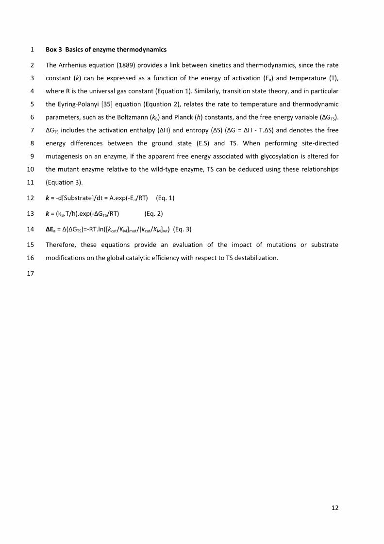
Box 3 Basics of enzyme thermodynamics 1
The Arrhenius equation (1889) provides a link between kinetics and thermodynamics, since the rate 2
constant (k) can be expressed as a function of the energy of activation (Ea) and temperature (T), 3
where R is the universal gas constant (Equation 1). Similarly, transition state theory, and in particular 4
the Eyring-Polanyi [35] equation (Equation 2), relates the rate to temperature and thermodynamic 5
parameters, such as the Boltzmann (kB) and Planck (h) constants, and the free energy variable (ΔGTS). 6
ΔGTS includes the activation enthalpy (ΔH) and entropy (ΔS) (ΔG = ΔH - T.ΔS) and denotes the free 7
energy differences between the ground state (E.S) and TS. When performing site-directed 8
mutagenesis on an enzyme, if the apparent free energy associated with glycosylation is altered for 9
the mutant enzyme relative to the wild-type enzyme, TS can be deduced using these relationships 10
(Equation 3). 11
k = -d[Substrate]/dt = A.exp(-Ea/RT) (Eq. 1) 12
k = (kB.T/h).exp(-ΔGTS/RT) (Eq. 2) 13
ΔEa = Δ(ΔGTS)=-RT.ln([kcat/KM]mut/[kcat/KM]wt) (Eq. 3) 14
Therefore, these equations provide an evaluation of the impact of mutations or substrate 15
modifications on the global catalytic efficiency with respect to TS destabilization. 16
17

13
1
The driving force behind enzyme TS is local energy expenditure, which is the price of TS 2
stabilization. This energy is in turn derived from the ability of enzymes to form quite intricate 3
interactions with the substrate donor moieties [36]. Accordingly, tight donor recognition is the result 4
of efficient electron sharing and the formation of strong, low-barrier hydrogen bonds (< 2.5 Å), two 5
factors that are synonymous with efficient enzyme catalysis. 6
When discussing enzyme catalysis it is also relevant to mention enzyme dynamics because these 7
constitute a key feature of the process [37,38]. Indeed, attempts to investigate catalytic phenomena, 8
such as the modulation of H/T using methods like X-ray crystallography, have often failed to provide 9
any useful information due to the omission of dynamics. Nevertheless, the role of dynamics in TS 10
formation is less clear [39], although it is plausible that they contribute to TS properties. 11
12
On TS properties 13
In the case of β-glycopyranosidases, the structures adopted by TS along the reaction pathway have 14
been comprehensively described by Davies et al. [40], whereas nothing is yet known about the TS 15
developed by furanosidases. For these latter, the only relevant information available is that of the 16
quite extensive work performed by the Lowary group on furanose conformations [41,42]. On the 17
basis of this current knowledge, it is clear that the TS along reaction pathways display coplanar 18
geometry between C5, O5, C1 and C2 in pyranoses (or C4, O4, C1 and C2 in furanoses), which infers 19
the formation of an oxocarbenium ion-like state (sp2-hybridization) [43]. In this case, the anomeric 20
carbon is subject to electrophilic migration (Figure 1) towards the nucleophile catalyst [44,45]. To 21
favor orbital overlap between the electron lone pair of the endocyclic oxygen and C1 (necessary for 22
cationic character establishment) the sugar undergoes ring distortion, moving away from the lowest 23
energy chair conformation [46], as illustrated by structural [47,48] and computational [49] analyses. 24
Recently, in silico approaches have been employed to demonstrate that maximum charge 25
development and TS coordinate points do not necessarily occur at the same time point [50]. 26
Regarding the energetic properties of TS, the contribution of the 2-hydroxyl group is a well-known 27
feature of retaining β-glycosidases (5-10 kcal.mol-1, compared to < 2 kcal.mol-1 for other hydroxyl 28
groups) [18,51,52]. This is because in β-glycosidases the 2-hydroxyl group hydrogen bonds to the 29
catalytic nucleophile, thus favoring a greater share of positive charge and directly affecting 30
oxocarbenium cation formation [53], though to different extents depending on the GH family [19]. In 31
the case of retaining α-glycosidases and α-glycosyltransferases [54], this contribution plays a lesser 32
role (5.2 and 1.9 kcal.mol-1), probably because of different electronic patterns within the trio 33

14
constituted by the nucleophile’s carboxylic acid function, the endocyclic oxygen and the anomeric 1
carbon of the sugar moiety [18]. In retaining β-glycosidases, the nucleophile carboxyl oxygen 2
establishes a syn interaction with the 2-hydroxyl group and the anomeric center, whereas in 3
retaining α-glycosidases the equivalent syn interaction involves the endocyclic oxygen and the 4
anomeric carbon center. A direct consequence of this in retaining β- or α-glycosidases is a greater 5
share of positive charge localized either on the anomeric center or on the endocyclic oxygen 6
respectively. Taking this difference into account, when considering TS electronic patterns it is 7
plausible that this feature could be a key determinant of the principal activity displayed by any given 8
glycosidase. Indeed, it is noteworthy that many ‘true’ non-Leloir TGs are α-retaining enzymes (e.g. 9
glucansucrases, CGTases), which form a β-linked covalent intermediate that displays inherently 10
greater reactivity compared to its α-counterpart [55]. Furthermore, α-retaining GHs are all equipped 11
with anti-protonators, which means that unlike syn-protonators the interaction of the acid/base 12
catalyst with the lone pair of the endocyclic oxygen is impossible [56,57]. In principle, the absence of 13
this interaction is detrimental for TS stabilization, although some GHs display compensatory 14
interactions (e.g. provided by conserved tyrosines in some β-retaining glycosidases) [57,58]. In other 15
work, it has been shown that the presence of a hydrophobic platform within the subsite -1, present 16
in almost all GHs (α or β, retaining or inverting), might play a critical role in TS stabilization [58]. 17
Therefore, for any given GH the study of the impact of charge distribution at TS and the anomery of 18
the glycosyl-enzyme intermediate on the selectivity between water and sugar acceptors should be a 19
useful source of information on the enzyme’s H/T partition. 20
From a temporal point of view, TS are highly transient displaying lifetimes estimated to be within 21
a single bond vibration timescale (i.e. approximately 10 fs, or 10-15 s) [59], far lower than the global 22
kcat, which occurs on a millisecond timescale in most GHs. Regarding water molecules, their diffusion 23
occurs over approximately 1 ps (10-12 s) and does not constitute a rate-limiting step, unlike bond 24
breaking and formation that are much more critical (see below). 25
26
Differences between TS1 and TS2 27
From a practical point of view the kinetic isotope effect (KIE), also called ‘isotope fractionation’ [60], 28
has so far proved to be the only experimental approach that can provide details about TS formation 29
and properties (i.e. geometry and electronic environment) [59,61]. Using this technique it has been 30
shown that in the reaction catalyzed by Agrobacterium sp. β-glucosidase the oxocarbenium ion 31
character is stronger for the deglycosylation TS than for that of the glycosylation step [62], despite 32
the fact that the TS1 and TS2 in retaining GHs are usually considered to share very similar features. 33
Nevertheless, it is clear that the study of TS, in particular the activation barrier of TS2, is hampered 34

15
by the lack of experimental approaches that can provide sound data. 1
Recently, Quantum Mechanics/Molecular Mechanics (QM/MM) approaches have been 2
developed to provide insight into the properties of TS and the extent of bond breaking at each 3
individual step [63,64]. Accordingly, based on the findings of QM/MM it has been postulated that TS2 4
is more dissociative, meaning that the (C1-nucleophile) bond is almost broken before the nascent C1-5
OR bond (with OR from acceptor HOR, with R= H for water) is formed [65]. This provides interesting 6
insight into the enzyme-catalyzed chemistry of the second reaction step and is consistent with the 7
fact that water-mediated deglycosylation is rate-limiting. Moreover, QM/MM has revealed that 8
conserved, non-catalytic active site residues, which are involved in hydrogen bonding with the sugar 9
moiety, contribute to TS stabilization to different extents, this being dependent on the exact position 10
of the hydroxyl moiety and the reaction step under consideration [66]. This is consistent with 11
previous experimental findings that reported on the different contributions of the sugar hydroxyl 12
groups [52]. Despite these encouraging results, QM and MM are still in their infancy and thus 13
findings need to be more extensively corroborated by experimental data. 14
Unfortunately, in the case of retaining GHs, the study of TS is always limited to those developed 15
during hydrolytic reactions, despite the fact that other reagents, such as hydroxylated molecules, can 16
act as acceptors for the deglycosylation step (i.e. transglycosylation). Therefore, in the quest to 17
elucidate the determinants of H/T modulation it is rather evident that water- and carbohydrate-18
mediated deglycosylation involve different behaviours. Although diffusion issues should be 19
considered as important, thermodynamics are at heart of the enzyme-catalyzed chemical reaction 20
and are probably much more critical, as underlined by in silico approaches. Therefore, the key 21
questions regarding the H/T partition appear to concern the properties of the deglycosylation 22
transition state (TS2ROH) and the impact thereupon of the nature of the reacting acceptor substrate 23
(ROH). 24
25
NATURALLY-OCCURRING TRANSGLYCOSYLASES: ELUCIDATING NATURE’S 26
DESIGN STRATEGY 27
In the following section, naturally-occurring TGs are defined as retaining GHs that display a dominant 28
or exclusive ability to transfer glycosyl units onto acceptor sugars (e.g. xyloglucan endo-29
transglycosylases or XET, sucrase-type enzymes, cyclodextrin glucanotransferases or CGTases and 30
trans-sialidases or trS). For practical reasons, in the specific case of TGs the partition between 31
hydrolysis and transglycosylation is described by the ratio T/H rather than the more usual H/T ratio. 32
Moreover, herein we only discuss enzymes for which there is a sufficient amount of knowledge 33
concerning structure-function relationships. 34

16
In guise of a general introduction to this section, the reader is referred to Table 1 that underlines 1
the fact that sugar-transferring enzymes are usually catalytically-less efficient (e.g. kcat/KM values) 2
than hydrolytic counterpart enzymes (85- to 1165-fold lower for GH1 β-glycosidase and GH13 3
sucrose-acting enzymes, respectively). This catalytic sluggishness is likely to be correlated with more 4
energy-demanding TS (for both glycosylation and deglycosylation steps), which lower overall catalytic 5
turnover. In this respect, it is also useful to recall that in a previous study that set out to correlate 6
enzyme and substrate flexibility with catalytic performance, it was proposed that the enzymes we 7
observe today are the result of evolutionary processes that have transformed intrinsically slow, 8
broad specificity prototypes into more efficient catalysts [67]. Of course, this is a rather simplistic 9
view of enzyme evolution and other data suggest that enzymes might have evolved in both directions 10
[68,69], and indeed some GHs (mainly from plants) display both hydrolysis and transglycosylation 11
activities and thus present intermediate cases (i.e. mixed activity) [70–75]. 12
13
14

17
Table 1 Comparison of catalytic constants between glycosynthetic and hydrolytic natural GHs 1
GH family
Enzyme Substrate kcat
a
(s-1
) KM
a
(mM) kcat/KM
a
(s-1
.mM-1
) Reference
1
Rice OsBGlu31 (exo)
Ferulic acid (acceptor)b 1.21 0.05 25.42
[70] pNP-β-D-Glcp (donor)
b,c 1.21 9.33 0.13
Agrobacterium β-glucosidase (exo)
pNP-β-D-Glcp 169 0.078 2170 [62]
13
Bc strain 251 CGTase (endo)
β-cyclization 329 - - [76]
hydrolysis 3.9 - -
Barley α-amylase (endo)
Blue starch 248 0.52
(mg.mL-1
) 477
(s-1
.mL.mg-1
) [77]
CNP-β-D-maltoheptaoside
c 122 1.1 111
NpAS (exo) Sucrose (< 20 mM)
d 0.55 1.9 3.45
[78] Sucrose (> 20 mM) 1.28 50.2 0.0255
XagSUH (exo)
Sucrose 66.5 2.24 29.7 [79]
16
PttXET16-34 (endo)
XGOGlc8 (transglycosylation)e 0.08 0.4 0.2
[80] TmNXG1 (XEH)
XGOGlc8 (hydrolysis)e 0.071 0.08 0.85
XGOGlc8 (transglycosylation)e 0.015 0.5 0.028
32
Wheat FT (1-SST) (exo)
Sucrose (1-kestose production)
0.78 551 - [81]
Wheat VI (exo) Sucrose (hydrolysis) 608 15 -
33
TctrS (exo) Sialyllactose (tranglycosylation)
12.6 1.2 10.5 [82]
TctrS (exo) Sialyllactose (hydrolyse)
0.18 0.29 0.62 [83]
TrSA (exo) 151.4 0.27 554.7 a Determined in the optimal operating conditions for each enzyme. Specific activity is provided when the kcat value is
unavailable. b Kinetic parameters were determined either for the acceptor (with 30 mM donor) or for the donor (with 0.25 mM
acceptor)
c pNP, 4-nitrophenyl; and CNP, 2-chloro-4-nitrophenyl.
d For low sucrose concentration (< 20 mM), hydrolysis is dominant.
e Xyloglucan-oligosaccharides mixture composed of XXXG, XLXG, XXLG, and XLLG moieties (using the nomenclature
developed by Fry et al. [84]) and based on (D-Glcp)8 backbone.
2

18
1
Xyloglucan endo-transglycosylases 2
In terms of understanding the determinants of the T/H partition, XETs and their hydrolytic 3
counterparts, xyloglucan endo-hydrolases (XEH), are extremely interesting enzymes that are usually 4
referred to as xyloglucan endo-transglycosylase/hydrolases or XTHs, even though biochemical 5
evidence reveals that most of them are XETs, displaying very little hydrolytic ability. XTHs are mainly 6
grouped in family GH16 (members of GH-B clan), which also contains other hydrolytic GHs enzymes 7
that display a wide variety of substrate specificities [85,86]. The molecular phylogeny of XTH genes, 8
their catalytic properties and in vivo functional differences provide criteria for the definition of three 9
major groups. Members of groups I and II exclusively exhibit XET activity, which is also the 10
predominant feature of group III-B. However, members of group III-A (XEH) are mainly hydrolytic 11
[80,87–89]. With regard to XETs, these are known to be important for plant cell wall remodelling, 12
since they catalyze the non-hydrolytic cleavage and religation of xyloglucan molecules through a 13
ping-pong bi-bi mechanism that is subject to competitive inhibition, since competing substrates can 14
act as both the donor and the acceptor [90,91]. 15
As explained earlier, the canonical double-displacement mechanism of glycosyl transfer involves 16
the formation of a covalent glycosyl-enzyme intermediate. In XTHs, glycosylation is rapid (< 2 min) 17
and procures a relatively long-lived glycosyl-enzyme intermediate, whose formation is associated 18
with an estimated free energy change (ΔG0, Figure 3) of formation of approximately 1.5-2.0 kcal.mol-1 19
[92–94]. Indeed, the glycosyl-enzyme intermediate of PttXET16-34 (a XET from hybrid poplar Populus 20
tremula x tremuloides) is approximately 3 h with a khydr. = 1.10-4 s-1. Deglycosylation of the glycosyl-21
PttXET16-34 intermediate is brought about by the presence of suitable sugar acceptors, such as 22
xylogluco-oligosaccharides. When this criterion is fulfilled, it has been shown that the xylogluco-23
oligosaccharyl-XET adduct can be fully deglycosylated in 30 min. In this respect, it is also noteworthy 24
that when PttXET16-34 was supplied with activated β-D-xyloglucan-oligosaccharidic donors (e.g. LG = 25
2-chloro-4-nitrophenyl or fluoride), no activity (neither transglycosylation nor hydrolysis) was 26
observed [95,96]. This underlines the fact that the energetic barrier of TS1 can only be overcome by 27
the presence of a sugar LG in the positive subsites, as is the case for deglycosylation (TS2). This 28
requirement is removed in the case of the PttXET16-34-based glycosynthase, since the reaction only 29
proceeds through the ‘pseudo’ second step (TS2ROH) of the canonical retaining-mechanism. 30
Moreover, it is remarkable that donor substrate binding is dominated by the higher affinity of the 31
xyloglucan moiety for the positive subsites, an interaction that is driven by the presence of aromatic 32
residues. This increased affinity for the positive subsites is thought to be necessary (though not 33
sufficient per se) for transglycosylation [91,95]. 34

19
Despite a lack of sequence identity within family GH16, all of its members display a typical β-1
jellyroll fold that is composed of two large curved β-sheets, stacked in a sandwich-like manner. 2
However, in the case of XTHs specific structural features reflect the specialization of these enzymes 3
toward their highly branched substrates [95]. Notably, according to Brumer et al., starting from an 4
ancestral (hydrolytic) licheninase active on linear 1,3-1,4-β-glucans [97], the deletion of a loop 5
procured the ability to bind highly branched substrates, such as xyloglucan, a characteristic that is 6
shared by both GH16 endo-glucanases (EG) and XTHs that display hydrolytic and/or 7
transglycosylation activities. Examples of this are PtEG16 from Populus trichocarpa, which is able to 8
hydrolyze the xylogluco-oligosaccharide XXXGXXXG, and its counterpart PttXET16-34, which performs 9
transglycosylation using the same substrate [90,97]. Moreover, the extension of the C-terminal 10
domain differentiates the XTHs from EGs. This XTH feature provides exclusive specificity for 11
xyloglucan (i.e. branched substrates) to this group of GH16 enzymes [91,97]. Finally, regarding XETs 12
and XEHs, X-ray structure data have revealed that in some cases these differ in two loops located 13
between β-strands β6 and β7, and between β8 and β9, in the vicinity of the active site [80]. The 14
importance of this last observation has been demonstrated through the creation of a β8/β9 loop 15
deletion in the Tropaeloum majus XEH (TmNXG1-ΔYNIIG mutant), a loop that forms part of subsite +1 16
and interacts with the D-glucosyl residue. This mutation procured an increased T/H ratio in the initial 17
phase of the reaction, with a 2-fold increase in transglycosylation rate being coupled to a 5.7-fold 18
decrease in hydrolysis rate. 19
Structural and molecular dynamics work performed on PttXET16-34 and TmNXG1 has revealed a 20
correlation between the nature of the principal activity and subsite binding interactions, which are 21
combined with subtle differences in dynamic behavior [98]. As a matter of fact, in XETs, the number 22
of H-bonds formed between the enzyme and the acceptor moiety is greater than in XEHs, whereas in 23
XEHs the number of H-bonds formed with the donor moiety is higher. Moreover, a determinant of 24
transglycosylation in XETs appears to be more flexibility in subsite -1, which is detrimental for overall 25
activity, except when a sugar is present in subsite +1. 26
27
Sucrase-type enzymes 28
Sucrases are exo-enzymes that include glucansucrases (GS) and fructansucrases (FS). Using sucrose as 29
a substrate, these enzymes are able to synthesize homopolysaccharides composed of D-glucosyl or D-30
fructosyl subunits respectively, with different linkage specificities [99,100]. GS are classified in both 31
GH13 and GH70 family, with GH13 GS being designated amylosucrases (AS). Transglucosylating AS 32
and GS have been extensively studied both in our group [99,101] and in Lubbert Dijkhuizen’s group 33
[100,102]. Although AS, GS and FS act on the same substrate, they actually exhibit different protein 34

20
folds, with AS and GS being characterized by a (β/α)8-barrel architecture and belonging to clan GH-H 1
(α-amylase superfamily), which is divided into 40 subfamilies [103], and FS belonging to clan GH-J (5-2
bladed-β-propeller). Nevertheless, all three enzyme groups operate through a retaining mechanism. 3
Until recently, the structure of GS remained elusive [104–106], thus hampering progress in the 4
understanding of structure-function relationships in these enzymes [107]. Nevertheless, using 5
sequence-based approaches it was possible to identify transition state stabilizers (histidines) that are 6
present in both GH13 and GH70, being conserved in α-amylases, CGTases and GS. These residues are 7
essential for overall catalysis (hydrolysis and transglycosylation) and their mutation is often highly 8
detrimental for activity (< 0.5% residual activity) [107]. 9
Concerning GH70 GS, the analysis of the impact of mutations of key catalytic residues and others 10
located in the positive subsites has led to the conclusion that such mutations can be grouped into 11
one of three categories: those affecting (i) D-glucosidic linkage specificity, (ii) glucan solubility and (iii) 12
overall enzyme activity [100]. Structural data analysis revealed that subsite +1 residues form H-bonds 13
with the D-fructosyl moiety, as do residues in subsite +2 with the D-glucosyl moiety, these latter 14
playing an important role in determining the linkage ratio [107]. Results from the study of a 15
reuteransucrase (GH70) from Lactobacillus reuteri suggest that steric hindrances play a major role in 16
chain elongation, since the deletion of a variable N-terminal domain procured an increase in 17
transglycosylation (3- to 4-fold) at the expense of hydrolysis [108]. Similarly, the creation of a single 18
point mutation (N1179E) within the same subgroup of enzymes led to a T/H ratio increase [109]. In 19
another study, it was reported that a GH70 4,6-α-glucanotransferase is able to perform a 20
disproportionation reaction on α-(1,4)-linked malto-oligosaccharides, but is unable to use sucrose as 21
a substrate, despite the high energy (6.6 kcal.mol-1) associated with its glycosidic linkage [110]. In the 22
light of this observation it was proposed that this enzyme represents an evolutionary intermediate 23
between GH13 and GH70 [111]. 24
Compared to GH70 GS, the data available for GH13 AS is more abundant. These enzymes all 25
display a similar 5-domain structure with a deep pocket at the bottom of which sucrose binds to 26
subsites -1 and +1 [112,113]. Three arginines (R226, R415 and R446), located in positive subsites 27
+2/+3, +4 and +1 respectively, are particularly important in the transglucosylation reaction, since 28
these play a crucial role in the docking and positioning of acceptors [114,115]. In a study of the AS 29
from Neisseria polysaccharea (NpAS, subfamily 4 of GH13) the positive subsites were submitted to 30
mutagenesis with the aim of improving transglucosylation using unnatural acceptors. Although quite 31
impressive increases in transglucosylation were achieved (395-fold increase), which were 32
accompanied by decreased apparent KM values, no evidence of significant structural changes that 33
would alter sucrose binding was detected [116]. Therefore, it was concluded that modified loop 34

21
flexibility and enzyme dynamics are likely to be the determinants of altered substrate recognition 1
and thus responsible for the establishment of a catalytically-productive state. Overall, this study 2
revealed a certain plasticity of subsite +1, because it was possible to isolate mutants that could 3
glucosylate a series of different acceptors, and suggested that the improvement of transglucosylation 4
using unnatural acceptors was facilitated by improved interactions in the positive subsites. In 5
another study, recognition of D-glucosyl moieties in subsite -1 was investigated. This revealed that 6
despite the fact that AS exhibits slow rates, the D-glucosyl is specifically recognized by a complex 7
network of interactions [117]. To further understand the transglycosylating character of NpAS, it is 8
useful to compare this enzyme with a hydrolytic counterpart, such as the sucrose hydrolase from 9
Xanthomonas axonopodis pv. glycines (XagSUH). Although this GH13 member shares 57% sequence 10
identity with NpAS and is structurally similar (identical 5-domain structure with rmsd value of 1.78 Å) 11
[79], XagSUH catalyzes sucrose hydrolysis and is incapable of catalyzing transglucosylation [118]. One 12
main difference between XagSUH and NpAS has been revealed by acquiring structural snapshots 13
along the catalytic coordinate. This revealed in XagSUH that upon sucrose binding a pocket-shaped 14
active site is formed through rigid-body movements of the B and B’ domains towards the active site. 15
Moreover, it is noteworthy that the majority of active site residues are conserved between the two 16
enzymes, except for three arginines (R226, R415 and R446) that are substituted in XagSUH by other 17
residues (glycine or leucine). Significantly, as mentioned earlier these arginines are essential in NpAS 18
for transglucosylation, although the introduction of homologous arginines in XagSUH by mutagenesis 19
failed to confer transglucosylation properties to the enzyme, an observation that is consistent with 20
the fact that improvements in transglycosylation first require diminution of hydrolysis [79], especially 21
given the fact that the value of kcat on sucrose is 120-fold higher than that of NpAS (Table 1). 22
Surprisingly, this fact was not evoked by the authors, who suggested that hydrolysis in XagSUH might 23
be caused by a collateral effect of D-fructose release, which would disorder the B-domain and thus 24
expose the enzyme-bound D-glucosyl moiety to bulk solvent and thus hydrolysis. 25
In summary, the identification of the key factors that determine the T/H partition in GS has 26
proved to be quite difficult, although it appears evident that interactions in positive subsites play an 27
important role. In this respect, and taking into account the fact that the active site in these enzymes 28
is often buried, it has been suggested that the presence of an acceptor group in sucrases during 29
catalysis protects the covalent intermediate from water-mediated attack [119]. This could be true if 30
sucrose was able to bind in subsites -1 and +1 in the presence of the acceptor (implying that the 31
acceptor is bound elsewhere). However, this hypothesis assumes that upon formation of the 32
covalent intermediate the acceptor is somehow displaced towards subsite +1 and that there is 33
considerable flexibility within the active site, allowing for example the unhindered departure of the 34

22
D-fructose LG. Unfortunately, at least in the case of AS the ‘U-shaped’ active site structure does not 1
appear to allow for this possibility. Moreover, even if the active site of all AS were highly flexible and 2
accessible, the hypothesis would not explain how water-mediated deglycosylation is avoided, 3
especially in an enzyme such as the AS from Deinococcus radiodurans, which despite its open active 4
site topology, still mainly performs transglucosylation [119,120]. Therefore, alternative hypotheses 5
are required, not to explain how water is prevented from entering active sites, but rather to explain 6
how the presence of water is rendered irrelevant with respect to deglycosylation. 7
Regarding fructose-specific sucrases (FS), which can act on sucrose and/or fructans, these are 8
gathered within families GH68 and GH32 (clan GH-J). The FS in GH68 (i.e. levansucrases, 9
inulosucrases) usually display a dominant hydrolytic activity, accounting for 70-80% of substrate 10
(levan, inulin) conversion. A previous study performed on the single domain levansucrase, SacB from 11
Bacillus subtilis, revealed that the addition of transitional and complete C-terminal domains from 12
other FS leads to reductions in hydrolysis (down to 10% of substrate conversion), accompanied by a 13
5-fold increase in transfructosylation. Upon analysis of the chimeric enzymes the authors remarked 14
that the kcat value associated with hydrolysis was unaltered and thus attributed the increase in the 15
T/H ratio to more favorable positive subsite interactions provided by a structural adjustment in the 16
catalytic site mediated by the addition of extra domains [121]. 17
The GH32 family comprises both fructan-acting (β-D-fructofuranosidases and inulinases) and 18
sucrose-acting enzymes, and compared to GH68 FS contains an additional β-sandwich domain. The 19
GH32 sucrases or invertases (as they are often known) are able to transfer the D-fructosyl moiety of 20
sucrose either onto water (hydrolysis) leading to the production of fructose (i.e. inverted sugar), or 21
onto a sucrose acceptor (transglycosylation) thus catalyzing the synthesis of fructan. In the latter 22
case, the enzymes are designated as fructosyltransferases (FTs). Within the plant kingdom, sucrose 23
can be degraded by vacuolar (VIs) or cell wall invertases, and from a phylogenetic standpoint FTs and 24
VIs belong to the same GH32 subgroup, sharing high sequence identity (ca. 65%) and structural 25
homology. Using phylogenetic tree analysis, it has been proposed that FTs have evolved from 26
ancestral VIs [69]. Among the different VIs, it is noteworthy that three amino acid sequence motifs 27
are highly conserved: (i) the sucrose-binding box motif WMNDPNG, which contains the catalytic 28
nucleophile D, (ii) the EC motif, which includes the catalytic acid/base E and (iii) the RDP motif, with D 29
being identified as a TS-stabilizing residue [122,123]. The first N in the sucrose-binding box is involved 30
in a hydrogen bond network, forming links with the nucleophile D and W (Figure 4). 31
32

23
1
Figure 4 Cartoon representation of the X-ray structure of the active site of the Saccharomyces 2
invertase (PDB ID: 4EQV [124]), showing the catalytic triad (red) and the H-bond network of the 3
sucrose-binding box. This motif in VIs is shown along with the key amino acid substitutions that 4
characterize FTs. PyMOL Molecular Graphics System, v0.99 (Schrödinger, LLC) was used to prepare 5
the figure. 6
7

24
1
Importantly, within the sucrose-binding box, W is always replaced by a Y in FTs from the same 2
subgroup. Likewise, the first N is very often substituted in FTs by S. Engineering of these alternative 3
residues into VIs demonstrated that the disruption of the hydrogen bond network involving the 4
nucleophilic aspartate (i.e. W23Y and N25S) enhanced transglycosylation up to 17-fold when 5
compared to wild-type VIs [81]. Similarly, other studies performed on VIs from yeast [125] or onion 6
[69] led to similar conclusions, although the increase in transglycosylation was more modest. 7
Furthermore, a shift of optimum pH from 3.8-4.8 to 4.8-5.7 was observed for a yeast VI mutant 8
(W19Y-N21S), consistent with an alteration of the ionization state of the catalytic residues [125]. 9
Interestingly, the reverse experiment involving the substitution of Y by W in two different FTs failed 10
to procure a more hydrolytic VI-like enzyme [126,127], which suggests that it is much easier to 11
disrupt rather than create a hydrogen bond network ! 12
From a kinetic point of view, compared to FTs (Table 1) VIs are more efficient catalysts. FTs do 13
not display a saturation profile (i.e. KM of hundreds of mM relative to 2-20 mM range for VIs), but are 14
nonetheless very good at transfructosylation (70-80% substrate conversion) compared to VIs (2-5% 15
of substrate conversion). When considering mutated VIs, these can be seen as intermediate cases, 16
since for most of the available examples KM values were increased from 4- to 34-fold [69,81,125], 17
resulting in severely reduced kcat/KM values, an alteration that is indicative of higher TS1 energy 18
levels. 19
Acceptor substrate selectivity among GH32 has also been investigated using a mutagenesis 20
approach to modify residues located in subsites +1 or +2. However, this type of mutation has so far 21
failed to confer significant transglycosylation ability to invertases, although in at least one case both 22
regioselectivity (β-(2,6)/β-(2,1)) and catalytic efficiency were significantly altered [125,128]. On the 23
other hand, the mutagenesis of putative positive subsite residues in a FT proved to be quite 24
detrimental for transglycosylation [127]. Therefore, based on available data on FT/VIs it is possible to 25
conclude that the modification of positive subsite determinants can be used to improve acceptor 26
recognition and positioning for transglycosylation, but this is insufficient to destabilize water-27
mediated deglycosylation (i.e. TS2water) in invertases. To achieve this, it is much better to target the 28
proton network in the negative subsite (Figure 3). 29
30
Cyclodextrin glucanotransferases and α-amylases 31
Involved in starch depolymerization, CGTases and their hydrolytic counterparts, α-amylases, belong 32
to GH13 and thus to clan GH-H. These enzymes share a common structural architecture, which is 33

25
defined by three domains, A, B and C, although CGTases possess two extra domains D and E. It has 1
been proposed that CGTases have evolved from α-amylases, since the latter display greater sequence 2
diversity and are more widespread through the different taxonomic groups [68]. Regarding the 3
natural function of CGTases, it is likely that by providing cyclodextrins of defined size (i.e. α, β or γ), 4
CGTases procure ‘tailored’ substrates for α-amylases and thus accelerate starch saccharification. 5
Crystallographic analysis of CGTases has revealed an extensive active site structure, extending 6
from at least a subsite -7 to a subsite +3. Due to their architecture, these endo-enzymes are able to 7
catalyze intra-molecular transglycosylation (β-cyclization) through the transfer of the covalently 8
bound sugar unit onto the 4-hydroxyl group of the non-reducing end of the same donor molecule 9
[129]. Compared to GS (exo-enzymes with only one negative subsite), negative subsite interactions in 10
CGTases are much more developed and, taking into account the high transfer rates that characterize 11
these enzymes (102-103 IU.mg-1, Table 1), it is probable that the transition state energy barrier is 12
lower than that of GS. 13
In addition to the synthesis of cyclodextrins, CGTases have also been shown to be capable of 14
hydrolysis or to perform the transfer of the bound glycosyl intermediate onto another α-glucan chain 15
(i.e. disproportionation) [130]. In order to prevent hydrolysis, it appears that CGTases have acquired 16
positive subsites that favor sugar recognition. This is illustrated by mutagenesis work that was 17
performed on the positive subsites (+2 and +3) of the Bacillus circulans 251 CGTase (BcCGTase). The 18
substitution of F183 and F259 in BcCGTase by N or S resulted in a 10- to 300-fold decrease in 19
transglycosylation activity (β-cyclization) and a 3- to 20-fold increase in hydrolysis [131]. Similarly, the 20
simultaneous mutation of equivalent residues (F184Q and F260W) in the CGTase from 21
Thermoanerobacterium thermosulfurigenes strain EM1 (Tabium CGTase) and the addition of a third 22
mutation (A231V) converted this enzyme into an α-amylase-like hydrolytic enzyme [132]. 23
Impressively, this mutant no longer displayed detectable CGTase activity, with the T/H ratio being 24
0.0012 (compared to 5 for the parental CGTase). Consistent with these results, another study 25
focusing on the positive subsites in liquefying (hydrolytic) and maltogenic (transglycosylating) α-26
amylases revealed that increased hydrophobicity in subsites +2/+3 of the α-amylase from Bacillus 27
licheniformis (BLA) increased the T/H ratio, reducing the hydrolysis rate (associated with a higher KM 28
value) on starch by one third [133]. Likewise, the sequence comparison of hydrolytic and maltogenic 29
α-amylases revealed the presence in subsite +1 of a conserved histidine or glutamate residue, 30
respectively [134]. The introduction of the substitution H235E in BLA created a transglycosylation 31
activity, which is undetectable in the wild-type enzyme, but did not drastically affect the efficiency of 32
hydrolysis (72% residual) [135]. Overall, mutations in the positive subsites of CGTases generally 33
provoke a diminution of transglycosylation activity [130], whereas negative subsite mutations mostly 34

26
alter cyclodextrin specificity (α, β and γ ratio, for cyclodextrins composed of 6, 7 and 8 glucose units 1
respectively). In this respect, it is noteworthy that a five-residue loop localized in subsites -3/-4 of α-2
amylases has been described as a key determinant (steric hindrance) of the T/H partition, since it is 3
absent in CGTases. To test this hypothesis, the loop in the α-amylase Novamyl (residues 191 to 195) 4
was deleted and positive subsite mutations (F189L/T190Y) were introduced. These modifications 5
procured CGTase-like behavior [136], but the reverse experiment (i.e. introduction of a loop in US132 6
CGTase) failed, since it yielded a mutant that was unable to catalyze hydrolysis or even initial β-7
cyclization [137]. This failure once again underlines the complexity of the phenomenon and supports 8
the notion that hydrolysis is driven by optimized interactions in the negative subsites, which in turn 9
contribute to the formation of TS. In this respect, it is interesting to mention that the successful 10
conversion of the aforementioned Tabium CGTase into a hydrolase was almost certainly facilitated 11
by the fact that the parental enzyme already displays unusually high hydrolytic ability. This implies 12
that in Tabium CGTase the donor interactions required for hydrolysis are already in place and thus it 13
is simply a case of deleting the determinants of transfer activity. 14
More generally, these studies highlight the role of aromatic/hydrophobic residues in positive 15
subsites. Notably, it appears obvious that the presence of aromatic residues provides both a stacking 16
platform for better acceptor docking [138] and a hydrophobic barrier, which limits the presence of 17
water in the active site, with both of these factors favoring transglycosylation. Similarly, such 18
features were also suggested to be part of an evolutionary relationship between α-amylases and 4-α-19
glucanotransferase within family GH57 [139]. 20
Assuming that CGTases are indeed the consequence of the evolution of α-amylases, presumably 21
the former have somehow dealt with the well-developed negative subsite interactions that favor 22
hydrolysis [140]. Theoretically, the existence of intermediate CGTases that display high ‘residual’ 23
hydrolytic activity, such as the GH13 Tabium CGTase [132] or the one from Bacillus sp. SK 13.002 24
strain [141], should provide clues as to how this has been achieved. However, in reality unravelling 25
subtle molecular differences might actually prove to be a considerable challenge [142]. 26
27
Transferring vs hydrolyzing sialidases 28
Sialidases (SA) and trans-sialidases (trS) are members of family GH33 and belong to the GH-E clan. 29
These enzymes catalyze either the hydrolysis or the synthesis of sialyl-glycoconjugates respectively, 30
operating via a classical ping-pong bi-bi mechanism with acid/base catalysis [143–145]. trS exhibits 31
both activities, although when a suitable acceptor is available transglycosylation is approximately 10-32
fold higher than hydrolysis [146]. Moreover, in the case of TctrS, the trS from Trypanosoma cruzi, the 33

27
KM value for the acceptor is lower than that of the donor (10 µM and in the millimolar range for the 1
lactose and sialic acid moieties respectively) [82,144]. Both SA and trS possess similar catalytic 2
domains that display six-bladed β-propeller topology, which are connected via a long α-helix and a 3
large hydrophobic interface to a domain displaying a β-sandwich fold and lectin-like topology (Figure 4
5A). This latter does not appear to be directly involved in transglycosylation activity [147]. The 5
molecular architecture of the active sites of these enzymes displays several common features, 6
including eight strictly invariant residues and a hydrophobic pocket that binds the N-acetyl group of 7
the sialic acid moiety, suggesting a mutual evolutionary origin and a similar mode of action for the 8
entire family [83,144,146]. 9
10
11

28
1
Figure 5 (A) Superimposition of the structures of the sialidase from T. rangeli (TrSA - orange, PDB 2
ID: 1N1T [148]) and the trans-sialidase from T. cruzi (TctrS - blue, PDB ID: 1MS3 [149]); (B) zoom on 3
the residues that are mutated, creating TrSA10mut (TrSA numbering) and (C) hydrogen bonding of 4
TctrS (blue) and TrSA (orange) with DANA and lactose (PDB ID: 1MS0 [149]). Bold/cartoon (B), or 5
underlined (C) amino acids are those that are mutated to create TrSA5mut. (B) The additional 6
substitutions are those introduced (in silico only) to create TrSA10mut and are close to the nucleophilic 7
tyrosine (Y343, TrSA numbering), drawn with grey sticks. (C) The base catalyst (D60, TrSA numbering) 8
is depicted, but Y343 is not shown. Graphics were prepared using PyMOL Molecular Graphics System, 9
v0.99 (Schrödinger, LLC) and PoseView [150]. 10
11

29
1
It has been suggested that subtle structural differences are likely to be responsible for the 2
different selectivities of hydrolysis and transglycosylation reactions catalysed by Trypanosoma 3
rangeli SA (TrSA) and TctrS respectively. Although these enzymes share 70% amino acid identity, their 4
active sites display distinctive features [144,147–149]. TctrS exhibits a narrower, more hydrophobic 5
substrate-binding pocket. This implies that the reactive center is less solvent-exposed and results in 6
an alternative hydrogen bonding pattern with the sialyl donor moiety. Additionally, residue Q284 in 7
TrSA is replaced by P283 in TctrS (Figure 5B and C), a substitution that alters the conformation of the 8
neighboring W312 residue (W313 in TrSA). In TctrS, W312 and Y119 (S120 in TrSA) form the two 9
lateral walls of the acceptor binding site, providing the basis for stacking interactions with the sugar 10
acceptor. Moreover, it is noteworthy TctrS appears to display greater active site flexibility than TrSA 11
[83,147,148,151], a point that is exemplified by the study of the inherent motions of Y119 and the 12
strictly conserved Y342 (catalytic nucleophile) residues. According to Demir and Roitberg, structural 13
rearrangements that are triggered by ‘allosteric’ binding of the sialyl-conjugate donor forming a 14
covalent sialyl-enzyme intermediate lead to the creation of a productive acceptor sugar binding site 15
[151]. 16
Overall, finely-tuned enzyme-donor substrate interactions, conformational flexibility (notably 17
loops), solvent exposure and the presence of an acceptor sugar-binding site are all crucial to obtain 18
trans-sialidase activity. Therefore, to switch between hydrolysis and transglycosylation, TrSA has 19
been submitted to mutagenesis, introducing five mutations designed to modify the structure and 20
dynamics of the negative subsite and to create a suitable positive subsite. This work provided 21
TrSA5mut, a mutant that displayed detectable trans-sialidase activity, although this was only 1% of that 22
exhibited by the true trans-sialidase, TctrS [83]. Further mutation of TrSA5mut, introducing either I37L 23
or G342A (Figure 5B), which affect the negative subsite, procured a higher transglycosylation rate, 24
which was 11% of that exhibited by TctrS. Therefore, it appears that the acquisition of improved 25
trans-sialidase activity requires alterations in the negative subsite, notably to alter the flexibility of 26
the tyrosine nucleophile residue and thus diminish hydrolytic activity. In this respect, it is also 27
significant that while TrSA is inhibited by DANA (Ki = 1.5 µM for TrSA), a structural analog of the 28
transition state sialic acid oxocarbenium ion (Figure 5C), the mutated TrSA described above is less 29
sensitive to inhibition (Ki = 1.54 mM) [83], as is the case for TctrS (Ki = 12.3 mM). This implies that the 30
acquisition of trans-sialidase activity may involve a modification of the TS that is developed during 31
the glycosylation step. 32
More recently, using QM/MM approaches Roitberg et al. evaluated the free energy profiles for 33
the conversion of the Michaelis complex to the covalent glycosyl-enzyme intermediate in TrSA, 34

30
TrSA5mut and TctrS [152,153]. In SA enzymes, the free energy barrier (ΔGTS1) to reach the glycosylated-1
enzyme intermediate (15.2 and 15.0 kcal.mol-1 for TrSA and TrSA5mut, respectively) is approximately 5 2
kcal.mol-1 lower than that of TctrS (20.8 kcal.mol-1). Moreover, the change in free energy (ΔG0, Figure 3
3) associated with the glycosylation step of the TctrS-catalyzed reaction is close to zero (-0.89 4
kcal.mol-1), compared to -10.9 and -9.8 kcal.mol-1 for TrSA and TrSA5mut respectively, these values 5
being linked to the higher stability of the glycosyl-enzyme intermediates. However, the 6
deglycosylation step appears to be favorable for trS-like enzymes, with the difference being 7
approximately 5 kcal.mol-1 (i.e. 21.6, 24.8 and 26.1 kcal.mol-1 for TctrS, TrSA5mut and TrSA, 8
respectively). Based on these findings further in silico design of an efficient trS was performed, giving 9
rise to the hypothetical mutant TrSA10mut, which contains five additional substitutions (Figure 5B). 10
According to the in silico results, in TrSA10mut residues I37L and G342A (TrSA numbering), both located 11
in the vicinity of the catalytic nucleophile tyrosine (Y343), would be responsible for the predicted 12
increased T/H ratio. Moreover, it was speculated that TrSA10mut would only weakly stabilize the 13
covalent intermediate and when compared to TctrS would display a lower free energy barrier for 14
deglycosylation step (-3.2 and 19.1 kcal.mol-1, respectively). However, regarding the free energy 15
barrier of the glycosylation step, it was predicted that this would be similar (ΔGTS1 = 16.0 kcal.mol-1) to 16
that of a typical hydrolytic SA. 17
Finally, it is noteworthy that on the edge of its acceptor substrate binding cleft TctrS displays a 18
seven-amino acid loop (VTNKKKQ) whose composition, physico-chemical properties and dynamics 19
differ from the equivalent loop (IADMGGR) in TrSA. Using an enzyme engineering approach it was 20
shown that the loop in TctrS promotes transglycosylation, increasing product yield and reduces 21
hydrolysis, effects that were attributed to a perturbation of the water binding network [154]. 22
23
ENGINEERED TRANSGLYCOSYLASES 24
Although TGs have only been identified in a few GHs families, hydrolytic GHs from other families 25
have been submitted to protein engineering in order to modify their H/T balance. In the following 26
section, the different strategies that have been adopted are described along with the results that 27
have been obtained. 28
29
Modification of negative subsite interactions 30
Enzyme engineering 31
One of the very first protein engineering studies aimed specifically at increasing the T/H ratio was 32
performed on a GH1 β-glycosidase from Thermus thermophilus using a random 33
31
mutagenesis/screening methodology. The mutation of two conserved residues F401 and N282 in this 1
enzyme increased KM values (> 6-fold) and significantly improved transglycosylation (up to 78% 2
synthesis yield compared to 8% for the wild-type enzyme) [155]. In a follow-up study, using a site-3
directed approach the same authors probed the importance of conserved residues in the donor (-1) 4
subsite [156,157] and revealed that these play an important role in TS stabilization (29- to 3577-fold 5
decrease kcat/KM values), but do not induce major structural changes. 6
Working on AMY1, a GH13 α-amylase (clan GH-H), it was shown that the mutation of a subsite -2 7
residue (M53W) leads to increased lifetime of the glycosyl-enzyme and thus to the acquired ability to 8
perform transglycosylation using pNP-α-D-maltoheptaose as the donor [77]. It is also noteworthy that 9
the introduction of a range of mutations at position 53 procured kcat/KM values that were 59- to 5000-10
fold lower than that of the parental enzyme (mainly due to up to a 20-fold increase in KM values). 11
Likewise, it is significant that the presence of tryptophan at position 53 is a common occurrence in 12
GH13 CGTases, which is consistent with the impact of the mutation M53W in AMY1. 13
The mutation of conserved negative subsites residues produces a similar effect to the one 14
described above in other GH families. This is exemplified by protein engineering work performed on 15
a GH18 chitinase from Serratia marcescens (SmChiA). The latter possesses a long active site cleft 16
positioned at the top of a (β/α)8 barrel, the negative subsite of which was targeted with the aim of 17
prolonging the retention time of the donor glycosyl moiety [158]. The introduction of the mutation 18
W167A (subsite -3) procured a higher transglycosylation yield (45% of the substrate converted into 19
transglycosylation products, compared to 8% for wild-type SmChiA) and subsequent determination 20
of the 3D structure of the mutated enzyme revealed that repositioning of D313 (subsite -1) had 21
occurred. This is significant because D313 is involved in the stabilization of the oxazolinium 22
intermediate and interacts with E315, a residue that is putatively responsible for water molecule 23
activation during hydrolysis. Therefore, the mutation W167A might both prolong residency of the 24
donor glycosyl moiety and/or diminish hydrolysis. Similar examples of such a coupled effect (i.e. 25
improved transglycosylation and diminished hydrolysis) are provided by work performed on 26
chitinases from Serratia proteamaculans (SpChiD) [159] and Aspergillus fumigatus (AfChiB) [160], 27
with mutations being introduced at the catalytic center and in subsite -1 respectively. Furthermore, 28
QM/MM calculations performed on a hyper-transglycosylating variant (D142N) of ChiB from S. 29
marcescens (SmChiB) predicted that the mutation, which is within a highly conserved DxDxE motif, 30
would affect both TS stabilization and the catalytic water molecule [161]. 31
A further example concerns two homologous α-galactosidases (AgaA and AgaB) from family 32
GH36 (clan GH-D) [162]. Despite being highly related (97% identity), AgaA displays a relatively low KM 33
value for raffinose (KM = 3.8 mM) and exhibits high hydrolytic activity and no detectable ability to 34

32
catalyze transglycosylation. On the other hand, AgaB displays a higher KM value (200 mM) for 1
raffinose and exhibits the ability to catalyze autocondensation reactions (i.e. transglycosylation). In 2
this context, the mutation of residue 355 (Ala in AgaA and Glu in AgaB) provides the means to switch 3
between the two phenotypes, with for example the substitution A355E in AgaA procuring AgaB-like 4
behavior and vice versa. Although residue 355 is located far from the active site (20 Å), structural 5
analysis revealed that the presence of a Glu at position 355 provokes the displacement of the 6
conserved W336, which is present in subsite -1 where it provides the basis for sugar stacking. This 7
modification widens the active site and thus probably disturbs the binding of raffinose. 8
Regarding another example of a galactose-acting enzyme family, random mutagenesis and 9
screening performed on the GH42 β-galactosidase from Geobacillus stearothermophilus (BgaB) 10
pinpointed a residue (R109) for subsequent site-saturation mutagenesis. This ultimately procured a 11
mutant (R109W) that displayed improved ability (23% yield compared to 2% for the parental 12
enzyme) to transfer D-galactosyl moieties onto lactose [163]. R109 is a highly conserved amino acid 13
among GH42 β-galactosidases that according to 3D structure analyses is involved in hydrogen 14
bonding with the D-galactosyl moiety. Therefore, mutation of this residue probably leads to the 15
destabilization of donor binding in subsite -1 (KM values on lactose increase from 1.8 to 114 mM), 16
coupled to decreased hydrolysis (15% residual) and thus alterations in the T/H ratio that favor 17
transglycosylation. Overall, in terms of TS it is likely that the mutation R109W increases the TS energy 18
barriers for glycosylation and deglycosylation, thus rendering water-mediated deglycosylation less 19
competitive. 20
Regarding rational engineering work focused on the nucleophile catalyst, several studies have 21
revealed that modifications of the latter can also have drastic effects on the ability of water to 22
deglycosylate the glycosyl-enzyme intermediate. Recently, it was reported that the introduction of a 23
sulfinate function (i.e. SOO-), to replace the catalytic nucleophile of the GH13 dextran glucosidase, 24
provoked a drastic drop in kcat (0.27%), an acidic pKa shift (from 3.9 to 1.5) and an increase in 25
transglucosylation yields [164]. According to the authors of this work, the observed effects can be 26
attributed to differences in the TS energy barriers between water and acceptor-mediated enzyme 27
deglycosylation. In this respect, shortening or lengthening (E78D or carboxymethylation of the 28
mutant E78C) of the nucleophile residue in the GH11 xylanase from Bacillus circulans was also shown 29
to be detrimental for global catalytic efficiency, nucleophile shortening having a greater impact 30
(1600-5000-fold decrease) than lengthening (16-100-fold) [165]. However, in this study no 31
information concerning the impact on the T/H ratio was reported. Nevertheless, in a very recent 32
study, nucleophile shortening (E134D) in a GH16 EG was shown to introduce glycosynthase-like 33
activity [166]. The resulting enzyme, which retained 2% residual hydrolytic activity and displayed a 34
33
modified pKa value (5.8 instead of 7.0 for the parental enzyme) for its acid/base catalytic residue, was 1
described as a hydrolase-glycosynthase intermediate [167]. Unfortunately, no information regarding 2
the reactivity of the glycosyl-enzyme covalent intermediate towards water or sugar acceptors was 3
reported. 4
5
Substrate modifications 6
The previous section described how modifications in enzyme negative subsites can favor 7
transglycosylation. In a similar manner, several authors have revealed that substrate modifications 8
can procure the same overall effect (i.e. altering the TS energy barrier for water-mediated 9
deglycosylation). An excellent example of this was reported for the GH51 α-L-arabinofuranosidase 10
from Thermobacillus xylanilyticus (TxAbf). This enzyme was shown to display much better 11
transglycosylation yields in the presence of the non-natural donor sugar pNP-β-D-Galf (75% when 12
using Bn-α-D-Xylp as the acceptor) than with pNP-α-L-Araf (7%) [168,169]. Compared to pNP-α-L-Araf, 13
pNP-β-D-Galf possesses an extra hydroxymethyl moiety at position C5, a difference that is sufficient 14
to decrease by 100-fold the hydrolytic rate and radically increase the KM value (> 50 mM, compared to 15
0.72 mM on pNP-α-L-Araf), changes that are clearly indicative of modified glycosylation and 16
deglycosylation steps. It is noteworthy, that similar results were subsequently observed for the GH51 17
Abf from Clostridium thermocellum [170]. 18
Another example, described over 20 years ago, concerns the GH1 β-glucosidase from 19
Agrobacterium faecalis (Abg). When acting on pNP-β-D-Xylp, the value of kcat/KM was divided by 20
approximately 140-fold compared to that obtained with pNP-β-D-Fucp (a substrate that contains an 21
extra methyl group at C5), while the T/H ratio for the autocondensation reaction was 4.3 [62]. 22
Moreover, it was shown that when Abg acts on dNP-3-deoxy-3-fluoro-glucoside, the reaction 23
mechanism is altered and is described by a biphasic profile of Vi = f(S) plot, with transglycosylation 24
(autocondensation) occurring above a certain threshold concentration [52]. Through the 25
measurement of kcat values (Box 3) it was possible to demonstrate that this effect was due to an 26
increase of TS energy barriers associated with glycosylation (6.4 kcal.mol-1) and deglycosylation (12.0 27
kcal.mol-1) steps, respectively. 28
Glycosynthases, which are crippled enzymes in which the catalytic nucleophile is usually mutated 29
into A, S or G, are also good examples of how the donor substrate can diminish the potency of water. 30
In the glycosynthase-mediated reaction, glycosyl fluorides displaying inverted anomery are used as 31
donors. These mimic the covalent glycosyl-enzyme intermediate and, in the presence of an acceptor, 32
allow deglycosylation to occur. Importantly, once the transglycosylation product is formed, it cannot 33

34
be hydrolyzed by the enzyme, since this is catalytically-impotent, being unable to perform 1
glycosylation. However, this rather neat account of how glycosynthases work fails to explain why 2
water does not compete with the acceptor. In fact, the rather elusive answer to this question 3
concerns the donor-borne fluorine atom [171]. This atom is likely to extensively perturb the donor-4
enzyme interaction that is necessary to overcome the TS energy barrier and thus only counter-5
interactions provided by the presence of a sugar in the positive subsite are sufficient to decrease the 6
energy of TS2 [25]. In this respect, it is also interesting to note that a correlation has been observed 7
between the ability to create efficient glycosynthases from GHs and the behavior of the 8
corresponding parental GH with 2-fluorosugars [25]. When high ktrans and ktrans/kH2O were measured in 9
the presence of such inhibitors, it was predicted that the enzyme would form the basis of a good 10
glycosynthase. Actually, it is highly probable that, in both cases, ‘equivalent’ TS destabilizations are 11
responsible for this improved selectivity for sugar acceptors over water molecules. 12
In summary, although very few studies have actually measured catalytic efficiencies of GHs in the 13
presence of different donors and compared transglycosylation rates, it appears likely that the 14
destabilization of the hydrogen bonding network in negative subsites is responsible for increased 15
transglycosylation, irrespective of whether this is achieved through enzyme mutagenesis or donor 16
substrate modification. 17
18
Positive subsite interactions and impact on the deglycosylation step 19
Work performed on lysozyme in the 1970’s provided the first evidence that acceptor specificity might 20
influence transglycosylation [172]. The role of residues in positive subsites +1/+2 (originally denoted 21
E and F) were shown to be important for transglycosylation activity by contributing to higher binding 22
free energy of the incoming acceptor [173,174]. Similarly, a hypothetical link between 23
transglycosylation and acceptor binding interactions was proposed on the basis of work performed 24
on the GH10 xylanase from Streptomyces lividans (XlnA) [175]. In this work, the mutation of a subsite 25
+3 residue (N173D) decreased transglycosylation activity in the presence of xylo-oligosaccharide (DP 26
> 3) acceptors, thus revealing the sensitivity of this reaction to changes in acceptor binding. In 27
another study involving a GH10 xylanase (Xyn10A from Pseudomonas cellulosa) the introduction of 28
alanines in subsites +2 (N182), +3 (Y255) and +4 (Y220) provoked a strong decrease in 29
transglycosylation activity [176]. This loss of activity was correlated with increased kcat and KM on 30
xylan (21- and 22-fold, respectively), suggesting that decreased ‘affinity’ in the positive subsite region 31
accelerates the leaving group departure and facilitates the access of water to the active site. 32
To better understand the determinants of transglycosylation in members of family GH5, endo-β-33

35
(1,4)-mannanases have been subjected to positive subsite engineering. The results of this work 1
revealed that stacking interactions (subsite +1 of Man C) and hydrogen bonding with the acceptor 2
moiety (revealed by the deleterious effect of R171K in subsite + 2 of TrMan5A) are critical features 3
for efficient transglycosylation [177,178]. 4
The question of acceptor affinity has also been addressed in the case of the aforementioned 5
chitinase, SmChiA [161]. By further mutating the transglycosylating variant, SmChiA-D313N, 6
introducing the subsite +2 mutation F396W, it was possible to further enhance transglycosylation by 7
several fold (not numerically quantified). Unfortunately, in this study the effect of the mutation 8
F396W alone was not studied, making it impossible to state whether it needs to be combined with 9
D313N in order to observe an effect on transglycosylation. Nevertheless, this example is particularly 10
interesting, because it underlines the importance of the aromatic surface area. The mutation F396W 11
increases this parameter, which might explain the higher rate of the transglycosylation reaction 12
(compared to D313N alone) and the lower catalytic efficiency (the relative kcat/KM for D313N/F396W 13
and D313N are 1.4 and 4.9% respectively) of this enzyme [161]. Consistent with this result are 14
independant observations that the removal of aromatic residues located in the positive subsites of 15
other chitinases, such as that of Serratia proteamaculans (SpChiD), Aspergillus fumigatus (AfChiB1) or 16
Cycas revolute (CrChi-A), decreases or annihilates transglycosylation activity [159,160,179]. In the 17
case of AfChiB1, computational simulation suggested that the complete loss of transglycosylation 18
coupled to the maintenance of hydrolysis in the mutant W137E would be due to the loss of a 19
stacking interaction between W137 and the acceptor sugar. According to the authors, this interaction 20
would be necessary for the efficient attack of the oxazolinium ion intermediate. 21
Another example of how modifications in the acceptor binding site of GHs can lead to T/H 22
modulation in favor of transglycosylation has been provided for family GH85 endo-β-N-23
acetylglucosaminidases (ENGase). The enhancement of transglycosylation, procured by either 24
increased hydrophobicity in the positive subsite (achieved by the mutation Y217F in the ENGase from 25
Mucor hiemalis) or mutation of so-called ‘gate keeper’ amino acids (W216 and W244 in the ENGase 26
from Arthrobacter protophormiae), was partially attributed to alterations in active site dynamics 27
[180,181]. 28
Although the introduction of aromatic side-chains substitutions into the acceptor binding regions 29
of GHs is frequently used to modulate the T/H ratio, this is neither an exclusive nor a general strategy. 30
Indeed, the introduction of other mutations (P402D and F328A) into the +1 region of the active site 31
of a family GH36 α-D-galactosidase (subsite +1) actually led to an enlargement of the entrance to the 32
active site and consequently influenced the orientation of the bound acceptor. Such mutations led to 33
4- to 16-fold increases in the yield of transglycosylation products, although in absolute terms the 34

36
amounts produced were modest [182]. Another example reveals that the presence of aromatic 1
residues in the positive subsite does not automatically favor transglycosylation. Indeed, the 2
introduction of the mutation F116A into the positive subsite of the GH39 β-xylosidase from Bacillus 3
halodurans (BhXyl39) revealed that the effect of this mutation on transglycosylation was dependent 4
on the nature of the acceptor molecule, with transglycosylation being increased when octanol was 5
used, but decreased in the case of pentanol [183]. 6
In summary, generally-speaking one can affirm that although certain mutations in the positive 7
subsites of GHs enhance the T/H ratio, such mutations are much less likely to drastically affect 8
hydrolysis. Consequently, positive subsite mutations mostly exhibit a limited potential to increase 9
transglycosylation, since any beneficial effects that maybe associated with positive subsite mutations 10
are masked to some extent by the persistence of hydrolysis. 11
12
Water ‘activation’ and channels 13
Evidently, water is a key external element in hydrolytic reactions catalyzed by GHs. Therefore, in 14
order to better diminish hydrolysis, it is quite logical that scientists have attempted to elucidate the 15
molecular determinants of water access to the active site and the way in which ‘catalytic water’ is 16
bound in a productive manner for catalysis. 17
In the case of T4 lysozyme, which is actually an inverting GH, the Oγ of T26 was identified as a 18
solvent binding determinant. Consistent with this hypothesis, mutation of T26 to histidine (T26H) 19
procured transglycosylase activity, with a T/H ratio of 10:1 being observed after a 60 min reaction 20
period [184]. This quite surprising feat (considering that the parental enzyme is an inverting one) is 21
explicable if one considers that the Nε of H26, which lies close to the putative catalytic water binding 22
site, permits the formation of a covalent glycosyl-enzyme intermediate. 23
In other studies, tyrosine residues have been identified as water binding determinants. For 24
example, the introduction of a tyrosine near to the active site (V286Y) of the Bacillus licheniformis α-25
amylase led to a 5-fold increase in hydrolytic activity on starch compared to the wild-type enzyme, 26
consistent with the hypothesis that the tyrosine OH group favors water access to the active site 27
[133]. Similarly, the key role of tyrosine residues has been demonstrated in other GH13 α-amylases 28
[185,186] and in endo-xylanases [187,188], with the elimination (mutation to F, A, S or N) of tyrosine 29
generally leading to the loss of anchoring points for water molecules, coupled to increased 30
hydrophobicity and thus overall lower hydrolysis. It is also noteworthy, that the mutation of the 31
water-binding tyrosine (Y198F) in a GH8 exo-acting, inverting xylanase (Rex) led to the creation of an 32
unusual glycosynthase that displays a high (4.7 s-1) fluoride release rate when fed with α-xylobiosyl 33

37
fluoride despite the fact that the enzyme’s general base was present [189]. However, in this case the 1
mutation did not remove the nucleophilic water per se, but rather led to the non-productive 2
orientation of the water’s lone electron pair, due to the absence of the H-bond ordinarily furnished 3
by Y198 [171]. 4
Tyrosines that provide catalytic assistance have also been revealed in GH85 enzymes, in 5
particular in the Arthrobacter protophormiae ENGase A. The creation in this enzyme of mutations 6
Y205F (negative subsite) and Y299F (acceptor region) led to 2.5- and 3-fold increases respectively in 7
transglycosylation yield, although hydrolysis was not affected in the same way. Indeed mutation of 8
Y205 significantly reduced hydrolysis (44% residual activity), consistent with the postulate that the 9
hydroxyl moiety of Y205 activates the catalytic water molecule. However, mutation of Y299 did not 10
affect hydrolysis [181], an observation that implies that the substitution Y299F mainly improves 11
acceptor binding and thus transglycosylation. 12
Overall, the mutation of residues that interact with catalytic water in GHs appears to be a good 13
strategy to reduce hydrolysis and thus modulate the H/T ratio. Attractively, such a strategy reduces 14
the risk of major active site perturbations, while targeting a critical element of hydrolysis. 15
Another way to alter the hydrolytic potency of GHs is to actually modify water access to the 16
active site. Although protein are generally tightly packed and surrounded by bulk water, internal 17
water molecules are observed in cavities and channels, which in some cases may provide access to 18
the active site, following paths that are determined by a combination of hydrogen bonding, 19
electrostatic interactions and hydrophobic effects [190]. An elegant example of such a channel was 20
found in a substrate-complexed form of the GH13 α-amylase from Pseudoalteromonas haloplanktis. 21
X-ray crystallography revealed a series of seven well-ordered water molecules that followed a path 22
from the surface to the enzyme’s active site (Figure 6) [191]. Similarly, evidence that the alteration of 23
water channels can provide the means to modulate the H/T ratio was shown in the case of the 24
neopullulanase from Bacillus stearothermophilus. Here, the introduction of a hydrophobic residue at 25
the entry point of water into the catalytic active site was sufficient to increase transglycosylation. 26
Inversely, the introduction of hydrophilicity produced the opposite effect, increasing hydrolysis [192]. 27
Likewise, in another α-amylase (human salivary type), a water channel composed of aromatic 28
residues was also demonstrated to be relevant for catalysis [193]. Two recent studies probed the role 29
of water channels with respect to the H/T balance in GH1 β-glucosidases from Thermotoga maritima 30
and Thermus thermophilus respectively [194,195]. In the latter, internal water dynamics were 31
extensively studied using hydrogen-deuterium exchange mass spectrometry (DXMS) and molecular 32
dynamics techniques [195]. Finally, structural analysis of a 1,3-α-3,6-anhydro-L-galactosidase from 33
family GH117 has also revealed the presence of a putative water channel that runs from the protein’s 34
38
surface to the active site [196]. 1
2
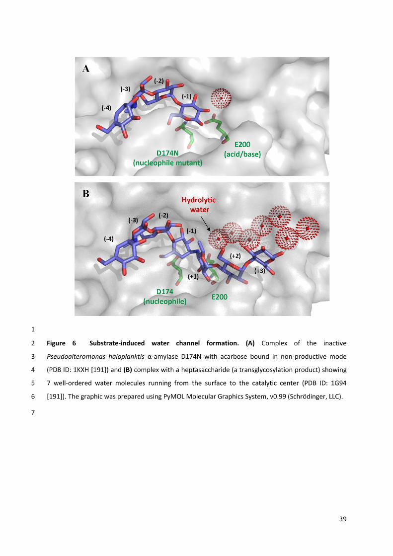
39
1
Figure 6 Substrate-induced water channel formation. (A) Complex of the inactive 2
Pseudoalteromonas haloplanktis α-amylase D174N with acarbose bound in non-productive mode 3
(PDB ID: 1KXH [191]) and (B) complex with a heptasaccharide (a transglycosylation product) showing 4
7 well-ordered water molecules running from the surface to the catalytic center (PDB ID: 1G94 5
[191]). The graphic was prepared using PyMOL Molecular Graphics System, v0.99 (Schrödinger, LLC). 6
7

40
1
From an evolutionary perspective, the presence of specific water binding determinants (e.g. 2
water-binding tyrosine residues) or water delivery systems within the active site can be considered as 3
extra features that contribute to the enzyme’s hydrolytic potency. Nevertheless, the presence of 4
such features is manifestly facultative for activity, because TS stabilization is the key element for 5
catalysis to occur. Indeed, the lack of specific water interactions during the deglycosylation step may 6
explain why this step is usually rate-limiting in the majority of GHs. 7
8
9
Figure 7 Over a century of knowledge pertaining to glycoside hydrolases and their ability to 10
catalyse both hydrolysis and transglycosylation. 11
12
Conclusions 13
Hopefully, this quite extensive review of available literature allows the reader to better perceive a 14
certain number of generic features that allow GHs to perform transglycosylation in adverse reaction 15
conditions, dominated by the presence of water (Figure 7). 16
Unsurprisingly, the first lesson that can be drawn from the examples that are described herein is 17
that TGs are probably characterized by modified transition states when compared to their GH 18
counterparts, as illustrated by our ‘locked door’ analogy. This leads to the conclusion that the first 19
condition that must be met if a GH is to be converted into an efficient TG is that new transition state 20
structures or electronic displacement systems must be developed that lower the efficiency of water-21
mediated deglycosylation, thus radically reducing the inherent advantage of ubiquitous water over 22
other suitably-nucleophilic acceptors. The analysis of available data indicates that the destabilization 23
of TS2water is generally brought about by altering interactions in the negative subsite, which inevitably 24
41
results in higher transition state energy barriers (c.f. enhanced stiffness of the lock mechanism) for 1
both deglycosylation and glycosylation (TS2 and TS1 respectively), since they display similar 2
properties. In turn, this translates into less efficient catalysis (i.e. low values of kcat/KM). 3
The second lesson that can be drawn from the cumulative knowledge presented here is that 4
transglycosylation is also promoted by the presence of substrate-specific interactions in the positive 5
subsites (c.f. the better grip on the door handle). These are useful, because they partially 6
compensate for the destabilization of the negative subsite interactions, possibly providing a situation 7
where TS2acceptor energy is lower than that of TS2water. Moreover, these can be very specific for certain 8
acceptor molecules, thus their presence does not necessarily imply more general access for 9
acceptors, including water. In this respect, it is noteworthy that the conversion of endo-GHs into TG 10
(i.e. endo-TGs) might not be accompanied by significantly lowered kcat/KM values, because in these 11
enzymes both donor and acceptor regions are involved in donor substrate recognition, with relative 12
contributions probably being GH family-dependent. Consequently, understanding the evolutionary 13
pathway that has provided endo-TGs may prove to be more complicated than the study of exo-TGs. 14
Finally, despite the fact that the natural solvent for most enzymes is water, most of this remains 15
as bulk water. For catalysis many GHs display specific features that either fix specific water molecules 16
within the vicinity of the catalytic center and/or deliver water to the active site via channels. In both 17
cases, these water-specific molecular determinants can be modified in order to reduce the presence 18
of catalytic water, thus further promoting transglycosylation. 19
In the case of natural TGs, evolutionary processes have no doubt resulted in the combination of 20
the aforementioned features. However, it is certain that the foremost of these must be the 21
destabilization of TS2water, a modification that we consider to be the main driving force of the GH→TG 22
transition. Unfortunately, from an enzyme engineering perspective, the rational modification of TS 23
structures (geometry and/or electronic features) remains impossible due to insufficient knowledge of 24
these. Nevertheless, in this respect, the use of combined approaches, associating QM/MM and 25
experimental validation (e.g. the specific design of transition state inhibitors for GHs and TG 26
counterparts), should provide new information in the near future. Moreover, with the increasing 27
integration of knowledge in the fields of chemistry and biology, it is likely that new comprehension of 28
carbohydrate reactivity in chemically-catalyzed glycosylation will rapidly contribute to better 29
understanding of the enzyme-substrate interactions that are necessary for transglycosylation to 30
occur. 31
From an evolutionary standpoint, it appears that TGs have mostly evolved from their hydrolytic 32
counterparts, although endo-acting XTH is an exception to this general rule [97]. Therefore, it is 33
intriguing to speculate upon the circumstances that might have led to the conversion of catalytically-34

42
optimized GHs into today’s relatively inefficient TGs. Logically, glycosynthesis should have been left 1
to the dedicated glycosynthetic enzymes (i.e. GTs) that we know today. Unless evolutionary 2
divergence between GHs and TGs occurred at a point when these were not available? 3
Despite the long history of carbohydrate chemistry, the fact that some fundamental features of 4
glycosylation (e.g. the factors that influence anomeric center reactivity and stereo-electronic effects) 5
are still subject to research [197,198] underlines the complexity of this important field and reveals 6
the fact that there are still considerable opportunities for progress. In the case of synthetic 7
glycochemistry, the extension of the available palette of TGs is undoubtedly an attractive grail for 8
enzyme engineers. From this point of view, the knowledge that is presented herein should provide a 9
rational starting point for would-be enzyme engineers wishing to convert GHs into TGs. 10
11
ACKNOWLEDGEMENTS 12
The authors would like to thank Drs. Leila Lo Leggio, Marco Moracci, Antoni Planas, Morten Sørlie 13
and Hugues Driguez for their critical appraisal of this manuscript. 14
15
REFERENCES 16
1 Varki, A., Cummings, R. D., Esko, J. D., Freeze, H. H., Stanley, P., Bertozzi, C. R., Hart, G. W. and 17 Etzler, M. E. (2009) In Essentials of Glycobiology, Cold Sprin Harbor Lab Press 18
2 Edelman, J. (1956) The formation of oligosaccharides by enzymic transglycosylation. Adv. 19 Enzym. Relat. Areas Mol. Biol. 17, 189–232 20
3 Breton, C., Fournel-Gigleux, S. and Palcic, M. M. (2012) Recent structures, evolution and 21 mechanisms of glycosyltransferases. Curr. Opin. Struct. Biol. 22, 540–549 22
4 Lairson, L. L., Henrissat, B., Davies, G. J. and Withers, S. G. (2008) Glycosyltransferases: 23 structures, functions, and mechanisms. Annu. Rev. Biochem. 77, 521–555 24
5 Field, R. A. (2011) Glycobiology: challenging reaction equilibria. Nat. Chem. Biol. 7, 658–659 25 6 Gantt, R. W., Peltier-Pain, P., Cournoyer, W. J. and Thorson, J. S. (2011) Using simple donors to 26
drive the equilibria of glycosyltransferase-catalyzed reactions. Nat. Chem. Biol. 7, 685–691 27 7 Cote, G. L. and Tao, B. Y. (1990) Oligosaccharide synthesis by enzymatic transglycosylation. 28
Glycoconjugate J. 7, 145–162 29 8 Mackenzie, L. F., Wang, Q., Warren, R. A. J. and Withers, S. G. (1998) Glycosynthases: mutant 30
glycosidases for oligosaccharide synthesis. J. Am. Chem. Soc. 120, 5583–5584 31 9 Malet, C. and Planas, A. (1998) From β-glucanase to β-glucansynthase: glycosyl transfer to α-32
glycosyl fluorides catalyzed by a mutant endoglucanase lacking its catalytic nucleophile. FEBS 33 Lett. 440, 208–212 34
10 Moracci, M., Trincone, A. and Rossi, M. (2001) Glycosynthases: new enzymes for 35 oligosaccharide synthesis. J. Mol. Catal. B Enzym. 11, 155–163 36
11 Perugino, G., Trincone, A., Rossi, M. and Moracci, M. (2004) Oligosaccharide synthesis by 37 glycosynthases. Trends Biotechnol. 22, 31–37 38
12 Faijes, M. and Planas, A. (2007) In vitro synthesis of artificial polysaccharides by glycosidases 39 and glycosynthases. Carbohydr. Res. 342, 1581–1594 40

43
13 Rakić, B. and Withers, S. G. (2009) Recent developments in glycoside synthesis with 1 glycosynthases and thioglycoligases. Aust. J. Chem. 62, 510–520 2
14 Cobucci-Ponzano, B. and Moracci, M. (2012) Glycosynthases as tools for the production of 3 glycan analogs of natural products. Nat. Prod. Rep. 29, 697–709 4
15 Schmaltz, R. M., Hanson, S. R. and Wong, C.-H. (2011) Enzymes in the synthesis of 5 glycoconjugates. Chem. Rev. 111, 4259–4307 6
16 Kobayashi, S. and Makino, A. (2009) Enzymatic polymer synthesis: an opportunity for green 7 polymer chemistry. Chem. Rev. 109, 5288–5353 8
17 Kadokawa, J. (2011) Precision polysaccharide synthesis catalyzed by enzymes. Chem. Rev. 9 111, 4308–4345 10
18 Zechel, D. L. and Withers, S. G. (2000) Glycosidase mechanisms: anatomy of a finely tuned 11 catalyst. Acc. Chem. Res. 33, 11–18 12
19 Vasella, A., Davies, G. J. and Böhm, M. (2002) Glycosidase mechanisms. Curr. Opin. Chem. 13 Biol. 6, 619–629 14
20 Vuong, T. V and Wilson, D. B. (2010) Glycoside hydrolases: catalytic base/nucleophile 15 diversity. Biotechnol. Bioeng. 107, 195–205 16
21 Henrissat, B. (1991) A classification of glycosyl hydrolases based sequence similarities amino 17 acid. Biochem. J. 280, 309–316 18
22 Henrissat, B. and Davies, G. (1997) Structural and sequence-based classification of glycoside 19 hydrolases. Curr. Opin. Struct. Biol. 7, 637–644 20
23 Lombard, V., Golaconda Ramulu, H., Drula, E., Coutinho, P. M. and Henrissat, B. (2014) The 21 carbohydrate-active enzymes database (CAZy) in 2013. Nucleic Acids Res. 42, D490–D495 22
24 Street, I. P., Kempton, J. B. and Withers, S. G. (1992) Inactivation of a β-glucosidase through 23 the accumulation of a stable 2-deoxy-2-fluoro-α-D-glucopyranosyl-enzyme intermediate: a 24 detailed investigation. Biochemistry 31, 9970–9978 25
25 Ducros, M.-A., Tarling, C. A., Zechel, D. L., Brzozowski, A. M., Frandsen, T. P., Ossowski, I. Von, 26 Withers, S. G. and Davies, G. J. (2003) Anatomy of glycosynthesis : structure and kinetics of 27 the Humicola insolens Cel7B E197A and E197S glycosynthase mutants. Chem. Biol. 10, 619–28 628 29
26 Koshland Jr., D. E. (1953) Stereochemistry and the mechanism of enzymatic reactions. Biol. 30 Rev. 28, 416–436 31
27 Eisenthal, R., Danson, M. J. and Hough, D. W. (1997) Catalytic efficiency and kcat/KM: a useful 32 comparator? Trends Biotechnol. 25, 247–249 33
28 Johnson, K. A. (2013) A century of enzyme kinetic analysis, 1913 to 2013. FEBS Lett. 587, 34 2753–2766. 35
29 Fischer, E. (1894) Einfluss der configuration auf die wirkung der enzyme. Ber. Dtsch. Chem. 36 Ges. 27, 2985–2993 37
30 Koshland, D. E. (1995) The key–lock theory and the induced fit theory. Angew. Chem. Int. Ed. 38 33, 2375–2378 39
31 Davies, G. J., Wilson, K. S. and Henrissat, B. (1997) Nomenclature for sugar-binding subsites in 40 glycosyl hydrolases. Biochem. J. 321, 557–559 41
32 Wolfenden, R. and Snider, M. J. (2001) The depth of chemical time and the power of enzymes 42 as catalysts. Acc. Chem. Res. 34, 938–945 43
33 Pauling, L. (1946) Molecular architecture and biological reactions. Chem. Eng. News 24, 1375–44 1377 45
34 Wolfenden, R., Lu, X. and Young, G. (1998) Spontaneous hydrolysis of glycosides. J. Am. Chem. 46 Soc. 120, 6814–6815 47
35 Eyring, H. (1935) The activated complex in chemical reactions. J. Chem. Phys. 3, 107–115 48 36 Warshel, A. (1981) Electrostatic basis of structure-function correlation in proteins. Acc. Chem. 49
Res. 14, 284–290 50 37 Tokuriki, N. and Tawfik, D. S. (2009) Protein dynamism and evolvability. Science 324, 203–207 51 38 Chouard, T. (2011) Breaking the protein rules. Nature 471, 151–153 52

44
39 Glowacki, D. R., Harvey, J. N. and Mulholland, A. J. (2012) Taking Ockham’s razor to enzyme 1 dynamics and catalysis. Nat. Chem. 4, 169–176 2
40 Davies, G. J., Ducros, V. M.-A., Varrot, A. and Zechel, D. L. (2003) Mapping the conformational 3 itinerary of β-glycosidases by X-ray crystallography. Biochem. Soc. Trans. 31, 523–527 4
41 Taha, H. A., Richards, M. R. and Lowary, T. L. (2013) Conformational analysis of furanoside-5 containing mono- and oligosaccharides. Chem. Rev. 113, 1851–1876 6
42 Houseknecht, J. B., Altona, C., Hadad, C. M. and Lowary, T. L. (2002) Conformational analysis 7 of furanose rings with PSEUROT: parametrization for rings possessing the arabino, lyxo , ribo, 8 and xylo stereochemistry and application to arabinofuranosides. J. Org. Chem 67, 4647–4651 9
43 Sinnott, M. L. (1990) Catalytic mechanism of enzymic glycosyl transfer. Chem. Rev. 90, 1171–10 1202 11
44 Vocadlo, D. J. and Davies, G. J. (2008) Mechanistic insights into glycosidase chemistry. Curr. 12 Opin. Chem. Biol. 12, 539–555 13
45 Hovel, K., Shallom, D., Niefind, K., Belakhov, V., Shoham, G., Baasov, T., Shoham, Y. and 14 Schomburg, D. (2003) Crystal structure and snapshots along the reaction pathway of a family 15 51 α-L-arabinofuranosidase. EMBO J. 22, 4922–4932 16
46 Davies, G. J., Planas, A. and Rovira, C. (2012) Conformational analyses of the reaction 17 coordinate of glycosidases. Acc. Chem. Res. 45, 308–316 18
47 Sulzenbacher, G., Driguez, H., Henrissat, B., Schülein, M. and Davies, G. J. (1996) Structure of 19 the Fusarium oxysporum endoglucanase I with a nonhydrolyzable substrate analogue: 20 substrate distortion gives rise to the preferred axial orientation for the leaving group. 21 Biochemistry 35, 15280–15287 22
48 Davies, G. J., Mackenzie, L., Varrot, A., Dauter, M. and Brzozowski, A. M. (1998) Snapshots 23 along an enzymatic reaction coordinate : analysis of a retaining β-glycosidase. Biochemistry 24 37, 11707–11713 25
49 Biarnés, X., Nieto, J., Planas, A. and Rovira, C. (2006) Substrate distortion in the Michaelis 26 complex of Bacillus 1,3-1,4-β-glucanase. Insight from first principles molecular dynamics 27 simulations. J. Biol. Chem. 281, 1432–1441 28
50 Biarnés, X., Ardèvol, A., Iglesias-Fernández, J., Planas, A. and Rovira, C. (2011) Catalytic 29 itinerary in 1,3-1,4-β-glucanase unraveled by QM/MM metadynamics. Charge is not yet fully 30 developed at the oxocarbenium ion-like transition state. J. Am. Chem. Soc. 133, 20301–20309 31
51 Zechel, D. L., Reid, S. P., Stoll, D., Nashiru, O., Warren, R. A. J. and Withers, S. G. (2003) 32 Mechanism, mutagenesis, and chemical rescue of a β-mannosidase from Cellulomonas fimi. 33 Biochemistry 42, 7195–7204 34
52 Namchuk, M. N. and Withers, S. G. (1995) Mechanism of Agrobacterium β-glucosidase: kinetic 35 analysis of the role of noncovalent enzyme/substrate interactions. Biochemistry 34, 16194–36 16202 37
53 Notenboom, V., Birsan, C., Nitz, M., Rose, D. R., Warren, R. a and Withers, S. G. (1998) Insights 38 into transition state stabilization of the β-1,4-glycosidase Cex by covalent intermediate 39 accumulation in active site mutants. Nat. Struct. Biol. 5, 812–818 40
54 Sierks, M. R. and Svensson, B. (1992) Kinetic identification of a hydrogen bonding pair in the 41 glucoamylase-maltose transition state complex. Protein Eng. 5, 185–188 42
55 Kim, Y.-W., Zhang, R., Chen, H. and Withers, S. G. (2010) O-glycoligases, a new category of 43 glycoside bond-forming mutant glycosidases, catalyse facile syntheses of isoprimeverosides. 44 Chem. Commun. 46, 8725–8727 45
56 Heightman, T. D. and Vasella, A. T. (1999) Recent insights into inhibition, structure, and 46 mechanism of configuration-retaining glycosidases. Angew. Chem. Int. Ed. 38, 750–770 47
57 Nerinckx, W., Desmet, T., Piens, K. and Claeyssens, M. (2005) An elaboration on the syn-anti 48 proton donor concept of glycoside hydrolases: electrostatic stabilisation of the transition state 49 as a general strategy. FEBS Lett. 579, 302–312 50
58 Nerinckx, W., Desmet, T. and Claeyssens, M. (2003) A hydrophobic platform as a 51 mechanistically relevant transition state stabilising factor appears to be present in the active 52

45
centre of all glycoside hydrolases. FEBS Lett. 538, 1–7 1 59 Schramm, V. L. (2011) Enzymatic transition states, transition-state analogs, dynamics, 2
thermodynamics, and lifetimes. Annu. Rev. Biochem. 80, 703–732 3 60 Bigeleisen, J. and Mayer, M. G. (1947) Calculation of equilibrium constants for isotopic 4
exchange reactions. J. Chem. Phys. 15, 261–267 5 61 Cleland, W. W. (2003) The use of isotope effects to determine enzyme mechanisms. J. Biol. 6
Chem. 278, 51975–51984 7 62 Kempton, J. B. and Withers, S. G. (1992) Mechanism of Agrobacterium β-glucosidase: kinetic 8
studies. Biochemistry 31, 9961–9969 9 63 Wang, J., Hou, Q., Sheng, X., Gao, J., Liu, Y. and Liu, C. (2013) Theoretical study on the 10
deglycosylation mechanism of rice BGlu1 β-glucosidase. Int. J. Quantum Chem. 113, 1071–11 1075 12
64 Wang, J., Hou, Q., Dong, L., Liu, Y. and Liu, C. (2011) QM/MM studies on the glycosylation 13 mechanism of rice BGlu1 β-glucosidase. J. Mol. Graph. Model. 30, 148–152 14
65 Brás, N. F., Ramos, M. J. and Fernandes, P. A. (2010) DFT studies on the β-glycosidase catalytic 15 mechanism: the deglycosylation step. J. Mol. Struct. THEOCHEM 946, 125–133 16
66 Badieyan, S., Bevan, D. R. and Zhang, C. (2012) Probing the active site chemistry of β-17 glucosidases along the hydrolysis reaction pathway. Biochemistry 51, 8907–8918 18
67 Demetrius, L. (1998) Role of enzyme-substrate flexibility in catalytic activity: an evolutionary 19 perspective. J. Theor. Biol. 194, 175–194 20
68 del-Rio, G., Morett, E. and Soberon, X. (1997) Did cyclodextrin glycosyltransferases evolve 21 from α-amylases? FEBS Lett. 416, 221–224 22
69 Ritsema, T., Hernández, L., Verhaar, A., Altenbach, D., Boller, T., Wiemken, A. and Smeekens, 23 S. (2006) Developing fructan-synthesizing capability in a plant invertase via mutations in the 24 sucrose-binding box. Plant J. 48, 228–237 25
70 Luang, S., Cho, J.-I., Mahong, B., Opassiri, R., Akiyama, T., Phasai, K., Komvongsa, J., Sasaki, N., 26 Hua, Y., Matsuba, Y., Ozeki, Y., Jeon, J.-S. and Cairns, J. R. K. (2013) Rice Os9BGlu31 is a 27 transglucosidase with the capacity to equilibrate phenylpropanoid, flavonoid, and 28 phytohormone glycoconjugates. J. Biol. Chem. 288, 10111–10123 29
71 Schröder, R., Atkinson, R. G. and Redgwell, R. J. (2009) Re-interpreting the role of endo-β-30 mannanases as mannan endotransglycosylase/hydrolases in the plant cell wall. Ann. Bot. 104, 31 197–204 32
72 Johnston, S. L., Prakash, R., Chen, N. J., Kumagai, M. H., Turano, H. M., Cooney, J. M., 33 Atkinson, R. G., Paull, R. E., Cheetamun, R., Bacic, A., Brummell, D. A. and Schröder, R. (2013) 34 An enzyme activity capable of endotransglycosylation of heteroxylan polysaccharides is 35 present in plant primary cell walls. Planta 237, 173–187 36
73 Franková, L. and Fry, S. C. (2011) Phylogenetic variation in glycosidases and glycanases acting 37 on plant cell wall polysaccharides, and the detection of transglycosidase and trans-β-xylanase 38 activities. Plant J. 67, 662–681 39
74 Franková, L. and Fry, S. C. (2012) Trans-α-xylosidase and trans-β-galactosidase activities, 40 widespread in plants, modify and stabilize xyloglucan structures. Plant J. 71, 45–60 41
75 Fry, S. C. (2004) Primary cell wall metabolism: tracking the careers of wall polymers in living 42 plant cells. New Phytol. 161, 641–675 43
76 van der Veen, B. A., Uitdehaag, J. C., Penninga, D., van Alebeek, G. J., Smith, L. M., Dijkstra, B. 44 W. and Dijkhuizen, L. (2000) Rational design of cyclodextrin glycosyltransferase from Bacillus 45 circulans strain 251 to increase α-cyclodextrin production. J. Mol. Biol. 296, 1027–1038 46
77 Mori, H., Bak-Jensen, K. S. and Svensson, B. (2002) Barley α-amylase Met53 situated at the 47 high-affinity subsite −2 belongs to a substrate binding motif in the β→α loop 2 of the catalytic 48 (β/α)8-barrel and is critical for activity and substrate specificity. Eur. J. Biochem. 269, 5377–49 5390 50
78 Potocki de Montalk, G., Remaud-Simeon, M., Willemot, R. M., Sarçabal, P., Planchot, V. and 51 Monsan, P. (2000) Amylosucrase from Neisseria polysaccharea: novel catalytic properties. 52
46
FEBS Lett. 471, 219–223 1 79 Kim, M.-I., Kim, H.-S., Jung, J. and Rhee, S. (2008) Crystal structures and mutagenesis of 2
sucrose hydrolase from Xanthomonas axonopodis pv. glycines: insight into the exclusively 3 hydrolytic amylosucrase fold. J. Mol. Biol. 380, 636–647 4
80 Baumann, M. J., Eklöf, J. M., Michel, G., Kallas, A. M., Teeri, T. T., Czjzek, M. and Brumer, H. 5 (2007) Structural evidence for the evolution of xyloglucanase activity from xyloglucan endo-6 transglycosylases: biological implications for cell wall metabolism. Plant Cell 19, 1947–1963 7
81 Schroeven, L., Lammens, W., Van Laere, A. and Van den Ende, W. (2008) Transforming wheat 8 vacuolar invertase into a high affinity sucrose: sucrose 1-fructosyltransferase. New Phytol. 9 180, 822–831 10
82 Ribeirāo, M., Pereira-Chioccola, V. L., Eichinger, D., Rodrigues, M. M. and Schenkman, S. 11 (1997) Temperature differences for trans-glycosylation and hydrolysis reaction reveal an 12 acceptor binding site in the catalytic mechanism of Trypanosoma cruzi trans-sialidase. 13 Glycobiology 7, 1237–1246 14
83 Paris, G., Ratier, L., Amaya, M. F., Nguyen, T., Alzari, P. M. and Frasch, A. C. C. (2005) A 15 sialidase mutant displaying trans-sialidase activity. J. Mol. Biol. 345, 923–934 16
84 Fry, S. C., York, W. S., Albersheim, P., Darvill, A., Hayashi, T., Joseleau, J.-P., Kato, Y., Lorences, 17 E. P., Maclachlan, G. A., McNeil, M., Mort, A. J., Reid, J. S. G., Seitz, H. U., Selvendran, R. R., 18 Voragen, A. G. J. and White A. R. (1993) An unambigous nomenclature for xyloglucan-derived 19 oligosaccharides. Physiol. Plant. 89, 1–3 20
85 Gilbert, H. J., Stålbrand, H. and Brumer, H. (2008) How the walls come crumbling down: recent 21 structural biochemistry of plant polysaccharide degradation. Curr. Opin. Plant Biol. 11, 338–22 348 23
86 Strohmeier, M., Hrmova, M., Fischer, M., Harvey, A. J., Fincher, G. B. and Pleiss, J. (2004) 24 Molecular modeling of family GH16 glycoside hydrolases: Potential roles for xyloglucan 25 transglucosylases/hydrolases in cell wall modification in the poaceae. Protein Sci. 13, 3200–26 3213 27
87 Campbell, P. and Braam, J. (1999) Xyloglucan endotransglycosylases: diversity of genes, 28 enzymes and potential wall-modifying functions. Trends Plant. Sci. 4, 361–366 29
88 Rose, J. K. C., Braam, J., Fry, S. C. and Nishitani, K. (2002) The XTH family of enzymes involved 30 in xyloglucan endotransglucosylation and endohydrolysis: current perspectives and a new 31 unifying nomenclature. Plant Cell Physiol. 43, 1421–1435 32
89 Eklöf, J. M. and Brumer, H. (2010) The XTH gene family: an update on enzyme structure, 33 function, and phylogeny in xyloglucan remodeling. Plant Physiol. 153, 456–466 34
90 Saura-Valls, M., Fauré, R., Ragàs, S., Piens, K., Brumer, H., Teeri, T. T., Cottaz, S., Driguez, H. 35 and Planas, A. (2006) Kinetic analysis using low-molecular mass xyloglucan oligosaccharides 36 defines the catalytic mechanism of a Populus xyloglucan endotransglycosylase. Biochem. J. 37 395, 99–106 38
91 Saura-Valls, M., Fauré, R., Brumer, H., Teeri, T. T., Cottaz, S., Driguez, H. and Planas, A. (2008) 39 Active-site mapping of a Populus xyloglucan endo-transglycosylase with a library of xylogluco-40 oligosaccharides. J. Biol. Chem 283, 21853–21863 41
92 Piens, K., Fauré, R., Sundqvist, G., Baumann, M. J., Saura-Valls, M., Teeri, T. T., Cottaz, S., 42 Planas, A., Driguez, H. and Brumer, H. (2008) Mechanism-based labeling defines the free 43 energy change for formation of the covalent glycosyl-enzyme intermediate in a xyloglucan 44 endo-transglycosylase. J. Biol. Chem. 283, 21864–21872 45
93 Steele, N. M. and Fry, S. C. (1999) Purification of xyloglucan endotransglycosylases (XETs): a 46 generally applicable and simple method based on reversible formation of an enzyme–47 substrate complex. Biochem. J. 340, 207–211 48
94 Sulová, Z., Takáčová, M., Steele‹ , N. M., Fry, S. C. and Farkaš, V. (1998) Xyloglucan 49 endotransglycosylase: evidence for the existence of a relatively stable glycosyl–enzyme 50 intermediate. Biochem. J. 330, 1475–1480 51
95 Johansson, P., Brumer, H., Baumann, M. J., Kallas, A. M., Henriksson, H., Denman, S. E., Teeri, 52

47
T. T. and Jones, T. A. (2004) Crystal structures of a poplar xyloglucan endotransglycosylase 1 reveal details of transglycosylation acceptor binding. Plant Cell 16, 874–886 2
96 Fauré, R., Saura-Valls, M., Brumer, H., Planas, A., Cottaz, S. and Driguez, H. (2006) Synthesis of 3 a library of xylogluco-oligosaccharides for active-site mapping of xyloglucan endo-4 transglycosylase. J. Org. Chem. 71, 5151–5161 5
97 Eklöf, J. M., Shojania, S., Okon, M., McIntosh, L. P. and Brumer, H. (2013) Structure-function 6 analysis of a broad specificity Populus trichocarpa endo-β-glucanase reveals an evolutionary 7 link between bacterial licheninases and plant XTH gene products. J. Biol. Chem. 288, 15786–8 15799 9
98 Mark, P., Baumann, M. J., Eklöf, J. M., Gullfot, F., Michel, G., Kallas, A. M., Teeri, T. T., Brumer, 10 H. and Czjzek, M. (2009) Analysis of nasturtium TmNXG1 complexes by crystallography and 11 molecular dynamics provides detailed insight into substrate recognition by family GH16 12 xyloglucan endo-transglycosylases and endo-hydrolases. Proteins 75, 820–836 13
99 Monsan, P., Remaud-Siméon, M. and André, I. (2010) Transglucosidases as efficient tools for 14 oligosaccharide and glucoconjugate synthesis. Curr. Opin. Microbiol. 13, 293–300 15
100 van Hijum, S. A. F. T., Kralj, S., Ozimek, L. K., Dijkhuizen, L. and van Geel-schutten, I. G. H. 16 (2006) Structure-Function relationships of glucansucrase and fructansucrase enzymes from 17 lactic acid bacteria. Microbiol. Mol. Biol. Rev. 70, 157–176 18
101 Monchois, V., Willemot, R. M. and Monsan, P. (1999) Glucansucrases: mechanism of action 19 and structure-function relationships. FEMS Microbiol. Rev. 23, 131–151 20
102 Leemhuis, H., Pijning, T., Dobruchowska, J. M., van Leeuwen, S. S., Kralj, S., Dijkstra, B. W. and 21 Dijkhuizen, L. (2013) Glucansucrases: three-dimensional structures, reactions, mechanism, α-22 glucan analysis and their implications in biotechnology and food applications. J. Biotechnol. 23 163, 250–272 24
103 Stam, M. R., Danchin, E. G. J., Rancurel, C., Coutinho, P. M. and Henrissat, B. (2006) Dividing 25 the large glycoside hydrolase family 13 into subfamilies: towards improved functional 26 annotations of α-amylase-related proteins. Protein Eng. Des. Sel. 19, 555–562 27
104 Vujicic-Zagar, A., Pijning, T., Kralj, S., López, C. A. and Eeuwema, W. (2010) Crystal structure of 28 a 117 kDa glucansucrase fragment provides insight into evolution and product specificity of 29 GH70 enzymes. Proc. Natl. Acad. Sci. U.S.A. 107, 21406–21411 30
105 Ito, K., Ito, S., Shimamura, T., Weyand, S., Kawarasaki, Y., Misaka, T., Abe, K., Kobayashi, T., 31 Cameron, A. D. and Iwata, S. (2011) Crystal structure of glucansucrase from the dental caries 32 pathogen Streptococcus mutans. J. Mol. Biol. 408, 177–186 33
106 Brison, Y., Pijning, T., Malbert, Y., Fabre, É., Mourey, L., Morel, S., Potocki-Véronèse, G., 34 Monsan, P., Tranier, S., Remaud-Siméon, M. and Dijkstra, B. W. (2012) Functional and 35 structural characterization of α-(1→2) branching sucrase derived from DSR-E glucansucrase. J. 36 Biol. Chem. 287, 7915–7924 37
107 Leemhuis, H., Pijning, T., Dobruchowska, J. M., Dijkstra, B. W. and Dijkhuizen, L. (2012) 38 Glycosidic bond specificity of glucansucrases: on the role of acceptor substrate binding 39 residues. Biocatal. Biotransfor. 30, 366–376 40
108 Kralj, S., van Geel-Schutten, G. H., van der Maarel, M. J. E. C. and Dijkhuizen, L. (2004) 41 Biochemical and molecular characterization of Lactobacillus reuteri 121 reuteransucrase. 42 Microbiol. 150, 2099–2112 43
109 Kralj, S., van Leeuwen, S. S., Valk, V., Eeuwema, W., Kamerling, J. P. and Dijkhuizen, L. (2008) 44 Hybrid reuteransucrase enzymes reveal regions important for glucosidic linkage specificity 45 and the transglucosylation/hydrolysis ratio. FEBS J. 275, 6002–6010 46
110 Goldberg, R. N., Tewaril, Y. B. and Ahluwaliaq, J. C. (1989) Thermodynamics of the hydrolysis 47 of sucrose. J. Biol. Chem. 264, 9901–9904 48
111 Kralj, S., Grijpstra, P., van Leeuwen, S. S., Leemhuis, H., Dobruchowska, J. M., van der Kaaij, R. 49 M., Malik, A., Oetari, A., Kamerling, J. P. and Dijkhuizen, L. (2011) 4,6-α-Glucanotransferase, a 50 novel enzyme that structurally and functionally provides an evolutionary link between 51 glycoside hydrolase enzyme families 13 and 70. Appl. Environ. Microbiol. 77, 8154–8163 52

48
112 Skov, L. K., Mirza, O., Henriksen, A., De Montalk, G. P., Remaud-Siméon, M., Sarçabal, P., 1 Willemot, R. M., Monsan, P. and Gajhede, M. (2001) Amylosucrase, a glucan-synthesizing 2 enzyme from the α-amylase family. J. Biol. Chem. 276, 25273–25278 3
113 Guérin, F., Barbe, S., Pizzut-Serin, S., Potocki-Véronèse, G., Guieysse, D., Guillet, V., Monsan, 4 P., Mourey, L., Remaud-Siméon, M., André, I., and Tranier, S. (2012) Structural investigation of 5 the thermostability and product specificity of amylosucrase from the bacterium Deinococcus 6 geothermalis. J. Biol. Chem. 287, 6642–6654 7
114 Albenne, C., Skov, L. K., Mirza, O., Gajhede, M., Feller, G., D’Amico, S., André, G., Potocki-8 Véronèse, G., van der Veen, B., Monsan, P., and Remaud-Simeon, M. (2004) Molecular basis 9 of the amylose-like polymer formation catalyzed by Neisseria polysaccharea amylosucrase. J. 10 Biol. Chem. 279, 726–734 11
115 Cambon, E., Barbe, S., Pizzut-Serin, S., Remaud-Simeon, M. and André, I. (2014) Essential role 12 of amino acid position 226 in oligosaccharide elongation by amylosucrase from Neisseria 13 polysaccharea. Biotechnol. Bioeng. 111, 1719–1728 14
116 Champion, E., Guérin, F., Moulis, C., Barbe, S., Tran, T. H., Morel, S., Descroix, K., Monsan, P., 15 Mourey, L., Mulard, L., Tranier, S., Remaud-Siméon, M. and André, I. (2012) Applying pairwise 16 combinations of amino acid mutations for sorting out highly efficient glucosylation tools for 17 chemo-enzymatic synthesis of bacterial oligosaccharides. J. Am. Chem. Soc. 134, 18677–18 18688 19
117 Daudé, D., Champion, E., Morel, S., Guieysse, D., Remaud-Siméon, M. and André, I. (2013) 20 Probing substrate promiscuity of amylosucrase from Neisseria polysaccharea. ChemCatChem 21 5, 2288–2295 22
118 Kim, H., Park, H., Heu, S. and Jung, J. (2004) Molecular and functional characterization of a 23 unique sucrose hydrolase from Xanthomonas axonopodis pv. glycines. J. Bacteriol. 186, 411–24 418 25
119 Skov, L. K., Pizzut-Serin, S., Remaud-Siméon, M., Ernst, H. A., Gajhede, M. and Mirza, O. (2013) 26 The structure of amylosucrase from Deinococcus radiodurans has an unusual open active-site 27 topology. Acta Crystallogr. Sect. F 69, 973–978 28
120 Pizzut-Serin, S., Potocki-Véronèse, G., van der Veen, B. A., Albenne, C., Monsan, P. and 29 Remaud-Siméon, M. (2005) Characterisation of a novel amylosucrase from Deinococcus 30 radiodurans. FEBS Lett. 579, 1405–1410 31
121 Olvera, C., Centeno-Leija, S., Ruiz-Leyva, P. and López-Munguía, A. (2012) Design of chimeric 32 levansucrases with improved transglycosylation activity. Appl. Environ. Microbiol. 78, 1820–33 1825 34
122 Lammens, W., Le Roy, K., Schroeven, L., Van Laere, A., Rabijns, A. and Van den Ende, W. 35 (2009) Structural insights into glycoside hydrolase family 32 and 68 enzymes: functional 36 implications. J. Exp. Bot. 60, 727–740 37
123 Van den Ende, W., Lammens, W., Van Laere, A., Schroeven, L. and Le Roy, K. (2009) Donor and 38 acceptor substrate selectivity among plant glycoside hydrolase family 32 enzymes. FEBS J. 39 276, 5788–5798 40
124 Sainz-Polo, M. A., Ramírez-Escudero, M., Lafraya, A., González, B., Marín-Navarro, J., Polaina, 41 J. and Sanz-Aparicio, J. (2013) Three-dimensional structure of Saccharomyces invertase: role 42 of a non-catalytic domain in oligomerization and substrate specificity. J. Biol. Chem. 288, 43 9755–9766 44
125 Lafraya, Á., Sanz-Aparicio, J., Polaina, J. and Marín-Navarro, J. (2011) Fructo-oligosaccharide 45 synthesis by mutant versions of Saccharomyces cerevisiae invertase. Appl. Environ. Microbiol. 46 77, 6148–6157 47
126 Ritsema, T., Verhaar, A., Vijn, I. and Smeekens, S. (2005) Using natural variation to investigate 48 the function of individual amino acids in the sucrose-binding box of fructan:fructan 6G-49 fructosyltransferase (6G-FFT) in product formation. Plant Mol. Biol. 58, 597–607 50
127 Altenbach, D., Rudiño-Pinera, E., Olvera, C., Boller, T., Wiemken, A. and Ritsema, T. (2009) An 51 acceptor-substrate binding site determining glycosyl transfer emerges from mutant analysis of 52
49
a plant vacuolar invertase and a fructosyltransferase. Plant Mol. Biol. 69, 47–56 1 128 Álvaro-Benito, M., Sainz-Polo, M. A., González-Pérez, D., González, B., Plou, F. J., Fernández-2
Lobato, M. and Sanz-Aparicio, J. (2012) Structural and kinetic insights reveal that the amino 3 acid pair Gln-228/Asn-254 modulates the transfructosylating specificity of Schwanniomyces 4 occidentalis β-fructofuranosidase, an enzyme that produces prebiotics. J. Biol. Chem. 287, 5 19674–19686 6
129 Uitdehaag, J. C. M. (1999) The cyclization mechanism of cyclodextrin glycosyltransferase 7 (CGTase) as revealed by a γ-cyclodextrin-CGTase complex at 1.8-Å resolution. J. Biol. Chem. 8 274, 34868–34876 9
130 Leemhuis, H., Kelly, R. M. and Dijkhuizen, L. (2010) Engineering of cyclodextrin 10 glucanotransferases and the impact for biotechnological applications. Appl. Microbiol. 11 Biotechnol. 85, 823–835 12
131 Van der Veen, B. A., Leemhuis, H., Kralj, S., Uitdehaag, J. C., Dijkstra, B. W. and Dijkhuizen, L. 13 (2001) Hydrophobic amino acid residues in the acceptor binding site are main determinants 14 for reaction mechanism and specificity of cyclodextrin-glycosyltransferase. J. Biol. Chem. 276, 15 44557–44562 16
132 Kelly, R. M., Leemhuis, H. and Dijkhuizen, L. (2007) Conversion of a cyclodextrin 17 glucanotransferase into an α-amylase: assessment of directed evolution strategies. 18 Biochemistry 46, 11216–11222 19
133 Rivera, M. H., López-Munguía, A., Soberón, X. and Saab-Rincón, G. (2003) α-Amylase from 20 Bacillus licheniformis mutants near to the catalytic site: effects on hydrolytic and 21 transglycosylation activity. Protein Eng. 16, 505–514 22
134 Kim, T., Park, C., Cho, H., Cha, S., Kim, J., Lee, S., Moon, T., Kim, J., Oh, B. and Park, K. (2000) 23 Role of the glutamate 332 residue in the transglycosylation activity of Thermus maltogenic 24 amylase. Biochemistry 39, 6773–6780 25
135 Tran, P. L., Cha, H.-J., Lee, J.-S., Park, S.-H., Woo, E.-J. and Park, K.-H. (2014) Introducing 26 transglycosylation activity in Bacillus licheniformis α-amylase by replacement of His235 with 27 Glu. Biochem. Biophys. Res. Commun. 451, 541–547 28
136 Beier, L., Svendsen, A., Andersen, C., Frandsen, T. P., Borchert, T. V and Cherry, J. R. (2000) 29 Conversion of the maltogenic α-amylase Novamyl into a CGTase. Protein Eng. 13, 509–513 30
137 Jemli, S., Ben-Ali, M., Ben-Hlima, H., Khemakhem, B. and Bejar, S. (2012) Mutations affecting 31 the activity of the cyclodextrin glucanotransferase of Paenibacillus pabuli US132: insights into 32 the low hydrolytic activity of cyclodextrin glucanotransferases. Biologia 67, 636–643 33
138 Asensio, J. L., Ardá, A., Cañada, F. J. and Jiménez-Barbero, J. (2013) Carbohydrate-aromatic 34 interactions. Acc. Chem. Res. 46, 946–954 35
139 Janeček, S. and Blesák, K. (2011) Sequence-structural features and evolutionary relationships 36 of family GH57 α-amylases and their putative α-amylase-like homologues. Prot. J. 30, 429–435 37
140 Janeček, S., Svensson, B. and MacGregor, E. A. (2003) Relation between domain evolution, 38 specificity, and taxonomy of the α-amylase family members containing a C-terminal starch-39 binding domain. Eur. J. Biochem. 270, 635–645 40
141 Sun, T., Letsididi, R., Pan, B. and Jiang, B. (2013) Production of a novel cyclodextrin 41 glycosyltransferase from Bacillus sp . SK13.002. Afr. J. Microbiol. Res. 7, 2311–2315 42
142 Bornscheuer, U. T. and Kazlauskas, R. J. (2004) Catalytic promiscuity in biocatalysis: using old 43 enzymes to form new bonds and follow new pathways. Angew. Chem., Int. Ed. 43, 6032–6040 44
143 Watts, A. G., Damager, I., Amaya, M. L., Buschiazzo, A., Alzari, P., Frasch, A. C. and Withers, S. 45 G. (2003) Trypanosoma cruzi trans-sialidase operates through a covalent sialyl-enzyme 46 intermediate: tyrosine is the catalytic nucleophile. J. Am. Chem. Soc. 125, 7532–7533 47
144 Amaya, M. F., Watts, A. G., Damager, I., Wehenkel, A., Nguyen, T., Buschiazzo, A., Paris, G., 48 Frasch, A. C., Withers, S. G. and Alzari, P. M. (2004) Structural insights into the catalytic 49 mechanism of Trypanosoma cruzi trans-sialidase. Structure 12, 775–784 50
145 Damager, I., Buchini, S., Amaya, M. F., Buschiazzo, A., Alzari, P., Frasch, A. C., Watts, A. and 51 Withers, S. G. (2008) Kinetic and mechanistic analysis of Trypanosoma cruzi trans-sialidase 52

50
reveals a classical ping-pong mechanism with acid/base catalysis. Biochemistry 47, 3507–3512 1 146 Montagna, G., Cremona, M. L., Paris, G., Amaya, M. F., Buschiazzo, A., Alzari, P. M. and Frasch, 2
A. C. C. (2002) The trans-sialidase from the african trypanosome Trypanosoma brucei. Eur. J. 3 Biochem. 269, 2941–2950 4
147 Buschiazzo, A., Tavares, G. A., Campetella, O., Spinelli, S., Cremona, M. L., París, G., Amaya, M. 5 F., Frasch, A. C. and Alzari, P. M. (2000) Structural basis of sialyltransferase activity in 6 trypanosomal sialidases. EMBO J. 19, 16–24 7
148 Amaya, M. F., Buschiazzo, A., Nguyen, T. and Alzari, P. M. (2003) The high resolution 8 structures of free and inhibitor-bound Trypanosoma rangeli sialidase and its comparison with 9 T. cruzi trans-sialidase. J. Mol. Biol. 325, 773–784 10
149 Buschiazzo, A., Amaya, M. F., Cremona, M. L., Frasch, A. C. and Alzari, P. M. (2002) The crystal 11 structure and mode of action of trans-sialidase, a key enzyme in Trypanosoma cruzi 12 pathogenesis. Mol. Cell 10, 757–768 13
150 Stierand, K. and Rarey, M. (2011) Consistent two-dimensional visualization of protein-ligand 14 complex series. J. Cheminform., Chemistry Central Ltd 3, 21 15
151 Demir, O. and Roitberg, A. E. (2009) Modulation of catalytic function by differential plasticity 16 of the active site: case study of Trypanosoma cruzi trans-sialidase and Trypanosoma rangeli 17 sialidase. Biochemistry 48, 3398–3406 18
152 Pierdominici-Sottile, G., Horenstein, N. A. and Roitberg, A. E. (2011) A free energy study of the 19 catalytic mechanism of Trypanosoma cruzi trans-sialidase. From the Michaelis complex to the 20 covalent intermediate. Biochemistry 50, 10150–10158 21
153 Pierdominici-Sottile, G., Palma, J. and Roitberg, A. E. (2013) Free energy computations identify 22 the mutations required to confer trans-sialidase activity into Trypanosoma rangeli sialidase. 23 Proteins 82, 424–435 24
154 Jers, C., Michalak, M., Larsen, D. M., Kepp, K. P., Li, H., Guo, Y., Kirpekar, F., Meyer, A. S. and 25 Mikkelsen, J. D. (2014) Rational design of a new Trypanosoma rangeli trans-sialidase for 26 efficient sialylation of glycans. PLoS One 9, e83902 27
155 Feng, H.-Y., Drone, J., Hoffmann, L., Tran, V., Tellier, C., Rabiller, C. and Dion, M. (2005) 28 Converting a β-glycosidase into a β-transglycosidase by directed evolution. J. Biol. Chem. 280, 29 37088–37097 30
156 Teze, D., Hendrickx, J., Czjzek, M., Ropartz, D., Sanejouand, Y.-H., Tran, V., Tellier, C. and Dion, 31 M. (2014) Semi-rational approach for converting a GH1 β-glycosidase into a β-32 transglycosidase. Protein Eng. Des. Sel. 27, 13–19 33
157 Teze, D., Daligault, F., Ferrières, V., Sanejouand, Y.-H. and Tellier, C. (2014) Semi-rational 34 approach for converting a GH36 α-glycosidase into an α-transglycosidase. Glycobiology, 35 doi:10.1093/glycob/cwu124 36
158 Aronson, N. N., Halloran, B. A., Alexeyev, M. F., Zhou, X. E., Wang, Y., Meehan, E. J. and Chen, 37 L. (2006) Mutation of a conserved tryptophan in the chitin-binding cleft of Serratia 38 marcescens chitinase A enhances transglycosylation. Biosci., Biotechnol., Biochem. 70, 243–39 251 40
159 Madhuprakash, J., Tanneeru, K., Purushotham, P., Guruprasad, L. and Podile, A. R. (2012) 41 Transglycosylation by chitinase D from Serratia proteamaculans improved through altered 42 substrate interactions. J. Biol. Chem. 287, 44619–44627 43
160 Lü, Y., Yang, H., Hu, H., Wang, Y., Rao, Z. and Jin, C. (2009) Mutation of Trp137 to glutamate 44 completely removes transglycosyl activity associated with the Aspergillus fumigatus AfChiB1. 45 Glycoconjugate J. 26, 525–534 46
161 Zakariassen, H., Hansen, M. C., Jøranli, M., Eijsink, V. G. H. and Sørlie, M. (2011) Mutational 47 effects on transglycosylating activity of family 18 chitinases and construction of a 48 hypertransglycosylating mutant. Biochemistry 50, 5693–5703 49
162 Merceron, R., Foucault, M., Haser, R., Mattes, R., Watzlawick, H. and Gouet, P. (2012) The 50 molecular mechanism of thermostable α-galactosidases AgaA and AgaB explained by X-ray 51 crystallography and mutational studies. J. Biol. Chem. 287, 39642–39652 52

51
163 Placier, G., Watzlawick, H., Rabiller, C. and Mattes, R. (2009) Evolved β-galactosidases from 1 Geobacillus stearothermophilus with improved transgalactosylation yield for galacto-2 oligosaccharide production. Appl. Environ. Microbiol. 75, 6312–6321 3
164 Saburi, W., Kobayashi, M., Mori, H., Okuyama, M. and Kimura, A. (2013) Replacement of the 4 catalytic nucleophile aspartyl residue of dextran glucosidase by cysteine sulfinate enhances 5 transglycosylation activity. J. Biol. Chem. 288, 31670–31677 6
165 Lawson, S. L., Wakarchuk, W. W. and Withers, S. G. (1996) Effects of both shortening and 7 lengthening the active site nucleophile of Bacillus circulans xylanase on catalytic activity. 8 Biochemistry 35, 10110–10108 9
166 Andrés, E., Aragunde, H. and Planas, A. (2014) Screening glycosynthase libraries with a 10 fluoride chemosensor assay independently of enzyme specificity: identification of a 11 transitional hydrolase to synthase mutant. Biochem. J. 458, 355–363 12
167 Aragunde, H., Castilla, E., Biarnés, X., Faijes, M. and Planas, A. (2014) A transitional hydrolase 13 to glycosynthase mutant by Glu to Asp substitution at the catalytic nucleophile in a retaining 14 glycosidase. Carbohydr. Res. 389, 85–92 15
168 Rémond, C., Plantier-Royon, R., Aubry, N., Maes, E., Bliard, C. and O’Donohue, M. J. (2004) 16 Synthesis of pentose-containing disaccharides using a thermostable α-L-arabinofuranosidase. 17 Carbohydr. Res. 339, 2019–2025 18
169 Rémond, C., Plantier-Royon, R., Aubry, N. and O’Donohue, M. J. (2005) An original 19 chemoenzymatic route for the synthesis of β-D-galactofuranosides using an α-L-20 arabinofuranosidase. Carbohydr. Res. 340, 637–644 21
170 Chlubnova, I., Filipp, D., Spiwok, V., Dvorakova, H., Daniellou, R., Nugier-Chauvin, C., Kralova, 22 B. and Ferrières, V. (2010) Enzymatic synthesis of oligo-D-galactofuranosides and L-23 arabinofuranosides: from molecular dynamics to immunological assays. Org. Biomol. Chem. 8, 24 2092–2102 25
171 Hidaka, M., Fushinobu, S., Honda, Y., Wakagi, T., Shoun, H. and Kitaoka, M. (2010) Structural 26 explanation for the acquisition of glycosynthase activity. J. Biochem. 147, 237–244 27
172 Pollock, J. J. and Sharon, N. (1970) Acceptor specificity of the lysozyme-catalyzed 28 transglycosylation reaction. Biochemistry 9, 3913–3925 29
173 Toshima, G., Kawamura, S., Araki, T. and Torikata, T. (2003) Histidine-114 at subsites E and F 30 can explain the characteristic enzymatic activity of guinea hen egg-white lysozyme. Biosci., 31 Biotechnol., Biochem. 67, 540–546 32
174 Kawamura, S., Eto, M., Imoto, T., Ikemizu, S., Araki, T. and Torikata, T. (2004) Functional and 33 structural effects of mutagenic replacement of Asn37 at subsite F on the lysozyme-catalyzed 34 reaction. Biosci., Biotechnol., Biochem. 68, 593–601 35
175 Moreau, A., Shareck, F., Kluepfel, D. and Morosoli, R. (1994) Alteration of the cleavage mode 36 and of the transglycosylation reactions of the xylanase A of Streptomyces lividans 1326 by 37 site-directed mutagenesis of the Asn173 residue. Eur. J. Biochem. 219, 261–266 38
176 Armand, S., Andrews, S. R., Charnock, S. J. and Gilbert, H. J. (2001) Influence of the aglycone 39 region of the substrate binding cleft of Pseudomonas xylanase 10A on catalysis. Biochemistry 40 40, 7404–7409 41
177 Rosengren, A., Hägglund, P., Anderson, L., Pavon-Orozco, P., Peterson-Wulff, R., Nerinckx, W. 42 and Stålbrand, H. (2012) The role of subsite +2 of the Trichoderma reesei β-mannanase 43 TrMan5A in hydrolysis and transglycosylation. Biocatal. Biotransform. 30, 338–352 44
178 Dilokpimol, A., Nakai, H., Gotfredsen, C. H., Baumann, M. J., Nakai, N., Abou Hachem, M. and 45 Svensson, B. (2011) Recombinant production and characterisation of two related GH5 endo-β-46 1,4-mannanases from Aspergillus nidulans FGSC A4 showing distinctly different 47 transglycosylation capacity. BBA-Proteins Proteom., Elsevier B.V. 1814, 1720–1729 48
179 Taira, T., Fujiwara, M., Dennhart, N., Hayashi, H., Onaga, S., Ohnuma, T., Letzel, T., Sakuda, S. 49 and Fukamizo, T. (2010) Transglycosylation reaction catalyzed by a class V chitinase from 50 cycad, Cycas revoluta: a study involving site-directed mutagenesis, HPLC, and real-time ESI-51 MS. BBA-Proteins Proteom. 1804, 668–675 52

52
180 Umekawa, M., Huang, W., Li, B., Fujita, K., Ashida, H., Wang, L.-X. and Yamamoto, K. (2008) 1 Mutants of Mucor hiemalis endo-β-N-acetylglucosaminidase show enhanced 2 transglycosylation and glycosynthase-like activities. J. Biol. Chem. 283, 4469–4479 3
181 Yin, J., Li, L., Shaw, N., Li, Y., Song, J. K., Zhang, W., Xia, C., Zhang, R., Joachimiak, A., Zhang, H.-4 C., Wang, L.-X., Liu, Z.-J. and Wang, P. (2009) Structural basis and catalytic mechanism for the 5 dual functional endo-β-N-acetylglucosaminidase A. PLoS One 4, e4658 6
182 Bobrov, K. S., Borisova, A. S., Eneyskaya, E. V, Ivanen, D. R., Shabalin, K. A., Kulminskaya, A. A. 7 and Rychkov, G. N. (2013) Improvement of the efficiency of transglycosylation catalyzed by α-8 galactosidase from Thermotoga maritima by protein engineering. Biochemistry (Moscow) 78, 9 1112–1123 10
183 Ochs, M., Belloy, N., Dauchez, M., Muzard, M., Plantier-Royon, R. and Rémond, C. (2013) Role 11 of hydrophobic residues in the aglycone binding subsite of a GH39 β-xylosidase in alkyl 12 xylosides synthesis. J. Mol. Catal. B: Enzym. 96, 21–26 13
184 Kuroki, R., Weaver, L. H. and Matthews, B. W. (1999) Structural basis of the conversion of T4 14 lysozyme into a transglycosidase by reengineering the active site. Proc. Natl. Acad. Sci. U.S.A. 15 96, 8949–8954 16
185 Matsui, I., Yoneda, S., Ishikawa, K., Miyairi, S., Fukui, S., Umeyama, H. and Honda, K. (1994) 17 Roles of the aromatic residues conserved in the active center of Saccharomycopsis α-amylase 18 for transglycosylation and hydrolysis activity. Biochemistry 33, 451–458 19
186 Mizuno, M., Tonozuka, T., Uechi, A., Ohtaki, A., Ichikawa, K., Kamitori, S., Nishikawa, A. and 20 Sakano, Y. (2004) The crystal structure of Thermoactinomyces vulgaris R‐47 α-amylase II (TVA 21 II) complexed with transglycosylated product. Eur. J. Biochem. 271, 2530–2538 22
187 Collins, T., De Vos, D., Hoyoux, A., Savvides, S. N., Gerday, C., Van Beeumen, J. and Feller, G. 23 (2005) Study of the Active Site Residues of a Glycoside Hydrolase Family 8 Xylanase. J. Mol. 24 Biol. 354, 425–435 25
188 Wakarchuk, W. W., Campbell, R. L., Sung, W. L., Davoodi, J. and Yaguchi, M. (1994) Mutational 26 and crystallographic analyses of the active site residues of the Bacillus circulans xylanase. 27 Protein Sci. 3, 467–475 28
189 Honda, Y., Fushinobu, S., Hidaka, M., Wakagi, T., Shoun, H., Taniguchi, H. and Kitaoka, M. 29 (2008) Alternative strategy for converting an inverting glycoside hydrolase into a 30 glycosynthase. Glycobiology 18, 325–330 31
190 Purkiss, A., Skoulakis, S. and M., G. J. (2001) The protein–solvent interface: a big splash. Phil. 32 Trans. R. Soc. Lond. A 359, 1515–1527 33
191 Aghajari, N., Roth, M. and Haser, R. (2002) Crystallographic evidence of a transglycosylation 34 reaction: ternary complexes of a psychrophilic α-amylase. Biochemistry 41, 4273–4280 35
192 Kuriki, T., Kaneko, H., Yanase, M., Takata, H., Shimada, J., Handa, S., Takada, T., Umeyama, H. 36 and Okada, S. (1996) Controlling substrate preference and transglycosylation activity of 37 neopullulanase by manipulating steric constraint and hydrophobicity in active center. J. Biol. 38 Chem. 271, 17321–17329 39
193 Ramasubbu, N., Ragunath, C., Sundar, K., Mishra, P. J. and Kandra, L. (2005) Structure-function 40 relationships in human salivary α-amylase: role of aromatic residues. Biologia 60, 47–56 41
194 Frutuoso, M. A. and Marana, S. R. (2013) A single amino acid residue determines the ratio of 42 hydrolysis to transglycosylation catalyzed by β-glucosidases. Protein Pept. Lett. 20, 102–106 43
195 Teze, D., Hendrickx, J., Dion, M., Tellier, C., Woods, V. L., Tran, V. and Sanejouand, Y.-H. (2013) 44 Conserved water molecules in family 1 glycosidases: a DXMS and molecular dynamics study. 45 Biochemistry 52, 5900–5910 46
196 Rebuffet, E., Groisillier, A., Thompson, A., Jeudy, A., Barbeyron, T., Czjzek, M. and Michel, G. 47 (2011) Discovery and structural characterization of a novel glycosidase family of marine origin. 48 Environ. Microbiol. 13, 1253–1270. 49
197 Jensen, H. H. and Bols, M. (2006) Stereoelectronic substituent effects. Acc. Chem. Res. 39, 50

53
259–265 1 198 Mydock, L. K. and Demchenko, A. V. (2010) Mechanism of chemical O-glycosylation: from 2
early studies to recent discoveries. Org. Biomol. Chem. 8, 497–510 3 4