global small rna chaperone hfq a nd regulatory small rnas control
TRANSCRIPT

Global Small RNA Chaperone Hfq and Regulatory Small RNAs AreImportant Virulence Regulators in Erwinia amylovora
Quan Zeng, R. Ryan McNally, George W. Sundin
Department of Plant, Soil and Microbial Sciences and Center for Microbial Pathogenesis, Michigan State University, East Lansing, Michigan, USA
Hfq is a global small RNA (sRNA) chaperone that interacts with Hfq-regulated sRNAs and functions in the posttranscriptionalregulation of gene expression. In this work, we identified Hfq to be a virulence regulator in the Gram-negative fire blight patho-gen Erwinia amylovora. Deletion of hfq in E. amylovora Ea1189 significantly reduced bacterial virulence in both immature pearfruits and apple shoots. Analysis of virulence determinants in strain Ea1189�hfq showed that Hfq exerts pleiotropic regulationof amylovoran exopolysaccharide production, biofilm formation, motility, and the type III secretion system (T3SS). Furthercharacterization of biofilm regulation by Hfq demonstrated that Hfq limits bacterial attachment to solid surfaces while promot-ing biofilm maturation. Characterization of T3SS regulation by Hfq revealed that Hfq positively regulates the translocation andsecretion of the major type III effector DspE and negatively controls the secretion of the putative translocator HrpK and the typeIII effector Eop1. Lastly, 10 Hfq-regulated sRNAs were identified using a computational method, and two of these sRNAs, RprAand RyhA, were found to be required for the full virulence of E. amylovora.
Erwinia amylovora is a Gram-negative bacterial plant pathogenand the causal agent of fire blight, a disease that occurs on
rosaceous species, such as apples and pears. During infection, E.amylovora enters host plants through natural openings in flowersor shoot tips and is able to rapidly move within plant hosts in thevascular tissue and establish systemic infections. To date, manyvirulence factors of E. amylovora have been characterized, with themajor determinants including the type III secretion system(T3SS), amylovoran exopolysaccharide production, biofilm for-mation, and motility (1). E. amylovora pathogenesis on apple treesis manifested through several distinct stages and interactions withliving and nonliving host cells. The stigma surface of flowers is theprimary site of multiplication of E. amylovora prior to infection offlowers through nectarthodes and internal invasion of the host(2). Infection of flowers by E. amylovora requires a functionalT3SS, and motility is an important virulence factor affecting mi-gration of cells downward from the stigma to the nectarthodes(3–5). The T3SS of E. amylovora is mainly required for the trans-location of the effector protein DspE into plant cells; DspE is re-quired for the pathogenicity of E. amylovora, multiplication inplanta, and disease promotion by the alteration of host cell de-fenses (6–8). Genes associated with production of the T3SS andtype III effector genes, including the alternate sigma factor hrpL,type III pilus hrpA, translocator hrpN, and effector dspE, are ex-pressed at between 6 and 48 h of inoculation to flower stigmas (9),which correlates well with regulatory studies of the HrpL regulonperformed in vitro (10). In addition to the T3SS and DspE, a thirdpathogenicity factor produced by E. amylovora is the exopolysac-charide amylovoran, which is an important component of bio-films (11, 12). One role of amylovoran is apparently to protectcells from exposure to antimicrobial compounds produced by thehost in response to pathogen infection (11).
Infection of apple leaves and shoots by E. amylovora is a secondcommon mode of infection that is initiated following cell entrythrough wounds. Leaf infection also requires the T3SS, as the ini-tial interaction is with living parenchymal cells in leaf tissue. Sys-temic infection of apple and other hosts by E. amylovora is accom-plished following invasion of xylem (the water-conducting tubes
of plants), phloem, and the cortical parenchyma of stems. Xylemis composed of nonliving cells, and plant pathogens such as Xan-thomonas albilineans that exclusively infect xylem do not possess aT3SS (13), suggesting that this determinant is of less importancefor systemic infection by E. amylovora. In contrast, biofilm forma-tion, which is not required for leaf infection, is critically importantto the establishment of large cell populations in xylem tubes and tosystemic movement of E. amylovora out of leaves and into applestems (12, 14).
In order to successfully establish infections, E. amylovora uti-lizes a complicated regulatory network involving two-componentsignal transduction systems, alternate sigma factors, quorumsensing, and the second messenger cyclic di-GMP to collectivelycontrol the expression of virulence genes at different stages ofinfection (1, 4, 10, 15–18). In addition, the requirement at differ-ent times of infection for the T3SS and biofilm formation and theimportance of cellular motility suggest that E. amylovora cellsmust be able to rapidly alter the production of distinct virulencefactors in response to specific host cues.
One method utilized by bacteria to facilitate rapid responses toenvironmental changes is through the use of regulatory smallRNAs (sRNAs). These noncoding RNAs range from 50 to 400nucleotides (nt) and target specific mRNA transcripts in cells, ef-fecting posttranscriptional regulation of the target mRNAs by ei-ther altering their translational efficiency or affecting mRNA sta-bility, or both (19–21). The stability and functional activation ofsRNAs are controlled by the RNA chaperone protein Hfq (22).
Hfq forms a hexameric ring structure and preferentially binds
Received 2 November 2012 Accepted 29 January 2013
Published ahead of print 1 February 2013
Address correspondence to George W. Sundin, [email protected].
Supplemental material for this article may be found at http://dx.doi.org/10.1128/JB.02056-12.
Copyright © 2013, American Society for Microbiology. All Rights Reserved.
doi:10.1128/JB.02056-12
1706 jb.asm.org Journal of Bacteriology p. 1706–1717 April 2013 Volume 195 Number 8
Dow
nloa
ded
from
http
s://j
ourn
als.
asm
.org
/jour
nal/j
b on
19
Oct
ober
202
1 by
98.
245.
85.8
4.

to U-rich sequences of sRNAs on the proximal side of its centralcore and to A-rich sequences on the distal face (23, 24). ThesRNAs bound by Hfq target specific mRNAs in the bacterial celland exert posttranscriptional regulatory effects. Two roles of Hfqin sRNA-mRNA interactions have been implicated: first, Hfq-sRNA binding enhances the stability of sRNAs; second, Hfq-sRNAbinding also facilitates the imperfect base pairings of sRNAs to the5= untranslated regions (5= UTRs) of their target mRNAs (20, 21,25). The binding of sRNAs to the 5= UTRs would either lead totranslational repression of the target mRNAs, when the binding ofsRNAs blocks the ribosomal binding site (RBS), or lead to trans-lational activation, when the binding of sRNAs competes withinhibitory intramolecular base-pairing interactions (21). In addi-tion, RsmB/CsrB-type sRNAs also play critical roles in the post-transcriptional regulation by binding to and sequestering theRsmA/CsrA (26, 27).
Because Hfq is a global sRNA chaperone and binds to sRNAswith diverse functions, its regulation in bacteria is often pleiotro-pic. Genes controlled by Hfq encode diverse traits, including cellmembrane protein composition, cell surface structures, stress tol-erance, motility, and sugar, nitrogen, and fatty acid metabolism(28). Recent reports have also shown that Hfq and sRNAs playimportant roles in virulence regulation in animal pathogens (28,29). For example, in Vibrio cholerae, Hfq is required for intestinalcolonization of suckling mice (30), and in enterohemorrhagicEscherichia coli (EHEC), Hfq negatively controls T3SS-encodinggenes in the locus of enterocyte effacement (LEE) (31). A reduced-virulence phenotype in hfq mutants was also commonly observedin other Gram-negative pathogens, including Brucella abortus,Francisella tularensis, Neisseria meningitidis, Pseudomonas aerugi-nosa, and Yersinia pestis, and in the Gram-positive pathogen Lis-teria monocytogenes (28). Although Hfq has been implicated incontrolling virulence in many bacterial pathogens, the regulatorytargets of Hfq vary among species, from regulation of the T3SS(31, 32) and stress tolerance (33, 34) to biofilm formation (35). Insome cases, the regulatory function of Hfq is still not clear (28).
Although the contribution of Hfq and Hfq-regulated sRNAs tovirulence has been described in detail in bacterial pathogens ofanimals, the role of Hfq and Hfq-regulated sRNAs in virulenceand host colonization has been reported in only one bacterialplant pathogen, Agrobacterium tumefaciens (36). We hypothesizedthat Hfq and Hfq-regulated sRNAs would regulate the criticalcomponents of pathogenesis in E. amylovora, including the T3SSand amylovoran exopolysaccharide biosynthesis. In this study, weconstructed an hfq deletion mutant in E. amylovora Ea1189 anddemonstrated that Hfq is an important virulence regulator. Bycombining virulence assays, electron microscopy analyses, andeffector translocation and secretion assays, we showed that thevirulence regulation of Hfq is exerted through its control of amy-lovoran biosynthesis, biofilm formation, motility, and the T3SS.Finally, 10 potential Hfq-regulated sRNAs were identified in the E.amylovora genome, and 2 of them were shown to be important forvirulence.
MATERIALS AND METHODSBacterial strains, plasmids, primers, and culture conditions. The bacte-rial strains and plasmids used in this study and their relevant characteris-tics are listed in Table 1. The sequences of the oligonucleotide primersused for cloning and mutations are listed in Table S1 in the supplementalmaterial. All strains were stored at �80°C in 15% glycerol and cultured in
Luria-Bertani (LB) medium at 28°C. For biofilm assays, strains were cul-tured in 0.5� LB broth. For Northern blot assays and protein secretionassays, strains were cultured in LB broth at 28°C overnight and then cul-tured in Hrp-inducing minimal medium (41), which induces the expres-sion of the T3SS regulon. When required, antibiotics were added to themedia at the following concentrations: gentamicin, 15 �g ml�1; chloram-phenicol, 30 �g ml�1; kanamycin, 50 �g ml�1; and ampicillin, 100�g ml�1.
Deletion mutagenesis of hfq and sRNA-encoding genes. E. amylo-vora chromosomal deletion mutants were constructed using the red re-combinase method (38). Briefly, recombination fragments consisting of50-nucleotide homology arms of flanking regions of hfq or sRNA-encod-ing genes flanking a chloramphenicol resistance cassette were amplifiedfrom the plasmid pKD4. PCR products were purified by gel purificationand electroporated into E. amylovora Ea1189 expressing recombinasegenes from the helper plasmid pKD46. Mutants were selected on LB me-dium amended with kanamycin. Mutations of target genes were con-firmed by PCR and sequencing. Deletion of sRNA-encoding genes in E.amylovora was based on their sequence homologies to correspondingsRNA-encoding genes in E. coli.
Virulence assays. The virulence of wild-type strain Ea1189 and mu-tant strains was tested using an immature pear fruit assay and an appleshoot assay as previously described (17, 42). Briefly, for the immature pearfruit assay, bacteria were inoculated on wounded immature pears at aconcentration of 1 � 104 CFU ml�1, and the pears were incubated at 25°Cunder high-relative-humidity conditions. Lesion diameters were mea-sured at 3, 5, and 7 days postinoculation. Bacterial populations withinimmature pear fruits were quantified at 3, 27, and 51 h postinoculation.
TABLE 1 Bacterial strains and plasmids used in this study and theirrelevant characteristics
Strain or plasmid Relevant characteristicsaSource orreference
StrainsEscherichia coli DH5� F� �80dlacZ�M15 �(lacZYA-
argF)U169 endA1 recA1hsdR17(rK
� mK�) deoR thi-1
supE44 gyrA96 relA1 ��
Invitrogen
Erwinia amylovoraEa1189 Wild type 37Ea1189�hfq hfq deletion mutant, Cmr This studyEa1189�hrpL hrpL deletion mutant, Cmr 10Ea1189�ams Deletion of 12-gene ams operon, Cmr 37Ea1189�T3SS Deletion of 24-gene T3SS pathogenicity
island, Kmr37
Ea1189�ryhA ryhA sRNA deletion mutant, Cmr This studyEa1189�rprA rprA sRNA deletion mutant, Cmr This studyEa1189�spf spf sRNA deletion mutant, Cmr This studyEa1189�micA micA sRNA deletion mutant, Cmr This studyEa1189�omrAB omrAB sRNA deletion mutant, Cmr This studyEa1189�ryhB ryhB sRNA deletion mutant, Cmr This studyEa1189�sroB sroB sRNA deletion mutant, Cmr This studyEa1189�ryeA ryeA sRNA deletion mutant, Cmr This studyEa1189�glmZ glmZ sRNA deletion mutant, Cmr This study
PlasmidspKD4 Apr Kmr, mutagenesis cassette template 38pKD46 Apr, expresses bacteriophage � red
recombinase38
pML123 RSF1010-derived expression and lacfusion broad-host-range vector, Gmr
39
pMLhfq 530-bp fragment containing hfq with itsnative promoter cloned at XbaI/SacIin pML123, Gmr
This study
pLRT201 pMJH20 expressing DspE(1-737)-CyaA 40pLRT8 pMJH20 expressing DspE(1-15)-CyaA 40
a Cmr, Kmr, Gmr, and Apr, chloramphenicol, kanamycin, gentamicin, and ampicillinresistance, respectively.
Hfq and sRNAs in E. amylovora
April 2013 Volume 195 Number 8 jb.asm.org 1707
Dow
nloa
ded
from
http
s://j
ourn
als.
asm
.org
/jour
nal/j
b on
19
Oct
ober
202
1 by
98.
245.
85.8
4.

For the apple shoot assay, the youngest apple leaves were inoculated bycutting with scissors dipped in a bacterial suspension of 2 � 108 CFUml�1. The progression of symptoms was observed at 3, 8, and 14 dayspostinoculation. All assays were repeated three times, with five biologicalreplicates in each experiment. Statistical analyses of treatment means wasdone by one-way analysis of variance, and mean separation (P 0.05) wasaccomplished using Fisher’s protected-least-significant-difference test.
Amylovoran production assay and motility assays. The amylovoranconcentration in supernatants of bacterial cultures was quantified using aturbidity assay with cetylpyrimidinium chloride (CPC) as previously de-scribed (42). Briefly, cells from overnight LB medium cultures were har-vested by centrifugation, washed with phosphate-buffered saline (PBS),and inoculated into MBMA medium (43) with 1% sorbitol. The superna-tant of the MBMA culture was tested for the amylovoran concentration byadding 50 �l of CPC (50 mg ml�1) per ml of supernatant sample, followedby measuring the optical density at 600 nm (OD600). The experimentswere repeated three times with four biological replicates in each experi-ment.
To measure bacterial swarming motility, cells from overnight cultureswere collected by centrifugation, resuspended in PBS, and diluted in ster-ile water. The diluted bacterial suspension was plated onto the center ofswarming agar plates (10 g tryptone, 5 g NaCl, 3 g agar per liter). Swarm-ing diameters were measured at 18 h postinoculation. The experimentswere repeated three times with four biological replicates in each experi-ment.
Biofilm quantification by crystal violet staining and analysis usingSEM. To quantify the amount of biofilm by crystal violet staining, bacte-rial strains were cultured in 0.5� LB broth in a 24-well plate with a glasscoverslip placed in each well at a 30° angle. After 48 h of incubation at28°C, the bacterial culture was removed from the wells, 10% crystal violetwas added to the wells, and the plate was incubated at room temperaturefor 1 h. Glass coverslips were rinsed with water, air dried for 2 h, and elutedwith 200 �l of elusion solution (40% methanol, 10% glacial acetic acid).The solubilized crystal violet in the elution solution was quantified bymeasuring the light absorbance at OD600 using a Safire microplate reader(Tecan, Research Triangle Park, NC). The experiment was repeated threetimes with 12 replicates in each experiment.
For the observation of biofilm formation using scanning electron mi-croscopy (SEM), strains to be tested were cultured in 100 �l of 0.5� LBbroth in a 96-well plate with a 300-mesh transmission electron micros-copy (TEM) gold grid in each well (G300-Au; Electron Microscopy Sci-ences, Hatfield, PA). The plates were incubated at 28°C for 48 h, and 100�l of paraformaldehyde-glutaraldehyde (2.5% of each compound in 0.1M sodium cacodylate buffer; Electron Microscopy Sciences) was added toeach well. The mixture was incubated at room temperature for 1 h, andgrids were successively dehydrated in 25, 50, 75, and 90% ethanol for 30min each and in 100% ethanol three times for 15 min each. Grids werethen dried to the critical point using a critical point drier (Balzers CPD,Lichtenstein) and mounted on aluminum mounting stubs (Electron Mi-croscopy Sciences). Samples were then coated with osmium using a pureosmium coater (Neoc-an; Meiwa Shoji Co. Ltd., Japan). Images weretaken on a JEOL 6400V scanning electron microscope (Japan ElectronOptics Laboratories) equipped with an LaB6 emitter (Noran EDS) usinganalySIS software (Soft Imaging System, GmbH).
Protein purification and analyses. Strains were cultured in 50 ml LBbroth overnight at 28°C. Cells were harvested by centrifugation, washedwith 20 ml of PBS, and resuspended in 100 ml Hrp-inducing minimalmedium. After 48 h of induction in Hrp-inducing minimal medium, cul-ture supernatant was collected by centrifugation, phenylmethylsulfonylfluoride (PMSF) was added to a concentration of 0.5 mM, and the result-ing solution was filtered through a 0.22-�m-pore-size filter (Stericup;Millipore, Billerica, MA) to obtain a cell-free supernatant. The cell-freesupernatant was concentrated to 1 ml using an Amicon 15-ml centrifugalfilter unit (10-kDa-molecular-mass cutoff). Proteins was extracted fromthe cell-free supernatant using a previously described method (44) with
modifications. Proteins from the concentrated supernatant were ex-tracted twice by mixing with 0.5 volume of water-saturated phenol at 4°Cwith agitation for 30 min. Phases of the mixture were separated by cen-trifugation; the lower phases of the phenol fractions from each extractionwere combined, and the proteins in the phenol fraction were precipitatedby adding 5 volumes of 100 mM ammonium acetate in methanol. Sampleswere incubated overnight at �20°C, followed by centrifugation at13,000 � g at 4°C for 30 min. The protein pellets were resuspended in 50�l of water and reprecipitated by adding 500 �l of cold acetone. Sampleswere incubated overnight at �20°C. Protein pellets were collected by cen-trifugation at 13,000 � g at 4°C for 30 min and resuspended in 50 �l ofwater with 0.5 mM PMSF. Protein concentrations were measured with abicinchoninic acid (BCA) protein assay kit (Thermo Scientific, Rockford,IL) and were adjusted to 1 �g �l�1. For the SDS-PAGE analysis of proteinsfrom the E. amylovora secretome, proteins were separated using a Mini-PROTEAN 3 system (Bio-Rad, Hercules, CA), and gels were stained withCoomassie blue. Protein bands of interest were excised from the Coomas-sie blue-stained polyacrylamide gels and identified by mass spectrometry(MS) using a Thermo Scientific LTQ linear ion trap mass spectrometer.For Western blot analysis, 8 �g of proteins of each sample was analyzedusing anti-CyaA antibody (Santa Cruz Biotechnology, Santa Cruz, CA).Anti-DnaK antibody was used as a lysis control. The band intensities ofproteins of interest following Western blot analysis were quantified usingImageJ software (http://rsbweb.nih.gov.ij/).
Hypersensitive response and DspE-CyaA translocation assay. Forhypersensitive response (HR) assays, strains were cultured in LB brothovernight, harvested by centrifugation, and washed with 0.5� PBS twice.Cells were resuspended in 0.5� PBS and adjusted to a concentration of1 � 107 CFU ml�1. Approximately 100 �l of cell suspension was infil-trated into 9-week-old Nicotiana benthamiana leaves using a needlelesssyringe, and the HR was observed at 16 h after infiltration.
The DspE-CyaA translocation assay was performed as previously de-scribed (40). Briefly, bacterial strains carrying DspE-CyaA fusion plas-mids pRLT201 and pRLT8 were cultured in LB medium overnight,washed with 0.5� PBS, and resuspended in 0.5� PBS. Cells were adjustedto a concentration of 6 � 108 CFU ml�1 and were infiltrated into theyoungest three fully expanded leaves of an 8-week-old Nicotiana tabacumplant. After 20 h postinoculation, leaf disks were collected using a 1-cmhole puncher and were immediately frozen in liquid nitrogen. Cyclic AMP(cAMP) was extracted from the leaf disks by grinding leaf disks in liquidnitrogen and resuspending them in 325 �l 1.1 M HCl. cAMP levels in thesupernatants were then quantified using a cyclic AMP enzyme immuno-assay kit (Cayman Chemical Co., Ann Arbor, MI). Protein levels in leafpellets were measured using the Bradford method. The final cAMP con-centration of each sample was the measured cAMP level adjusted by theamount of proteins (pg cAMP/�g protein).
RNA isolation, qRT-PCR, and Northern blot analysis. Total bacterialRNA from cultures grown in Hrp-inducing medium was isolated by usingthe RNeasy minikit method (Qiagen, Valencia, CA) and treated withTurbo DNA-free DNase (Ambion, Austin, TX). cDNA was synthesizedfrom 1 �g of DNase-treated total RNA using TaqMan reverse transcrip-tion (RT) reagents (Applied Biosystems, Foster City, CA). SYBR greenPCR master mix (Applied Biosystems) was used for real-time PCRs toquantify the cDNA levels of target genes. The oligonucleotide primer se-quences used in quantitative RT-PCRs (qRT-PCRs) are listed in Table S1in the supplemental material. recA was used as an endogenous control fordata analysis (45). Data were collected using a StepOne Plus real-timePCR system (Applied Biosystems) and analyzed using the Relative Expres-sion software tool as described previously (36).
5= rapid amplification of cDNA ends (RACE) assay. Twelve micro-grams of total bacterial RNA from E. amylovora Ea1189 was treated withtobacco acid pyrophosphatase (Epicentre, Madison, WI) at 37°C for 0.5 h,following which 300 pmol of RNA oligonucleotide linker was added. Anextraction with a 25:24:1 (vol/vol) solution of water-saturated phenol-chloroform-isoamyl alcohol (P-C-I) was added to the tobacco acid pyro-
Zeng et al.
1708 jb.asm.org Journal of Bacteriology
Dow
nloa
ded
from
http
s://j
ourn
als.
asm
.org
/jour
nal/j
b on
19
Oct
ober
202
1 by
98.
245.
85.8
4.

phosphatase-treated sample in a 2:1 (vol/vol) ratio, followed by vigorousshaking for 30 s and centrifugation at 13,000 � g for 15 min. RNA waspelleted from the aqueous phase by adding 3 volumes of ethanol contain-ing 0.3 M sodium acetate, followed by incubation on ice for 1 h andcentrifugation at 13,000 � g at 4°C for 40 min. The RNA pellet was thendissolved in 14 �l of RNase-free H2O. Purified RNA-linker mix was de-natured at 90°C for 2 min and was ligated by T4 RNA ligase (New EnglandBioLabs, Ipswich, MA). The ligated RNA-linker mix was purified by theP-C-I extraction again and was dissolved in 10 �l of RNase-free H2O.cDNA was synthesized by SuperScript III reverse transcriptase (Invitro-gen, Carlsbad, CA) using random hexamers following the instructions ofthe kit. The cDNA of ryhA and rprA was amplified by PCR using the totalcDNA as the template and RNA linker primer and primers specific for theryhA and rprA genes. cDNA of ryhA and rprA was gel purified and se-quenced to map the 5= end of the transcript.
Nucleotide sequence accession numbers. The ryhA and rprA se-quences from E. amylovora Ea1189 were deposited in GenBank with ac-cession numbers KC357251 and KC357252, respectively.
RESULTSHfq is an important virulence regulator in Erwinia amylovora.To determine whether Hfq plays a role in virulence regulation ofE. amylovora, an hfq deletion mutant (EAM0436) was constructedand the levels of virulence of wild-type strain E. amylovora Ea1189and E. amylovora mutant strain Ea1189�hfq were compared inimmature pears and in apple shoots (Fig. 1). In the immature pearfruit assay, Ea1189 caused dramatic necrosis symptoms with asso-ciated ooze production at 5 days postinoculation (dpi), whileEa1189�hfq caused significantly reduced necrosis symptoms thatwere restricted to the inoculation site (Fig. 1A). In apple shoots at
8 dpi, Ea1189 caused complete necrosis of the inoculated leaf andsurrounding leaves and exhibited systemic spreading within theshoot, resulting in an overall wilt symptom (Fig. 1B). In contrast,Ea1189�hfq caused only slight necrosis at the inoculation site (de-noted by the arrow) and showed no systemic movement to otherparts of the plant (Fig. 1B). Enumeration of bacterial populationsat the early stage of infection indicated that Ea1189�hfq failed torapidly multiply following inoculation and exhibited significantlyreduced populations compared to wild-type Ea1189 at 3, 27, and51 h postinoculation (Fig. 1C). The reduced-virulence phenotypeand reduced growth following inoculation into pears ofEa1189�hfq were partially complemented by plasmid pMLhfq,which encoded the hfq gene from Ea1189 (Fig. 1A and C).
Hfq regulates amylovoran production, motility, and biofilmformation. We next conducted experiments to assess the involve-ment of Hfq in regulating critical virulence factors in E. amylovora.We first compared the amylovoran production, motility, and bio-film formation in strains Ea1189, Ea1189�hfq, and Ea1189�hfq/pMLhfq. Production of amylovoran was detected in Ea1189 butnot in Ea1189�ams, a deletion mutant of the amylovoran biosyn-thesis operon (Fig. 2A). Compared to Ea1189, Ea1189�hfq exhib-ited a significant reduction in amylovoran production that wascomplemented by pMLhfq (Fig. 2A). Similar to the observation ofamylovoran production, bacterial swarming motility was also sig-nificantly reduced in Ea1189�hfq to about 0.5 that in Ea1189(Fig. 2B). The reduced-motility phenotype in Ea1189�hfq wasalso restored by pMLhfq (Fig. 2B).
To confirm the regulation of amylovoran biosynthesis and mo-
FIG 1 Effect of Hfq on E. amylovora virulence. (A) Virulence of E. amylovora Ea1189, Ea1189�hfq, and Ea1189�hfq/pMLhfq in immature pears at 5 dpi. (B)Virulence of Ea1189, Ea1189�hfq, and Ea1189�hfq/pMLhfq in apple shoots, at 8 dpi. Arrows denote the disease symptoms. (C) Populations of Ea1189,Ea1189�hfq, Ea1189�hfq/pMLhfq, and Ea1189�hrpL bacteria in immature pear fruits measured at 3 h, 27 h, and 51 h postinoculation. An identical amount ofinoculum (1 � 104 CFU) was inoculated into each wounded pear. Sample means were compared by an analysis of variance and separated using the Student t test.The presence of different letters indicates that the means were significantly different (P 0.05).
Hfq and sRNAs in E. amylovora
April 2013 Volume 195 Number 8 jb.asm.org 1709
Dow
nloa
ded
from
http
s://j
ourn
als.
asm
.org
/jour
nal/j
b on
19
Oct
ober
202
1 by
98.
245.
85.8
4.

tility by Hfq, the expression of two genes in the amylovoran bio-synthesis operon, amsG and amsK, along with one gene encodingflagellin, fliC, was compared in strains Ea1189, Ea1189�hfq, andEa1189�hfq/pMLhfq. Consistent with the phenotypic observa-tions, the mRNA levels of amsG, amsK, and fliC were all reduced inEa1189�hfq and restored in Ea1189�hfq/pMLhfq (Fig. 2C). Inter-estingly, although amylovoran is critical for biofilm formationand Ea1189�hfq produced significantly less amylovoran thanEa1189, a significant increase in biofilm formation was observedin Ea1189�hfq compared to Ea1189, detected by crystal violetstaining of biofilm formation on glass coverslips (Fig. 2D). Thisincreased biofilm phenotype of Ea1189�hfq was restored to wild-type levels in Ea1189�hfq/pMLhfq. Together, these results indi-cate that Hfq controls different virulence determinants in E. amy-lovora.
Hfq limits cell attachment to solid surfaces while promotingcell aggregation and biofilm maturation. The crystal violet stain-ing method used as described above measures the amount of cellsattaching to and presumably forming biofilms on glass coverslipsbut does not provide additional information regarding the biofilm
structure and complexity. To test whether the increased biofilmformed by strain Ea1189�hfq was similar in structure to thatformed by the wild type, we examined the biofilm structures ofEa1189, Ea1189�hfq, and Ea1189�hfq/pMLhfq produced in vitroon 300-mesh gold grids using SEM. After 52 h, the majority ofEa1189 cells formed highly structured cell aggregates that attachedto the grid rim and expanded into the center space of the mesh(Fig. 3A). In addition to the aggregated cells, single, nonaggre-gated cells individually attached to the grid surfaces were ob-served. Compared to Ea1189, the majority of cells of Ea1189�hfqobserved were nonaggregated individual cells that formed a lawnevenly covering the grid surfaces. Fewer cell aggregates with lesscomplex structures were observed for Ea1189�hfq than forEa1189 (Fig. 3B). The complemented strain Ea1189�hfq/pMLhfqshowed fewer cells attached to the grid surfaces and formed fewercell aggregates than Ea1189 (Fig. 3C). Greatly reduced cell attach-ment and aggregation on the TEM grids were observed in the�ams strain (unpublished data). Detailed structures of cell aggre-gates of Ea1189, Ea1189�hfq, and Ea1189�hfq/pMLhfq were fur-ther characterized by SEM under higher magnification (�14,000).
FIG 2 Effect of Hfq on virulence-related functions in E. amylovora. (A) Amylovoran production of E. amylovora Ea1189, Ea1189�hfq, Ea1189�hfq/pMLhfq, andEa1189�ams. Bacterial strains were cultured in MBMA medium for 2 days, and the amount of amylovoran produced was quantified using the cetylpyrimidiniumchloride (CPC) assay. (B) Swarming motility of Ea1189, Ea1189�hfq, and Ea1189�hfq/pMLhfq. Bacterial strains were inoculated at the center of swarming agarplates (0.3% agar), and the swarming diameters were measured at 18 h postinoculation. Asterisks indicate significant differences (P 0.05) compared to Ea1189.(C) Relative amount of amsG, amsK, and fliC mRNA in Ea1189, Ea1189�hfq, and Ea1189�hfq/pMLhfq compared to Ea1189, measured by qRT-PCR. (D) Biofilmformation of Ea1189, Ea1189�hfq, Ea1189�hfq/pMLhfq, and Ea1189�ams. Bacterial strains were incubated with glass coverslips in static cultures of 0.5� LBbroth. The biofilm formed on the coverslips was stained with crystal violet and quantified by measuring light absorbance at OD600. Asterisks indicate significantdifferences (P 0.05) compared to Ea1189.
Zeng et al.
1710 jb.asm.org Journal of Bacteriology
Dow
nloa
ded
from
http
s://j
ourn
als.
asm
.org
/jour
nal/j
b on
19
Oct
ober
202
1 by
98.
245.
85.8
4.
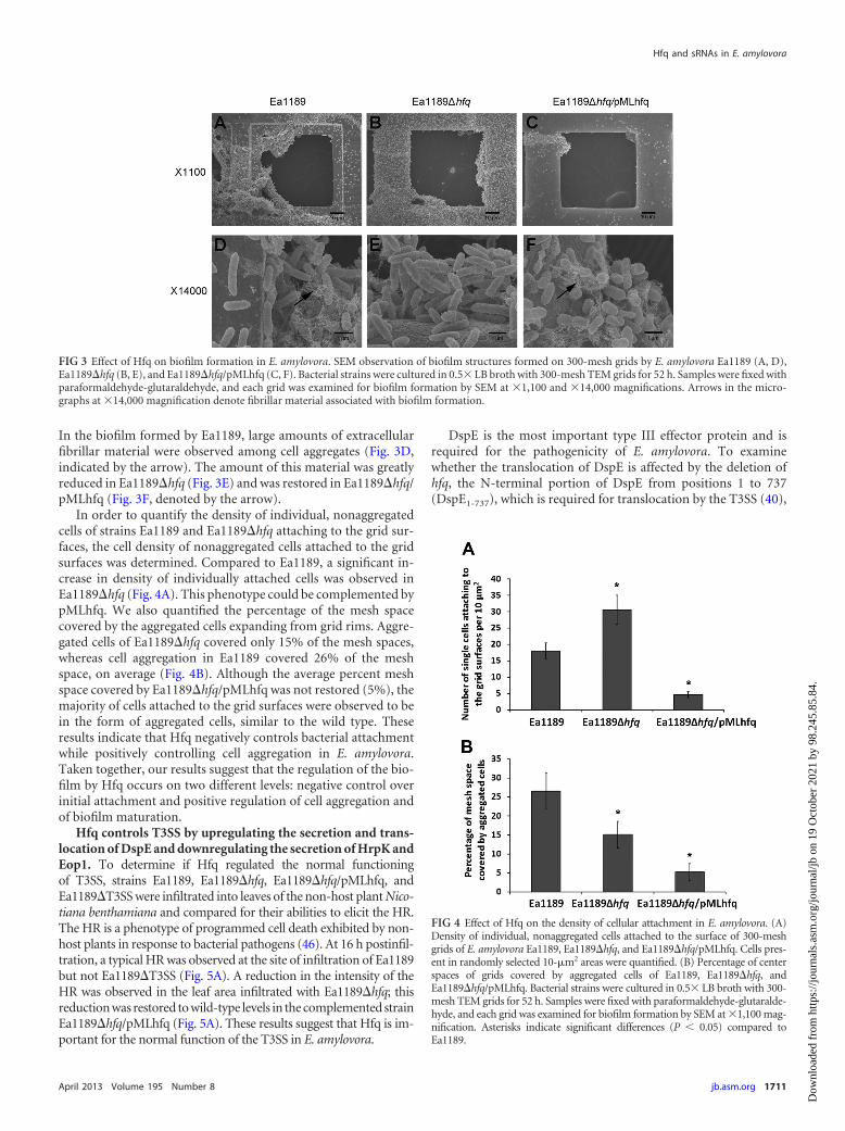
In the biofilm formed by Ea1189, large amounts of extracellularfibrillar material were observed among cell aggregates (Fig. 3D,indicated by the arrow). The amount of this material was greatlyreduced in Ea1189�hfq (Fig. 3E) and was restored in Ea1189�hfq/pMLhfq (Fig. 3F, denoted by the arrow).
In order to quantify the density of individual, nonaggregatedcells of strains Ea1189 and Ea1189�hfq attaching to the grid sur-faces, the cell density of nonaggregated cells attached to the gridsurfaces was determined. Compared to Ea1189, a significant in-crease in density of individually attached cells was observed inEa1189�hfq (Fig. 4A). This phenotype could be complemented bypMLhfq. We also quantified the percentage of the mesh spacecovered by the aggregated cells expanding from grid rims. Aggre-gated cells of Ea1189�hfq covered only 15% of the mesh spaces,whereas cell aggregation in Ea1189 covered 26% of the meshspace, on average (Fig. 4B). Although the average percent meshspace covered by Ea1189�hfq/pMLhfq was not restored (5%), themajority of cells attached to the grid surfaces were observed to bein the form of aggregated cells, similar to the wild type. Theseresults indicate that Hfq negatively controls bacterial attachmentwhile positively controlling cell aggregation in E. amylovora.Taken together, our results suggest that the regulation of the bio-film by Hfq occurs on two different levels: negative control overinitial attachment and positive regulation of cell aggregation andof biofilm maturation.
Hfq controls T3SS by upregulating the secretion and trans-location of DspE and downregulating the secretion of HrpK andEop1. To determine if Hfq regulated the normal functioningof T3SS, strains Ea1189, Ea1189�hfq, Ea1189�hfq/pMLhfq, andEa1189�T3SS were infiltrated into leaves of the non-host plant Nico-tiana benthamiana and compared for their abilities to elicit the HR.The HR is a phenotype of programmed cell death exhibited by non-host plants in response to bacterial pathogens (46). At 16 h postinfil-tration, a typical HR was observed at the site of infiltration of Ea1189but not Ea1189�T3SS (Fig. 5A). A reduction in the intensity of theHR was observed in the leaf area infiltrated with Ea1189�hfq; thisreduction was restored to wild-type levels in the complemented strainEa1189�hfq/pMLhfq (Fig. 5A). These results suggest that Hfq is im-portant for the normal function of the T3SS in E. amylovora.
DspE is the most important type III effector protein and isrequired for the pathogenicity of E. amylovora. To examinewhether the translocation of DspE is affected by the deletion ofhfq, the N-terminal portion of DspE from positions 1 to 737(DspE1-737), which is required for translocation by the T3SS (40),
FIG 3 Effect of Hfq on biofilm formation in E. amylovora. SEM observation of biofilm structures formed on 300-mesh grids by E. amylovora Ea1189 (A, D),Ea1189�hfq (B, E), and Ea1189�hfq/pMLhfq (C, F). Bacterial strains were cultured in 0.5� LB broth with 300-mesh TEM grids for 52 h. Samples were fixed withparaformaldehyde-glutaraldehyde, and each grid was examined for biofilm formation by SEM at �1,100 and �14,000 magnifications. Arrows in the micro-graphs at �14,000 magnification denote fibrillar material associated with biofilm formation.
FIG 4 Effect of Hfq on the density of cellular attachment in E. amylovora. (A)Density of individual, nonaggregated cells attached to the surface of 300-meshgrids of E. amylovora Ea1189, Ea1189�hfq, and Ea1189�hfq/pMLhfq. Cells pres-ent in randomly selected 10-�m2 areas were quantified. (B) Percentage of centerspaces of grids covered by aggregated cells of Ea1189, Ea1189�hfq, andEa1189�hfq/pMLhfq. Bacterial strains were cultured in 0.5� LB broth with 300-mesh TEM grids for 52 h. Samples were fixed with paraformaldehyde-glutaralde-hyde, and each grid was examined for biofilm formation by SEM at �1,100 mag-nification. Asterisks indicate significant differences (P 0.05) compared toEa1189.
Hfq and sRNAs in E. amylovora
April 2013 Volume 195 Number 8 jb.asm.org 1711
Dow
nloa
ded
from
http
s://j
ourn
als.
asm
.org
/jour
nal/j
b on
19
Oct
ober
202
1 by
98.
245.
85.8
4.
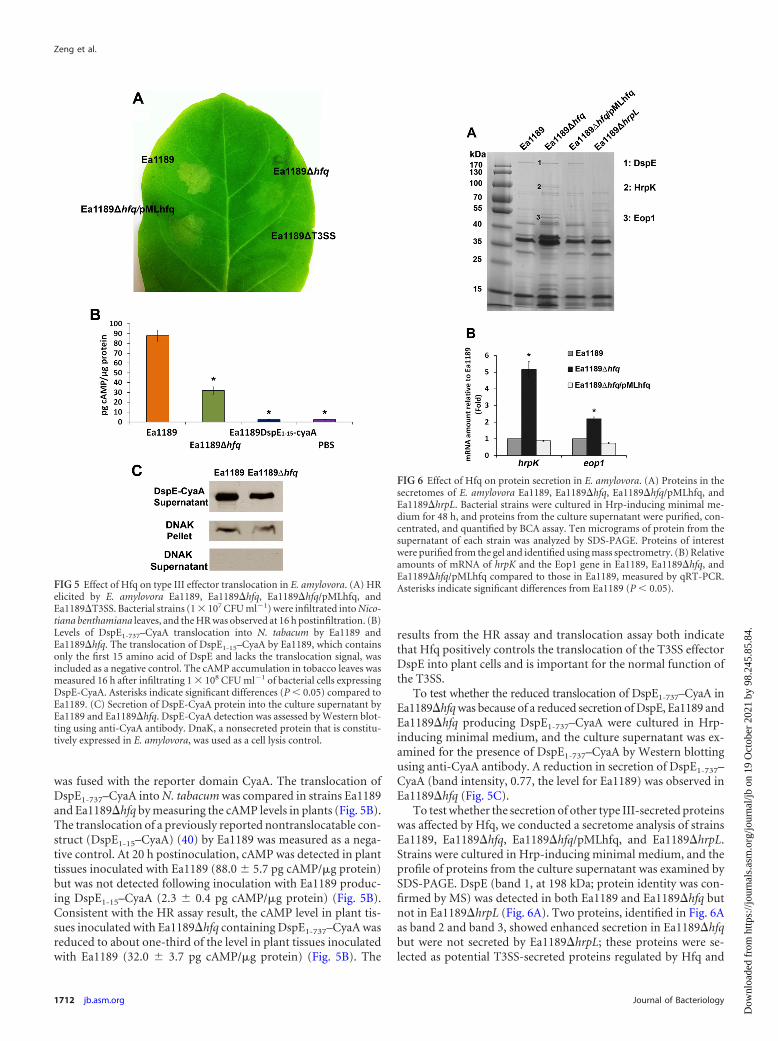
was fused with the reporter domain CyaA. The translocation ofDspE1-737–CyaA into N. tabacum was compared in strains Ea1189and Ea1189�hfq by measuring the cAMP levels in plants (Fig. 5B).The translocation of a previously reported nontranslocatable con-struct (DspE1-15–CyaA) (40) by Ea1189 was measured as a nega-tive control. At 20 h postinoculation, cAMP was detected in planttissues inoculated with Ea1189 (88.0 5.7 pg cAMP/�g protein)but was not detected following inoculation with Ea1189 produc-ing DspE1-15–CyaA (2.3 0.4 pg cAMP/�g protein) (Fig. 5B).Consistent with the HR assay result, the cAMP level in plant tis-sues inoculated with Ea1189�hfq containing DspE1-737–CyaA wasreduced to about one-third of the level in plant tissues inoculatedwith Ea1189 (32.0 3.7 pg cAMP/�g protein) (Fig. 5B). The
results from the HR assay and translocation assay both indicatethat Hfq positively controls the translocation of the T3SS effectorDspE into plant cells and is important for the normal function ofthe T3SS.
To test whether the reduced translocation of DspE1-737–CyaA inEa1189�hfq was because of a reduced secretion of DspE, Ea1189 andEa1189�hfq producing DspE1-737–CyaA were cultured in Hrp-inducing minimal medium, and the culture supernatant was ex-amined for the presence of DspE1-737–CyaA by Western blottingusing anti-CyaA antibody. A reduction in secretion of DspE1-737–CyaA (band intensity, 0.77, the level for Ea1189) was observed inEa1189�hfq (Fig. 5C).
To test whether the secretion of other type III-secreted proteinswas affected by Hfq, we conducted a secretome analysis of strainsEa1189, Ea1189�hfq, Ea1189�hfq/pMLhfq, and Ea1189�hrpL.Strains were cultured in Hrp-inducing minimal medium, and theprofile of proteins from the culture supernatant was examined bySDS-PAGE. DspE (band 1, at 198 kDa; protein identity was con-firmed by MS) was detected in both Ea1189 and Ea1189�hfq butnot in Ea1189�hrpL (Fig. 6A). Two proteins, identified in Fig. 6Aas band 2 and band 3, showed enhanced secretion in Ea1189�hfqbut were not secreted by Ea1189�hrpL; these proteins were se-lected as potential T3SS-secreted proteins regulated by Hfq and
FIG 5 Effect of Hfq on type III effector translocation in E. amylovora. (A) HRelicited by E. amylovora Ea1189, Ea1189�hfq, Ea1189�hfq/pMLhfq, andEa1189�T3SS. Bacterial strains (1 � 107 CFU ml�1) were infiltrated into Nico-tiana benthamiana leaves, and the HR was observed at 16 h postinfiltration. (B)Levels of DspE1-737–CyaA translocation into N. tabacum by Ea1189 andEa1189�hfq. The translocation of DspE1-15–CyaA by Ea1189, which containsonly the first 15 amino acid of DspE and lacks the translocation signal, wasincluded as a negative control. The cAMP accumulation in tobacco leaves wasmeasured 16 h after infiltrating 1 � 108 CFU ml�1 of bacterial cells expressingDspE-CyaA. Asterisks indicate significant differences (P 0.05) compared toEa1189. (C) Secretion of DspE-CyaA protein into the culture supernatant byEa1189 and Ea1189�hfq. DspE-CyaA detection was assessed by Western blot-ting using anti-CyaA antibody. DnaK, a nonsecreted protein that is constitu-tively expressed in E. amylovora, was used as a cell lysis control.
FIG 6 Effect of Hfq on protein secretion in E. amylovora. (A) Proteins in thesecretomes of E. amylovora Ea1189, Ea1189�hfq, Ea1189�hfq/pMLhfq, andEa1189�hrpL. Bacterial strains were cultured in Hrp-inducing minimal me-dium for 48 h, and proteins from the culture supernatant were purified, con-centrated, and quantified by BCA assay. Ten micrograms of protein from thesupernatant of each strain was analyzed by SDS-PAGE. Proteins of interestwere purified from the gel and identified using mass spectrometry. (B) Relativeamounts of mRNA of hrpK and the Eop1 gene in Ea1189, Ea1189�hfq, andEa1189�hfq/pMLhfq compared to those in Ea1189, measured by qRT-PCR.Asterisks indicate significant differences from Ea1189 (P 0.05).
Zeng et al.
1712 jb.asm.org Journal of Bacteriology
Dow
nloa
ded
from
http
s://j
ourn
als.
asm
.org
/jour
nal/j
b on
19
Oct
ober
202
1 by
98.
245.
85.8
4.
were further characterized using MS. Protein 2, an 80-kDa pro-tein, and protein 3, a 44-kDa protein, were identified as HrpK, aputative type III translocator protein, and Eop1, a type III secretedeffector protein, respectively. Using a qRT-PCR assay, signifi-cantly enhanced levels of mRNA of hrpK and the Eop1 gene com-pared to the levels for the wild-type strain were also detected (Fig.6B). The enhanced mRNA levels could be restored in the hfq-complemented strain (Fig. 6B). These results indicate that Hfqcontrols the production and secretion of T3SS-secreted proteins,including HrpK and Eop1.
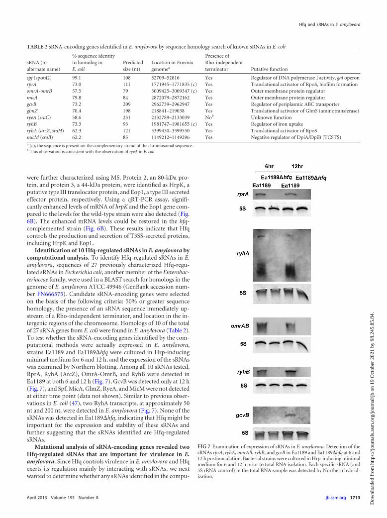
Identification of 10 Hfq-regulated sRNAs in E. amylovora bycomputational analysis. To identify Hfq-regulated sRNAs in E.amylovora, sequences of 27 previously characterized Hfq-regu-lated sRNAs in Escherichia coli, another member of the Enterobac-teriaceae family, were used in a BLAST search for homologs in thegenome of E. amylovora ATCC 49946 (GenBank accession num-ber FN666575). Candidate sRNA-encoding genes were selectedon the basis of the following criteria: 50% or greater sequencehomology, the presence of an sRNA sequence immediately up-stream of a Rho-independent terminator, and location in the in-tergenic regions of the chromosome. Homologs of 10 of the totalof 27 sRNA genes from E. coli were found in E. amylovora (Table 2).To test whether the sRNA-encoding genes identified by the com-putational methods were actually expressed in E. amylovora,strains Ea1189 and Ea1189�hfq were cultured in Hrp-inducingminimal medium for 6 and 12 h, and the expression of the sRNAswas examined by Northern blotting. Among all 10 sRNAs tested,RprA, RyhA (ArcZ), OmrA-OmrB, and RyhB were detected inEa1189 at both 6 and 12 h (Fig. 7), GcvB was detected only at 12 h(Fig. 7), and Spf, MicA, GlmZ, RyeA, and MicM were not detectedat either time point (data not shown). Similar to previous obser-vations in E. coli (47), two RyhA transcripts, at approximately 50nt and 200 nt, were detected in E. amylovora (Fig. 7). None of thesRNAs was detected in Ea1189�hfq, indicating that Hfq might beimportant for the expression and stability of these sRNAs andfurther suggesting that the sRNAs identified are Hfq-regulatedsRNAs.
Mutational analysis of sRNA-encoding genes revealed twoHfq-regulated sRNAs that are important for virulence in E.amylovora. Since Hfq controls virulence in E. amylovora and Hfqexerts its regulation mainly by interacting with sRNAs, we nextwanted to determine whether any sRNAs identified in the compu-
TABLE 2 sRNA-encoding genes identified in E. amylovora by sequence homology search of known sRNAs in E. coli
sRNA (oralternate name)
% sequence identityto homolog inE. coli
Predictedsize (nt)
Location in Erwiniagenomea
Presence ofRho-independentterminator Putative function
spf (spot42) 99.1 108 52709–52816 Yes Regulator of DNA polymerase I activity, gal operonrprA 73.0 111 1771945–1771835 (c) Yes Translational activator of RpoS, biofilm formationomrA-omrB 57.5 79 3009425–3009347 (c) Yes Outer membrane protein regulatormicA 79.8 84 2872079–2872162 Yes Outer membrane protein regulatorgcvB 73.2 209 2962739–2962947 Yes Regulator of periplasmic ABC transporterglmZ 70.4 198 218841–219038 Yes Translational activator of GlmS (aminotransferase)ryeA (sraC) 58.6 251 2132789–2133039 Nob Unknown functionryhB 73.3 93 1981747–1981655 (c) Yes Regulator of iron uptakeryhA (arcZ, sraH) 62.3 121 3399430–3399550 Yes Translational activator of RpoSmicM (sroB) 62.2 85 1149212–1149296 Yes Negative regulator of DpiA/DpiB (TCSTS)a (c), the sequence is present on the complementary strand of the chromosomal sequence.b This observation is consistent with the observation of ryeA in E. coli.
FIG 7 Examination of expression of sRNAs in E. amylovora. Detection of thesRNAs rprA, ryhA, omrAB, ryhB, and gcvB in Ea1189 and Ea1189�hfq at 6 and12 h postinoculation. Bacterial strains were cultured in Hrp-inducing minimalmedium for 6 and 12 h prior to total RNA isolation. Each specific sRNA (and5S rRNA control) in the total RNA sample was detected by Northern hybrid-ization.
Hfq and sRNAs in E. amylovora
April 2013 Volume 195 Number 8 jb.asm.org 1713
Dow
nloa
ded
from
http
s://j
ourn
als.
asm
.org
/jour
nal/j
b on
19
Oct
ober
202
1 by
98.
245.
85.8
4.

tational analysis play a role in regulating virulence in E. amylovora.Seven out of the nine sRNA deletion mutants constructed showeda level of virulence similar to that of strain Ea1189 in an immaturepear assay (Fig. 8A and B). However, two sRNA mutants, thosewith the �ryhA and �rprA mutations, showed significantly re-duced virulence compared to Ea1189 (Fig. 8A and B).
The transcriptional start sites of ryhA and rprA were deter-mined using a 5=RACE assay (see Fig. S1 in the supplemental ma-terial). Based on our identification of the ryhA and rprA transcrip-tional start sites, we determined that the deletions constructed instrains Ea1189�rprA and Ea1189�ryhA encompassed 80% and100% of the sequences of these sRNAs (see Fig. S2 in the supple-mental material). In addition, an examination of the expression offlanking genes mtgA and arcB for ryhA and ppsA, respectively, andEAM1647 for rprA indicated that the deletions of ryhA and rprAdid not affect the expression levels of these genes (see Fig. S3 in thesupplemental material).
At 5 dpi, the mean lesion diameter of Ea1189�ryhA was re-duced to a level similar to that of Ea1189�hfq. The mean lesiondiameter of Ea1189�rprA was also reduced to about half of that ofEa1189 but was still significantly higher than the lesion diametersof Ea1189�hfq and Ea1189�ryhA (Fig. 8B). The Ea1189�hrpLmutant, with deletion of the alternate sigma factor that regulatesT3SS-encoding genes, was nonpathogenic (Fig. 8B). The reduced-virulence phenotype observed in Ea1189�rprA and in Ea1189�ryhA was complemented by plasmids carrying rprA and ryhA, re-spectively (see Fig. S4 in the supplemental material). These resultsindicate that the Hfq-regulated sRNAs RyhA and RprA may work
together with the sRNA chaperone Hfq and collaboratively con-trol virulence in E. amylovora.
DISCUSSION
In this study, we showed that Hfq regulated all of the known es-sential pathogenicity determinants in E. amylovora, including typeIII secretion, translocation of the major effector DspE, and pro-duction of amylovoran EPS. Hfq is a known regulator of virulencein many bacterial pathogens and is also important in the responseto various environmental stress factors in pathogens and otherbacteria (28). However, the role of Hfq as a virulence regulator isnot universal among pathogens. For example, in Staphylococcusaureus, an hfq mutant, virulence for the worm Caenorhabditis el-egans was not affected (48), and in Neisseria gonorrhoeae, the vir-ulence of an hfq mutant was not altered in cell culture models (49).
Biofilm formation is critical for E. amylovora to develop largepopulations in apple xylem and to move systemically through thehost (12, 14, 42). The Ea1189�hfq mutant appears to be uniqueamong E. amylovora biofilm mutants in that although the mutantproduced a decreased amount of amylovoran, it exhibited analtered enhanced biofilm phenotype. However, further examina-tion of biofilm formation by this mutant indicated thatEa1189�hfq exhibited a hyperattachment phenotype that pre-sumably led to an artifact result suggesting an increase in bio-film formation on a polystyrene surface. In addition, althoughEa1189�hfq cells were capable of aggregation, the fibrillar matrixmaterial characteristic of wild-type E. amylovora Ea1189 biofilmswas not present. Thus, it is likely that Ea1189�hfq cells are not
FIG 8 Analysis and quantification of virulence of E. amylovora deletion mutants of hfq and sRNAs. (A) Virulence of E. amylovora Ea1189, Ea1189�hfq, andEa1189�hrpL and sRNA mutants Ea1189�ryhA, Ea1189�rprA, Ea1189�spf, Ea1189�micA, Ea1189�omrAB, Ea1189�ryhB, Ea1189�sroB, Ea1189�ryeA, andEa1189�glmZ in immature pears at 5 dpi. (B) Average lesion diameters of immature pears ( standard error) inoculated with each E. amylovora deletion mutant.Sample means were compared by an analysis of variance and separated using the Student t test. The presence of different letters indicates that the means weresignificantly different (P 0.05).
Zeng et al.
1714 jb.asm.org Journal of Bacteriology
Dow
nloa
ded
from
http
s://j
ourn
als.
asm
.org
/jour
nal/j
b on
19
Oct
ober
202
1 by
98.
245.
85.8
4.

capable of formation of mature biofilms. Since biofilm formationis important for the establishment of large populations of E. amy-lovora in apple xylem (12, 14, 42), we hypothesize that the lack ofbiofilm formation by Ea1189�hfq resulted in an inability of thesecells to establish in apple xylem and move systemically through thehost. Furthermore, our results suggest that multiple in vitro and invivo experiments are necessary for the characterization of mutantsaffecting biofilm formation in bacterial pathogens.
The regulatory effect of Hfq on biofilm formation had beenstudied in a few bacterial species prior to this study. In Moraxellacatarrhalis, when the �hfq and wild-type M. catarrhalis strainswere mixed and inoculated in a continuous-flow biofilm system,the number of �hfq cells clearly predominated over the number ofwild-type cells in the population recovered from the biofilm afteran overnight incubation (50). This observation is consistent withour finding that Ea1189�hfq exhibited a hyperattaching pheno-type on polystyrene solid surfaces compared to the wild-typestrain Ea1189. Similarly, increased biofilm formation was ob-served in the absence of Hfq in Yersinia pestis (35). However, inuropathogenic Escherichia coli (UPEC), a mutation of hfq causedreduced biofilm formation when examined by a crystal violetstaining method, which suggests a general activating effect of Hfqon biofilm formation (51). These observations indicate that bio-film regulation by Hfq may vary in different pathogens.
Biofilm formation is a complex developmental process thattypically involves four phases: planktonic phase, attachmentphase, maturation phase, and detachment phase (52). The transi-tion between different phases is tightly regulated by multiplemechanisms, including the second messenger molecule cyclic di-GMP, and by quorum sensing (53, 54). Our observation that Hfqnegatively controls bacterial attachment while positively control-ling the production of amylovoran and biofilm maturation mayprovide insight into the transition of biofilm development pro-cesses. The attachment or adhesion to solid surfaces is the initialand prerequisite step of biofilm formation. This could be seen inour observation that much reduced attachment and biofilm for-mation were observed in Ea1189�hfq/pMLhfq compared toEa1189 when a multicopy plasmid, pMLhfq, was used for comple-mentation. However, although attachment is an important step ofbiofilm formation, it has been proposed in animal pathogens thatbacterial adhesion to host epithelial cells may also come at a costdue to the possibility of inducing host immunity (55). To mini-mize host recognition and possible host immune responses whileestablishing colonization, bacterial animal pathogens likely regu-late the transition from initial attachment to biofilm maturation.Our work indicates that Hfq and potentially Hfq-regulated sRNAsmay play important roles in this transition.
The delivery of effector proteins from bacteria into plant cellsrequires the processes of secretion and translocation. The fact thatthe translocation of DspE-CyaA in Ea1189�hfq was reduced to�0.3-fold of that of wild-type Ea1189 while its secretion was re-duced to only �0.77-fold of that of Ea1189 suggests that Hfq mayalso affect the translocation of DspE, in addition to regulating itssecretion. Increased secretion of HrpK, a type III translocator pro-tein (56), was observed in Ea1189�hfq. The disruption of the pro-duction of HrpK, which is important for effector translocation,may be the reason for the reduced DspE translocation observed inEa1189�hfq. Similar to the downregulation of the expression ofhrpK and the Eop1 gene observed in this work, Hfq was also re-ported to repress the production of type III effectors encoded on
the LEE pathogenicity island in EHEC strains (31). Further studyis needed to characterize the detailed mechanism of control of theT3SS by Hfq and small RNAs.
We observed pleiotropic regulation of amylovoran produc-tion, biofilm formation, motility, and the T3SS in E. amylovora byHfq. These observations imply that Hfq-regulated sRNAs maylikewise be important virulence regulators in E. amylovora. Todetermine the role of Hfq-regulated sRNAs in virulence regula-tion, we first identified 10 sRNAs using computational analysisand found 2 of them (RprA and RyhA) that contribute to thevirulence of E. amylovora. Both RprA and RyhA were previouslydemonstrated to positively regulate translation of the stationary-phase sigma factor RpoS in E. coli (57). To test whether the re-duced virulence in strains Ea1189�rprA and Ea1189�ryhA wasdue to a potential downregulation of rpoS, a �rpoS mutant wasconstructed and its virulence phenotype was compared with thatof Ea1189 in an immature pear fruit assay (data not shown). Asimilar level of virulence was observed in Ea1189�rpoS andEa1189, which is consistent with a previous observation that RpoSis not involved in the induction of fire blight disease symptoms byE. amylovora (58). This suggests that the regulation of virulence byRprA and RyhA is likely mediated via targets different from thosein E. coli. We are currently examining the regulatory mechanismsof these sRNAs. Also of note, prior to our study, Schmidtke et al.identified one virulence-related sRNA, sX12, in the plant-patho-genic bacterium Xanthomonas campestris (59). Neither RprA norRyhA in E. amylovora shares any sequence homology with sX12.
In conclusion, we provide evidence that, similar to its role inanimal pathogens, the RNA chaperone Hfq also plays importantroles in controlling virulence in the plant-pathogenic bacterium E.amylovora. Compared to previously characterized regulators suchas HrpL, the master regulator for T3SS (10), and RcsBCD, the keyregulator of amylovoran production (42), Hfq appears to morebroadly regulate many virulence determinants, including amylo-voran production, biofilm formation, T3SS translocation and se-cretion, and bacterial motility. Thus, Hfq and the sRNAs that itregulates likely play a central role in the fine-tuning of virulencegene expression in E. amylovora. During fire blight pathogenesis,E. amylovora cells colonize and grow on the relatively nutrient-rich stigma surface, on the high-osmotic flower nectarthode,within the leaf apoplast, and in the potentially low-nutrient vas-cular system (1). Transitions between these various host environ-ments may occur over relatively short time scales, thus necessitat-ing the ability to rapidly alter the production of virulence factorsvia posttranscriptional regulation. Our identification of pleiotro-pic virulence effects in Ea1189�hfq and of individual sRNAs withstrong effects on virulence provides a promising start for charac-terization of the detailed regulatory mechanisms of Hfq and Hfq-regulated sRNAs in the virulence of E. amylovora.
ACKNOWLEDGMENTS
This work was supported by a special grant from the United States De-partment of Agriculture CSREES, Project GREEEN, a Michigan plantagriculture initiative at Michigan State University, and Michigan Ag-BioResearch.
REFERENCES1. Malnoy M, Martens S, Norelli JL, Barny MA, Sundin GW, Smits TH,
Duffy B. 2012. Fire blight: applied genomic insights of the pathogen andhost. Annu. Rev. Phytopathol. 50:475– 494.
Hfq and sRNAs in E. amylovora
April 2013 Volume 195 Number 8 jb.asm.org 1715
Dow
nloa
ded
from
http
s://j
ourn
als.
asm
.org
/jour
nal/j
b on
19
Oct
ober
202
1 by
98.
245.
85.8
4.

2. Thomson SV. 1986. The role of the stigma in fire blight infections. Phy-topathology 76:476 – 482.
3. Bayot RG, Ries SM. 1986. Role of motility in apple blossom infection byErwinia amylovora and studies of fire blight control with attractant andrepellent compounds. Phytopathology 76:441– 445.
4. Oh CS, Kim JF, Beer SV. 2005. The Hrp pathogenicity island of Erwiniaamylovora and identification of three novel genes required for systemicinfection. Mol. Plant Pathol. 6:125–138.
5. Venisse JS, Malnoy M, Faize M, Paulin JP, Brisset MN. 2002. Modula-tion of defense responses of Malus spp. during compatible and incompat-ible interactions with Erwinia amylovora. Mol. Plant Microbe Interact.15:1204 –1212.
6. Boureau T, ElMaarouf-Bouteau H, Garnier A, Brisset MN, Perino C,Pucheu I, Barny MA. 2006. DspA/E, a type III effector essential forErwinia amylovora pathogenicity and growth in planta, induces cell deathin host apple and nonhost tobacco plants. Mol. Plant Microbe Interact.19:16 –24.
7. DebRoy S, Thilmony R, Kwack YB, Nomura K, He SY. 2004. A familyof conserved bacterial effectors inhibits salicylic acid-mediated basal im-munity and promotes disease necrosis in plants. Proc. Natl. Acad. Sci.U. S. A. 101:9927–9932.
8. Gaudriault S, Malandrin L, Paulin JP, Barny MA. 1997. DspA, anessential pathogenicity factor of Erwinia amylovora showing homologywith AvrE of Pseudomonas syringae, is secreted via the Hrp secretion path-way in a DspB-dependent way. Mol. Microbiol. 26:1057–1069.
9. Pester D, Milcevicova R, Schaffer J, Wilhelm E, Blumel S. 2012. Erwiniaamylovora expresses fast and simultaneously hrp/dsp virulence genes dur-ing flower infection on apple trees. PLoS One 7:e32583. doi:10.1371/journal.pone.0032583.
10. McNally RR, Toth IK, Cock PJ, Pritchard L, Hedley PE, Morris JA,Zhao Y, Sundin GW. 2012. Genetic characterization of the HrpL regulonof the fire blight pathogen Erwinia amylovora reveals novel virulence fac-tors. Mol. Plant Pathol. 13:160 –173.
11. Geider K. 2006. Twenty years of molecular genetics with Erwinia amylo-vora: answers and new questions about EPS-synthesis and other virulencefactors. Acta Hortic. 704:397– 402.
12. Koczan JM, McGrath MJ, Zhao Y, Sundin GW. 2009. Contribution ofErwinia amylovora exopolysaccharides amylovoran and levan to biofilmformation: implications in pathogenicity. Phytopathology 99:1237–1244.
13. Pieretti I, Royer M, Barbe V, Carrere S, Koebnik R, Cociancich S,Couloux A, Darrasse A, Gouzy J, Jacques MA, Lauber E, Manceau C,Mangenot S, Poussier S, Segurens B, Szurek B, Verdier V, Arlat M, RottP. 2009. The complete genome sequence of Xanthomonas albilineans pro-vides new insights into the reductive genome evolution of the xylem-limited Xanthomonadaceae. BMC Genomics 10:616. doi:10.1186/1471-2164-10-616.
14. Koczan JM, Lenneman BR, McGrath MJ, Sundin GW. 2011. Cell surfaceattachment structures contribute to biofilm formation and xylem coloni-zation by Erwinia amylovora. Appl. Environ. Microbiol. 77:7031–7039.
15. Castiblanco L, Edmunds A, Waters CM, Sundin GW. 2011. Character-ization of quorum sensing and cyclic-di-GMP signaling systems in Er-winia amylovora. Phytopathology 101:S2.2. doi:10.1094/PHYTO-101-10-S2.1.
16. Wang D, Qi M, Calla B, Korban SS, Clough SJ, Cock PJ, Sundin GW,Toth I, Zhao Y. 2012. Genome-wide identification of genes regulated bythe Rcs phosphorelay system in Erwinia amylovora. Mol. Plant MicrobeInteract. 25:6 –17.
17. Zhao Y, Blumer SE, Sundin GW. 2005. Identification of Erwinia amylo-vora genes induced during infection of immature pear tissue. J. Bacteriol.187:8088 – 8103.
18. Zhao Y, Wang D, Nakka S, Sundin GW, Korban SS. 2009. Systems levelanalysis of two-component signal transduction systems in Erwinia amylovora:role in virulence, regulation of amylovoran biosynthesis and swarming mo-tility. BMC Genomics 10:245. doi:10.1186/1471-2164-10-245.
19. Frohlich KS, Vogel J. 2009. Activation of gene expression by small RNA.Curr. Opin. Microbiol. 12:674 – 682.
20. Gottesman S, Storz G. 2011. Bacterial small RNA regulators: versatileroles and rapidly evolving variations. Cold Spring Harb. Perspect. Biol.3:pii�a003798. doi10.1101/cshperspect.a003798.
21. Storz G, Vogel J, Wassarman KM. 2011. Regulation by small RNAs inbacteria: expanding frontiers. Mol. Cell 43:880 – 891.
22. Vogel J, Luisi BF. 2011. Hfq and its constellation of RNA. Nat. Rev.Microbiol. 9:578 –589.
23. Link TM, Valentin-Hansen P, Brennan RG. 2009. Structure of Esche-richia coli Hfq bound to polyriboadenylate RNA. Proc. Natl. Acad. Sci.U. S. A. 106:19292–19297.
24. Sauer E, Weichenrieder O. 2011. Structural basis for RNA 3=-end recog-nition by Hfq. Proc. Natl. Acad. Sci. U. S. A. 108:13065–13070.
25. Waters LS, Storz G. 2009. Regulatory RNAs in bacteria. Cell 136:615–628.
26. Romeo T, Vakulskas CA, Babitzke P. 2013. Post-transcriptional regula-tion on a global scale: form and function of Csr/Rsm systems. Environ.Microbiol. 15:313–324.
27. Zeng Q, Ibekwe AM, Biddle E, Yang CH. 2010. Regulatory mechanismsof exoribonuclease PNPase and regulatory small RNA on T3SS of Dickeyadadantii. Mol. Plant Microbe Interact. 23:1345–1355.
28. Chao Y, Vogel J. 2010. The role of Hfq in bacterial pathogens. Curr. Opin.Microbiol. 13:24 –33.
29. Toledo-Arana A, Repoila F, Cossart P. 2007. Small noncoding RNAscontrolling pathogenesis. Curr. Opin. Microbiol. 10:182–188.
30. Ding Y, Davis BM, Waldor MK. 2004. Hfq is essential for Vibrio choleraevirulence and downregulates sigma expression. Mol. Microbiol. 53:345–354.
31. Shakhnovich EA, Davis BM, Waldor MK. 2009. Hfq negatively regulatestype III secretion in EHEC and several other pathogens. Mol. Microbiol.74:347–363.
32. Pfeiffer V, Sittka A, Tomer R, Tedin K, Brinkmann V, Vogel J. 2007. Asmall non-coding RNA of the invasion gene island (SPI-1) represses outermembrane protein synthesis from the Salmonella core genome. Mol. Mi-crobiol. 66:1174 –1191.
33. Chambers JR, Bender KS. 2011. The RNA chaperone Hfq is important forgrowth and stress tolerance in Francisella novicida. PLoS One 6:e19797.doi:10.1371/journal.pone.0019797.
34. Christiansen JK, Larsen MH, Ingmer H, Sogaard-Andersen L, Kalli-politis BH. 2004. The RNA-binding protein Hfq of Listeria monocyto-genes: role in stress tolerance and virulence. J. Bacteriol. 186:3355–3362.
35. Bellows L, Koestler BJ, Karaba SM, Walters CM, Lanthem WW. 2012.Hfq-dependent, co-ordinate control of cyclic diguanylate synthesis andcatabolism in the plague pathogen Yersinia pestis. Mol. Microbiol. 86:661–674.
36. Wilms I, Moller P, Stock AM, Gurski R, Lai EM, Narberhaus F. 2012.Hfq influences multiple transport systems and virulence in the plantpathogen Agrobacterium tumefaciens. J. Bacteriol. 194:5209 –5217.
37. Zhao Y, Sundin GW, Wang D. 2009. Construction and analysis ofpathogenicity island deletion mutants of Erwinia amylovora. Can. J. Mi-crobiol. 55:457– 464.
38. Datsenko KA, Wanner BL. 2000. One-step inactivation of chromosomalgenes in Escherichia coli K-12 using PCR products. Proc. Natl. Acad. Sci.U. S. A. 97:6640 – 6645.
39. Labes M, Puhler A, Simon R. 1990. A new family of RSF1010-derivedexpression and lac-fusion broad-host-range vectors for gram-negativebacteria. Gene 89:37– 46.
40. Triplett LR, Melotto M, Sundin GW. 2009. Functional analysis of the Nterminus of the Erwinia amylovora secreted effector DspA/E reveals fea-tures required for secretion, translocation, and binding to the chaperoneDspB/F. Mol. Plant Microbe Interact. 22:1282–1292.
41. Huynh TV, Dahlbeck D, Staskawicz BJ. 1989. Bacterial blight of soybean:regulation of a pathogen gene determining host cultivar specificity. Sci-ence 245:1374 –1377.
42. Wang D, Korban SS, Pusey PL, Zhao Y. 2011. Characterization of theRcsC sensor kinase from Erwinia amylovora and other enterobacteria.Phytopathology 101:710 –717.
43. Bellemann P, Bereswill S, Berger S, Geider K. 1994. Visualization ofcapsule formation by Erwinia amylovora and assays to determine amylo-voran synthesis. Int. J. Biol. Macromol. 16:290 –296.
44. Nissinen RM, Ytterberg AJ, Bogdanove AJ, Van Wijk KJ, Beer SV. 2007.Analyses of the secretomes of Erwinia amylovora and selected hrp mutantsreveal novel type III secreted proteins and an effect of HrpJ on extracellu-lar hairpin levels. Mol. Plant Pathol. 8:55– 67.
45. Takle GW, Toth IK, Brurberg MB. 2007. Evaluation of reference genesfor real-time RT-PCR expression studies in the plant pathogen Pectobac-terium atrosepticum. BMC Plant Biol. 7:50. doi:10.1186/1471-2229-7-50.
46. Dangl JL, Jones JD. 2001. Plant pathogens and integrated defence re-sponses to infection. Nature 411:826 – 833.
47. Mandin P, Gottesman S. 2010. Integrating anaerobic/aerobic sensing and
Zeng et al.
1716 jb.asm.org Journal of Bacteriology
Dow
nloa
ded
from
http
s://j
ourn
als.
asm
.org
/jour
nal/j
b on
19
Oct
ober
202
1 by
98.
245.
85.8
4.

the general stress response through the ArcZ small RNA. EMBO J. 29:3094 –3107.
48. Bohn C, Rigoulay C, Bouloc P. 2007. No detectable effect of RNA-binding protein Hfq absence in Staphylococcus aureus. BMC Microbiol.7:10. doi:10.1186/1471-2180-7-10.
49. Dietrich M, Munke R, Gottschald M, Ziska E, Boettcher JP, MollenkopfH, Friedrich A. 2009. The effect of hfq on global gene expression andvirulence in Neisseria gonorrhoeae. FEBS J. 276:5507–5520.
50. Attia AS, Sedillo JL, Wang W, Liu W, Brautigam CA, Winkler W,Hansen EJ. 2008. Moraxella catarrhalis expresses an unusual Hfq protein.Infect. Immun. 76:2520 –2530.
51. Kulesus RR, Diaz-Perez K, Slechta ES, Eto DS, Mulvey MA. 2008.Impact of the RNA chaperone Hfq on the fitness and virulence potential ofuropathogenic Escherichia coli. Infect. Immun. 76:3019 –3026.
52. Monroe D. 2007. Looking for chinks in the armor of bacterial biofilms.PLoS Biol. 5:e307. doi:10.1371/journal.pbio.0050307.
53. Srivastava D, Waters CM. 2012. A tangled web: regulatory connectionsbetween quorum sensing and cyclic di-GMP. J. Bacteriol. 194:4485– 4493.
54. Waters CM, Lu W, Rabinowitz JD, Bassler BL. 2008. Quorum sensing
controls biofilm formation in Vibrio cholerae through modulation of cy-clic di-GMP levels and repression of vpsT. J. Bacteriol. 190:2527–2536.
55. Kline KA, Falker S, Dahlberg S, Normark S, Henriques-Normark B.2009. Bacterial adhesins in host-microbe interactions. Cell Host Microbe5:580 –592.
56. Petnicki-Ocwieja T, van Dijk K, Alfano JR. 2005. The hrpK operon ofPseudomonas syringae pv. tomato DC3000 encodes two proteins secretedby the type III (Hrp) protein secretion system: HopB1 and HrpK, a puta-tive type III translocator. J. Bacteriol. 187:649 – 663.
57. Soper T, Mandin P, Majdalani N, Gottesman S, Woodson SA. 2010.Positive regulation by small RNAs and the role of Hfq. Proc. Natl. Acad.Sci. U. S. A. 107:9602–9607.
58. Anderson M, Pollitt CE, Roberts IS, Eastgate JA. 1998. Identificationand characterization of the Erwinia amylovora rpoS gene: RpoS is notinvolved in induction of fireblight disease symptoms. J. Bacteriol. 180:6789 – 6792.
59. Schmidtke C, Findeiss S, Sharma CM, Kuhfuss J, Hoffmann S, Vogel J,Stadler PF, Bonas U. 2012. Genome-wide transcriptome analysis of theplant pathogen Xanthomonas identifies sRNAs with putative virulencefunctions. Nucleic Acids Res. 40:2020 –2031.
Hfq and sRNAs in E. amylovora
April 2013 Volume 195 Number 8 jb.asm.org 1717
Dow
nloa
ded
from
http
s://j
ourn
als.
asm
.org
/jour
nal/j
b on
19
Oct
ober
202
1 by
98.
245.
85.8
4.