glial cell activity within the ventrolateral
TRANSCRIPT
Georgia State University Georgia State University
ScholarWorks @ Georgia State University ScholarWorks @ Georgia State University
Biology Honors Theses Department of Biology
Spring 5-6-2012
Glial Cell Activity within the Ventrolateral Periaqueductal Gray of Glial Cell Activity within the Ventrolateral Periaqueductal Gray of
Male and Female Rats Male and Female Rats
Jean-Marc A. Sauzier
Lori N. Eidson
Follow this and additional works at: https://scholarworks.gsu.edu/biology_hontheses
Recommended Citation Recommended Citation Sauzier, Jean-Marc A. and Eidson, Lori N., "Glial Cell Activity within the Ventrolateral Periaqueductal Gray of Male and Female Rats." Thesis, Georgia State University, 2012. https://scholarworks.gsu.edu/biology_hontheses/3
This Thesis is brought to you for free and open access by the Department of Biology at ScholarWorks @ Georgia State University. It has been accepted for inclusion in Biology Honors Theses by an authorized administrator of ScholarWorks @ Georgia State University. For more information, please contact [email protected].
GLIAL CELL ACTIVITY WITHIN THE VENTROLATERAL PERIAQUEDUCTAL GRAY
OF MALE AND FEMALE RATS
by
Jean-Marc A. Sauzier, Lori N. Eidson, Anne Z. Murphy
Under the Direction of Dr. Anne Z. Murphy
ABSTRACT
Morphine is one of the most commonly prescribed medications for the relief of prolonged pain.
Both basic science and clinical studies indicate that females require 2-3 times more morphine
than males to achieve the same analgesic effect. To date, the mechanisms underlying sex
differences in opiate responsiveness are unknown. Recent studies suggest that glial cells are
potent modulators of morphine-based analgesia, and in particular, decrease the analgesic effect
of opiates. Therefore, we tested the hypothesis that the sexually dimorphic effects of morphine
were due to sex differences in glial cell activity. Our studies focused on the midbrain
periaqueductal gray (PAG) as this region of the brain is critical for the analgesic effects of
morphine. Adult male and female Sprague Dawley rats (250g- 400g) were procured from
Charles River Laboratories, and were allowed 7 days to acclimate to the new facility. On the day
of the experiment, animals received a subcutaneous injection of morphine (5mg/kg) or were
handled in a similar manner. Thirty or 60 minutes after injections or handling, animals were
perfused with a 4% paraformaldehyde and 2.5% acrolein tissue fixative solution. Brains were
removed and stored in 20% sucrose until ready for sectioning. Brains were sectioned at 25µm
using a freezing microtome, and immunohistochemical localization of markers for astrocyte glial
cell activity was performed. Antibodies to glial fibrillary acidic protein (GFAP) were used to
label activated astrocytes. If our hypothesis is correct, then females will have significantly
greater density of the astrocyte cell activity marker GFAP as compared with males. Sex
differences in PAG glial cell activity may provide the biological bases for the sexually dimorphic
effect of morphine. This research may lead to better treatment for females experiencing
prolonged chronic or neuropathic pain.
INDEX WORDS: Glia, Astrocytes, Chronic Pain
GLIAL CELL ACTIVITY WITHIN THE VENTROLATERAL PERIAQUEDUCTAL
GRAY OF MALE AND FEMALE RATS
by
Jean-Marc A. Sauzier
An Honors Thesis Submitted in Partial Fulfillment of the Requirements for the Degree of
Biology Department
in the College of Arts and Sciences
Georgia State University
2012

Copyright by
Jean-Marc Andre Sauzier
2012
GLIAL CELL ACTIVITY WITHIN THE VENTROLATERAL PERIAQUEDUCTAL GRAY
OF MALE AND FEMALE RATS
by
Jean-Marc A. Sauzier, Lori N. Eidson, Anne Z. Murphy
Honors Thesis Director: Dr. Anne Z. Murphy
Honors College Dean: Dr. Larry Berman
Electronic Version Approved:
The Honors College
Georgia State University
April 2012

iv
DEDICATION
To my family, immediate and extended, whose support is always constant. This would not have
come to pass without all your input.
v
Acknowledgements
Firstly, I would like to thank Dr. Anne Murphy, my advisor, for allowing me access to the higher
level of education offered at the university and pushing me to fulfill my potential. I would like to
thank Lori Eidson, whose jedi glia knowledge and advice gave the backbone to this project. I
would also like to thank Nicole Victoria, who gave me the fundamental steps needed to start this
project. One acknowledgement is not enough, again to the three of you, thank you for your time
and effort spent on training, enlightening and preparing me for the bigger picture. Thanks to past
and present Murphy lab members for any and all contributions. I would like to thank the
Neuroscience Institute and the Biology department. Lastly I would like to thank the GSU
Department of Animal Resources, whose time and management allows for the most painless
method of learning and care taking of animals.
vi
TABLE OF CONTENTS
DEDICATION……………………………………………………………………………………iv
ACKNOWLEDGEMENTS………………………………………………………………………v
LIST OF FIGURES……………………………………………………………………………...vii
CHAPTER
1. INTRODUCTION………………………………………………………………………...1
2. METHODS………………………………………………………………………………..2
3. RESULTS…………………………………………………………………………………5
4. DISCUSSION……………………………………………………………………………..5
5. FIGURES……………………………………………………………………………….....7
6. REFERENCES………………………………………………………………………..…10

vii
LIST OF FIGURES
Figure 1 Percent Maximum Possible Effect (%MPE) of varying doses of morphine in males vs.
females Complete Freund’s Adjuvant (CFA) -treated rats …………………………………….7
Figure 2 Periaqueductal gray-rostral ventromedial medullary (PAG-RVM) descending pain
pathway…………………………………………...…………………………...………….……7
Figure 3 Florescent images of glial fibrillary acidic protein (GFAP) immunoreactivity within the
periaqueductal gray (PAG) of male rats treated subcutaneously (sc) with either saline (5ml/kg) or
morphine (5mg/kg)…………………………………………...………………………………...8
Figure 4 Glial fibrillary acidic protein (GFAP) immunoreactivity within the periaqueductal gray
(PAG) of male rats treated subcutaneously (sc) with either saline (5ml/kg) or morphine
(5mg/kg)………………………………………………………………………...……………...8
Figure 5 Glial fibrillary acidic protein (GFAP) immunoreactivity within the periaqueductal gray
(PAG) of male and female rats that were handled or treated with morphine (5mg/kg; sc)…...9
Figure 6 Representative fluorescent images of glial fibrillary acidic protein (GFAP)
immunoreactivity in the periaqueductal gray (PAG) of male and female rats that were handled or
treated with morphine (5mg/kg;sc)……………...…………………………………………….9
1
INTRODUCTION
Morphine is one of the most commonly prescribed drugs for the relief of prolonged pain.
Morphine does not produce the same degree of analgesia in males as compared to females
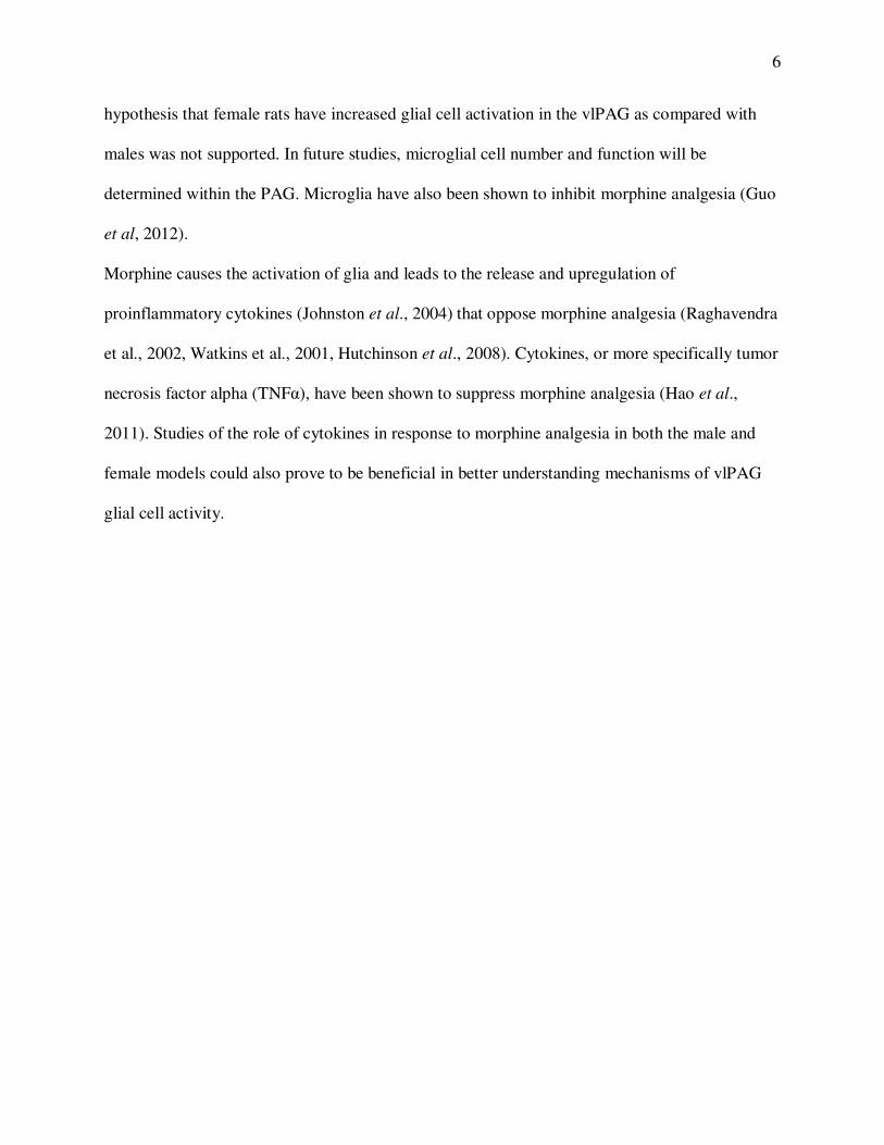
(Kepler et al., 1989; Cicero et al., 1996). Previous work in our lab has shown that females
require 2-3 times more morphine to produce the same analgesic effect as males (Fig 1; Wang et
al., 2006.). To date, the mechanisms underlying sex differences in opiate responsiveness are
unknown. The periaqueductal gray (PAG) is a brain region that has been shown to be important
for the analgesic effects of morphine (Loyd and Murphy, 2008). The PAG sends extensive
projections to the rostral ventromedial medulla (RVM) of the brainstem, which in turn sends
descending projections to the dorsal horn of the spinal cord. The PAG-RVM-spinal cord pathway
is an essential circuit for antinociception (A.I Basbaum et al., 1978; H.L Fields et al., 1991).
These regions also contain a high density of mu opioids receptors (MOR) (Loyd et al., 2007).
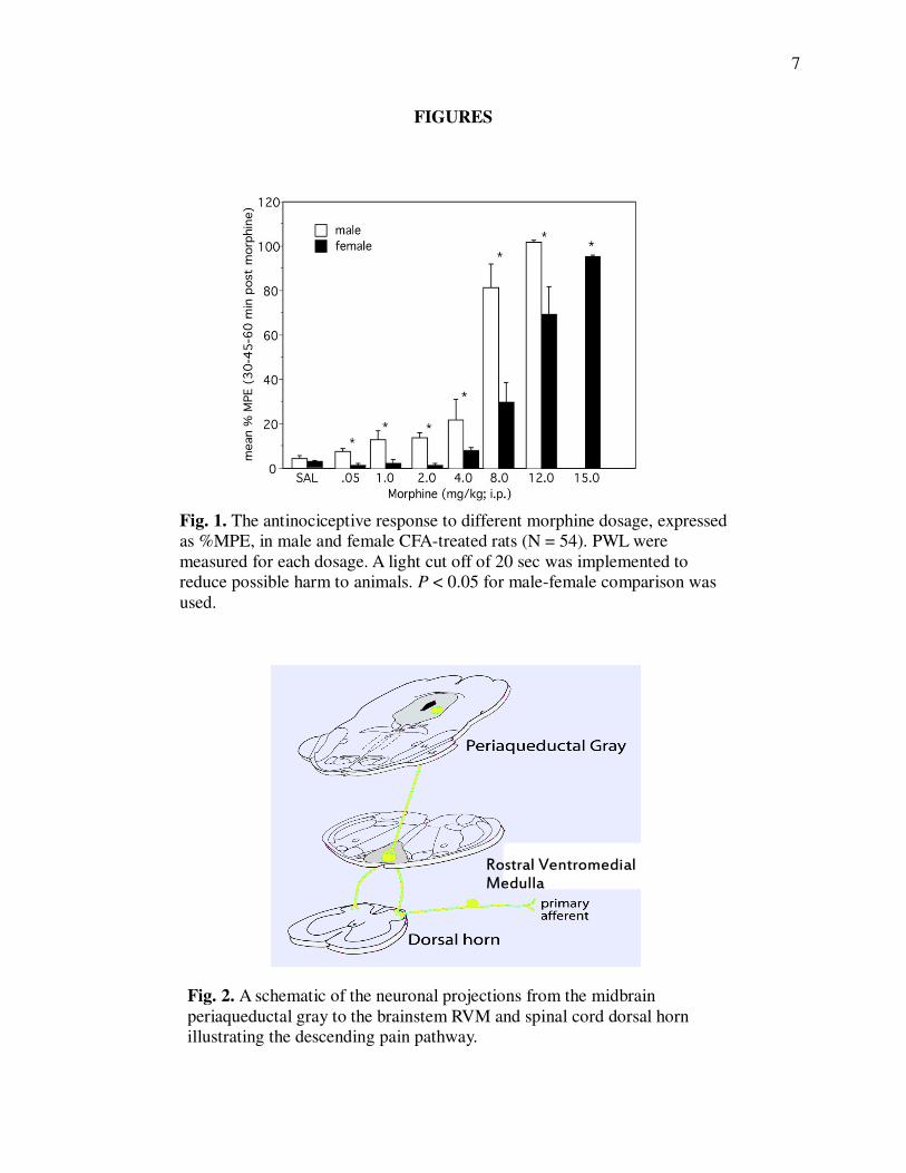
Opioids modulate pain by binding to the mu opioid receptor in the ventrolateral PAG (vlPAG).
vlPAG projection neurons excite cells in the rostral ventromedial medulla (RVM) that project to
the dorsal horn of the spinal cord (Fig 2: Loyd and Murphy, 2008) where they inhibit incoming
pain signals.
Recent studies suggest that glial cells are potent modulators of morphine-based analgesia. In
particular, several studies have now shown that glial cells decrease the analgesic effect of opiates
(Hao et al., 2011; Wei et al., 2012). The two primary glia cells involved in this phenomenon are
astrocytes and microglia. Microglia are shown to be the first responders to even minor
pathological changes in the CNS (Kreutzberg G.W., 1996). Activation of these cells is a key
factor in the defense against brain tumors, infectious diseases, inflammation, ischaemia, trauma,
and neurodegeneration (Kreutzberg G.W., 1996). Astrocytes have also been implicated in the
2
proinflamatory response to injuy (Hao et al., 2011; Wei et al., 2012). Recent studies by our lab
show that both microglia and astrocyte activity within the PAG increases in response to
morphine (Fig 3 & 4: Eidson & Murphy; under review). As increased levels of glia activity are
associated with decreased morphine analgesia, we tested the hypothesis that the sexually
dimorphic effects of morphine were due to sex differences in glial cell activity within the vlPAG.
If our hypothesis is correct, then administration of morphine to females should result in
significantly higher levels of astrocyte activation as compared to males. For these studies we
used immunohistochemistry to label glial fibrillary acidic protein (GFAP). GFAP is a commonly
used marker of astrocyte activity, as it has been shown to correlate with a change in morphology
and release of proinflammatory cytokines, indicative of an active phenotype (Raghavendra et al.,
2004). Sex differences in vlPAG glial cell activity may provide the biological bases for the
sexually dimorphic effect of morphine. Given the strong evidence indicating that glial cell
activity prevents morphine from being an effective analgesic, this research may lead to better
treatment for females experiencing prolonged, chronic, or neuropathic pain.
Materials and Methods
Subjects. Aged matched adult male and female (2 months; 150-350g) Sprague-Dawley rats
(Charles River Laboratories; USA) were procured and were allowed 7 days to acclimate to the
new facility. Same-sex rats were pair-housed in separate rooms with 12:12 hour light: dark cycle
(lights on at 8:00A.M.). Vaginal lavages were taken, and vaginal cytology was analyzed daily (for
2-3 weeks) to ensure that females were cycling normally, and to determine the stage of the
estrous cycle the rats were in on the day of sacrifice. Rats had access to food and water ad
3
libitum throughout the experiment. These studies were done in accordance with the Georgia State
University Animal Care and Use Committee (IACUC).
Morphine Administration. Adult male and female Sprague Dawley rats were administered
morphine sulfate (experimental group) in 0.9% sterile saline (5 mg/kg, sc; NIDA; Bethesda, MD)
or were restrained in a similar manner (handled control). Following morphine administration,
animals were placed back into their home cage until sacrifice.
Perfusion. The handled control and morphine groups were further assigned to two groups. The
first group received a lethal dose of sodium pentobarbital (60mg/kg; ip) 15 mins after injection
or handling. The second group was euthanized 60 mins following morphine administration. The
animal was confirmed to be unconscious and was perfused transcardially with 200 ml of 0.9%
sodium chloride containing 2% sodium nitrite solution to clear the blood from brain. Following
the saline, brains were immediately fixed by perfusing 150 ml of 4% aqueous paraformaldehyde
fixative solution containing 2.5% acrolein (Polysciences Inc.; Warrington, PA) into the heart.
The fixative was rinsed using 200 ml of 0.9% sodium chloride/sodium nitrite solution.
Immediately after perfusion, brains were removed and stored in a 30% sucrose solution at 4 C
until the time of sectioning (at least 24 hours). Brains were coronally sectioned using a freezing
microtome (Leica 2000R) at 25µm and stored in cryoprotectant solution (Watson et al., 1986) at
-20 C until immunohistochemical staining.
Immunohistochemistry. Glial cell activity was localized to the vlPAG region. Tissue samples
were rinsed in potassium phosphate buffer saline solution (KPBS) to remove cryoprotectant
solution. Tissue was then incubated for 20 min in 1% sodium borohydride in KPBS. Astrocyte
activity levels were determined by incubating the tissue samples in primary antibody, glial
fibrillary acidic protein (GFAP), for one hour at room temperature followed by 48 hours at 4 C.
4
Rabbit α-GFAP (Abcam, 1:5000 for 3,3’-diaminobenzidine reaction, and 1:3000 for
fluorescence) in KPBS containing 1% Triton-X solution. The primary antibody was washed out
with KPBS, and the tissue was incubated for one hour in biotinylated goat anti-rabbit IgG
secondary antibody (Jackson Immunoresearch, 1:600). Tissue was rinsed in KPBS and incubated
in avindin-biotin peroxidase complex (ABC Elite Kit, Vector labs). Following rinsing in KPBS
and sodium acetate solution (0.175M; pH 6.5), GFAP reactivity was visualized as a black
chromagen reaction product using 3, 3’- diaminobenzidine solution containing nickel sulfate and
0.8% hydrogen peroxide in sodium acetate buffer. The reaction was terminated using three rinses
of sodium acetate buffer. Sections were sorted to rostral-caudal levels, mounted onto gelatin-
subbed slides, and allowed to air dry (at least 10 h). Tissue was dehydrated in a graded series of
ethanol solutions, cleared in xylenes and cover-slipped using Permount.
Densitometry. Previous studies have shown that the PAG is not a homogenous structure (Van
Bockstaele et al. 1991, Bandler et al., 1994). In this study the densitometry of GFAP staining
was determined for four rostro-caudal levels of PAG (Bregma -6.24, -6.72, -7.04, -8.00). GFAP
immunoreactivity in the vlPAG was compared across treatment groups using previously
described semi-quantitative densitometry (loyd et al., 2008, Laprairie et al., 2009). Images, 12-
bit grayscale, including the region of interest (ROI) were captured using a QImaging Retiga EXi
CCD camera (Surrey, BC, Canada) and iVision Image analysis software (Biovision
Technologies, Exton, PA). Grayscale values were inverted for each image so that higher values
represent increased staining levels. Drawing tools were used in iVision to outline the ROI for
data sampling, and using the “measure” function determine an average grayscale pixel value for
the outlined area. Values were corrected for nonspecific binding by subtracting an adjacent
measure of gray matter in the ROI to represent background. Densitometry values are presented
5
as mean ± S.E.M. Analysis of variance (ANOVA) was used to test for significant main effects of
sex (male, female), PAG level (Bregma –6.24 through −8.00); treatment (Handled, sc morphine
15 min, sc morphine 60 min) where relevant. P≤0.05 was considered significant for all analyses.
RESULTS
Male and female vlPAG GFAP immunoreactivity increased in a similar pattern in response to
morphine, with no significant differences noted at different Bregma levels within vlPAG region
(Fig 4). There was a significant main effect of treatment (handled v. morphine; ANOVA: F (2, 24)
= 5.767; p= 0.009). Post-hoc analysis revealed that 60 minutes of morphine increased vlPAG
astrocyte activity as compared with handled controls (t-test; p= 0.0131), and sc morphine 15
minute (t-test; p= 0.0136) in both males and females. There was no significant difference
between the handled and sc morphine 15 groups (t-test; p=0.6204). There was not a significant
main effect of sex (ANOVA: F (1, 24) = 0.148; p=0.7034), and no significant interaction (ANOVA:
F (2, 24) = 0.079; p=0.9245).
DISCUSSION
Recent studies suggest that glial cells are potent modulators of morphine-based analgesia, and in
particular, decrease the analgesic effect of opiates (Raghavendra et al., 2002, Wei et al., 2008,
Watkins et al., 2001). Morphine acts through the PAG-RVM pathway to produce analgesia. The
results of our study indicate that morphine does not differentially activate vlPAG astrocytes in
male and female rats. Indeed, both males and females showed a similar activation pattern in
response to morphine. Administration of morphine resulted in a 2 fold increase in activation at
60 mins as compared to handled. Consistent with previous work done in our lab, morphine
causes increased vlPAG glial cell activity in males (Eidson & Murphy; under review). Our
6
hypothesis that female rats have increased glial cell activation in the vlPAG as compared with
males was not supported. In future studies, microglial cell number and function will be
determined within the PAG. Microglia have also been shown to inhibit morphine analgesia (Guo
et al, 2012).
Morphine causes the activation of glia and leads to the release and upregulation of
proinflammatory cytokines (Johnston et al., 2004) that oppose morphine analgesia (Raghavendra
et al., 2002, Watkins et al., 2001, Hutchinson et al., 2008). Cytokines, or more specifically tumor
necrosis factor alpha (TNFα), have been shown to suppress morphine analgesia (Hao et al.,
2011). Studies of the role of cytokines in response to morphine analgesia in both the male and
female models could also prove to be beneficial in better understanding mechanisms of vlPAG
glial cell activity.
7
FIGURES
Fig. 1. The antinociceptive response to different morphine dosage, expressed
as %MPE, in male and female CFA-treated rats (N = 54). PWL were
measured for each dosage. A light cut off of 20 sec was implemented to
reduce possible harm to animals. P < 0.05 for male-female comparison was
used.
Rostral Ventromedial
Medulla
Fig. 2. A schematic of the neuronal projections from the midbrain
periaqueductal gray to the brainstem RVM and spinal cord dorsal horn
illustrating the descending pain pathway.
8
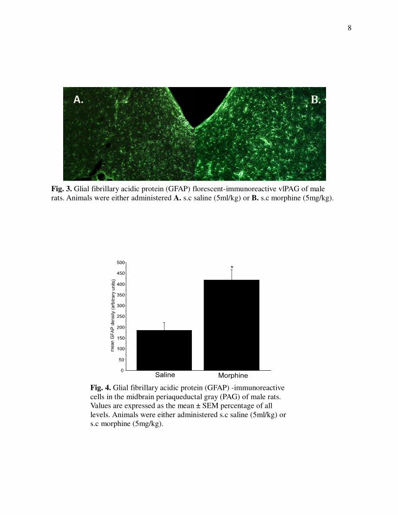
Fig. 3. Glial fibrillary acidic protein (GFAP) florescent-immunoreactive vlPAG of male
rats. Animals were either administered A. s.c saline (5ml/kg) or B. s.c morphine (5mg/kg).
A. B.
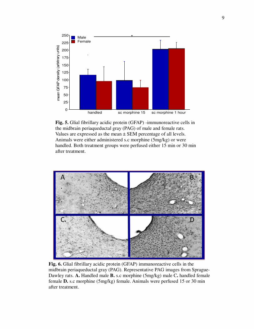
Fig. 4. Glial fibrillary acidic protein (GFAP) -immunoreactive
cells in the midbrain periaqueductal gray (PAG) of male rats.
Values are expressed as the mean ± SEM percentage of all
levels. Animals were either administered s.c saline (5ml/kg) or
s.c morphine (5mg/kg).
9
0
25
50
75
100
125
150
175
200
225
250
handled sc morphine 15 sc morphine 1 hour
FemaleMale
mean G
FA
P d
ensity (arb
itra
ry u
nits)
*
Fig. 5. Glial fibrillary acidic protein (GFAP) -immunoreactive cells in
the midbrain periaqueductal gray (PAG) of male and female rats.
Values are expressed as the mean ± SEM percentage of all levels.
Animals were either administered s.c morphine (5mg/kg) or were
handled. Both treatment groups were perfused either 15 min or 30 min
after treatment.
Fig. 6. Glial fibrillary acidic protein (GFAP) immunoreactive cells in the
midbrain periaqueductal gray (PAG). Representative PAG images from Sprague-
Dawley rats. A. Handled male B. s.c morphine (5mg/kg) male C. handled female
female D. s.c morphine (5mg/kg) female. Animals were perfused 15 or 30 min
after treatment.
A
.
B.
C. D
.
10
REFERENCES
Bandler R. and Shipley M.T. 1994. “Columnar organization in the midbrain periaqueductal gray:
modules for emotional expression?” Trends Neurosci. 17, 9: 379-389.
Basbaum A.I. and Fields H.L. 1978. “Endogenous pain control mechanisms: review and
hypothesis.” Annals of Neurobiology, 4, 5: 451-462.
Cicero T.J., Nock B. and Meyer E.R. 1996. “Gender-related differences in the antinociceptive
properties of morphine.” J. Pharmacol. Exp. Ther. 279: 767-773.
Fields H.L., Heinricher M.M., and Mason P. 1991. “Neurotransmitters in nociceptive modulatory
circuits.” Annu. Rev. Neurosci. 14: 219-245.
Guo W., Wang H., Zou S., Dubner R., and Ren K. 2012. “ Chemokine signaling involving
chemokine (c-c motif) ligand 2 plays a role in descending pain facilitation.” J. Neurosci. 28: 2;
193-207.
Hao S., Liu S., Zheng X., Zheng W., Ouyang H., Mata M. and Fink D.J. 2011. “The role of
TNFα in the periaqueductal gray during naloxone- precipitated morphine withdrawal in rats.”
Neuropharm. 36: 664-676.
Hutchinson M.R., Coats B.D., Lewis S.S., Zhang Y., Sprunger D.B., Rezvani N., Baker E.M.,
Jekich B.M., Wieseler J.L., Somogyi A.A., Martin D., Poole S., Judd C.M., Maier S.F., Watkins
L.R. 2008. “Proinflammatory cytokines oppose opioid-induced acute and chronic analgesia.”
Brain Beh Immun. 22, 8: 1178-1189.
Johnston I.N., Milligan E.D., Wieseler-Frank J., Frank M.G., Zapata V., Campisi J., et al. 2004.
“A role for proinflammatory cytokines and fractalkine in analgesia, tolerance, and subsequent
pain facilitation induced by chronic intrathecal morphine.” J. Neurosci. 24: 7353–7365.
Kepler K. L., Kest B., Paul D., Kiefel J.M., Cooper M.L. and Bodnar R. J. 1989. “Roles of
gender, gonadectomy and estrous phase in the analgesic effects of intracerebroventricular
morphine in rats.’ Pharmacol. Biochem. Behav. 34: 119-127.
Kreutzberg G.W. 1996. “Microglia: a sensor for pathological events in the CNS.” Trends
Neurosci. 19, 8: 312-318.
Laprairie J.L., and Murphy A.Z. 2009. “Neonaltal injury alters adult pain sensitivity by
increasing opioid tone in the periaqueductal gray.” Front Behav. Neurosci. 3: 31.
Loyd D.R., Morgan M.M., and Murphy A.Z. 2008. “Sexually dimorphic activation of the
periaqueductal gray- rostral ventromedial medulluary circuit during the development of tolerance
to morphine in the rat.” Eur. J. Neurosci. 27: 1517-1524.
11
Loyd D.R. and Murphy A.Z. 2008. “The role of the periaqueductal gray in the modulation of
pain in males and females: are the anatomy and physiology really that different?” Neural
Plasticity. vol. 2009. Article ID 462879, 12 pages.
Raghavendra V. and Deleo J.A. 2004. “The role of astrocytes and microglia in persistent pain.”
Advances in Molecular and Cell Biology, 31: 951–966.
Raghavendra V., Rutkowski M.D., and Deleo J.A. 2002. “The role of spinal neuroimmune
activation in morphine tolerance/hyperalgesia in neuropathic and sham-operated rats.” J.
Neurosci. 22: 9980-9989.
Van Bockstaele E.J., Aston-Jones G., Pieribone V.A., Ennis M., and Shipley M.T. 1991.
“Subregions of the periaqueductal gray topographically innervate the rostral ventral medulla in
the rat.” J. Comp. Neurol. 309, 3: 305-327.
Wang, X., Traub, R.J. and Murphy, A.Z. 2006. “Persistent pain model reveals sex difference in
morphine potency.” Am. J. Physiology, 291: 300-306.
Watkins L.R., Milligan E.D., and Maier S.F. 2001. “Spinal cord glia: new players in pain.” Pain.
93, 3: 201-205.
Watson R.E., Wiegand S.J., Clough R.W., Hoffman G.E. 1986. “Use of cryoprotectant to
maintain longterm peptide immunoreactivity and tissue morphology.” Peptides. 7: 155-159.
Wei F., Guo W., Zou S., Ren K. and Dubner R. 2008. “Supraspinal glial- neuronal interactions
contribute to descending pain facilitation.” J. Neurosci. 28: 10482-10495.