differential expression of cell surface antigens and glial fibrillary
TRANSCRIPT

[CANCER RESEARCH 46, 6406-6412, December 1986]
Differential Expression of Cell Surface Antigens and Glial Fibrillary Acidic Proteinin Human Astrocytoma Subsets1
Wolfgang J. Rettig,2 Pilar Garin Chesa,3 H. Richard Beresford, Hans-Joachim Feickert, Mark T. Jennings,
James Cohen, Herbert F. Oettgen, and Lloyd J. OldMemorial Sloan-Kettering Cancer Cancer. New York, New York 10021 [W. J. R., P. C. C., H. R. B., H-J. F., M. T. J., H. F. O., L. J. O.], and Department of Zoology,University College London, Gotver Street, London WC1E6BT [J. C.]
ABSTRACT
We have characterized five distinct cell surface antigens of humanastrocytomas and correlated their expression with the expression of glialfibrillar) acidic protein (GFAP) and four previously defined cell surfacemarkers of astrocytomas. One of the newly studied antigens, A4, whichwas originally detected on rat central nervous system (but not peripheralnervous system) neurons, is expressed on GFAP* human astrocytomacells, but not on GFAP" astrocytomas or a wide range of other neuroec-
todermal, epithelial, and hematopoietic cells. Antigens F19 (M, 140,000/90,000 glycoprotein) and F24 (M, 90,000 glycoprotein) also show restricted distribution and are expressed on subsets of neuroectodermaland mesenchymal cells. Antigens G253 (M, 95,000 glycoprotein) and S5(M, 120,000 glycoprotein) are more widely distributed on the culturedcell panel. The distribution of these antigens was determined on a seriesof 22 astrocytoma cell lines and in normal brain tissue and the resultswere compared with the distribution of 5 additional glial cell markers:GFAP and cell surface antigens AOIO (A/r 110,000 glycoprotein); AJ8(M, 100,000 glycoprotein); LK26 (M, 35,000 glycoprotein); and Thy-1.Distinct patterns of expression on cultured astrocytomas and in neuraltissues were identified for all antigenic systems studied, and cell surfaceexpression of antigen A4 was found to correlate closely with GFAPphenotype of cultured astrocytomas. The antigens described in this studyprovide new markers to study normal glial differentiation and to correlatethe phenotypes and biological behavior of distinct subsets of astrocytomas.
INTRODUCTION
Distinct pathways and stages of cellular differentiation areassociated with specific patterns of cell surface antigen expression. This principle was first established through analysis ofnormal and malignant cells of hematopoietic origin and hasbeen extended recently to other cell lineages (1-4). The findingof an ordered progression of surface phenotype changes duringnormal differentiation has permitted classification of leukemiasand lymphomas (2, 5) and malignant melanomas (3) into subsets that show antigenic similarity with normal cells at distinctstages of hematopoietic or neuroectodermal differentiation. Formost other human neoplasms, however, little is known aboutcoordinate patterns of antigen expression and their correlationwith normal cellular differentiation.
We have previously defined several restricted cell surfaceantigens of human astrocytomas (6). Two antigens, AJ8 andAOIO, showed reciprocal (although overlapping) patterns ofexpression on a panel of cultured astrocytomas; the AJ8+/AOIO" and AJ8~/A010+ phenotypes were found to correlatewith expression in these cell lines of GFAP,4 the best-known
molecular marker of glial differentiation. The present study
Received 1/31/86; revised 5/1/86, 7/30/86; accepted 8/19/86.The costs of publication of this article were defrayed in part by the payment
of page charges. This article must therefore be hereby marked advertisement inaccordance with 18 U.S.C. Section 1734 solely to indicate this fact.
' This work was supported by grants from the National Cancer Institute (CA-08748) and by the Oliver S. and Jennie R. Donaldson Charitable Trust, Inc.
2 Recipient of a Fellowship from the Deutsche Forschungsgemeinschaft.3 Recipient of a Fulbright Fellowship from the Spanish Ministry of Education.4 The abbreviations used are: GFAP, glial fibrillary acidic protein; MAb,
monoclonal antibody; MHA, mixed hemadsorption.
extends the serological analysis of glial tumors to includeseveral newly defined antigenic systems. The most restricted ofthese antigens, designated as A4, was first described in the rat(7) and serves as a marker that distinguishes central (A4+) andperipheral neurons (A4~). We now show that A4 is expressed
on a proportion of human astrocytomas and that A4 expressionis closely correlated with the expression of GFAP. Expressionof six additional surface antigenic systems was also found toidentify astrocytoma subsets. Our findings support the notionthat distinct patterns of cell surface antigen expression characterize subsets of glial neoplasms and that different tumor phenotypes may correspond to antigenic phenotypes of normalcells at distinct stages of glial differentiation.
MATERIALS AND METHODS
Cell Lines and Cell Culture. Nineteen human astrocytoma cell lines(SK-MG and SK-GS series) were established from tumor specimens ofdifferent patients obtained from North Shore University Hospital andNew York University Hospital; all tumors were diagnosed as grade IIIor grade IV astrocytomas by routine pathological evaluation. Stockcultures of newly established cell lines were stored in liquid nitrogen atearly passage levels (passages 5-20); in the present study, cell lines ofearly and late passage levels were analyzed. Additional cell lines wereobtained from the collection of Dr. J. Fogh (Sloan-Kettering Institute)and from the human tumor cell line bank of our laboratory. Cell lineswere routinely tested for Mycoplasma contamination and only Myco-piasma-free cultures were used for analysis.
Monoclonal Antibodies and Serological Procedures. MAb G253 wasderived from a (BALB/c x C57BL/6)F| mouse immunized with SK-GS-1 cells, MAb S5 from a mouse immunized with SK-MG-17 cells,and MAbs F24 and F19 from a mouse immunized with lung fibroblasts,following published fusion, hybrid selection, and cloning procedures(8). The Ig subclass, as determined by double diffusion in agar withanti-immunoglobulin heavy chain-specific antisera (Bionetics, Kensington, MD), was found to be IgG2a for MAb G253 and IgGl for MAbsF19, F24, and S5. Generation and initial characterization of MAbs A4,AOIO, AJ8, K117, and LK26 have been described previously (6, 7, 9,10). A second antibody to the A4 antigen, MAb C5 (IgG), was alsoused in this study and results for cell lines and tissue typing wereidentical to those obtained with MAb A4.
MHA rosetting assays for the detection of surface antigens oncultured cells have been described (11,12). Briefly, 200-300 cells/wellwere seeded into Falcon 3034 Microtest II plates (Falcon Labware,Oxnard, CA), and cultured for 24-48 h prior to serological analysis.Microcultures were incubated with serial dilutions of antibody for 1 h.After repeated washes, cells were incubated for 45 min with indicatorcells, prepared by conjugating purified rabbit anti-mouse Ig (DakoCorp., Santa Barbara, CA) or goat anti-mouse IgM (Dako) with humanerythrocytes using 0.01% chromium chloride. Finally, plates werewashed and reactivity was scored microscopically by determining theproportion of individual target cells that showed erythrocyte rosettingat each antibody dilution step tested; the highest antibody dilutiongiving rosette formation was defined as the reciprocal titration endpoint. MHA assays were also evaluated for the percentage of cellswithin antigen-positive cultures showing rosette formation, but noheterogeneity in antigen expression was observed. Unrelated MAbswere used as negative controls in all MHA assays; they gave no rosetteformation.
6406
Research. on November 18, 2018. © 1986 American Association for Cancercancerres.aacrjournals.org Downloaded from
SURFACE ANTIGENIC PATTERNS OF HUMAN ASTROCYTOMAS
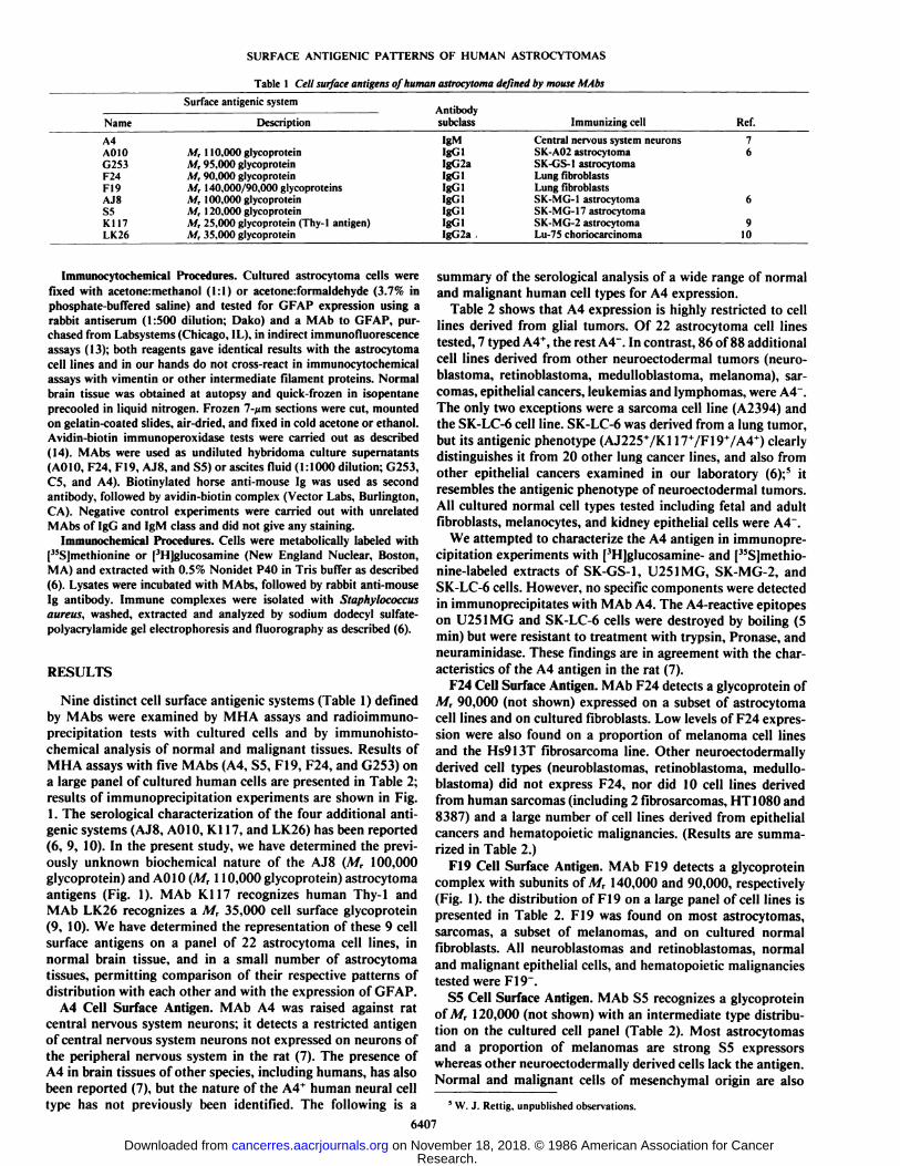
Table 1 Cell surface antigens of human astrocytoma defined by mouse MAbs
NameA4AGIOG253F24FI9AJ8SSK117LK26Surface
antigenicsystemDescriptionM,
110,000glycoproteinM,95,000glycoproteinM,90,000glycoproteinM,140,000/90,000glycoproteinsM,100.000glycoproteinM,120,000glycoproteinM,25,000 glycoprotein (Thy-1antigen)M,35,000 glycoproteinAntibody
subclassIgMIgGlIgG2aIgGlIgGlIgGlIgGlIgGlIgG2aImmunizingcellCentral
nervous systemneuronsSK-A02astrocytomaSK-GS-1astrocytomaLungfibroblastsLungfibroblastsSK-MG-1astrocytomaSK-MG-17astrocytomaSK-MG-2astrocytomaLu-75
choriocarcinomaRef.766910
Immunocytochemical Procedures. Cultured astrocytoma cells werefixed with acetone:methanol (1:1) or acetone:formaldehyde (3.7% inphosphate-buffered saline) and tested for GFAP expression using arabbit antiserum (1:500 dilution; Dako) and a MAb to GFAP, purchased from Labsystems (Chicago, IL), in indirect immunofluorescenceassays (13); both reagents gave identical results with the astrocytomacell lines and in our hands do not cross-react in immunocytochemicalassays with vimentin or other intermediate filament proteins. Normalbrain tissue was obtained at autopsy and quick-frozen in isopentaneprecooled in liquid nitrogen. Frozen 7-/jm sections were cut, mountedon gelatin-coated slides, air-dried, and fixed in cold acetone or ethanol.Avidin-biotin immunoperoxidase tests were carried out as described(14). MAbs were used as undiluted hybridoma culture supernatants(A010, F24, F19, AJ8, and S5) or ascites fluid (1:1000 dilution; G253,C5, and A4). Biotinylated horse anti-mouse Ig was used as secondantibody, followed by avidin-biotin complex (Vector Labs, Burlington,CA). Negative control experiments were carried out with unrelatedMAbs of IgG and IgM class and did not give any staining.
Immunochemical Procedures. Cells were metabolically labeled with["Sjmethionine or [3H]glucosamine (New England Nuclear, Boston,
MA) and extracted with 0.5% Nonidet P40 in Tris buffer as described(6). Lysates were incubated with MAbs, followed by rabbit anti-mouseIg antibody. Immune complexes were isolated with Staphylococcusaureus, washed, extracted and analyzed by sodium dodecyl sulfate-polyacrylamide gel electrophoresis and fluorography as described (6).
RESULTS
Nine distinct cell surface antigenic systems (Table 1) definedby MAbs were examined by MHA assays and radioimmuno-precipitation tests with cultured cells and by immunohisto-chemical analysis of normal and malignant tissues. Results ofMHA assays with five MAbs (A4, S5, F19, F24, and G253) ona large panel of cultured human cells are presented in Table 2;results of immunoprecipitation experiments are shown in Fig.1. The serological characterization of the four additional antigenic systems (AJ8, AGIO, Kl 17, and LK26) has been reported(6, 9, 10). In the present study, we have determined the previously unknown biochemical nature of the AJ8 (MT 100,000glycoprotein) and A010 (M, 110,000 glycoprotein) astrocytomaantigens (Fig. 1). MAb K117 recognizes human Thy-1 andMAb LK26 recognizes a M, 35,000 cell surface glycoprotein(9, 10). We have determined the representation of these 9 cellsurface antigens on a panel of 22 astrocytoma cell lines, innormal brain tissue, and in a small number of astrocytomatissues, permitting comparison of their respective patterns ofdistribution with each other and with the expression of GFAP.
A4 Cell Surface Antigen. MAb A4 was raised against ratcentral nervous system neurons; it detects a restricted antigenof central nervous system neurons not expressed on neurons ofthe peripheral nervous system in the rat (7). The presence ofA4 in brain tissues of other species, including humans, has alsobeen reported (7), but the nature of the A4* human neural cell
type has not previously been identified. The following is a
summary of the serological analysis of a wide range of normaland malignant human cell types for A4 expression.
Table 2 shows that A4 expression is highly restricted to celllines derived from glial tumors. Of 22 astrocytoma cell linestested, 7 typed A4+, the rest A4". In contrast, 86 of 88 additional
cell lines derived from other neuroectodermal tumors (neuroblastoma, retinoblastoma, medulloblastoma, melanoma), sarcomas, epithelial cancers, leukemias and lymphomas, were A4".
The only two exceptions were a sarcoma cell line (A2394) andthe SK-LC-6 cell line. SK-LC-6 was derived from a lung tumor,but its antigenic phenotype (AJ225+/K117+/F19+/A4+) clearly
distinguishes it from 20 other lung cancer lines, and also fromother epithelial cancers examined in our laboratory (6);5 it
resembles the antigenic phenotype of neuroectodermal tumors.All cultured normal cell types tested including fetal and adultfibroblasts, melanocytes, and kidney epithelial cells were A4".
We attempted to characterize the A4 antigen in immunoprecipitation experiments with ['Hjglucosamine- and ["Sjmethio-nine-labeled extracts of SK-GS-1, U251MG, SK-MG-2, andSK-LC-6 cells. However, no specific components were detectedin immunoprecipitates with MAb A4. The A4-reactive epitopeson U251MG and SK-LC-6 cells were destroyed by boiling (5min) but were resistant to treatment with trypsin, Pronase, andneuraminidase. These findings are in agreement with the characteristics of the A4 antigen in the rat (7).
F24 Cell Surface Antigen. MAb F24 detects a glycoprotein ofM, 90,000 (not shown) expressed on a subset of astrocytomacell lines and on cultured fibroblasts. Low levels of F24 expression were also found on a proportion of melanoma cell linesand the Hs913T fibrosarcoma line. Other neuroectodermallyderived cell types (neuroblastomas, retinoblastoma, medulloblastoma) did not express F24, nor did 10 cell lines derivedfrom human sarcomas (including 2 fibrosarcomas, HT 1080 and8387) and a large number of cell lines derived from epithelialcancers and hematopoietic malignancies. (Results are summarized in Table 2.)
F19 Cell Surface Antigen. MAb F19 detects a glycoproteincomplex with subunits of M, 140,000 and 90,000, respectively(Fig. 1). the distribution of F19 on a large panel of cell lines ispresented in Table 2. F19 was found on most astrocytomas,sarcomas, a subset of melanomas, and on cultured normalfibroblasts. All neuroblastomas and retinoblastomas, normaland malignant epithelial cells, and hematopoietic malignanciestested were F19~.
S5 Cell Surface Antigen. MAb S5 recognizes a glycoproteinof M, 120,000 (not shown) with an intermediate type distribution on the cultured cell panel (Table 2). Most astrocytomasand a proportion of melanomas are strong S5 expressorswhereas other neuroectodermally derived cells lack the antigen.Normal and malignant cells of mesenchymal origin are also
5 W. J. Rettig, unpublished observations.
6407
Research. on November 18, 2018. © 1986 American Association for Cancercancerres.aacrjournals.org Downloaded from

SURFACE ANTIGENIC PATTERNS OF HUMAN ASTROCYTOMAS
Table 2 Reactivity of MAbs to human cell surface antigens with a panel of over 100 independently derived human tumor cell lines andsnort-term cultures of normal cells
Summary of MHA assays using serial, 5-fold dilutions of antibody (starting dilution 1:500 of nu/nu serum or ascites) to determine highest antibody dilution(titration end point) giving rosette formation (see Fig. 2). Results of limitimi experiments are indicated as follows: •.strong reactivity with reciprocal titers of 5 xIO3-! x IO7;Ó.weak reactivity with reciprocal liters of 5 x 10²-5 X 10'; O, no reactivity at starting dilution of MAb. Each symbol represents typing results with adistinct cell line. Derivation and designation of cell lines have been described in previous studies (6, 8-10, 12); panel of epithelial cancer lines includes renal, bladder,lung, pancreatic, prostate, breast, colon, and ovarian cancers; leukemias and lymphomas were of T-. B-, and null cell and myeloid phenotypes. Astrocytoma cell linesare specified in Table 3.
Monoclonal antibody
Cultured cell type A4 F24 F19 S5 G253Neuroectoderm-derived cells
Astrocytoma
NeuroblastomaRetinoblastomaMcdulloblastomaMelanoma
Sarcomas
Normal fibrobiasts
Epithelial cells
•••••••Oooooooooooooooooooooooooooooooooooo
•ooooooo00
oocco
oooooooo oco»««««oooooo««
••©oooooooooooooo@@©oooco00
•ooooooo0000
ooooooooooo•••00000co
•*o««*oooooooooooooooo00o•••••ooooo
•MO •000
Normal kidney epithelial cellsSV40-positive keratinocytesEpithelialcancersHematopoietic
cellsLeukemias/lymphomasEpstein-Ban-
virus-positive B-cells000oooooooooo
oocccocooooooooooooooooo0000oooooooo
oooooooooocoo
oooooooooooooooooooooooooooooooooo0000oooooooo
oooooooooo000oooooooo
oooooooooooooooooooooooooooooooooooooooo
oooooooooooooCO•••ooooo
ooooooooooooooooooooooooooooooo•ooooooo
ooooooocoo*••
M••oooooo©ooooooooooooo
oooooooooo
.140•90
.110•100
A B C D E FFig. I. Fluorogram of immunoprecipitates obtained with MAbs to cell surface
antigens using extracts of I'Hjglucosamine-labeled fibrobiasts (lanes A and lì).U251MG (lane £'),SK-MG-7 (lane D). or SK-GS-1 astrocytoma cells (lanes Eund F). Immunoprecipitates were separated on sodium dodecyl sulfate-polyacryl-amide gels under reducing conditions. MAbs used for immunoprecipitation testswere: lane A. F19; lane C, A010: lane D, AJ8: lane E, G253; lane F. A4; and laneH. unrelated control M Ab S V63 ( 10). Molecular weights of immunoprecipitatedcomponents are indicated on the right (molecular weight x 10"').
S5*. Normal kidney epithelial cells and a large proportion ofcell lines derived from epithelial cancers as well as hemato-
poietic malignancies are negative for S5 expression. Only 3 of36 epithelial cancer lines (choriocarcinoma GCC-SV and renalcancers SK-RC-1 and SK-RC-18) and one of 9 leukemia linestested (null cell leukemia NALL-1) were strong S5 expressors.
G253 Cell Surface Antigen. MAb G253 detects a glycoproteinof M, 95,000 (Fig. 1) that also shows an intermediate typedistribution (Table 2). Normal cultured kidney epithelial cells
were strongly G253+, skin fibrobiasts were weakly reactive, and
most cell lines derived from neuroectodermal tumors, epithelial—95 cancers, and sarcomas were strongly G253+. Leukemias and
lymphomas, Epstein-Barr virus-transformed B-cells, and other
cell lines growing in suspension culture (retinoblastomas andneuroblastomas) were G253~.
Thy-1 and LK26 Expression on Astrocytoma Cell Lines. MAbK117 recognizes human Thy-1 antigen, a M, 25,000 cell surfaceglycoprotein expressed on normal and malignant cells of neuroectodermal and mesenchymal origin (9). Typing of the astrocytoma panel showed that all cell lines included in this studywere strongly reactive with MAb Kl 17. With regard to LK.26,two astrocytoma lines (U373MG and SK-MG-9) were found toexpress this antigen at moderate levels and three additionallines (SK-MG-4, SK-MG-11, and T98) showed only weakreactivity. MAb LK26 was raised against a human choriocarcinoma cell line and has been shown to react with culturedepithelial cells (10) and with a small range of normal epithelialtissues.6 More significantly, we show in this study that a proportion of glial cells in normal adult brain are LK26+ (see
below).Coordinate Expression of GFAP and Surface Antigens on Glial
Tumor Cells. The panel of 22 astrocytoma cell lines was typedfor expression of the nine cell surface antigens included in thisstudy and for expression of GFAP. Fig. 2 shows examples ofMHA titration experiments with MAbs A4, A010, G253, F19,and AJ8 on four representative astrocytoma cell lines. A characteristic of these five surface antigens and of antigens S5 andF24 is that they are expressed on distinct subsets of the glialtumor panel (Table 3). A close correlation was observed betweenA4 and GFAP expression, with 21 of 22 cell lines showing
6 P. Garin Chesa and W. J. Rettig, manuscript in preparation.
6408
Research. on November 18, 2018. © 1986 American Association for Cancercancerres.aacrjournals.org Downloaded from

SURFACE ANTIGENIC PATTERNS OF HUMAN ASTROCYTOMAS
TARGET CELLS
SK-MG-22 SK-MG-5 T 98 SK-GS-1
loo -\•—•—•
cr>
AJ8
F19
G253
A010
A4
ANTIBODY DILUTION (x10ó)
Fig. 2. Titration of MAb reactivity with human astrocyloma cell lines by MHA rosetting assay. Serial 5-fold dilutions of MAbs (range: 1/1.000-1/3,125,000 ofmi nn sera or asciles fluid) were tested for rosette formation of indicator cells (erythrocytes coated with secondary antibody) with the target cells; at each dilutionstep, the proportion of target cells showing rosette formation was determined microscopically.
concordance for these 2 markers: 6 lines were GFAP+/A4+, 15lines were GFAP-/A4~, and one line was GFAP~/A4+. A010
expression correlated with both GFAP and A4 expression: 7 ofthe 11 A010+ cell lines were A4* (of these, 6 were also GFAP*),whereas none of the 11 A010~ cell lines expressed either A4 or
GFAP.We have compared our typing results for AJ8 and A010
expression in the astrocytoma panel (Table 3) with the resultsdescribed by Cairncross et al. (6) and good general agreementwas observed. A direct comparison of the AJ8 and A010 typingreagents used in the two studies established that the reagents(nu/nu sera and ascites fluid) used in the present study containapproximately 10-fold higher concentration of specific MAbs
than those used by Cairncross et al. (6); thus, it is not surprisingthat we now find weak or moderate reactivity with some celllines previously typed as unreactive or weakly positive with therespective MAb. (We disagree, however, with the results reported by Cairncross et al. (6) for the SK-MG-9 cell line whichwe found to be consistently AJ8+/A010~, both by MHA assay
and by radioimmunoprecipitation tests.) Furthermore, we havecompared early and late passages of SK-MG and SK-GS astrocytoma lines for surface antigen expression and no differenceswere observed, suggesting considerable phenotypic stability oflong-term cultured astrocytomas. In this and previous studies(3,6,8-10) we have evaluated the reciprocal titration end pointsdetermined in MHA assays with different cell lines as a measure
6409
Research. on November 18, 2018. © 1986 American Association for Cancercancerres.aacrjournals.org Downloaded from

SURFACE ANTIGENIC PATTERNS OF HUMAN ASTROCYTOMAS
Table 3 Patterns of GFAP and cell surface antigen expression in a panel of 22 independently derived human astrocytoma cell linesSummary of MHA (¡(rationexperiments (see legend to Table 2) with seven MAbs to cell surface antigens and indirect immunofluorescence typing with rabbit anti-
GFAP antibody and anti-GFAP MAb (see "Materials and Me(hods"). Resuhs of MHA (itration experiments are symbolized as follows: +++, strong reactivity(reciprocal liters 1 x lO'-l x IO7); ++, intermediate reactivity (reciprocal titers 1 x 10M x 10s); +, weak reactivity (reciprocal titers 5 x 10²-l X IO4); -, no
reactivity at s(ar(ing dilution of MAb ( 1:500 nu/nu serum or ascites fluid). Each symbol represents the results of at least three separate experiments; test results didnot vary by more than a single, S-fold (¡(rationstep. Cell line SK-MG-22 has previously been referred to as SK-MS (6).
Cell surfaceantigenS5
AJ8 F19 F24 G253 A010glycoprotein glycoprotein glycoprotein glycopro(ein glycoprotein glycoprotein
Cell line 120 100 140/90 90 95 110A4SK-MG-13+++ +++ +++ - - --SK-MG-7+++ +++++SK-MG-8++ +++ ++++SK-MG-21++ +++ +++ ++SK-MG-22+++ +++ ++++++SK-MG-1
5 +++ +++ +++ ++++SK-MG-11 +++ +++ +++ ++++SK-MG-5
+++ + +++ ++SK-MG-3+++ ++ ++++++CL*
\/tf(^aK-ivnj-oSK-MG-9
+++ +++ +++++SK-MG-16+++ +++ +++ +++ +-1-CIS
\nf 13N-JV1O-ISK-MG-14
+++ +++ +++ +++ ++++++T98+++ ++ ++ ++++SK-MG-1
2 +++ +++ +++ + +++ ++++SK-MG-4-1- +++ + +++ ++SK-MG-23+ + +++ + ++++++U251MG
+ +++ +++ ++++++U373MG+++ ++++++SK-MG-2+++ ++++++SK-GS-1
- +++ +++ +++GFAP_——————————————+—++++-1-
of quantitative differences in antigen expression. In agreementwith this interpretation, we have found that in radioimmuno-precipitation assays MAb A010 precipitates the characteristicM, 110,000 glycoprotein from SK-GS-1, SK-MG-2, andU251MG cell extracts but not from extracts of T98, SK-MG-16, or SK-MG-1 cells; in contrast MAb AJ8 precipitates theM, 100,000 glycoprotein from SK-MG-7, SK-MG-22, and SK-MG-13 cells but not from SK-GS-1, SK-MG-2, U251MG, SK-MG-23, T98, or SK-MG-5 cells. Similarly, all other MAbs(except MAb A4; see above) were successfully used to precipitate the respective glycoproteins from several high-expressorcell lines (identified by MHA titration experiments) but failedto precipitate any detectable antigen from cell lines that areunreactive or weakly reactive by MHA assays.
Antigen Expression in Normal and Malignant Human BrainTissues. In order to examine antigen expression in normalneural cells in vivo, MAbs were tested with frozen sections ofhuman brain tissues by the avidin-biotin immunoperoxidasemethod. Tissue samples were taken from several regions ofnormal brain, including cerebral cortex, thalamus, basal ganglia, hypothalamus, medulla oblongata, and cerebellum. MAbA4 showed strong staining of gray matter in all areas tested,labeling both cell bodies with a cytoplasmic pattern and neu-
ropil (Fig. 3, A and B); in addition, MAb A4 stained cellprocesses in white matter (Fig. 3, A and B). The strong reactivityof MAb A4 with neuronal cells made it difficult to determinewhether glial cells also express A4 antigen. MAb A010 showeda more restricted staining pattern in normal brain. Neurons inseveral areas tested, including cerebellum, medulla oblongata,and globus pallidus showed A010 staining localized to neuronalcell bodies and cell processes (Fig. 3, D and E). In contrast tothe uniform neuronal reactivity seen for A4, A010 reactivitywas detected only in subpopulations of CNS neurons. (TheA010 distribution in the CNS will be described in detail elsewhere.) No A010 reactivity was seen with glial cells.
MAbs G253 and LK26 showed reactivity with glial cells butnot neurons, as judged by the characteristic morphology of the
antigen-expressing cells (Fig. 3/) in all areas of the brain tested.However, distinct staining patterns distinguish these two anti-genie systems. MAb G253 labeled a large proportion of glialcells and blood vessels whereas reactivity with MAb LK26 wasrestricted to a small subpopulation of glial cells; these LK26+
glial cells have not yet been further characterized. MAb AJ8showed strong reactivity with glial cell bodies and cell processes(Fig. 3//), but did not seem to label any neurons. No stainingwas seen with MAbs S5, F19, and F24 in any of the normalbrain tissues tested.
To determine whether any of the cell surface antigens detected on cultured astrocytoma cells but not on normal glialcells in vivo are expressed on astrocytoma cells in vivo, frozentissues from four high-grade astrocytomas were tested by immunoperoxidase procedures with the respective MAbs. Threetumors were found to be A4+/A010+ (Fig. 3, C and F) and did
not express any of the other markers and the fourth tumor wasLK26+ and negative for the other markers.
DISCUSSION
GFAP is the most extensively studied molecular marker ofmature astrocytes; it is thought to be acquired during late stagesof glial differentiation (15). We have now defined a human cellsurface antigen, A4, which shows concordant expression withGFAP on a large panel of cultured glial tumors. The A4 antigen,initially described in the rat as a neuronal antigen that distinguishes neurons of the central nervous system (antigen positive)from those of the peripheral nervous system (antigen negative),is highly restricted to human astrocytoma cell lines. Furthermore, it is predominantly expressed on GFAP* cell lines,permitting classification of cultured astrocytomas into GFAP*/A4+ and GFAP~/A4~ subsets. Immunohistochemical tests with
normal human brain were carried out to examine whethernormal astrocytes also express A4. However, neurons of thecentral nervous system uniformly express A4 and the strongneuronal staining made it difficult to determine whether glial
6410
Research. on November 18, 2018. © 1986 American Association for Cancercancerres.aacrjournals.org Downloaded from

SURFACE ANTIGENIC PATTERNS OF HUMAN ASTROCYTOMAS
s
3$
.- ,«y¿. •.-3$&»'¿
^^Ä£&
c * ¿.i*'v<Ã}X-1^, ,,
-F:'V^:
i
Fig. 3. Distribution of cell surface antigens of cultured human astrocytomas in normal adult brain and astrocytoma tissues. Immunoperoxidase staining of frozensections, hematoxylin counterstain.. I. globus pallidus tested with MAb A4, low-power magnification to show strong uniform reactivity in areas of gray matter (solidarrowheads) and discrete filamentous staining in white matter (open arrowhead); B, caudate nucleus tested with MAb A4 (arrowheads as in IK C, high-gradeastrocytoma tested with MAb A4, strong membrane staining; /'. medulla oblongata tested with MAb AGIO, low-power magnification to show neuronal staining inthe principal nucleus of the inferior olivary complex (gyrated pattern); no staining of surrounding neuropil; E. globus pallidus tested with MAb A010, higher-powermagnification to show A010 staining of individual neuronal cell bodies and processes (arrowheads); /•',high-grade astrocytoma tested with MAb A010, strong
membrane staining; C and //. cerebellum tested with MAb AJ8, staining of a small proportion of cells in the granular layer (G) and of radial fibers in the molecularlayer (H); /, cerebral cortex tested with MAb G253, reactivity with glial cell. Original magnifications: A, B, and D, x 40; C and E-H, x 200; /, x 400.
cells in general or a proportion of glial cells also express thisantigen. Therefore, it is significant that we have demonstratedA4 expression in a proportion of astrocytoma tissues, indicatingthat the A4+ phenotype of cultured astrocytomas is not merely
a tissue culture artifact. We have previously described a M,145,000 surface antigen of human astrocytomas, detected by
MAb AJ225, which also shows a highly restricted distribution(6). Our serological findings clearly distinguish this antigenfrom A4. Other astrocytoma antigens, described by us andothers (6, 16, 17) can also be distinguished from A4 by theirbroader representation on neuroectodermal cells. Similarly,F19, F24, S5, and G253 appear to be distinct from the previ-
6411
Research. on November 18, 2018. © 1986 American Association for Cancercancerres.aacrjournals.org Downloaded from

SURFACE ANTIGENIC PATTERNS OF HUMAN ASTROCYTOMAS
ously described antigens. AJ8 shares many characteristics withcommon acute lymphoblastic leukemia antigen ( 18), and studiesin our laboratory indicate that the two antigens are identical.7
The biochemical nature of both rat and human A4 moleculeshas only partially been determined. However, the difference insubcellular localization between GFAP, a cytoskeletal component, and A4, a cell surface component, clearly distinguishesthese two markers. Thus, GFAP and A4 seem to be coordinatelyexpressed as part of a glial differentiation program. Based onthe distribution of GFAP and A4 alone, two major subsets aredefined on the astrocytoma cell panel, one being GFAP+/A4+and one being GFAP~/A4~; by including additional surface
antigenic markers, such as A010, AJ8, and S5, several intermediate phenotypes can be identified. Among these cell surfaceantigens, A4/A010 and AJ8/S5 tend to be expressed on reciprocal subsets of the astrocytoma panel. Cell surface antigenicsystems that are expressed only on subsets of certain types ofmalignancies have been described by several investigators (fora review, see Ref. 19). Relatively few tumors have been shownto express different antigenic systems in coordinate concordantor reciprocal patterns (2, 3. 6, 10), and these may parallelantigenic phenotypes of normal cells at distinct stages of cellulardifferentiation. However, malignant transformation of subsetsof glial cells and preservation of their characteristic antigenicpatterns is only one possible mechanism to explain the antigenicdiversity observed in the present study. With regard to the S5,F19. and F24 antigens which are expressed on distinct subsetsof cultured astrocytomas but not detected on glial cells in vivo,it will be important to determine whether their expression isrelated to the increased proliferative activity or the transformedstate of the tumor cells. Alternatively, S5, F19, and F24 expression on astrocytoma cell lines may be induced by specific tissueculture conditions. If this is true, an additional explanation isrequired for the clear distinction between antigen-positive andantigen-negative lines. In the case of the AGIO antigen, whichis also expressed on a number of cultured astrocytomas but notby normal glial cells in vivo, we have already shown in thisstudy that antigen expression is not limited to cultured astrocytomas but is also found in a proportion of astrocytomas invivo. Similarly, A4 is expressed both on cultured astrocytomasand in astrocytoma tissues and possibly also on normal glialcells (20). In order to determine the contributions of differentiation- and transformation-related mechanisms in generatingspecific tumor phenotypes it will be necessary to compare thepatterns of antigen expression seen in normal fetal and adultbrain with the antigenic phenotypes of a larger series of astrocytoma tissues. This typing may lead to a classification of glialtumors not only with respect to their cellular origin but also totheir biological properties.
7C. L. Finstad and L. J. Old. manuscript in preparation.
ACKNOWLEDGMENTS
We are grateful to Drs. J. Ransohoff and R. Carras for providingtumor specimens. We acknowledge the expert technical assistance ofS. Walker and G. Lark and excellent secretarial help provided by J.Rios.
REFERENCES
1. Boyse, E. A., and Old, L. J. Some aspects of normal and abnormal cellsurface genetics. Annu. Rev. Genet., 3: 269-290, 1969.
2. Greaves, M. F. "Target" cells, cellular phenotypes, and lineage fidelity inhuman leukemia. J. Cell. Physiol., /: 113-125, 1982.
3. Houghton, A. N., Eisinger, M., Albino, A. P., Cairncross, J. G.. and Old, L.J. Surface antigens of melanocytes and melanomas. Markers of melanocytedifferentiation and melanoma subsets. J. Exp. Med., 156: 1755-1766, 1982.
4. Raff, M. C., Miller, R. H., and Noble, M. A glial progenitor cell that developsin vitro into an astrocyte or an oligodendrocyte depending on culture medium.Nature (Lond.), 303: 390-396, 1983.
5. Foon, K. A., and Todd, R. F. Immunologie classification of leukemia andlymphoma. Blood. 68: 1-31. 1986.
6. Cairncross. J. G.. Mattes, M. J., Beresford. H. R., Albino, A. P. Houghton,A. N., Lloyd, K. O.. and Old, L. J. Cell surface antigens of human astrocytoma defined by mouse monoclonal antibodies: identification of astrocytomasubsets. Proc. Nati. Acad. Sci. USA, 79: 5641-5645. 1982.
7. Cohen, J., and Selvendran, S. Y. A neuronal cell-surface antigen is found inthe CNS but not in peripheral neurons. Nature (Lond.), 291:421-423, 1981.
8. Dippold, W. G., Lloyd. K. O.. Li, L. T. C, Ikeda, H., Oettgen, H. F., andOld, L. J, Cell surface antigens of human malignant melanoma: definitionof six antigenic systems with mouse monoclonal antibodies. Proc. Nati. Acad.Sci. USA. 77:6114-6118, 1980.
9. Rettig. W. J.. Dracopoli, N. C.. Garin Chesa, P., Spengler, B. A., Beresford,H. R., Davies, P., Biedler, J. L., and Old, L. J. Role of human chromosome11 in determining surface antigenic phenotype of normal and malignant cells.Somatic cell genetic analysis of eight antigens, including putative humanThy-1. J. Exp. Med., 162: 1603-1619, 1985.
10. Rettig, W. J.. Cordon-Cardo, C., Koulos, J. P., Lewis, J. L. Jr., Oettgen, H.F.. and Old. L. J. Cell surface antigens of human trophoblast and choriocar-cinoma defined by monoclonal antibodies. Int. J. Cancer. 35:469-475,1985.
11. Carey. T. E.. Takahashi. T.. Resnick. L. A., Oettgen, H. F.. and Old, L. J.Cell surface antigens of human malignant melanoma. I. Mixed hemadsorp-tion assay for humoral immunity to cultured autologous melanoma cells.Proc. Nati. Acad. Sci. USA, 73: 3278-3282, 1976.
12. Mattes, M. J., Tanimoto, M., Pollack, M. S., and Maurer, D. H. Preparingmonolayers of non-adherent mammalian cells. J. Immunol. Methods, 61:145-150, 1983.
13. Coons, A. H.. Leduc, E. H., and Connolly, J. M. Studies on antibodyproduction: a method for the histochemical demonstration of specific antibody and its application to a study of the hyperimmune rabbit. J. Exp. Med..102: 49-60. 1955.
14. Hsu, S. M., Raine, L., and Fanger, H. The use of avidin-biotin-peroxidasecomplex (ABC) in immunoperoxidase techniques. A comparison betweenABC and unlabeled antibody (PAP) procedures. J. Histochem. Cytochem.,29:577-580, 1982.
15. Juurlink, B. H., Fedoroff, S., Hall, C., and Nathaniel, E. J. Astrocyte celllineage. I. Astrocyte progenitor cells in mouse neopallium. J. Comp. Neurol..200:375-391. 1981.
16. Schnegg, J. F., Diserens. A. C.. Carrel, S.. Accolla. R. S., and de Tribolet,N. Human glioma-associated antigens detected by monoclonal antibodies.Cancer Res.. 41: 1209-1213, 1981.
17. Wikstrand, C. J., Bigner. S. H., and Bigner. D. D. Characterization of threerestricted specificity monoclonal antibodies raised against the human gliomacell line D-54MG. J. Neuroimmunol.. 6: 169-186, 1984.
18. Ritz. J., Pesando. J.. Notis-McConarty. J., Lazarus. H.. and Schlossman. S.F. A monoclonal antibody to human acute lymphoblastic leukemia antigen.Nature (Lond.), 283: 583-585, 1980.
19. Lloyd. K. O. Human tumor antigens, in: R. B. Herberman (ed.), Basic andClinical Tumor Immunology, pp. 159-214. The Hague, Netherlands: Mar-tinus Nijhoff, 1983.
20. Miller. R. H.. Williams, B. P., Cohen. Y.. and Raff. M. C. A4: an antigenicmarker of neural tube-derived cells. J. Neurocytol.. 13: 329-338. 1984.
6412
Research. on November 18, 2018. © 1986 American Association for Cancercancerres.aacrjournals.org Downloaded from

1986;46:6406-6412. Cancer Res Wolfgang J. Rettig, Pilar Garin Chesa, H. Richard Beresford, et al. Fibrillary Acidic Protein in Human Astrocytoma SubsetsDifferential Expression of Cell Surface Antigens and Glial
Updated version
http://cancerres.aacrjournals.org/content/46/12_Part_1/6406
Access the most recent version of this article at:
E-mail alerts related to this article or journal.Sign up to receive free email-alerts
Subscriptions
Reprints and
To order reprints of this article or to subscribe to the journal, contact the AACR Publications
Permissions
Rightslink site. Click on "Request Permissions" which will take you to the Copyright Clearance Center's (CCC)
.http://cancerres.aacrjournals.org/content/46/12_Part_1/6406To request permission to re-use all or part of this article, use this link
Research. on November 18, 2018. © 1986 American Association for Cancercancerres.aacrjournals.org Downloaded from