genomic responses during acute human anaphylaxis are ... filegenomic responses during acute human...
TRANSCRIPT

Genomic Responses during Acute Human AnaphylaxisAre Characterized by Upregulation of InnateInflammatory Gene NetworksShelley F. Stone 1,2*.", Anthony Bosco3.", Anya Jones3, Claire L. Cotterell1,2, Pauline E. van Eeden1,2,
Glenn Arendts1,2, Daniel M. Fatovich1,2, Simon G. A. Brown1,2
1 Centre for Clinical Research in Emergency Medicine, Harry Perkins Institute of Medical Research and the University of Western Australia, Perth, Australia, 2 Department of
Emergency Medicine, Royal Perth Hospital, Perth, Australia, 3 Telethon Kids Institute and the Centre for Child Health Research, University of Western Australia, Perth,
Australia
Abstract
Background: Systemic spread of immune activation and mediator release is required for the development of anaphylaxis inhumans. We hypothesized that peripheral blood leukocyte (PBL) activation plays a key role.
Objective: To characterize PBL genomic responses during acute anaphylaxis.
Methods: PBL samples were collected at three timepoints from six patients presenting to the Emergency Department (ED)with acute anaphylaxis and six healthy controls. Gene expression patterns were profiled on microarrays, differentiallyexpressed genes were identified, and network analysis was employed to explore underlying mechanisms.
Results: Patients presented with moderately severe anaphylaxis after oral aspirin (2), peanut (2), bee sting (1) and unknowncause (1). Two genes were differentially expressed in patients compared to controls at ED arrival, 67 genes at 1 hour post-arrival and 2,801 genes at 3 hours post-arrival. Network analysis demonstrated that three inflammatory modules wereupregulated during anaphylaxis. Notably, these modules contained multiple hub genes, which are known to play a centralrole in the regulation of innate inflammatory responses. Bioinformatics analyses showed that the data were enriched forLPS-like and TNF activation signatures.
Conclusion: PBL genomic responses during human anaphylaxis are characterized by dynamic expression of innateinflammatory modules. Upregulation of these modules was observed in patients with different reaction triggers. Ourfindings indicate a role for innate immune pathways in the pathogenesis of human anaphylaxis, and the hub genesidentified in this study represent logical candidates for follow-up studies.
Citation: Stone SF, Bosco A, Jones A, Cotterell CL, van Eeden PE, et al. (2014) Genomic Responses during Acute Human Anaphylaxis Are Characterized byUpregulation of Innate Inflammatory Gene Networks. PLoS ONE 9(7): e101409. doi:10.1371/journal.pone.0101409
Editor: Simon Patrick Hogan, Cincinnati Children’s Hospital Medical Center, University of Cincinnati College of Medicine, United States of America
Received April 9, 2014; Accepted May 13, 2014; Published July 1, 2014
Copyright: � 2014 Stone et al. This is an open-access article distributed under the terms of the Creative Commons Attribution License, which permitsunrestricted use, distribution, and reproduction in any medium, provided the original author and source are credited.
Data Availability: The authors confirm that all data underlying the findings are fully available without restriction. The raw microarray data are available from theGene Expression Omnibus repository (accession number GSE47655).
Funding: The study was supported by a grant received by SFS and SGAB from the US Food Allergy and Anaphylaxis Network, now called Food Allergy Researchand Education (www.foodallergy.org). SGAB is supported by a NHMRC Career Development Fellowship Award ID1023265. AB is supported by a BrightSparkFoundation McCusker Fellowship. Additional funding was received from the Royal Perth Hospital Medical Research Foundation. The funders had no role in studydesign, data collection and analysis, decision to publish or preparation of the manuscript.
Competing Interests: The authors have declared that no competing interests exist.
* Email: [email protected]
. These authors contributed equally to this work.
" SFS and AB are joint senior authors on this work.
Introduction
Anaphylaxis is a severe allergic reaction affecting multiple organ
systems, characterized by generalized erythema-urticaria, plus
cardiovascular compromise (hypotension) and/or respiratory
features (breathlessness, bronchospasm and hypoxemia). Foods,
insect stings and drugs cause roughly equal proportions of
reactions. Allergen crosslinking of allergen-specific IgE bound by
high affinity (FceRI) receptors to mast cells in the gut, skin and
perivascular tissues including coronary vessels is the predominant
triggering mechanism. An array of preformed and newly
synthesized biochemical mediators with overlapping biological
effects are then released [1]. However, the mechanism by which
minute amounts of allergen administered locally (e.g. a sting to the
skin, or minute amount of ingested food) leads to massive levels of
systemic mediator release and death within minutes of exposure is
not fully understood [2].
Several groups of immune mediators have independent
associations with reaction severity, suggesting a synergistic
involvement of multiple inflammatory pathways in human
PLOS ONE | www.plosone.org 1 July 2014 | Volume 9 | Issue 7 | e101409

anaphylaxis [3]. Possible amplification mechanisms include
mediators from triggered mast cells having a direct effect on
other mast cells [4], and the involvement of other immune cells,
including peripheral blood leukocytes (PBL). The concept of a
‘‘mast cell-leukocyte cytokine cascade’’ has been proposed in the
context of allergic airway inflammation [5], and neutrophils and
basophils have been found to have pivotal roles in mouse models
of anaphylaxis [6,7]. However, mouse models are largely IgG-
mediated and there is no evidence for the involvement of
circulating leukocytes in human anaphylaxis.
We therefore aimed to improve our understanding of the
pathophysiology of human anaphylaxis by investigating gene
expression patterns in PBL collected during anaphylaxis.
Methods
Study populationPatients were recruited in the Royal Perth Hospital ED as part
of the Critical Illness and Shock Study [8]. Because the need for
emergency care took priority, waiver of initial consent was
approved under the provision of paragraph 2.3.6 of the National
Health and Medical Research Council Ethical Conduct guidelines
(2007). Once treatment was started, fully informed written consent
was obtained as soon as possible and patients were given the
option of declining further involvement and having all research
samples collected up to that point destroyed. Ethics approval,
including waiver of initial consent, was obtained from the Royal
Perth Hospital Human Research Ethics Committee (EC 2009/
080).
We enrolled a convenience sample of six consecutive adult
patients, presenting when a research nurse was on duty with
typical anaphylaxis according to the National Institutes Allergy
and Infections Diseases/Food Allergy and Anaphylaxis Network
definition of anaphylaxis [9], and who had not received any
treatment prior to ED arrival. A structured datasheet was used to
record demographics, reaction features, likely causation (if known),
co-morbidities, physiological observations and treatments. Reac-
tion severity was graded according to our established grading
system [10]. Samples were also collected from six age-sex matched
healthy controls with no history of anaphylaxis.
Sample collection and storageBlood samples were collected at enrolment (arrival in the ED),
1 hour and 3 hours after enrolment in both patients and controls,
and stabilized in PAXgene tubes (PreAnalytiX GmbH, Switzer-
land). The PAXgene tubes were placed at 4uC then transferred to
220uC within 72 hours, before final storage at 280uC.
RNA extractionRNA was extracted with the PAXgene Blood RNA Extraction
Kits (PreAnalytiX GmbH, Switzerland) by automation with the
Qiacube instrument (Qiagen, Australia). The purity and integrity
of the RNA was assessed on a NanoDrop (Thermo Scientific,
Australia) and Bioanalyzer (Agilent), and was very high (median
OD 260/280 ratio of 2.1 (IQR 2.06–2.14); median RIN 8.4 (IQR
8.1–8.8)).
Gene expression profilingTotal RNA samples were labeled and hybridized to Affymetrix
Human Gene 1.0 ST microarrays at the Ramaciotti Centre for
Gene Function Analysis (University of New South Wales). The
microarray data was high quality (mean6sd; pm mean = 454689;
all probeset mean = 6.6260.02; pos vs neg auc = 0.8360.01; mad
residual mean = 0.3560.04; relative log expression mean
= 0.260.04). The raw microarray data are available from the
Gene Expression Omnibus repository (accession number
GSE47655).
The microarray data was analyzed in the R environment for
statistical computing. The data was preprocessed employing the
Factor Analysis for Robust Microarray Summarization algorithm
(qFARMS; laplacian prior was used) [11]. A custom chip
description file was used to map probe sets to genes based on
current annotation of the genome (hugene10sthsentrezg; version
16) [12]. The informative/non-informative calls algorithm was
employed to identify relevant gene expression signals and filter out
noise [13]. The final filtered data set comprised 5,292 genes and
36 samples, and this filtered data set was used for all downstream
analyses.
Differentially expressed genes were identified using Bayesian/
moderated t-statistics (LIMMA), and those genes with a False
Discovery Rate (FDR) adjusted p-value of less than 0.05 were
deemed significant [14]. Molecular signatures from the Molecular
Signatures Database (http://www.broadinstitute.org/gsea/msigdb/
index.jsp) were tested for differential expression employing Gene Set
Analysis, with FDR control for multiple testing [15]. A coexpression
network was constructed employing weighted gene coexpression
network analysis (WGCNA) [16–18]. Modules of coexpressed genes
were tested for differential expression in anaphylaxis cases versus
controls employing Correlation Adjusted MEan RAnk gene set
analysis (CAMERA) [19]. The wiring diagram of the disease-
associated modules was reconstructed in Ingenuity Systems software
using mechanistic data from prior studies [18]. Genes with no
previously documented molecular interactions were removed from
the analysis. Biological functions and pathways enriched in the data
were identified using the database for annotation, visualization and
integrated discovery (DAVID) [20]. Additional pathways analyses
were performed with Ingenuity Systems software. Ingenuity Systems
Upstream Regulator analysis was employed to infer putative driver
genes or drugs/compounds that may give rise to the observed gene
expression changes, and an overlap p-value was calculated based on
the number of differentially expressed genes identified in the data
that are known to be regulated by the upstream regulator.
Results
PatientsDetails for each patient are presented in Table 1. Reactions
were of moderate severity (i.e. without hypotension or hypoxemia)
at the time of ED arrival. Reactions satisfied consensus clinical
criteria for a diagnosis of anaphylaxis [9], and were attributed to
aspirin (n = 2), peanut (n = 2), bee sting (n = 1) and unknown cause
(n = 1). All patients were untreated at T0 and were treated with
combinations of steroids, intravenous (IV) fluids and epinephrine
between T0 and T1.
Gene expression profiling of acute anaphylaxis in wholeblood
Gene expression levels were initially compared between patients
with acute anaphylaxis and healthy controls at each timepoint. At
ED arrival, only two genes were differentially expressed; one gene
was upregulated (Interferon-inducible transmembrane protein 1
(IFITM1)) during acute anaphylaxis in comparison to controls and
the other gene was downregulated (HLA-DQA1, Fig. 1). At one
hour post arrival, 67 genes were differentially expressed; 44 were
upregulated and 23 were downregulated during anaphylaxis.
Strikingly, at 3 hours post arrival, 2801 genes were differentially
Genomic Responses during Acute Human Anaphylaxis
PLOS ONE | www.plosone.org 2 July 2014 | Volume 9 | Issue 7 | e101409

expressed; 1104 of which were upregulated and 1697 were
downregulated.Differential expression of biological pathways duringacute anaphylaxis
At ED arrival, not enough genes were differentially expressed
for meaningful pathways analysis. However, a molecular signature
Table 1. Reaction features.
ID Age/Sex Time (mins)* Treatment Cause Symptoms
10299 36F 90 Steroids IV fluid Aspirin Generalized urticaria, periorbital edema, angioedema,dyspnea, dizziness, chest/throat tightness, markedtachycardia
10325 21M 90 Steroids IV fluids epinephrine Aspirin Periorbital edema, wheeze, chest/throat tightness
10331 22F 125 Steroids IV fluids epinephrine Peanut Generalized urticaria, angioedema, dyspnea, chest/throattightness, abdominal pain
10391 57F 90 Steroids epinephrine Peanut Generalized urticaria and erythema, periorbital edema,wheeze, nausea
10129 31M 48 Steroids IV fluids epinephrine Bee Generalized urticaria and erythema, angioedema,dyspnea, wheeze, chest/throat tightness
10188 18M 45 IV fluid epinephrine Unknown Periorbital edema, wheeze, chest/throat tightness
*Time from illness onset to first blood sample collected in the ED.doi:10.1371/journal.pone.0101409.t001
Figure 1. Identification of differentially expressed genes in PBL during acute anaphylaxis at ED arrival. Gene expression patterns in PBLfrom anaphylaxis patients and healthy controls were profiled on microarrays, and differentially expressed genes were identified with LIMMA. The dataare plotted along axes of statistical significance and fold change (Volcano Plot).doi:10.1371/journal.pone.0101409.g001
Genomic Responses during Acute Human Anaphylaxis
PLOS ONE | www.plosone.org 3 July 2014 | Volume 9 | Issue 7 | e101409

derived from interferon-producing killer dendritic cells [21] (e.g.
granzyme A (GZMA), granzyme B (GZMB), killer cell lectin-like
receptor C1 (KLRC1), KLRD1, KLRG1, natural killer cell group
7 (NKG7), perforin (PRF1)) was upregulated (false discovery rate
(FDR),0.001).
At one hour post arrival, bioinformatics analyses identified
activation signatures downstream of lipopolysaccharide (LPS),
tumor necrosis factor (TNF), prostaglandin E2, and Interleukin-1B
(IL-1B) stimulation, and drugs (dexamethasone, prednisolone,
norepinephrine) (overlap p-value ,161028) (Table S1). T cell
related pathways were downregulated in the response.
At 3 hours post arrival, upregulated genes were mainly involved
in the inflammatory response (DAVID p = 8.3610214), activation
of the mitogen-activated protein kinase (MAPK) cascade (DAVID
p = 5.061029), response to LPS (DAVID p = 2.161028), innate
immune response (DAVID p = 7.961027), apoptosis/cell death
(DAVID p = 1.861026), organization of the actin cytoskeleton
(DAVID p = 2.561026) and chemotaxis (DAVID p = 4.061026).
Major inflammatory signaling pathways that were upregulated
included toll like receptor (TLR), triggering receptor expressed on
myeloid cells (TREM1), NFkB, and multiple cytokines (Interferon-
c (IFNc), IL-1, IL-4, IL-6, IL-8, IL-10, transforming growth factor
(TGFb) and TNF) (Table S2). The downregulated genes were
mainly involved in T cell signaling/activation, and the protein
translation/synthesis machinery (Table S3). LPS and TNF were
again the main upstream regulators.
Differential expression of gene coexpression networksduring acute anaphylaxis
A coexpression network was constructed to obtain a systems
level view of the anaphylactic inflammatory response. This analysis
utilized information gleaned from gene correlation patterns across
the samples to elucidate the topology of the underlying gene
networks. Gene networks are organized into smaller functional
units of highly correlated genes known as modules, which carry
out specific biological functions. Altered module behavior is
thought to give rise to disease states [22]. The resulting
coexpression network comprised 5,292 genes organized into 10
modules (data not shown). These modules were tested for
differential expression in anaphylaxis cases versus controls at each
individual time point, employing gene set analysis. This statistical
method tests the association of a set of genes with a phenotype of
interest, deriving a single p-value for the gene set [23]. At ED
arrival, module #1 was upregulated, however this did not reach
statistical significance (p = 0.06), because it was not consistently
hyper-expressed across all of the patients (Fig. 2A, Fig. 3). Module
#2 was significantly upregulated at 1 and 3 hours post ED arrival
(Fig. 2B, Fig 4), and module #3 was also upregulated at 3 hours
post ED arrival (Fig. 2C, Fig 5).
Module #1 was enriched for NK receptors and genes involved
in cytotoxic functions (eomesodermin (EOMES), KLRC1,
KLRD1, KLRG1, NKG7, granulysin, GZMA, GZMB, PRF1,
IL-2RB, IL-12RB2). Upstream regulator analysis suggested that
this module was driven by cytokines that promote Th1 and
cytotoxic responses (IL-15, IL-2, IL-21, IL-12 overlap p,
16102162161029) [24].
Module #2 was enriched for genes involved in the MAPK
cascade (MAPK14, MAP2K1, MAP2K4, MAP2K6, MAP3K2,
MAP3K3, MAP3K5, MAP4K4, MAPKAPK2, DAVID
p = 2.261026), positive regulation of cell death (e.g. apoptotic
peptidase activating factor 1 (APAF1), caspase recruitment domain
family 6 (CARD6), caspase 1 (CASP1), CASP4, CASP8 and
FADD-like apoptosis regulator (CFLAR), death-associated protein
kinase 2 (DAPK2), DAVID p = 1.061025), and the inflammatory
response (e.g. chemokine (C-X-C motif) ligand 1 (CXCL1),
complement component (3b/4b) receptor 1 (CR1), complement
component 3a receptor (C3AR1), IL-1 receptor accessory protein
(IL1RAP), IL18RAP, (thrombospondin) THBS1, (DAVID
p = 1.361023). Classical pathways identified in this module were
NFkB signaling, TLR signaling (TLR2, TLR4, TLR8, IL-1
receptor-associated kinase (IRAK), MAPK, NFkB), p38 Map
Kinase, IL-1 and IL-6 signaling (Table S4). Upstream regulator
analysis demonstrated that LPS was the most prominent activation
signature identified in Module #2 (overlap p,3.9610212). Other
upstream regulators implicated in this module were transgluta-
minase 2 (TGM2), TNF, oncostatin M (OSM), tumor protein p53
(TP53), IFNc, IL-4, IL-1B, and others. A reconstruction of
Module #2 showing the top six hub genes is illustrated in Fig. 6.
The top six hub genes in this module were nuclear factor of kappa
light polypeptide gene enhancer in B-cells inhibitor alpha
(NFKBIA; also known as IkBa), MAPK14 (also known as p38),
matrix metallopeptidase-9 (MMP9), E1A binding protein p300
(EP300; also known as p300, KAT3B, RSTS2), hepatocyte growth
factor (HGF; also known as hepapoietin A, scatter factor) and
CREB binding protein (CREBBP; also known as CBP, RSTS,
KAT3A).
Module #3 was enriched for genes involved in the inflamma-
tory response (e.g. arachidonate 5-lipoxygenase (ALOX5),
CCAAT/enhancer binding protein beta (CEBPB), IL1B, oroso-
mucoid 1 (ORM1), NLR family, pyrin domain containing 3
(NLRP3), S100A8, S100A9, S100A12, DAVID p = 1.4610216),
chemotaxis (e.g. CCR1, CXCR1, CXCR2, CXCR4, CXCL16,
formyl peptide receptor 1 (FPR1), platelet factor 4 (PF4), DAVID
p = 2.561029), actin cytoskeleton organization (e.g. actinin 1
(ACTN1), ACTN4, actin related protein 2/3 complex subunit 1A
(ARPC1A), ARPC5, DAVID p = 6.461026), and programmed
cell death (e.g. caspase recruitment domain family 8 (CARD8),
caspase 5 (CASP5), cathepsin D (CTSD), death effector domain 2
(DEDD2), superoxide dismutase 2 (SOD2), TNR receptor 1
(TNF-RI), TNF-related apoptosis inducing ligand (TRAIL),
DAVID p = 3.861025). Classical pathways identified in this
module were TREM1 signaling, leukocyte extravasation signaling,
Fcc receptor-mediated signaling, N-formylmethionyl-leucyl-phe-
nylalanine (fMLP) signaling in neutrophils, acute phase response,
IL-6 signaling, and TLR signaling (TLR1, TLR5, TLR6, CD14,
MyD88) (Table S5). Upstream regulator analysis demonstrated
that the most prominent activation signature in this module was
also LPS (overlap p = 6.1610230). Other upstream regulators
implicated in driving the response include TNF, IFNc, TGFb,
CEBPA, TGM2, STAT3, TP53, IL-6, prostaglandin E2, IL-1b,
IFNa and others. A reconstruction of Module #3 showing the top
six hub genes is illustrated in Fig. 7. The top six hub genes in this
module were IL-1b (IL-1B), MAPK1, signal transducer and
activator of transcription 3 (STAT3; also known as acute-phase
response factor), MAPK3 (also known as ERK-1), FBJ murine
osteosarcoma viral oncogene homolog (FOS; also known as p55,
AP1, C-FOS) and prostaglandin-endoperoxide synthase 2
(PTGS2; also known as COX2). A full list of hub genes (defined
as at least five links) for Module #2 and #3 are available in Table
S6.
Discussion
We found that limited initial gene expression in PBL was
followed by a striking upregulation of the innate immune response
with a maximal response observed three hours after ED arrival,
approximately four to six hours after reaction onset. Coexpression
Genomic Responses during Acute Human Anaphylaxis
PLOS ONE | www.plosone.org 4 July 2014 | Volume 9 | Issue 7 | e101409

network analysis identified 3 functionally coherent modules that
were upregulated at one or more time points during anaphylaxis:
N Module #1 was upregulated at ED arrival and was enriched
for NK receptors and genes involved in cytotoxic function. A
molecular signature for interferon-producing killer dendritic
cells [21] was detected in module #1 and IFITM1 was
significantly upregulated compared to controls at ED arrival
(IFITM proteins are induced by Type I and II interferons).
N Module #2 was upregulated at both 1 and 3 hours post-arrival
and was enriched for genes involved in the MAPK cascade,
and positive regulation of cell death. Major hub genes in this
module include CREBBP, EP300, HGF, MAPK14, MMP9,
and NFKBIA.
N Module #3 was upregulated at 3 hours post-arrival and was
Figure 2. Time course of module gene expression in anaphylaxis patients and healthy controls. The expression pattern of each modulewas summarized using principal components analysis, and the first component was plotted. Statistical analysis by correlation adjusted gene setanalysis. *p = 0.06, **p,0.01.doi:10.1371/journal.pone.0101409.g002
Figure 3. Module # 1 was upregulated at ED arrival in a subsetof the anaphylaxis patients. Network analysis was employed toidentify gene coexpression modules in PBL responses during acuteanaphylaxis. The heatmap illustrates the expression of module # 1 atED arrival for individual patients and controls. Hierarchical clusteranalysis was employed to cluster genes and samples based on thesimilarity of their expression patterns. (PN – peanut anaphylaxis, AS –aspirin anaphylaxis, BE – bee anaphylaxis, UN – unknown anaphylaxis,CTRL – healthy control).doi:10.1371/journal.pone.0101409.g003
Figure 4. Module # 2 was upregulated at both 1 and 3 hourspost ED arrival in anaphylaxis patients. Network analysis wasemployed to identify gene coexpression modules in PBL responsesduring acute anaphylaxis. The heatmap illustrates the expression ofmodule # 2 at 3 hours post ED arrival for individual patients andcontrols. Hierarchical cluster analysis was employed to cluster genesand samples based on the similarity of their expression patterns. (PN –peanut anaphylaxis, AS – aspirin anaphylaxis, BE – bee anaphylaxis, UN– unknown anaphylaxis, CTRL – healthy control).doi:10.1371/journal.pone.0101409.g004
Genomic Responses during Acute Human Anaphylaxis
PLOS ONE | www.plosone.org 5 July 2014 | Volume 9 | Issue 7 | e101409
enriched for genes involved in the inflammatory response and
chemotaxis, including TREM1 signaling (myeloid cell activa-
tion) and leukocyte extravasation signaling. Major hub genes
in this module were FOS, IL-1B, MAPK1, MAPK3, PTGS2,
and STAT3.
N Bioinformatics analyses identified prominent LPS-like and
TNF activation signatures in the data, with gene activation
patterns similar to that seen during sepsis, including TLR-
mediated responses.
Little is known about the role of innate immune responses
during anaphylaxis. TLR-mediated responses were prominent in
our analysis. These are known to be triggered by both pathogen-
associated molecular patterns (PAMPs) and danger-associated
molecular pattern (DAMPs) [25]. DAMPs include endogenous
danger signals released from damaged and necrotic cells and
alarmins such as granulosyins, defensins, lactoferrin, the S100
proteins and high mobility group box 1 (HMGB1) [26,27]. In our
study, module # 3 contained several members of the S100 family
(S100A8, S100A9, S100A12), and HMGB1 and HMGB2 were
also detected in the responses. S100A8 and S100A9 are
endogenous TLR4 ligands that promote endotoxin-induced shock
[28]. HMGB1 is a nuclear protein that is released or secreted
following trauma or severe cellular stress and triggers inflamma-
tion and recruits leukocytes to the site of tissue damage [29].
HMGB1 can bind to TLR4 and the CXCR4 receptor when
complexed with CXCL12 [30].
We observed upregulation of genes that regulate TLR responses
(MyD88, MAPK) and TLR genes themselves (TLR1, TLR2,
TLR4, TLR5, TLR6, TLR8). Both modules #2 and #3
identified upregulation of TLR signaling but differentiated
between the specific TLRs triggered (i.e. module #2 TLR2,
TLR4, TLR8 and module #3 TLR1, TLR5, TLR6). This
difference may represent a shift in the concentration of specific
activating/regulatory molecules over time or changes in the
activation of specific populations of peripheral blood cells. For
example, monocytes and neutrophils express all TLR family
members except TLR3 and TLR7, with TLR2 and TLR4 most
highly expressed on monocytes [31,32], whereas plasmacytoid
dendritic cells (pDC) do not express TLR1, TLR2, TLR3, TLR4,
TLR5 or TLR6, but express TLR7 and 9 [33].
Activation of TLRs results in the production of a large set of
NFkB-dependent proinflammatory cytokines and type I IFNs
induced via IFN regulatory factors. The type 1 IFN system may be
activated during anaphylaxis through cell damage releasing self-
nucleic acids, forming complexes with cellular alarmins and other
proteins which facilitate endocytosis by pDC and induction of type
1 IFNs via TLR7 and TLR9 signaling [34–36].
Immune activation by infectious agents results in a remarkable
crosstalk occurring among different cell types, leading to the
amplification and/or modulation of the ongoing innate immune
response [37]. Mast cells produce TNFa in response to TLR4
engagement by LPS [38], and type I IFN and various chemokines
in response to TLR3 engagement by double stranded RNA [39].
This results in the activation and chemotaxis of peripheral blood
cells such as neutrophils, DCs, monocytes and natural NK cells
[40–42]. Activated pDCs and NK cells are also a potential source
of IFNa during anaphylaxis. Holtzman and coworkers have shown
that type I IFN signaling upregulates expression of the high affinity
IgE receptor on dendritic cells, suggesting that type I IFNs may
augment IgE-dependent immune pathways [43], and trigger both
mild/moderate and severe asthma exacerbations [24,44].
We observed a striking upregulation of gene expression over the
three hours following ED arrival, with only 2 genes differentially
expressed on arrival in the ED, 67 genes at one hour later and
2,801 genes after three hours. The small number of differentially
expressed genes on arrival may have been because the majority of
the early immune response was occurring in tissues at this time.
After one hour, upregulated genes included those downstream
from prostaglandin E2, IL-1B and TNF signaling. All of these
immune mediators are produced by activated mast cells [45–47].
As patients received adrenaline and/or steroids, it is not surprising
that genes downstream of these drug-signaling pathways were
upregulated after one hour. At three hours post-ED arrival,
patients were no longer experiencing clinical symptoms and were
preparing to be discharged from the ED. By this time (4–6 hours
after reaction onset) a large number of differentially expressed
genes were evident. Similarly, Calvano et al found similar changes
in PBL gene expression patterns for innate immune responses,
peaked in human subjects 4–6 hours after bolus injection of
bacterial endotoxin [48]. Many of the same genes identified by
Calvano et al, were present in our network analysis, including
genes that initiate (IL-1B, CEBP, CREBBP) and limit/resolve the
immune response (NFKBIA, STAT3, SOCS3, IL-1RAP). We
identified upregulation of major inflammatory pathways, including
TLR and TREM1, suggesting early involvement of the innate
immune system and neutrophil activation. This is consistent with
mouse models indicating a pivotal role for neutrophils in the
anaphylaxis as generators of platelet activating factor (PAF) [49].
Genes involved in apoptosis/cell death were also upregulated,
possibly indicating the timely apoptosis and clearance of neutro-
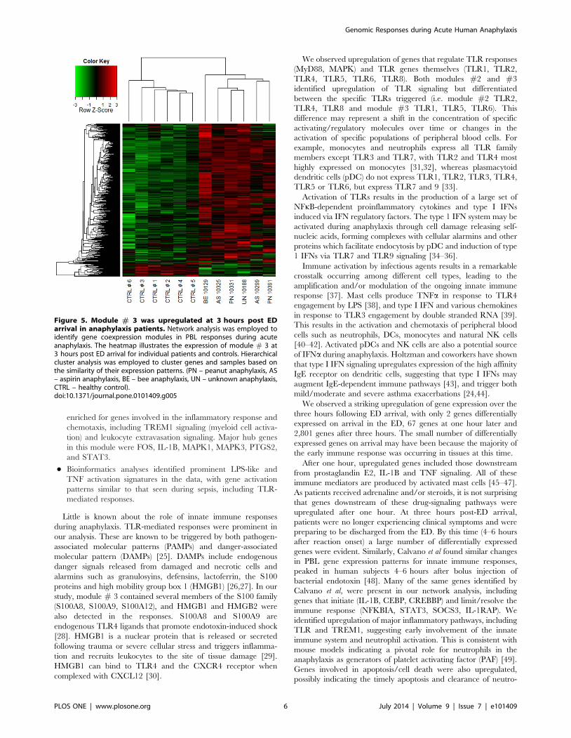
Figure 5. Module # 3 was upregulated at 3 hours post EDarrival in anaphylaxis patients. Network analysis was employed toidentify gene coexpression modules in PBL responses during acuteanaphylaxis. The heatmap illustrates the expression of module # 3 at3 hours post ED arrival for individual patients and controls. Hierarchicalcluster analysis was employed to cluster genes and samples based onthe similarity of their expression patterns. (PN – peanut anaphylaxis, AS– aspirin anaphylaxis, BE – bee anaphylaxis, UN – unknown anaphylaxis,CTRL – healthy control).doi:10.1371/journal.pone.0101409.g005
Genomic Responses during Acute Human Anaphylaxis
PLOS ONE | www.plosone.org 6 July 2014 | Volume 9 | Issue 7 | e101409

phils and other PBL that is essential for resolution of an
inflammatory response [50]. Unfortunately, we were unable to
collect samples from our patient cohort beyond three hours to
assess the timing of complete resolution of the immune response
and return to baseline.
A number of major hub genes that both initiate and resolve
inflammatory responses were identified in module #2 and module
#3. Many of the identified hub genes interact or directly activate
each other, activate overlapping groups of target genes and are
engaged in both negative and positive crosstalk. For example, the
NFkB signaling pathway induces IL-1B, MMP9 and IkBa and
p38 MAPK upregulates genes coding for IL-1B and PTGS2
(COX-2). P38a is required for activation of the transcription factor
CREB and it contributes to the induction of several genes,
including those encoding chemokines, cytokines and regulators of
extracellular matrix remodeling and cell adhesion [51]. CREBBP
and EP300 are co-activators that assist with CREB-induced
transcription, which is involved in cell proliferation, survival,
apoptosis and the innate immune response. NFKBIA (IkBa)
tightly regulates the activity of NFkB with the modulation of
NFKBIA regarded as an anti-inflammatory and immunosuppres-
sive mechanism in asthma [52]. The balanced activation of the
p38MAPK-pathway and STAT3-mediated signal transduction is
essential for both induction and propagation of the inflammatory
Figure 6. A reconstruction of module # 2. Mechanistic data from previous studies was utilized to build a molecular interaction network. Forclarity, the figure illustrates the top six most interactive hubs and their neighbours. The genes are colour coded according to the hub that they areconnected to.doi:10.1371/journal.pone.0101409.g006
Genomic Responses during Acute Human Anaphylaxis
PLOS ONE | www.plosone.org 7 July 2014 | Volume 9 | Issue 7 | e101409

macrophage response as well as for the control of the resolution
phase, which is largely driven by IL-10 and sustained STAT3
activation [53]. STAT3 is activated through phosphorylation in
response to various cytokines and growth factors including IFNs,
HGF and IL-6. IL-1b plays a central role in innate immunity and
has been shown to induce urticarial rashes in autoinflammatory
diseases and play a role in bronchial asthma, contact hypersen-
sitivity and atopic dermatitis [54].
Both MMP9 and HGF may be important for vascular repair
after acute damage. Neutrophils are a potent source of MMP9,
one of the matrix metalloproteinase family, which are major
proteins involved in tissue remodeling. Gene expression and
plasma concentrations of MMP9 have been shown to be
significant higher in ischemic stroke patients compared to healthy
controls [55], and in severe sepsis [56]. In a mouse model, TLR2
activation of neutrophils led to the release of MMP9, which was
protective against experimentally-induced asthma [57]. HGF has
been proposed as a modulator of cardiac tissue repair [58]. The
expression of HGF and its secretion into the blood circulation are
promoted during the early phase of myocardial infarction [59]. By
promoting angiogenesis and inhibiting apoptosis, endogenous
HGF may play an important role in cardioprotection as well as in
the regeneration of endothelial cells and cardiomyocytes after
myocardial infarction [60].
Figure 7. A reconstruction of module # 3. Mechanistic data from previous studies was utilized to build a molecular interaction network. Forclarity, the figure illustrates the top six most interactive hubs and their neighbours. The genes are colour coded according to the hub that they areconnected to.doi:10.1371/journal.pone.0101409.g007
Genomic Responses during Acute Human Anaphylaxis
PLOS ONE | www.plosone.org 8 July 2014 | Volume 9 | Issue 7 | e101409

This study has a number of limitations that must be
acknowledged. The number of patients studied was small and
patients experienced moderately severe anaphylaxis with no cases
of hypotension or hypoxia. The attributed causes were also
heterogenous, including possible IgE- and non-IgE-triggered
anaphylaxis, and the effect of emergency treatment on gene
expression was not controlled for. Patients also differed in the time
taken to arrive in the ED after reaction onset, although it should
be noted that all patients were untreated at ED arrival and all
presented with similar skin and respiratory features. Although the
time course design of the study increased the statistical power of
our analyses, a follow up study is required in a larger number of
patients to determine how variations in gene network patterns
differ in relation to variations in reaction triggers and reaction
severity. The gene expression profiling data was based on a mixed
cell population from peripheral blood, therefore variations in the
cellular composition of the samples may potentially limit the
precision of the analysis. Whilst the data showed evidence of a
cytotoxic response (including type I IFNs) and neutrophil
activation during human anaphylaxis, the exact populations of
PBL involved requires confirmation by other techniques such as
flow cytometry. Nevertheless, this exploratory analysis of gene
expression patterns during human anaphylaxis indicates a major
role for the innate immune system in disease pathogenesis, and the
hub genes identified in this study represent logical candidates for
follow-up in detailed mechanistic studies.
Supporting Information
Table S1 Top 10 canonical pathways and upstream regulators
associated with acute human anaphylaxis at one hour post ED
arrival. Differentially expressed genes were identified and analyzed
in Ingenuity Systems software. Due to the limited number of
differentially expressed genes at this time point, up- and down-
regulated genes were analyzed together. Upstream regulators are
only included when the activation state was predicted from
Ingenuity Systems. The activation state can only be predicted
when the direction of the gene expression changes are consistent
with prior studies. q = molecules associated with this pathway
were mainly upregulated. Q = molecules associated with this
pathway were mainly downregulated.
(DOCX)
Table S2 Canonical pathways and upstream regulators associ-
ated with the genes that were upregulated during acute human
anaphylaxis at three hours post ED arrival. Differentially
expressed genes were identified and analyzed in Ingenuity Systems
software. The analysis was restricted to the upregulated genes
only. Upstream regulators are only included when the activation
state was predicted from Ingenuity Systems. The activation state
can only be predicted when the direction of the gene expression
changes are consistent with prior studies.
(DOCX)
Table S3 Canonical pathways and upstream regulators associ-
ated with the genes that were downregulated during acute human
anaphylaxis at three hours post ED arrival. Differentially
expressed genes were identified and analyzed in Ingenuity Systems
software. The analysis was restricted to the downregulated genes
only. Upstream regulators are only included when the activation
state was predicted from Ingenuity Systems. The activation state
can only be predicted when the direction of the gene expression
changes are consistent with prior studies.
(DOCX)
Table S4 Canonical pathways and upstream regulators associ-
ated with the genes in module # 2. Anaphylaxis-associated
module # 2 was analyzed in Ingenuity Systems software. The
module contains both up and down regulated genes. q =
molecules associated with this pathway were mainly upregulated.
Upstream regulators are only included when the activation state
was predicted from Ingenuity Systems. The activation state can
only be predicted when the direction of the gene expression
changes are consistent with prior studies.
(DOCX)
Table S5 Canonical pathways and upstream regulators associ-
ated with the genes in module # 3. Anaphylaxis-associated
module # 3 was analyzed in Ingenuity Systems software. The
module contains both up and down regulated genes. q =
molecules associated with this pathway were mainly upregulated.
Upstream regulators are only included when the activation state
was predicted from Ingenuity Systems. The activation state can
only be predicted when the direction of the gene expression
changes are consistent with prior studies.
(DOCX)
Table S6 Hub genes identified in Module #2 and Module #3.
(DOCX)
Acknowledgments
We acknowledge the assistance of the medical and nursing staff at Royal
Perth Hospital for identifying patients for the study. The authors would
also like to thank Miss Leah Stone, Graduate Research Assistant, for
performing laboratory assays.
Author Contributions
Conceived and designed the experiments: SFS CLC PvE AB SGAB.
Performed the experiments: CLC PvE. Analyzed the data: SFS AB AJ.
Contributed reagents/materials/analysis tools: AB AJ DMF GA SGAB.
Contributed to the writing of the manuscript: SFS AB SGAB GA DMF.
References
1. Stone SF, Brown SG (2012) Mediators released during human anaphylaxis. Curr
Allergy Asthma Rep 12:33–41.
2. Golden DB (2007) What is anaphylaxis? Curr Opin Allergy Clin Immunol
7:331–336.
3. Brown SGA, Stone SF, Fatovich DM, Burrows SA, Holdgate A, et al (2013)
Anaphylaxis: clinical patterns, mediator release and severity. J Allergy Clin
Immunol 132:1141–1149.
4. Kajiwara N, Sasaki T, Bradding P, Cruse G, Sagara H, et al (2010) Activation of
human mast cells through the platelet-activating factor receptor. J Allergy Clin
Immunol 125:1137–1145.
5. Williams CM, Galli SJ (2000) The diverse potential effector and immunoreg-
ulatory roles of mast cells in allergic disease. J Allergy Clin Immunol 105:847–
859.
6. Jonsson F, Mancardi DA, Kita Y, Karasuyama H, Iannascoli B, et al (2011)
Mouse and human neutrophils induce anaphylaxis. J Clin Invest 121:1484–
1496.
7. Mancardi DA, Albanesi M, Jonsson F, Iannascoli B, Van Rooijen N, et al (2013)
The high-affinity human IgG receptor FcgammaRI (CD64) promotes IgG-
mediated inflammation, anaphylaxis and anti-tumor immunotherapy. Blood
121:1563–1573.
8. Arendts G, Stone SF, Fatovich DM, van Eeden P, MacDonald E, et al (2012)
Critical illness in the emergency department: lessons learnt from the first 12
months of enrolments in the Critical Illness and Shock Study. Emerg Med
Australas 24:31–36.
9. Sampson HA, Munoz-Furlong A, Campbell RL, Adkinson NF Jr, Bock SA, et al
(2006) Second symposium on the definition and management of anaphylaxis:
summary report–Second National Institute of Allergy and Infectious Disease/
Food Allergy and Anaphylaxis Network symposium. J Allergy Clin Immunol
117:391–397.
10. Brown SG (2004) Clinical features and severity grading of anaphylaxis. J Allergy
Clin Immunol 114:371–376.
Genomic Responses during Acute Human Anaphylaxis
PLOS ONE | www.plosone.org 9 July 2014 | Volume 9 | Issue 7 | e101409

11. Hochreiter S, Clevert DA, Obermayer K (2006) A new summarization method
for Affymetrix probe level data. Bioinformatics 22:943–949.12. Dai M, Wang P, Boyd AD, Kostov G, Athey B, et al (2005) Evolving gene/
transcript definitions significantly alter the interpretation of GeneChip data.
Nucleic Acids Res 33:e175.13. Talloen W, Clevert DA, Hochreiter S, Amaratunga D, Bijnens L, et al (2007) I/
NI-calls for the exclusion of non-informative genes: a highly effective filteringtool for microarray data. Bioinformatics 23:2897–2902.
14. Smyth GK (2004) Linear models and empirical bayes methods for assessing
differential expression in microarray experiments. Stat Appl Genet Mol Biol3:Article3.
15. Efron B, Tibshirani R (2007) On testing the significance of sets of genes. AnnAppl Stat 1:107–129.
16. Langfelder P, Horvath S (2008) WGCNA: an R package for weightedcorrelation network analysis. BMC bioinformatics 9:559.
17. Bosco A, McKenna KL, Firth MJ, Sly PD, Holt PG (2009) A network modeling
approach to analysis of the Th2 memory responses underlying human atopicdisease. J Immunol 182:6011–6021.
18. Bosco A, Ehteshami S, Panyala S, Martinez FD (2012) Interferon regulatoryfactor 7 is a major hub connecting interferon-mediated responses in virus-
induced asthma exacerbations in vivo. J Allergy Clin Immunol 129:88–94.
19. Wu D, Smyth GK (2012) Camera: a competitive gene set test accounting forinter-gene correlation. Nucleic Acids Res 40:e133.
20. Jiao X, Sherman BT, Huang da W, Stephens R, Baseler MW, et al (2012)DAVID-WS: a stateful web service to facilitate gene/protein list analysis.
Bioinformatics 28:1805–1806.21. Chan CW, Crafton E, Fan HN, Flook J, Yoshimura K, et al (2006) Interferon-
producing killer dendritic cells provide a link between innate and adaptive
immunity. Nat Med 12:207–213.22. Chen Y, Zhu J, Lum PY, Yang X, Pinto S, et al (2008) Variations in DNA
elucidate molecular networks that cause disease. Nature 452:429–435.23. Subramanian A, Tamayo P, Mootha VK, Mukherjee S, Ebert BL, et al (2005)
Gene set enrichment analysis: a knowledge-based approach for interpreting
genome-wide expression profiles. Proc Natl Acad Sci U S A 102:15545–15550.24. Bosco A, Ehteshami S, Stern DA, Martinez FD (2010) Decreased activation of
inflammatory networks during acute asthma exacerbations is associated withchronic airflow obstruction. Mucosal Immunol 3:399–409.
25. Ward PA (2012) New approaches to the study of sepsis. EMBO Mol Med4:1234–1243.
26. Oppenheim JJ, Tewary P, de la Rosa G, Yang D (2007) Alarmins initiate host
defense. Adv Exp Med Biol 601:185–194.27. Yang D, de la Rosa G, Tewary P, Oppenheim JJ (2009) Alarmins link
neutrophils and dendritic cells. Trends Immunol 30:531–537.28. Ehrchen JM, Sunderkotter C, Foell D, Vogl T, Roth J (2009) The endogenous
Toll-like receptor 4 agonist S100A8/S100A9 (calprotectin) as innate amplifier of
infection, autoimmunity, and cancer. J Leukoc Biol 86:557–566.29. Bae JS (2012) Role of high mobility group box 1 in inflammatory disease: focus
on sepsis. Arch Pharm Res 35:1511–1523.30. Venereau E, Schiraldi M, Uguccioni M, Bianchi ME (2013) HMGB1 and
leukocyte migration during trauma and sterile inflammation. Mol Immunol55:76–82.
31. Prince LR, Whyte MK, Sabroe I, Parker LC (2011) The role of TLRs in
neutrophil activation. Current Opin Pharmacol 11:397–403.32. Hornung V, Rothenfusser S, Britsch S, Krug A, Jahrsdorfer B, et al (2002)
Quantitative expression of toll-like receptor 1–10 mRNA in cellular subsets ofhuman peripheral blood mononuclear cells and sensitivity to CpG oligodeox-
ynucleotides. J Immunol 168:4531–4537.
33. Schreibelt G, Tel J, Sliepen KH, Benitez-Ribas D, Figdor CG, et al (2010) Toll-like receptor expression and function in human dendritic cell subsets:
implications for dendritic cell-based anti-cancer immunotherapy. CancerImmunol Immunother 59:1573–1582.
34. Kawasaki T, Kawai T, Akira S (2011) Recognition of nucleic acids by pattern-
recognition receptors and its relevance in autoimmunity. Immunol Rev 243:61–73.
35. Yanai H, Ban T, Wang Z, Choi MK, Kawamura T, et al (2009) HMGBproteins function as universal sentinels for nucleic-acid-mediated innate immune
responses. Nature 462:99–103.36. Lande R, Gregorio J, Facchinetti V, Chatterjee B, Wang Y-H, et al (2007)
Plasmacytoid dendritic cells sense self-DNA coupled with antimicrobial peptide.
Nature 449:564–569.
37. Moretta A, Marcenaro E, Sivori S, Della Chiesa M, Vitale M, et al (2005) Early
liaisons between cells of the innate immune system in inflamed peripheral tissues.Trends Immunol 26:668–675.
38. Supajatura V, Ushio H, Nakao A, Akira S, Okumura K, et al (2002) Differential
responses of mast cell Toll-like receptors 2 and 4 in allergy and innate immunity.J Clin Invest 109:1351–1359.
39. Kulka M, Alexopoulou L, Flavell RA, Metcalfe DD (2004) Activation of mastcells by double-stranded RNA: evidence for activation through Toll-like receptor
3. J Allergy Clin Immunol 114:174–182.
40. Burke SM, Issekutz TB, Mohan K, Lee PW, Shmulevitz M, et al (2008) Human
mast cell activation with virus-associated stimuli leads to the selective chemotaxis
of natural killer cells by a CXCL8-dependent mechanism. Blood 111:5467–5476.
41. Moller A, Lippert U, Lessmann D, Kolde G, Hamann K, et al (1993) Humanmast cells produce IL-8. J Immunol 151:3261–3266.
42. Baghestanian M, Hofbauer R, Kiener HP, Bankl HC, Wimazal F, et al (1997)The c-kit ligand stem cell factor and anti-IgE promote expression of monocyte
chemoattractant protein-1 in human lung mast cells. Blood 90:4438–4449.
43. Grayson MH, Cheung D, Rohlfing MM, Kitchens R, Spiegel DE, et al (2007)Induction of high-affinity IgE receptor on lung dendritic cells during viral
infection leads to mucous cell metaplasia. J Exp Med 204:2759–2769.
44. Holt PG, Sly PD (2011) Interaction between adaptive and innate immune
pathways in the pathogenesis of atopic asthma: operation of a lung/bone
marrow axis. Chest 139:1165–1171.
45. Nakamura Y, Kambe N, Saito M, Nishikomori R, Kim YG, et al (2009) Mast
cells mediate neutrophil recruitment and vascular leakage through the NLRP3inflammasome in histamine-independent urticaria. J Exp Med 206:1037–1046.
46. Okayama Y (2005) Mast cell-derived cytokine expression induced via Fcreceptors and Toll-like receptors. Chem Immunol Allergy 87:101–110.
47. Boyce JA (2007) Mast cells and eicosanoid mediators: a system of reciprocal
paracrine and autocrine regulation. Immunol Rev 217:168–185.
48. Calvano SE, Xiao W, Richards DR, Felciano RM, Baker HV, et al (2005) A
network-based analysis of systemic inflammation in humans. Nature 437:1032–1037.
49. Jonsson F, Mancardi DA, Zhao W, Kita Y, Iannascoli B, et al (2012) HumanFcgammaRIIA induces anaphylactic and allergic reactions. Blood 119:2533–
2544.
50. Kobayashi SD, Voyich JM, Somerville GA, Braughton KR, Malech HL, et al(2003) An apoptosis-differentiation program in human polymorphonuclear
leukocytes facilitates resolution of inflammation. J Leukoc Biol 73:315–322.
51. Kang YJ, Chen JM, Otsuka M, Mols J, Ren SX, et al (2008) Macrophage
deletion of p38 alpha partially impairs lipopolysaccharide-induced cellular
activation. J Immunol 180(7):5075–5082.
52. Auphan N, Didonato JA, Rosette C, Helmberg A, Karin M (1995)
Immunosuppression by Glucocorticoids - Inhibition of Nf-Kappa-B Activitythrough Induction of I-Kappa-B Synthesis. Science 270:286–290.
53. Bode JG, Ehlting C, Haussinger D (2012) The macrophage response towardsLPS and its control through the p38(MAPK)-STAT3 axis. Cell Signal 24:1185–
1194.
54. Krause K, Metz M, Makris M, Zuberbier T, Maurer M (2012) The role ofinterleukin-1 in allergy-related disorders. Curr Opin Allergy Clin Immunol
12:477–484.
55. Oh SH, Kim OJ, Shin DA, Song J, Yoo H, et al (2012) Alteration of
immunologic responses on peripheral blood in the acute phase of ischemicstroke: Blood genomic profiling study. J Neuroimmunol 249:60–65.
56. Yazdan-Ashoori P, Liaw P, Toltl L, Webb B, Kilmer G, et al (2011) Elevated
plasma matrix metalloproteinases and their tissue inhibitors in patients withsevere sepsis. J Crit Care 26:556–565.
57. Page K, Ledford JR, Zhou P, Wills-Karp M (2009) A TLR2 agonist in germancockroach frass activates MMP-9 release and is protective against allergic
inflammation in mice. J Immunol 183:3400–3408.
58. Sala V, Crepaldi T (2011) Novel therapy for myocardial infarction: can HGF/Met be beneficial? Cell Mol Life Sci 68:1703–1717.
59. Matsumori A, Furukawa Y, Hashimoto T, Ono K, Shioi T, et al (1996)Increased circulating hepatocyte growth factor in the early stage of acute
myocardial infarction. Biochem Biophys Res Commun 221:391–395.
60. Madonna R, Cevik C, Nasser M, De Caterina R (2012) Hepatocyte growth
factor: Molecular biomarker and player in cardioprotection and cardiovascular
regeneration. Thromb Haemost 107:656–661.
Genomic Responses during Acute Human Anaphylaxis
PLOS ONE | www.plosone.org 10 July 2014 | Volume 9 | Issue 7 | e101409