gadolinium-containing phosphatidylserine liposomes for ... · supplementary key words macrophages...
TRANSCRIPT
This article is available online at http://www.jlr.org Journal of Lipid Research Volume 50, 2009 2157
Copyright © 2009 by the American Society for Biochemistry and Molecular Biology, Inc.
fundamental role of this process in atherosclerosis ( 3 ). Macrophage identifi cation strategies are particularly at-tractive for molecular imaging in light of their ubiquitous presence and positive correlation with complications of atherosclerosis. A number of different targeted contrast agents specifi c for macrophage proteins have been tested in atherosclerosis. In such approaches, the contrast strat-egy is typically paramagnetic gadolinium (Gd) chelates or iron (Fe) oxide particles and is passively incorporated to various carrier molecules. The carrier moieties by virtue of preferential uptake by, or delivery to, specifi c pathways or receptors of relevance to the macrophage allow for “high-payload” delivery of the contrast agent and facilitate im-aging with adequate signal to background noise ratios. Examples of such approaches include HDL (4) and LDL (5) lipoproteins incorporating Gd, polyethyleneglycol (PEG) grafted immunomicels ( 6, 7 ), and liposome vesi cles ( 8 ) conjugated to antibodies targeting the mac-rophage scavenger receptor. In general, these approaches have been shown to provide enhanced detection of macrophage-rich areas, allowing monitoring of macrophage-rich areas. Although these strategies are attractive, they do have several disadvantages, including considerable diffi -culties in preparation and handling, expense (e.g., anti-bodies), nonspecifi city of targeting, and steric hinderances pertaining to the carrier moieties, such as PEG that may reduce targeted interaction of the antibody to sites of rec-ognition ( 9, 10 ). It is well known that exteriorized phos-phatidylserine (PS) residues present in apoptotic cells promote macrophage recognition by macrophage scaven-ger receptor/CD36 followed by rapid phagocytosis ( 11, 12 ). Accordingly, we hypothesized that mimicking apopto-sis with liposomes containing PS may represent an attrac-tive approach for molecular imaging of atherosclerosis.
Abstract Exteriorized phosphatidylserine (PS) residues in apoptotic cells trigger rapid phagocytosis by macrophage scavenger receptor pathways. Mimicking apoptosis with liposomes containing PS may represent an attractive approach for molecular imaging of atherosclerosis. We investigated the utility of paramagnetic gadolinium lipo-somes enriched with PS (Gd-PS) in imaging atherosclerotic plaque. Gd-PS-containing Gd-conjugated lipids, fl uorescent rhodamine, and PS were prepared and characterized. Cel-lular uptake in RAW macrophages (fl uorescent uptake of rhodamine) was studied on a fl uorescence plate reader, while Gd-PS-induced alteration in T1 relaxivity was evalu-ated using a 1.5 T MRI scanner. RAW cells demonstrate PS-dependent uptake of across a range of concentrations (2, 6, 12, and 20%) in comparison to control liposomes with no PS (0%). In vivo performance of Gd-PS was evaluated in the ApoE � / � mouse model by collection of serial T1 weighted gradient echo MR images using an 11.7 T MRI system and revealed rapid and signifi cant enhancement of the aortic wall that was seen for at least 4 h after injection. Gd-PS-enriched liposomes enhance atherosclerotic plaque and colocalize with macrophages in experimental atheroscle-rosis. —Maiseyeu, A., G. Mihai, T. Kampfrath, O. P. Simonetti, C. K. Sen, S. Roy, S. Rajagopalan, and S. Parthasarathy. Gadolinium-containing phosphatidylserine liposomes for molecular imaging of atherosclerosis. J. Lipid Res. 2009. 50: 2157–2163.
Supplementary key words macrophages • apoptosis mimicking • cel-lular uptake • colocalization with macrophages • ApoE � / � mouse model
Molecular imaging approaches in atherosclerosis have the advantage of visualizing specifi c events that may be of relevance in the progression and complications of the dis-ease ( 1, 2 ). Imaging strategies that allow visualization of infl ammatory cells are particularly attractive in view of the
This study was supported by an RO1 grant (ES013406) from the National Institute of Environmental Health Sciences. Additional funding support was received from the Regenerative Medicine Thematic Program at OSU and start-up funds.
Manuscript received 30 July 2008 and in revised form 1 October 2008 and in re-revised form 6 November 2008.
Published, JLR Papers in Press, November 17, 2008 DOI 10.1194/jlr.M800405-JLR200
Gadolinium-containing phosphatidylserine liposomes for molecular imaging of atherosclerosis
Andrei Maiseyeu, * Georgeta Mihai, * Thomas Kampfrath, * Orlando P. Simonetti, * Chandan K. Sen, † Sashwati Roy, * Sanjay Rajagopalan, * and Sampath Parthasarathy 1,†
Davis Heart and Lung Research Institute* and Department of Surgery, † Ohio State University, Columbus, OH
Abbreviations: DOPE-Rho, phosphatidylethanolamine-Lissamine-Rhodamine B; Gd, gadolinium; Gd-DTPA-SA, Gd-diethylenetriamine-pentaacetic acid distearylamide; LDH, lactate dehydrogenase; MSR, macrophage scavenger receptor; NENH, normalized enhancement; PC, phosphatidylcholine; PEG, polyethyleneglycol; PG, phosphatidyl-glycerol; PS, phosphatidylserine; SNR, signal-to-noise ratio.
1 To whom correspondence should be addressed. e-mail: [email protected]
by guest, on June 14, 2018w
ww
.jlr.orgD
ownloaded from

2158 Journal of Lipid Research Volume 50, 2009
size of 230 nm with a narrow size distribution. Lipids concentra-tion was determined by the Stewart method ( 15 ).
Cell culture RAW 264.7 macrophages were obtained from American Type
Culture Collection (Manassas, VA). Cells were cultured in com-plete RPMI media containing 10% FBS, 1% penicillin-streptomy-cin, 1% glutamine, and 1% sodium pyruvate at 37°C, 5% CO 2 . For fl uorescent and cytoxicity studies, cells were plated in opaque 96-well plates with 10 4 cells per well. After 24 h of incubation, the cells were washed with PBS (1×), and each well was incubated with 100 � l phenol red-free medium containing 50 � g of appro-priate liposome formulation or Magnevist (as a control in cytox-icity experiments). After 12 h, cells were washed with prewarmed PBS (2×, 100 � l) and assayed on fl uorescence plate reader (Victor V; Perkin-Elmer). For in vitro MRI experiments, cells were placed in 300 cm 2 culture fl asks (8 × 10 6 cells per fl ask, 80 ml of media) and after 24 h of incubation, the media were removed and replaced with fresh media. Liposomes (1 ml, 40 mg/ml of lipids) were then added and gently swirled, and cells were incu-bated in a wet atmosphere at 37°C, 5% CO 2 for 12 h. Then, the old media were aspirated off and cells were washed twice with 80 ml of prewarmed PBS. Next, deionized water was added (50 ml per fl ask), and fl asks were agitated on an orbital shaker for 1 h. Resulting suspension of cell lysate was transferred into 50 ml Fal-con tubes and centrifuged at 1,500 g for 5 min. Pellet was iso-lated, frozen in dry ice, and lyophilized. Resulting dry powder was reconstituted by sonication in 1 ml of 10% Triton X-100. Mix-ture was transferred to Eppendorf tubes and imaged in a 1.5 T MRI scanner (Avanto, Siemens, Erlangen, Germany).
Cellular toxicity assay Cells were prepared as indicated above (cell culture section),
and the same liposomes but containing no fl uorescent lipids were added to each well at three different doses: 6 � g of lipo-somes containing 0.1 � g of Gd, 60 � g of liposomes containing 1.0 � g of Gd, and 120 � g of liposomes containing 2.0 � g of Gd. Two controls were used: the same amount of liposomes without Gd-DTPA-SA and Magnevist, diluted with HEPES buffer corre-sponding to Gd concentrations of 0.1, 1.0, and 2.0 � g per well. The lactate dehydrogenase (LDH) assay was carried out using the CytoTox-ONE homogeneous Membrane Integrity Assay (Pro-mega, Madison, WI)
Microscopy Cells were grown in 6-well plates in conditions described
above. Cultured macrophages were treated with fl uorescent lipo-somes at concentration 0.45 mg total lipids per well. After an ap-propriate incubation period, cells were fi xed by the addition of cold 2% paraformaldehyde and fl uorescence was analyzed. Opti-cal imaging of cells treated with fl uorescent liposomes was per-formed on a Nikon Eclipse FN1 microscope (Japan) using a 40×/0.80 W water immersed objective. The data were processed using Metamorph software (version 7.1.2.0; Metamorph, Down-ingtown, PA).
Confocal fl uorescence microscopy and immunohistochemistry
At the end of the MRI experiment, mice were euthanized, and the aortas were immediately removed and embedded in optical cutting temperature compound. Sectioned abdominal aortas (8 � M) were immersed into ice-cold ethanol for 5 min and then air dried and rehydrated with PBS. After treatment with 0.02% Tri-ton X-100 and successive washing with PBS, slides were blocked with goat serum and then incubated with FITC-conjugated rat
Here, we describe a non-PEGylated, antibody-free, tar-geted contrast agent approach for macrophage imaging ( Fig. 1 ).
MATERIALS AND METHODS
Reagents Egg-phosphatidylcholine (PC), egg-phosphatidylglycerol (PG),
brain-PS, and phosphatidylethanolamine-Lissamine-Rhodamine B (DOPE-Rho) were purchased from Avanti Polar Lipids (Alabaster, AL). Cholesterol and all other chemicals were ob-tained from Sigma-Aldrich (St. Louis, MO); Gd-diethylenetri-aminepentaacetic acid distearylamide (Gd-DTPA-SA) was synthesized according References 13 and 14 , RPMI 1640, FBS, and PBS were purchased from Cellgro Mediatech (Herndon, VA); and penicillin-streptomycin, glutamine, and sodium pyru-vate were from Gibco/Invitrogen (Carlsbad, CA).
Paramagnetic liposomes Liposomes were prepared by lipid fi lm hydration ( 10, 12, 14 ).
Briefl y, solutions of lipids in chloroform were mixed and solvent was evaporated in vacuo. The molar composition of vesicles was 28.95% to 18.95% PC, 28.95% to 18.95% PG, 0% to 20% PS, 0.1% or 0.5% DOPE-Rho, 35% cholesterol, and 7% Gd-DTPA-SA. The vesicles containing different concentrations of PS were made by decreasing of amount of PC and PG simultaneously to accom-modate increases in PS, while amount of DOPE-Rho, cholesterol, and Gd-DTPA-SA remained constant. Residue was pumped for 2–3 h and hydrated with 1 ml of HEPES buffer (20 mM HEPES and 135 mM NaCl, pH 7.2) at 55°C. After vigorous vortexing, re-sulting liposome suspension was passed through 400 and 200 nm Nucleopore membranes (21×) using mini extruder (Avanti Polar Lipids). Fluorescent paramagnetic liposomes always contained 0.1 mol% and 0.5 mol% DOPE-Rho for in vitro and in vivo ex-periments, respectively, and 7% of Gd-DTPA-SA. Control lipo-somes contained the same kind and amount of lipids except PS. The absence of later was compensated for by an equal amount of PG. For all cell studies, liposomes were prepared in concentra-tion 10 mg/ml total lipid, and for in vivo MRI experiments, 25 mg/ml of total lipid concentration was used. The liposome par-ticle size was determined using Dynamic Light Scattering on Nano S Zetasizer (Malvern Instruments). Liposomes had mean
Fig. 1. Schematic representation of hypothesis. PS-enriched lipo-somes are recognized by macrophages and thereby can serve as targeted contrast agents to macrophage-rich areas, such as athero-sclerotic plaque.
by guest, on June 14, 2018w
ww
.jlr.orgD
ownloaded from
Phosphatidylserine liposomes for plaque imaging 2159
magnet and while still placed in the birdcage coil holder was tail vein injected with contrast agent and immediately placed back in the magnet for the postcontrast scans. Each mouse was imaged for approximately 1.5–2 h after contrast administration using re-petitive acquisitions of the 5:56 min gradient echo sequence. Just minutes before 4, 8, 12, and 24 h postcontrast, each mouse was anesthetized and placed back in the magnet, and a single MRI scan was acquired using the T1 weighted gradient echo sequence.
Image analysis MR images were analyzed using ImageJ (NIH). For each
mouse, four slices common to the all time points were identifi ed by checking the shape and position of the spinal column. For each time point, the thickened aortic wall was identifi ed, and one irregular 25–35 pixel region of interest, which covered the entire aortic wall, less the region next to vena cava, was hand drawn on each slice. Signal-to-noise ratio (SNR) of each region of interest was calculated as the average signal intensity divided by the stan-dard deviation of the noise level. The SNRs of the four slices were averaged and displayed as a function of time to depict the time course of the signal intensity changes due to contrast uptake in the atherosclerotic wall. Percentage of normalized enhance-ment (NENH) was calculated by the formula: % NENH = 100 × (SNR post /SNR pre ); SNR post and SNR pre are the SNRs obtained af-ter and before contrast agent administration, respectively.
Statistics All values in this article represent mean ± SD unless otherwise
specifi ed. One-way ANOVA methodology was used to compare multiple variables or time points with P < 0.05 representing statis-tically signifi cant differences. A Bonferroni posthoc correction was used to assess differences between groups. All statistics were performed using GraphPad Prism (Version 4.0; GraphPad Soft-ware, La Jolla, CA).
RESULTS
Optimization of liposome formulations and evaluation of its T1 by MRI
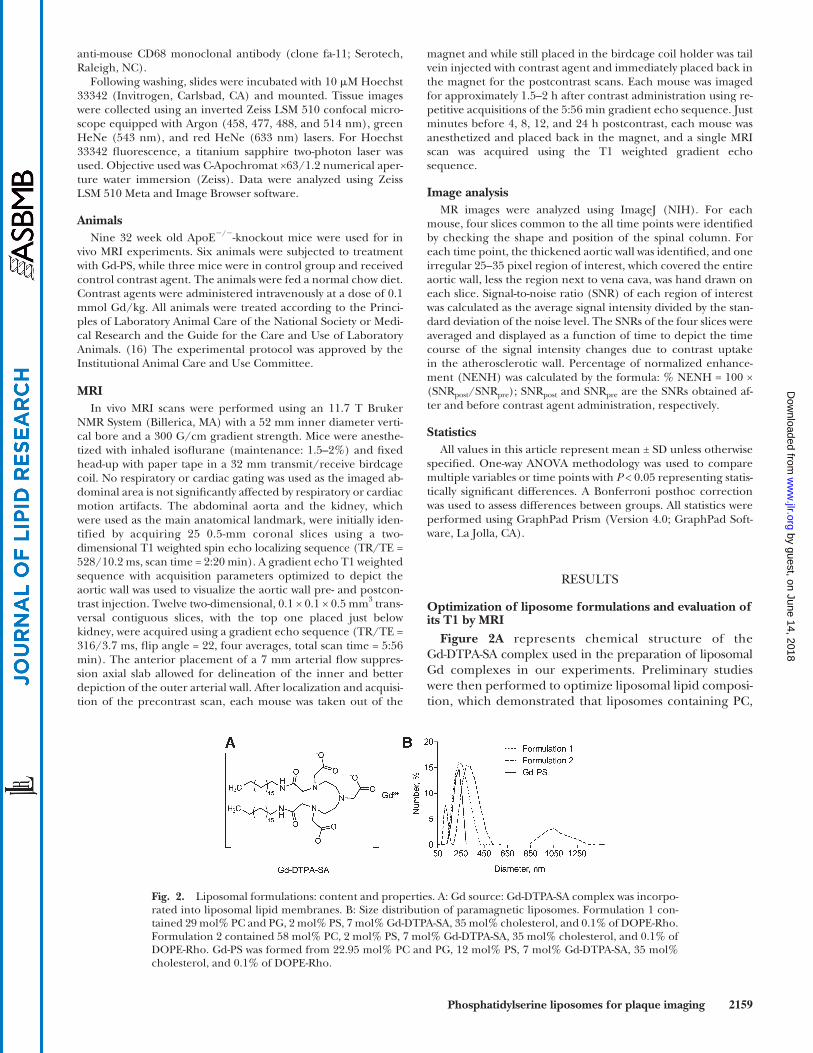
Figure 2A represents chemical structure of the Gd-DTPA-SA complex used in the preparation of liposomal Gd complexes in our experiments. Preliminary studies were then performed to optimize liposomal lipid composi-tion, which demonstrated that liposomes containing PC,
anti-mouse CD68 monoclonal antibody (clone fa-11; Serotech, Raleigh, NC).
Following washing, slides were incubated with 10 � M Hoechst 33342 (Invitrogen, Carlsbad, CA) and mounted. Tissue images were collected using an inverted Zeiss LSM 510 confocal micro-scope equipped with Argon (458, 477, 488, and 514 nm), green HeNe (543 nm), and red HeNe (633 nm) lasers. For Hoechst 33342 fl uorescence, a titanium sapphire two-photon laser was used. Objective used was C-Apochromat ×63/1.2 numerical aper-ture water immersion (Zeiss). Data were analyzed using Zeiss LSM 510 Meta and Image Browser software.
Animals Nine 32 week old ApoE � / � -knockout mice were used for in
vivo MRI experiments. Six animals were subjected to treatment with Gd-PS, while three mice were in control group and received control contrast agent. The animals were fed a normal chow diet. Contrast agents were administered intravenously at a dose of 0.1 mmol Gd/kg. All animals were treated according to the Princi-ples of Laboratory Animal Care of the National Society or Medi-cal Research and the Guide for the Care and Use of Laboratory Animals. ( 16 ) The experimental protocol was approved by the Institutional Animal Care and Use Committee.
MRI In vivo MRI scans were performed using an 11.7 T Bruker
NMR System (Billerica, MA) with a 52 mm inner diameter verti-cal bore and a 300 G/cm gradient strength. Mice were anesthe-tized with inhaled isofl urane (maintenance: 1.5–2%) and fi xed head-up with paper tape in a 32 mm transmit/receive birdcage coil. No respiratory or cardiac gating was used as the imaged ab-dominal area is not signifi cantly affected by respiratory or cardiac motion artifacts. The abdominal aorta and the kidney, which were used as the main anatomical landmark, were initially iden-tified by acquiring 25 0.5-mm coronal slices using a two-dimensional T1 weighted spin echo localizing sequence (TR/TE = 528/10.2 ms, scan time = 2:20 min). A gradient echo T1 weighted sequence with acquisition parameters optimized to depict the aortic wall was used to visualize the aortic wall pre- and postcon-trast injection. Twelve two-dimensional, 0.1 × 0.1 × 0.5 mm 3 trans-versal contiguous slices, with the top one placed just below kidney, were acquired using a gradient echo sequence (TR/TE = 316/3.7 ms, fl ip angle = 22, four averages, total scan time = 5:56 min). The anterior placement of a 7 mm arterial fl ow suppres-sion axial slab allowed for delineation of the inner and better depiction of the outer arterial wall. After localization and acquisi-tion of the precontrast scan, each mouse was taken out of the
Fig. 2. Liposomal formulations: content and properties. A: Gd source: Gd-DTPA-SA complex was incorpo-rated into liposomal lipid membranes. B: Size distribution of paramagnetic liposomes. Formulation 1 con-tained 29 mol% PC and PG, 2 mol% PS, 7 mol% Gd-DTPA-SA, 35 mol% cholesterol, and 0.1% of DOPE-Rho. Formulation 2 contained 58 mol% PC, 2 mol% PS, 7 mol% Gd-DTPA-SA, 35 mol% cholesterol, and 0.1% of DOPE-Rho. Gd-PS was formed from 22.95 mol% PC and PG, 12 mol% PS, 7 mol% Gd-DTPA-SA, 35 mol% cholesterol, and 0.1% of DOPE-Rho.
by guest, on June 14, 2018w
ww
.jlr.orgD
ownloaded from
2160 Journal of Lipid Research Volume 50, 2009
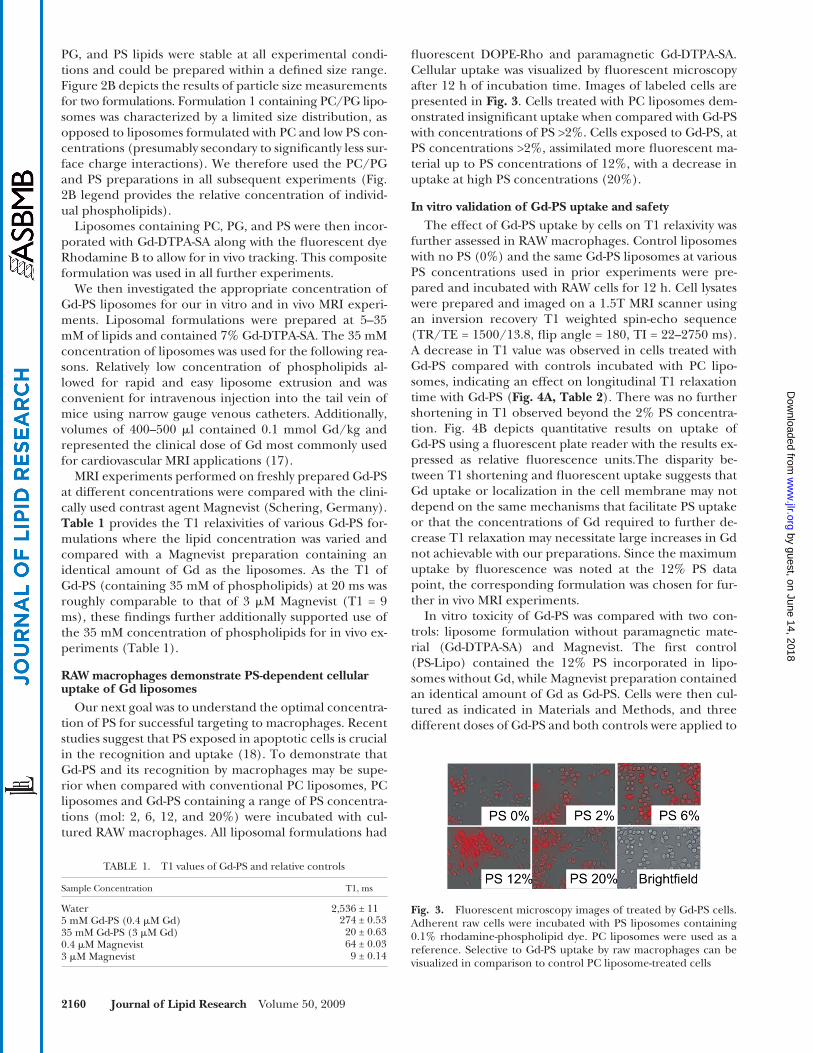
fl uorescent DOPE-Rho and paramagnetic Gd-DTPA-SA. Cellular uptake was visualized by fl uorescent microscopy after 12 h of incubation time. Images of labeled cells are presented in Fig. 3 . Cells treated with PC liposomes dem-onstrated insignifi cant uptake when compared with Gd-PS with concentrations of PS >2%. Cells exposed to Gd-PS, at PS concentrations >2%, assimilated more fl uorescent ma-terial up to PS concentrations of 12%, with a decrease in uptake at high PS concentrations (20%).
In vitro validation of Gd-PS uptake and safety The effect of Gd-PS uptake by cells on T1 relaxivity was
further assessed in RAW macrophages. Control liposomes with no PS (0%) and the same Gd-PS liposomes at various PS concentrations used in prior experiments were pre-pared and incubated with RAW cells for 12 h. Cell lysates were prepared and imaged on a 1.5T MRI scanner using an inversion recovery T1 weighted spin-echo sequence (TR/TE = 1500/13.8, fl ip angle = 180, TI = 22–2750 ms). A decrease in T1 value was observed in cells treated with Gd-PS compared with controls incubated with PC lipo-somes, indicating an effect on longitudinal T1 relaxation time with Gd-PS ( Fig. 4A, Table 2 ). There was no further shortening in T1 observed beyond the 2% PS concentra-tion. Fig. 4B depicts quantitative results on uptake of Gd-PS using a fl uorescent plate reader with the results ex-pressed as relative fl uorescence units.The disparity be-tween T1 shortening and fl uorescent uptake suggests that Gd uptake or localization in the cell membrane may not depend on the same mechanisms that facilitate PS uptake or that the concentrations of Gd required to further de-crease T1 relaxation may necessitate large increases in Gd not achievable with our preparations. Since the maximum uptake by fl uorescence was noted at the 12% PS data point, the corresponding formulation was chosen for fur-ther in vivo MRI experiments.
In vitro toxicity of Gd-PS was compared with two con-trols: liposome formulation without paramagnetic mate-rial (Gd-DTPA-SA) and Magnevist. The fi rst control (PS-Lipo) contained the 12% PS incorporated in lipo-somes without Gd, while Magnevist preparation contained an identical amount of Gd as Gd-PS. Cells were then cul-tured as indicated in Materials and Methods, and three different doses of Gd-PS and both controls were applied to
PG, and PS lipids were stable at all experimental condi-tions and could be prepared within a defi ned size range. Figure 2B depicts the results of particle size measurements for two formulations. Formulation 1 containing PC/PG lipo-somes was characterized by a limited size distribution, as opposed to liposomes formulated with PC and low PS con-centrations (presumably secondary to signifi cantly less sur-face charge interactions). We therefore used the PC/PG and PS preparations in all subsequent experiments ( Fig. 2B legend provides the relative concentration of individ-ual phospholipids).
Liposomes containing PC, PG, and PS were then incor-porated with Gd-DTPA-SA along with the fl uorescent dye Rhodamine B to allow for in vivo tracking. This composite formulation was used in all further experiments.
We then investigated the appropriate concentration of Gd-PS liposomes for our in vitro and in vivo MRI experi-ments. Liposomal formulations were prepared at 5–35 mM of lipids and contained 7% Gd-DTPA-SA. The 35 mM concentration of liposomes was used for the following rea-sons. Relatively low concentration of phospholipids al-lowed for rapid and easy liposome extrusion and was convenient for intravenous injection into the tail vein of mice using narrow gauge venous catheters. Additionally, volumes of 400–500 � l contained 0.1 mmol Gd/kg and represented the clinical dose of Gd most commonly used for cardiovascular MRI applications ( 17 ).
MRI experiments performed on freshly prepared Gd-PS at different concentrations were compared with the clini-cally used contrast agent Magnevist (Schering, Germany). Table 1 provides the T1 relaxivities of various Gd-PS for-mulations where the lipid concentration was varied and compared with a Magnevist preparation containing an identical amount of Gd as the liposomes. As the T1 of Gd-PS (containing 35 mM of phospholipids) at 20 ms was roughly comparable to that of 3 � M Magnevist (T1 = 9 ms), these fi ndings further additionally supported use of the 35 mM concentration of phospholipids for in vivo ex-periments (Table 1).
RAW macrophages demonstrate PS-dependent cellular uptake of Gd liposomes
Our next goal was to understand the optimal concentra-tion of PS for successful targeting to macrophages. Recent studies suggest that PS exposed in apoptotic cells is crucial in the recognition and uptake ( 18 ). To demonstrate that Gd-PS and its recognition by macrophages may be supe-rior when compared with conventional PC liposomes, PC liposomes and Gd-PS containing a range of PS concentra-tions (mol: 2, 6, 12, and 20%) were incubated with cul-tured RAW macrophages. All liposomal formulations had
TABLE 1. T1 values of Gd-PS and relative controls
Sample Concentration T1, ms
Water 2,536 ± 115 mM Gd-PS (0.4 � M Gd) 274 ± 0.5335 mM Gd-PS (3 � M Gd) 20 ± 0.630.4 � M Magnevist 64 ± 0.033 � M Magnevist 9 ± 0.14
Fig. 3. Fluorescent microscopy images of treated by Gd-PS cells. Adherent raw cells were incubated with PS liposomes containing 0.1% rhodamine-phospholipid dye. PC liposomes were used as a reference. Selective to Gd-PS uptake by raw macrophages can be visualized in comparison to control PC liposome-treated cells
by guest, on June 14, 2018w
ww
.jlr.orgD
ownloaded from

Phosphatidylserine liposomes for plaque imaging 2161
injection. Increase in signal was discernible at 10 min, with both agents probably related to a fi rst pass perfusion ef-fect, followed by a decrease probably related to redistribu-tion and then a steady state phase related to equilibration. Signal decreased rapidly from 55 min onward in the con-trol liposomes, while the decrease in the Gd-PS was grad-ual beyond 120 min with obvious retention in the vessel wall at 8 h. In contrast, the signal intensity in the control liposomes was no different from the precontrast phase (100% NENH; Fig. 6C ).
Colocalization of macrophages and Gd-PS in vivo As evidence of colocalization atherosclerosis-associated
macrophages with Gd-PS in vivo, we performed confocal fl uorescence imaging on sectioned mice aortas. Abdomi-nal aortas were removed and immunostained 24 h post-contrast injection. To localize atherosclerosis-derived macrophages, we used CD-68 FITC-conjugated antibody and Hoechst 33342 to show nuclei of cells. Fluorescently labeled Gd-PSs were visible in the peri-adventitial region (red fl uorescence from rhodamine). The CD-68 staining indicated the presence of macrophages in the plaque area (green from FITC). Colocalization of rhodamine with some of the FITC appeared yellow and was detected pri-marily in the peri-adventitia ( Fig. 7 ). The obtained images demonstrate in vivo delivery of Gd-PS to macrophage-rich areas. These results along with MRI data strongly support our central hypothesis of targeted macrophage behavior of Gd-PS in vivo.
DISCUSSION
In this study, we describe a novel approach to imaging macrophages using PS-containing liposomes. This ap-proach resulted in signifi cant enhancement of atheroscle-rotic plaque in vivo for a prolonged duration and demonstrates the feasibility of such an approach for mo-lecular characterization of high-risk plaque. Liposomal composition can be easily adjusted by including different lipids into the vesicle double layer and allows for inclusion of paramagnetic material and fl uorescence tags for in vivo
them with following the incubation and measurement of the number of nonviable cells. LDH release from all treated cells was minimal at 0.1 � g Gd (6 � g lipids) and almost without any difference between Gd-PS and controls ( Fig. 5 ). There was little difference in toxicity between all three doses for PS-Lipo. This suggests that the toxicity may be related only to Gd in the sample. This also can be con-sidered as normal cell growth and proliferation taking place in spite of presence of phospholipids. At 1.0 � g Gd (60 � g lipids), Gd-PS showed less toxic effect than Magn-evist; however, at the higher concentrations, they both re-sulted in comparable cytotoxcity.
In vivo MRI validation of Gd-PS liposomes as contrast agent strategy
In vivo MRI imaging was performed in ApoE � / � mice using a gradient echo approach (see Materials and Meth-ods). The abdominal aorta was imaged axially starting be-low the kidney to the aortic bifurcation using a gradient echo sequence, before and after in vivo administration of Gd-PS or control liposomes. The presence of plaque in all the animals was confi rmed following euthanizing, and rep-resentative Hematoxylin and Eosin (H and E) staining de-picting plaque are provided as part of the fi gure as well. Figure 6A depicts representative axial MRI images to assess contrast opacifi cation with Gd-PS, while Fig. 6B represents representative images following nonPS liposomes contain-ing Gd. Enhancement of plaque seen in control nonPS li-posomes was seen at 60 min postcontrast administration, with prolonged retention of signal intensity at 8 h after
Fig. 4. Uptake of PS-enriched liposomes across a range of con-centrations (2, 6, 12, and 20%) in comparison to control liposomes with no PS (0%). A: Raw cells were treated with Gd-PS liposomes, and T1 relaxivities of cell lysate pellets were measured at 1.5 T after 12 h of incubation. There was a signifi cant drop in T1 values of Gd-PS when compared with control showed gadolinum uptake in-duced by PS-containing liposomes, but not by PC liposomes. B: Treatment of cultured cells with the same formulations of Gd-PS and control for 12 h and following fl uorescent assay on the fl uores-cence plate reader revealed the maximum uptake at 12% PS. The obtained relative fl uorescence unit (RFU) values represent an ex-cellent concordance with T1 assay and fl uorescence microscopy studies ( Fig. 3 ). * P < 0.05.
TABLE 2. T1 values resulting from Gd-PS-treated cells
Liposome Formulation Containing PS (%) T1, ms
0 771.56 ± 3.232 387.70 ± 0.606 388.99 ± 0.5812 392.65 ± 2.3320 467.29 ± 4.78
Fig. 5. LDH assay. Assay results calculated to show percentage of cytoxicity as a result of added Gd-PS and comparison to liposomal formulation without paramagnetic Gd-DTPA-SA (PS-lipo). For ref-erence, cells were treated with Magnevist containing the same con-centration of Gd as Gd-PS liposomes.
by guest, on June 14, 2018w
ww
.jlr.orgD
ownloaded from

2162 Journal of Lipid Research Volume 50, 2009
riorized PS residues as a consequence of apoptosis ( 11, 18, 19 ). We did not see signifi cant decreases in T1 shortening beyond 2% PS, while we did see a continuing increase in fl uorescent uptake of Gd-PS in our in vitro studies at least to the 12% PS concentration. This discordance in results using fl uorescence and MRI-based measures may relate to potential differences in the measurements with MRI and fl uorescence. Gd entry into the cell is gauged by changes in T1 values (an exponential variable), whereas changes in fl uorescence measurements are typically linear over the ranges studied.
In our study, we obtained high-resolution images of plaque-containing regions of abdominal aorta at different time points. After injection of Gd-PS, signifi cant enhance-ment of aortic wall was induced initially by blood fl ow bearing contrast agent (fi rst pass enhancement). This was followed by a more gradual and sustained increase in sig-nal at the later time points (>50 min), which likely repre-sents targeted accumulation of paramagnetic material in the plaque macrophages as evidenced by our colocaliza-tion experiments. Our experiments comparing this with Gd-liposomes not containing PS clearly show that the pres-ence of PS clearly allows accumulation of the contrast agent in the arterial wall. Although we did not study addi-tional time points beyond 480 min, it is possible that there may be persistent contrast opacifi cation owing to seques-tration of the agent in infl ammatory cells, such as mac-rophages. It is worth noting that inherent error associated with SNR analysis of plaque should be taken into account
monitoring of tissue/cell-specifi c delivery. In our ap-proach, we used a simple route to prepare nonPEGylated liposomes containing a well-known lipophilic Gd chelate-Gd-DTPA-SA ( 13, 14 ) and fl uorescent organic dye Rhoda-mine B to assess cellular uptake. Based on our studies, PS liposomes appear to be avidly taken up by macrophages. Our approach takes advantage of well-described pathways for PS-containing epitopes, as these are analogous to exte-
Fig. 7. Confocal microscopy images of ApoE � / � mouse abdomi-nal aorta 24 h after injection of fl uorescent paramagnetic lipo-somes Gd-PS. A: Red indicates rhodamine from Gd-PS accumulated in adventitia macrophage-rich areas. B: Macrophages associated CD68-FITC immunohistochemical stain (green). C: Nuclear stain Hoechst 33342 (blue) was used to localize nuclei. D: Overlay (yel-low) proves Gd-PS liposomes were taken by macrophages. Bar = 20 � m.
Fig. 6. Results of in vivo testing of Gd-PS on ApoE � / � knockout mouse model. Representative T1-weighted MR images prior to and after Gd-PS and control liposomes administration at different time points. Magnifi ed areas of the region of interest are outlined in the insets. After injection, signifi cant contrast enhancement appears in the plaque (white arrows) as shown in the insets of the aorta. Delin-eation of aortic wall after contrast enhancement is clearly im-proved. The bottom-right panels are the matched histopathological sections stained with H and E. The locations of the inferior vena cava (IVC), lumen (L), and abdominal fat (F) are indicated. A: MRI of atherosclerotic plaque with Gd-PS. B: Control imaging ex-periment with liposomes containing no PS. Maximum enhance-ment at 1 h and rapid disappearance of signal over next 2 h. C: NENH values of wall enhancement with Gd-PS and control lipo-somes without PS. * P < 0.05.
by guest, on June 14, 2018w
ww
.jlr.orgD
ownloaded from
Phosphatidylserine liposomes for plaque imaging 2163
and the observed PS-dependent enhancement is statisti-cally signifi cant, yet somewhat small ( Fig. 6C ).
A number of studies in experimental models of athero-sclerosis and human studies have demonstrated the criti-cal importance of recognizing macrophage content within plaque. The extent of macrophage infi ltration is directly correlated to subsequent complications associated with plaque disruption ( 20 ). Previous approaches that have fo-cused on macrophage imaging using MRI approaches have sought to recognize specifi c receptors on macrophage surface, such as the scavenger receptors ( 6, 7 ) or other antigenic determinants associated with oxidative modifi ca-tion of lipoproteins ( 20 ). In contrast, our approach seeks to take advantage of a specifi c high-effi ciency endogenous mechanisms by which macrophages engulf apoptotic cells and do not depend on antigen-antibody interactions to deliver Gd. Based on our data, we now demonstrate signifi -cant vascular wall enhancement that persists many hours after contrast material injection. These fi ndings suggest that simple modifi cation of existing Gd-containing con-trast agents could render them as lesion-specifi c targeted contrast agents. Such an approach will not involve the use of antibodies or other targeting methods that are not clini-cally approved and moreover require the usage of PEG protection. Previous reports using PS-containing lipo-somes incorporated PEG chains grafted on to the lipo-some and/or micelle surface ( 6–8 ). While the incorporation of PEG has a benefi cial impact on prolonging circulation times and in enhancing intravascular signal characteristics when administered in vivo, it may have other unintended consequences ( 9, 21, 22 ). Specifi cally, the existence of a PEG-mediated shield effect has been described previously and has been theorized to hamper or abort targeted inter-action of the carrier/vector to the antigenic determinants by blocking sites of recognition. Therefore, in our ap-proach, we deliberately avoided PEG as our intent was to optimize delivery to a specifi c population of cells. Our data seem to indicate prolonged vascular retention for several hours after administration and may represent an incre-mental advance over current formulations used clinically that have minimal effects on wall enhancement or effects that are transient (<1 h).
In conclusion, liposomes containing PS may represent a novel approach to target macrophages in the atheroscle-rosis. Further modifi cations to enhance uptake and reten-tion times may represent a simple and clinically relevant strategy to image atherosclerosis as phospholipid formula-tions are widely used clinically and may not pose undue concerns with regards to safety.
REFERENCES
1 . Wickline , S. A. , A. M. Neubauer , P. M. Winter , S. D. Caruthers , and G. M. Lanza . 2007 . Molecular imaging and therapy of athero-sclerosis with targeted nanoparticles. J. Magn. Reson. Imaging . 25 : 667 – 680 .
2 . Jaffer , F. A. , P. Libby , and R. Weissleder . 2007 . Molecular imaging of cardiovascular disease. Circulation . 116 : 1052 – 1061 .
3 . Jaffer , F. A. , and R. Weissleder . 2005 . Molecular imaging in the clinical arena. JAMA . 293 : 855 – 862 .
4 . Frias , J. C. , K. J. Williams , E. A. Fisher , and Z. A. Fayad . 2004 . Recombinant HDL-like nanoparticles: a specifi c contrast agent for MRI of atherosclerotic plaques. J. Am. Chem. Soc. 126 : 16316 – 16317 .
5 . Corbin , I. R. , H. Li , J. Chen , S. Lund-Katz , R. Zhou , J. D. Glickson , and G. Zheng . 2006 . Low-density lipoprotein nanoparticles as mag-netic resonance imaging contrast agents. Neoplasia . 8 : 488 – 498 .
6 . Amirbekian , V. , M. J. Lipinski , K. C. Briley-Saebo , S. Amirbekian , J. G. S. Aguinaldo , D. B. Weinreb , E. Vucic , J. C. Frias , F. Hyafi l , V. Mani , et al . 2007 . Detecting and assessing macrophages in vivo to evaluate atherosclerosis noninvasively using molecular MRI. Proc. Natl. Acad. Sci. USA . 104 : 961 – 966 .
7 . Mulder , W. J. M. , G. J. Strijkers , K. C. Briley-Saboe , J. C. Frias , J. G. S. Aguinaldo , E. Vucic , V. Amirbekian , C. Tang , P. T. K. Chin , K. Nicolay , et al . 2007 . Molecular imaging of macrophages in athero-sclerotic plaques using bimodal PEG-micelles. Magn. Reson. Med. 58 : 1164 – 1170 .
8 . Mulder , W. J. , K. Douma , G. A. Koning , M. A. van Zandvoort , E. Lutgens , M. J. Daemen , K. Nicolay , and G. J. Strijkers . 2006 . Liposome-enhanced MRI of neointimal lesions in the ApoE-KO mouse. Magn. Reson. Med. 55 : 1170 – 1174 .
9 . Sofou , S. 2007 . Surface-active liposomes for targeted cancer ther-apy. Nanomedicine . 2 : 711 – 724 .
10 . Klibanov , A. L. , K. Maruyama , A. M. Beckerleg , V. P. Torchilin , and L. Huang . 1991 . Activity of amphipathic poly(ethylene glycol) 5000 to prolong the circulation time of liposomes depends on the liposome size and is unfavorable for immunoliposome binding to target. Biochim. Biophys. Acta . 1062 : 142 – 148 .
11 . Henson , P. M. , D. L. Bratton , and V. A. Fadok . 2001 . Apoptotic cell removal. Curr. Biol. 11 : R795 – R805 .
12 . Tait , J. F. , and C. Smith . 1999 . Phosphatidylserine receptors: role of CD36 in binding of anionic phospholipid vesicles to monocytic cells. J. Biol. Chem. 274 : 3048 – 3054 .
13 . Prosser , R. S. , H. Bryant , R. G. Bryant , and R. R. Vold . 1999 . Lanthanide chelates as bilayer alignment tools in NMR studies of membrane-associated peptides. J. Magn. Reson. 141 : 256 – 260 .
14 . Kabalka , G. W. , M. A. Davis , T. H. Moss , E. Buonocore , K. Hubner , E. Holmberg , K. Maruyama , and L. Huang . 1991 . Gadolinium-labeled liposomes containing various amphiphilic Gd-DTPA de-rivatives: targeted MRI contrast enhancement agents for the liver. Magn. Reson. Med. 19 : 406 – 415 .
15 . Stewart , J. C. 1980 . Colorimetric determination of phospholipids with ammonium ferrothiocyanate. Anal. Biochem. 104 : 10 – 14 .
16 . National Institutes of Health . 1985 . Guide for the Care and Use of Laboratory Animals. NIH Publication No. 86–23, revised 1985. NIH, Washington, DC.
17 . Bogaert , J. S. D., and A. M. Taylor. 2005 . Clinical Cardiac MRI. Springer, New York.
18 . Ravichandran , K. S. , and U. Lorenz . 2007 . Engulfment of apoptotic cells: signals for a good meal. Nat. Rev. Immunol. 7 : 964 – 974 .
19 . Huynh , M. L. N. , V. A. Fadok , and P. M. Henson . 2002 . Phosphatidylserine-dependent ingestion of apoptotic cells pro-motes TGF-beta 1 secretion and the resolution of infl ammation. J. Clin. Invest. 109 : 41 – 50 .
20 . Briley-Saebo , K. C. , P. X. Shaw , W. J. Mulder , S. H. Choi , E. Vucic , J. G. Aguinaldo , J. L. Witztum , V. Fuster , S. Tsimikas , and Z. A. Fayad . 2008 . Targeted molecular probes for imaging atherosclerotic le-sions with magnetic resonance using antibodies that recognize oxidation-specifi c epitopes. Circulation 117: 3206–3215.
21 . Photos , P. J. , L. Bacakova , B. Discher , F. S. Bates , and D. E. Discher . 2003 . Polymer vesicles in vivo: correlations with PEG molecular weight. J. Control. Release . 90 : 323 – 334 .
22 . Maruyama , K. , T. Yuda , A. Okamoto , S. Kojima , A. Suginaka , and M. Iwatsuru . 1992 . Prolonged circulation time in vivo of large unila-mellar liposomes composed of distearoyl phosphatidylcholine and cholesterol containing amphipathic poly(ethylene glycol). Biochim. Biophys. Acta . 1128 : 44 – 49 .
by guest, on June 14, 2018w
ww
.jlr.orgD
ownloaded from