ps, it’s complicated: the roles of phosphatidylserine and
TRANSCRIPT
FungiJournal of
Review
PS, It’s Complicated: The Roles of Phosphatidylserineand Phosphatidylethanolamine in the Pathogenesis ofCandida albicans and Other Microbial Pathogens
Chelsi D. Cassilly ID and Todd B. Reynolds *
Department of Microbiology, University of Tennessee, Knoxville, TN 37996, USA; [email protected]* Correspondence: [email protected]; Tel.: +865-974-4025
Received: 19 January 2018; Accepted: 13 February 2018; Published: 20 February 2018
Abstract: The phospholipids phosphatidylserine (PS) and phosphatidylethanolamine (PE) playimportant roles in the virulence of Candida albicans and loss of PS synthesis or synthesis of PE fromPS (PS decarboxylase) severely compromises virulence in C. albicans in a mouse model of systemiccandidiasis. This review discusses synthesis of PE and PS in C. albicans and mechanisms by whichthese lipids impact virulence in this fungus. This is further compared to how PS and PE synthesisimpact virulence in other fungi, parasites and bacteria. Furthermore, the impact of PS asymmetry onvirulence and extracellular vesicle formation in several microbes is reviewed. Finally, the potentialfor PS and PE synthases as drug targets in these various kingdoms is also examined.
Keywords: phospholipid; phosphatidylserine; phosphatidylethanolamine; virulence
1. Introduction
Understanding the roles for lipids in the virulence of microbial pathogens has long been an areaof interest. Virulence is a broad area of study, encompassing both host and microbial factors, however,within the last decade the role of microbial physiology in virulence has become more appreciated. Manymicrobes have complex life cycles or reside in a variety of locations and must sense their environmentin order to survive and reproduce. This adjustment to environmental stimuli (e.g., nutrient availability,temperature, pH) plays a large role in the metabolism and virulence of microbes [1–3].
Lipids are one of the four main macromolecules (along with nucleic acids, proteins andcarbohydrates) essential for cells to function. Depending on their properties, lipids can have manyroles in the cell including control of membrane structure and fluidity [4,5], signaling [6], facilitatingmembrane-associated functions [4,7], virulence [8–12] and drug resistance [4,13,14]. A great deal ofresearch has been conducted to help better understand the role that lipids play in virulence acrossspecies and even within strains of the same species [15–17].
Within the broad category of lipids are many different subtypes, including sphingolipids,phospholipids and sterols. Nearly all of these have been implicated in virulence across a widerange of pathogens [11,18–22]. Furthermore, some microbes have been shown to have the abilityto take up host fatty acids which alter the microbes’ membranes, allowing them to resist antibioticsand other stressors [23–26]. While there are many reviews describing the general role of lipids inmicrobial pathogenesis [18,19,27–29], this review will focus on a specific subset of aminophospholipids,phosphatidylserine (PS) and phosphatidylethanolamine (PE) and their roles in microbial pathogenesis.PS and PE have been subject to fewer studies than some other phospholipid classes regarding theirroles in virulence. However, a number of more recent reports reveal interesting roles for PS andPE in the virulence of Candida albicans as well as a variety of protozoan and prokaryotic pathogens.This communication will briefly review PS and PE synthesis and then cover the role of PS and PE as
J. Fungi 2018, 4, 28; doi:10.3390/jof4010028 www.mdpi.com/journal/jof
J. Fungi 2018, 4, 28 2 of 14
regulators of virulence in C. albicans. We will compare this to what has been learned in other eukaryoticpathogens and a few prokaryotes.
2. Phosphatidylserine and Phosphatidylethanolamine Synthesis in Microbes
2.1. Phosphatidylserine Synthesis Is Similar between Fungi and Bacteria
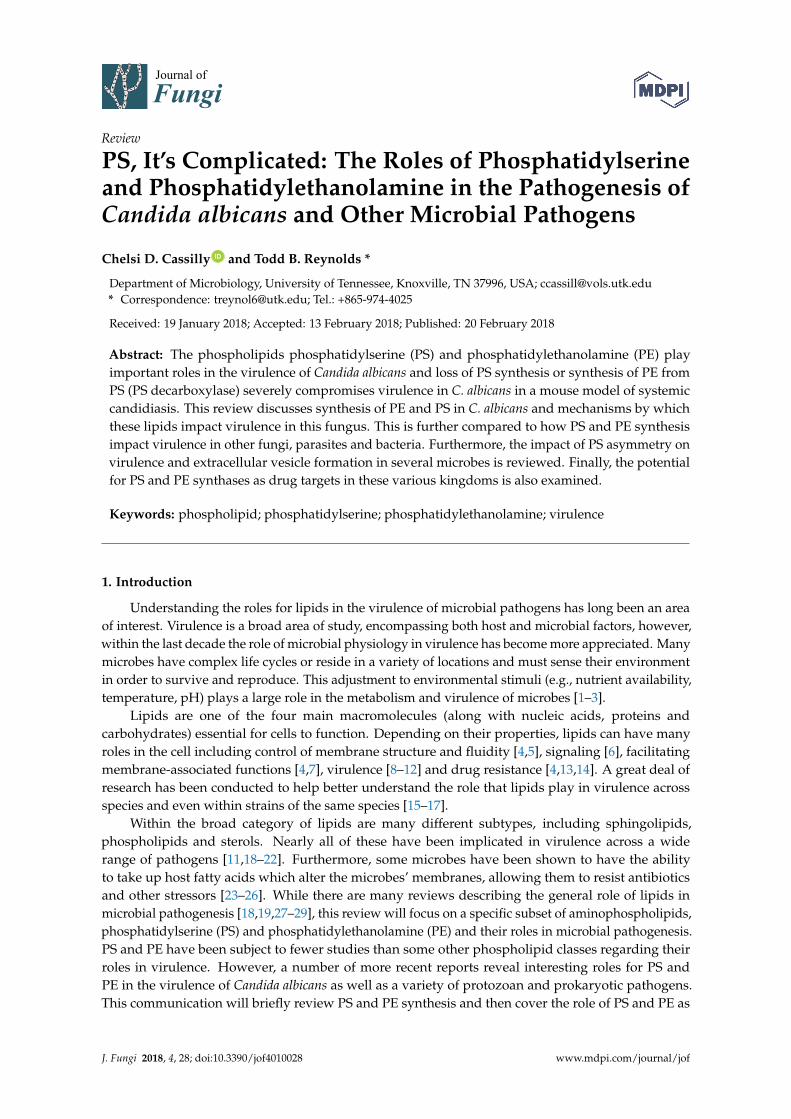
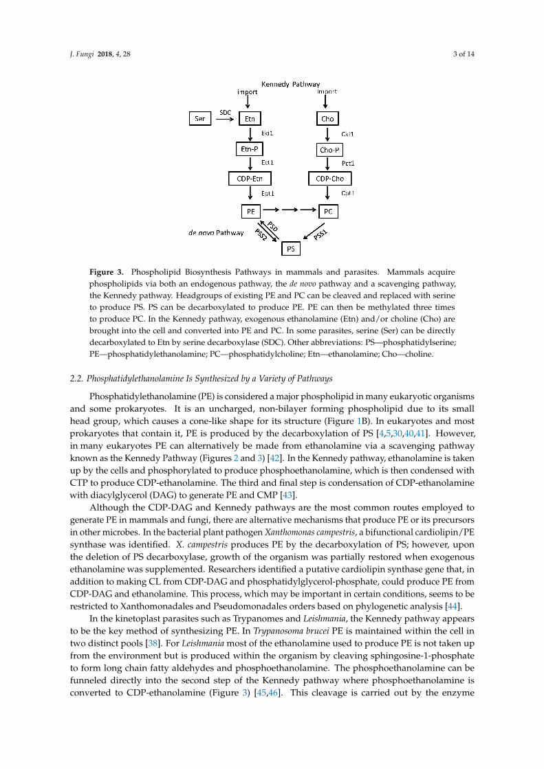
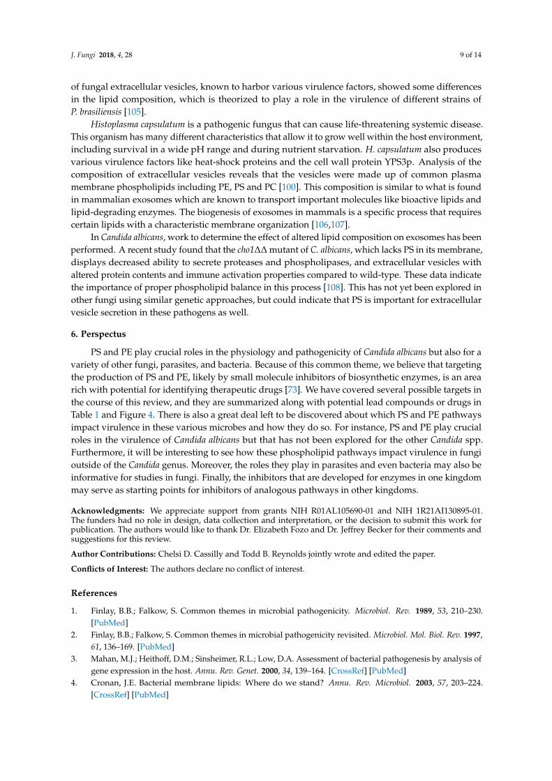
PS is a negatively charged phospholipid with a glycerol backbone and two fatty acid tails(Figure 1A). In bacteria and fungi, PS is produced from two substrates: cytidine diphosphatediacylglycerol (CDP-DAG) and serine (Figure 2). Although the enzymes responsible for this reactioncan differ greatly in primary sequence between fungi and many prokaryotes (excepting somebacteria like Bacillus subtilis or Sinorhizobium meliloti whose PS synthase is similar to S. cerevisiae)the mechanism by which they produce PS is similar [4,30–36]. In mammals and many parasites likeTrypanosoma brucei, PS is produced through a base-exchange reaction. In mammals, head groups ofexisting phosphatidylcholine (PC) and PE are cleaved off by two different enzymes, phosphatidylserinesynthase-2 (PSS2) and phosphatidylserine synthase-1 (PSS1) respectively and replaced with serine toproduce PS [34,37–39] (Figure 3).
J. Fungi 2018, 4, x FOR PEER REVIEW 2 of 14
regarding their roles in virulence. However, a number of more recent reports reveal interesting roles for PS and PE in the virulence of Candida albicans as well as a variety of protozoan and prokaryotic pathogens. This communication will briefly review PS and PE synthesis and then cover the role of PS and PE as regulators of virulence in C. albicans. We will compare this to what has been learned in other eukaryotic pathogens and a few prokaryotes.
2. Phosphatidylserine and Phosphatidylethanolamine Synthesis in Microbes
2.1. Phosphatidylserine Synthesis Is Similar between Fungi and Bacteria
PS is a negatively charged phospholipid with a glycerol backbone and two fatty acid tails (Figure 1A). In bacteria and fungi, PS is produced from two substrates: cytidine diphosphate diacylglycerol (CDP-DAG) and serine (Figure 2). Although the enzymes responsible for this reaction can differ greatly in primary sequence between fungi and many prokaryotes (excepting some bacteria like Bacillus subtilis or Sinorhizobium meliloti whose PS synthase is similar to S. cerevisiae) the mechanism by which they produce PS is similar [4,30–36]. In mammals and many parasites like Trypanosoma brucei, PS is produced through a base-exchange reaction. In mammals, head groups of existing phosphatidylcholine (PC) and PE are cleaved off by two different enzymes, phosphatidylserine synthase-2 (PSS2) and phosphatidylserine synthase-1 (PSS1) respectively and replaced with serine to produce PS [34,37–39] (Figure 3).
Figure 1. The structure of (A) phosphatidylserine and (B) phosphatidylethanolamine.
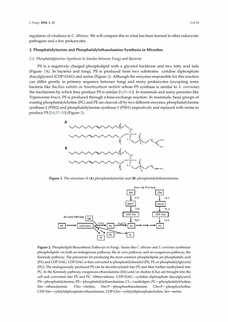
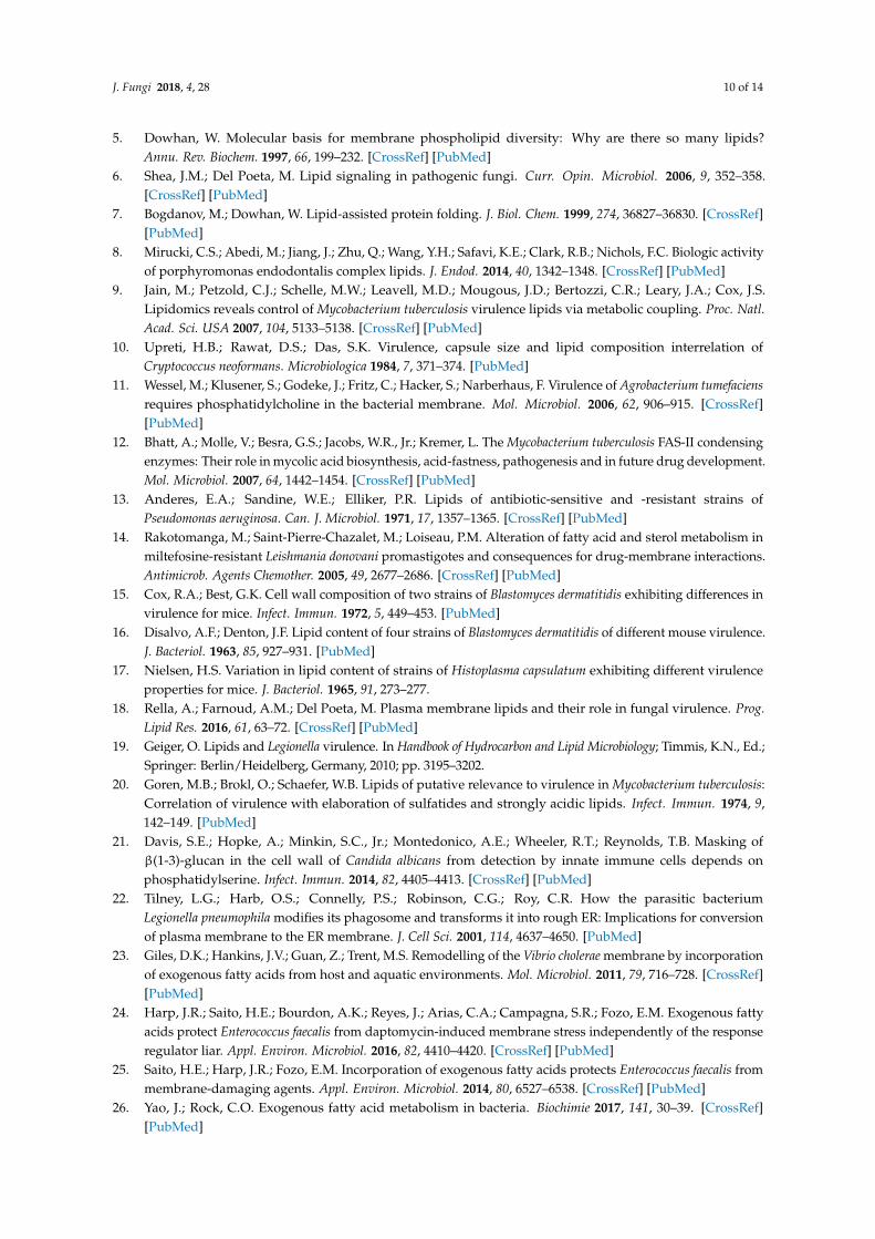
Figure 2. Phospholipid Biosynthesis Pathways in Fungi. Yeasts like C. albicans and S. cerevisiae synthesize phospholipids via both an endogenous pathway, the de novo pathway and an exogenous pathway, the Kennedy pathway. The precursors for producing the most common phospholipids are
Figure 1. The structure of (A) phosphatidylserine and (B) phosphatidylethanolamine.
J. Fungi 2018, 4, x FOR PEER REVIEW 2 of 14
regarding their roles in virulence. However, a number of more recent reports reveal interesting roles for PS and PE in the virulence of Candida albicans as well as a variety of protozoan and prokaryotic pathogens. This communication will briefly review PS and PE synthesis and then cover the role of PS and PE as regulators of virulence in C. albicans. We will compare this to what has been learned in other eukaryotic pathogens and a few prokaryotes.
2. Phosphatidylserine and Phosphatidylethanolamine Synthesis in Microbes
2.1. Phosphatidylserine Synthesis Is Similar between Fungi and Bacteria
PS is a negatively charged phospholipid with a glycerol backbone and two fatty acid tails (Figure 1A). In bacteria and fungi, PS is produced from two substrates: cytidine diphosphate diacylglycerol (CDP-DAG) and serine (Figure 2). Although the enzymes responsible for this reaction can differ greatly in primary sequence between fungi and many prokaryotes (excepting some bacteria like Bacillus subtilis or Sinorhizobium meliloti whose PS synthase is similar to S. cerevisiae) the mechanism by which they produce PS is similar [4,30–36]. In mammals and many parasites like Trypanosoma brucei, PS is produced through a base-exchange reaction. In mammals, head groups of existing phosphatidylcholine (PC) and PE are cleaved off by two different enzymes, phosphatidylserine synthase-2 (PSS2) and phosphatidylserine synthase-1 (PSS1) respectively and replaced with serine to produce PS [34,37–39] (Figure 3).
Figure 1. The structure of (A) phosphatidylserine and (B) phosphatidylethanolamine.
Figure 2. Phospholipid Biosynthesis Pathways in Fungi. Yeasts like C. albicans and S. cerevisiae synthesize phospholipids via both an endogenous pathway, the de novo pathway and an exogenous pathway, the Kennedy pathway. The precursors for producing the most common phospholipids are
Figure 2. Phospholipid Biosynthesis Pathways in Fungi. Yeasts like C. albicans and S. cerevisiae synthesizephospholipids via both an endogenous pathway, the de novo pathway and an exogenous pathway, theKennedy pathway. The precursors for producing the most common phospholipids are phosphatidic acid(PA) and CDP-DAG. CDP-DAG is then converted to phosphatidylinositol (PI), PS, or phosphatidylglycerol(PG). The endogenously produced PS can be decarboxylated into PE and then further methylated intoPC. In the Kennedy pathway, exogenous ethanolamine (Etn) and/or choline (Cho) are brought into thecell and converted into PE and PC. Abbreviations: CDP-DAG—cytidine diphosphate diacylglycerol;PS—phosphatidylserine; PE—phosphatidylethanolamine; CL—cardiolipin; PC—phosphatidylcholine;Etn—ethanolamine; Cho—choline, Etn-P—phosphoethanolamine, Cho-P—phosphocholine,CDP-Etn—cytidyldiphosphate-ethanolamine, CDP-Cho—cytidyldiphosphatecholine, Ser—serine.
J. Fungi 2018, 4, 28 3 of 14
J. Fungi 2018, 4, x FOR PEER REVIEW 3 of 14
phosphatidic acid (PA) and CDP-DAG. CDP-DAG is then converted to phosphatidylinositol (PI), PS, or phosphatidylglycerol (PG). The endogenously produced PS can be decarboxylated into PE and then further methylated into PC. In the Kennedy pathway, exogenous ethanolamine (Etn) and/or choline (Cho) are brought into the cell and converted into PE and PC. Abbreviations: CDP-DAG—cytidine diphosphate diacylglycerol; PS—phosphatidylserine; PE—phosphatidylethanolamine; CL—cardiolipin; PC—phosphatidylcholine; Etn—ethanolamine; Cho—choline, Etn-P—phosphoethanolamine, Cho-P—phosphocholine, CDP-Etn—cytidyldiphosphate-ethanolamine, CDP-Cho—cytidyldiphosphatecholine, Ser—serine.
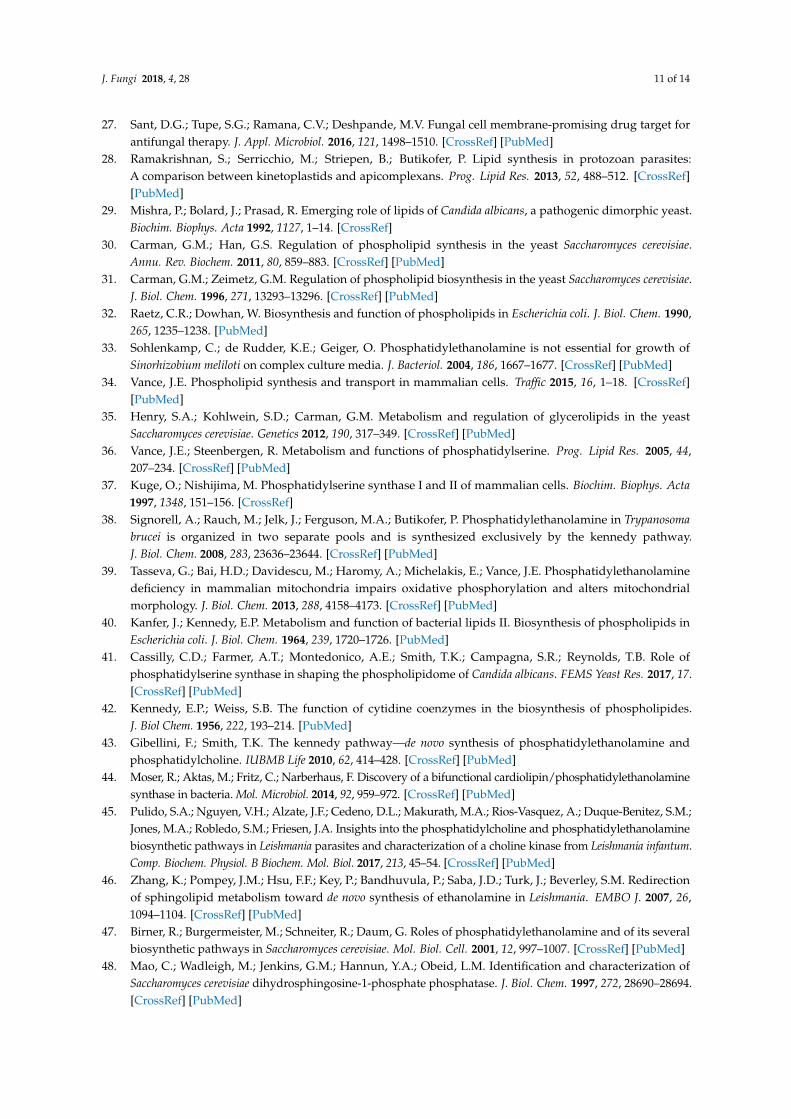
Figure 3. Phospholipid Biosynthesis Pathways in mammals and parasites. Mammals acquire phospholipids via both an endogenous pathway, the de novo pathway and a scavenging pathway, the Kennedy pathway. Headgroups of existing PE and PC can be cleaved and replaced with serine to produce PS. PS can be decarboxylated to produce PE. PE can then be methylated three times to produce PC. In the Kennedy pathway, exogenous ethanolamine (Etn) and/or choline (Cho) are brought into the cell and converted into PE and PC. In some parasites, serine (Ser) can be directly decarboxylated to Etn by serine decarboxylase (SDC). Other abbreviations: PS—phosphatidylserine; PE—phosphatidylethanolamine; PC—phosphatidylcholine; Etn—ethanolamine; Cho—choline.
2.2. Phosphatidylethanolamine Is Synthesized by a Variety of Pathways
Phosphatidylethanolamine (PE) is considered a major phospholipid in many eukaryotic organisms and some prokaryotes. It is an uncharged, non-bilayer forming phospholipid due to its small head group, which causes a cone-like shape for its structure (Figure 1B). In eukaryotes and most prokaryotes that contain it, PE is produced by the decarboxylation of PS [4,5,30,40,41]. However, in many eukaryotes PE can alternatively be made from ethanolamine via a scavenging pathway known as the Kennedy Pathway (Figures 2 and 3) [42]. In the Kennedy pathway, ethanolamine is taken up by the cells and phosphorylated to produce phosphoethanolamine, which is then condensed with CTP to produce CDP-ethanolamine. The third and final step is condensation of CDP-ethanolamine with diacylglycerol (DAG) to generate PE and CMP [43].
Although the CDP-DAG and Kennedy pathways are the most common routes employed to generate PE in mammals and fungi, there are alternative mechanisms that produce PE or its precursors in other microbes. In the bacterial plant pathogen Xanthomonas campestris, a bifunctional cardiolipin/PE synthase was identified. X. campestris produces PE by the decarboxylation of PS; however, upon the deletion of PS decarboxylase, growth of the organism was partially restored when exogenous ethanolamine was supplemented. Researchers identified a putative cardiolipin synthase gene that, in addition to making CL from CDP-DAG and phosphatidylglycerol-phosphate, could
Figure 3. Phospholipid Biosynthesis Pathways in mammals and parasites. Mammals acquirephospholipids via both an endogenous pathway, the de novo pathway and a scavenging pathway,the Kennedy pathway. Headgroups of existing PE and PC can be cleaved and replaced with serineto produce PS. PS can be decarboxylated to produce PE. PE can then be methylated three timesto produce PC. In the Kennedy pathway, exogenous ethanolamine (Etn) and/or choline (Cho) arebrought into the cell and converted into PE and PC. In some parasites, serine (Ser) can be directlydecarboxylated to Etn by serine decarboxylase (SDC). Other abbreviations: PS—phosphatidylserine;PE—phosphatidylethanolamine; PC—phosphatidylcholine; Etn—ethanolamine; Cho—choline.
2.2. Phosphatidylethanolamine Is Synthesized by a Variety of Pathways
Phosphatidylethanolamine (PE) is considered a major phospholipid in many eukaryotic organismsand some prokaryotes. It is an uncharged, non-bilayer forming phospholipid due to its smallhead group, which causes a cone-like shape for its structure (Figure 1B). In eukaryotes and mostprokaryotes that contain it, PE is produced by the decarboxylation of PS [4,5,30,40,41]. However,in many eukaryotes PE can alternatively be made from ethanolamine via a scavenging pathwayknown as the Kennedy Pathway (Figures 2 and 3) [42]. In the Kennedy pathway, ethanolamine is takenup by the cells and phosphorylated to produce phosphoethanolamine, which is then condensed withCTP to produce CDP-ethanolamine. The third and final step is condensation of CDP-ethanolaminewith diacylglycerol (DAG) to generate PE and CMP [43].
Although the CDP-DAG and Kennedy pathways are the most common routes employed togenerate PE in mammals and fungi, there are alternative mechanisms that produce PE or its precursorsin other microbes. In the bacterial plant pathogen Xanthomonas campestris, a bifunctional cardiolipin/PEsynthase was identified. X. campestris produces PE by the decarboxylation of PS; however, uponthe deletion of PS decarboxylase, growth of the organism was partially restored when exogenousethanolamine was supplemented. Researchers identified a putative cardiolipin synthase gene that, inaddition to making CL from CDP-DAG and phosphatidylglycerol-phosphate, could produce PE fromCDP-DAG and ethanolamine. This process, which may be important in certain conditions, seems to berestricted to Xanthomonadales and Pseudomonadales orders based on phylogenetic analysis [44].
In the kinetoplast parasites such as Trypanomes and Leishmania, the Kennedy pathway appearsto be the key method of synthesizing PE. In Trypanosoma brucei PE is maintained within the cell intwo distinct pools [38]. For Leishmania most of the ethanolamine used to produce PE is not taken upfrom the environment but is produced within the organism by cleaving sphingosine-1-phosphateto form long chain fatty aldehydes and phosphoethanolamine. The phosphoethanolamine can befunneled directly into the second step of the Kennedy pathway where phosphoethanolamine isconverted to CDP-ethanolamine (Figure 3) [45,46]. This cleavage is carried out by the enzyme
J. Fungi 2018, 4, 28 4 of 14
sphinosine-1-phosphate lyase (Dpl1), which is also found in other microbes such as S. cerevisiae.In yeast, Dpl1 can support growth of yeast in the absence of PS decarboxylase (psd1∆ psd2∆) andexogenous ethanolamine, indicating that it can support PE synthesis by the Kennedy pathway inyeast as well. However, under normal conditions, Dpl1 is not a major source for PE synthesis in thisorganism [47–49]. Yeast also have the ability to take up and acylate lyso-PE to produce PE or remodelexisting PE species [50–55]. Candida albicans has a Dpl1 homolog but a role for it in PE synthesis hasnot been tested.
The apicomplexan parasites also have unusual characteristics regarding PS and PE. The malariaparasite Plasmodium falciparum can acquire ethanolamine for the Kennedy pathway by directlydecarboxylating serine into ethanolamine, by means of the serine decarboxylase (SDC) enzyme,an enzymatic activity shared with plants but not animals or fungi (Figure 3) [56].
In the intracellular apicomplexan parasite Toxoplasma gondii, PE is produced via the Kennedypathway and via the decarboxylation of PS in the mitochondria [57] as is seen in fungi or mammals.However, T. gondii also has unusual versions of the canonical base-exchanging PS synthase and PSdecarboxylase enzymes. First, in addition to an internal, membrane-bound PS decarboxylase, it hasa second, soluble PS decarboxylase enzyme (TgPSD1) that is secreted extracellularly from T. gondiicells and appears to decarboxylate PS to PE in the parasitophorous vacuole, an organelle within thehost where T. gondii reproduces [57]. This is unusual because both PS decarboxylase and PS synthaseare typically membrane bound enzymes with multiple transmembrane domains, although there havebeen other reports of hyper-expressed PS decarboxylase enzymes dissociating from the cytoplasmicmembrane in bacteria [58] and in Plasmodium falciparum [59,60]. The function of secreted TgPSD1within the parasitophorous vacuole is not entirely clear but it could potentially help damage thehost cell membrane to allow T. gondii to escape the parasitophorous vacuole when it lyses the cell.Furthermore, the secreted enzyme may bind liposomes and host membranes to allow for membranebiogenesis and parasite replication. Third, the secreted enzyme may suppress PS exposure on theapoptotic host cell, thereby avoiding phagocytosis and allowing the parasite to replicate and avoidthe immune system. While these are all possible roles, the exact reasons for its unique function stillremain to be elucidated [61]. T. gondii also appears to have the ability to take up host PE (possibly viaa permease) when production of PE is inhibited intracellularly, further increasing the survival andfitness of this organism [57].
3. PS and PE Can Act as Modulators of Virulence in Candida, Bacteria and Parasites
3.1. Candida albicans Requires PE Synthesis from PS to Be Virulent
Candida albicans is a commensal fungus that normally inhabits the gut and skin of healthy people.However, immunocompromised individuals are at a high risk of developing bloodstream infectionswhere C. albicans can infect the deep organs leading to sepsis [62–64]. C. albicans is known to produceseveral virulence factors including hyphae, adhesins, lipases, proteases and the more recently describedcandidalysin [65,66]. It is also able to hide itself to a limited extent from the innate immune systemby a process called masking. Yeast cell walls contain four main components: chitin, mannosylatedproteins (mannan), β(1-6)-glucan and β(1-3)-glucan. These components are differentially enrichedinto two layers, with chitin, β(1-6)-glucan and β(1-3)-glucan in the inner layer. Mannan makes up themajority of the outer surface layer of the cell wall, and β-glucans and chitin are “masked” beneath.β-(1,3)-glucan is a pathogen associate molecular pattern (PAMP) that can be detected by the innateimmune receptor Dectin-1 as a signal that the host is infected by a fungal pathogen [67]. Dectin-1is found on macrophages, dendritic cells, neutrophils and some other immune cells. The proposedlayered topology, where mannan masks the immunogenic molecule β(1-3)-glucan is a method of innateimmune system evasion by this yeast [21]. Disruption of this layering (i.e., unmasking) makes it easierfor the host to recognize β(1,3)-glucan and detect the fungus [68–70].
J. Fungi 2018, 4, 28 5 of 14
It has been shown that the fungal phosphatidylserine (PS) synthase, Cho1, is absolutely requiredfor virulence of C. albicans. In a mouse model of systemic infection, the cho1∆∆ deletion mutant isunable to cause infection, while mice infected with wild-type or cho1∆/∆::CHO1 reintegrant strains diewithin two weeks [71]. In addition, cho1∆∆ exhibits significant reduction in kidney colonization and iscompletely cleared from the mice, even when they are made neutropenic with cyclophosphamide [72].In contrast, mice infected with wild-type C. albicans show high kidney burden before succumbing toinfection [71].
In addition to a complete loss of PS, the cho1∆∆ mutation also causes a loss of PE synthesizedfrom PS (Figure 2) [41]. This suggested that the avirulence could be caused by loss of PE as wellas PS. A major difference between cho1∆∆ and psd1∆∆ psd2∆∆ is that only cho1∆∆ has increasedβ(1-3)-glucan unmasking in its cell wall, increasing host immune recognition of this microbe [21]. Thus,other underlying factors related to loss of PE play a role in the loss of virulence but cell wall unmaskingdriven by the loss of PS may contribute to avirulence in the cho1∆∆ mutant, as well. The mechanismsresponsible for cell wall unmasking in the cho1∆∆ mutant are currently under investigation.
These defects in virulence in the cho1∆∆ and psd1∆∆ psd2∆∆ mutants are manifest despite thepresence of an alternative Kennedy pathway for PE synthesis (Figure 2). This brings up questions as towhether Kennedy pathway synthesized PE is able to compensate for PS-derived PE or if cells are unableto make sufficient PE by the Kennedy pathway. Furthermore, this also opens the question of howmuch of a role the loss of PS alone plays in virulence and whether this impact occurs via unmasking.
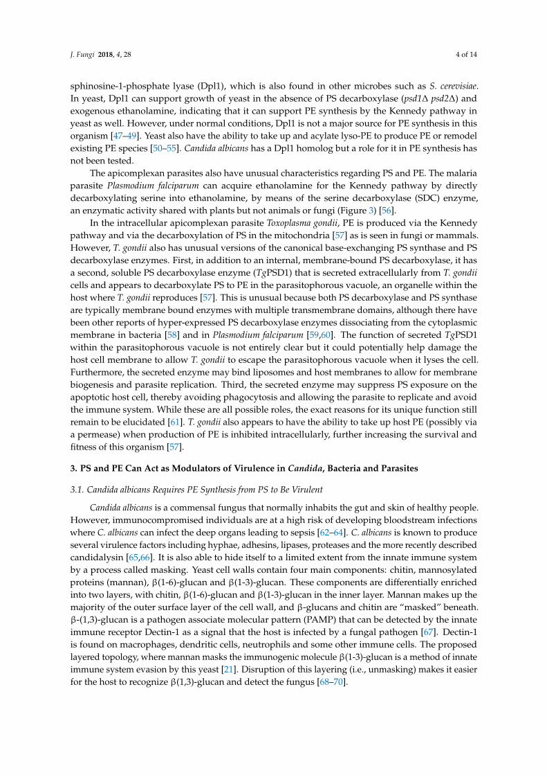
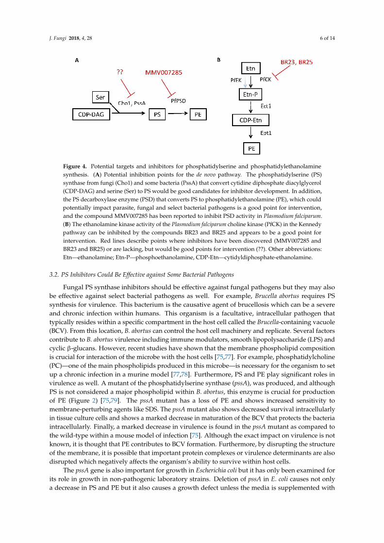
Due to these virulence defects and the reasons that follow, Cho1 represents a good drug targetin C. albicans (Table 1 and Figure 4A) [71,73]. First, as loss of Cho1 renders C. albicans avirulent inmice, inhibition of Cho1 is predicted to render C. albicans nonpathogenic in humans. Secondly, sincethe mammalian PS synthase enzymes are not orthologous with the fungal PS synthase, an inhibitorof Cho1 should be very specific for fungi without affecting mammalian Pss1p and Pss2p (compareFigures 2 and 3). Identification of small molecule inhibitors of Cho1 as potential therapeutics is apriority [73]. Third, Cho1 is conserved throughout pathogenic fungi, so an inhibitor could be broadspectrum [74].
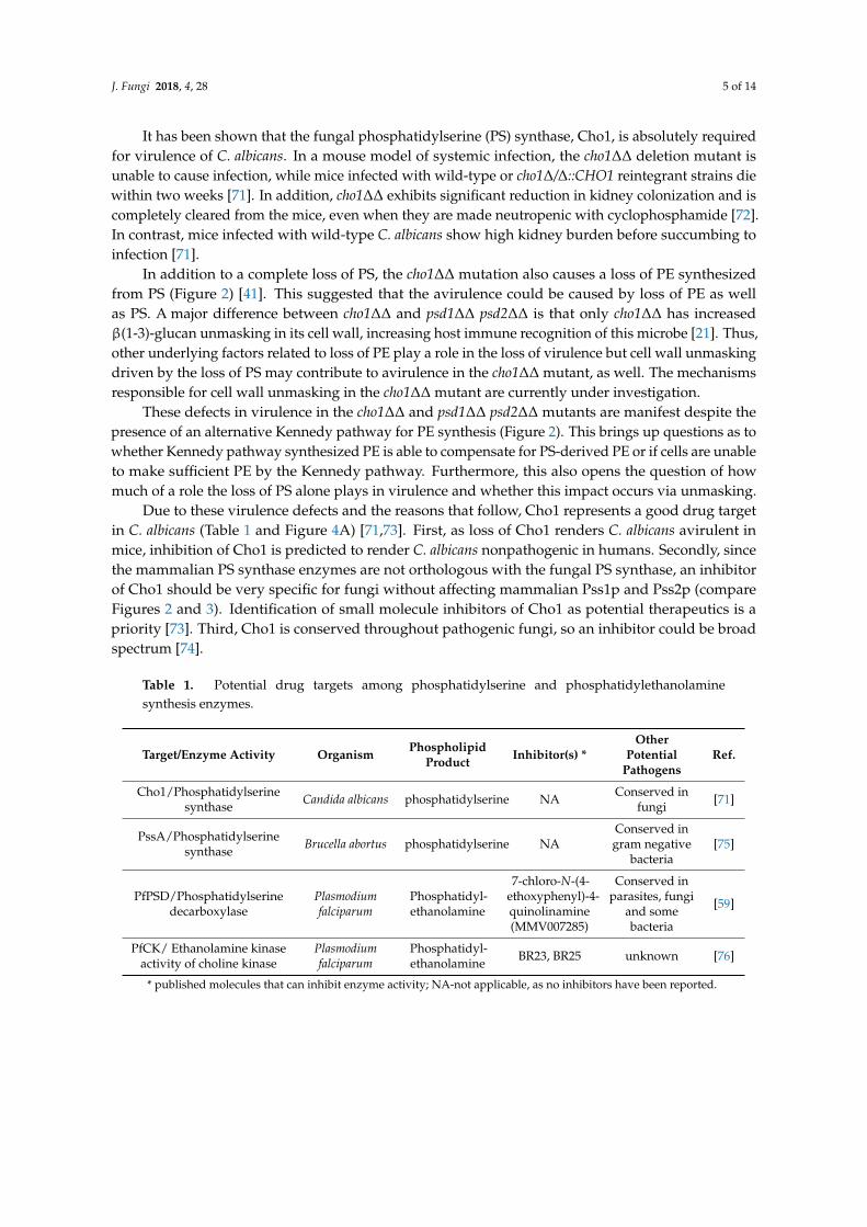
Table 1. Potential drug targets among phosphatidylserine and phosphatidylethanolaminesynthesis enzymes.
Target/Enzyme Activity Organism PhospholipidProduct Inhibitor(s) *
OtherPotential
PathogensRef.
Cho1/Phosphatidylserinesynthase Candida albicans phosphatidylserine NA Conserved in
fungi [71]
PssA/Phosphatidylserinesynthase Brucella abortus phosphatidylserine NA
Conserved ingram negative
bacteria[75]
PfPSD/Phosphatidylserinedecarboxylase
Plasmodiumfalciparum
Phosphatidyl-ethanolamine
7-chloro-N-(4-ethoxyphenyl)-4-quinolinamine(MMV007285)
Conserved inparasites, fungi
and somebacteria
[59]
PfCK/ Ethanolamine kinaseactivity of choline kinase
Plasmodiumfalciparum
Phosphatidyl-ethanolamine BR23, BR25 unknown [76]
* published molecules that can inhibit enzyme activity; NA-not applicable, as no inhibitors have been reported.
J. Fungi 2018, 4, 28 6 of 14J. Fungi 2018, 4, x FOR PEER REVIEW 6 of 14
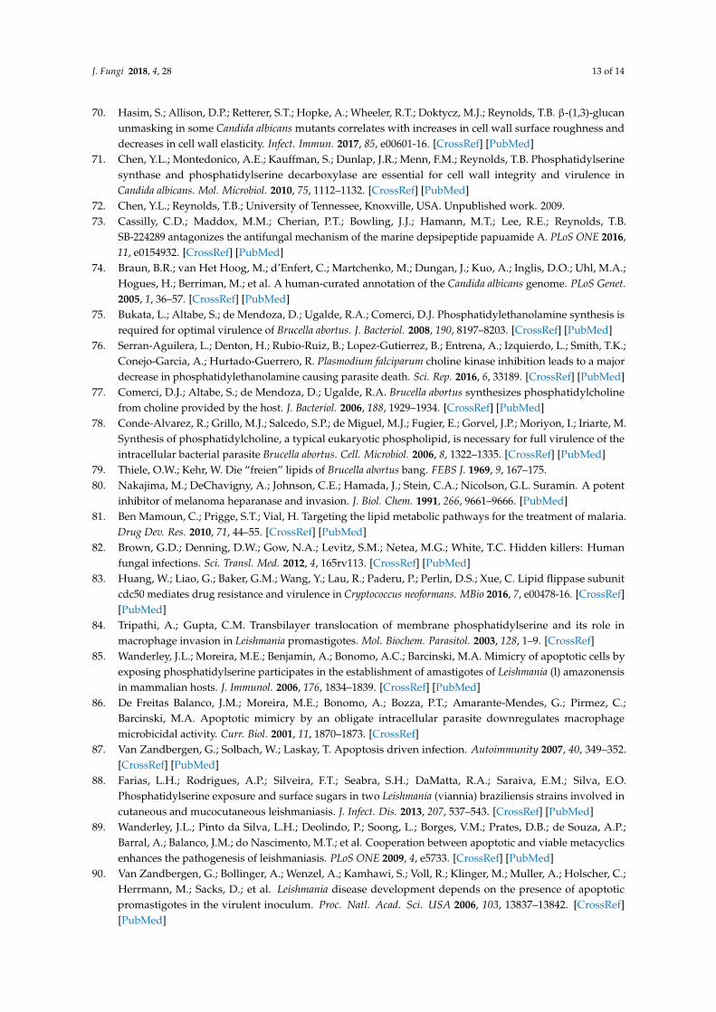
Figure 4. Potential targets and inhibitors for phosphatidylserine and phosphatidylethanolamine synthesis. (A) Potential inhibition points for the de novo pathway. The phosphatidylserine (PS) synthase from fungi (Cho1) and some bacteria (PssA) that convert cytidine diphosphate diacylglycerol (CDP-DAG) and serine (Ser) to PS would be good candidates for inhibitor development. In addition, the PS decarboxylase enzyme (PSD) that converts PS to phosphatidylethanolamine (PE), which could potentially impact parasite, fungal and select bacterial pathogens is a good point for intervention, and the compound MMV007285 has been reported to inhibit PSD activity in Plasmodium falciparum. (B) The ethanolamine kinase activity of the Plasmodium falciparum choline kinase (PfCK) in the Kennedy pathway can be inhibited by the compounds BR23 and BR25 and appears to be a good point for intervention. Red lines describe points where inhibitors have been discovered (MMV007285 and BR23 and BR25) or are lacking, but would be good points for intervention (??). Other abbreviations: Etn—ethanolamine; Etn-P—phosphoethanolamine, CDP-Etn—cytidyldiphosphate-ethanolamine.
3.2. PS Inhibitors Could Be Effective against Some Bacterial Pathogens
Fungal PS synthase inhibitors should be effective against fungal pathogens but they may also be effective against select bacterial pathogens as well. For example, Brucella abortus requires PS synthesis for virulence. This bacterium is the causative agent of brucellosis which can be a severe and chronic infection within humans. This organism is a facultative, intracellular pathogen that typically resides within a specific compartment in the host cell called the Brucella-containing vacuole (BCV). From this location, B. abortus can control the host cell machinery and replicate. Several factors contribute to B. abortus virulence including immune modulators, smooth lipopolysaccharide (LPS) and cyclic β-glucans. However, recent studies have shown that the membrane phospholipid composition is crucial for interaction of the microbe with the host cells [75,77]. For example, phosphatidylcholine (PC)—one of the main phospholipids produced in this microbe—is necessary for the organism to set up a chronic infection in a murine model [77,78]. Furthermore, PS and PE play significant roles in virulence as well. A mutant of the phosphatidylserine synthase (pssA), was produced, and although PS is not considered a major phospholipid within B. abortus, this enzyme is crucial for production of PE (Figure 2) [75,79]. The pssA mutant has a loss of PE and shows increased sensitivity to membrane-perturbing agents like SDS. The pssA mutant also shows decreased survival intracellularly in tissue culture cells and shows a marked decrease in maturation of the BCV that protects the bacteria intracellularly. Finally, a marked decrease in virulence is found in the pssA mutant as compared to the wild-type within a mouse model of infection [75]. Although the exact impact on virulence is not known, it is thought that PE contributes to BCV formation. Furthermore, by disrupting the structure of the membrane, it is possible that important protein complexes or virulence determinants are also disrupted which negatively affects the organism’s ability to survive within host cells.
The pssA gene is also important for growth in Escherichia coli but it has only been examined for its role in growth in non-pathogenic laboratory strains. Deletion of pssA in E. coli causes not only a decrease in PS and PE but it also causes a growth defect unless the media is supplemented with
Figure 4. Potential targets and inhibitors for phosphatidylserine and phosphatidylethanolaminesynthesis. (A) Potential inhibition points for the de novo pathway. The phosphatidylserine (PS)synthase from fungi (Cho1) and some bacteria (PssA) that convert cytidine diphosphate diacylglycerol(CDP-DAG) and serine (Ser) to PS would be good candidates for inhibitor development. In addition,the PS decarboxylase enzyme (PSD) that converts PS to phosphatidylethanolamine (PE), which couldpotentially impact parasite, fungal and select bacterial pathogens is a good point for intervention,and the compound MMV007285 has been reported to inhibit PSD activity in Plasmodium falciparum.(B) The ethanolamine kinase activity of the Plasmodium falciparum choline kinase (PfCK) in the Kennedypathway can be inhibited by the compounds BR23 and BR25 and appears to be a good point forintervention. Red lines describe points where inhibitors have been discovered (MMV007285 andBR23 and BR25) or are lacking, but would be good points for intervention (??). Other abbreviations:Etn—ethanolamine; Etn-P—phosphoethanolamine, CDP-Etn—cytidyldiphosphate-ethanolamine.
3.2. PS Inhibitors Could Be Effective against Some Bacterial Pathogens
Fungal PS synthase inhibitors should be effective against fungal pathogens but they may alsobe effective against select bacterial pathogens as well. For example, Brucella abortus requires PSsynthesis for virulence. This bacterium is the causative agent of brucellosis which can be a severeand chronic infection within humans. This organism is a facultative, intracellular pathogen thattypically resides within a specific compartment in the host cell called the Brucella-containing vacuole(BCV). From this location, B. abortus can control the host cell machinery and replicate. Several factorscontribute to B. abortus virulence including immune modulators, smooth lipopolysaccharide (LPS) andcyclic β-glucans. However, recent studies have shown that the membrane phospholipid compositionis crucial for interaction of the microbe with the host cells [75,77]. For example, phosphatidylcholine(PC)—one of the main phospholipids produced in this microbe—is necessary for the organism to setup a chronic infection in a murine model [77,78]. Furthermore, PS and PE play significant roles invirulence as well. A mutant of the phosphatidylserine synthase (pssA), was produced, and althoughPS is not considered a major phospholipid within B. abortus, this enzyme is crucial for productionof PE (Figure 2) [75,79]. The pssA mutant has a loss of PE and shows increased sensitivity tomembrane-perturbing agents like SDS. The pssA mutant also shows decreased survival intracellularlyin tissue culture cells and shows a marked decrease in maturation of the BCV that protects the bacteriaintracellularly. Finally, a marked decrease in virulence is found in the pssA mutant as compared tothe wild-type within a mouse model of infection [75]. Although the exact impact on virulence is notknown, it is thought that PE contributes to BCV formation. Furthermore, by disrupting the structureof the membrane, it is possible that important protein complexes or virulence determinants are alsodisrupted which negatively affects the organism’s ability to survive within host cells.
The pssA gene is also important for growth in Escherichia coli but it has only been examined forits role in growth in non-pathogenic laboratory strains. Deletion of pssA in E. coli causes not onlya decrease in PS and PE but it also causes a growth defect unless the media is supplemented with
J. Fungi 2018, 4, 28 7 of 14
divalent cations like Ca2+ or Mn2+ [80]. Further investigations into the role for PS synthase in thepathogenesis of Gram negative organisms is warranted as it may be important for virulence in a varietyof these pathogens (Table 1 and Figure 4A).
3.3. PE Synthesis Inhibitors Could Be Effective against Eukaryotic Pathogens
The PS decarboxylase enzymes that convert PS to PE (Psd1 or Psd2) are required for virulenceof C. albicans as described above, thus these enzymes may be good drug targets in addition to Cho1.However, work on development of eukaryotic PS decarboxylase inhibitors has made greater progressin parasites than in fungi.
Plasmodium parasites are the causative agents of malaria, which is one of the most importanthealth problems in the developing world. Finding new treatments with novel modes of actions tobetter combat this pathogen is a major area of current research because of the rising resistance toexisting anti-malarial therapies. The Plasmodium PS decarboxylase (PSD) enzyme has been suggestedas a drug target since PE is an essential phospholipid in Plasmodium. Indeed, inhibition of PSD resultsin growth arrest of the parasite [59,76,81]. Recent findings have shown that the PSD enzyme fromPlasmodium falciparum, (found in both soluble and membrane-bound forms in this organism) cancomplement yeast psd mutants [59]. Furthermore, screening a library of known malaria inhibitorsidentified a particular compound, 7-chloro-N-(4-ethoxyphenyl)-4-quinolinamine (MMV007285), withpotent activity against Plasmodium and the ability to inhibit the catalytic function of PSD (Table 1 andFigure 4A) [59].
In addition to PS decarboxylase, the choline kinase (CK) is crucial for PE synthesis in Plasmodiumand has been suggested as another drug target [76]. This enzyme is involved in the Kennedy Pathwaywhere choline and ethanolamine are taken up from the environment and used to produce PC and PE,respectively (Figure 3). Recent work has found that known anti-cancer compounds BR23 and BR25that inhibit human choline kinase cause a dramatic drop in the levels of PE within P. falciparum but notPC, as these compounds influence PE synthesis more than PC synthesis in this parasite [76]. The CKof P. falciparum is involved in both choline and ethanolamine phosphorylation but the drugs seem toprimarily impact ethanolamine phosphorylation, rather than choline phosphorylation, explaining thedifferential effects on PE and PC synthesis. Treatment with either drug led to arrested development ofthe parasite, likely as a result of the loss of membrane PE and ultimately were lethal [76]. These findingsdemonstrate the importance of PE biosynthesis in survival and pathogenicity of some microbes andare some of the first studies where small molecule inhibition of an ethanolamine kinase in a pathogenhas led to promising lead inhibitory compounds (Table 1 and Figure 4B).
Finally, although P. falciparum has two different pathways to make PE, (both CDP-DAG andKennedy, Figures 2 and 3), loss of either pathway appears to be sufficient to compromise its growth.This is surprising and indicates several possible explanations: (1) the molecular species of PE madefrom the two pathways differ and each is crucial for virulence; (2) localization of PE synthesis for eachpathway differs (PSD is in the mitochondria while the Kennedy pathway synthesizes PE in the ER)and PE made in one location is not sufficient to make up for the other; (3) the volume of PE made byeither pathway alone is not sufficient to support virulence [59,76].
The ability of PS decarboxylase inhibitors to block fungal infections needs to be tested since Psd1and Psd2 are required for virulence. The effectiveness of choline/ethanolamine kinase inhibitors inthis fungus is unclear, as the role of the Kennedy pathway in the virulence of fungi like Candida albicanshas not yet been reported.
4. PS Symmetry in the Membrane Plays a Role in Virulence
The lipid composition in the microbe’s membranes can play a role in promoting virulence but thesymmetry of lipids can also play an important role. For example, the symmetry of PS can impact amicrobe’s uptake by host cells (e.g., phagocytosis by host macrophages).
J. Fungi 2018, 4, 28 8 of 14
4.1. Cryptococcus neoformans Lipid Flippase Impacts Virulence
Cryptococcus neoformans is a facultative intracellular fungal pathogen that is a leading cause offungal pathogenesis worldwide [82]. A recent study has demonstrated that Cdc50, a regulatorysubunit for lipid flippases that are responsible for maintaining asymmetry in the phospholipid bilayer,is required for virulence [83]. Upon deletion of Cdc50, C. neoformans becomes more sensitive tofluconazole, caspofungin and SDS, likely due to a change in membrane integrity. In mice, the cdc50∆mutant is unable to cause a robust infection and is cleared from the lungs, further implicating thisprotein as a virulence factor. The exact mechanism behind this loss of virulence is currently still underinvestigation. However, PS is normally localized to the inner leaflet of the plasma membrane and isnot exposed to the outside of cell, but in the cdc50∆ mutant there is increased exposure of PS to outsideof the membrane. This provides support for the importance of proper PS symmetry for the virulenceof this fungus. These results suggest that enzymes needed to maintain PS asymmetry represent gooddrug targets within Cryptococcus. In addition, since the cdc50∆ mutant showed greater susceptibility tocaspofungin, a common antifungal drug that Cryptococcus is naturally resistant to, further explorationof phospholipid flippases or membrane asymmetry could improve the effectiveness of echinocandinsagainst the fungus. The role of such flippases remains to be studied in Candida spp. and could be anexciting area of study.
4.2. PS Exposure in Parasites Facilitates Invasion of Host Cells
Changes in PS symmetry can actually improve virulence in some parasites. Leishmania braziliensisis known to have multiple virulence factors associated with disease including cell surface molecules likelipophosphoglycan (LPG) and carbohydrates. In addition, PS also serves as a ligand for mononuclearmacrophages. Leishmania tropica promastigote forms expose higher levels of PS on their surface duringthe infective growth phases [84]. Furthermore, amastigotes of Leishmania amazonensis with higher levelsof PS on the cell surface had increased infectivity in vivo and in vitro [85]. These findings indicate that ahigher concentration of PS on the surface of these organisms increases the chances of being internalizedby the host macrophages [85]. The PS exposed on the membrane of the parasite is thought to playa role in apoptotic mimicry, allowing L. brasiliensis to establish an infection within the host [86,87].When PS exposed on the surface of L. brasiliensis, Leishmania tropica or L. amazonensis is blocked withannexin V, the infectivity of the parasite in murine peritoneal macrophages is decreased [84–86,88,89].
Interestingly, PS exposure seems to have importance even beyond the initial entry into hostmacrophages as well. In L. amazonensis and L. major, subpopulations of PS-positive and PS-negativepromastigotes cooperate to produce a sustained and successful infection of host macrophages [89,90].L. amazonensis amastigotes with high levels of PS exposed on their cell surfaces are able to inducecytokine production as well as inhibit NO production [85]. These findings implicate PS productionand exposure as an excellent drug target within Leishmania.
Similar instances of apoptotic mimicry have also been reported for Trypanosoma cruzi [91,92],Toxoplasma gondii [93] and even enveloped viruses [94–97], reinforcing the model that PS exposure canregulate infection. Targeting proteins responsible for this PS exposure [98,99] or enzymes involved inPS synthesis, could be a viable option for future therapies across a wide variety of pathogens.
5. PS and PE May Play a Role in Extracellular Vesicles in Candida and Other Fungi
Another potential contributor to virulence in fungi and other microbes is the use of extracellularvesicles as delivery systems for virulence factors. Extracellular vesicles been observed in C. albicans,Candida parapsilosis, Sporothrix schenckii, Saccharomyces cerevisiae [100], Cryptococcus neoformans [101,102]and Paracoccidioides brasiliensis [103]. The role of extracellular vesicles in parasites and bacteria is moreextensively reviewed in [104].
Lipid profiles from 4 different strains of P. brasiliensis [103] revealed that the concentration of PCwas higher in virulent strains than in the avirulent strain. Further studies into the lipid composition
J. Fungi 2018, 4, 28 9 of 14
of fungal extracellular vesicles, known to harbor various virulence factors, showed some differencesin the lipid composition, which is theorized to play a role in the virulence of different strains ofP. brasiliensis [105].
Histoplasma capsulatum is a pathogenic fungus that can cause life-threatening systemic disease.This organism has many different characteristics that allow it to grow well within the host environment,including survival in a wide pH range and during nutrient starvation. H. capsulatum also producesvarious virulence factors like heat-shock proteins and the cell wall protein YPS3p. Analysis of thecomposition of extracellular vesicles reveals that the vesicles were made up of common plasmamembrane phospholipids including PE, PS and PC [100]. This composition is similar to what is foundin mammalian exosomes which are known to transport important molecules like bioactive lipids andlipid-degrading enzymes. The biogenesis of exosomes in mammals is a specific process that requirescertain lipids with a characteristic membrane organization [106,107].
In Candida albicans, work to determine the effect of altered lipid composition on exosomes has beenperformed. A recent study found that the cho1∆∆ mutant of C. albicans, which lacks PS in its membrane,displays decreased ability to secrete proteases and phospholipases, and extracellular vesicles withaltered protein contents and immune activation properties compared to wild-type. These data indicatethe importance of proper phospholipid balance in this process [108]. This has not yet been explored inother fungi using similar genetic approaches, but could indicate that PS is important for extracellularvesicle secretion in these pathogens as well.
6. Perspectus
PS and PE play crucial roles in the physiology and pathogenicity of Candida albicans but also for avariety of other fungi, parasites, and bacteria. Because of this common theme, we believe that targetingthe production of PS and PE, likely by small molecule inhibitors of biosynthetic enzymes, is an arearich with potential for identifying therapeutic drugs [73]. We have covered several possible targets inthe course of this review, and they are summarized along with potential lead compounds or drugs inTable 1 and Figure 4. There is also a great deal left to be discovered about which PS and PE pathwaysimpact virulence in these various microbes and how they do so. For instance, PS and PE play crucialroles in the virulence of Candida albicans but that has not been explored for the other Candida spp.Furthermore, it will be interesting to see how these phospholipid pathways impact virulence in fungioutside of the Candida genus. Moreover, the roles they play in parasites and even bacteria may also beinformative for studies in fungi. Finally, the inhibitors that are developed for enzymes in one kingdommay serve as starting points for inhibitors of analogous pathways in other kingdoms.
Acknowledgments: We appreciate support from grants NIH R01AL105690-01 and NIH 1R21AI130895-01.The funders had no role in design, data collection and interpretation, or the decision to submit this work forpublication. The authors would like to thank Dr. Elizabeth Fozo and Dr. Jeffrey Becker for their comments andsuggestions for this review.
Author Contributions: Chelsi D. Cassilly and Todd B. Reynolds jointly wrote and edited the paper.
Conflicts of Interest: The authors declare no conflict of interest.
References
1. Finlay, B.B.; Falkow, S. Common themes in microbial pathogenicity. Microbiol. Rev. 1989, 53, 210–230.[PubMed]
2. Finlay, B.B.; Falkow, S. Common themes in microbial pathogenicity revisited. Microbiol. Mol. Biol. Rev. 1997,61, 136–169. [PubMed]
3. Mahan, M.J.; Heithoff, D.M.; Sinsheimer, R.L.; Low, D.A. Assessment of bacterial pathogenesis by analysis ofgene expression in the host. Annu. Rev. Genet. 2000, 34, 139–164. [CrossRef] [PubMed]
4. Cronan, J.E. Bacterial membrane lipids: Where do we stand? Annu. Rev. Microbiol. 2003, 57, 203–224.[CrossRef] [PubMed]
J. Fungi 2018, 4, 28 10 of 14
5. Dowhan, W. Molecular basis for membrane phospholipid diversity: Why are there so many lipids?Annu. Rev. Biochem. 1997, 66, 199–232. [CrossRef] [PubMed]
6. Shea, J.M.; Del Poeta, M. Lipid signaling in pathogenic fungi. Curr. Opin. Microbiol. 2006, 9, 352–358.[CrossRef] [PubMed]
7. Bogdanov, M.; Dowhan, W. Lipid-assisted protein folding. J. Biol. Chem. 1999, 274, 36827–36830. [CrossRef][PubMed]
8. Mirucki, C.S.; Abedi, M.; Jiang, J.; Zhu, Q.; Wang, Y.H.; Safavi, K.E.; Clark, R.B.; Nichols, F.C. Biologic activityof porphyromonas endodontalis complex lipids. J. Endod. 2014, 40, 1342–1348. [CrossRef] [PubMed]
9. Jain, M.; Petzold, C.J.; Schelle, M.W.; Leavell, M.D.; Mougous, J.D.; Bertozzi, C.R.; Leary, J.A.; Cox, J.S.Lipidomics reveals control of Mycobacterium tuberculosis virulence lipids via metabolic coupling. Proc. Natl.Acad. Sci. USA 2007, 104, 5133–5138. [CrossRef] [PubMed]
10. Upreti, H.B.; Rawat, D.S.; Das, S.K. Virulence, capsule size and lipid composition interrelation ofCryptococcus neoformans. Microbiologica 1984, 7, 371–374. [PubMed]
11. Wessel, M.; Klusener, S.; Godeke, J.; Fritz, C.; Hacker, S.; Narberhaus, F. Virulence of Agrobacterium tumefaciensrequires phosphatidylcholine in the bacterial membrane. Mol. Microbiol. 2006, 62, 906–915. [CrossRef][PubMed]
12. Bhatt, A.; Molle, V.; Besra, G.S.; Jacobs, W.R., Jr.; Kremer, L. The Mycobacterium tuberculosis FAS-II condensingenzymes: Their role in mycolic acid biosynthesis, acid-fastness, pathogenesis and in future drug development.Mol. Microbiol. 2007, 64, 1442–1454. [CrossRef] [PubMed]
13. Anderes, E.A.; Sandine, W.E.; Elliker, P.R. Lipids of antibiotic-sensitive and -resistant strains ofPseudomonas aeruginosa. Can. J. Microbiol. 1971, 17, 1357–1365. [CrossRef] [PubMed]
14. Rakotomanga, M.; Saint-Pierre-Chazalet, M.; Loiseau, P.M. Alteration of fatty acid and sterol metabolism inmiltefosine-resistant Leishmania donovani promastigotes and consequences for drug-membrane interactions.Antimicrob. Agents Chemother. 2005, 49, 2677–2686. [CrossRef] [PubMed]
15. Cox, R.A.; Best, G.K. Cell wall composition of two strains of Blastomyces dermatitidis exhibiting differences invirulence for mice. Infect. Immun. 1972, 5, 449–453. [PubMed]
16. Disalvo, A.F.; Denton, J.F. Lipid content of four strains of Blastomyces dermatitidis of different mouse virulence.J. Bacteriol. 1963, 85, 927–931. [PubMed]
17. Nielsen, H.S. Variation in lipid content of strains of Histoplasma capsulatum exhibiting different virulenceproperties for mice. J. Bacteriol. 1965, 91, 273–277.
18. Rella, A.; Farnoud, A.M.; Del Poeta, M. Plasma membrane lipids and their role in fungal virulence. Prog.Lipid Res. 2016, 61, 63–72. [CrossRef] [PubMed]
19. Geiger, O. Lipids and Legionella virulence. In Handbook of Hydrocarbon and Lipid Microbiology; Timmis, K.N., Ed.;Springer: Berlin/Heidelberg, Germany, 2010; pp. 3195–3202.
20. Goren, M.B.; Brokl, O.; Schaefer, W.B. Lipids of putative relevance to virulence in Mycobacterium tuberculosis:Correlation of virulence with elaboration of sulfatides and strongly acidic lipids. Infect. Immun. 1974, 9,142–149. [PubMed]
21. Davis, S.E.; Hopke, A.; Minkin, S.C., Jr.; Montedonico, A.E.; Wheeler, R.T.; Reynolds, T.B. Masking ofβ(1-3)-glucan in the cell wall of Candida albicans from detection by innate immune cells depends onphosphatidylserine. Infect. Immun. 2014, 82, 4405–4413. [CrossRef] [PubMed]
22. Tilney, L.G.; Harb, O.S.; Connelly, P.S.; Robinson, C.G.; Roy, C.R. How the parasitic bacteriumLegionella pneumophila modifies its phagosome and transforms it into rough ER: Implications for conversionof plasma membrane to the ER membrane. J. Cell Sci. 2001, 114, 4637–4650. [PubMed]
23. Giles, D.K.; Hankins, J.V.; Guan, Z.; Trent, M.S. Remodelling of the Vibrio cholerae membrane by incorporationof exogenous fatty acids from host and aquatic environments. Mol. Microbiol. 2011, 79, 716–728. [CrossRef][PubMed]
24. Harp, J.R.; Saito, H.E.; Bourdon, A.K.; Reyes, J.; Arias, C.A.; Campagna, S.R.; Fozo, E.M. Exogenous fattyacids protect Enterococcus faecalis from daptomycin-induced membrane stress independently of the responseregulator liar. Appl. Environ. Microbiol. 2016, 82, 4410–4420. [CrossRef] [PubMed]
25. Saito, H.E.; Harp, J.R.; Fozo, E.M. Incorporation of exogenous fatty acids protects Enterococcus faecalis frommembrane-damaging agents. Appl. Environ. Microbiol. 2014, 80, 6527–6538. [CrossRef] [PubMed]
26. Yao, J.; Rock, C.O. Exogenous fatty acid metabolism in bacteria. Biochimie 2017, 141, 30–39. [CrossRef][PubMed]
J. Fungi 2018, 4, 28 11 of 14
27. Sant, D.G.; Tupe, S.G.; Ramana, C.V.; Deshpande, M.V. Fungal cell membrane-promising drug target forantifungal therapy. J. Appl. Microbiol. 2016, 121, 1498–1510. [CrossRef] [PubMed]
28. Ramakrishnan, S.; Serricchio, M.; Striepen, B.; Butikofer, P. Lipid synthesis in protozoan parasites:A comparison between kinetoplastids and apicomplexans. Prog. Lipid Res. 2013, 52, 488–512. [CrossRef][PubMed]
29. Mishra, P.; Bolard, J.; Prasad, R. Emerging role of lipids of Candida albicans, a pathogenic dimorphic yeast.Biochim. Biophys. Acta 1992, 1127, 1–14. [CrossRef]
30. Carman, G.M.; Han, G.S. Regulation of phospholipid synthesis in the yeast Saccharomyces cerevisiae.Annu. Rev. Biochem. 2011, 80, 859–883. [CrossRef] [PubMed]
31. Carman, G.M.; Zeimetz, G.M. Regulation of phospholipid biosynthesis in the yeast Saccharomyces cerevisiae.J. Biol. Chem. 1996, 271, 13293–13296. [CrossRef] [PubMed]
32. Raetz, C.R.; Dowhan, W. Biosynthesis and function of phospholipids in Escherichia coli. J. Biol. Chem. 1990,265, 1235–1238. [PubMed]
33. Sohlenkamp, C.; de Rudder, K.E.; Geiger, O. Phosphatidylethanolamine is not essential for growth ofSinorhizobium meliloti on complex culture media. J. Bacteriol. 2004, 186, 1667–1677. [CrossRef] [PubMed]
34. Vance, J.E. Phospholipid synthesis and transport in mammalian cells. Traffic 2015, 16, 1–18. [CrossRef][PubMed]
35. Henry, S.A.; Kohlwein, S.D.; Carman, G.M. Metabolism and regulation of glycerolipids in the yeastSaccharomyces cerevisiae. Genetics 2012, 190, 317–349. [CrossRef] [PubMed]
36. Vance, J.E.; Steenbergen, R. Metabolism and functions of phosphatidylserine. Prog. Lipid Res. 2005, 44,207–234. [CrossRef] [PubMed]
37. Kuge, O.; Nishijima, M. Phosphatidylserine synthase I and II of mammalian cells. Biochim. Biophys. Acta1997, 1348, 151–156. [CrossRef]
38. Signorell, A.; Rauch, M.; Jelk, J.; Ferguson, M.A.; Butikofer, P. Phosphatidylethanolamine in Trypanosomabrucei is organized in two separate pools and is synthesized exclusively by the kennedy pathway.J. Biol. Chem. 2008, 283, 23636–23644. [CrossRef] [PubMed]
39. Tasseva, G.; Bai, H.D.; Davidescu, M.; Haromy, A.; Michelakis, E.; Vance, J.E. Phosphatidylethanolaminedeficiency in mammalian mitochondria impairs oxidative phosphorylation and alters mitochondrialmorphology. J. Biol. Chem. 2013, 288, 4158–4173. [CrossRef] [PubMed]
40. Kanfer, J.; Kennedy, E.P. Metabolism and function of bacterial lipids II. Biosynthesis of phospholipids inEscherichia coli. J. Biol. Chem. 1964, 239, 1720–1726. [PubMed]
41. Cassilly, C.D.; Farmer, A.T.; Montedonico, A.E.; Smith, T.K.; Campagna, S.R.; Reynolds, T.B. Role ofphosphatidylserine synthase in shaping the phospholipidome of Candida albicans. FEMS Yeast Res. 2017, 17.[CrossRef] [PubMed]
42. Kennedy, E.P.; Weiss, S.B. The function of cytidine coenzymes in the biosynthesis of phospholipides.J. Biol Chem. 1956, 222, 193–214. [PubMed]
43. Gibellini, F.; Smith, T.K. The kennedy pathway—de novo synthesis of phosphatidylethanolamine andphosphatidylcholine. IUBMB Life 2010, 62, 414–428. [CrossRef] [PubMed]
44. Moser, R.; Aktas, M.; Fritz, C.; Narberhaus, F. Discovery of a bifunctional cardiolipin/phosphatidylethanolaminesynthase in bacteria. Mol. Microbiol. 2014, 92, 959–972. [CrossRef] [PubMed]
45. Pulido, S.A.; Nguyen, V.H.; Alzate, J.F.; Cedeno, D.L.; Makurath, M.A.; Rios-Vasquez, A.; Duque-Benitez, S.M.;Jones, M.A.; Robledo, S.M.; Friesen, J.A. Insights into the phosphatidylcholine and phosphatidylethanolaminebiosynthetic pathways in Leishmania parasites and characterization of a choline kinase from Leishmania infantum.Comp. Biochem. Physiol. B Biochem. Mol. Biol. 2017, 213, 45–54. [CrossRef] [PubMed]
46. Zhang, K.; Pompey, J.M.; Hsu, F.F.; Key, P.; Bandhuvula, P.; Saba, J.D.; Turk, J.; Beverley, S.M. Redirectionof sphingolipid metabolism toward de novo synthesis of ethanolamine in Leishmania. EMBO J. 2007, 26,1094–1104. [CrossRef] [PubMed]
47. Birner, R.; Burgermeister, M.; Schneiter, R.; Daum, G. Roles of phosphatidylethanolamine and of its severalbiosynthetic pathways in Saccharomyces cerevisiae. Mol. Biol. Cell. 2001, 12, 997–1007. [CrossRef] [PubMed]
48. Mao, C.; Wadleigh, M.; Jenkins, G.M.; Hannun, Y.A.; Obeid, L.M. Identification and characterization ofSaccharomyces cerevisiae dihydrosphingosine-1-phosphate phosphatase. J. Biol. Chem. 1997, 272, 28690–28694.[CrossRef] [PubMed]

J. Fungi 2018, 4, 28 12 of 14
49. Saba, J.D.; Nara, F.; Bielawska, A.; Garrett, S.; Hannun, Y.A. The BST1 gene of Saccharomyces cerevisiae is thesphingosine-1-phosphate lyase. J. Biol. Chem. 1997, 272, 26087–26090. [CrossRef] [PubMed]
50. Deng, L.; Fukuda, R.; Kakihara, T.; Narita, K.; Ohta, A. Incorporation and remodeling ofphosphatidylethanolamine containing short acyl residues in yeast. Biochim. Biophys. Acta 2010, 1801,635–645. [CrossRef] [PubMed]
51. Flis, V.V.; Daum, G. Lipid transport between the endoplasmic reticulum and mitochondria. Cold Spring Harb.Perspect. Biol. 2013, 5, a013235. [CrossRef] [PubMed]
52. Riekhof, W.R.; Voelker, D.R. Uptake and utilization of lyso-phosphatidylethanolamine by Saccharomycescerevisiae. J. Biol. Chem. 2006, 281, 36588–36596. [CrossRef] [PubMed]
53. Riekhof, W.R.; Wu, J.; Gijon, M.A.; Zarini, S.; Murphy, R.C.; Voelker, D.R. Lysophosphatidylcholinemetabolism in Saccharomyces cerevisiae: The role of P-type ATPases in transport and a broad specificityacyltransferase in acylation. J. Biol. Chem. 2007, 282, 36853–36861. [CrossRef] [PubMed]
54. Riekhof, W.R.; Wu, J.; Jones, J.L.; Voelker, D.R. Identification and characterization of the majorlysophosphatidylethanolamine acyltransferase in Saccharomyces cerevisiae. J. Biol. Chem. 2007, 282, 28344–28352.[CrossRef] [PubMed]
55. Burgermeister, M.; Birner-Grunberger, R.; Nebauer, R.; Daum, G. Contribution of different pathways to thesupply of phosphatidylethanolamine and phosphatidylcholine to mitochondrial membranes of the yeastSaccharomyces cerevisiae. Biochim. Biophys. Acta 2004, 1686, 161–168. [CrossRef] [PubMed]
56. Elabbadi, N.; Ancelin, M.L.; Vial, H.J. Phospholipid metabolism of serine in plasmodium-infectederythrocytes involves phosphatidylserine and direct serine decarboxylation. Biochem. J. 1997, 324 Pt. 2,435–445. [CrossRef]
57. Hartmann, A.; Hellmund, M.; Lucius, R.; Voelker, D.R.; Gupta, N. Phosphatidylethanolamine synthesis inthe parasite mitochondrion is required for efficient growth but dispensable for survival of Toxoplasma gondii.J. Biol. Chem. 2014, 289, 6809–6824. [CrossRef] [PubMed]
58. Tyhach, R.J.; Hawrot, E.; Satre, M.; Kennedy, E.P. Increased synthesis of phosphatidylserine decarboxylasein a strain of Escherichia coli bearing a hybrid plasmid. Altered association of enzyme with the membrane.J. Biol. Chem. 1979, 254, 627–633. [PubMed]
59. Choi, J.Y.; Kumar, V.; Pachikara, N.; Garg, A.; Lawres, L.; Toh, J.Y.; Voelker, D.R.; Ben Mamoun, C.Characterization of plasmodium phosphatidylserine decarboxylase expressed in yeast and applicationfor inhibitor screening. Mol. Microbiol. 2016, 99, 999–1014. [CrossRef] [PubMed]
60. Baunaure, F.; Eldin, P.; Cathiard, A.M.; Vial, H. Characterization of a non-mitochondrial type Iphosphatidylserine decarboxylase in Plasmodium falciparum. Mol. Microbiol. 2004, 51, 33–46. [CrossRef][PubMed]
61. Gupta, N.; Hartmann, A.; Lucius, R.; Voelker, D.R. The obligate intracellular parasite Toxoplasma gondiisecretes a soluble phosphatidylserine decarboxylase. J. Biol. Chem. 2012, 287, 22938–22947. [CrossRef][PubMed]
62. Bustamante, C.I. Treatment of Candida infection: A view from the trenches! Curr. Opin. Infect. Dis. 2005, 18,490–495. [CrossRef] [PubMed]
63. Eggimann, P.; Garbino, J.; Pittet, D. Management of Candida species infections in critically ill patients.Lancet Infect. Dis. 2003, 3, 772–785. [CrossRef]
64. Cassone, A.; Cauda, R. Candida and candidiasis in hiv-infected patients: Where commensalism, opportunisticbehavior and frank pathogenicity lose their borders. Aids 2012, 26, 1457–1472. [CrossRef] [PubMed]
65. Moyes, D.L.; Wilson, D.; Richardson, J.P.; Mogavero, S.; Tang, S.X.; Wernecke, J.; Hofs, S.; Gratacap, R.L.;Robbins, J.; Runglall, M.; et al. Candidalysin is a fungal peptide toxin critical for mucosal infection. Nature2016, 532, 64–68. [CrossRef] [PubMed]
66. Kumamoto, C.A.; Vinces, M.D. Contributions of hyphae and hypha-co-regulated genes to Candida albicansvirulence. Cell. Microbiol. 2005, 7, 1546–1554. [CrossRef] [PubMed]
67. Brown, G.D.; Gordon, S. Immune recognition. A new receptor for β-glucans. Nature 2001, 413, 36–37.[CrossRef] [PubMed]
68. Wheeler, R.T.; Fink, G.R. A drug-sensitive genetic network masks fungi from the immune system.PLoS Pathog. 2006, 2, e35. [CrossRef] [PubMed]
69. Wheeler, R.T.; Kombe, D.; Agarwala, S.D.; Fink, G.R. Dynamic, morphotype-specific Candida albicans β-glucanexposure during infection and drug treatment. PLoS Pathog. 2008, 4, e1000227. [CrossRef] [PubMed]
J. Fungi 2018, 4, 28 13 of 14
70. Hasim, S.; Allison, D.P.; Retterer, S.T.; Hopke, A.; Wheeler, R.T.; Doktycz, M.J.; Reynolds, T.B. β-(1,3)-glucanunmasking in some Candida albicans mutants correlates with increases in cell wall surface roughness anddecreases in cell wall elasticity. Infect. Immun. 2017, 85, e00601-16. [CrossRef] [PubMed]
71. Chen, Y.L.; Montedonico, A.E.; Kauffman, S.; Dunlap, J.R.; Menn, F.M.; Reynolds, T.B. Phosphatidylserinesynthase and phosphatidylserine decarboxylase are essential for cell wall integrity and virulence inCandida albicans. Mol. Microbiol. 2010, 75, 1112–1132. [CrossRef] [PubMed]
72. Chen, Y.L.; Reynolds, T.B.; University of Tennessee, Knoxville, USA. Unpublished work. 2009.73. Cassilly, C.D.; Maddox, M.M.; Cherian, P.T.; Bowling, J.J.; Hamann, M.T.; Lee, R.E.; Reynolds, T.B.
SB-224289 antagonizes the antifungal mechanism of the marine depsipeptide papuamide A. PLoS ONE 2016,11, e0154932. [CrossRef] [PubMed]
74. Braun, B.R.; van Het Hoog, M.; d’Enfert, C.; Martchenko, M.; Dungan, J.; Kuo, A.; Inglis, D.O.; Uhl, M.A.;Hogues, H.; Berriman, M.; et al. A human-curated annotation of the Candida albicans genome. PLoS Genet.2005, 1, 36–57. [CrossRef] [PubMed]
75. Bukata, L.; Altabe, S.; de Mendoza, D.; Ugalde, R.A.; Comerci, D.J. Phosphatidylethanolamine synthesis isrequired for optimal virulence of Brucella abortus. J. Bacteriol. 2008, 190, 8197–8203. [CrossRef] [PubMed]
76. Serran-Aguilera, L.; Denton, H.; Rubio-Ruiz, B.; Lopez-Gutierrez, B.; Entrena, A.; Izquierdo, L.; Smith, T.K.;Conejo-Garcia, A.; Hurtado-Guerrero, R. Plasmodium falciparum choline kinase inhibition leads to a majordecrease in phosphatidylethanolamine causing parasite death. Sci. Rep. 2016, 6, 33189. [CrossRef] [PubMed]
77. Comerci, D.J.; Altabe, S.; de Mendoza, D.; Ugalde, R.A. Brucella abortus synthesizes phosphatidylcholinefrom choline provided by the host. J. Bacteriol. 2006, 188, 1929–1934. [CrossRef] [PubMed]
78. Conde-Alvarez, R.; Grillo, M.J.; Salcedo, S.P.; de Miguel, M.J.; Fugier, E.; Gorvel, J.P.; Moriyon, I.; Iriarte, M.Synthesis of phosphatidylcholine, a typical eukaryotic phospholipid, is necessary for full virulence of theintracellular bacterial parasite Brucella abortus. Cell. Microbiol. 2006, 8, 1322–1335. [CrossRef] [PubMed]
79. Thiele, O.W.; Kehr, W. Die “freien” lipids of Brucella abortus bang. FEBS J. 1969, 9, 167–175.80. Nakajima, M.; DeChavigny, A.; Johnson, C.E.; Hamada, J.; Stein, C.A.; Nicolson, G.L. Suramin. A potent
inhibitor of melanoma heparanase and invasion. J. Biol. Chem. 1991, 266, 9661–9666. [PubMed]81. Ben Mamoun, C.; Prigge, S.T.; Vial, H. Targeting the lipid metabolic pathways for the treatment of malaria.
Drug Dev. Res. 2010, 71, 44–55. [CrossRef] [PubMed]82. Brown, G.D.; Denning, D.W.; Gow, N.A.; Levitz, S.M.; Netea, M.G.; White, T.C. Hidden killers: Human
fungal infections. Sci. Transl. Med. 2012, 4, 165rv113. [CrossRef] [PubMed]83. Huang, W.; Liao, G.; Baker, G.M.; Wang, Y.; Lau, R.; Paderu, P.; Perlin, D.S.; Xue, C. Lipid flippase subunit
cdc50 mediates drug resistance and virulence in Cryptococcus neoformans. MBio 2016, 7, e00478-16. [CrossRef][PubMed]
84. Tripathi, A.; Gupta, C.M. Transbilayer translocation of membrane phosphatidylserine and its role inmacrophage invasion in Leishmania promastigotes. Mol. Biochem. Parasitol. 2003, 128, 1–9. [CrossRef]
85. Wanderley, J.L.; Moreira, M.E.; Benjamin, A.; Bonomo, A.C.; Barcinski, M.A. Mimicry of apoptotic cells byexposing phosphatidylserine participates in the establishment of amastigotes of Leishmania (l) amazonensisin mammalian hosts. J. Immunol. 2006, 176, 1834–1839. [CrossRef] [PubMed]
86. De Freitas Balanco, J.M.; Moreira, M.E.; Bonomo, A.; Bozza, P.T.; Amarante-Mendes, G.; Pirmez, C.;Barcinski, M.A. Apoptotic mimicry by an obligate intracellular parasite downregulates macrophagemicrobicidal activity. Curr. Biol. 2001, 11, 1870–1873. [CrossRef]
87. Van Zandbergen, G.; Solbach, W.; Laskay, T. Apoptosis driven infection. Autoimmunity 2007, 40, 349–352.[CrossRef] [PubMed]
88. Farias, L.H.; Rodrigues, A.P.; Silveira, F.T.; Seabra, S.H.; DaMatta, R.A.; Saraiva, E.M.; Silva, E.O.Phosphatidylserine exposure and surface sugars in two Leishmania (viannia) braziliensis strains involved incutaneous and mucocutaneous leishmaniasis. J. Infect. Dis. 2013, 207, 537–543. [CrossRef] [PubMed]
89. Wanderley, J.L.; Pinto da Silva, L.H.; Deolindo, P.; Soong, L.; Borges, V.M.; Prates, D.B.; de Souza, A.P.;Barral, A.; Balanco, J.M.; do Nascimento, M.T.; et al. Cooperation between apoptotic and viable metacyclicsenhances the pathogenesis of leishmaniasis. PLoS ONE 2009, 4, e5733. [CrossRef] [PubMed]
90. Van Zandbergen, G.; Bollinger, A.; Wenzel, A.; Kamhawi, S.; Voll, R.; Klinger, M.; Muller, A.; Holscher, C.;Herrmann, M.; Sacks, D.; et al. Leishmania disease development depends on the presence of apoptoticpromastigotes in the virulent inoculum. Proc. Natl. Acad. Sci. USA 2006, 103, 13837–13842. [CrossRef][PubMed]
J. Fungi 2018, 4, 28 14 of 14
91. Damatta, R.A.; Seabra, S.H.; Deolindo, P.; Arnholdt, A.C.; Manhaes, L.; Goldenberg, S.; de Souza, W.Trypanosoma cruzi exposes phosphatidylserine as an evasion mechanism. FEMS Microbiol. Lett. 2007, 266,29–33. [CrossRef] [PubMed]
92. Freire-de-Lima, C.G.; Nascimento, D.O.; Soares, M.B.; Bozza, P.T.; Castro-Faria-Neto, H.C.; de Mello, F.G.;DosReis, G.A.; Lopes, M.F. Uptake of apoptotic cells drives the growth of a pathogenic trypanosome inmacrophages. Nature 2000, 403, 199–203. [CrossRef] [PubMed]
93. Seabra, S.H.; de Souza, W.; Damatta, R.A. Toxoplasma gondii exposes phosphatidylserine inducing a TGF-β1
autocrine effect orchestrating macrophage evasion. Biochem. Biophys. Res. Commun. 2004, 324, 744–752.[CrossRef] [PubMed]
94. Moller-Tank, S.; Kondratowicz, A.S.; Davey, R.A.; Rennert, P.D.; Maury, W. Role of the phosphatidylserinereceptor TIM-1 in enveloped-virus entry. J. Virol. 2013, 87, 8327–8341. [CrossRef] [PubMed]
95. Jemielity, S.; Wang, J.J.; Chan, Y.K.; Ahmed, A.A.; Li, W.; Monahan, S.; Bu, X.; Farzan, M.; Freeman, G.J.;Umetsu, D.T.; et al. TIM-family proteins promote infection of multiple enveloped viruses throughvirion-associated phosphatidylserine. PLoS Pathog. 2013, 9, e1003232. [CrossRef] [PubMed]
96. Soares, M.M.; King, S.W.; Thorpe, P.E. Targeting inside-out phosphatidylserine as a therapeutic strategy forviral diseases. Nat. Med. 2008, 14, 1357–1362. [CrossRef] [PubMed]
97. Vanlandschoot, P.; Leroux-Roels, G. Viral apoptotic mimicry: An immune evasion strategy developed by thehepatitis b virus? Trends Immunol. 2003, 24, 144–147. [CrossRef]
98. Dos Santos, M.G.; Muxel, S.M.; Zampieri, R.A.; Pomorski, T.G.; Floeter-Winter, L.M. Transbilayer dynamicsof phospholipids in the plasma membrane of the Leishmania genus. PLoS ONE 2013, 8, e55604. [CrossRef][PubMed]
99. Araujo-Santos, J.M.; Gamarro, F.; Castanys, S.; Herrmann, A.; Pomorski, T. Rapid transport of phospholipidsacross the plasma membrane of Leishmania infantum. Biochem. Biophys. Res. Commun. 2003, 306, 250–255.[CrossRef]
100. Albuquerque, P.C.; Nakayasu, E.S.; Rodrigues, M.L.; Frases, S.; Casadevall, A.; Zancope-Oliveira, R.M.;Almeida, I.C.; Nosanchuk, J.D. Vesicular transport in Histoplasma capsulatum: An effective mechanism fortrans-cell wall transfer of proteins and lipids in ascomycetes. Cell. Microbiol. 2008, 10, 1695–1710. [CrossRef][PubMed]
101. Rodrigues, M.L.; Nakayasu, E.S.; Oliveira, D.L.; Nimrichter, L.; Nosanchuk, J.D.; Almeida, I.C.; Casadevall, A.Extracellular vesicles produced by Cryptococcus neoformans contain protein components associated withvirulence. Eukaryot. Cell 2008, 7, 58–67. [CrossRef] [PubMed]
102. Oliveira, D.L.; Freire-de-Lima, C.G.; Nosanchuk, J.D.; Casadevall, A.; Rodrigues, M.L.; Nimrichter, L.Extracellular vesicles from Cryptococcus neoformans modulate macrophage functions. Infect. Immun. 2010, 78,1601–1609. [CrossRef] [PubMed]
103. Manocha, M.S.; San-Blas, G.; Centeno, S. Lipid composition of Paracoccidioides brasiliensis: Possible correlationwith virulence of different strains. J. Gen. Microbiol. 1980, 117, 147–154. [CrossRef] [PubMed]
104. Schorey, J.S.; Cheng, Y.; Singh, P.P.; Smith, V.L. Exosomes and other extracellular vesicles in host-pathogeninteractions. EMBO Rep. 2015, 16, 24–43. [CrossRef] [PubMed]
105. Vallejo, M.C.; Nakayasu, E.S.; Longo, L.V.; Ganiko, L.; Lopes, F.G.; Matsuo, A.L.; Almeida, I.C.; Puccia, R.Lipidomic analysis of extracellular vesicles from the pathogenic phase of Paracoccidioides brasiliensis.PLoS ONE 2012, 7, e39463. [CrossRef]
106. Laulagnier, K.; Motta, C.; Hamdi, S.; Roy, S.; Fauvelle, F.; Pageaux, J.F.; Kobayashi, T.; Salles, J.P.; Perret, B.;Bonnerot, C.; et al. Mast cell- and dendritic cell-derived exosomes display a specific lipid composition andan unusual membrane organization. Biochem. J. 2004, 380, 161–171. [CrossRef] [PubMed]
107. Subra, C.; Laulagnier, K.; Perret, B.; Record, M. Exosome lipidomics unravels lipid sorting at the level ofmultivesicular bodies. Biochimie 2007, 89, 205–212. [CrossRef] [PubMed]
108. Wolf, J.M.; Espadas, J.; Luque-Garcia, J.; Reynolds, T.; Casadevall, A. Lipid biosynthetic genes affect Candidaalbicans extracellular vesicle morphology, cargo, and immunostimulatory properties. Eukaryot. Cell 2015, 14,745–754. [CrossRef] [PubMed]
© 2018 by the authors. Licensee MDPI, Basel, Switzerland. This article is an open accessarticle distributed under the terms and conditions of the Creative Commons Attribution(CC BY) license (http://creativecommons.org/licenses/by/4.0/).