g proteins and their cell type specific functions
DESCRIPTION
GPCR and their cell type specific functionsTRANSCRIPT

Mammalian G Proteins and Their Cell Type Specific Functions
NINA WETTSCHURECK AND STEFAN OFFERMANNS
Institute of Pharmacology, University of Heidelberg, Heidelberg, Germany
I. Introduction 1160A. Basic principles of G protein-mediated signaling 1160B. G protein �-subunits and ��-complexes 1163
II. Cardiovascular System 1167A. Autonomic control of heart function 1167B. Myocardial hypertrophy 1168C. Smooth muscle tone 1169D. Platelet activation 1171
III. Endocrine System and Metabolism 1173A. Hypothalamo-pituitary system 1173B. Pancreatic �-cells 1174C. Thyroid gland/parathyroid gland 1175D. Regulation of carbohydrate and lipid metabolism 1175
IV. Immune System 1177A. Leukocyte migration/homing 1177B. Immune cell effector functions 1178
V. Nervous System 1179A. Inhibitory modulation of synaptic transmission 1179B. Modulation of synaptic transmission by the Gq/G11-mediated signaling pathway 1179C. Roles of Gz and Golf in the nervous system 1180
VI. Sensory Systems 1180A. Visual system 1181B. Olfactory/pheromone system 1181C. Gustatory system 1182
VII. Development 1182A. G13-mediated signaling in embryonic angiogenesis 1183B. Gq/G11-mediated signaling during embryonic myocardial growth 1183C. Neural crest development 1183
VIII. Cell Growth and Transformation 1184A. Constitutively active mutants of G�q/G�11 family members 1184B. The oncogenic potential of G�s 1184C. Gi-mediated cell transformation 1185D. Cellular growth induced by G�12/G�13 1185
IX. Concluding Remarks 1185
Wettschureck, Nina, and Stefan Offermanns. Mammalian G Proteins and Their Cell Type Specific Functions.Physiol Rev 85: 1159–1204, 2005; doi:10.1152/physrev.00003.2005.—Heterotrimeric G proteins are key players intransmembrane signaling by coupling a huge variety of receptors to channel proteins, enzymes, and other effectormolecules. Multiple subforms of G proteins together with receptors, effectors, and various regulatory proteinsrepresent the components of a highly versatile signal transduction system. G protein-mediated signaling is employedby virtually all cells in the mammalian organism and is centrally involved in diverse physiological functions such asperception of sensory information, modulation of synaptic transmission, hormone release and actions, regulation ofcell contraction and migration, or cell growth and differentiation. In this review, some of the functions ofheterotrimeric G proteins in defined cells and tissues are described.
Physiol Rev 85: 1159–1204, 2005;doi:10.1152/physrev.00003.2005.
www.prv.org 11590031-9333/05 $18.00 Copyright © 2005 the American Physiological Society
on April 23, 2015
Dow
nloaded from

I. INTRODUCTION
All cells possess transmembrane signaling systemsthat allow them to receive information from extracellularstimuli like hormones, neurotransmitters, or sensorystimuli. This fundamental process allows cells to commu-nicate with each other. All transmembrane signaling sys-tems share two basic elements, a receptor which is able torecognize an extracellular stimulus as well as an effectorwhich is controlled by the receptor and which can gener-ate an intracellular signal. Many transmembrane signalingsystems like receptor tyrosine kinases incorporate thesetwo elements in one molecule. In contrast, the G protein-mediated signaling system is relatively complex consist-ing of a receptor, a heterotrimeric G protein, and aneffector. This modular design of the G protein-mediatedsignaling system allows convergence and divergence atthe interfaces of receptor and G protein as well as of Gprotein and effector. In addition, each component, thereceptor, the G protein as well as the effector can beregulated independently by additional proteins, solublemediators, or on the transcriptional level. The relativelycomplex organization of the G protein-mediated trans-membrane signaling system provides the basis for a hugevariety of transmembrane signaling pathways that aretailored to serve particular functions in distinct cell types.It is probably this versatility of the G protein-mediatedsignaling system that has made it by far the most oftenemployed transmembrane signaling mechanism. In thisreview we summarize some of the biological roles of Gprotein-mediated signaling processes in the mammalianorganism which are based on their cell type-specific func-tion. Although we have tried to cover a wide variety ofcellular systems and functions, the plethora of availabledata forced us to restrict this review. Particular emphasisis placed on cellular G protein functions that have beenstudied in primary cells or in the context of the wholeorganism using genetic approaches.
A. Basic Principles
of G Protein-Mediated Signaling
More than 1,000 G protein-coupled receptors (GPCRs)are encoded in mammalian genomes. While most of themcode for sensory receptors like taste or olfactory recep-tors, �400–500 of them recognize nonsensory ligands likehormones, neurotransmitters, or paracrine factors (53,185, 519, 534, 649). For more than 200 GPCRs, the phys-iological ligands are known (Table 1). GPCRs for whichno endogenous ligand has been found are “orphan”GPCRs (376, 389, 688).
Upon activation of a receptor by, e.g., its endogenousligand, coupling of the activated receptor to the hetero-trimeric G protein is facilitated. Multiple site-directed mu-
tagenesis experiments have been performed on G protein-coupled receptors, and they have revealed various cyto-plasmic domains of the receptors that are involved in thespecific interaction between the receptors and the G pro-tein. However, despite the determination of the structureof rhodopsin at atomic resolution (504), it is still not clearhow specificity of the receptor-G protein interaction isachieved and how a ligand-induced conformationalchange in the receptor molecule results in G proteinactivation (177, 212, 213, 565, 674).
The heterotrimeric G protein consists of an �-subunitthat binds and hydrolyzes GTP as well as of a �- and a�-subunit that form an undissociable complex (233, 255,475). Several subtypes of �-, �-, and �-subunits have beendescribed (Table 2). To dynamically couple activated re-ceptors to effectors, the heterotrimeric G protein under-goes an activation-inactivation cycle (Fig. 1). In the basalstate, the ��-complex and the GDP-bound �-subunit areassociated, and the heterotrimer can be recognized by anappropriate activated receptor. Coupling of the activatedreceptor to the heterotrimer promotes the exchange ofGDP for GTP on the G protein �-subunit. The GTP-bound�-subunit dissociates from the activated receptor as wellas from the ��-complex, and both the �-subunit and the��-complex are now free to modulate the activity of avariety of effectors like ion channels or enzymes. Signal-ing is terminated by the hydrolysis of GTP by the GTPaseactivity, which is inherent to the G protein �-subunit. Theresulting GDP-bound �-subunit reassociates with the ��-complex to enter a new cycle if activated receptors arepresent. For recent excellent reviews on basic structuraland functional aspects of G proteins, see References 49,83, 361, and 526.
While the kinetics of G protein activation throughGPCRs has been well described for quite a while, onlyrecently has the regulation of the deactivation processbeen understood in more detail. Based on the observationthat the GTPase activity of isolated G proteins is muchlower than that observed under physiological conditions,the existence of mechanisms that accelerate the GTPaseactivity had been postulated. Various effectors have in-deed been found to enhance GTPase activity of the Gprotein �-subunit, thereby contributing to the deactiva-tion and allowing for rapid modulation of G protein-me-diated signaling (23, 45, 348, 571). More recently, a familyof proteins called “regulators of G protein signaling” (RGSproteins) has been identified, which is also able to in-crease the GTPase activity of G protein �-subunits (272,481, 550). There are �30 RGS proteins currently known,which have selectivities for G protein �-subfamilies. Thephysiological role of RGS proteins is currently under in-vestigation. Besides their role in the modulation of Gprotein-mediated signaling kinetics, they also influencethe specificity of the signaling process and in some casesmay have effector functions.
1160 NINA WETTSCHURECK AND STEFAN OFFERMANNS
Physiol Rev • VOL 85 • OCTOBER 2005 • www.prv.org
on April 23, 2015
Dow
nloaded from

TABLE 1. Physiological ligands of G protein-coupled receptors
Endogenous Ligand(s) ReceptorCoupling to G Protein
Subclass(es) Reference Nos.
Amino acids, dicarboxylic acidsGlutamate mGluR1,5 Gq/11 117
mGluR2,3,4,6,7,8 Gi/o 117�-Aminobutyric acid (GABA) GABAB1 (binding), GABAB2 (signaling) Gi/o 64�-Ketoglutarate GPR99 Gq/11 248Succinate GPR91 Gq/11, Gi/o 248L-Arginine, L-lysine GPRC6A Gq/11 ? 673
Biogenic AminesAcetylcholine M1,M3,M5 Gq/11 675
M2, M4 Gi/o 675Epinephrine, norepinephrine �1A,�1B,�1D Gq/11 252
�2A,�2B,�2C Gi/o 252�1,�2,�3 Gs 399
Dopamine D1,D5 Gs 487D2,D3,D4 Gi/o 487
Histamine H1 Gq/11 262H2 Gs 262H3,H4 Gi/o 262
Melatonin MT1,MT2,MT3 Gi/o 151Serotonin 5-HT1A/B/D/E/F Gi/o 281
5-HT2A/B/C Gq/11 2815-HT4,5-HT6,5-HT7 Gs 281, 466, 4675-HT5A/B Gi/o, Gs 281
Trace amines TA1, TA2 Gs 57, 80Ions
Ca2� CaSR Gq/11, Gi/o 218H� SPC1, G2A Gq/11, G12/13 403, 465
GPR4, TDAG-8 Gs 403, 658Nucleotides/nucleosides
Adenosine A1, A3 Gi/o 184A2A, A2B Gs 184
ADP P2Y12, P2Y13 Gi/o 273, 422ADP/ATP P2Y1 Gq/11 183ATP P2Y11 Gq/11, Gs 183UDP P2Y6 Gq/11 183UDP-glucose P2Y14 Gi/o 1UTP/ATP P2Y2, P2Y4 Gq/11 183
LipidsAnandamide, 2-arachidonoyl glycerol CB1, CB2 Gi/o 28011-Cis-retinal (covalently bound for
light-dependent receptor activation;see below)
Rhodopsin Gt-r 717Opsins (green, blue, red) Gt-c 471Melanopsin Gq/11 ? 436, 505, 530
Fatty acids (C2–C5) GPR41, GPR43 Gi/o, Gq/11 72(C12–C20) GPR40 Gq/11 70(C14–C22) GPR120 Gq/11 265
5-Oxo-ETE TG1019, GPR170 Gi/o 69Leukotrinene B4 (LTB4) BLT Gi/o 68LTC4, LTD4 CysLT1, CysLT2 Gq/11 68LXA4 FPRL1 (ALXR) Gi/o 68Lysophosphatidic acid (LPA) LPA1/2/3 (Edg2/4/7) Gi, Gq/11, G12/13 110Platelet-activating factor (PAF) PAF Gq/11 528Prostacyclin (PGI2) IP Gs 238, 470Prostaglandin D2 (PGD2) DP Gs 238, 470
CRTH2 Gi 238, 470Prostaglandin F2� (PGF) FP Gq/11 238, 470Prostaglandin E2 (PGE2) EP1 Gq/11 238, 470
EP2, EP4 Gs 238, 470EP3 Gs, Gq/11, Gi 238, 470
Spingosine-1-phosphate (S1P) S1P1/2/3/4/5 (Edg1/5/3/6/8) Gi, Gq/11, G12/13 293Spingosylphosphorylcholine (SPC) SPC1 (OGR1), SPC2 (GPR4) Gi 706, 735Thromboxane A2 (TxA2) TP Gq/11, G12/13 238, 470
Peptides/proteinsAdrenocorticotrophin (ACTH) MC2 Gs 682Adrenomedullin AM1 (CL�RAMP2), AM2 (CL�RAMP3) Gs 525
CELLULAR FUNCTIONS OF G PROTEINS 1161
Physiol Rev • VOL 85 • OCTOBER 2005 • www.prv.org
on April 23, 2015
Dow
nloaded from

TABLE 1—Continued
Endogenous Ligand(s) ReceptorCoupling to G Protein
Subclass(es) Reference Nos.
Amylin AMY1 (CT�RAMP1), AMY2
(CT�RAMP2), AMY3 (CT�RAMP3)Gs 525
Angiotensin II AT1 Gq/11, G12/13, Gi/o 134AT2 ?
Apelin APJ Gi/o 627Bradykinin B1, B2 Gq/11 378Calcitonin CT Gs, Gq/11 525Calcitonin gene-related peptide (CGRP) CGRP1 (CL�RAMP1) Gs, Gq/11 525CC chemokines CCR1,2,3,4,5,6,7,8,9,10 Gi/o 466, 467CXC chemokines CXCR1,2,3,4,5,6 Gi/o 466, 467CX3C chemokines XCL1, XCL2, CX3L1 Gi/o 466, 467Cholecyctokinin (CCK-8) CCK1, CCK2 Gq/11, Gs 582Complement C3a/C5a C3a, C5a Gi/o 208Corticitropin-releasing factor (CRF),
urocortinCRF1, CRF2 Gs 239
Endothelin-1, -2, -3 ETA (ET-1, ET-2), ETB (ET-1, -2, -3) Gq/11, G12/13, Gs 131Follicle-stimulating hormone (FSH) FSH Gs 648Formyl-Met-Leu-Phe (fMLP) FPR Gi/o 373Galanin, galanin-like peptide GAL1, GAL3 Gi/o 657
GAL2 Gi/o, Gq/11, G12/13 657Gastric inhibitory peptide GIP Gs 332, 431Gastrin CCK2 Gq/11 582Gastrin-releasing peptide (GRP),
bombesinBB2 Gq/11 36
Ghrelin GHS-R Gq/11 342Glucagon Glucagon Gs 431Glucagon-like peptide GLP1, GLP2 Gs 431Gonadotropin-releasing hormone GnRH Gq/11 445Growth hormone-releasing hormone GHRH Gs 206, 431Kisspeptins, metastin GPR54 Gq/11 137, 575Luteinizing hormone,
choriogonadotropinLSH Gs, Gi 648
Melanin-concentrating hormone MCH1 Gi/o? 63MCH2 Gq/11
Melanocortins MC1, MC3, MC4, MC5 Gs 682Motilin GPR38 Gq/11 173Neurokinin-A/-B (NK-A/-B) NK2 (NK-A), NK3 (NK-B) Gq/11 513Neuromedin-B, bombesin BB1 Gq/11 496Neuromedin U NMU1 (FM-3), NMU2 (FM-4) Gq/11 67Neuropeptide FF & AF NPFF1, NPFF2 Gi/o 54Neuropeptide W-23, W-30 GRP7, GPR8 Gi/o 580Neuropeptide Y (NPY) etc. Y1, Y2, Y4, Y5, Y6 Gi/o 440Neurotensin NTS1, NTS2 Gq/11 654Opioids (�-endorphin, Met/Leu-
enkephalin, dynorphin A, nociceptin/orphanin FQ)
�, �, �, ORL1 Gi/o 370
Orexin A/B OX1, OX2 Gs, Gq/11 442Oxytocin OT Gq/11, Gi/o 256Parathyroid hormone (related peptide) PTH/PTHrP Gs, Gq/11 203Prokineticin-1,2 PK-R1, PK-R2 Gq/11 591Prolactin-releasing peptide PrRP (GPR10) Gq/11 613Relaxin, insulin-like 3 LGR7, LGR8 Gs 35Secretin Secretin Gs 431Somatostatin SST1, SST2, SST3, SST4, SST5 Gi/o 511Substance P (SP) NK1 Gq/11 513Thyrotropin (TSH) TSH Gs, Gq/11, Gi, G12/13 648Thyrotropin-releasing hormone (TRH) TRH-1, TRH-2 Gq/11 614Urotensin II UT-II (GPR14) Gq/11 150Vasoactive intestinal polypeptide (VIP),
PACAPVPAC1, VPAC2, PAC1 Gs 651
Vasopressin V1a, V1b Gq/11 256V2 Gs 256
Proteases (the new NH2-terminal domainproduced by proteolytic cleavageserves as a tethered ligand)
Thrombin and others PAR-1, PAR-3, PAR-4 Gq/11, G12/13, Gi/o 271Trypsin and others PAR-2 Gq/11 271
1162 NINA WETTSCHURECK AND STEFAN OFFERMANNS
Physiol Rev • VOL 85 • OCTOBER 2005 • www.prv.org
on April 23, 2015
Dow
nloaded from

The interaction of G proteins with the inner side ofthe plasma membrane is facilitated by lipid modificationsof both the �-subunit as well as of the �-subunit of the��-complex (97, 448, 589, 728). Recent data provide evi-dence that heterotrimeric G proteins of the Gi family arealso involved in receptor-independent processes (52,413), which appear to be critically involved in the posi-tioning of the mitotic spindle and the attachment of mi-crotubules to the cell cortex (234). These processes alsoinvolve a group of proteins that carry a so-called GoLocomotif which functions as a guanine nucleotide dissocia-tion inhibitor (684).
B. G Protein �-Subunits and ��-Complexes
The functional versatility of the G protein-mediatedsignaling system is based on its modular architecture andon the fact that there are numerous subtypes of G pro-teins. The �-subunits that define the basic properties of aheterotrimeric G protein can be divided into four families,G�s, G�i/G�o, G�q/G�11, and G�12/G�13 (Table 2). Eachfamily consists of various members that often show veryspecific expression patterns. Members of one family arestructurally similar and often share some of their func-tional properties. The ��-complex of mammalian G pro-teins is assembled from a repertoire of 5 G protein �-sub-units and 12 �-subunits (Table 2). While �1- to �4-subunitsform a tight complex with �-subunits which can only beseparated under denaturing conditions, the �5-subunitinteraction with �-subunits is comparably weak (347,543). The �5-subunit is an exception in that it can also befound in a complex with a subgroup of RGS proteins(689). The ��-complex was initially regarded as a more
passive partner of the G protein �-subunit. However, ithas become clear that ��-complexes freed from the Gprotein �-subunit can regulate various effectors (112).These ��-mediated signaling events include the regula-tion of ion channels (488), of particular isoforms of ad-enylyl cyclase and phospholipase C (169, 615), as well asof phosphoinositide-3-kinase isoforms (641). With a fewexceptions, the ability of different ��-combinations toregulate effector functions does not dramatically differ(112).
Most receptors are able to activate more than one Gprotein subtype. The activation of a G protein-coupledreceptor therefore usually results in the activation ofseveral signal transduction cascades via G protein �-sub-units as well as through the freed ��-complex. The pat-tern of G proteins activated by a given receptor deter-mines the cellular and biological response, and activatedreceptors that lead to functionally similar or identicalcellular effects usually activate the same G protein sub-types. The G protein receptor interaction in general doesnot occur in an absolutely specific or in a completelypromiscuous manner. Some receptors appear to interactonly with certain G protein subforms, and in some cellularsystems, the compositions of defined G protein-mediatedsignaling pathways can be very specific. However, thereare some characteristic patterns of receptor-G proteincoupling that have been described for the majority ofreceptors (Fig. 2).
The G proteins of the Gi/Go family are widely ex-pressed and especially the �-subunits of Gi1, Gi2 and Gi3
have been shown to mediate receptor-dependent inhi-bition of various types of adenylyl cyclases (615). Be-cause the expression levels of Gi and Go are relatively
TABLE 1—Continued
Sensory Stimuli ReceptorCoupling to G Protein
Subclass(es) Reference Nos.
Light�500 nm (max. absorption) Rhodopsin (11-cis-retinal) Gt-r 717�426 nm (max. absorption) Blue-opsin (11-cis-retinal) Gt-c 471�530 nm (max. absorption) Green-opsin (11-cis-retinal) Gt-c 471�560 nm (max. absorption) Red-opsin (11-cis-retinal) Gt-c 471�425–480 nm (max. absorption) Melanopsin (11-cis-retinal) Gq/11? 436, 505, 530
TasteUmami T1R1 � T1R3 Ggust? 457, 477, 730
mGluR4 Gi/o 95Sweet T1R2 � T1R3 Ggust? 457, 477, 730Bitter T2 receptor group (many; �25 in
human, �36 in mouse)Ggust? 94, 457, 463
Odorants many (�350 in human, �1,000 inmouse)
Golf 77, 457
Pheromones V1 group (few in human, �150 inmouse)
Gi2 ? 428, 457
V2 group (none in human, �150 inmouse)
Go ? 428, 457
In the reference column, reviews are cited whenever possible to limit the number of references.
CELLULAR FUNCTIONS OF G PROTEINS 1163
Physiol Rev • VOL 85 • OCTOBER 2005 • www.prv.org
on April 23, 2015
Dow
nloaded from

high, their receptor-dependent activation results in therelease of relatively high amounts of ��-complexes.Activation of Gi/Go is therefore believed to be the majorcoupling mechanism that results in the activation of��-mediated signaling processes (112, 543). The func-tion of members of the Gi/Go family has often beenstudied using a toxin from Clostridium botulinum
(pertussis toxin; PTX) which is able to ADP-ribosylatemost of the members of the G�i/G�o family close totheir COOH termini. COOH-terminally ADP-ribosylatedG�i/G�o is unable to interact with the receptor. ThusPTX treatment results in the uncoupling of the receptorand Gi/Go. The structural similarity between the 3 G�i
subforms suggests that they may have partially over-lapping functions. In contrast to other G proteins, theeffects of Go, which is particularly abundant in the
nervous system, appears to be primarily mediated by its��-complex. Whether G�o can regulate effectors di-rectly is currently not clear. A less widely expressedmember of the G�i/G�o family is G�z (438), which incontrast to Gi and Go is not a substrate for PTX. G�z isexpressed in various tissues including the nervous sys-tem and platelets. It shares some functional similaritieswith Gi-type G proteins but has recently been shown tointeract specifically with various other proteins includ-ing Rap1GAP and certain RGS proteins (438). Several�-subunits like gustducin and transducins belong to theG�i/G�o family and are involved in specific sensoryfunctions (24, 126).
The Gq/G11 family of G proteins couples receptors to�-isoforms of phospholipase C (169, 538). The �-subunitsof Gq and G11 are almost ubiquitously expressed while the
TABLE 2. Heterotrimeric G proteins
Name Gene Expression Effector(s)
�-SubunitsG�s class
G�s GNAS Ubiquitous AC (all types) 1G�sXL (GNASXL) Neuroendocrine AC 1G�olf GNAL Olfactory epithelium, brain AC 1
G�i/o classG�i1 GNAI1 Widely distributed AC (types I,III,V,VI,VIII,IX) 2 (directly regulated)
various other effectots are regulated via G��released from activated Gi1-3 (see below)
G�i2 GNAI2 UbiquitousG�i3 GNAI3 Widely distributedG�o GNAO Neuronal, neuroendocrine VDCC2, GIRK1 (via G��; see below)G�z GNAZ Neuronal, platelets AC (e.g., V,VI) 2 (directly regulated); Rap1GAPG�gust GNAT3 Taste cells, brush cells PDE 1?; other effectors via G��?G�t-r GNAT1 Retinal rods, taste cells PDE 6 (�-subunit rod) 1G�t-c GNAT2 Retinal cones PDE 6 (�-subunit cone) 1
G�q/11 classG�q GNAQ Ubiquitous PLC-�1-4 1G�11 GNA11 Almost ubiquitous PLC-�1-4 1G�14 GNA14 Kidney, lung, spleen PLC-�1-4 1G�15/16 GNA16 (Gna15) Hematopoietic cells PLC-�1-4 1
G�12/13 classG�12 GNA12 Ubiquitous PDZ-RhoGEF/LARG, Btk, Gap1m, cadherinG�13 GNA13 Ubiquitous p115RhoGEF, PDZ-RhoGEF/LARG, radixin
�-Subunits�1 GNB1 Widely, retinal rods AC type I 2 AC types II,IV,VII 1 PLC-�
(�3��2��1) 1 GIRK1–4 (Kir3.1–3.4) 1 receptorkinases (GRK 2 and 3) 1 PI-3-K, �, � 1 T typeVDCC (Cav3.2) 2 (G�2�2) N-,P/Q-,R-type VDCC(Cav2.1–2.3) 2
�2 GNB2 Widely distributed�3 GNB3 Widely, retinal cones�4 GNB4 Widely distributed�5 GNB5 Mainly brain
�-Subunits�1, �rod GNGT1 Retinal rods, brain,�14, �cone GNGT2 Retinal cones, brain�2, �6 GNG2 Widely�3 GNG3 Brain, blood�4 GNG4 Brain and other tissues�5 GNG5 Widely�7 GNG7 Widely�8, �9 GNG8 Olfactory/vomeronasal epithelium�10 GNG10 Widely�11 GNG11 Widely�12 GNG12 Widely�13 GNG13 Brain, taste buds
AC, adenylyl cyclase; PDE, phosphodiesterase; PLC, phospholipase C; GIRK, G protein-regulated inward rectifier potassium channel; VDCC,voltage-dependent Ca2� channel; PI-3-K, phosphatidylinositol 3-kinase; GRK, G protein-regulated kinase; RhoGEF, Rho guanine nucleotide exchangefactor.
1164 NINA WETTSCHURECK AND STEFAN OFFERMANNS
Physiol Rev • VOL 85 • OCTOBER 2005 • www.prv.org
on April 23, 2015
Dow
nloaded from

other members of this family like G�14 and G�15/16 (G�15
being the murine, G�16 the human ortholog) show a ratherrestricted expression pattern. Receptors that are able tocouple to the Gq/G11 family do not appear to discriminatebetween Gq and G11 (490, 660, 696, 705). Similarly, there isobviously no difference between the abilities of both Gprotein �-subunits to regulate phospholipase C �-iso-forms. While G�q and G�11 both are good activators of�1-, �3-, and �4-isoforms of phospholipase C (PLC), thePLC �2-isoform is a poor effector for both (538). Thebiological significance of the diversity among the G�q
gene family is currently not clear. While the importance ofGq and G11 in various biological processes has been wellestablished, the roles of G�14 and G�15/16, which showvery specific expression patterns, are not clear. Mice car-rying inactivating mutations of the G�14 and G�15 geneshave no or very minor phenotypical changes (132; H. Jiangand M. I. Simon, personal communication). In contrast,mice lacking G�q or both G�q and G�11 have multipledefects (489, 494, 495) (see below).
The G proteins G12 and G13, which are often activatedby receptors coupling to Gq/G11, constitute the G12/G13
family and are expressed ubiquitously (139, 607). Theanalysis of cellular signaling processes regulated throughG12 and G13 has been difficult since specific inhibitors ofthese G proteins are not available. In addition, G12/G13-
coupled receptors usually also activate other G proteins.Most information on the cellular functions regulated byG12/G13 therefore came from indirect experiments em-ploying constitutively active mutants of G�12/G�13. Thesestudies showed that G12/G13 can induce a variety of sig-naling pathways leading to the activation of variousdownstream effectors including phospholipase A2,Na�/H� exchanger, or c-jun NH2-terminal kinase (139,193, 276, 523, 607). Another important cellular function ofG12/G13 is their ability to regulate the formation of acto-myosin-based structures and to modulate their contractil-ity by increasing the activity of the small GTPase RhoA(79). Activation of RhoA by G�12 and G�13 is mediated bya subgroup of guanine nucleotide exchange factors(GEFs) for Rho which include p115-RhoGEF, PDZ-Rho-GEF, and LARG (194, 236, 618). While the RhoGEF activ-ity of PDZ-RhoGEF and LARG appears to be activated byboth G�12 and G�13, p115-RhoGEF activity is stimulatedonly by G�13. Recently, an interesting link between G12/G13 and cadherin-mediated signaling was described, bothG�12 and G�13 interact with the cytoplasmic domain ofsome type I and type II class cadherins, causing therelease of �-catenin from cadherins (434, 435). Variousother proteins including Bruton’s tyrosine kinase, the RasGTPase-activating protein Gap1m, radixin, heat shockprotein 90, AKAP110, protein phosphatase type 5, orHax-1 have also been shown to interact with G�12 and/orG�13 (309, 359, 485, 532, 638, 709).
The ubiquitously expressed G protein Gs couplesmany receptors to adenylyl cyclase and mediates recep-tor-dependent adenylyl cyclase activation resulting in in-creases in the intracellular cAMP concentration. The�-subunit of Gs, G�s, is encoded by GNAS, a compleximprinted gene that gives rise to several gene productsdue to the presence of various promoters and splice vari-ants (Fig. 3). In addition to G�s, two transcripts encodingXL�s and Nesp55 are generated by promoters upstream ofthe G�s promoter. While the chromogranin-like proteinNesp55 is structurally and functionally not related to G�s,XL�s is structurally identical to G�s but has an extra longNH2-terminal extension that is encoded by a specific firstexon (329). In contrast to G�s, XL�s has a limited expres-sion pattern being mainly expressed in the adrenal gland,heart, pancreatic islets, brain, and the pars intermedia ofthe pituitary (509). However, XL�s shares with G�s theability to bind to ��-subunits and to mediate receptor-dependent stimulation of cAMP production (33, 339). In-terestingly, the first exon of the Gnasxl gene encodesanother protein termed ALEX (338), which is able tointeract with the XL domain of XL�s and to inhibit itsactivity (188, 338). Interestingly, Nesp55 and XL�s aredifferentially imprinted. While the promoter of Nesp55 isDNA-methylated on the paternally inherited allele result-ing in the expression only from the maternally inheritedallele, the promoter driving XL�s expression is methyl-
FIG. 1. Functional cycle of G protein activity. The complex of a7-transmembrane domain receptor and an agonist (Ag) promotes therelease of GDP from the �-subunit of the heterotrimeric G proteinresulting in the formation of GTP-bound G�. GTP-G� and G�� dissociateand are able to modulate effector functions. The spontaneous hydrolysisof GTP to GDP can be accelerated by various effectors as well as byregulators of G protein signaling (RGS) proteins. GDP-bound G� thenreassociates with G��.
CELLULAR FUNCTIONS OF G PROTEINS 1165
Physiol Rev • VOL 85 • OCTOBER 2005 • www.prv.org
on April 23, 2015
Dow
nloaded from

ated on the maternal allele, and XL�s is only expressedfrom the paternal allele (244, 245, 515). Several othertranscripts like Nespas of which some are believed to be
untranslated show ubiquitous expression and are derivedfrom the paternal allele due to differentially methylatedpromoter regions (243, 294, 396, 619). In contrast, the
FIG. 2. Typical patterns of receptor/G protein coupling. Although there are many exceptions, three basic patterns of receptor-G protein couplinghave been found which critically define the cellular response after ligand-dependent receptor activation. �2, �2-adrenergic receptor; D1–5, dopaminereceptor subtypes 1 to 5; GIRK, G protein-regulated inward rectifier potassium channel; 5-HT1,2, serotonin receptor subtypes 1 and 2; M1–5,muscarinic acetylcholine receptor subtypes 1 to 5; mGluR1–7, metabotropic glutamate receptor subtypes 1 to 7; PLC-�, phospholipase C-�; PI-3-K,phosphoinositide-3-kinase; PIP2, phosphatidylinositol 4,5-bisphosphate; IP3, inositol 1,4,5-trisphosphate; DAG, diacylglycerol; PKC, protein kinase C;Rho-GEF, Rho-guanine nucleotide exchange factor; TP, thromboxane A2 receptor; IP, prostacyclin receptor.
FIG. 3. Model of the GNAS gene complex withsome of its transcripts. Some of the transcripts gen-erated from the maternal and paternal allele areshown on the top and bottom, respectively. Openboxes indicate noncoding sequences; closed boxesindicate coding sequences. Exon 3 of the GNAS gene(hatched box) is alternatively spliced out giving riseto long and short forms of G�s. Promoters active onthe maternal and paternal allele are indicated by ar-rows. While Nesp is only expressed from the maternalallele, XL�s and Nespas are expressed from the pa-ternal allele. The GNAS promoter is biallelically ac-tive; however, in a few tissues only the maternal alleleis expressed (see text). Several other transcripts ofthe GNAS gene complex have been described; how-ever, their function is unclear. Exon sequences areshown in black and white for coding and noncodingsequences, while transcripts are shown in gray andwhite for coding and noncoding sequences.
1166 NINA WETTSCHURECK AND STEFAN OFFERMANNS
Physiol Rev • VOL 85 • OCTOBER 2005 • www.prv.org
on April 23, 2015
Dow
nloaded from

promoter driving the expression of G�s has been shownto be biallelically active and to lack differential methyl-ation (85, 244, 245, 733). However, in a few tissues such asthe renal proximal tubules, the thyroid, pituitary, andovaries, the paternal G�s expression is silenced by an asyet undefined mechanism (209, 242, 394, 414, 723).
II. CARDIOVASCULAR SYSTEM
A. Autonomic Control of Heart Function
Cardiac regulation by the sympathetic system is me-diated by �-adrenergic receptors that are coupled primar-ily to Gs (Fig. 4). cAMP produced in response to Gs
activation directly modulates the gating of hyperpolariza-tion-activated, cyclic nucleotide-gated channels and acti-vates protein kinase A (PKA) which in turn phosphory-lates several proteins involved in excitation-contractioncoupling including L-type Ca2� channels, phospholam-ban, or troponin I (44). These cellular changes are be-lieved to underlie the well-known effects of sympatheticcardiac activation including positive chronotropic,dromotropic, lusitropic, and inotropic effects (545).Transgenic overexpression of the short form of G�s
(G�s-S) in the murine heart had no effect on the basalcardiac function but resulted in an enhanced efficacy of�-adrenoceptor Gs signaling, and chronotropic and ino-tropic responses to catecholamines were increased (299).Once G�s-overexpressing mice become older, they de-velop clinical and pathological signs of cardiomyopathy(300). These pathological processes are accompanied by alack of normal heart rate variability as well as of protec-tive desensitization mechanisms (635, 650). The develop-ment of cardiomyopathy after prolonged overexpressionof G�s is in line with the current concept of the patho-physiological mechanisms underlying the development ofchronic heart failure. The insufficient cardiac output char-
acteristic for heart failure typically goes along with anincreased sympathetic tone resulting in chronic catechol-amine stimulation of cardiomyocytes, which is believed tobe deleterious (71). Although the �1-adrenoceptor is thepredominant subtype expressed in cardiomyocytes, also�2-adrenoceptors are expressed in the heart (545). Inter-estingly, there is increasing evidence that �1- and �2-adrenoceptors play different roles in catecholamine-in-duced cardiomyopathy. Mice overexpressing human �2-adrenoceptors have only slightly altered cardiac functionand appear to have normal life expectancy (259, 260, 443,544) while mice overexpressing �1-adrenoceptors developsevere hypertrophy and die of heart failure (162). The �1-and �2-adrenoceptors also differ with regard to their sig-nal transduction. While �1-adrenergic receptors are Gs
coupled, �2-adrenoceptors are also able to couple to Gi-type G proteins (700, 701) (Fig. 4). The additional activa-tion of Gi via �2-adrenergic receptors may explain theobserved differences in signaling induced via �1- and�2-adrenergic receptors (116, 125, 232, 405, 725, 736). Thisled to the hypothesis that �2-adrenoceptor stimulationexerts some sort of protection against cardiac hypertro-phy and failure, especially under conditions of chronicactivation of the �-adrenergic system and that this is dueto signaling via Gi. The well-documented upregulation ofGi in human heart failure (174, 482) may be a mechanismto counteract deleterious Gs-mediated signaling. The po-tential cardioprotective role of Gi is also supported bystudies in mice. While the overexpression of �2-adrener-gic receptors in normal cardiomyocytes is well tolerated,mice which lack in addition the major Gi �-subunit, G�i2,die within a few days after birth (180). Mice that overex-press �2-adrenoceptors in cardiomyocytes and whichcarry only one intact G�i2 gene allele develop more pro-nounced cardiac hypertrophy and earlier heart failurecompared with �2-adrenoceptor transgenic animals withnormal G�i2 levels.
FIG. 4. Role of heterotrimeric G proteins in mediating autonomic control of heart function by the sympathetic and parasympathetic system.�1/�2, �1- and �2-adrenergic receptor; M2, muscarinic receptor; If, pacemaker channel; GIRK, G protein-regulated inward rectifier potassium channel;VDCC, voltage-dependent calcium channel; PKA, protein kinase A.
CELLULAR FUNCTIONS OF G PROTEINS 1167
Physiol Rev • VOL 85 • OCTOBER 2005 • www.prv.org
on April 23, 2015
Dow
nloaded from

The muscarinic acetylcholine (M2) receptor that iscoupled to Gi/Go G proteins mediates the parasympa-thetic regulation of the heart (Fig. 4). The negative chro-notropic and dromotropic effects of the parasympatheticsystem are believed to result from the Gi-mediated inhi-bition of adenylyl cyclase, resulting in an inhibition of thecAMP production as well as by the activation of G protein-regulated inward rectifier potassium channels (GIRK) by��-subunits released from activated Gi/Go (601). Theatrial GIRK consists of Kir3.1 and Kir3.4 subunits. Micelacking either of the two channel subunits have normalbasal heart rates but show reduced vagal and adenosine-mediated slowing of heart rate and markedly reducedheart rate variability, which is thought to be determinedby the vagal tone (47, 680). The involvement of G��complexes in regulation of GIRK channels has been wellestablished using electrophysiological and biochemicalapproaches (349, 398, 679). Mice in which the amount offunctional G�� protein was reduced by more than 50% incardiomyocytes also show an impaired parasympatheticheart rate control (207). The central role of G�i in inhib-itory regulation of heart rate and atrioventricular conduc-tance has led to attempts to treat cardiac arrhythmias byatrioventricular nodal gene transfer of G�i2 in a model ofpersistent atrial fibrillation in swine (146). While wild-typeG�i2 did not change basal heart rate, a constitutivelyactive mutant of G�i2 resulted in a significant decrease inheart rate. When tested for their effects in a model fortachycardia-induced cardiomyopathy, the condition wassignificantly improved by wild-type G�i2 and even moreby constitutively active G�i2 (37). In addition to the stim-ulatory regulation of potassium channels, muscarinic reg-ulation of heart function also involves inhibition of volt-age-dependent L-type Ca2� channels via an unknownmechanism. In mice lacking the �-subunit of Go, inhibi-tory muscarinic regulation of cardiac L-type Ca2� chan-nels was abrogated, although G�o represents only a minorfraction of all G proteins in the heart (639). Interestingly,mice which lack the �-subunit of Gi2 (G�i2) also show aseverely affected inhibitory regulation of L-type Ca2�
channels via muscarinic M2 receptors (101, 468). Thissuggests that both G proteins, Go and Gi2, are involved inthe regulation of cardiac L-type Ca2� channels.
B. Myocardial Hypertrophy
Myocardial hypertrophy is the chronic adaptive re-sponse of the heart to injury or increased hemodynamicload. It is characterized by increased cardiomyocyte sizeand protein content, as well as altered gene expression,recapitulating an embryonic phenotype (109, 301). Suchpathological myocardial hypertrophy was shown to beassociated with increased cardiac mortality (191, 285,535), raising the question whether prevention of patho-
logical hypertrophy is beneficial or not (191). Severalmechanosensitive mechanisms involving stretch-acti-vated ion channels, integrins or Z-disc proteins were sug-gested to mediate myocardial hypertrophy in response topressure overload (191, 285, 535). In addition, GPCR ago-nists like norepinephrine/phenylephrine, angiotensin II,or endothelin-1 were shown to induce a hypertrophicphenotype in cultured rat embryonic cardiomyocytes (4,341, 560, 581). These ligands are known to activate Gq/G11-coupled receptors, such as the �1-adrenergic recep-tor, the angiotensin AT1 receptor, or the endothelin ETA
receptor (362, 561, 592). Activation of G�q by Pasteurella
multocida toxin (559) or expression of wild-type G�q (5,362) induces the hypertrophic phenotype in cultured car-diomyocytes, while inhibition of Gq/G11 by the RGS do-main of GRK2 inhibited agonist-induced hypertrophy(423). In vivo, cardiac-restricted expression of wild-type(128) or constitutively active G�q (437) results in cardiachypertrophy. In addition, in vivo overexpression of typi-cally Gq/G11-coupled receptors (444, 474) or their down-stream effectors (65, 454, 656) induces hypertrophy. Con-versely, in vivo inhibition of Gq/G11 by overexpression ofRGS4, a GTPase-activating G protein for Gq/G11 and Gi/Go
(547), or by overexpression of the COOH terminus of G�q
(10) results in a reduced hypertrophic response, and car-diomyocyte-specific inactivation of the genes encodingG�q/G�11 completely abrogates the hypertrophic re-sponse elicited by pressure overload (677). Interestingly,an impaired hypertrophic response due to inhibition ofGq/G11-mediated signaling does not negatively influencelong-term cardiac function (166), suggesting that hyper-trophy in response to pressure overload is not necessarilyrequired to maintain cardiac function. In addition to pres-sure overload-induced myocardial hypertrophy, the Gq/G11-mediated signaling pathway was also implicated inthe pathogenesis of diabetic cardiomyopathy. G�q levelsand PKC activity were shown to be enhanced in thestreptozotocin-induced diabetic rat heart (714), and heartspecific overexpression of RGS4 protected mice againstdifferent models of diabetic cardiomyopathy. In contrast,heart-specific expression of a RGS-resistant G�q causedsensitization towards diabetic cardiomyopathy (235). Thedownstream signaling processes in Gq/G11-mediated hy-pertrophy are complex and not fully understood (Fig. 5).Intracellular Ca2� mobilization in response to activationof Gq/G11-coupled receptors promotes Ca2�/calmodulin(CaM)-dependent activation of calcineurin, which in turnmediates dephosphorylation and nuclear translocation oftranscription factors of the NFAT (nuclear factor of acti-vated T cells) family. Although activation of the cal-cineurin/NFAT signaling pathway is clearly sufficient toinduce myocardial hypertrophy, it is not completely clearwhether inhibition of this signaling pathway prevents hy-pertrophy (for review, see Refs. 190, 191). In addition, avariety of other effectors have been implicated in myo-
1168 NINA WETTSCHURECK AND STEFAN OFFERMANNS
Physiol Rev • VOL 85 • OCTOBER 2005 • www.prv.org
on April 23, 2015
Dow
nloaded from

cardial hypertrophy, such as protein kinase C (PKC) iso-forms, mitogen-activated protein (MAP) kinases, thephosphatidylinositol (PI) 3-kinase/Akt/GSK-3 pathway orsmall GTPases (for review, see Refs. 148, 191, 285, 535).
GPCRs known to mediate myocardial hypertrophycan also activate G12/G13 family G proteins, resulting inthe activation of RhoA (215, 257). RhoA was suggested tobe involved in the hypertrophic responses to phenyleph-rine, endothelin, chronic hypertension, or overexpressionof G�q (128, 264, 278, 360, 562, 567). Expression of inhib-itory COOH-terminal peptides of G�12 and G�13, as wellas expression of the G12/G13 specific RGS domain ofp115RhoGEF, inhibited phenylephrine-mediated JNK ac-tivation in neonatal cardiomyocytes (423). In addition,overexpression of a constitutively active mutant of G�13
induced a hypertrophic response in neonatal cardiomyo-cytes, with increased expression of the hypertrophy-asso-
ciated embryonic gene program (179). However, no invivo data on the role of G12/G13 in myocardial hypertrophyare available.
C. Smooth Muscle Tone
Smooth muscle tone is controlled by the phosphory-lation state of the regulatory light chain (MLC20) of myo-sin II (for review, see Refs. 269, 593, 594). MLC20 isphosphorylated by the Ca2�/CaM-dependent myosin lightchain kinase (MLCK), leading to enhanced velocity andforce of actomyosin cross-bridging. Dephosphorylation ofMLC20 is mediated by myosin phosphatase, an enzymethat is negatively regulated by the Rho/Rho-kinase path-way. Thus increased contractility can be achievedthrough Ca2�-mediated MLCK activation and throughRho-dependent inhibition of MLC20 dephosphorylation. Avariety of transmitters and hormones regulate smoothmuscle tone through GPCRs (Fig. 6). Typical vasocon-strictor receptors, such as the angiotensin AT1 receptor,the endothelin ETA receptor, or the �1-adrenergic recep-tor, act on Gq/G11-coupled receptors (159, 215, 724) toenhance intracellular Ca2� concentration, leading toMLCK activation. Increased intracellular Ca2� levels arenot only due to IP3-mediated Ca2� release from the sar-coplasmic reticulum, but also to Ca2� influx through cat-ion channels or voltage-gated Ca2� channels (for review,see Refs. 269, 593). In addition, many Gq/G11-coupledreceptors have been shown to activate RhoA, therebycontributing to Ca2�-independent smooth muscle con-traction (593, 594). Smooth muscle specific overexpres-sion of a COOH-terminal G�q peptide, which is believed toinhibit the receptor/G protein interaction, ameliorates hy-pertension induced by long-term treatment with phenyl-ephrine, serotonin, or angiotensin II (331). Mice lackingRGS2, a GTPase activating G protein which acceleratesthe inactivation of Gq/G11, suffer from hypertension (261).Interestingly, it was recently shown that the nitric oxide/cGMP cascade, which constitutes the main relaxant path-way in smooth muscle cells, negatively regulates Gq/G11
signaling by cGMP kinase-mediated phosphorylation andactivation of RGS2 (625). However, in addition to thisperipheral vascular mechanism, an increased sympathetictone might contribute to elevated arterial blood pressurein RGS2-deficient mice (227).
In vitro, most Gq/G11-coupled vasoconstrictor recep-tors also activate G12/G13 family G proteins, like the re-ceptors for endothelin-1, vasopressin, angiotensin II (215,257), thrombin (411), or thromboxane A2 (491, 492). Con-stitutively active forms of G�12 and G�13 induced a pro-nounced, RhoA-dependent contraction in cultured vascu-lar smooth muscle cells, and receptor-mediated contrac-tions were strongly inhibited by dominant negative formsof G�12 and G�13 (215). These data suggest that also the
FIG. 5. Gq/G11 family G proteins are centrally involved in myocar-dial hypertrophy. G protein-coupled receptors like the �1-adrenergicreceptor (�1), the angiotensin AT1 receptor, or the endothelin ETA
receptor act through Gq/G11 to induce hypertrophy via activation ofdownstream effectors including the calcineurin/NFAT pathway, PKCisoforms, MAP kinases, the PI-3-kinase/Akt/GSK-3 pathway or smallGTPases. CaM, calmodulin; DAG, diacylglycerol; IP3, inositol 1,4,5-trisphosphate; MAPK, mitogen-activated protein kinases; NFAT, nuclearfactor of activated T cells; PI-3-K, phosphoinositide-3-kinase; PIP2, phos-phatidylinositol 4,5-bisphosphate; PKC, protein kinase C; PLC-�, phos-pholipase C-�.
CELLULAR FUNCTIONS OF G PROTEINS 1169
Physiol Rev • VOL 85 • OCTOBER 2005 • www.prv.org
on April 23, 2015
Dow
nloaded from
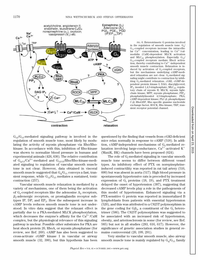
G12/G13-mediated signaling pathway is involved in theregulation of smooth muscle tone, most likely by modu-lating the activity of myosin phosphatase via Rho/Rho-kinase. In accordance with this, inhibition of Rho-kinasewas shown to normalize blood pressure in humans andexperimental animals (426, 636). The relative contributionof Gq/11/Ca2�-mediated and G12/13/Rho/Rho-kinase-medi-ated signaling to regulation of vascular smooth muscletone is not clear. However, data obtained in visceralsmooth muscle suggested that Gq/G11 conveys a fast, tran-sient response, while G12/G13 mediates a sustained, toniccontraction (257).
Vascular smooth muscle relaxation is mediated by avariety of mechanisms, one of them being the activationof Gs-coupled receptors like the adenosine A2 receptors,�2-adrenergic receptors, or prostaglandin receptor sub-types IP, DP, and EP2. How the subsequent increase incAMP levels reduces smooth muscle tone is not under-stood. In vitro data suggest that the relaxant effect ispartially due to a PKA-mediated MLCK phosphorylation,which decreases the enzyme’s affinity for the Ca2�/CaMcomplex, but the physiological relevance of this signalingpathway is unclear. Possible other substrates for PKA areheat shock protein 20, RhoA, or myosin phosphatase (forreview, see Ref. 269). cAMP has also been suggested tocross-activate cGMP kinase I in vascular or airwaysmooth muscle (32, 390), but this hypothesis has been
questioned by the finding that vessels from cGKI-deficientmice relax normally in response to cAMP (518). In addi-tion, cAMP-independent mechanisms of Gs-mediated re-laxation involving large-conductance, Ca2�-activated K�
(MaxiK, BK) channels have been proposed (624).The role of Gi-mediated signaling in vascular smooth
muscle tone seems to differ between different vesseltypes. An inhibitory effect of PTX on norepinephrine-induced contractility was reported in rat tail artery (516,600) but was absent in aorta (517). High blood pressure inspontaneously hypertensive rats is preceded by increasedexpression of Gi proteins (18, 19), and PTX treatmentdelayed the onset of hypertension (387), suggesting thatdecreased cAMP levels play a role in the pathogenesis ofthis model of hypertension. Enhanced signaling via aPTX-sensitive G protein was reported in immortalized Blymphoblasts from patients with essential hypertension(520), and this was attributed to a C825T polymorphism inthe gene coding for G�3, a constituent of the Gi hetero-trimer (586). The C825T polymorphism was suggested tobe associated with an increased risk of hypertension,obesity, and arteriosclerosis in some (for review, see Ref.585) but not in all studies (283, 616, 617). However, thesignificance of genetic association studies in general re-mains controversial (20, 199, 291).
Very similar to vascular smooth muscle, also airwaysmooth muscle tone is mainly regulated by Gq/G11 family
FIG. 6. Heterotrimeric G proteins involvedin the regulation of smooth muscle tone. Gq/G11-coupled receptors increase the intracellu-lar Ca2� concentration, leading to Ca2�/cal-modulin (CaM)-dependent MLCK activationand MLC20 phosphorylation. Especially G12/G13-coupled receptors mediate RhoA activa-tion, thereby contributing to Ca2�-independentsmooth muscle contraction. Relaxation is in-duced by activation of Gs-coupled receptors,but the mechanisms underlying cAMP-medi-ated relaxation are not clear. Gi-mediated sig-naling might contribute to contraction by inhib-iting Gs-mediated relaxation. cGKI, cGMP-de-pendent protein kinase I; DAG, diacylglycerol;IP3, inositol 1,4,5-trisphosphate; MLC20, regula-tory chain of myosin II; MLCK, myosin light-chain kinase; MPP, myosin phosphatase; PIP2,phosphatidylinositol 4,5-bisphosphate; PKA,cAMP-dependent kinase; PLC-�, phospholipaseC-�; RhoGEF, Rho specific guanine nucleotideexchange factor; ROCK, Rho kinase; TRP, tran-sient receptor potential channel.
1170 NINA WETTSCHURECK AND STEFAN OFFERMANNS
Physiol Rev • VOL 85 • OCTOBER 2005 • www.prv.org
on April 23, 2015
Dow
nloaded from

G proteins, which mediate bronchoconstriction, and Gs
family G proteins, which mediate bronchorelaxation. Ace-tylcholine released from postganglionic parasympatheticnerves controls resting tone mainly via the Gq/G11-cou-pled M3 receptor subtype (90), but also other Gq/G11-coupled receptors are expressed in airway smooth mus-cles, like the H1 histamine receptor (133, 222), the leuko-triene CysLT1 receptor (314), the B2 bradykinin receptor(421, 630), the ETB endothelin receptor (216, 241, 441),and others. Airway hyperreactivity in the A/J mouse strainwas suggested to be due to enhanced agonist affinity andincreased G protein coupling efficiency of the M3 musca-rinic receptor (205), and Gq protein was shown to beupregulated in antigen-induced airway hyperresponsiverats (106). Mice lacking the �-subunit of Gq showed im-paired metacholine-induced airway responses and lackedthe typical increase in metacholine sensitivity after aller-gen sensitization and reexposition (55). Not much isknown about the role of G�12 and G�13 in airway smoothmuscle tone regulation. The fact that repetitive antigenchallenge significantly increases the expression of theseproteins in airway smooth muscle suggests a role in aller-gic asthma (105, 108), but direct evidence for an involve-ment of G12/G13 is still lacking. Gs-coupled receptors playan important role in the relaxation of contracted airwaysmooth muscle, most prominently the �2-adrenergic re-ceptor, but also the prostaglandin E2 receptor EP2 (512)or the prostacyclin IP receptor (41) (for review, see Ref.628). The Gi family of G proteins contributes to the reg-ulation of airway smooth muscle contractility mainly byinhibiting the relaxant effects of Gs. The inhibitory effectof PTX on acetylcholine-induced bronchoconstriction isnegligible in normal rats, but significant in rats sufferingfrom antigen-induced airway hyperresponsiveness (107).In these mice, G�i3 protein is upregulated in bronchialsmooth muscle cells, suggesting that the relative contri-
bution of Gi-mediated constriction is increased in antigen-challenged airway smooth muscle (107).
D. Platelet Activation
Platelets are small cell fragments that circulate in theblood and adhere at places of vascular injury to the vesselwall where they become activated resulting in the forma-tion of a platelet plug that is responsible for primaryhemostasis. Platelets can also become activated underpathological conditions, e.g., on ruptured atheroscleroticplaques leading to arterial thrombosis. Platelet adhesionand activation is initiated by their interaction with adhe-sive macromolecules like collagen and von Willebrandfactor (vWF) at the subendothelial surface (303, 554).While collagen is able to induce firm adhesion of plateletsto the subendothelium (666), the recruitment of addi-tional platelets to the growing platelet plaque requires thelocal accumulation of diffusible mediators that are pro-duced or released once platelet adhesion has been initi-ated, and some level of activation through platelet adhe-sion receptors has occurred (3). These mediators includeADP/ATP and thromboxane A2 (TxA2), which are se-creted or released from activated platelets as well asthrombin, which is produced on the surface of activatedplatelets. These platelet stimuli have in common theiraction through G protein-coupled receptors. While ADPinduces the activation of Gq and Gi via P2Y1 and P2Y12
receptors (197, 354), the activated TxA2 receptor (TP)couples to Gq and G12/G13 (337, 492) (Fig. 7). G protein-coupled protease-activated receptors (PARs) that are ac-tivated by thrombin are functionally coupled to Gq, G12/G13, and in some cases to Gi (121). In response to thesesecondary mediators of platelet activation, platelets im-mediately undergo a shape change reaction during whichthey become spherical and extrude pseudopodia-like
FIG. 7. Role of heterotrimeric G pro-teins in mediating platelet activation bysoluble mediators like ADP, thrombox-ane A2 (TxA2), thrombin, and epineph-rine. Major roles are played by the G pro-teins Gq, G13, and Gi which couple recep-tors to the indicated effector molecules.The subsequent signaling processes even-tually lead to platelet responses likeshape change, degranulation, and aggre-gation (for details, see text). TP, TxA2
receptor; PAR, protease-activated recep-tor; P2Y1/P2Y12, purinergic receptors;�2A, �2A-adrenergic receptor; RhoGEF,Rho guanine nucleotide exchange factor;PLC-�2/3, phospholipase C-�2/3; PI-3-K,phosphoinositide-3-kinase; PIP2, phos-phatidylinositol 4,5-bisphosphate; IP3,inositol 1,4,5-trisphosphate; DAG, diacyl-glycerol; PKC, protein kinase C; PIP3,phosphatidylinositol 3,4,5-trisphosphate.
CELLULAR FUNCTIONS OF G PROTEINS 1171
Physiol Rev • VOL 85 • OCTOBER 2005 • www.prv.org
on April 23, 2015
Dow
nloaded from

structures. In addition, the glycoprotein IIb/IIIa (integrin�IIb�3) undergoes a conformational change resulting inbinding of fibrinogen/vWF and subsequent platelet aggre-gation. Finally, the formation and release of TxA2, throm-bin, and ADP is further stimulated. Thus secondary me-diators increase through G protein-coupled receptorstheir own formation resulting in an amplification of theireffects, and eventually all G protein-mediated signalingpathways induced via these receptors become activated.The multiple positive feedback mechanisms operatingduring platelet activation have obscured the exact analy-sis of the roles individual G protein-mediated signalingpathways play during the platelet activation process.Progress has recently been made using genetic mousemodels in understanding the role of individual G protein-mediated signaling pathways during platelet activation.
The requirement of Gq-mediated signaling for ago-nist-induced platelet activation has been demonstrated bythe phenotype of G�q-deficient platelets, which fail toaggregate and to secrete in response to thrombin, ADP,and TxA2 due to a lack of agonist-induced phospholipaseC activation. This dramatic phenotype found in G�q-defi-cient platelets is due to the fact that platelets lack G�11
(313), which is in most other cells coexpressed with G�q
and can compensate G�q deficiency. Mice lacking G�q
have increased bleeding times and are protected againstcollagen/epinephrine-induced thromboembolism (494).Although Gq-mediated signaling appears to be absolutelyrequired for platelet activation, there is clear evidencethat also Gi type G proteins need to be activated to inducefull activation of integrin �IIb�3. In mice lacking the�-subunit of Gi2, the response of ADP that acts throughthe Gi-coupled P2Y12 receptor is reduced (304). However,also the effects of mediators like thrombin and TxA2,which primarily signal through Gq and G12/G13 were foundto be inhibited in platelets lacking G�i2 (304, 712). Thissupports the view that platelet activation by thrombin andthromboxane A2 requires in part the action of secondarymediators like ADP, which are released after activation ofGq-mediated signaling pathways through TxA2 and throm-bin receptors. An important role of the Gi-mediated sig-naling pathway in platelet activation is also suggested bystudies in platelets lacking the Gq-coupled P2Y1 receptoror after pharmacological blockade of P2Y1 (170, 251, 310,379, 568). These platelets do not aggregate in response tolow and intermediate concentrations of ADP unless Gq-mediated signaling is induced via activation of anotherreceptor. Similarly, platelets lacking P2Y12 or in whichP2Y12 was pharmacologically blocked did not aggregate inresponse to ADP unless the Gi-mediated pathway wasactivated via a different receptor (181, 568). Thus there isclear evidence that Gq and Gi synergize to induce plateletactivation. It is currently not clear how Gi contributes tointegrin �IIb�3 activation in platelets, but a decrease incAMP levels is unlikely to be involved (129, 529, 569, 712).
Another member of the Gi family of heterotrimeric Gproteins, Gz, has been implicated in platelet activationinduced by epinephrine acting on �2-adrenergic recep-tors. In contrast to ADP, TxA2, and thrombin, epinephrineis alone not able to fully activate mouse platelets. How-ever, it is able to potentiate the effect of other plateletstimuli. In G�z-deficient platelets, the inhibitory effect ofepinephrine on adenylyl cyclase and epinephrine-potenti-ating effects were strongly impaired while the effects ofother platelet activators appear to be unaffected (713).
Despite the central role of Gq in platelet activation, itwas recently demonstrated that induction of Gi- and G12/G13-mediated signaling pathways is sufficient to induceintegrin �IIb�3 activation (149, 483). Interestingly, inG�13-deficient platelets, but not in G�12-deficient plate-lets, the potency of various stimuli including TxA2, throm-bin, and collagen to induce platelet shape change andaggregation is markedly reduced (455). These defects areaccompanied by a defect in the activation of RhoA and adelayed phosphorylation of the myosin light chain as wellas by an inability to form stable platelet thrombi underhigh sheer stress conditions (455). In addition, mice car-rying platelets that lack G�13 have an increased bleedingtime and are protected against the formation of arterialthrombi induced in a carotid artery thrombosis model(455). These data indicate that in addition to Gq and Gi
also G13 is crucially involved in the signaling processesmediating platelet activation via G protein-coupled recep-tors both in hemostasis and thrombosis. These findingsalso indicated that G13-mediated signaling is not onlyinvolved in the response of platelets to relatively lowstimulus concentrations that induce platelet shapechange but is also required for normal responsiveness ofplatelets at higher stimulus concentrations. A reducedpotency of platelet activators in the absence of G13-medi-ated signaling becomes in particular limiting under highflow conditions that lead to a rapid clearance of solublestimuli from the site of platelet activation and formationof mediators. In addition, the defective activation ofRhoA-mediated signaling in the absence of G13 appears tocontribute to the observed defect in the stabilization ofplatelet aggregates under high sheer stress ex vivo as wellas in vivo. In fact, RhoA-mediated signaling has beensuggested to be required for platelet aggregation underhigh sheer conditions as well as for the irreversible ag-gregation of platelets in suspension (450, 570).
These studies have clearly shown that three G pro-teins are major mediators of platelet activation via Gprotein-coupled receptors: Gq, Gi2, and G13. However,even in the absence of either Gq, Gi2, or G13 some plateletactivation can still be induced, while in the absence ofboth G�q and G�13, platelets are unresponsive to throm-bin, TxA2, or ADP. This indicates that the activation ofGi-mediated signaling alone is not sufficient to induce anyplatelet activation (456). The optimal activation of plate-
1172 NINA WETTSCHURECK AND STEFAN OFFERMANNS
Physiol Rev • VOL 85 • OCTOBER 2005 • www.prv.org
on April 23, 2015
Dow
nloaded from

lets under physiological and pathological conditions ob-viously requires the parallel signaling through several het-erotrimeric G proteins.
III. ENDOCRINE SYSTEM AND METABOLISM
The endocrine system consists of a variety of glandsand other structures that produce, store, and secrete hor-mones directly into the systemic circulation, thereby con-trolling electrolyte and water homeostasis, metabolism,growth, reproduction, etc. GPCRs contribute to endocrinefunctions in a twofold way: 1) by mediating hormonal endorgan effects and 2) by controlling hormone secretionitself. Hormone secretion, as well as secretion from neu-ronal or exocrine cells, typically involves elevation ofcytosolic Ca2� and/or cAMP (for review, see Refs. 160,738). In most secretory cells, Ca2� influx through voltage-operated Ca2� channels is the dominant mode of regula-tion, like in adrenal chromaffin cells (160), while in othercells, such as anterior pituitary gonadotropes, Ca2� mo-bilization from internal stores is the critical step (633). Inyet another endocrine cell, such as lactotroph cells, bothincreased intracellular Ca2� levels and cAMP productioncontribute to secretion (130, 186, 620). Accordingly, withrare exceptions, activation of Gs and/or Gq/G11 family Gproteins enhances secretion regardless of the endocrinecell type involved.
A. Hypothalamo-Pituitary System
Hormone release from the anterior pituitary is tightlycontrolled by hypothalamic releasing hormones and re-lease inhibiting factors, all of which act through GPCRs.The receptors for corticotropin-releasing hormone (263)and growth hormone-releasing hormone (GHRH) (588)primarily act through Gs, while receptors for gonado-tropin-releasing hormone (445), thyrotropin-releasinghormone (211, 720), and the many prolactin-releasing fac-tors (186) mainly act through Gq/G11 family G proteins,and only partly through Gs. In addition to their secreta-gogue effects, hypothalamic releasing hormones regulatehormone synthesis and cell proliferation (81, 430, 531,583, 587, 647). Anterior pituitary secretion and prolifera-tion is not only stimulated by the classical hypothalamicreleasing hormones, but also by a variety of other factors,such as the gastrointestinal peptide hormone ghrelin,which enhances growth hormone (GH) secretion viathe predominantly Gq/G11-coupled growth hormonesecretagogue receptor GHS-R (343, 588), or members ofthe pituitary adenylate cyclase-activating polypeptide(PACAP)/glucagon superfamily, which exert secreta-gogue effects on a variety of pituitary cell types via theirGs-coupled receptors (579). The in vivo relevance of Gs
family G proteins in anterior pituitary function was stud-
ied in mice and in patients with inactivating or activatingGs mutants. Somatotroph-specific overexpression of chol-era toxin, which irreversibly activates Gs by ADP ribosy-lation, caused somatotroph hyperplasia, increased GHlevels and gigantism in mice (82). In humans, activatingmutations of GNAS can be found in �40% of GH produc-ing pituitary tumors (363, 406), as well as in 10% ofnonfunctioning pituitary adenomas (406, 632, 685). Theseactivating mutations of GNAS encode substitutions ofeither Arg-201 or Gln-227, two residues that are critical forthe GTPase reaction (187, 223, 363, 406). In GH-secretingtumors, the mutation is almost always in the maternalallele, presumably because G�s is mainly expressed fromthe maternal allele (“paternally imprinted”) in pituitarycells (242). Activating GNAS mutations were also, thoughrarely, found in corticotroph (541, 686), but not in thyro-troph tumors (147, 406). Such activating somatic GNAS
mutations are not necessarily restricted to the pituitary,but are often part of the McCune-Albright syndrome,which is defined by the trias fibrous dysplasia of bone,cafe-au-lait skin pigmentation, and endocrine hyperfunc-tions of variable degree (for review, see Refs. 599, 669).Endocrine hyperfunction is due to constitutive activationof Gs signaling in other endocrine glands, leading to ad-renal hyperplasia with Cushing syndrome (60, 182), pre-cocious puberty (138, 574), or hyperthyroidism (see sect.IIIC). In melanocytes, increased Gs activity mimics theactivity of melanocyte stimulating hormone, leading totypical cafe-au-lait hyperpigmentation (334).
Heterozygous inactivating GNAS mutations result inAlbright hereditary osteodystrophy (AHO), a congenitaldisorder characterized by obesity, short stature, brachy-dactyly, subcutaneous ossifications, and neurobehavioraldeficits of variable severity (for review, see Refs. 13, 366,599, 669). In addition to these defects, patients with ma-ternally inherited mutations show multihormone resis-tance (termed pseudohypoparathyroidism type Ia,PHP1a) in tissues with a paternally imprinted GNAS al-lele, such as proximal tubules of the kidney, thyroid, orovaries (209, 242, 414, 723). In these tissues, the effects ofGs-coupled hormone receptors, like those for parathyroidhormone, thyroid stimulating hormones, or the gonado-tropins, are impaired. Clinically, this results in variabledegrees of hypocalcemia and hyperphosphatemia, hypo-thyroidism (see also sect. IIIC), and delayed or incompletesexual development and reproductive dysfunction inwomen (13, 366, 599, 669). These abnormalities of thereproductive system are easily explained by malfunctionof receptors for follicle-stimulating hormone and luteiniz-ing hormone. In addition, at least in mice, the Gs-coupledorphan receptor GPR3 is crucially involved in the main-tenance of meiotic arrest in oocytes (432, 433).
The phenotype of humans heterozygous for an in-activating GNAS mutation is partly reproduced in micecarrying a targeted disruption of Gnas exon 2. In these
CELLULAR FUNCTIONS OF G PROTEINS 1173
Physiol Rev • VOL 85 • OCTOBER 2005 • www.prv.org
on April 23, 2015
Dow
nloaded from

animals, PTH resistance was only found if the mutationwas maternally inherited, and only these animalsshowed reduced G�s expression in the renal cortex(723). In humans, renal PTH resistance without Al-bright hereditary osteodystrophy (PHPIb) can also bedue to other GNAS mutations, such as a mutant whichresults in a biallelic paternal imprinting phenotype(395), or a mutant unable to interact with the PTHreceptor (697). Yet another GNAS mutation causes im-paired signaling via the PTH and TSH receptors, butenhanced signaling via the likewise Gs-coupled recep-tor for luteinizing hormone, leading to enhanced tes-tosterone production. This paradoxical combination ofgain and loss of function is explained by the fact thatthe underlying GNAS mutation results in a constitu-tively active form of G�s which, however, is tempera-ture sensitive. The mutant is stable only at the rela-tively low temperature in the testis, but rapidly de-graded at 37°C, leading to G�s deficiency (287). Withrespect to pituitary function, patients with inactivatingGNAS mutations show variable degrees of GHRH resis-tance (415), GH deficiency (210), or hypoprolactinemia(88). In accordance with the important role of Gs familyG proteins in lactotrophs and somatotrophs, hypotha-lamic inhibiting hormones, like dopamine or somatosta-tin, act through Gi-coupled receptors (311, 536).
Releasing hormone secretion itself is influenced byGPCRs, and several former orphan receptors were re-cently shown to positively regulate releasing hormonesecretion. Kisspeptins for example, a family of peptidesderived from the metastasis suppressor gene Kiss-1, wereshown to enhance hypothalamic gonadotropin-releasinghormone secretion via the GPR54 receptor (137, 220, 472),and genetic inactivation of GPR54 in mice or mutation inhumans causes hypogonadotropic hypogonadism (137,196, 220, 575). The peptide hormone ghrelin induces pitu-itary growth hormone release not only directly via activa-tion of GHS-R on somatotroph cells, but also acts as areleasing factor for hypothalamic GHRH (343). BothGPR54 and GHS-R are known to activate Gq/G11 family Gproteins (343, 345), suggesting that releasing hormonerelease is controlled by the same mechanisms as pituitaryhormone release. In line with this notion, mice lackingboth G�q alleles and one G�11 allele selectively in thenervous system show severe somatotroph hypoplasiawith dwarfism due to reduced hypothalamic GHRH pro-duction, which is probably secondary to impaired GHS-Rsignaling (676).
B. Pancreatic �-Cells
The tight regulation of blood glucose levels is mainlyachieved by the on-demand release of insulin from pan-creatic �-cells. High glucose levels result in enhanced
intracellular glucose metabolism with ATP accumulationand consecutive closure of ATP-sensitive K� channels,leading to the opening of voltage-operated Ca2� channelsand Ca2�-mediated insulin exocytosis (27, 119). In addi-tion to the ATP-dependent mechanism of insulin release,several GPCRs have been shown to either amplify or toinhibit glucose-induced insulin release (for review, seeRefs. 161, 364, 558), and these receptors and their respec-tive ligands play an important role in the regulation ofislet function by, e.g., the autonomous system (for review,see Ref. 7). Neuropeptides and hormones that potentiateinsulin secretion mainly act though Gs-coupled receptors,like glucose-dependent insulinotropic polypeptide, secre-tin, cholecystokinin, PACAP, glucagon, vasoactive intes-tinal polypeptide, or glucagon-like peptide-1 (GLP-1) (forreview, see Refs. 161, 418, 542). The potentiating effect ofGs on glucose-induced insulin release (576, 608, 609)might either be mediated by phosphorylation of voltage-operated Ca2� channels (295) or through the opening ofnonselective cation channels (275). Transgenic expres-sion of a constitutively active G�s mutant in mouse �-cellscaused increased islet cAMP production and insulin se-cretion, but these changes were only detectable in thepresence of phosphodiesterase inhibitors, suggesting thatincreased G�s activity is normally compensated by up-regulation of cAMP degrading enzymes like phosphodies-terases (408). Conversely, activation of receptors coupledto Gi or Go, like the �2-adrenergic receptor or receptorsfor somatostatin, neuropeptide Y, prostaglandin E2, orgalanin, inhibits insulin secretion in a PTX-sensitive man-ner (319, 328, 365, 514, 542).
Not only Gs family members, but also Gq/G11 family Gproteins, can mediate potentiation of glucose-induced in-sulin release. Acetylcholine released from postganglionicparasympathetic nerves or muscarinic agonists actthrough the Gq/G11-coupled M3 receptor (58, 157) to en-hance insulin release during the cephalic phase of insulinsecretion (8, 447, 691). This effect was shown to dependon PLC activation and consecutive inositol 1,4,5-trisphos-phate (IP3)-mediated intracellular Ca2� elevation (46, 469,726) and PKC activation (22). The exact pathways leadingto increased insulin secretion are not clear, but activationof L-type Ca2� channels (58), modulation of ATP-sensitiveK� channels (469), activation of CaM-kinase II (427, 537),or enhanced plasma membrane Na� permeability (254)have been suggested. In addition to acetylcholine, a vari-ety of other local mediators act through Gq/G11-coupledreceptors to enhance insulin release, like cholecystokininvia the CCK1 receptor (652), bombesin via the BB2 recep-tor (521, 646), arginine vasopressin via the V1b receptor(375, 501, 539), or endothelin via the ETA receptor (224).Fatty acids such as palmitate potentiate insulin secretionat high glucose levels independently of ATP formation(527, 663), and Ca2� influx via voltage-operated Ca2�
channels or intracellular Ca2� mobilization was suggested
1174 NINA WETTSCHURECK AND STEFAN OFFERMANNS
Physiol Rev • VOL 85 • OCTOBER 2005 • www.prv.org
on April 23, 2015
Dow
nloaded from

to mediate these effects. The former orphan GPCR GPR40was shown to mediate the effects of saturated and unsat-urated fatty acids (C�6) on intracellular Ca2� mobiliza-tion in pancreatic �-cells (70, 296, 346). These effectswere not PTX sensitive, suggesting that Gq/G11-mediatedintracellular Ca2� mobilization was involved (70, 296).Another recently deorphanized GPCR probably coupledto Gq/G11 is GPR120, which was suggested to be involvedin fatty acid-induced release of GLP-1 from intestinalcells, thereby contributing to GLP-1-mediated insulin re-lease (265).
C. Thyroid Gland/Parathyroid Gland
Thyroid stimulating hormone (TSH) regulates thy-roid cell proliferation as well as thyroid hormone synthe-sis and release through the G protein-coupled TSH recep-tor (508). In vitro, the TSH receptor was shown to coupleto all four G protein families (15, 16, 368), but the majorsignal transduction pathway in vivo seems to be the Gs/cAMP cascade, which was shown to activate iodide or-ganification, thyroid hormone production, secretion, andthyroid cell mitogenesis (153–155, 546). In TSH receptor-deficient mice, direct stimulation of adenylyl cyclase re-stores the ability to concentrate and organify iodide, sug-gesting that expression of the sodium-iodide symporter iscontrolled by G�s (417). In vitro, overexpression of con-stitutive active G�s in a thyroid cell line (462) or activa-tion of Gs by transgenic expression of cholera toxin in themouse thyroid (727) caused hyperplasia and increasedhormone secretion. In line with this, spontaneous activat-ing mutations of GNAS can cause thyroid cell hyperfunc-tion in humans, leading to hyperthyroidism, goiter, andbenign adenoma (175, 406, 424, 502). Malignant transfor-mation of thyroid cells has also been observed (165, 611,711), but seems to require additional mutational or epige-netic events (115). Much more frequent than activatingGNAS mutations are the activating mutations of the TSHreceptor itself (508), which mainly lead to constitutiveactivation of the Gs/cAMP cascade, or, in some cases, toactivation of both Gs- and Gq/G11-coupled pathways (48,643). Since the paternal GNAS allele is partly imprinted inthyroid cells (209, 394), inactivating GNAS mutants inher-ited from the maternal side cause TSH resistance withmoderately elevated TSH levels and low thyroid hormonelevels (171, 381, 382, 670, 719). Mild TSH resistance canalso be observed in patients with PHP1B due to a GNAS
mutation with paternal specific epigenotype of both exon1A regions (34, 394, 395). In addition to adenylyl cyclaseactivation, TSH stimulates PLC activity (344, 369, 644),but the TSH concentrations needed for PLC stimulationare 100-fold higher than those needed for adenylyl cyclasestimulation (643).
The parathyroid gland controls Ca2� homeostasisthrough parathyroid hormone (PTH), which enhances
Ca2� (re)absorption in gut and kidney, as well as Ca2�
release from bone. High extracellular Ca2� concentra-tions activate the G protein-coupled extracellular Ca2�
sensing receptor (CaR), leading to inhibition of PTH pro-duction and secretion in parathyroid cells (for review, seeRefs. 74, 268, 661). Activation of the CaR causes a PTX-insensitive (240) stimulation of PLC-� isoforms, with con-secutive increments in inositol phosphates, DAG, andintracellular Ca2� levels (73, 75, 478). In addition, anactivation of phospholipases A2 and D (333) as well as aPTX-sensitive suppression of cAMP formation was ob-served (98). These studies suggested that the CaR couplesboth to Gq/G11 and Gi/Go family G proteins, and thisnotion was supported by the finding that CaR activationinduced incorporation of radiolabeled GTP into G�q andG�i in Madin-Darby kidney (MDCK) cells, which endog-enously express low levels of the CaR (25). The fact thatincreased intracellular Ca2� levels result in decreased,not increased, hormone release is quite exceptional, andparathyroid cells are, besides renin-secreting juxtaglo-merular cells (572), the only endocrine cells showing suchinverse coupling. However, the molecular mechanism un-derlying Ca2�-mediated inhibition of PTH secretion is notunderstood (for review, see Refs. 74, 86, 268, 661).
D. Regulation of Carbohydrate and
Lipid Metabolism
Normal blood glucose levels are maintained both byregulating the activity of enzymes involved in carbohy-drate metabolism and by controlling glucose uptake intoperipheral tissues. Insulin is a major regulator of bothprocesses, but also a variety of GPCR agonists, like cat-echolamines and glucagon, contribute to glucose ho-meostasis. In hepatocytes, activation of Gs-coupled recep-tors like the �2-adrenergic receptor or glucagon receptorscauses PKA-mediated phosphorylation of key enzymeswhich regulate glycogen synthesis, glycogen breakdown,glycolysis, or gluconeogenesis (167, 168). Together, thesechanges lead to enhanced hepatic glucose release. Com-parable changes can be induced by activation of Gq/G11-coupled receptors like the �1-adrenergic receptor or re-ceptors for vasopressin and angiotensin, and these effectsare probably mediated by Ca2�/calmodulin-dependent al-teration of enzymatic activity (167, 168). In the periphery,glucose uptake into skeletal muscle and adipocytes ismediated by translocation of glucose transporter subtype4 (GLUT4) from intracellular vesicles to the plasma mem-brane (665). A variety of GPCRs have been demonstratedto modify insulin-induced GLUT4 translocation, and evena direct interaction between the insulin receptor and het-erotrimeric G proteins was suggested (412). HeterozygousGNAS-deficient mice show, in addition to other metabolicabnormalities (for effects on lipolysis, see below), an
CELLULAR FUNCTIONS OF G PROTEINS 1175
Physiol Rev • VOL 85 • OCTOBER 2005 • www.prv.org
on April 23, 2015
Dow
nloaded from

increased sensitivity towards insulin, which was attrib-uted to enhanced insulin-dependent glucose uptake intothe skeletal muscle (104, 721). In line with this, transgenicexpression of a constitutively active mutant of G�s
(G�sQ227L) in fat, liver, and skeletal muscle decreasedglucose tolerance (284), suggesting that G�s-mediated sig-naling negatively regulates insulin-induced GLUT4 trans-location. In contrast, Gi family G proteins seem to facili-tate insulin effects. Pretreatment of isolated adipocytesand soleus muscle with PTX results in reduced insulin-stimulated glucose uptake (111, 325), and mice in whichG�i2 was downregulated in liver and adipose tissue usingan antisense RNA approach show insulin resistance withhyperinsulinemia, decreased glucose tolerance, and insu-lin resistance (459–461). In the latter mice, both insulin-induced GLUT4 translocation to the plasma membraneand activation of glycogen synthase and antilipolytic me-diators were impaired (461). The decrease in G�i2 levelswas accompanied by an increase in protein tyrosine phos-phatase-1B (PTP-1B) activity, an enzyme known to de-phosphorylate phosphorylated tyrosine residues on theinsulin receptor and on insulin receptor substrate-1. Thissuggests that the inhibition of insulin signaling in micewith reduced G�i2 levels is due to disinhibition of PTP-1B(461). Mice expressing a constitutively active mutant ofG�i2 (G�i2Q205L) in fat, liver, and skeletal muscle dis-played reduced fasting blood glucose levels and increasedglucose tolerance (103). Adipocytes from these miceshowed enhanced insulin-induced glucose uptake andGLUT4 translocation, as well as increased PI 3-kinase andAkt activities (596). In addition, PTP-1B is suppressed inG�i2 overexpressing mice (626), and streptozotocin-in-duced diabetic changes are ameliorated (732). Up to nowit is not clear at which levels the insulin receptor-medi-ated pathway interacts with the Gi-mediated pathway. Itwas suggested that Gi/Go family G proteins physicallyinteract and are phosphorylated by the activated insulinreceptor (for review, see Ref. 510). In addition, Gi/Go
might be involved in insulin-mediated autophosphoryla-tion of the insulin receptor (351).
Data from 3T3 L1 adipocytes strongly point to aninvolvement of Gq/G11 family G proteins in basal andinsulin-induced GLUT4 translocation. Overexpression ofwild-type or constitutively active G�q increased basalGLUT4 translocation (290, 326), while microinjection ofG�q/G�11 antibodies or RGS2 protein inhibited insulin-induced GLUT4 translocation (290, 326). In line with thesefindings, inhibition of GRK2, a negative regulator of Gq/G11 signaling, increased insulin-stimulated GLUT4 trans-location, while adenovirus-mediated overexpression ofGRK2 reduced translocation as well as 2-deoxyglucoseuptake (637). The exact mechanisms underlying Gq/G11-mediated GLUT4 translocation are not fully understood.Several Gq/G11-coupled receptors are able to stimulateglucose uptake via GLUT4 translocation, such as recep-
tors for endothelin-1 (ET-1), norepinephrine, platelet-ac-tivating factor, or bradykinin (335, 336, 698). Microinjec-tion of an anti-G�q/G�11 antibody or of RGS2 proteincauses inhibition of ET-1-induced GLUT4 translocation(289). Of note, chronic ET-1 treatment inhibits insulin-stimulated glucose uptake and GLUT4 translocation in3T3-L1 adipocytes, and decreased tyrosine phosphoryla-tion of insulin receptor substrates was suggested to me-diate this heterologous desensitization (292). The Gq/G11-mediated effect on GLUT4 translocation was shown to bePI 3-kinase dependent in some (289, 290) but not in allstudies (59, 326). Recently, a role for the ADP ribosylationfactor 6 and the Ca2�-activated tyrosine kinase Pyk2 wassuggested (59, 371, 506).
Other examples for G protein-regulated metabolicprocesses are adipocyte lipolysis and lipogenesis. Activa-tion of Gs-coupled receptors like those for cat-echolamines, ACTH, glucagons, TSH, or PTH enhanceslipolysis via PKA-dependent phosphorylation of hormone-sensitive lipase (274, 401, 606). In adipocytes from pa-tients heterozygous for an inactivating GNAS mutation,the lipolytic effect in response to epinephrine is impaired,and this was suggested to contribute to obesity observedin AHO patients (87). Heterozygous disruption of Gnas
exon 2 in mice causes not only enhanced insulin sensitiv-ity (721), but also a variety of other metabolic defects thatdepend on the parental origin of the inherited mutation(722). While mice with a maternally inherited inactivatingmutation of the Gnas gene are obese and hypometabolic,paternally inherited Gnas mutations cause abnormal lean-ness with decreased serum, liver, and muscle triglycer-ides, and lipid oxidation in adipocytes is enhanced (104,722). Since these mice have increased urine norepineph-rine excretion, it was suggested that increased sympa-thetic stimulation of adipocytes caused the changes inlipid metabolism (104, 722). The reason for the restrictionto paternally inherited mutations is not completely clear.Deletion of Gnas exon 2 does not only affect the expres-sion of G�s, but also of alternative gene products likeXL�s (Fig. 3) (245, 667). An involvement of XL�s in thehypermetabolic phenotype seems likely for three reasons.First, XL�s is only expressed from the paternal allele(104) and is therefore well suited to explain a paternallyinherited phenotype. Second, XL�s-deficient mice show aphenotype similar to paternally exon 2-deficient mice(522). Third, mice with paternal inheritance of a Gnas
exon 1 deletion, which does not affect XL�s expression,do not show the respective phenotype (668). Interest-ingly, studies in XL�s-deficient mice suggest that G�s andXL�s can exert opposing effects on whole body metabo-lism (522, 668). In addition to Gs family G proteins, alsothe Gq/G11 family was implicated in the regulation oflipolysis. Expression of antisense RNA to G�q in liver andwhite fat caused hyperadiposity, which was suggested to
1176 NINA WETTSCHURECK AND STEFAN OFFERMANNS
Physiol Rev • VOL 85 • OCTOBER 2005 • www.prv.org
on April 23, 2015
Dow
nloaded from

be attributed to an impaired lipolytic response towards�1-adrenergic agonists (198).
Inhibition of adipocyte lipolysis is mediated by theinsulin receptor or by activation of Gi-coupled receptorslike the �2-adrenergic receptor, receptors for adenosineor prostaglandin (401) or the recently deorphanized re-ceptor for nicotinic acid, GPR109A (HM74a/PUMA-G)(634). G�i was suggested to be directly involved in theantilipolytic effect of insulin, since insulin-dependent in-hibition of lipolysis and activation of glucose oxidation inadipocytes were shown to be PTX sensitive (219).
IV. IMMUNE SYSTEM
A. Leukocyte Migration/Homing
Directed cell movement in response to an increasedconcentration of chemoattractant underlies the correcttargeting of leukocytes to lymphatic organs during anti-gen surveillance and also allows them to migrate to sitesof infection and/or inflammation (for review, see Refs.653, 694). Known lymphocyte chemoattractants either be-long to the large family of chemokines (355, 500) or to thelysophospholipid family, like sphingosine-1-phosphate(S1P) or lysophosphatidic acid (LPA) (123, 221, 282). Bothchemokine and lysophospholipid receptors are GPCRs.While chemokine receptors primarily act through Gi fam-ily G proteins (355), lysophospholipid receptors activateGi, G12/G13, and Gq/G11 family G proteins depending ontype and activation state of the respective cell (377, 584,612).
Inactivation of Gi family G proteins by PTX pretreat-ment strongly impairs lymphocyte migration in vitro (28,598) and causes defective homing to spleen, lymph nodes,and Peyer’s patches in vivo (31, 124, 597, 662), suggestingthat Gi family G proteins are involved in these processes. Inline with this, inactivation of Gi by transgenic expression ofthe S1 subunit of PTX in murine thymocytes resulted inaccumulation of mature T cells in the thymus, with greatlyreduced levels of T cells in peripheral lymphatic organs (91,92). To investigate the role of Gi-mediated signaling in moredetail, mouse lines carrying inactivating mutations of G�i
subtypes were generated. Both G�i2 and G�i3 are expressedin the murine thymus (91), but only inactivation of G�i2
mimicked the phenotype of PTX expressing mice with re-spect to thymic accumulation of mature T cells. NeitherG�i2- nor G�i3-deficient mice showed defects in T cell hom-ing to the periphery (552), suggesting that the homing de-fects induced by PTX probably result from the combinedinactivation of Gi2 and Gi3.
Despite the predominant role of Gi signaling in che-mokine-induced lymphocyte migration, an involvement ofGq/G11 family G proteins was suggested by a variety of invitro studies (11, 21, 353, 590, 716). In contrast to other
tissues, hematopoietic cells do not only express the�-subunits G�q and G�11, but also G�15, which corre-sponds to human G�16 (17, 683). Mice deficient for G�11,G�15, or both G�q and G�15 were normal under basalconditions and after antigenic challenge, and only a minorsignaling defect in response to complement C5a wasfound in G�15-deficient macrophages (132).
The G12/G13 effector RhoA was repeatedly shownto be involved in the regulation of lymphocyte adhesionand migration (367, 631), but direct evidence for aninvolvement of the G12/G13 family is still lacking. Indi-rect evidence for a role of G13 in lymphocyte migrationcomes from studies in Lsc-deficient mice. The murineRho-specific guanine nucleotide exchange factor Lsc(p115RhoGEF in humans) is expressed exclusively in he-matopoietic cells and couples G�13 to the activation ofRhoA (236). Lsc-deficient mice show impaired agonist-induced actin polymerization and motility, as well as ab-normal B-cell homing and altered T- and B-cell prolifera-tion (214). Defective migration was also observed in micelacking the proton-sensing receptor G2A (465), which wasshown to couple to G�13 (316). G2A-deficient macro-phages (659) and T cells (533) showed reduced migrationtowards lysophosphatidylcholine (LPC), while G2A over-expression in a macrophage cell line enhanced migrationtowards LPC (533, 715). How LPC effects are affected bythe absence of G2A is currently not clear (690). In thelatter cells, LPC-induced chemotaxis was inhibited byoverexpression of dominant negative mutants of G�q/G�11 or G�12/G�13, as well as expression of RGS domainsor GRK2 (specific for Gq/G11) or of p115RhoGEF (specificfor G12/G13), while PTX treatment was without effect(715).
Also neutrophils respond to a variety of chemoattrac-tants, such as N-formyl-Met-Leu-Phe (fMLP), C5a, plateletactivating factor (PAF), or the chemokine interleukin(IL)-8, with polarization and directed migration. This ef-fect was shown to be PTX sensitive (43, 217, 577, 598) andto be mainly mediated via ��-subunits (479, 480) (Fig. 8).Lentiviral-mediated knockdown of different G proteinsubunits in a macrophage cell line revealed that comple-ment C5a-induced migration critically depends on G�2,but not on G�1, G�i2, or G�i3 (286). Intracellular effectorsof G�� in neutrophils are the �-isoform of PI 3-kinase(PI3K�) and the �2- and �3-isoforms of phospholipase C(PLC-�2, -�3). While neutrophils from mice lackingPLC-�2 and -�3 show normal, or even enhanced, chemo-tactic responses (388), migration of PI3K�-deficient neu-trophils was severely impaired (266, 388, 566). Moreover,PI3K�-deficient neutrophils failed to accumulate at sitesof inflammation in a septic peritonitis model (266), sug-gesting that PI3K� mediates chemotactic responses bothin vitro and in vivo. In addition to PI3K�, RhoA activityseems to be required for fMLP-induced neutrophil chemo-kinesis and chemotaxis (review in Ref. 484), and espe-
CELLULAR FUNCTIONS OF G PROTEINS 1177
Physiol Rev • VOL 85 • OCTOBER 2005 • www.prv.org
on April 23, 2015
Dow
nloaded from

cially the deadhesion of the uropod was attributed toRhoA (12, 397). Interestingly, fMLP receptor-mediatedchemotaxis of HL60 cells was recently suggested to in-volve not only Gi, but also G12/G13 (702). In these cells,activation of Gi defined “frontness” by activating PI3K andRac, whereas activation of G12/G13 defined “backness” byinducing RhoA-dependent actomyosin interaction (702).
B. Immune Cell Effector Functions
Activation of immune cells by antigen contact ini-tiates complex signaling cascades leading to proliferation,differentiation, or initiation of effector functions like cy-tokine production, mediator release, phagocytosis, etc.Although these functions are not directly G protein me-diated, G proteins might have important modulatory roles(312, 400).
In neutrophils, chemoattractant-induced O2� forma-
tion is mediated via Gi-coupled receptors (39) and isstrongly impaired both in neutrophils from mice lackingPLC-�2 and -�3 (388, 695, 699) and in PI3K�-deficientneutrophils (266, 388, 566). PI3K-mediated formation ofphosphatidylinositol 3,4,5-trisphosphate (PIP3) was shownto activate the small GTPase Rac, which contributes toneutrophil migration by actin polymer formation in theleading edge (540, 653) and to O2
� formation by NADPH
oxidase activation (144). Recently, a new Rac GEF termedP-Rex1 was shown to mediate Rac activation in responseto PIP3 and G�� in neutrophils (672).
In lymphocytes, the balance between the TH1-drivencellular immunity and TH2-driven humoral response ismainly shaped by the cytokine pattern present during Thelper cell activation. PTX has long been known to pro-mote TH1 responses (246, 270, 464), and this was sug-gested to be due to a negative regulation of IL-12 produc-tion by Gi, especially in dendritic cells (246, 279). Micelacking G�i2 develop a diffuse inflammatory colitis resem-bling ulcerative colitis in humans (277, 552), and adeno-carcinomas secondary to chronic inflammation were of-ten observed (552). In these mice, levels of proinflamma-tory TH1-type cytokines and of IL-12 were increased (277),and these changes clearly preceded the onset of bowelinflammation (497, 498). In addition, antigen presentingcells like CD8a� dendritic cells showed a highly increasedbasal production of IL-12 (246), suggesting that colitis inthese mice is a TH1-driven disease and that production ofproinflammatory TH1 cytokines is constitutively sup-pressed through a Gi-mediated pathway. Accordingly, ac-tivation of Gs-mediated signaling opposes these effects.Cholera toxin, which activates Gs by ADP-ribosylation,can be used as an adjuvant to promote TH2 responses(419, 687, 707), and systemic administration of choleratoxin during induction of TH1-based autoimmune diseasescan shift the immune response to the nonpathogenic TH2phenotype (610). In general, the Gs family was suggestedto attenuate proinflammatory signaling, but direct evi-dence is lacking. In vitro, application of cAMP (62, 321,671) or activation of typically Gs-coupled receptors, likethose for vasoactive intestinal polypeptide or PACAP(135, 201), was shown to inhibit immune cell functions.Immune cells from mice lacking the Gs-coupled adeno-sine A2A receptor respond with enhanced cytokine tran-scription and NF�B activation to activation of Toll-likereceptors (404), and activation of Gs by cholera toxininhibits signaling in T cells (476, 595) or natural killer cells(678).
The relevance of Gq/G11 and G12/G13 family G pro-teins in lymphocyte activation and proliferation is un-clear. In vitro studies suggested an involvement of bothfamilies in the activation of Bruton’s tyrosine kinase, aprotein that is required for normal B-cell development andactivation (42, 309, 402). The Gq/G11 family, especiallyhuman G�16, has been implicated in T-cell activation in avariety of in vitro studies (392, 603, 734), but the physio-logical relevance of these findings is unclear. In vivo, noobvious immunological defects were observed inG�11
�/�, G�15�/�, or G�15
�/�;G�q�/� mice (132). How-
ever, after antigenic challenge, G�q-deficient mice showedimpaired eosinophil recruitment to the lung, probably dueto an impaired production of granulocyte macrophagecolony stimulating factor GM-CSF by resident airway leu-
FIG. 8. Gi family G proteins are centrally involved in neutrophilmigration and activation and mediate their effects via the �2- and�3-isoforms of phospholipase C (PLC-�2/�3), phosphoinositide 3-ki-nases (PI-3-K), or the RacGEF P-Rex1. Btk, Bruton’s kinase; DAG,diacylglycerol; IP3, inositol 1,4,5-trisphosphate; PIP2, phosphatidylinosi-tol 4,5-bisphosphate; PI(3,4,5)P3, phosphatidylinositol 3,4,5-trisphos-phate; PKC, protein kinase C.
1178 NINA WETTSCHURECK AND STEFAN OFFERMANNS
Physiol Rev • VOL 85 • OCTOBER 2005 • www.prv.org
on April 23, 2015
Dow
nloaded from

kocytes (56). Indirect evidence for a role of Gq/G11 and/orG12/G13 in T-cell activation comes from RGS2-deficientmice, which show impaired T-cell proliferation and IL-2production after T-cell receptor stimulation, as well asdefective antiviral immunity in vivo (499). Inactivation ofthe murine TxA2 receptor, which is typically coupled toGq/G11 and/or G12/G13 family G proteins (337, 492), causeda lymphoproliferative syndrome due to prolongedT-cell/DC interaction (317). Genetic inactivation of theproton-sensitive G2A receptor (465), which was shown toactivate G12/G13, causes a late-onset autoimmune syn-drome in mice (372), suggesting that these G proteins maybe involved in the regulation of lymphocyte activation.
V. NERVOUS SYSTEM
G proteins play multiple roles in the nervous systemas most neurotransmitters also activate G protein-cou-pled metabotropic receptors to modulate neuronal activ-ity. In contrast to ionotropic receptors, GPCRs that arepresent pre- and postsynaptically mediate comparativelyslow responses. Only a few well-studied examples aredescribed below.
A. Inhibitory Modulation of Synaptic Transmission
The regulation of neurotransmitter release at presyn-aptic terminals is an important mechanism underlying themodulation of synaptic transmission in the nervous sys-tem. Inhibitory regulation of neurotransmitter release ismediated by various GPCRs like �2-adrenoceptors, �- and�-opioid receptors, GABAB receptors, adenosine A1, orendocannabinoid CB1-receptors. These receptors have incommon that they couple to G proteins of the Gi/Go
family. A major mechanism by which these G proteinsmediate the inhibition of transmitter release is the inhib-itory modulation of the action potential-evoked Ca2� en-try to the presynaptic terminal which is required to triggerneurotransmitter release. N- and P/Q-type calcium chan-nels that are concentrated at nerve terminals as well asR-type calcium channels have been shown to be inhibitedvia Gi/Go-coupled receptors (145). This inhibition is due tothe interaction of the G protein ��-complex with the�1-subunit of Cav2.1–2.3 (89, 420). The �-subunit of thechannel as well as strong depolarization can reduce thisinhibition (61, 439). Most G�� combinations are similarlyeffective in inhibiting channels of the Cav2 family (202,288, 557). There is some evidence that ��-mediated inhi-bition of Cav2 channels is not the only mechanismthrough which GPCRs mediate inhibition of neurotrans-mitter release (449). Also, G protein-coupled inwardlyrectifying K� channels (GIRKs) are localized at presynap-tic terminals; however, their physiological role in regula-tion of transmitter release from presynaptic terminals is
less clear (156, 524). GIRKs are well-established effectorsfor G protein ��-subunits (113, 356, 398, 718), and mostG�� combinations appear to be similarly effective in ac-tivating GIRKs (681, 708). There is also increasing evi-dence that the G��-mediated inhibition of neurotransmit-ter release at the presynaptic terminus involves mecha-nisms downstream of the regulation of Ca2� (51). Thismay involve the direct interaction between G�� and thecore vesicle fusion machinery as suggested by the obser-vation that G�� can directly bind to SNARE proteins likesyntaxin and SNAP25 as well as cysteine string protein(CSP) (50, 51, 305, 410). Evidence exists that presynapticCa2� channels, syntaxin1, and the �-subunit of Go arecomponents of a functional complex at the presynapticnerve terminal release site (384, 602).
Both Gi-type G proteins as well as Go are highlyabundant in the nervous system. Mice lacking the �-sub-unit of Go are smaller and weaker than their littermatesand have a greatly reduced life expectancy (308, 639). Inaddition, these animals have tremors and occasional sei-zures, and they show an increased motor activity with anextreme turning behavior. G�o-deficient mice have alsobeen shown to be hyperalgesic (308). When neuronal cellsfrom G�o-deficient mice were analyzed by electrophysio-logical methods for the regulation of GIRKs and voltage-dependent Ca2� channels through GPCRs, it was foundthat the recovery kinetics after agonist washout weremuch slower in the absence of G�o. However, currentmodulation via various receptors was as effectively as inwild-type cells (225, 308). This indicates that other Gproteins especially Gi-type G proteins that are activatedby the same receptors can compensate for Go deficiency.
B. Modulation of Synaptic Transmission by the
Gq/G11-Mediated Signaling Pathway
In the nervous system, the G proteins Gq and G11 arewidely expressed (622), and they are involved in multiplepathways that modulate neuronal function. The modula-tion of synaptic transmission has best been described atthe parallel fiber (PF)-Purkinje cell (PC) synapse in thecerebellum. At the PF-PC synapse, Gq/G11 mediate theeffects of metabotropic glutamate group 1 receptors(mGluR1). The Gq/G11-mediated IP3-dependent transientincrease in the dendritic Ca2� concentration, which doesnot require changes in the membrane potential (178, 621),is important for the induction of long-term depression(LTD) at the PF-PC synapse (452). LTD in turn is believedto be one of the cellular mechanisms underlying cerebel-lar motor learning (66). Interestingly, while G�q-deficientmice develop an ataxia with clear signs of motor coordi-nation deficits, G�11-deficient animals show only verysubtle defects in motor coordination (237, 489). A detailedcomparison of both mouse lines showed that Ca2� re-
CELLULAR FUNCTIONS OF G PROTEINS 1179
Physiol Rev • VOL 85 • OCTOBER 2005 • www.prv.org
on April 23, 2015
Dow
nloaded from

sponses to mGluR1 activation were absent in G�q-defi-cient mice, whereas they are indistinguishable betweenwild-type and G�11-deficient animals (237). However, syn-aptically evoked LTD was decreased in G�11-deficientanimals, but to a lesser extent than in G�q-deficient mice.The predominant role of Gq in Purkinje cells can beexplained by the higher expression compared with G�q
(489). In fact, quantitative single-cell RT-PCR analysisshowed that Purkinje cells express at least 10-fold moreG�q than G�11 (237).
Lack of G�q also results in a defect in the regressionof supernumerary climbing fibers innervating Purkinjecells in the third postnatal week. This process is believedto be due to a defect in the modulation of the PF-PCsynapse. Similar cerebellar phenotypes as in G�q-deficientmice have been described in mice lacking the mGluR1 (9,323) as well as in mice lacking the �4-isoform of PLC,which is predominantly expressed in the rostral cerebel-lum (324, 453). Interestingly mGluR1, G�q, and PLC-�4can be found colocalized in dendritic spines of PCs (324,622, 664), suggesting that they are components of a sig-naling cascade involved in the modulation of the PF-PCsynapse.
Interestingly, hippocampal synaptic plasticity isequally impaired in G�q- and G�11-deficient mice (451).However, mGluR-dependent long-term depression in thehippocampal CA1-region was absent in G�q-deficient micebut appeared to be unaffected in mice lacking G�11 (340).Similarly, suppression of slow afterhyperpolarizations viamuscarinic and metabotropic glutamate receptors wasnearly abolished in G�q-deficient mice but was unchangedin mice lacking G�11 (350).
Besides the voltage-dependent, G protein-mediatedinhibition of calcium channels via G�� (see above), func-tional studies in calyx-type nerve terminals and in sym-pathetic neurons have identified a voltage-insensitive in-hibition of calcium currents via metabotropic receptorsthat is believed to involve Gq/G11 (322, 449). How Gq/G11
activation can lead to inhibition of voltage-dependent cal-cium channels is not clear. However, recent evidence sug-gests that the depletion of phosphatidylinositol 4,5-bisphos-phate (PIP2), the substrate of PLC plays a role (200).
There is also evidence that activation of Gq/G11-mediated signaling on the postsynaptic site is involvedin the induction of retrograde signaling via the endo-cannabinoid system. Gq/G11-coupled receptors likegroup I mGluRs as well as muscarinic M1 receptors canlead to the activation of the retrogradely acting canna-binoid system by stimulating the formation of endocan-nabinoids (136, 189, 380, 409).
C. Roles of Gz and Golf in the Nervous System
Gs is the ubiquitous G protein that couples recep-tors in a stimulatory fashion to adenylyl cyclases. How-
ever, in a few tissues the G protein Golf is the predom-inant G protein that couples receptors to adenylyl cy-clase. Apart from the olfactory system (see below),G�olf expression levels exceed those of G�s also in afew other defined brain regions like the nucleus accum-bens, the olfactory tubercle, and the striatum (40, 120,737). In the striatum, Golf appears to be critically in-volved in dopamine (D1) and adenosine (A2) receptor-mediated effects (258, 737). Data from various labora-tories show that in the striatum, the dopamine D1 re-ceptor, G�olf and adenylyl cyclase type V as well as theG protein �7-subunit are coexpressed. Strikingly, micelacking either of these signaling components showclear signs of motor abnormalities (40, 298, 573, 703).Mice lacking adenylyl cyclase V show an attenuatedD1-receptor/Golf-mediated adenylyl cyclase activationin the striatum (298). G�7-deficient mice have stronglyreduced levels of striatal G�olf, and activation of adeny-lyl cyclase via dopamine receptors is abolished in thestriatal cells (573). Thus, in rodents, striatal D1 recep-tors appear to activate adenylyl cyclase V through a Gprotein containing G�olf and �7. In humans, it has re-cently been shown that G�olf expression is markedlydiminished in the putamen of patients with Huntingtondisease, while the putamen of patients with Parkinsondisease showed significantly increased levels of G�olf
and G�7 (120).The G protein Gz is a member of the Gi/Go family. It
shares with other Gi/Go family members the ability toinhibit adenylyl cyclase (176, 267). It is expressed primar-ily in brain, retina, adrenal medulla, and platelets. G�z-deficient mice are viable and have no major phenotypicalabnormalities. However, G�z deficiency results in an ab-normal response to certain psychoactive drugs (253, 713).This includes a considerably pronounced cocaine-in-duced increase in locomotor activity as well as a reduc-tion in the short-term antinociceptive effects of morphine(713). In addition, G�z-deficient animals develop signifi-cantly increased tolerance to morphine (374). Interest-ingly, the behavioral effects of catecholamine reuptakeinhibitors that are used as antidepressant drugs wereabolished in mice lacking G�z (713). While mice lackingG�z clearly indicate that Gz plays a role in the nervoussystem and is involved in various drug responses, thefunction of Gz on a cellular level in the nervous systemremains unclear.
VI. SENSORY SYSTEMS
G protein-mediated signal transduction processesmediate the perception of many sensory stimuli. Odors,light, and especially sweet and bitter taste substances actdirectly on GPCRs that modulate the activity of primarysensory cells via often very specialized heterotrimeric Gproteins.
1180 NINA WETTSCHURECK AND STEFAN OFFERMANNS
Physiol Rev • VOL 85 • OCTOBER 2005 • www.prv.org
on April 23, 2015
Dow
nloaded from

A. Visual System
The human retina contains two types of photorecep-tor cells, rods and cones. While rods mediate mainlyachromatic night vision and contain one type of GPCR,rhodopsin, cones are responsible for chromatic day visionand contain three GPCRs (opsins), which are sensitive todifferent parts of the visible light spectrum. Rhodopsinand opsins are coupled to different but closely relatedheterotrimeric G proteins. Rod-transducin (Gt-r) andcone-transducin (Gt-c), which mediate the effects of rho-dopsins and opsins, respectively, couple these receptorsin a stimulatory manner to cGMP-phosphodiesterase(PDE) by binding and sequestering the inhibitory �-sub-unit of the retinal type 6 PDE (PDE6). Activation of PDElowers cytosolic cGMP levels leading to a decreased openprobability of cGMP-regulated cation channels in theplasma membrane, which eventually results in hyperpo-larization of the photoreceptor cells (24). In mice lackingG�t-r, the majority of retinal rods does not respond to lightany more, and these animals develop mild retinal degen-eration with increasing age (84). To ensure an adequatetime resolution of the light signal, the transducin-medi-ated signaling process initiated by light-induced receptoractivation requires an efficient termination mechanism. Atleast three proteins contribute to the rapid deactivation oftransducin by increasing its GTPase activity: RGS9 andG�5L, which form a complex, and the �-subunit of thetransducin effector cGMP-PDE (24). In mice lacking G�5,the levels of RGS9 and other G protein �-like (GGL)domain RGS proteins in various tissues including retinaare drastically reduced, suggesting that the formation ofthe RGS9 G�5 complex is required for expression andfunction of RGS9 (100). Animals lacking G�5 or RGS9show a strongly impaired termination of transducin-me-diated signaling (99, 352, 407).
The light-induced hyperpolarization of photoreceptorcells results in a decreased release of glutamate. Gluta-mate released from photoreceptor cells acts on twoclasses of second-order neurons in the retina, one thatdepolarizes in response to glutamate most likely via iono-tropic glutamate receptors (OFF bipolar cells) and an-other that hyperpolarizes (ON bipolar cells). ON bipolarcells are in the absence of light inhibited by glutamatereleased from rods and cones, and this effect is mediatedby the metabotropic glutamate receptor mGluR6. Light-induced hyperpolarization of rods results in decreasedglutamate release and disinhibition of ON bipolar cellsdue to a decreased activation of mGluR6. In mice lackingmGluR6 or the G�o splice variant G�o1, the modulation ofON bipolar cells in response to light is abolished (142, 143,425), indicating that Go is the principle mediator of glu-tamate-induced inhibition of ON bipolar cells, which oc-curs especially in the absence of light. The retina-specificRGS protein Ret-RGS1 is localized in the dendritic tips of
ON bipolar cells together with mGluR6 and G�o1 and mayplay a critical role in increasing the deactivation of Go1
resulting in an acceleration in the rising phase of the lightresponse of the ON bipolar cells. This mechanism hasbeen suggested to match the kinetics of ON bipolar cellactivation to that of the OFF bipolar cells that arisesdirectly from ligand-gated channel activation by gluta-mate (141).
B. Olfactory/Pheromone System
Chemosensation by the olfactory system is based onthe expression of a huge variety of GPCRs specifically inthe olfactory epithelium. Individual olfactory sensory neu-rons appear to often express only one olfactory receptor,and olfactory sensory neurons expressing one receptortype converge to the same subgroup of glomeruli in theolfactory bulb (457, 548). Rodents have �1,000 differentolfactory receptors, whereas the number in humans isconsiderably smaller and has been estimated to be �350(457). Despite this remarkable diversity on the level of thereceptor, the olfactory GPCRs appear to employ the sameG protein-mediated signal transduction pathway in olfac-tory sensory neurons. The G protein Golf is centrallyinvolved in signaling by olfactory receptors in response toodorant stimuli, and G�olf-deficient mice are anosmic ex-hibiting dramatically reduced electrophysiological re-sponses to all odors tested (40). Golf, a G protein relatedto Gs, couples olfactory receptors in the cilia to adenylylcyclase III, resulting in the increased formation of cAMP.cAMP then activates a cyclic nucleotide-gated (CNG) cat-ion channel consisting of three different subunits,CNGA2, CNGA4, and CNGB1. The increase in cellularCa2� activates a Ca2�-activated Cl� channel that furtherdepolarizes the cell membrane (457, 548). Most G�olf-deficient pups die a few days after birth due to insufficientfeeding. Rare surviving animals exhibit inadequate mater-nal behavior resulting in the death of all pups born toG�olf-deficient mothers. This indicates that normal nurs-ing and mothering behavior in rodents is greatly depen-dent on an intact olfactory system (40). The fundamentalrole of this signaling pathway is underscored by the anos-mic phenotype found not only in mice lacking G�olf, butalso adenylyl cyclase (693) as well as in mice lacking thesubunits of the olfactory CNG channel (30, 76, 731).
A second olfactory system called the accessory ol-factory system or the vomeronasal system exists in mostmammals (152, 330). The peripheral sensory structure ofthis system, the vomeronasal organ, is localized at thebottom of the nasal cavity. The vomeronasal system re-sponds to pheromones that mediate defined effects onindividuals of the same species and modulate social, ag-gressive, reproductive, and sexual behaviors. In the vome-ronasal organ, two families of GPCRs, which in mice
CELLULAR FUNCTIONS OF G PROTEINS 1181
Physiol Rev • VOL 85 • OCTOBER 2005 • www.prv.org
on April 23, 2015
Dow
nloaded from

consist of �150 members each, have been identified. TheV1 receptor family is expressed in vomeronasal sensoryneurons together with the G protein Gi2, whereas the V2receptor family is expressed in a different population ofneurons that coexpresses the G protein Go (428). In hu-mans, no intact genes coding for V2 family receptors havebeen found, and most V1 receptor genes are pseudogenes,suggesting that the function of the vomeronasal organ ofrodents is not fully preserved in humans and higher pri-mates. Similar to the olfactory receptors, individual mem-bers of the V1 and V2 receptor families appear to bespecifically expressed in individual sensory neurons thatproject to a small group of glomeruli in defined regions ofthe accessory olfactory bulb. The striking coexpression ofV1 receptor family members and Gi2 and of V2 receptorfamily members and Go suggests that these G proteinsmediate the cellular effects induced by activation of therespective pheromone receptors. Consistent with this,G�i2-deficient mice have a reduced number of vomerona-sal sensory neurons that normally express G�i2 and showalterations in the behaviors for which an intact vomero-nasal organ is believed to be required like maternal ag-gressive behavior as well as aggressive behavior in malesexposed to an intruder (486). In mice lacking G�o apo-ptotic death of vomeronasal sensory cells which usuallyexpress G�o has been observed (623). Despite these be-havioral and anatomical abnormalities in G�i2- and G�o-deficient mice, a direct involvement of G�i2 in signaling ofV1 receptor-expressing cells and of G�o in V2 receptor-expressing cells has not been demonstrated so far. Thetransient receptor potential channel TRP2, which is ex-pressed in vomeronasal sensory neurons, has been iden-tified as a critical downstream mediator of the signaltransduction pathway in vomeronasal sensory neurons(383, 605).
C. Gustatory System
Unlike the olfactory system, chemosensation bythe gustatory system involves only in part G protein-mediated signal transduction mechanisms. The tastequalities sweet, bitter, and amino acid (umami) signalthrough GPCRs, whereas salty and sour tastants actdirectly on ion channels (391). During recent years, twofamilies of candidate mammalian taste receptors, T1receptors and T2 receptors, have been implicated insweet, umami, and bitter detection. The T2 receptorsare a group of �30 GPCRs that are specifically ex-pressed in taste buds of the tongue and that are linkedto bitter taste in mice and humans (6, 78, 94, 429). TheT1 receptor family consists of only three GPCRs andhas been shown to form heterodimers. T1R1 and T1R3form a receptor that responds to amino acids carryingthe umami taste (477), and T1R2 forms heterodimers
with T1R3 that functions as a sweet receptor (386, 477).Studies in knockout mice support a role of T1R2/T1R3as a sweet receptor and of T1R1/T1R3 as an umamireceptor. In addition, T1R3 may form homodimers thatcan also mediate some sweet tastes, and there is evi-dence that also a splice variant of the mGluR4 gluta-mate receptor may be involved in umami sensation (95,96, 127, 730). The signal transduction mechanisms usedby different G protein-coupled taste receptors are lessclear. Gustducin, a G protein mainly expressed in tastecells, is believed to be able to couple receptors tophosphodiesterase resulting in a decrease of cyclic nu-cleotide levels. Mice lacking the �-subunit of gustducinshow impaired responses to bitter, sweet, as well asumami tastes (247, 555, 692). However, the residualbitter and sweet taste responses in G�gust-deficientmice and the finding that expression of a dominantnegative mutant of �-gustducin reduces this respon-siveness even further indicates that �-gustducin is notthe only �-subunit involved in sweet, bitter, and umamisignal transduction (416, 556). In addition, �-gustducin-deficient mice expressing the �-subunit of rod-transdu-cin as a transgene driven by the �-gustducin promoterpartially recovered responses to sweet and bitter com-pounds (247). However, in mice lacking the �-subunitof rod-transducin, responses to bitter and sweet com-pounds were normal, whereas the responses to variousumami compounds were impaired (249). Thus gustdu-cin is involved in sweet, bitter, and umami taste detec-tion, whereas rod-transducin mediates part of umamitaste sensation. Both gustducin and transducin are ableto activate phosphodiesterase, thereby leading to de-creased cGMP levels, disinhibition of cyclic nucleotide-inhibited channels, and calcium influx. In addition, bit-ter tastants have also been shown to activate PLC-�2
through G�� subunits (probably G�3�13) released fromgustducin or other G proteins (416), and PLC�2 hasrecently been shown to be involved in the activation oftransient receptor potential M5 (TRPM5) cation chan-nels, which cause depolarization of the cells (729).Interestingly, mice lacking PLC-�2 or TRPM5 are com-pletely insensitive not only to bitter tastants but also tosweet and umami (729), indicating that the G protein/PLC-�2/TRPM5 signaling cascade is centrally involvedin the sensation of these three taste qualities. However,other signal transduction pathways have been sug-gested, and it is not clear how gustducin or transducinfunctions are related to those of PLC�2 and TRPM5.
VII. DEVELOPMENT
Although heterotrimeric G proteins are expressedthroughout the prenatal development of the mammalianorganism, only a limited number of studies have so far
1182 NINA WETTSCHURECK AND STEFAN OFFERMANNS
Physiol Rev • VOL 85 • OCTOBER 2005 • www.prv.org
on April 23, 2015
Dow
nloaded from

addressed the role of G proteins in development. Mostinsights into the developmental role of G protein-medi-ated signaling pathways came from studies on mousemutants lacking individual G protein �-subunits. Somenull mutations like those of the genes encoding G�s, G�13,or G�q/G�11 are embryonic lethal (493, 495, 723) andclearly indicate an essential role during development.
A. G13-Mediated Signaling in
Embryonic Angiogenesis
Both G12 and G13 have been shown to induce cy-toskeletal rearrangements in a Rho-dependent manner(79, 563). Lack of G�13 in mice results in embryoniclethality at midgestation. At this stage, mouse embryosexpress both G�12 and G�13. G�13-deficient mouse em-bryos show a defective organization of the vascular sys-tem, which is most prominent in the yolk sac and in thehead mesenchyme (493). Vasculogenic blood vessel for-mation through the differentiation of progenitor cells intoendothelial cells was not affected by the loss of G�13.However, angiogenesis which includes sprouting, growth,migration, and remodeling of existing endothelial cells,was severely disturbed, and the maintenance of the integ-rity of newly developed vessels appeared to be defectivein G�13-deficient embryos. Chemokinetic effects ofthrombin, which acts through protease-activated recep-tors (PARs), were completely abrogated in fibroblastslacking G�13, indicating that G�13 is required for fullmigratory responses of cells to certain stimuli. Interest-ingly, approximately one-half of the embryos that lack theprotease-activated receptor 1 (PAR-1) also die at midges-tation with bleeding from multiple sites (118). This phe-notype of embryos lacking PAR-1 which is expressed inendothelial cells, can be rescued by a PAR-1 transgenewhose expression is driven by an endothelial-specific pro-moter (226). This clearly indicates that PAR-1 function isrequired for proper vascular development. The more se-vere embryonic defect of G�13 compared with PAR-1-deficient embryos suggests that G�13 function is not re-stricted to protease-activated receptor signaling.
The defects observed in G�13-deficient embryos andcells occurred in the presence of G�12, and loss of G�12
did not result in any obvious defects during development.Interestingly, G�12-deficient mice that carry only one intactG�13 allele also die in utero (228). This genetic evidenceindicates that G�13 and its closest relative, G�12, fulfill atleast partially nonoverlapping cellular and biologic func-tions, which are required for proper development.
B. Gq/G11-Mediated Signaling During Embryonic
Myocardial Growth
The G�q/G�11-mediated signaling pathway appears toplay a pivotal role in the regulation of the physiological
myocardial growth during embryogenesis. This is demon-strated by the phenotype of mice lacking both G�q andG�11. These mice die at embryonic day 11 due to a severethinning of the myocardial layer of the heart (495). Boththe trabecular ventricular myocardium as well as thesubepicardial layer appeared to be underdeveloped.There are several Gq/G11-coupled receptors that may beinvolved in the regulation of cardiac growth at midgesta-tion. Inactivation of the gene encoding the Gq/G11-coupledserotonin 5-HT2B receptors in mice resulted in cardiomy-opathy with a loss of ventricular mass due to a reductionin the number and size of cardiomyocytes (473), and lackof both endothelin A (ETA) and endothelin B (ETB) re-ceptors, which can signal through Gq/G11, resulted inmidgestational cardiac failure (710). Most likely there issome degree of signaling redundancy with several inputsinto the Gq/G11-mediated signaling pathway, and only de-letion of both the G�q and the G�11 gene results in severephenotypic defects during early heart development. Inter-estingly, one intact allele of the G�q or the G�11 gene wassufficient to overcome the early developmental block inheart development. However, newborn mice that haveonly one intact G�q or G�11 allele show an increasedincidence of cardiac defects ranging from septal defectsto univentricular hearts (495).
C. Neural Crest Development
Signaling through Gq/G11 has been implicated in theproliferation and/or migration of neural crest cells. ET-1and the Gq/G11-coupled endothelin A (ETA) receptor areessential for normal function of craniofacial and cardiacneural crest. ET-1 and ETA receptor-deficient mice dieshortly after birth due to respiratory failure (114, 357,358). Severe skeletal abnormalities could be observed intheir craniofacial region, including a homeotic transfor-mation of mandibular arch-derived structures into maxil-lary-like structures as well as absence of auditory ossiclesand tympanic ring (503, 553). A hypomorphic phenotypesimilar to, but less severe than ETA or ET-1 null micecould be observed in G�q (�/�);G�11 (�/�) mice (495).In G�q/G�11 double-deficient mice studied at embryonicday 9.5, a expression of ET-1-dependent transcriptionfactors like Dlx3, Dlx6, dHAND, and eHAND was ablated(297), a finding similar to those made in mice lacking ETA
or ET-1 (114, 629). This suggests that in the neural crest-derived pharyngeal arch mesenchyme, a signaling path-way involving ET-1, ETA receptors, and G�q/G�11 is oper-ating. ET-1, which is expressed in the pharyngealendoderm binds to ETA receptors in neural crest cells ofthe pharyngeal archs of embryonic day 9.5 embryos(114). Gq/G11 then couples ETA receptors via phospho-lipase C-� activation to the activation of the expression ofgenes encoding transcription factors including Dlx3,
CELLULAR FUNCTIONS OF G PROTEINS 1183
Physiol Rev • VOL 85 • OCTOBER 2005 • www.prv.org
on April 23, 2015
Dow
nloaded from

Dlx6, dHAND and eHAND (297). In contrast, G�q (�/�);G�11 (�/�) mice did not show any craniofacial abnormal-ities indicating that ETA receptor-mediated neural crestdevelopment requires a certain amount of G�q/G�11. It isalso possible that G�11 is expressed at lower levels thanG�q in the neural crest-derived mesenchyme of the pha-ryngeal archs, or that the ETA receptor couples preferen-tially to G�q.
The ET-3 and ETB receptor system has been shownto be involved in the development of neural crest cellstaking part in the formation of epidermal melanocytes aswell as the myenteric ganglia of the distal colon. In micelacking ET-3 or the ETB receptor, this results in white-spotted hair and skin color as well as a dilation of theproximal colon (38, 710). These defects are very similar tothose present in humans suffering from multigenicHirschsprung disease, which in various cases has beenshown to be caused by mutations in ETB or ET-3 genes(158, 204). Whether Gq/G11-mediated signaling is involvedin ET-3-induced differentiation of cutaneous and entericneural crest cells has not been studied directly so far.However, several hypermorphic alleles of the G�q andG�11 genes have recently been found in mouse mutantswith an aberrant accumulation of pigment-producing me-lanocytes (302, 642). Genetic evidence was provided thatthe action of G�q/G�11 depended on the ETB receptor.
VIII. CELL GROWTH AND TRANSFORMATION
During recent years it has become obvious thatGPCRs and heterotrimeric G proteins play importantroles in the regulation of cell growth and that some Gproteins can induce cellular transformation (140, 229,549). Many G protein �-subunits are able to transformcells under certain conditions when rendered constitu-tively active by inducing GTPase inactivating mutations.In some cases, activated G protein �-subunits have beenfound in human tumors.
A. Constitutively Active Mutants of G�q/G�11
Family Members
Transforming mutants of G�q/G�11 have not beenfound in human tumors. However, their potential onco-genic function has been studied in cell lines by expres-sion of constitutively active forms like G�qQ209L orG�11Q209L. These studies indicated that activated G�q/11
can lead to transformation of fibroblasts when expressedat low levels (318) but induces growth inhibition andapoptosis when expressed at higher levels (140). Interest-ingly, various Gq/G11-coupled receptors have been shownto possess highly transforming activity. This has veryearly been shown for the 5-HT2C serotonin receptor aswell as for the M1, M3, and M5 muscarinic receptors,
which are able to transform fibroblasts in the presence ofa receptor agonist (231, 315). It is well known that variousGq/G11-coupled neuropeptide receptors like those for gas-trin-releasing peptides, galanin, neuromedin B, vasopres-sin, and others are involved in the autocrine and paracrinestimulation of proliferation in small cell lung cancer cells(250). This suggests that endogenous Gq/G11-coupled re-ceptors can be tumorigenic in the presence of excessligands. In vitro experiments indicate that constitutivelyactive Gq/G11-coupled receptors can enhance mitogenesisand tumorigenicity (14), and the virally encoded KSHV-Gprotein-coupled receptor, which is a constitutively activeGq/G11-coupled receptor, has been shown to behave as aviral oncogene and angiogenesis activator and appears tobe involved in Kaposi sarcoma progression (26, 29, 458).
B. The Oncogenic Potential of G�s
Gs-mediated increases in cAMP can induce growthinhibition in some tissues while in others it can have atransforming potential. The gsp oncogene encodes aGTPase-deficient mutant of G�s and has been found in upto 30% of thyroid toxic adenomas and in a few thyroidcarcinomas. A constitutively active mutant of G�s hasalso been found in GH-secreting pituitary adenomas aswell as in the McCune-Albright syndrome (172, 599). In-terestingly, responses to constitutively active G�s are celltype specific. In some cells, an increase in cAMP andconsecutive activation of PKA can inhibit Raf kinase andprevent transformation of cells (102). In contrast, in var-ious neuronal and neuroendocrine cells, G�s-mediatedcAMP formation activates cell growth (164, 551). Thetransforming potential of Gs obviously depends on thecellular context that links Gs-mediated cAMP formationto inhibition or stimulation of ERK (604). While variouskinases upstream of ERK have been involved in cAMP/PKA-induced ERK regulation, the determinants of the netproliferative effect of cAMP remain elusive (604). A pro-posed role of the cAMP-activated Epac/Rap1 pathway incAMP-mediated ERK activation has been questioned(163).
The potential of Gs-mediated signaling to induce tu-mor formation in endocrine cells is also underlined by thefact that constitutively active mutants of various Gs-cou-pled receptors have been detected in human tumors. Upto 80% of hyperfunctioning human thyroid adenomas anda minority of differentiated thyroid carcinomas have beenshown to contain constitutively active TSH receptors(507, 508). Similarly, mutations resulting in constitutiveactivation of the luteinizing hormone receptor have beenshown to cause hyperplastic growth of Leydig cells in aform of familial male precautious puberty (578) as well asin Leydig cell tumors (393).
1184 NINA WETTSCHURECK AND STEFAN OFFERMANNS
Physiol Rev • VOL 85 • OCTOBER 2005 • www.prv.org
on April 23, 2015
Dow
nloaded from

C. Gi-Mediated Cell Transformation
The gip2 oncogene that encodes an active mutant ofG�i2 has been found in adrenal cortical tumors as well asin human ovarian sex cord stromal tumors (406). It is amatter of debate how frequent these G�i2 mutations occurin these tumors. Expression of constitutively active G�i2
in fibroblast cell lines leads to cell transformation, aneffect which has not been completely understood yet, butmay result from the derepression of the Ras/ERK pathwayin response to a decrease in cellular cAMP levels (446,640). Depending on the cellular system, Gi-mediated re-lease of free ��-subunits may also be involved in thegrowth-promoting effect of G�i2 (122).
D. Cellular Growth Induced by G�12/G�13
The G protein �-subunit G�12 was identified as anoncogene present in soft tissue sarcoma (93). This onco-gene, termed gep, encoded the wild-type form of G�12. Atthe same time, the potent transforming ability of consti-tutively active mutants of G�12 and G�13 could be shownin various systems (307, 645, 655, 703, 704). While noactivating mutation of G�12 or G�13 has been found inhuman tumors, increased expression levels of both pro-teins have been detected in various human cancers (230).Both G�12 and G�13 can induce activation of the smallGTPase RhoA via regulation of a subgroup of RhoGEFproteins (195, 236). Interestingly, RhoA has been found tobe overexpressed in various tumor types (2, 192, 320), andRho GTPases have been involved in carcinogenesis andcancer progression (385, 564). Recently, an interestinglink between G12/G13- and cadherin-mediated signalingwas described (327, 434, 435). Active G�12 and G�13 caninteract with the cytoplasmic domain of some type I andtype II class cadherins, like E-cadherin, N-cadherin, orcadherin-14, causing the release of �-catenin from cad-herins and its relocalization to the cytoplasm and nucleus,where it is involved in transcriptional activation. In addi-tion, activated G�12 can block cadherin-mediated cell ad-hesion in various cells including breast cancer cells (434).This downregulation of cadherin-mediated cell adhesionand the release of �-catenin from cadherins may be amechanism by which G�12/G�13 induces cellular transfor-mation and metastatic tumor progression.
IX. CONCLUDING REMARKS
The field of heterotrimeric G proteins was launchedmore than 30 years ago when the involvement of guaninenucleotides in the hormonal stimulation of adenylyl cy-clase was discovered. Since then, the field has enor-mously expanded, and G protein-mediated signal trans-duction has turned out to be the most widely used trans-
membrane signaling system in higher organisms. Itconsists of hundreds of receptors that signal to dozens ofG proteins and effectors. The modular nature of thissignal transduction system with multiple components en-ables cells to assemble vast, combinatorially complexsignaling units that allow different cells in different con-texts to respond adequately to extracellular signals. Thereis probably not a single cell in a mammalian organism thatdoes not employ several G protein-mediated signalingpathways. Often these pathways integrate the informationconveyed by several receptors recognizing different li-gands. Only in recent years did we gain a more systematicinsight into the role of individual G proteins on a cellularand supracellular level. However, many aspects of G pro-tein-mediated signaling, especially under in vivo condi-tions, still remain to be elucidated. While many compo-nents of the system have been identified, new approachesare required to determine the exact composition of indi-vidual signaling units and to define their exact cellularlocalization. This will probably lead to the identificationof even more proteins and nonprotein factors that mod-ulate G protein-mediated signaling. An increasing use ofin vivo models is necessary to test the significance ofsignaling mechanisms described in vitro under normalconditions as well as in disease states. The parallel appli-cation of genetic, genomic, and of new proteomic ap-proaches will be required to continue to define how the Gprotein-mediated signaling system works on a molecular,cellular, and systemic level. Such an integrated view willprovide the basis for a full understanding of the physio-logical and pathophysiological role of G protein-mediatedsignaling and will allow the full exploitation of this mul-tifaceted signaling system as a target for pharmacologicalinterventions.
ACKNOWLEDGMENTS
Address for reprint requests and other correspondence: S.Offermanns, Institute of Pharmacology, Univ. of Heidelberg, ImNeuenheimer Feld 366, D-69120 Heidelberg, Germany (E-mail:[email protected]).
REFERENCES
1. Abbracchio MP, Boeynaems JM, Barnard EA, Boyer JL,
Kennedy C, Miras-Portugal MT, King BF, Gachet C, Jacobson
KA, Weisman GA, and Burnstock G. Characterization of theUDP-glucose receptor (re-named here the P2Y14 receptor) addsdiversity to the P2Y receptor family. Trends Pharmacol Sci 24:52–55, 2003.
2. Abraham MT, Kuriakose MA, Sacks PG, Yee H, Chiriboga L,
Bearer EL, and Delacure MD. Motility-related proteins as mark-ers for head and neck squamous cell cancer. Laryngoscope 111:1285–1289, 2001.
3. Abrams CS and Brass LF. Platelet signal transduction. In: Hemo-
stasis and Thrombosis, edited by Colman RW, Hirsh J, Marder VJ,Clowes AW, and George JN. Philadelphia, PA: Lippincott William &Wilkins, 2001, p. 541–559.
4. Adams JW, Migita DS, Yu MK, Young R, Hellickson MS, Cas-
tro-Vargas FE, Domingo JD, Lee PH, Bui JS, and Henderson
CELLULAR FUNCTIONS OF G PROTEINS 1185
Physiol Rev • VOL 85 • OCTOBER 2005 • www.prv.org
on April 23, 2015
Dow
nloaded from

SA. Prostaglandin F2 alpha stimulates hypertrophic growth ofcultured neonatal rat ventricular myocytes. J Biol Chem 271: 1179–1186, 1996.
5. Adams JW, Sakata Y, Davis MG, Sah VP, Wang Y, Liggett SB,
Chien KR, Brown JH, and Dorn GW II. Enhanced Galphaqsignaling: a common pathway mediates cardiac hypertrophy andapoptotic heart failure. Proc Natl Acad Sci USA 95: 10140–10145,1998.
6. Adler E, Hoon MA, Mueller KL, Chandrashekar J, Ryba NJ,
and Zuker CS. A novel family of mammalian taste receptors. Cell
100: 693–702, 2000.7. Ahren B. Autonomic regulation of islet hormone secretion—impli-
cations for health and disease. Diabetologia 43: 393–410, 2000.8. Ahren B, Taborsky GJ Jr, and Porte D Jr. Neuropeptidergic
versus cholinergic and adrenergic regulation of islet hormone se-cretion. Diabetologia 29: 827–836, 1986.
9. Aiba A, Kano M, Chen C, Stanton ME, Fox GD, Herrup K,
Zwingman TA, and Tonegawa S. Deficient cerebellar long-termdepression and impaired motor learning in mGluR1 mutant mice.Cell 79: 377–388, 1994.
10. Akhter SA, Luttrell LM, Rockman HA, Iaccarino G, Lefkowitz
RJ, and Koch WJ. Targeting the receptor-Gq interface to inhibit invivo pressure overload myocardial hypertrophy. Science 280: 574–577, 1998.
11. Al-Aoukaty A, Rolstad B, Giaid A, and Maghazachi AA. MIP-3alpha, MIP-3beta and fractalkine induce the locomotion and themobilization of intracellular calcium, and activate the heterotri-meric G proteins in human natural killer cells. Immunology 95:618–624, 1998.
12. Alblas J, Ulfman L, Hordijk P, and Koenderman L. Activationof Rhoa and ROCK are essential for detachment of migratingleukocytes. Mol Biol Cell 12: 2137–2145, 2001.
13. Aldred MA and Trembath RC. Activating and inactivating muta-tions in the human GNAS1 gene. Hum Mutat 16: 183–189, 2000.
14. Allen LF, Lefkowitz RJ, Caron MG, and Cotecchia S. G pro-tein-coupled receptor genes as protooncogenes: constitutively ac-tivating mutation of the alpha 1B-adrenergic receptor enhancesmitogenesis and tumorigenicity. Proc Natl Acad Sci USA 88:11354–11358, 1991.
15. Allgeier A, Laugwitz KL, Van Sande J, Schultz G, and Dumont
JE. Multiple G protein coupling of the dog thyrotropin receptor.Mol Cell Endocrinol 127: 81–90, 1997.
16. Allgeier A, Offermanns S, Van Sande J, Spicher K, Schultz G,
and Dumont JE. The human thyrotropin receptor activates Gproteins Gs and Gq/11. J Biol Chem 269: 13733–13735, 1994.
17. Amatruda TT, 3rd Steele DA, Slepak VZ, and Simon MI. Galpha 16, a G protein alpha subunit specifically expressed in hema-topoietic cells. Proc Natl Acad Sci USA 88: 5587–5591, 1991.
18. Anand-Srivastava MB. Enhanced expression of inhibitory gua-nine nucleotide regulatory protein in spontaneously hypertensiverats. Relationship to adenylate cyclase inhibition. Biochem J 288:79–85, 1992.
19. Anand-Srivastava MB, Picard S, and Thibault C. Altered ex-pression of inhibitory guanine nucleotide regulatory proteins (Gialpha) in spontaneously hypertensive rats. Am J Hypertens 4:840–843, 1991.
20. Anonymous. Freely associating. Nat Genet 22: 1–2, 1999.21. Arai H and Charo IF. Differential regulation of G protein-medi-
ated signaling by chemokine receptors. J Biol Chem 271: 21814–21819, 1996.
22. Arkhammar P, Juntti-Berggren L, Larsson O, Welsh M, Nan-
berg E, Sjoholm A, Kohler M, and Berggren PO. Protein kinaseC modulates the insulin secretory process by maintaining a properfunction of the beta-cell voltage-activated Ca2� channels. J Biol
Chem 269: 2743–2749, 1994.23. Arshavsky V and Bownds MD. Regulation of deactivation of
photoreceptor G protein by its target enzyme and cGMP. Nature
357: 416–417, 1992.24. Arshavsky VY, Lamb TD, and Pugh EN Jr. G proteins and
phototransduction. Annu Rev Physiol 64: 153–187, 2002.25. Arthur JM, Collinsworth GP, Gettys TW, Quarles LD, and
Raymond JR. Specific coupling of a cation-sensing receptor to G
protein alpha-subunits in MDCK cells. Am J Physiol Renal Physiol
273: F129–F135, 1997.26. Arvanitakis L, Geras-Raaka E, Varma A, Gershengorn MC,
and Cesarman E. Human herpesvirus KSHV encodes a constitu-tively active G protein-coupled receptor linked to cell proliferation.Nature 385: 347–350, 1997.
27. Ashcroft FM, Harrison DE, and Ashcroft SJ. Glucose inducesclosure of single potassium channels in isolated rat pancreaticbeta-cells. Nature 312: 446–448, 1984.
28. Bacon KB and Camp RD. Interleukin (IL)-8-induced in vitro hu-man lymphocyte migration is inhibited by cholera and pertussistoxins and inhibitors of protein kinase C. Biochem Biophys Res
Commun 169: 1099–1104, 1990.29. Bais C, Santomasso B, Coso O, Arvanitakis L, Raaka EG,
Gutkind JS, Asch AS, Cesarman E, Gershengorn MC, Mesri
EA, and Gerhengorn MC. G protein-coupled receptor of Kaposi’ssarcoma-associated herpesvirus is a viral oncogene and angiogen-esis activator. Nature 391: 86–89, 1998.
30. Baker H, Cummings DM, Munger SD, Margolis JW, Franzen L,
Reed RR, and Margolis FL. Targeted deletion of a cyclic nucle-otide-gated channel subunit (OCNC1): biochemical and morpho-logical consequences in adult mice. J Neurosci 19: 9313–9321, 1999.
31. Bargatze RF and Butcher EC. Rapid G protein-regulated activa-tion event involved in lymphocyte binding to high endothelialvenules. J Exp Med 178: 367–372, 1993.
32. Barman SA, Zhu S, Han G, and White RE. cAMP activates BKCachannels in pulmonary arterial smooth muscle via cGMP-depen-dent protein kinase. Am J Physiol Lung Cell Mol Physiol 284:L1004–L1011, 2003.
33. Bastepe M, Gunes Y, Perez-Villamil B, Hunzelman J, Wein-
stein LS, and Juppner H. Receptor-mediated adenylyl cyclaseactivation through XLalpha(s), the extra-large variant of the stim-ulatory G protein alpha-subunit. Mol Endocrinol 16: 1912–1919,2002.
34. Bastepe M, Pincus JE, Sugimoto T, Tojo K, Kanatani M,
Azuma Y, Kruse K, Rosenbloom AL, Koshiyama H, and Jup-
pner H. Positional dissociation between the genetic mutation re-sponsible for pseudohypoparathyroidism type Ib and the associ-ated methylation defect at exon A/B: evidence for a long-rangeregulatory element within the imprinted GNAS1 locus. Hum Mol
Genet 10: 1231–1241, 2001.35. Bathgate RA, Samuel CS, Burazin TC, Gundlach AL, and
Tregear GW. Relaxin: new peptides, receptors and novel actions.Trends Endocrinol Metab 14: 207–213, 2003.
36. Battey J and Wada E. Two distinct receptor subtypes for mam-malian bombesin-like peptides. Trends Neurosci 14: 524–528, 1991.
37. Bauer A, McDonald AD, Nasir K, Peller L, Rade JJ, Miller JM,
Heldman AW, and Donahue JK. Inhibitory G protein overexpres-sion provides physiologically relevant heart rate control in persis-tent atrial fibrillation. Circulation 110: 3115–3120, 2004.
38. Baynash AG, Hosoda K, Giaid A, Richardson JA, Emoto N,
Hammer RE, and Yanagisawa M. Interaction of endothelin-3 withendothelin-B receptor is essential for development of epidermalmelanocytes and enteric neurons. Cell 79: 1277–1285, 1994.
39. Becker EL, Kanaho Y, and Kermode JC. Nature and functioningof the pertussis toxin-sensitive G protein of neutrophils. Biomed
Pharmacother 41: 289–297, 1987.40. Belluscio L, Gold GH, Nemes A, and Axel R. Mice deficient in
G(olf) are anosmic. Neuron 20: 69–81, 1998.41. Belvisi MG, Saunders M, Yacoub M, and Mitchell JA. Expres-
sion of cyclo-oxygenase-2 in human airway smooth muscle is as-sociated with profound reductions in cell growth. Br J Pharmacol
125: 1102–1108, 1998.42. Bence K, Ma W, Kozasa T, and Huang XY. Direct stimulation of
Bruton’s tyrosine kinase by G(q)-protein alpha-subunit. Nature 389:296–299, 1997.
43. Bengtsson T, Stendahl O, and Andersson T. The role of thecytosolic free Ca2� transient for fMet-Leu-Phe induced actin poly-merization in human neutrophils. Eur J Cell Biol 42: 338–343, 1986.
44. Bers DM. Cardiac excitation-contraction coupling. Nature 415:198–205, 2002.
1186 NINA WETTSCHURECK AND STEFAN OFFERMANNS
Physiol Rev • VOL 85 • OCTOBER 2005 • www.prv.org
on April 23, 2015
Dow
nloaded from

45. Berstein G, Blank JL, Jhon DY, Exton JH, Rhee SG, and Ross
EM. Phospholipase C-beta 1 is a GTPase-activating protein forGq/11, its physiologic regulator. Cell 70: 411–418, 1992.
46. Best L and Malaisse WJ. Stimulation of phosphoinositide break-down in rat pancreatic islets by glucose and carbamylcholine.Biochem Biophys Res Commun 116: 9–16, 1983.
47. Bettahi I, Marker CL, Roman MI, and Wickman K. Contributionof the Kir3.1 subunit to the muscarinic-gated atrial potassium chan-nel IKACh. J Biol Chem 277: 48282–48288, 2002.
48. Biebermann H, Schoneberg T, Hess C, Germak J, Gudermann
T, and Gruters A. The first activating TSH receptor mutation intransmembrane domain 1 identified in a family with nonautoim-mune hyperthyroidism. J Clin Endocrinol Metab 86: 4429–4433,2001.
49. Birnbaumer L. Signal transduction by G proteins: basic principles,molecular diversity, and structural basis of their actions. In: Hand-
book of Cell Signaling, edited by Bradshaw RA and Dennis EA.Boston, MA: Academic, 2004, p. 557–569.
50. Blackmer T, Larsen EC, Bartleson C, Kowalchyk JA, Yoon EJ,
Preininger AM, Alford S, Hamm HE, and Martin TF. G proteinbetagamma directly regulates SNARE protein fusion machinery forsecretory granule exocytosis. Nat Neurosci 8: 421–425, 2005.
51. Blackmer T, Larsen EC, Takahashi M, Martin TF, Alford S,
and Hamm HE. G protein betagamma subunit-mediated presynap-tic inhibition: regulation of exocytotic fusion downstream of Ca2�
entry. Science 292: 293–297, 2001.52. Blumer JB and Lanier SM. Accessory proteins for G protein-
signaling systems: activators of G protein signaling and other non-receptor proteins influencing the activation state of G proteins.Receptors Channels 9: 195–204, 2003.
53. Bockaert J, Claeysen S, Becamel C, Pinloche S, and Dumuis
A. G protein-coupled receptors: dominant players in cell-cell com-munication. Int Rev Cytol 212: 63–132, 2002.
54. Bonini JA, Jones KA, Adham N, Forray C, Artymyshyn R,
Durkin MM, Smith KE, Tamm JA, Boteju LW, Lakhlani PP,
Raddatz R, Yao WJ, Ogozalek KL, Boyle N, Kouranova EV,
Quan Y, Vaysse PJ, Wetzel JM, Branchek TA, Gerald C, and
Borowsky B. Identification and characterization of two G protein-coupled receptors for neuropeptide FF. J Biol Chem 275: 39324–39331, 2000.
55. Borchers MT, Biechele T, Justice JP, Ansay T, Cormier S,
Mancino V, Wilkie TM, Simon MI, Lee NA, and Lee JJ. Metha-choline-induced airway hyperresponsiveness is dependent on Gal-phaq signaling. Am J Physiol Lung Cell Mol Physiol 285: L114–L120, 2003.
56. Borchers MT, Justice PJ, Ansay T, Mancino V, McGarry MP,
Crosby J, Simon MI, Lee NA, and Lee JJ. Gq signaling isrequired for allergen-induced pulmonary eosinophilia. J Immunol
168: 3543–3549, 2002.57. Borowsky B, Adham N, Jones KA, Raddatz R, Artymyshyn R,
Ogozalek KL, Durkin MM, Lakhlani PP, Bonini JA, Pathirana
S, Boyle N, Pu X, Kouranova E, Lichtblau H, Ochoa FY,
Branchek TA, and Gerald C. Trace amines: identification of afamily of mammalian G protein-coupled receptors. Proc Natl Acad
Sci USA 98: 8966–8971, 2001.58. Boschero AC, Szpak-Glasman M, Carneiro EM, Bordin S, Paul
I, Rojas E, and Atwater I. Oxotremorine-m potentiation of glu-cose-induced insulin release from rat islets involves M3 muscarinicreceptors. Am J Physiol Endocrinol Metab 268: E336–E342, 1995.
59. Bose A, Cherniack AD, Langille SE, Nicoloro SM, Buxton JM,
Park JG, Chawla A, and Czech MP. G(alpha)11 signaling throughARF6 regulates F-actin mobilization and GLUT4 glucose trans-porter translocation to the plasma membrane. Mol Cell Biol 21:5262–5275, 2001.
60. Boston BA, Mandel S, LaFranchi S, and Bliziotes M. Activatingmutation in the stimulatory guanine nucleotide-binding protein inan infant with Cushing’s syndrome and nodular adrenal hyperpla-sia. J Clin Endocrinol Metab 79: 890–893, 1994.
61. Bourinet E, Soong TW, Stea A, and Snutch TP. Determinants ofthe G protein-dependent opioid modulation of neuronal calciumchannels. Proc Natl Acad Sci USA 93: 1486–1491, 1996.
62. Bourne HR, Lichtenstein LM, Melmon KL, Henney CS, Wein-
stein Y, and Shearer GM. Modulation of inflammation and immu-nity by cyclic AMP. Science 184: 19–28, 1974.
63. Boutin JA, Suply T, Audinot V, Rodriguez M, Beauverger P,
Nicolas JP, Galizzi JP, and Fauchere JL. Melanin-concentratinghormone and its receptors: state of the art. Can J Physiol Phar-
macol 80: 388–395, 2002.64. Bowery NG, Bettler B, Froestl W, Gallagher JP, Marshall F,
Raiteri M, Bonner TI, and Enna SJ. International Union ofPharmacology. XXXIII. Mammalian gamma-aminobutyric acid(B)receptors: structure and function. Pharmacol Rev 54: 247–264,2002.
65. Bowman JC, Steinberg SF, Jiang T, Geenen DL, Fishman GI,
and Buttrick PM. Expression of protein kinase C beta in the heartcauses hypertrophy in adult mice and sudden death in neonates.J Clin Invest 100: 2189–2195, 1997.
66. Boyden ES, Katoh A, and Raymond JL. Cerebellum-dependentlearning: the role of multiple plasticity mechanisms. Annu Rev
Neurosci 27: 581–609, 2004.67. Brighton PJ, Szekeres PG, and Willars GB. Neuromedin U and
its receptors: structure, function, and physiological roles. Pharma-
col Rev 56: 231–248, 2004.68. Brink C, Dahlen SE, Drazen J, Evans JF, Hay DW, Nicosia S,
Serhan CN, Shimizu T, and Yokomizo T. International Union ofPharmacology. XXXVII. Nomenclature for leukotriene and lipoxinreceptors. Pharmacol Rev 55: 195–227, 2003.
69. Brink C, Dahlen SE, Drazen J, Evans JF, Hay DW, Rovati GE,
Serhan CN, Shimizu T, and Yokomizo T. International Union ofPharmacology. XLIV. Nomenclature for the oxoeicosanoid recep-tor. Pharmacol Rev 56: 149–157, 2004.
70. Briscoe CP, Tadayyon M, Andrews JL, Benson WG, Chambers
JK, Eilert MM, Ellis C, Elshourbagy NA, Goetz AS, Minnick
DT, Murdock PR, Sauls HR Jr, Shabon U, Spinage LD, Strum
JC, Szekeres PG, Tan KB, Way JM, Ignar DM, Wilson S, and
Muir AI. The orphan G protein-coupled receptor GPR40 is acti-vated by medium and long chain fatty acids. J Biol Chem 278:11303–11311, 2003.
71. Bristow MR. Why does the myocardium fail? Insights from basicscience. Lancet 352 Suppl 1: S8–S14, 1998.
72. Brown AJ, Goldsworthy SM, Barnes AA, Eilert MM, Tcheang
L, Daniels D, Muir AI, Wigglesworth MJ, Kinghorn I, Fraser
NJ, Pike NB, Strum JC, Steplewski KM, Murdock PR, Holder
JC, Marshall FH, Szekeres PG, Wilson S, Ignar DM, Foord
SM, Wise A, and Dowell SJ. The orphan G protein-coupledreceptors GPR41 and GPR43 are activated by propionate and othershort chain carboxylic acids. J Biol Chem 278: 11312–11319, 2003.
73. Brown EM, Gamba G, Riccardi D, Lombardi M, Butters R,
Kifor O, Sun A, Hediger MA, Lytton J, and Hebert SC. Cloningand characterization of an extracellular Ca(2�)-sensing receptorfrom bovine parathyroid. Nature 366: 575–580, 1993.
74. Brown EM and MacLeod RJ. Extracellular calcium sensing andextracellular calcium signaling. Physiol Rev 81: 239–297, 2001.
75. Brown EM, Vassilev PM, Quinn S, and Hebert SC. G protein-coupled, extracellular Ca(2�)-sensing receptor: a versatile regula-tor of diverse cellular functions. Vitam Horm 55: 1–71, 1999.
76. Brunet LJ, Gold GH, and Ngai J. General anosmia caused by atargeted disruption of the mouse olfactory cyclic nucleotide-gatedcation channel. Neuron 17: 681–693, 1996.
77. Buck L and Axel R. A novel multigene family may encode odorantreceptors: a molecular basis for odor recognition. Cell 65: 175–187,1991.
78. Bufe B, Hofmann T, Krautwurst D, Raguse JD, and Meyerhof
W. The human TAS2R16 receptor mediates bitter taste in responseto beta-glucopyranosides. Nat Genet 32: 397–401, 2002.
79. Buhl AM, Johnson NL, Dhanasekaran N, and Johnson GL. Galpha 12 and G alpha 13 stimulate Rho-dependent stress fiberformation and focal adhesion assembly. J Biol Chem 270: 24631–24634, 1995.
80. Bunzow JR, Sonders MS, Arttamangkul S, Harrison LM,
Zhang G, Quigley DI, Darland T, Suchland KL, Pasumamula S,
Kennedy JL, Olson SB, Magenis RE, Amara SG, and Grandy
DK. Amphetamine, 3,4-methylenedioxymethamphetamine, lysergicacid diethylamide, and metabolites of the catecholamine neuro-
CELLULAR FUNCTIONS OF G PROTEINS 1187
Physiol Rev • VOL 85 • OCTOBER 2005 • www.prv.org
on April 23, 2015
Dow
nloaded from

transmitters are agonists of a rat trace amine receptor. Mol Phar-
macol 60: 1181–1188, 2001.81. Burger LL, Haisenleder DJ, Dalkin AC, and Marshall JC.
Regulation of gonadotropin subunit gene transcription. J Mol En-
docrinol 33: 559–584, 2004.82. Burton FH, Hasel KW, Bloom FE, and Sutcliffe JG. Pituitary
hyperplasia and gigantism in mice caused by a cholera toxin trans-gene. Nature 350: 74–77, 1991.
83. Cabrera-Vera TM, Vanhauwe J, Thomas TO, Medkova M,
Preininger A, Mazzoni MR, and Hamm HE. Insights into Gprotein structure, function, and regulation. Endocr Rev 24: 765–781,2003.
84. Calvert PD, Krasnoperova NV, Lyubarsky AL, Isayama T,
Nicolo M, Kosaras B, Wong G, Gannon KS, Margolskee RF,
Sidman RL, Pugh EN Jr, Makino CL, and Lem J. Phototrans-duction in transgenic mice after targeted deletion of the rod trans-ducin alpha-subunit. Proc Natl Acad Sci USA 97: 13913–13918,2000.
85. Campbell R, Gosden CM, and Bonthron DT. Parental origin oftranscription from the human GNAS1 gene. J Med Genet 31: 607–614, 1994.
86. Canalejo A, Canadillas S, Ballesteros E, Rodriguez M, and
Almaden Y. Importance of arachidonic acid as a mediator ofparathyroid gland response. Kidney Int Suppl: S10–S13, 2003.
87. Carel JC, Le Stunff C, Condamine L, Mallet E, Chaussain JL,
Adnot P, Garabedian M, and Bougneres P. Resistance to thelipolytic action of epinephrine: a new feature of protein Gs defi-ciency. J Clin Endocrinol Metab 84: 4127–4131, 1999.
88. Carlson HE, Brickman AS, and Bottazzo GF. Prolactin defi-ciency in pseudohypoparathyroidism. N Engl J Med 296: 140–144,1977.
89. Catterall WA. Structure and regulation of voltage-gated Ca2�
channels. Annu Rev Cell Dev Biol 16: 521–555, 2000.90. Caulfield MP. Muscarinic receptors—characterization, coupling
and function. Pharmacol Ther 58: 319–379, 1993.91. Chaffin KE, Beals CR, Wilkie TM, Forbush KA, Simon MI, and
Perlmutter RM. Dissection of thymocyte signaling pathways by invivo expression of pertussis toxin ADP-ribosyltransferase. EMBO J
9: 3821–3829, 1990.92. Chaffin KE and Perlmutter RM. A pertussis toxin-sensitive pro-
cess controls thymocyte emigration. Eur J Immunol 21: 2565–2573,1991.
93. Chan AM, Fleming TP, McGovern ES, Chedid M, Miki T, and
Aaronson SA. Expression cDNA cloning of a transforming geneencoding the wild-type G alpha 12 gene product. Mol Cell Biol 13:762–768, 1993.
94. Chandrashekar J, Mueller KL, Hoon MA, Adler E, Feng L,
Guo W, Zuker CS, and Ryba NJ. T2Rs function as bitter tastereceptors. Cell 100: 703–711, 2000.
95. Chaudhari N, Landin AM, and Roper SD. A metabotropic glu-tamate receptor variant functions as a taste receptor. Nat Neurosci
3: 113–119, 2000.96. Chaudhari N, Yang H, Lamp C, Delay E, Cartford C, Than T,
and Roper S. The taste of monosodium glutamate: membranereceptors in taste buds. J Neurosci 16: 3817–3826, 1996.
97. Chen CA and Manning DR. Regulation of G proteins by covalentmodification. Oncogene 20: 1643–1652, 2001.
98. Chen CJ, Barnett JV, Congo DA, and Brown EM. Divalentcations suppress 3�,5�-adenosine monophosphate accumulation bystimulating a pertussis toxin-sensitive guanine nucleotide-bindingprotein in cultured bovine parathyroid cells. Endocrinology 124:233–239, 1989.
99. Chen CK, Burns ME, He W, Wensel TG, Baylor DA, and Simon
MI. Slowed recovery of rod photoresponse in mice lacking theGTPase accelerating protein RGS9–1. Nature 403: 557–560, 2000.
100. Chen CK, Eversole-Cire P, Zhang H, Mancino V, Chen YJ, He
W, Wensel TG, and Simon MI. Instability of GGL domain-con-taining RGS proteins in mice lacking the G protein beta-subunitGbeta5. Proc Natl Acad Sci USA 100: 6604–6609, 2003.
101. Chen F, Spicher K, Jiang M, Birnbaumer L, and Wetzel GT.
Lack of muscarinic regulation of Ca(2�) channels in G(i2)alphagene knockout mouse hearts. Am J Physiol Heart Circ Physiol 280:H1989–H1995, 2001.
102. Chen J and Iyengar R. Suppression of Ras-induced transforma-tion of NIH 3T3 cells by activated G alpha s. Science 263: 1278–1281, 1994.
103. Chen JF, Guo JH, Moxham CM, Wang HY, and Malbon CC.
Conditional, tissue-specific expression of Q205L G alpha i2 in vivomimics insulin action. J Mol Med 75: 283–289, 1997.
104. Chen M, Haluzik M, Wolf NJ, Lorenzo J, Dietz KR, Reitman
ML, and Weinstein LS. Increased insulin sensitivity in paternalGnas knockout mice is associated with increased lipid clearance.Endocrinology 145: 4094–4102, 2004.
105. Chiba Y and Misawa M. Increased expression of G12 and G13proteins in bronchial smooth muscle of airway hyperresponsiverats. Inflamm Res 50: 333–336, 2001.
106. Chiba Y, Sakai H, Arimoto T, Takada Y, Yoshikawa T, and
Misawa M. Gq protein level increases concurrently with antigen-induced airway hyperresponsiveness in rats. Respir Physiol 121:75–83, 2000.
107. Chiba Y, Sakai H, and Misawa M. Possible involvement of G(i3)protein in augmented contraction of bronchial smooth muscle fromantigen-induced airway hyperresponsive rats. Biochem Pharmacol
61: 921–924, 2001.108. Chiba Y, Takada Y, Miyamoto S, MitsuiSaito M, Karaki H, and
Misawa M. Augmented acetylcholine-induced, Rho-mediated Ca2�
sensitization of bronchial smooth muscle contraction in antigen-induced airway hyperresponsive rats. Br J Pharmacol 127: 597–600, 1999.
109. Chien KR, Knowlton KU, Zhu H, and Chien S. Regulation ofcardiac gene expression during myocardial growth and hypertro-phy: molecular studies of an adaptive physiologic response. FASEB
J 5: 3037–3046, 1991.110. Chun J, Goetzl EJ, Hla T, Igarashi Y, Lynch KR, Moolenaar W,
Pyne S, and Tigyi G. International Union of Pharmacology.XXXIV. Lysophospholipid receptor nomenclature. Pharmacol Rev
54: 265–269, 2002.111. Ciaraldi TP and Maisel A. Role of guanine nucleotide regulatory
proteins in insulin stimulation of glucose transport in rat adipo-cytes. Influence of bacterial toxins. Biochem J 264: 389–396, 1989.
112. Clapham DE and Neer EJ. G protein beta gamma subunits. Annu
Rev Pharmacol Toxicol 37: 167–203, 1997.113. Clapham DE and Neer EJ. New roles for G protein beta gamma-
dimers in transmembrane signalling. Nature 365: 403–406, 1993.114. Clouthier DE, Hosoda K, Richardson JA, Williams SC, Yanagi-
sawa H, Kuwaki T, Kumada M, Hammer RE, and Yanagisawa
M. Cranial and cardiac neural crest defects in endothelin-A recep-tor-deficient mice. Development 125: 813–824, 1998.
115. Collins MT, Sarlis NJ, Merino MJ, Monroe J, Crawford SE,
Krakoff JA, Guthrie LC, Bonat S, Robey PG, and Shenker A.
Thyroid carcinoma in the McCune-Albright syndrome: contributoryrole of activating Gs alpha mutations. J Clin Endocrinol Metab 88:4413–4417, 2003.
116. Communal C, Singh K, Sawyer DB, and Colucci WS. Opposingeffects of beta(1)- and beta(2)-adrenergic receptors on cardiacmyocyte apoptosis : role of a pertussis toxin-sensitive G protein.Circulation 100: 2210–2212, 1999.
117. Conn PJ and Pin JP. Pharmacology and functions of metabo-tropic glutamate receptors. Annu Rev Pharmacol Toxicol 37: 205–237, 1997.
118. Connolly AJ, Ishihara H, Kahn ML, Farese RV Jr, and Cough-
lin SR. Role of the thrombin receptor in development and evidencefor a second receptor. Nature 381: 516–519, 1996.
119. Cook DL and Hales CN. Intracellular ATP directly blocks K�
channels in pancreatic B-cells. Nature 311: 271–273, 1984.120. Corvol JC, Muriel MP, Valjent E, Feger J, Hanoun N, Girault
JA, Hirsch EC, and Herve D. Persistent increase in olfactory typeG protein alpha subunit levels may underlie D1 receptor functionalhypersensitivity in Parkinson disease. J Neurosci 24: 7007–7014,2004.
121. Coughlin SR. Protease-activated receptors in the cardiovascularsystem. Cold Spring Harb Symp Quant Biol 67: 197–208, 2002.
122. Crespo P, Xu N, Simonds WF, and Gutkind JS. Ras-dependentactivation of MAP kinase pathway mediated by G protein betagamma subunits. Nature 369: 418–420, 1994.
1188 NINA WETTSCHURECK AND STEFAN OFFERMANNS
Physiol Rev • VOL 85 • OCTOBER 2005 • www.prv.org
on April 23, 2015
Dow
nloaded from

123. Cyster JG. Chemokines, sphingosine-1-phosphate, and cell migra-tion in secondary lymphoid organs. Annu Rev Immunol 23: 127–159, 2005.
124. Cyster JG and Goodnow CC. Pertussis toxin inhibits migrationof B and T lymphocytes into splenic white pulp cords. J Exp Med
182: 581–586, 1995.125. Daaka Y, Luttrell LM, and Lefkowitz RJ. Switching of the
coupling of the beta2-adrenergic receptor to different G proteins byprotein kinase A. Nature 390: 88–91, 1997.
126. Damak S. G proteins mediating taste transduction. In: Handbook
of Cell Signaling, edited by Bradshaw RA and Dennis EA. Boston,MA: Academic, 2004, p. 657–661.
127. Damak S, Rong M, Yasumatsu K, Kokrashvili Z, Varadarajan
V, Zou S, Jiang P, Ninomiya Y, and Margolskee RF. Detectionof sweet and umami taste in the absence of taste receptor T1r3.Science 301: 850–853, 2003.
128. D’Angelo DD, Sakata Y, Lorenz JN, Boivin GP, Walsh RA,
Liggett SB, and Dorn GW II. Transgenic Galphaq overexpressioninduces cardiac contractile failure in mice. Proc Natl Acad Sci USA
94: 8121–8126, 1997.129. Daniel JL, Dangelmaier C, Jin J, Kim YB, and Kunapuli SP.
Role of intracellular signaling events in ADP-induced platelet ag-gregation. Thromb Haemost 82: 1322–1326, 1999.
130. Dannies PS, Gautvik KM, and Tashjian AH Jr. A possible roleof cyclic AMP in mediating the effects of thyrotropin-releasinghormone on prolactin release and on prolactin and growth hor-mone synthesis in pituitary cells in culture. Endocrinology 98:1147–1159, 1976.
131. Davenport AP. International Union of Pharmacology. XXIX. Up-date on endothelin receptor nomenclature. Pharmacol Rev 54:219–226, 2002.
132. Davignon I, Catalina MD, Smith D, Montgomery J, Swantek J,
Croy J, Siegelman M, and Wilkie TM. Normal hematopoiesisand inflammatory responses despite discrete signaling defects inGalpha15 knockout mice. Mol Cell Biol 20: 797–804, 2000.
133. Daykin K, Widdop S, and Hall IP. Control of histamine inducedinositol phospholipid hydrolysis in cultured human trachealsmooth muscle cells. Eur J Pharmacol 246: 135–140, 1993.
134. De Gasparo M, Catt KJ, Inagami T, Wright JW, and Unger T.
International Union of Pharmacology. XXIII. The angiotensin IIreceptors. Pharmacol Rev 52: 415–472, 2000.
135. Delgado M and Ganea D. Vasoactive intestinal peptide and pitu-itary adenylate cyclase-activating polypeptide inhibit interleukin-12transcription by regulating nuclear factor kappaB and Ets activa-tion. J Biol Chem 274: 31930–31940, 1999.
136. De Petrocellis L, Cascio MG, and Di Marzo V. The endocan-nabinoid system: a general view and latest additions. Br J Phar-
macol 141: 765–774, 2004.137. De Roux N, Genin E, Carel JC, Matsuda F, Chaussain JL, and
Milgrom E. Hypogonadotropic hypogonadism due to loss of func-tion of the KiSS1-derived peptide receptor GPR54. Proc Natl Acad
Sci USA 100: 10972–10976, 2003.138. De Sanctis C, Lala R, Matarazzo P, Andreo M, and de Sanctis
L. Pubertal development in patients with McCune-Albright syn-drome or pseudohypoparathyroidism. J Pediatr Endocrinol Metab
16 Suppl 2: 293–296, 2003.139. Dhanasekaran N and Dermott JM. Signaling by the G12 class of
G proteins. Cell Signal 8: 235–245, 1996.140. Dhanasekaran N, Tsim ST, Dermott JM, and Onesime D. Reg-
ulation of cell proliferation by G proteins. Oncogene 17: 1383–1394,1998.
141. Dhingra A, Faurobert E, Dascal N, Sterling P, and Vardi N. Aretinal-specific regulator of G protein signaling interacts with Gal-phao and accelerates an expressed metabotropic glutamate recep-tor 6 cascade. J Neurosci 24: 5684–5693, 2004.
142. Dhingra A, Jiang M, Wang TL, Lyubarsky A, Savchenko A,
Bar-Yehuda T, Sterling P, Birnbaumer L, and Vardi N. Lightresponse of retinal ON bipolar cells requires a specific splicevariant of Galpha(o). J Neurosci 22: 4878–4884, 2002.
143. Dhingra A, Lyubarsky A, Jiang M, Pugh EN Jr, Birnbaumer L,
Sterling P, and Vardi N. The light response of ON bipolar neuronsrequires G�o. J Neurosci 20: 9053–9058, 2000.
144. Dinauer MC. Regulation of neutrophil function by Rac GTPases.Curr Opin Hematol 10: 8–15, 2003.
145. Dolphin AC. G protein modulation of voltage-gated calcium chan-nels. Pharmacol Rev 55: 607–627, 2003.
146. Donahue JK, Heldman AW, Fraser H, McDonald AD, Miller
JM, Rade JJ, Eschenhagen T, and Marban E. Focal modifica-tion of electrical conduction in the heart by viral gene transfer. Nat
Med 6: 1395–1398, 2000.147. Dong Q, Brucker-Davis F, Weintraub BD, Smallridge RC, Carr
FE, Battey J, Spiegel AM, and Shenker A. Screening of candi-date oncogenes in human thyrotroph tumors: absence of activatingmutations of the G alpha q, G alpha 11, G alpha s, or thyrotropin-releasing hormone receptor genes. J Clin Endocrinol Metab 81:1134–1140, 1996.
148. Dorn GW II and Hahn HS. Genetic factors in cardiac hypertro-phy. Ann NY Acad Sci 1015: 225–237, 2004.
149. Dorsam RT, Kim S, Jin J, and Kunapuli SP. Coordinated sig-naling through both G12/13 and G(i) pathways is sufficient toactivate GPIIb/IIIa in human platelets. J Biol Chem 277: 47588–47595, 2002.
150. Douglas SA, Dhanak D, and Johns DG. From “gills to pills”:urotensin-II as a regulator of mammalian cardiorenal function.Trends Pharmacol Sci 25: 76–85, 2004.
151. Dubocovich ML, Masana MI, and Benloucif S. Molecular phar-macology and function of melatonin receptor subtypes. Adv Exp
Med Biol 460: 181–190, 1999.152. Dulac C and Torello AT. Molecular detection of pheromone
signals in mammals: from genes to behaviour. Nat Rev Neurosci 4:551–562, 2003.
153. Dumont JE. The action of thyrotropin on thyroid metabolism.Vitam Horm 29: 287–412, 1971.
154. Dumont JE, Jauniaux JC, and Roger PP. The cyclic AMP-mediated stimulation of cell proliferation. Trends Biochem Sci 14:67–71, 1989.
155. Dumont JE, Willems C, Van Sande J, and Neve P. Regulation ofthe release of thyroid hormones: role of cyclic AMP. Ann NY Acad
Sci 185: 291–316, 1971.156. Dutar P, Petrozzino JJ, Vu HM, Schmidt MF, and Perkel DJ.
Slow synaptic inhibition mediated by metabotropic glutamate re-ceptor activation of GIRK channels. J Neurophysiol 84: 2284–2290,2000.
157. Duttaroy A, Zimliki CL, Gautam D, Cui Y, Mears D, and Wess
J. Muscarinic stimulation of pancreatic insulin and glucagon re-lease is abolished in m3 muscarinic acetylcholine receptor-defi-cient mice. Diabetes 53: 1714–1720, 2004.
158. Edery P, Attie T, Amiel J, Pelet A, Eng C, Hofstra RM, Mar-
telli H, Bidaud C, Munnich A, and Lyonnet S. Mutation of theendothelin-3 gene in the Waardenburg-Hirschsprung disease (Shah-Waardenburg syndrome). Nat Genet 12: 442–444, 1996.
159. Eguchi S, Matsumoto T, Motley ED, Utsunomiya H, and
Inagami T. Identification of an essential signaling cascade formitogen-activated protein kinase activation by angiotensin II incultured rat vascular smooth muscle cells. Possible requirement ofGq-mediated p21ras activation coupled to a Ca2�/calmodulin-sen-sitive tyrosine kinase. J Biol Chem 271: 14169–14175, 1996.
160. Eiden LE. Signaling during exocytosis. In: Handbook of Cell Sig-
nalling, edited by Bradshaw RA. New York: Elsevier Science, 2003,p. 375–392.
161. Emami S, Regnauld K, Ferrand N, Astesano A, Pessah M,
Phan H, Boissard C, Garel JM, and Rosselin G. Stimulatorytransducing systems in pancreatic islet cells. Ann NY Acad Sci 865:118–131, 1998.
162. Engelhardt S, Hein L, Wiesmann F, and Lohse MJ. Progressivehypertrophy and heart failure in beta1-adrenergic receptor trans-genic mice. Proc Natl Acad Sci USA 96: 7059–7064, 1999.
163. Enserink JM, Christensen AE, de Rooij J, van Triest M,
Schwede F, Genieser HG, Doskeland SO, Blank JL, and Bos
JL. A novel Epac-specific cAMP analogue demonstrates indepen-dent regulation of Rap1 and ERK. Nat Cell Biol 4: 901–906, 2002.
164. Erhardt P, Troppmair J, Rapp UR, and Cooper GM. Differentialregulation of Raf-1 and B-Raf and Ras-dependent activation ofmitogen-activated protein kinase by cyclic AMP in PC12 cells. Mol
Cell Biol 15: 5524–5530, 1995.
CELLULAR FUNCTIONS OF G PROTEINS 1189
Physiol Rev • VOL 85 • OCTOBER 2005 • www.prv.org
on April 23, 2015
Dow
nloaded from

165. Esapa C, Foster S, Johnson S, Jameson JL, Kendall-Taylor P,
and Harris PE. G protein and thyrotropin receptor mutations inthyroid neoplasia. J Clin Endocrinol Metab 82: 493–496, 1997.
166. Esposito G, Rapacciuolo A, Naga Prasad SV, Takaoka H,
Thomas SA, Koch WJ, and Rockman HA. Genetic alterationsthat inhibit in vivo pressure-overload hypertrophy prevent cardiacdysfunction despite increased wall stress. Circulation 105: 85–92,2002.
167. Exton JH. Mechanisms involved in alpha-adrenergic effects ofcatecholamines on liver metabolism. J Cyclic Nucleotide Res 5:277–287, 1979.
168. Exton JH. Mechanisms of hormonal regulation of hepatic glucosemetabolism. Diabetes Metab Rev 3: 163–183, 1987.
169. Exton JH. Regulation of phosphoinositide phospholipases by hor-mones, neurotransmitters, and other agonists linked to G proteins.Annu Rev Pharmacol Toxicol 36: 481–509, 1996.
170. Fabre JE, Nguyen M, Latour A, Keifer JA, Audoly LP, Coff-
man TM, and Koller BH. Decreased platelet aggregation, in-creased bleeding time and resistance to thromboembolism in P2Y1-deficient mice. Nat Med 5: 1199–1202, 1999.
171. Farfel Z and Bourne HR. Pseudohypoparathyroidism: mutationaffecting adenylate cyclase. Miner Electrolyte Metab 8: 227–236,1982.
172. Farfel Z, Bourne HR, and Iiri T. The expanding spectrum of Gprotein diseases. N Engl J Med 340: 1012–1020, 1999.
173. Feighner SD, Tan CP, McKee KK, Palyha OC, Hreniuk DL,
Pong SS, Austin CP, Figueroa D, MacNeil D, Cascieri MA,
Nargund R, Bakshi R, Abramovitz M, Stocco R, Kargman S,
O’Neill G, Van Der Ploeg LH, Evans J, Patchett AA, Smith RG,
and Howard AD. Receptor for motilin identified in the humangastrointestinal system. Science 284: 2184–2188, 1999.
174. Feldman AM, Cates AE, Bristow MR, and Van Dop C. Alteredexpression of alpha-subunits of G proteins in failing human hearts.J Mol Cell Cardiol 21: 359–365, 1989.
175. Feuillan PP, Shawker T, Rose SR, Jones J, Jeevanram RK,
and Nisula BC. Thyroid abnormalities in the McCune-Albrightsyndrome: ultrasonography and hormonal studies. J Clin Endocri-
nol Metab 71: 1596–1601, 1990.176. Fields TA and Casey PJ. Signalling functions and biochemical
properties of pertussis toxin-resistant G proteins. Biochem J 321:561–571, 1997.
177. Filipek S, Stenkamp RE, Teller DC, and Palczewski K. Gprotein-coupled receptor rhodopsin: a prospectus. Annu Rev
Physiol 65: 851–879, 2003.178. Finch EA and Augustine GJ. Local calcium signalling by inositol-
1,4,5-trisphosphate in Purkinje cell dendrites. Nature 396: 753–756,1998.
179. Finn SG, Plonk SG, and Fuller SJ. G alpha 13 stimulates geneexpression and increases cell size in cultured neonatal rat ventric-ular myocytes. Cardiovasc Res 42: 140–148, 1999.
180. Foerster K, Groner F, Matthes J, Koch WJ, Birnbaumer L,
and Herzig S. Cardioprotection specific for the G protein Gi2 inchronic adrenergic signaling through beta 2-adrenoceptors. Proc
Natl Acad Sci USA 100: 14475–14480, 2003.181. Foster CJ, Prosser DM, Agans JM, Zhai Y, Smith MD, Lachow-
icz JE, Zhang FL, Gustafson E, Monsma FJ Jr, Wiekowski MT,
Abbondanzo SJ, Cook DN, Bayne ML, Lira SA, and Chintala
MS. Molecular identification and characterization of the plateletADP receptor targeted by thienopyridine antithrombotic drugs.J Clin Invest 107: 1591–1598, 2001.
182. Fragoso MC, Domenice S, Latronico AC, Martin RM, Pereira
MA, Zerbini MC, Lucon AM, and Mendonca BB. Cushing’ssyndrome secondary to adrenocorticotropin-independent macro-nodular adrenocortical hyperplasia due to activating mutations ofGNAS1 gene. J Clin Endocrinol Metab 88: 2147–2151, 2003.
183. Fredholm BB, Abbracchio MP, Burnstock G, Dubyak GR,
Harden TK, Jacobson KA, Schwabe U, and Williams M. To-wards a revised nomenclature for P1 and P2 receptors. Trends
Pharmacol Sci 18: 79–82, 1997.184. Fredholm BB, APIJ, Jacobson KA, Klotz KN, and Linden J.
International Union of Pharmacology. XXV. Nomenclature andclassification of adenosine receptors. Pharmacol Rev 53: 527–552,2001.
185. Fredriksson R, Lagerstrom MC, Lundin LG, and Schioth HB.
The G protein-coupled receptors in the human genome form fivemain families. Phylogenetic analysis, paralogon groups, and finger-prints. Mol Pharmacol 63: 1256–1272, 2003.
186. Freeman ME, Kanyicska B, Lerant A, and Nagy G. Prolactin:structure, function, and regulation of secretion. Physiol Rev 80:1523–1631, 2000.
187. Freissmuth M and Gilman AG. Mutations of GS alpha designedto alter the reactivity of the protein with bacterial toxins. Substi-tutions at ARG187 result in loss of GTPase activity. J Biol Chem
264: 21907–21914, 1989.188. Freson K, Jaeken J, Van Helvoirt M, de Zegher F, Wittevron-
gel C, Thys C, Hoylaerts MF, Vermylen J, and Van Geet C.
Functional polymorphisms in the paternally expressed XLalphasand its cofactor ALEX decrease their mutual interaction and en-hance receptor-mediated cAMP formation. Hum Mol Genet 12:1121–1130, 2003.
189. Freund TF, Katona I, and Piomelli D. Role of endogenouscannabinoids in synaptic signaling. Physiol Rev 83: 1017–1066,2003.
190. Frey N, McKinsey TA, and Olson EN. Decoding calcium signalsinvolved in cardiac growth and function. Nat Med 6: 1221–1227,2000.
191. Frey N and Olson EN. Cardiac hypertrophy: the good, the bad,and the ugly. Annu Rev Physiol 65: 45–79, 2003.
192. Fritz G, Just I, and Kaina B. Rho GTPases are over-expressed inhuman tumors. Int J Cancer 81: 682–687, 1999.
193. Fromm C, Coso OA, Montaner S, Xu N, and Gutkind JS. Thesmall GTP-binding protein Rho links G protein-coupled receptorsand Galpha12 to the serum response element and to cellular trans-formation. Proc Natl Acad Sci USA 94: 10098–10103, 1997.
194. Fukuhara S, Chikumi H, and Gutkind JS. RGS-containing Rho-GEFs: the missing link between transforming G proteins and Rho?Oncogene 20: 1661–1668, 2001.
195. Fukuhara S, Murga C, Zohar M, Igishi T, and Gutkind JS. Anovel PDZ domain containing guanine nucleotide exchange factorlinks heterotrimeric G proteins to Rho. J Biol Chem 274: 5868–5879, 1999.
196. Funes S, Hedrick JA, Vassileva G, Markowitz L, Abbondanzo
S, Golovko A, Yang S, Monsma FJ, and Gustafson EL. TheKiSS-1 receptor GPR54 is essential for the development of themurine reproductive system. Biochem Biophys Res Commun 312:1357–1363, 2003.
197. Gachet C. ADP receptors of platelets and their inhibition. Thromb
Haemost 86: 222–232, 2001.198. Galvin-Parton PA, Chen X, Moxham CM, and Malbon CC.
Induction of Galphaq-specific antisense RNA in vivo causes in-creased body mass and hyperadiposity. J Biol Chem 272: 4335–4341, 1997.
199. Gambaro G, Anglani F, and D’Angelo A. Association studies ofgenetic polymorphisms and complex disease. Lancet 355: 308–311,2000.
200. Gamper N, Reznikov V, Yamada Y, Yang J, and Shapiro MS.
Phosphatidylinositol 4,5-bisphosphate signals underlie receptor-specific Gq/11-mediated modulation of N-type Ca2� channels.J Neurosci 24: 10980–10992, 2004.
201. Ganea D. Regulatory effects of vasoactive intestinal peptide oncytokine production in central and peripheral lymphoid organs.Adv Neuroimmunol 6: 61–74, 1996.
202. Garcia DE, Li B, Garcia-Ferreiro RE, Hernandez-Ochoa EO,
Yan K, Gautam N, Catterall WA, Mackie K, and Hille B. Gprotein beta-subunit specificity in the fast membrane-delimitedinhibition of Ca2� channels. J Neurosci 18: 9163–9170, 1998.
203. Gardella TJ and Juppner H. Molecular properties of the PTH/PTHrP receptor. Trends Endocrinol Metab 12: 210–217, 2001.
204. Gariepy CE. Intestinal motility disorders and development of theenteric nervous system. Pediatr Res 49: 605–613, 2001.
205. Gavett SH and Wills-Karp M. Elevated lung G protein levels andmuscarinic receptor affinity in a mouse model of airway hyperre-activity. Am J Physiol Lung Cell Mol Physiol 265: L493–L500, 1993.
206. Gaylinn BD. Growth hormone releasing hormone receptor. Recep-
tors Channels 8: 155–162, 2002.
1190 NINA WETTSCHURECK AND STEFAN OFFERMANNS
Physiol Rev • VOL 85 • OCTOBER 2005 • www.prv.org
on April 23, 2015
Dow
nloaded from

207. Gehrmann J, Meister M, Maguire CT, Martins DC, Hammer
PE, Neer EJ, Berul CI, and Mende U. Impaired parasympatheticheart rate control in mice with a reduction of functional G proteinbetagamma-subunits. Am J Physiol Heart Circ Physiol 282: H445–H456, 2002.
208. Gerard NP and Gerard C. Complement in allergy and asthma.Curr Opin Immunol 14: 705–708, 2002.
209. Germain-Lee EL, Ding CL, Deng Z, Crane JL, Saji M, Ringel
MD, and Levine MA. Paternal imprinting of Galpha(s) in thehuman thyroid as the basis of TSH resistance in pseudohypopara-thyroidism type 1a. Biochem Biophys Res Commun 296: 67–72,2002.
210. Germain-Lee EL, Groman J, Crane JL, Jan de Beur SM, and
Levine MA. Growth hormone deficiency in pseudohypoparathy-roidism type 1a: another manifestation of multihormone resistance.J Clin Endocrinol Metab 88: 4059–4069, 2003.
211. Gershengorn MC and Osman R. Molecular and cellular biologyof thyrotropin-releasing hormone receptors. Physiol Rev 76: 175–191, 1996.
212. Gether U. Uncovering molecular mechanisms involved in activa-tion of G protein-coupled receptors. Endocr Rev 21: 90–113, 2000.
213. Gether U, Asmar F, Meinild AK, and Rasmussen SG. Structuralbasis for activation of G protein-coupled receptors. Pharmacol
Toxicol 91: 304–312, 2002.214. Girkontaite I, Missy K, Sakk V, Harenberg A, Tedford K,
Potzel T, Pfeffer K, and Fischer KD. Lsc is required for marginalzone B cells, regulation of lymphocyte motility and immune re-sponses. Nat Immunol 2: 855–862, 2001.
215. Gohla A, Schultz G, and Offermanns S. Role for G(12)/G(13) inagonist-induced vascular smooth muscle cell contraction. Circ Res
87: 221–227, 2000.216. Goldie RG, Henry PJ, Knott PG, Self GJ, Luttmann MA, and
Hay DW. Endothelin-1 receptor density, distribution, and functionin human isolated asthmatic airways. Am J Respir Crit Care Med
152: 1653–1658, 1995.217. Goldman DW, Chang FH, Gifford LA, Goetzl EJ, and Bourne
HR. Pertussis toxin inhibition of chemotactic factor-induced cal-cium mobilization and function in human polymorphonuclear leu-kocytes. J Exp Med 162: 145–156, 1985.
218. Goodman WG. Calcium-sensing receptors. Semin Nephrol 24: 17–24, 2004.
219. Goren HJ, Northup JK, and Hollenberg MD. Action of insulinmodulated by pertussis toxin in rat adipocytes. Can J Physiol
Pharmacol 63: 1017–1022, 1985.220. Gottsch ML, Cunningham MJ, Smith JT, Popa SM, Acohido
BV, Crowley WF, Seminara S, Clifton DK, and Steiner RA. Arole for kisspeptins in the regulation of gonadotropin secretion inthe mouse. Endocrinology 145: 4073–4077, 2004.
221. Graler MH and Goetzl EJ. Lysophospholipids and their G pro-tein-coupled receptors in inflammation and immunity. Biochim
Biophys Acta 1582: 168–174, 2002.222. Grandordy BM and Barnes PJ. Airway smooth muscle and dis-
ease workshop: phosphoinositide turnover. Am Rev Respir Dis
136: S17–S20, 1987.223. Graziano MP and Gilman AG. Synthesis in Escherichia coli of
GTPase-deficient mutants of Gs alpha. J Biol Chem 264: 15475–15482, 1989.
224. Gregersen S, Thomsen JL, and Hermansen K. Endothelin-1(ET-1)-potentiated insulin secretion: involvement of protein kinaseC and the ET(A) receptor subtype. Metabolism 49: 264–269, 2000.
225. Greif GJ, Sodickson DL, Bean BP, Neer EJ, and Mende U.
Altered regulation of potassium and calcium channels by GABA(B)and adenosine receptors in hippocampal neurons from mice lack-ing Galpha(o). J Neurophysiol 83: 1010–1018, 2000.
226. Griffin CT, Srinivasan Y, Zheng YW, Huang W, and Coughlin
SR. A role for thrombin receptor signaling in endothelial cellsduring embryonic development. Science 293: 1666–1670, 2001.
227. Gross V, Tank J, Obst M, Plehm R, Blumer KJ, Diedrich A,
Jordan J, and Luft FC. Autonomic nervous system and bloodpressure regulation in RGS2-deficient mice. Am J Physiol Regul
Integr Comp Physiol 288: R1134–R1142, 2005.
228. Gu JL, Muller S, Mancino V, Offermanns S, and Simon MI.
Interaction of G alpha(12) with G alpha(13) and G alpha(q) signal-ing pathways. Proc Natl Acad Sci USA 99: 9352–9357, 2002.
229. Gudermann T, Grosse R, and Schultz G. Contribution of recep-tor/G protein signaling to cell growth and transformation. Naunyn-
Schmiedebergs Arch Pharmacol 361: 345–362, 2000.230. Gutkind JS. Cell growth control by G protein-coupled receptors:
from signal transduction to signal integration. Oncogene 17: 1331–1342, 1998.
231. Gutkind JS, Novotny EA, Brann MR, and Robbins KC. Musca-rinic acetylcholine receptor subtypes as agonist-dependent onco-genes. Proc Natl Acad Sci USA 88: 4703–4707, 1991.
232. Hall RA, Premont RT, Chow CW, Blitzer JT, Pitcher JA,
Claing A, Stoffel RH, Barak LS, Shenolikar S, Weinman EJ,
Grinstein S, and Lefkowitz RJ. The beta2-adrenergic receptorinteracts with the Na�/H�-exchanger regulatory factor to controlNa�/H� exchange. Nature 392: 626–630, 1998.
233. Hamm HE. The many faces of G protein signaling. J Biol Chem
273: 669–672, 1998.234. Hampoelz B and Knoblich JA. Heterotrimeric G proteins: new
tricks for an old dog. Cell 119: 453–456, 2004.235. Harris IS, Treskov I, Rowley MW, Heximer S, Kaltenbronn K,
Finck BN, Gross RW, Kelly DP, Blumer KJ, and Muslin AJ. Gprotein signaling participates in the development of diabetic car-diomyopathy. Diabetes 53: 3082–3090, 2004.
236. Hart MJ, Jiang X, Kozasa T, Roscoe W, Singer WD, Gilman
AG, Sternweis PC, and Bollag G. Direct stimulation of the gua-nine nucleotide exchange activity of p115 RhoGEF by Galpha13.Science 280: 2112–2114, 1998.
237. Hartmann J, Blum R, Kovalchuk Y, Adelsberger H, Kuner R,
Durand GM, Miyata M, Kano M, Offermanns S, and Konnerth
A. Distinct roles of Galpha(q) and Galpha11 for Purkinje cellsignaling and motor behavior. J Neurosci 24: 5119–5130, 2004.
238. Hata AN and Breyer RM. Pharmacology and signaling of prosta-glandin receptors: multiple roles in inflammation and immune mod-ulation. Pharmacol Ther 103: 147–166, 2004.
239. Hauger RL, Grigoriadis DE, Dallman MF, Plotsky PM, Vale
WW, and Dautzenberg FM. International Union of Pharmacology.XXXVI. Current status of the nomenclature for receptors for cor-ticotropin-releasing factor and their ligands. Pharmacol Rev 55:21–26, 2003.
240. Hawkins D, Enyedi P, and Brown E. The effects of high extra-cellular Ca2� and Mg2� concentrations on the levels of inositol1,3,4,5-tetrakisphosphate in bovine parathyroid cells. Endocrinol-
ogy 124: 838–844, 1989.241. Hay DW, Luttmann MA, Muccitelli RM, and Goldie RG. Endo-
thelin receptors and calcium translocation pathways in humanairways. Naunyn-Schmiedebergs Arch Pharmacol 359: 404–410,1999.
242. Hayward BE, Barlier A, Korbonits M, Grossman AB, Jacquet
P, Enjalbert A, and Bonthron DT. Imprinting of the G(s)alphagene GNAS1 in the pathogenesis of acromegaly. J Clin Invest 107:R31–R36, 2001.
243. Hayward BE and Bonthron DT. An imprinted antisense tran-script at the human GNAS1 locus. Hum Mol Genet 9: 835–841, 2000.
244. Hayward BE, Kamiya M, Strain L, Moran V, Campbell R,
Hayashizaki Y, and Bonthron DT. The human GNAS1 gene isimprinted and encodes distinct paternally and biallelically ex-pressed G proteins. Proc Natl Acad Sci USA 95: 10038–10043, 1998.
245. Hayward BE, Moran V, Strain L, and Bonthron DT. Bidirec-tional imprinting of a single gene: GNAS1 encodes maternally,paternally, and biallelically derived proteins. Proc Natl Acad Sci
USA 95: 15475–15480, 1998.246. He J, Gurunathan S, Iwasaki A, Ash-Shaheed B, and Kelsall
BL. Primary role for Gi protein signaling in the regulation ofinterleukin 12 production and the induction of T helper cell type 1responses. J Exp Med 191: 1605–1610, 2000.
247. He W, Danilova V, Zou S, Hellekant G, Max M, Margolskee RF,
and Damak S. Partial rescue of taste responses of alpha-gustducinnull mice by transgenic expression of alpha-transducin. Chem
Senses 27: 719–727, 2002.
CELLULAR FUNCTIONS OF G PROTEINS 1191
Physiol Rev • VOL 85 • OCTOBER 2005 • www.prv.org
on April 23, 2015
Dow
nloaded from

248. He W, Miao FJ, Lin DC, Schwandner RT, Wang Z, Gao J, Chen
JL, Tian H, and Ling L. Citric acid cycle intermediates as ligandsfor orphan G protein-coupled receptors. Nature 429: 188–193, 2004.
249. He W, Yasumatsu K, Varadarajan V, Yamada A, Lem J, Ni-
nomiya Y, Margolskee RF, and Damak S. Umami taste re-sponses are mediated by alpha-transducin and alpha-gustducin.J Neurosci 24: 7674–7680, 2004.
250. Heasley LE. Autocrine and paracrine signaling through neuropep-tide receptors in human cancer. Oncogene 20: 1563–1569, 2001.
251. Hechler B, Leon C, Vial C, Vigne P, Frelin C, Cazenave JP, and
Gachet C. The P2Y1 receptor is necessary for adenosine 5’-diphos-phate-induced platelet aggregation. Blood 92: 152–159, 1998.
252. Hein L. Alpha-adrenergic system. In: Encyclopedic Reference of
Molecular Pharmacology, edited by Offermanns S and RosenthalW. Berlin: Springer, 2004, p. 27–30.
253. Hendry IA, Kelleher KL, Bartlett SE, Leck KJ, Reynolds AJ,
Heydon K, Mellick A, Megirian D, and Matthaei KI. Hypertol-erance to morphine in G(z alpha)-deficient mice. Brain Res 870:10–19, 2000.
254. Henquin JC, Garcia MC, Bozem M, Hermans MP, and Nenquin
M. Muscarinic control of pancreatic B cell function involves so-dium-dependent depolarization and calcium influx. Endocrinology
122: 2134–2142, 1988.255. Hepler JR and Gilman AG. G proteins. Trends Biochem Sci 17:
383–387, 1992.256. Hermosilla R. Vasopressin/oxytocin. In: Encyclopedic Reference
of Molecular Pharmacology, edited by Offermanns S and RosenthalW. Berlin: Springer, 2004, p. 957–961.
257. Hersch E, Huang J, Grider JR, and Murthy KS. Gq/G13 signal-ing by ET-1 in smooth muscle: MYPT1 phosphorylation via ETAand CPI-17 dephosphorylation via ETB. Am J Physiol Cell Physiol
287: C1209–C1218, 2004.258. Herve D, Le Moine C, Corvol JC, Belluscio L, Ledent C,
Fienberg AA, Jaber M, Studler JM, and Girault JA. Galpha-(olf) levels are regulated by receptor usage and control dopamineand adenosine action in the striatum. J Neurosci 21: 4390–4399,2001.
259. Heubach JF, Graf EM, Molenaar P, Jager A, Schroder F,
Herzig S, Harding SE, and Ravens U. Murine ventricular L-typeCa(2�) current is enhanced by zinterol via beta(1)-adrenoceptors,and is reduced in TG4 mice overexpressing the human beta(2)-adrenoceptor. Br J Pharmacol 133: 73–82, 2001.
260. Heubach JF, Trebess I, Wettwer E, Himmel HM, Michel MC,
Kaumann AJ, Koch WJ, Harding SE, and Ravens U. L-typecalcium current and contractility in ventricular myocytes frommice overexpressing the cardiac beta 2-adrenoceptor. Cardiovasc
Res 42: 173–182, 1999.261. Heximer SP, Knutsen RH, Sun X, Kaltenbronn KM, Rhee MH,
Peng N, Oliveira-dos-Santos A, Penninger JM, Muslin AJ,
Steinberg TH, Wyss JM, Mecham RP, and Blumer KJ. Hyper-tension and prolonged vasoconstrictor signaling in RGS2-deficientmice. J Clin Invest 111: 1259, 2003.
262. Hill SJ and Baker JG. Histaminergic system. In: Encyclopedic
Reference of Molecular Pharmacology, edited by Offermanns S andRosenthal W. Berlin: Springer, 2004, p. 456–460.
263. Hillhouse EW, Randeva H, Ladds G, and Grammatopoulos D.
Corticotropin-releasing hormone receptors. Biochem Soc Trans 30:428–432, 2002.
264. Hines WA and Thorburn A. Ras and rho are required for Galphaq-induced hypertrophic gene expression in neonatal rat cardiac myo-cytes. J Mol Cell Cardiol 30: 485–494, 1998.
265. Hirasawa A, Tsumaya K, Awaji T, Katsuma S, Adachi T,
Yamada M, Sugimoto Y, Miyazaki S, and Tsujimoto G. Freefatty acids regulate gut incretin glucagon-like peptide-1 secretionthrough GPR120. Nat Med 11: 90–94, 2005.
266. Hirsch E, Katanaev VL, Garlanda C, Azzolino O, Pirola L,
Silengo L, Sozzani S, Mantovani A, Altruda F, and Wymann
MP. Central role for G protein-coupled phosphoinositide 3-kinasegamma in inflammation. Science 287: 1049–1053, 2000.
267. Ho MK and Wong YH. Structure and function of the pertussis-toxin-insensitive Gz protein. Biol Signals Recept 7: 80–89, 1998.
268. Hofer AM and Brown EM. Extracellular calcium sensing andsignalling. Nat Rev Mol Cell Biol 4: 530–538, 2003.
269. Hofmann F. Smooth muscle tone regulation. In: Encyclopedic
Reference of Molecular Pharmacology, edited by Offermanns S andRosenthal W. Heidelberg: Springer-Verlag, 2004, p. 870–875.
270. Hofstetter HH, Shive CL, and Forsthuber TG. Pertussis toxinmodulates the immune response to neuroantigens injected in in-complete Freund’s adjuvant: induction of Th1 cells and experimen-tal autoimmune encephalomyelitis in the presence of high frequen-cies of Th2 cells. J Immunol 169: 117–125, 2002.
271. Hollenberg MD and Compton SJ. International Union of Phar-macology. XXVIII. Proteinase-activated receptors. Pharmacol Rev
54: 203–217, 2002.272. Hollinger S and Hepler JR. Cellular regulation of RGS proteins:
modulators and integrators of G protein signaling. Pharmacol Rev
54: 527–559, 2002.273. Hollopeter G, Jantzen HM, Vincent D, Li G, England L, Ra-
makrishnan V, Yang RB, Nurden P, Nurden A, Julius D, and
Conley PB. Identification of the platelet ADP receptor targeted byantithrombotic drugs. Nature 409: 202–207, 2001.
274. Holm C, Langin D, Manganiello V, Belfrage P, and Degerman
E. Regulation of hormone-sensitive lipase activity in adipose tissue.Methods Enzymol 286: 45–67, 1997.
275. Holz GG, Leech CA and Habener JF. Activation of a cAMP-regulated Ca(2�)-signaling pathway in pancreatic beta-cells by theinsulinotropic hormone glucagon-like peptide-1. J Biol Chem 270:17749–17757, 1995.
276. Hooley R, Yu CY, Symons M, and Barber DL. G alpha 13stimulates Na�-H� exchange through distinct Cdc42-dependentand RhoA-dependent pathways. J Biol Chem 271: 6152–6158, 1996.
277. Hornquist CE, Lu X, Rogers-Fani PM, Rudolph U, Shappell S,
Birnbaumer L, and Harriman GR. G(alpha)i2-deficient mice withcolitis exhibit a local increase in memory CD4� T cells and proin-flammatory Th1-type cytokines. J Immunol 158: 1068–1077, 1997.
278. Hoshijima M, Sah VP, Wang Y, Chien KR, and Brown JH. Thelow molecular weight GTPase Rho regulates myofibril formationand organization in neonatal rat ventricular myocytes. Involvementof Rho kinase. J Biol Chem 273: 7725–7730, 1998.
279. Hou W, Wu Y, Sun S, Shi M, Sun Y, Yang C, Pei G, Gu Y, Zhong
C, and Sun B. Pertussis toxin enhances Th1 responses by stimu-lation of dendritic cells. J Immunol 170: 1728–1736, 2003.
280. Howlett AC, Barth F, Bonner TI, Cabral G, Casellas P, Dev-
ane WA, Felder CC, Herkenham M, Mackie K, Martin BR,
Mechoulam R, and Pertwee RG. International Union of Pharma-cology. XXVII. Classification of cannabinoid receptors. Pharmacol
Rev 54: 161–202, 2002.281. Hoyer D, Clarke DE, Fozard JR, Hartig PR, Martin GR, Myle-
charane EJ, Saxena PR, and Humphrey PP. International Unionof Pharmacology classification of receptors for 5-hydroxytrypta-mine (serotonin). Pharmacol Rev 46: 157–203, 1994.
282. Huang MC, Graeler M, Shankar G, Spencer J, and Goetzl EJ.
Lysophospholipid mediators of immunity and neoplasia. Biochim
Biophys Acta 1582: 161–167, 2002.283. Huang X, Ju Z, Song Y, Zhang H, Sun K, Lu H, Yang Z, Jose PA,
Zhou G, Wang M, Wang W, Feng S, and Hui R. Lack of associ-ation between the G protein beta3 subunit gene and essentialhypertension in Chinese: a case-control and a family-based study. J
Mol Med 81: 729–735, 2003.284. Huang XP, Song X, Wang HY, and Malbon CC. Targeted expres-
sion of activated Q227L G(alpha)(s) in vivo. Am J Physiol Cell
Physiol 283: C386–C395, 2002.285. Hunter JJ and Chien KR. Signaling pathways for cardiac hyper-
trophy and failure. N Engl J Med 341: 1276–1283, 1999.286. Hwang JI, Fraser ID, Choi S, Qin XF, and Simon MI. Analysis
of C5a-mediated chemotaxis by lentiviral delivery of small interfer-ing RNA. Proc Natl Acad Sci USA 101: 488–493, 2004.
287. Iiri T, Herzmark P, Nakamoto JM, van Dop C, and Bourne HR.
Rapid GDP release from Gs alpha in patients with gain and loss ofendocrine function. Nature 371: 164–168, 1994.
288. Ikeda SR. Voltage-dependent modulation of N-type calcium chan-nels by G protein beta gamma subunits. Nature 380: 255–258, 1996.
289. Imamura T, Ishibashi K, Dalle S, Ugi S, and Olefsky JM.
Endothelin-1-induced GLUT4 translocation is mediated via Gal-pha(q/11) protein and phosphatidylinositol 3-kinase in 3T3-L1 adi-pocytes. J Biol Chem 274: 33691–33695, 1999.
1192 NINA WETTSCHURECK AND STEFAN OFFERMANNS
Physiol Rev • VOL 85 • OCTOBER 2005 • www.prv.org
on April 23, 2015
Dow
nloaded from
290. Imamura T, Vollenweider P, Egawa K, Clodi M, Ishibashi K,
Nakashima N, Ugi S, Adams JW, Brown JH, and Olefsky JM. Galpha-q/11 protein plays a key role in insulin-induced glucose trans-port in 3T3–L1 adipocytes. Mol Cell Biol 19: 6765–6774, 1999.
291. Ioannidis JP, Ntzani EE, Trikalinos TA, and Contopoulos-
Ioannidis DG. Replication validity of genetic association studies.Nat Genet 29: 306–309, 2001.
292. Ishibashi KI, Imamura T, Sharma PM, Huang J, Ugi S, and
Olefsky JM. Chronic endothelin-1 treatment leads to heterologousdesensitization of insulin signaling in 3T3-L1 adipocytes. J Clin
Invest 107: 1193–1202, 2001.293. Ishii I, Fukushima N, Ye X, and Chun J. Lysophospholipid
receptors: signaling and biology. Annu Rev Biochem 73: 321–354,2004.
294. Ishikawa Y, Bianchi C, Nadal-Ginard B, and Homcy CJ. Alter-native promoter and 5� exon generate a novel Gs alpha mRNA.J Biol Chem 265: 8458–8462, 1990.
295. Islam MS, Larsson O, Nilsson T, and Berggren PO. Effects ofcaffeine on cytoplasmic free Ca2� concentration in pancreaticbeta-cells are mediated by interaction with ATP-sensitive K� chan-nels and L-type voltage-gated Ca2� channels but not the ryanodinereceptor. Biochem J 306: 679–686, 1995.
296. Itoh Y, Kawamata Y, Harada M, Kobayashi M, Fujii R, Fuku-
sumi S, Ogi K, Hosoya M, Tanaka Y, Uejima H, Tanaka H,
Maruyama M, Satoh R, Okubo S, Kizawa H, Komatsu H, Mat-
sumura F, Noguchi Y, Shinohara T, Hinuma S, Fujisawa Y, and
Fujino M. Free fatty acids regulate insulin secretion from pancre-atic beta cells through GPR40. Nature 422: 173–176, 2003.
297. Ivey K, Tyson B, Ukidwe P, McFadden DG, Levi G, Olson EN,
Srivastava D, and Wilkie TM. Galphaq and Galpha11 proteinsmediate endothelin-1 signaling in neural crest-derived pharyngealarch mesenchyme. Dev Biol 255: 230–237, 2003.
298. Iwamoto T, Okumura S, Iwatsubo K, Kawabe J, Ohtsu K,
Sakai I, Hashimoto Y, Izumitani A, Sango K, Ajiki K, Toya Y,
Umemura S, Goshima Y, Arai N, Vatner SF, and Ishikawa Y.
Motor dysfunction in type 5 adenylyl cyclase-null mice. J Biol
Chem 278: 16936–16940, 2003.299. Iwase M, Bishop SP, Uechi M, Vatner DE, Shannon RP, Kudej
RK, Wight DC, Wagner TE, Ishikawa Y, Homcy CJ, and Vatner
SF. Adverse effects of chronic endogenous sympathetic drive in-duced by cardiac GS alpha overexpression. Circ Res 78: 517–524,1996.
300. Iwase M, Uechi M, Vatner DE, Asai K, Shannon RP, Kudej RK,
Wagner TE, Wight DC, Patrick TA, Ishikawa Y, Homcy CJ,
and Vatner SF. Cardiomyopathy induced by cardiac Gs alphaoverexpression. Am J Physiol Heart Circ Physiol 272: H585–H589,1997.
301. Izumo S, Nadal-Ginard B, and Mahdavi V. Protooncogene in-duction and reprogramming of cardiac gene expression producedby pressure overload. Proc Natl Acad Sci USA 85: 339–343, 1988.
302. Jackson IJ. The G-netics of dark skin. Nat Genet 36: 935–936,2004.
303. Jackson SP, Nesbitt WS, and Kulkarni S. Signaling events un-derlying thrombus formation. J Thromb Haemost 1: 1602–1612,2003.
304. Jantzen HM, Milstone DS, Gousset L, Conley PB, and
Mortensen RM. Impaired activation of murine platelets lacking Galpha(i2). J Clin Invest 108: 477–483, 2001.
305. Jarvis SE, Magga JM, Beedle AM, Braun JE, and Zamponi
GW. G protein modulation of N-type calcium channels is facilitatedby physical interactions between syntaxin 1A and Gbetagamma.J Biol Chem 275: 6388–6394, 2000.
307. Jiang H, Wu D, and Simon MI. The transforming activity ofactivated G alpha 12. FEBS Lett 330: 319–322, 1993.
308. Jiang M, Gold MS, Boulay G, Spicher K, Peyton M, Brabet P,
Srinivasan Y, Rudolph U, Ellison G, and Birnbaumer L. Multi-ple neurological abnormalities in mice deficient in the G protein Go.Proc Natl Acad Sci USA 95: 3269–3274, 1998.
309. Jiang Y, Ma W, Wan Y, Kozasa T, Hattori S, and Huang XY. TheG protein G alpha12 stimulates Bruton’s tyrosine kinase and arasGAP through a conserved PH/BM domain. Nature 395: 808–813,1998.
310. Jin J and Kunapuli SP. Coactivation of two different G protein-coupled receptors is essential for ADP-induced platelet aggrega-tion. Proc Natl Acad Sci USA 95: 8070–8074, 1998.
311. Johansen PW, Paulssen RH, Bjoro T, Gautvik KM, and
Gordeladze JO. Distinct guanine nucleotide binding protein al-pha-subunit receptor coupling in GH cell lines: effects of bro-mocriptine and hormones on effector enzyme modulation. Cell
Physiol Biochem 11: 339–352, 2001.312. Johnson EN and Druey KM. Heterotrimeric G protein signaling:
role in asthma and allergic inflammation. J Allergy Clin Immunol
109: 592–602, 2002.313. Johnson GJ, Leis LA, and Dunlop PC. Specificity of G alpha q
and G alpha 11 gene expression in platelets and erythrocytes.Expressions of cellular differentiation and species differences. Bio-
chem J 318: 1023–1031, 1996.314. Jonsson EW. Functional characterisation of receptors for cystei-
nyl leukotrienes in smooth muscle. Acta Physiol Scand Suppl 641:1–55, 1998.
315. Julius D, Livelli TJ, Jessell TM, and Axel R. Ectopic expressionof the serotonin 1c receptor and the triggering of malignant trans-formation. Science 244: 1057–1062, 1989.
316. Kabarowski JH, Feramisco JD, Le LQ, Gu JL, Luoh SW, Simon
MI, and Witte ON. Direct genetic demonstration of G alpha 13coupling to the orphan G protein-coupled receptor G2A leading toRhoA-dependent actin rearrangement. Proc Natl Acad Sci USA 97:12109–12114, 2000.
317. Kabashima K, Murata T, Tanaka H, Matsuoka T, Sakata D,
Yoshida N, Katagiri K, Kinashi T, Tanaka T, Miyasaka M,
Nagai H, Ushikubi F, and Narumiya S. Thromboxane A2 modu-lates interaction of dendritic cells and T cells and regulates ac-quired immunity. Nat Immunol 4: 694–701, 2003.
318. Kalinec G, Nazarali AJ, Hermouet S, Xu N, and Gutkind JS.
Mutated alpha subunit of the Gq protein induces malignant trans-formation in NIH 3T3 cells. Mol Cell Biol 12: 4687–4693, 1992.
319. Kalkbrenner F, Degtiar VE, Schenker M, Brendel S, Zobel A,
Heschler J, Wittig B, and Schultz G. Subunit composition ofG(o) proteins functionally coupling galanin receptors to voltage-gated calcium channels. EMBO J 14: 4728–4737, 1995.
320. Kamai T, Arai K, Tsujii T, Honda M, and Yoshida K. Overex-pression of RhoA mRNA is associated with advanced stage intesticular germ cell tumour. BJU Int 87: 227–231, 2001.
321. Kammer GM. The adenylate cyclase-cAMP-protein kinase A path-way and regulation of the immune response. Immunol Today 9:222–229, 1988.
322. Kammermeier PJ, Ruiz-Velasco V, and Ikeda SR. A voltage-independent calcium current inhibitory pathway activated by mus-carinic agonists in rat sympathetic neurons requires both Galphaq/11 and Gbeta gamma. J Neurosci 20: 5623–5629, 2000.
323. Kano M, Hashimoto K, Kurihara H, Watanabe M, Inoue Y,
Aiba A, and Tonegawa S. Persistent multiple climbing fiber in-nervation of cerebellar Purkinje cells in mice lacking mGluR1.Neuron 18: 71–79, 1997.
324. Kano M, Hashimoto K, Watanabe M, Kurihara H, Offermanns
S, Jiang H, Wu Y, Jun K, Shin HS, Inoue Y, Simon MI, and Wu
D. Phospholipase Cbeta4 is specifically involved in climbing fibersynapse elimination in the developing cerebellum. Proc Natl Acad
Sci USA 95: 15724–15729, 1998.325. Kanoh Y, Ishizuka T, Morita H, Ishizawa M, Miura A, Kajita K,
Kimura M, Suzuki T, Sakuma H, and Yasuda K. Effect ofpertussis toxin on insulin-induced signal transduction in rat adipo-cytes and soleus muscles. Cell Signal 12: 223–232, 2000.
326. Kanzaki M, Watson RT, Artemyev NO, and Pessin JE. Thetrimeric GTP-binding protein [G(q)/G(11)] alpha subunit is requiredfor insulin-stimulated GLUT4 translocation in 3T3L1 adipocytes.J Biol Chem 275: 7167–7175, 2000.
327. Kaplan DD, Meigs TE, and Casey PJ. Distinct regions of thecadherin cytoplasmic domain are essential for functional interac-tion with Galpha 12 and beta-catenin. J Biol Chem 276: 44037–44043, 2001.
328. Keahey HH, Boyd AE III, and Kunze DL. Catecholamine mod-ulation of calcium currents in clonal pancreatic beta-cells. Am J
Physiol Cell Physiol 257: C1171–C1176, 1989.
CELLULAR FUNCTIONS OF G PROTEINS 1193
Physiol Rev • VOL 85 • OCTOBER 2005 • www.prv.org
on April 23, 2015
Dow
nloaded from
329. Kehlenbach RH, Matthey J, and Huttner WB. XL alpha s is anew type of G protein. Nature 372: 804–809, 1994.
330. Keverne EB. The vomeronasal organ. Science 286: 716–720, 1999.331. Keys JR, Greene EA, Koch WJ, and Eckhart AD. Gq-coupled
receptor agonists mediate cardiac hypertrophy via the vasculature.Hypertension 40: 660–666, 2002.
332. Kieffer TJ. GIP or not GIP? That is the question. Trends Pharma-
col Sci 24: 110–112, 2003.333. Kifor O, Diaz R, Butters R, and Brown EM. The Ca2�-sensing
receptor (CaR) activates phospholipases C, A2, and D in bovineparathyroid and CaR-transfected, human embryonic kidney(HEK293) cells. J Bone Miner Res 12: 715–725, 1997.
334. Kim IS, Kim ER, Nam HJ, Chin MO, Moon YH, Oh MR, Yeo UC,
Song SM, Kim JS, Uhm MR, Beck NS, and Jin DK. Activatingmutation of GS alpha in McCune-Albright syndrome causes skinpigmentation by tyrosinase gene activation on affected melano-cytes. Horm Res 52: 235–240, 1999.
335. Kishi K, Hayashi H, Wang L, Kamohara S, Tamaoka K,
Shimizu T, Ushikubi F, Narumiya S, and Ebina Y. Gq-coupledreceptors transmit the signal for GLUT4 translocation via an insu-lin-independent pathway. J Biol Chem 271: 26561–26568, 1996.
336. Kishi K, Muromoto N, Nakaya Y, Miyata I, Hagi A, Hayashi H,
and Ebina Y. Bradykinin directly triggers GLUT4 translocation viaan insulin-independent pathway. Diabetes 47: 550–558, 1998.
337. Klages B, Brandt U, Simon MI, Schultz G, and Offermanns S.
Activation of G12/G13 results in shape change and Rho/Rho-kinase-mediated myosin light chain phosphorylation in mouse platelets.J Cell Biol 144: 745–754, 1999.
338. Klemke M, Kehlenbach RH, and Huttner WB. Two overlappingreading frames in a single exon encode interacting proteins–a novelway of gene usage. EMBO J 20: 3849–3860, 2001.
339. Klemke M, Pasolli HA, Kehlenbach RH, Offermanns S,
Schultz G, and Huttner WB. Characterization of the extra-large Gprotein alpha-subunit XLalphas. II. Signal transduction properties.J Biol Chem 275: 33633–33640, 2000.
340. Kleppisch T, Voigt V, Allmann R, and Offermanns S. G(alpha)q-deficient mice lack metabotropic glutamate receptor-dependentlong-term depression but show normal long-term potentiation inthe hippocampal CA1 region. J Neurosci 21: 4943–4948, 2001.
341. Knowlton KU, Michel MC, Itani M, Shubeita HE, Ishihara K,
Brown JH, and Chien KR. The alpha 1A-adrenergic receptorsubtype mediates biochemical, molecular, and morphologic fea-tures of cultured myocardial cell hypertrophy. J Biol Chem 268:15374–15380, 1993.
342. Kojima M, Hosoda H, Date Y, Nakazato M, Matsuo H, and
Kangawa K. Ghrelin is a growth-hormone-releasing acylated pep-tide from stomach. Nature 402: 656–660, 1999.
343. Korbonits M, Goldstone AP, Gueorguiev M, and Grossman
AB. Ghrelin—a hormone with multiple functions. Front Neuroen-
docrinol 25: 27–68, 2004.344. Kosugi S, Okajima F, Ban T, Hidaka A, Shenker A, and Kohn
LD. Substitutions of different regions of the third cytoplasmic loopof the thyrotropin (TSH) receptor have selective effects on consti-tutive, TSH-, and TSH receptor autoantibody-stimulated phospho-inositide and 3�,5�-cyclic adenosine monophosphate signal genera-tion. Mol Endocrinol 7: 1009–1020, 1993.
345. Kotani M, Detheux M, Vandenbogaerde A, Communi D,
Vanderwinden JM, Le Poul E, Brezillon S, Tyldesley R,
Suarez-Huerta N, Vandeput F, Blanpain C, Schiffmann SN,
Vassart G, and Parmentier M. The metastasis suppressor geneKiSS-1 encodes kisspeptins, the natural ligands of the orphan Gprotein-coupled receptor GPR54. J Biol Chem 276: 34631–34636,2001.
346. Kotarsky K, Nilsson NE, Flodgren E, Owman C, and Olde B. Ahuman cell surface receptor activated by free fatty acids andthiazolidinedione drugs. Biochem Biophys Res Commun 301: 406–410, 2003.
347. Kozasa T. Mechanism of beta-gamma effector interaction. In:Handbook of Cell Signaling, edited by Bradshaw RA and DennisEA. Boston, MA: Academic, 2004, p. 639–643.
348. Kozasa T, Jiang X, Hart MJ, Sternweis PM, Singer WD, Gil-
man AG, Bollag G, and Sternweis PC. p115 RhoGEF, a GTPase
activating protein for Galpha12 and Galpha13. Science 280: 2109–2111, 1998.
349. Krapivinsky G, Krapivinsky L, Wickman K, and Clapham DE.
G beta gamma binds directly to the G protein-gated K� channel,IKACh. J Biol Chem 270: 29059–29062, 1995.
350. Krause M, Offermanns S, Stocker M, and Pedarzani P. Func-tional specificity of G alpha q and G alpha 11 in the cholinergic andglutamatergic modulation of potassium currents and excitability inhippocampal neurons. J Neurosci 22: 666–673, 2002.
351. Kreuzer J, Nurnberg B, and Krieger-Brauer HI. Ligand-depen-dent autophosphorylation of the insulin receptor is positively reg-ulated by Gi-proteins. Biochem J 380: 831–836, 2004.
352. Krispel CM, Chen CK, Simon MI, and Burns ME. Prolongedphotoresponses and defective adaptation in rods of Gbeta5 �/�mice. J Neurosci 23: 6965–6971, 2003.
353. Kuang Y, Wu Y, Jiang H, and Wu D. Selective G protein couplingby C-C chemokine receptors. J Biol Chem 271: 3975–3978, 1996.
354. Kunapuli SP, Dorsam RT, Kim S, and Quinton TM. Plateletpurinergic receptors. Curr Opin Pharmacol 3: 175–180, 2003.
355. Kunkel EJ and Butcher EC. Chemokines and the tissue-specificmigration of lymphocytes. Immunity 16: 1–4, 2002.
356. Kurachi Y, Ito H, Sugimoto T, Katada T, and Ui M. Activationof atrial muscarinic K� channels by low concentrations of betagamma subunits of rat brain G protein. Pflugers Arch 413: 325–327,1989.
357. Kurihara Y, Kurihara H, Oda H, Maemura K, Nagai R,
Ishikawa T, and Yazaki Y. Aortic arch malformations and ven-tricular septal defect in mice deficient in endothelin-1. J Clin Invest
96: 293–300, 1995.358. Kurihara Y, Kurihara H, Suzuki H, Kodama T, Maemura K,
Nagai R, Oda H, Kuwaki T, Cao WH, Kamada N, Jishage K,
Ouchi Y, Azuma S, Toyoda Y, Ishikawa T, Kumada M, and
Yazaki Y. Elevated blood pressure and craniofacial abnormalitiesin mice deficient in endothelin-1 Nature 368: 703–710, 1994.
359. Kurose H. Galpha12 and Galpha13 as key regulatory mediator insignal transduction. Life Sci 74: 155–161, 2003.
360. Kuwahara K, Saito Y, Nakagawa O, Kishimoto I, Harada M,
Ogawa E, Miyamoto Y, Hamanaka I, Kajiyama N, Takahashi N,
Izumi T, Kawakami R, Tamura N, Ogawa Y, and Nakao K. Theeffects of the selective ROCK inhibitor, Y27632, on ET-1-inducedhypertrophic response in neonatal rat cardiac myocytes—possibleinvolvement of Rho/ROCK pathway in cardiac muscle cell hyper-trophy. FEBS Lett 452: 314–318, 1999.
361. Lambright DG. Heterotrimeric G protein signaling at atomic res-olution. In: Handbook of Cell Signaling, edited by Bradshaw RAand Dennis EA. Boston, MA: Academic, 2004, p. 575–5579.
362. LaMorte VJ, Thorburn J, Absher D, Spiegel A, Brown JH,
Chien KR, Feramisco JR, and Knowlton KU. Gq- and ras-depen-dent pathways mediate hypertrophy of neonatal rat ventricularmyocytes following alpha 1-adrenergic stimulation. J Biol Chem
269: 13490–13496, 1994.363. Landis CA, Masters SB, Spada A, Pace AM, Bourne HR, and
Vallar L. GTPase inhibiting mutations activate the alpha chain ofGs and stimulate adenylyl cyclase in human pituitary tumours.Nature 340: 692–696, 1989.
364. Lang J. Molecular mechanisms and regulation of insulin exocyto-sis as a paradigm of endocrine secretion. Eur J Biochem 259: 3–17,1999.
365. Lang J, Nishimoto I, Okamoto T, Regazzi R, Kiraly C, Weller
U, and Wollheim CB. Direct control of exocytosis by receptor-mediated activation of the heterotrimeric GTPases Gi and G(o) orby the expression of their active G alpha subunits. EMBO J 14:3635–3644, 1995.
366. Lania A, Mantovani G, and Spada A. G protein mutations inendocrine diseases. Eur J Endocrinol 145: 543–559, 2001.
367. Laudanna C, Campbell JJ, and Butcher EC. Role of Rho inchemoattractant-activated leukocyte adhesion through integrins.Science 271: 981–983, 1996.
368. Laugwitz KL, Allgeier A, Offermanns S, Spicher K, Van Sande
J, Dumont JE, and Schultz G. The human thyrotropin receptor:a heptahelical receptor capable of stimulating members of all fourG protein families. Proc Natl Acad Sci USA 93: 116–120, 1996.
1194 NINA WETTSCHURECK AND STEFAN OFFERMANNS
Physiol Rev • VOL 85 • OCTOBER 2005 • www.prv.org
on April 23, 2015
Dow
nloaded from

369. Laurent E, Mockel J, Van Sande J, Graff I, and Dumont JE.
Dual activation by thyrotropin of the phospholipase C and cyclicAMP cascades in human thyroid. Mol Cell Endocrinol 52: 273–278,1987.
370. Law PY, Wong YH, and Loh HH. Molecular mechanisms andregulation of opioid receptor signaling. Annu Rev Pharmacol Toxi-
col 40: 389–430, 2000.371. Lawrence JT and Birnbaum MJ. ADP-ribosylation factor 6 de-
lineates separate pathways used by endothelin 1 and insulin forstimulating glucose uptake in 3T3–L1 adipocytes. Mol Cell Biol 21:5276–5285, 2001.
372. Le LQ, Kabarowski JH, Weng Z, Satterthwaite AB, Harvill ET,
Jensen ER, Miller JF, and Witte ON. Mice lacking the orphan Gprotein-coupled receptor G2A develop a late-onset autoimmunesyndrome. Immunity 14: 561–571, 2001.
373. Le Y, Murphy PM, and Wang JM. Formyl-peptide receptors re-visited. Trends Immunol 23: 541–548, 2002.
374. Leck KJ, Bartlett SE, Smith MT, Megirian D, Holgate J, Pow-
ell KL, Matthaei KI, and Hendry IA. Deletion of guanine nucle-otide binding protein alpha z subunit in mice induces a gene dosedependent tolerance to morphine. Neuropharmacology 46: 836–846, 2004.
375. Lee B, Yang C, Chen TH, al-Azawi N, and Hsu WH. Effect ofAVP and oxytocin on insulin release: involvement of V1b receptors.Am J Physiol Endocrinol Metab 269: E1095–E1100, 1995.
376. Lee DK, George SR, and O’Dowd BF. Continued discovery ofligands for G protein-coupled receptors. Life Sci 74: 293–297, 2003.
377. Lee H, Liao JJ, Graeler M, Huang MC, and Goetzl EJ. Lyso-phospholipid regulation of mononuclear phagocytes. Biochim Bio-
phys Acta 1582: 175–177, 2002.378. Leeb-Lundberg LM, Marceau F, Muller-Esterl W, Pettibone
DJ, and Zuraw BL. International union of pharmacology. XLV.Classification of the kinin receptor family: from molecular mecha-nisms to pathophysiological consequences. Pharmacol Rev 57: 27–77, 2005.
379. Leon C, Hechler B, Freund M, Eckly A, Vial C, Ohlmann P,
Dierich A, LeMeur M, Cazenave JP, and Gachet C. Defectiveplatelet aggregation and increased resistance to thrombosis inpurinergic P2Y(1) receptor-null mice. J Clin Invest 104: 1731–1737,1999.
380. Levenes C, Daniel H, and Crepel F. Retrograde modulation oftransmitter release by postsynaptic subtype 1 metabotropic gluta-mate receptors in the rat cerebellum. J Physiol 537: 125–140, 2001.
381. Levine MA, Downs RW Jr, Moses AM, Breslau NA, Marx SJ,
Lasker RD, Rizzoli RE, Aurbach GD, and Spiegel AM. Resis-tance to multiple hormones in patients with pseudohypoparathy-roidism. Association with deficient activity of guanine nucleotideregulatory protein. Am J Med 74: 545–556, 1983.
382. Levine MA, Jap TS, and Hung W. Infantile hypothyroidism in twosibs: an unusual presentation of pseudohypoparathyroidism typeIa. J Pediatr 107: 919–922, 1985.
383. Leypold BG, Yu CR, Leinders-Zufall T, Kim MM, Zufall F, and
Axel R. Altered sexual and social behaviors in trp2 mutant mice.Proc Natl Acad Sci USA 99: 6376–6381, 2002.
384. Li Q, Lau A, Morris TJ, Guo L, Fordyce CB, and Stanley EF. Asyntaxin 1, Galpha(o), and N-type calcium channel complex at apresynaptic nerve terminal: analysis by quantitative immunocolo-calization. J Neurosci 24: 4070–4081, 2004.
385. Li X and Lim B. RhoGTPases and their role in cancer. Oncol Res
13: 323–331, 2003.386. Li X, Staszewski L, Xu H, Durick K, Zoller M, and Adler E.
Human receptors for sweet and umami taste. Proc Natl Acad Sci
USA 99: 4692–4696, 2002.387. Li Y and Anand-Srivastava MB. Inactivation of enhanced expres-
sion of G(i) proteins by pertussis toxin attenuates the developmentof high blood pressure in spontaneously hypertensive rats. Circ Res
91: 247–254, 2002.388. Li Z, Jiang H, Xie W, Zhang Z, Smrcka AV, and Wu D. Roles of
PLC-beta2 and -beta3 and PI3Kgamma in chemoattractant-medi-ated signal transduction. Science 287: 1046–1049, 2000.
389. Lin SH and Civelli O. Orphan G protein-coupled receptors: tar-gets for new therapeutic interventions. Ann Med 36: 204–214, 2004.
390. Lincoln TM, Cornwell TL, and Taylor AE. cGMP-dependentprotein kinase mediates the reduction of Ca2� by cAMP in vascularsmooth muscle cells. Am J Physiol Cell Physiol 258: C399–C407,1990.
391. Lindemann B. Receptors and transduction in taste. Nature 413:219–225, 2001.
392. Lippert E, Baltensperger K, Jacques Y, and Hermouet S. Galpha16 protein expression is up- and down-regulated followingT-cell activation: disruption of this regulation impairs activation-induced cell responses. FEBS Lett 417: 292–296, 1997.
393. Liu G, Duranteau L, Carel JC, Monroe J, Doyle DA, and
Shenker A. Leydig-cell tumors caused by an activating mutation ofthe gene encoding the luteinizing hormone receptor. N Engl J Med
341: 1731–1736, 1999.394. Liu J, Erlichman B, and Weinstein LS. The stimulatory G pro-
tein alpha-subunit Gs alpha is imprinted in human thyroid glands:implications for thyroid function in pseudohypoparathyroidismtypes 1A and 1B. J Clin Endocrinol Metab 88: 4336–4341, 2003.
395. Liu J, Litman D, Rosenberg MJ, Yu S, Biesecker LG, and
Weinstein LS. A GNAS1 imprinting defect in pseudohypoparathy-roidism type IB. J Clin Invest 106: 1167–1174, 2000.
396. Liu J, Yu S, Litman D, Chen W, and Weinstein LS. Identificationof a methylation imprint mark within the mouse Gnas locus. Mol
Cell Biol 20: 5808–5817, 2000.397. Liu L, Schwartz BR, Lin N, Winn RK, and Harlan JM. Require-
ment for RhoA kinase activation in leukocyte de-adhesion. J Im-
munol 169: 2330–2336, 2002.398. Logothetis DE, Kurachi Y, Galper J, Neer EJ, and Clapham
DE. The beta gamma subunits of GTP-binding proteins activate themuscarinic K� channel in heart. Nature 325: 321–326, 1987.
399. Lohse MJ. Beta-adrenergic system. In: Encyclopedic Reference of
Molecular Pharmacology, edited by Offermanns S and RosenthalW. Berlin: Springer, 2004, p. 169–172.
400. Lombardi MS, Kavelaars A, and Heijnen CJ. Role and modula-tion of G protein-coupled receptor signaling in inflammatory pro-cesses. Crit Rev Immunol 22: 141–163, 2002.
401. Londos C, Brasaemle DL, Schultz CJ, Adler-Wailes DC, Levin
DM, Kimmel AR, and Rondinone CM. On the control of lipolysisin adipocytes. Ann NY Acad Sci 892: 155–168, 1999.
402. Lowry WE and Huang XY. G Protein beta gamma subunits act onthe catalytic domain to stimulate Bruton’s agammaglobulinemiatyrosine kinase. J Biol Chem 277: 1488–1492, 2002.
403. Ludwig MG, Vanek M, Guerini D, Gasser JA, Jones CE,
Junker U, Hofstetter H, Wolf RM, and Seuwen K. Proton-sensing G protein-coupled receptors. Nature 425: 93–98, 2003.
404. Lukashev D, Ohta A, Apasov S, Chen JF, and Sitkovsky M.
Cutting edge: physiologic attenuation of proinflammatory tran-scription by the Gs protein-coupled A2A adenosine receptor invivo. J Immunol 173: 21–24, 2004.
405. Luttrell LM, Ferguson SS, Daaka Y, Miller WE, Maudsley S,
Della Rocca GJ, Lin F, Kawakatsu H, Owada K, Luttrell DK,
Caron MG, and Lefkowitz RJ. Beta-arrestin-dependent forma-tion of beta2 adrenergic receptor-Src protein kinase complexes.Science 283: 655–661, 1999.
406. Lyons J, Landis CA, Harsh G, Vallar L, Grunewald K, Feich-
tinger H, Duh QY, Clark OH, Kawasaki E, Bourne HR, and
McCormick F. Two G protein oncogenes in human endocrinetumors. Science 249: 655–659, 1990.
407. Lyubarsky AL, Naarendorp F, Zhang X, Wensel T, Simon MI,
and Pugh EN Jr. RGS9–1 is required for normal inactivation ofmouse cone phototransduction. Mol Vis 7: 71–78, 2001.
408. Ma YH, Landis C, Tchao N, Wang J, Rodd G, Hanahan D,
Bourne HR, and Grodsky GM. Constitutively active stimulatoryG protein alpha s in beta-cells of transgenic mice causes counter-regulation of the increased adenosine 3�,5�-monophosphate andinsulin secretion. Endocrinology 134: 42–47, 1994.
409. Maejima T, Hashimoto K, Yoshida T, Aiba A, and Kano M.
Presynaptic inhibition caused by retrograde signal from metabo-tropic glutamate to cannabinoid receptors. Neuron 31: 463–475,2001.
410. Magga JM, Jarvis SE, Arnot MI, Zamponi GW, and Braun JE.
Cysteine string protein regulates G protein modulation of N-typecalcium channels. Neuron 28: 195–204, 2000.
CELLULAR FUNCTIONS OF G PROTEINS 1195
Physiol Rev • VOL 85 • OCTOBER 2005 • www.prv.org
on April 23, 2015
Dow
nloaded from

411. Majumdar M, Seasholtz TM, Buckmaster C, Toksoz D, and
Brown JH. A rho exchange factor mediates thrombin and Gal-pha(12)-induced cytoskeletal responses. J Biol Chem 274: 26815–26821, 1999.
412. Malbon CC. Insulin signalling: putting the ’G-’ in protein-proteininteractions. Biochem J 380: 11–12, 2004.
413. Manning DR. Evidence mounts for receptor-independent activa-tion of heterotrimeric G proteins normally in vivo: positioning ofthe mitotic spindle in C. elegans. Sci STKE 2003: pe35, 2003.
414. Mantovani G, Ballare E, Giammona E, Beck-Peccoz P, and
Spada A. The Gsalpha gene: predominant maternal origin of tran-scription in human thyroid gland and gonads. J Clin Endocrinol
Metab 87: 4736–4740, 2002.415. Mantovani G, Maghnie M, Weber G, De Menis E, Brunelli V,
Cappa M, Loli P, Beck-Peccoz P, and Spada A. Growth hor-mone-releasing hormone resistance in pseudohypoparathyroidismtype Ia: new evidence for imprinting of the Gs alpha gene. J Clin
Endocrinol Metab 88: 4070–4074, 2003.416. Margolskee RF. Molecular mechanisms of bitter and sweet taste
transduction. J Biol Chem 277: 1–4, 2002.417. Marians RC, Ng L, Blair HC, Unger P, Graves PN, and Davies
TF. Defining thyrotropin-dependent and -independent steps of thy-roid hormone synthesis by using thyrotropin receptor-null mice.Proc Natl Acad Sci USA 99: 15776–15781, 2002.
418. Marie JC, Rosselin G, and Skoglund G. Pancreatic beta-cellreceptors and G proteins coupled to adenylyl cyclase. Ann NY
Acad Sci 805: 122–132, 1996.419. Marinaro M, Staats HF, Hiroi T, Jackson RJ, Coste M, Boyaka
PN, Okahashi N, Yamamoto M, Kiyono H, Bluethmann H,
Fujihashi K, and McGhee JR. Mucosal adjuvant effect of choleratoxin in mice results from induction of T helper 2 (Th2) cells andIL-4. J Immunol 155: 4621–4629, 1995.
420. Mark MD and Herlitze S. G protein mediated gating of inward-rectifier K� channels. Eur J Biochem 267: 5830–5836, 2000.
421. Marsh KA and Hill SJ. Bradykinin B2 receptor-mediated phos-phoinositide hydrolysis in bovine cultured tracheal smooth musclecells. Br J Pharmacol 107: 443–447, 1992.
422. Marteau F, Le Poul E, Communi D, Labouret C, Savi P,
Boeynaems JM, and Gonzalez NS. Pharmacological character-ization of the human P2Y13 receptor. Mol Pharmacol 64: 104–112,2003.
423. Maruyama Y, Nishida M, Sugimoto Y, Tanabe S, Turner JH,
Kozasa T, Wada T, Nagao T, and Kurose H. Galpha(12/13)mediates alpha(1)-adrenergic receptor-induced cardiac hypertro-phy. Circ Res 91: 961–969, 2002.
424. Mastorakos G, Mitsiades NS, Doufas AG, and Koutras DA.
Hyperthyroidism in McCune-Albright syndrome with a review ofthyroid abnormalities sixty years after the first report. Thyroid 7:433–439, 1997.
425. Masu M, Iwakabe H, Tagawa Y, Miyoshi T, Yamashita M,
Fukuda Y, Sasaki H, Hiroi K, Nakamura Y, and Shigemoto R.
Specific deficit of the ON response in visual transmission by tar-geted disruption of the mGluR6 gene. Cell 80: 757–765, 1995.
426. Masumoto A, Hirooka Y, Shimokawa H, Hironaga K, Setogu-
chi S, and Takeshita A. Possible involvement of Rho-kinase in thepathogenesis of hypertension in humans. Hypertension 38: 1307–1310, 2001.
427. Matsumoto K, Fukunaga K, Miyazaki J, Shichiri M, and Mi-
yamoto E. Ca2�/calmodulin-dependent protein kinase II and syn-apsin I-like protein in mouse insulinoma MIN6 cells. Endocrinology
136: 3784–3793, 1995.428. Matsunami H and Buck LB. A multigene family encoding a di-
verse array of putative pheromone receptors in mammals. Cell 90:775–784, 1997.
429. Matsunami H, Montmayeur JP, and Buck LB. A family of can-didate taste receptors in human and mouse. Nature 404: 601–604,2000.
430. Mayo KE, Godfrey PA, Suhr ST, Kulik DJ, and Rahal JO.
Growth hormone-releasing hormone: synthesis and signaling. Re-
cent Prog Horm Res 50: 35–73, 1995.431. Mayo KE, Miller LJ, Bataille D, Dalle S, Goke B, Thorens B,
and Drucker DJ. International Union of Pharmacology. XXXV.The glucagon receptor family. Pharmacol Rev 55: 167–194, 2003.
432. Mehlmann LM, Jones TL, and Jaffe LA. Meiotic arrest in themouse follicle maintained by a Gs protein in the oocyte. Science
297: 1343–1345, 2002.433. Mehlmann LM, Saeki Y, Tanaka S, Brennan TJ, Evsikov AV,
Pendola FL, Knowles BB, Eppig JJ, and Jaffe LA. The Gs-linked receptor GPR3 maintains meiotic arrest in mammalian oo-cytes. Science 306: 1947–1950, 2004.
434. Meigs TE, Fedor-Chaiken M, Kaplan DD, Brackenbury R, and
Casey PJ. Galpha12 and Galpha13 negatively regulate the adhesivefunctions of cadherin. J Biol Chem 277: 24594–24600, 2002.
435. Meigs TE, Fields TA, McKee DD, and Casey PJ. Interaction ofGalpha 12 and Galpha 13 with the cytoplasmic domain of cadherinprovides a mechanism for beta-catenin release. Proc Natl Acad Sci
USA 98: 519–524, 2001.436. Melyan Z, Tarttelin EE, Bellingham J, Lucas RJ, and Hankins
MW. Addition of human melanopsin renders mammalian cells pho-toresponsive. Nature 433: 741–745, 2005.
437. Mende U, Kagen A, Cohen A, Aramburu J, Schoen FJ, and
Neer EJ. Transient cardiac expression of constitutively activeGalphaq leads to hypertrophy and dilated cardiomyopathy by cal-cineurin-dependent and independent pathways. Proc Natl Acad Sci
USA 95: 13893–13898, 1998.438. Meng J and Casey PJ. Signaling through Gz. In: Handbook of Cell
Signaling, edited by Bradshaw RA and Dennis EA. Boston, MA:Academic, 2004, p. 601–604.
439. Meza U and Adams B. G protein-dependent facilitation of neuro-nal alpha1A, alpha1B, and alpha1E Ca channels. J Neurosci 18:5240–5252, 1998.
440. Michel MC, Beck-Sickinger A, Cox H, Doods HN, Herzog H,
Larhammar D, Quirion R, Schwartz T, and Westfall TX VI.
International Union of Pharmacology recommendations for thenomenclature of neuropeptide Y, peptide YY, and pancreaticpolypeptide receptors. Pharmacol Rev 50: 143–150, 1998.
441. Michoud MC, Tolloczko B, and Martin JG. Effects of purinenucleotides and nucleoside on cytosolic calcium levels in rat tra-cheal smooth muscle cells. Am J Respir Cell Mol Biol 16: 199–205,1997.
442. Mieda M and Yanagisawa M. Sleep, feeding, and neuropeptides:roles of orexins and orexin receptors. Curr Opin Neurobiol 12:339–345, 2002.
443. Milano CA, Allen LF, Rockman HA, Dolber PC, McMinn TR,
Chien KR, Johnson TD, Bond RA, and Lefkowitz RJ. Enhancedmyocardial function in transgenic mice overexpressing the beta2-adrenergic receptor. Science 264: 582–586, 1994.
444. Milano CA, Dolber PC, Rockman HA, Bond RA, Venable ME,
Allen LF, and Lefkowitz RJ. Myocardial expression of a consti-tutively active alpha 1B-adrenergic receptor in transgenic miceinduces cardiac hypertrophy. Proc Natl Acad Sci USA 91: 10109–10113, 1994.
445. Millar RP, Lu ZL, Pawson AJ, Flanagan CA, Morgan K, and
Maudsley SR. Gonadotropin-releasing hormone receptors. Endocr
Rev 25: 235–275, 2004.446. Miller MJ, Rioux L, Prendergast GV, Cannon S, White MA,
and Meinkoth JL. Differential effects of protein kinase A on Raseffector pathways. Mol Cell Biol 18: 3718–3726, 1998.
447. Miller RE. Pancreatic neuroendocrinology: peripheral neuralmechanisms in the regulation of the Islets of Langerhans. Endocr
Rev 2: 471–494, 1981.448. Milligan G and Grassie MA. How do G proteins stay at the
plasma membrane? Essays Biochem 32: 49–60, 1997.449. Mirotznik RR, Zheng X, and Stanley EF. G protein types in-
volved in calcium channel inhibition at a presynaptic nerve termi-nal. J Neurosci 20: 7614–7621, 2000.
450. Missy K, Plantavid M, Pacaud P, Viala C, Chap H, and Payras-
tre B. Rho-kinase is involved in the sustained phosphorylation ofmyosin and the irreversible platelet aggregation induced by PAR1activating peptide. Thromb Haemost 85: 514–520, 2001.
451. Miura M, Watanabe M, Offermanns S, Simon MI, and Kano M.
Group I metabotropic glutamate receptor signaling via Galphaq/Galpha 11 secures the induction of long-term potentiation in thehippocampal area CA1. J Neurosci 22: 8379–8390, 2002.
452. Miyata M, Finch EA, Khiroug L, Hashimoto K, Hayasaka S,
Oda SI, Inouye M, Takagishi Y, Augustine GJ, and Kano M.
1196 NINA WETTSCHURECK AND STEFAN OFFERMANNS
Physiol Rev • VOL 85 • OCTOBER 2005 • www.prv.org
on April 23, 2015
Dow
nloaded from
Local calcium release in dendritic spines required for long-termsynaptic depression. Neuron 28: 233–244, 2000.
453. Miyata M, Kim HT, Hashimoto K, Lee TK, Cho SY, Jiang H, Wu
Y, Jun K, Wu D, Kano M, and Shin HS. Deficient long-termsynaptic depression in the rostral cerebellum correlated with im-paired motor learning in phospholipase C beta4 mutant mice. Eur
J Neurosci 13: 1945–1954, 2001.454. Mochly-Rosen D, Wu G, Hahn H, Osinska H, Liron T, Lorenz
JN, Yatani A, Robbins J, and Dorn GW II. Cardiotrophic effectsof protein kinase C epsilon: analysis by in vivo modulation ofPKCepsilon translocation. Circ Res 86: 1173–1179, 2000.
455. Moers A, Nieswandt B, Massberg S, Wettschureck N, Gruner
S, Konrad I, Schulte V, Aktas B, Gratacap MP, Simon MI,
Gawaz M, and Offermanns S. G13 is an essential mediator ofplatelet activation in hemostasis and thrombosis. Nat Med 9: 1418–1422, 2003.
456. Moers A, Wettschureck N, Gruner S, Nieswandt B, and Offer-
manns S. Unresponsiveness of platelets lacking both G�q andG�13: implications for collagen-induced platelet activation. J Biol
Chem 279: 45354–45359, 2004.457. Mombaerts P. Genes and ligands for odorant, vomeronasal and
taste receptors. Nat Rev Neurosci 5: 263–278, 2004.458. Montaner S, Sodhi A, Molinolo A, Bugge TH, Sawai ET, He Y,
Li Y, Ray PE, and Gutkind JS. Endothelial infection with KSHVgenes in vivo reveals that vGPCR initiates Kaposi’s sarcomagenesisand can promote the tumorigenic potential of viral latent genes.Cancer Cell 3: 23–36, 2003.
459. Moxham CM, Hod Y, and Malbon CC. Gi alpha 2 mediates theinhibitory regulation of adenylylcyclase in vivo: analysis in trans-genic mice with Gi alpha 2 suppressed by inducible antisense RNA.Dev Genet 14: 266–273, 1993.
460. Moxham CM, Hod Y, and Malbon CC. Induction of G alphai2-specific antisense RNA in vivo inhibits neonatal growth. Science
260: 991–995, 1993.461. Moxham CM and Malbon CC. Insulin action impaired by defi-
ciency of the G protein subunit G ialpha2. Nature 379: 840–844,1996.
462. Muca C and Vallar L. Expression of mutationally activated Galpha s stimulates growth and differentiation of thyroid FRTL5cells. Oncogene 9: 3647–3653, 1994.
463. Mueller KL, Hoon MA, Erlenbach I, Chandrashekar J, Zuker
CS, and Ryba NJ. The receptors and coding logic for bitter taste.Nature 434: 225–229, 2005.
464. Munoz JJ and Mackay IR. Production of experimental allergicencephalomyelitis with the aid of pertussigen in mouse strainsconsidered genetically resistant. J Neuroimmunol 7: 91–96, 1984.
465. Murakami N, Yokomizo T, Okuno T, and Shimizu T. G2A is aproton-sensing G protein-coupled receptor antagonized by lyso-phosphatidylcholine. J Biol Chem 279: 42484–42491, 2004.
466. Murphy PM. International Union of Pharmacology. XXX. Updateon chemokine receptor nomenclature. Pharmacol Rev 54: 227–229,2002.
467. Murphy PM, Baggiolini M, Charo IF, Hebert CA, Horuk R,
Matsushima K, Miller LH, Oppenheim JJ, and Power CA.
International union of pharmacology. XXII. Nomenclature for che-mokine receptors. Pharmacol Rev 52: 145–176, 2000.
468. Nagata K, Ye C, Jain M, Milstone DS, Liao R, and Mortensen
RM. Galpha(i2) but not Galpha(i3) is required for muscarinic inhi-bition of contractility and calcium currents in adult cardiomyo-cytes. Circ Res 87: 903–909, 2000.
469. Nakano K, Suga S, Takeo T, Ogawa Y, Suda T, Kanno T, and
Wakui M. Intracellular Ca(2�) modulation of ATP-sensitive K(�)channel activity in acetylcholine-induced activation of rat pancre-atic beta-cells. Endocrinology 143: 569–576, 2002.
470. Narumiya S, Sugimoto Y, and Ushikubi F. Prostanoid receptors:structures, properties, and functions. Physiol Rev 79: 1193–1226,1999.
471. Nathans J. The evolution and physiology of human color vision:insights from molecular genetic studies of visual pigments. Neuron
24: 299–312, 1999.472. Navarro VM, Castellano JM, Fernandez-Fernandez R, Bar-
reiro ML, Roa J, Sanchez-Criado JE, Aguilar E, Dieguez C,
Pinilla L, and Tena-Sempere M. Developmental and hormonally
regulated messenger ribonucleic acid expression of KiSS-1 and itsputative receptor, GPR54, in rat hypothalamus and potent luteiniz-ing hormone-releasing activity of KiSS-1 peptide. Endocrinology
145: 4565–4574, 2004.473. Nebigil CG, Choi DS, Dierich A, Hickel P, Le Meur M, Mes-
saddeq N, Launay JM, and Maroteaux L. Serotonin 2B receptoris required for heart development. Proc Natl Acad Sci USA 97:9508–9513, 2000.
474. Nebigil CG, Jaffre F, Messaddeq N, Hickel P, Monassier L,
Launay JM, and Maroteaux L. Overexpression of the serotonin5-HT2B receptor in heart leads to abnormal mitochondrial functionand cardiac hypertrophy. Circulation 107: 3223–3229, 2003.
475. Neer EJ. Heterotrimeric G proteins: organizers of transmembranesignals. Cell 80: 249–257, 1995.
476. Nel AE, Vandenplas M, Wooten MM, Cooper R, Vandenplas S,
Rheeder A, and Daniels J. Cholera toxin partially inhibits theT-cell response to phytohaemagglutinin through the ADP-ribosyla-tion of a 45 kDa membrane protein. Biochem J 256: 383–390, 1988.
477. Nelson G, Hoon MA, Chandrashekar J, Zhang Y, Ryba NJ, and
Zuker CS. Mammalian sweet taste receptors. Cell 106: 381–390,2001.
478. Nemeth EF and Scarpa A. Rapid mobilization of cellular Ca2� inbovine parathyroid cells evoked by extracellular divalent cations.Evidence for a cell surface calcium receptor. J Biol Chem 262:5188–5196, 1987.
479. Neptune ER and Bourne HR. Receptors induce chemotaxis byreleasing the betagamma subunit of Gi, not by activating Gq or Gs.Proc Natl Acad Sci USA 94: 14489–14494, 1997.
480. Neptune ER, Iiri T, and Bourne HR. Galphai is not required forchemotaxis mediated by Gi-coupled receptors. J Biol Chem 274:2824–2828, 1999.
481. Neubig RR and Siderovski DP. Regulators of G protein signallingas new central nervous system drug targets. Nat Rev Drug Discov
1: 187–197, 2002.482. Neumann J, Schmitz W, Scholz H, von Meyerinck L, Doring V,
and Kalmar P. Increase in myocardial Gi-proteins in heart failure.Lancet 2: 936–937, 1988.
483. Nieswandt B, Schulte V, Zywietz A, Gratacap MP, and Offer-
manns S. Costimulation of Gi- and G12/G13-mediated signalingpathways induces integrin alpha IIbbeta 3 activation in platelets.J Biol Chem 277: 39493–39498, 2002.
484. Niggli V. Signaling to migration in neutrophils: importance oflocalized pathways. Int J Biochem Cell Biol 35: 1619–1638, 2003.
485. Niu J, Vaiskunaite R, Suzuki N, Kozasa T, Carr DW, Dulin N,
and Voyno-Yasenetskaya TA. Interaction of heterotrimeric G13protein with an A-kinase-anchoring protein 110 (AKAP110) medi-ates cAMP-independent PKA activation. Curr Biol 11: 1686–1690,2001.
486. Norlin EM, Gussing F, and Berghard A. Vomeronasal phenotypeand behavioral alterations in G alpha i2 mutant mice. Curr Biol 13:1214–1219, 2003.
487. Oak JN and Van Tol Dopamine HHM. Dopamine system. In:Encyclopedic Reference of Molecular Pharmacology, edited by Of-fermanns S and Rosenthal W. Berlin: Springer, 2004, p. 310–315.
488. Ofer W. G protein regulation of channels. In: Handbook of Cell
Signaling, edited by Bradshaw RA and Dennis EA. Boston, MA:Academic Press, 2004, p. 667–670.
489. Offermanns S, Hashimoto K, Watanabe M, Sun W, Kurihara
H, Thompson RF, Inoue Y, Kano M, and Simon MI. Impairedmotor coordination and persistent multiple climbing fiber innerva-tion of cerebellar Purkinje cells in mice lacking Galphaq. Proc Natl
Acad Sci USA 94: 14089–14094, 1997.490. Offermanns S, Heiler E, Spicher K, and Schultz G. Gq and G11
are concurrently activated by bombesin and vasopressin in Swiss3T3 cells. FEBS Lett 349: 201–204, 1994.
491. Offermanns S, Hu YH, and Simon MI. Galpha12 and Galpha13are phosphorylated during platelet activation. J Biol Chem 271:26044–26048, 1996.
492. Offermanns S, Laugwitz KL, Spicher K, and Schultz G. Gproteins of the G12 family are activated via thromboxane A2 andthrombin receptors in human platelets. Proc Natl Acad Sci USA 91:504–508, 1994.
CELLULAR FUNCTIONS OF G PROTEINS 1197
Physiol Rev • VOL 85 • OCTOBER 2005 • www.prv.org
on April 23, 2015
Dow
nloaded from

493. Offermanns S, Mancino V, Revel JP, and Simon MI. Vascularsystem defects and impaired cell chemokinesis as a result of Gal-pha13 deficiency. Science 275: 533–536, 1997.
494. Offermanns S, Toombs CF, Hu YH, and Simon MI. Defectiveplatelet activation in G alpha(q)-deficient mice. Nature 389: 183–186, 1997.
495. Offermanns S, Zhao LP, Gohla A, Sarosi I, Simon MI, and
Wilkie TM. Embryonic cardiomyocyte hypoplasia and craniofacialdefects in G alpha q/G alpha 11-mutant mice. EMBO J 17: 4304–4312, 1998.
496. Ohki-Hamazaki H. Neuromedin B. Prog Neurobiol 62: 297–312,2000.
497. Ohman L, Franzen L, Rudolph U, Birnbaumer L, and Horn-
quist EH. Regression of Peyer’s patches in G alpha i2 deficientmice prior to colitis is associated with reduced expression of Bcl-2and increased apoptosis. Gut 51: 392–397, 2002.
498. Ohman L, Franzen L, Rudolph U, Harriman GR, and Hultgren
Hornquist E. Immune activation in the intestinal mucosa beforethe onset of colitis in Galphai2-deficient mice. Scand J Immunol
52: 80–90, 2000.499. Oliveira-Dos-Santos AJ, Matsumoto G, Snow BE, Bai D,
Houston FP, Whishaw IQ, Mariathasan S, Sasaki T, Wakeham
A, Ohashi PS, Roder JC, Barnes CA, Siderovski DP, and Pen-
ninger JM. Regulation of T cell activation, anxiety, and maleaggression by RGS2. Proc Natl Acad Sci USA 97: 12272–12277,2000.
500. Olson TS and Ley K. Chemokines and chemokine receptors inleukocyte trafficking. Am J Physiol Regul Integr Comp Physiol
283: R7–R28, 2002.501. Oshikawa S, Tanoue A, Koshimizu TA, Kitagawa Y, and Tsu-
jimoto G. Vasopressin stimulates insulin release from islet cellsthrough V1b receptors: a combined pharmacological/knockout ap-proach. Mol Pharmacol 65: 623–629, 2004.
502. O’Sullivan C, Barton CM, Staddon SL, Brown CL, and Le-
moine NR. Activating point mutations of the gsp oncogene inhuman thyroid adenomas. Mol Carcinog 4: 345–349, 1991.
503. Ozeki H, Kurihara Y, Tonami K, Watatani S, and Kurihara H.
Endothelin-1 regulates the dorsoventral branchial arch patterningin mice. Mech Dev 121: 387–395, 2004.
504. Palczewski K, Kumasaka T, Hori T, Behnke CA, Motoshima H,
Fox BA, Le Trong I, Teller DC, Okada T, Stenkamp RE,
Yamamoto M, and Miyano M. Crystal structure of rhodopsin: a Gprotein-coupled receptor. Science 289: 739–745, 2000.
505. Panda S, Nayak SK, Campo B, Walker JR, Hogenesch JB, and
Jegla T. Illumination of the melanopsin signaling pathway. Science
307: 600–604, 2005.506. Park JG, Bose A, Leszyk J, and Czech MP. PYK2 as a mediator
of endothelin-1/G alpha 11 signaling to GLUT4 glucose transport-ers. J Biol Chem 276: 47751–47754, 2001.
507. Parma J, Duprez L, Van Sande J, Cochaux P, Gervy C, Mockel
J, Dumont J, and Vassart G. Somatic mutations in the thyro-tropin receptor gene cause hyperfunctioning thyroid adenomas.Nature 365: 649–651, 1993.
508. Paschke R and Ludgate M. The thyrotropin receptor in thyroiddiseases. N Engl J Med 337: 1675–1681, 1997.
509. Pasolli HA, Klemke M, Kehlenbach RH, Wang Y, and Huttner
WB. Characterization of the extra-large G protein alpha-subunitXLalphas. I. Tissue distribution and subcellular localization. J Biol
Chem 275: 33622–33632, 2000.510. Patel TB. Single transmembrane spanning heterotrimeric G pro-
tein-coupled receptors and their signaling cascades. Pharmacol
Rev 56: 371–385, 2004.511. Patel YC. Somatostatin and its receptor family. Front Neuroendo-
crinol 20: 157–198, 1999.512. Penn RB, Pascual RM, Kim YM, Mundell SJ, Krymskaya VP,
Panettieri RA Jr, and Benovic JL. Arrestin specificity for Gprotein-coupled receptors in human airway smooth muscle. J Biol
Chem 276: 32648–32656, 2001.513. Pennefather JN, Lecci A, Candenas ML, Patak E, Pinto FM,
and Maggi CA. Tachykinins and tachykinin receptors: a growingfamily. Life Sci 74: 1445–1463, 2004.
514. Persaud SJ, Jones PM, and Howell SL. Effects of Bordetella
pertussis toxin on catecholamine inhibition of insulin release from
intact and electrically permeabilized rat islets. Biochem J 258:669–675, 1989.
515. Peters J, Wroe SF, Wells CA, Miller HJ, Bodle D, Beechey CV,
Williamson CM, and Kelsey G. A cluster of oppositely imprintedtranscripts at the Gnas locus in the distal imprinting region ofmouse chromosome 2. Proc Natl Acad Sci USA 96: 3830–3835,1999.
516. Petitcolin MA, Spitzbarth-Regrigny E, Bueb JL, Capdeville-
Atkinson C, and Tschirhart E. Role of G(i)-proteins in norepi-nephrine-mediated vasoconstriction in rat tail artery smooth mus-cle. Biochem Pharmacol 61: 1169–1175, 2001.
517. Petitcolin MA, Vandeputte C, Spitzbarth-Regrigny E, Bueb
JL, Capdeville-Atkinson C, and Tschirhart E. Lack of involve-ment of pertussis toxin-sensitive G proteins in norepinephrine-induced vasoconstriction of rat aorta smooth muscle. Biochem
Pharmacol 61: 485–491, 2001.518. Pfeifer A, Klatt P, Massberg S, Ny L, Sausbier M, Hirneiss C,
Wang GX, Korth M, Aszodi A, Andersson KE, Krombach F,
Mayerhofer A, Ruth P, Fassler R, and Hofmann F. Defectivesmooth muscle regulation in cGMP kinase I-deficient mice. EMBO
J 17: 3045–3051, 1998.519. Pierce KL, Premont RT, and Lefkowitz RJ. Seven-transmem-
brane receptors. Nat Rev Mol Cell Biol 3: 639–650, 2002.520. Pietruck F, Moritz A, Montemurro M, Sell A, Busch S, Ross-
kopf D, Virchow S, Esche H, Brockmeyer N, Jakobs KH, and
Siffert W. Selectively enhanced cellular signaling by Gi proteins inessential hypertension. G alpha i2, G alpha i3, G beta 1, and G beta2 are not mutated. Circ Res 79: 974–983, 1996.
521. Piiper A, Stryjek-Kaminska D, Klengel R, and Zeuzem S. CCK,carbachol, and bombesin activate distinct PLC-beta isoenzymes viaGq/11 in rat pancreatic acinar membranes. Am J Physiol Gastroi-
ntest Liver Physiol 272: G135–G140, 1997.522. Plagge A, Gordon E, Dean W, Boiani R, Cinti S, Peters J, and
Kelsey G. The imprinted signaling protein XL alpha s is requiredfor postnatal adaptation to feeding. Nat Genet 36: 818–826, 2004.
523. Plonk SG, Park SK, and Exton JH. The alpha-subunit of theheterotrimeric G protein G13 activates a phospholipase D isozymeby a pathway requiring Rho family GTPases. J Biol Chem 273:4823–4826, 1998.
524. Ponce A, Bueno E, Kentros C, Vega-Saenz de Miera E, Chow
A, Hillman D, Chen S, Zhu L, Wu MB, Wu X, Rudy B, and
Thornhill WB. G protein-gated inward rectifier K� channel pro-teins (GIRK1) are present in the soma and dendrites as well as innerve terminals of specific neurons in the brain. J Neurosci 16:1990–2001, 1996.
525. Poyner DR, Sexton PM, Marshall I, Smith DM, Quirion R,
Born W, Muff R, Fischer JA, and Foord SM. International Unionof Pharmacology. XXXII. The mammalian calcitonin gene-relatedpeptides, adrenomedullin, amylin, and calcitonin receptors. Phar-
macol Rev 54: 233–246, 2002.526. Preininger AM and Hamm HE. G protein signaling: insights from
new structures. Sci STKE 2004: re3, 2004.527. Prentki M, Vischer S, Glennon MC, Regazzi R, Deeney JT, and
Corkey BE. Malonyl-CoA and long chain acyl-CoA esters as met-abolic coupling factors in nutrient-induced insulin secretion. J Biol
Chem 267: 5802–5810, 1992.528. Prescott SM, Zimmerman GA, Stafforini DM, and McIntyre
TM. Platelet-activating factor and related lipid mediators. Annu
Rev Biochem 69: 419–445, 2000.529. Pulcinelli FM, Pesciotti M, Pignatelli P, Riondino S, and Gaz-
zaniga PP. Concomitant activation of Gi and Gq protein-coupledreceptors does not require an increase in cytosolic calcium forplatelet aggregation. FEBS Lett 435: 115–118, 1998.
530. Qiu X, Kumbalasiri T, Carlson SM, Wong KY, Krishna V,
Provencio I, and Berson DM. Induction of photosensitivity byheterologous expression of melanopsin. Nature 433: 745–749, 2005.
531. Radecki SV, Deaver DR, and Scanes CG. Triiodothyronine re-duces growth hormone secretion and pituitary growth hormonemRNA in the chicken, in vivo and in vitro. Proc Soc Exp Biol Med
205: 340–346, 1994.532. Radhika V, Onesime D, Ha JH, and Dhanasekaran N. Galpha13
stimulates cell migration through cortactin-interacting proteinHax-1. J Biol Chem 279: 49406–49413, 2004.
1198 NINA WETTSCHURECK AND STEFAN OFFERMANNS
Physiol Rev • VOL 85 • OCTOBER 2005 • www.prv.org
on April 23, 2015
Dow
nloaded from
533. Radu CG, Yang LV, Riedinger M, Au M, and Witte ON. T cellchemotaxis to lysophosphatidylcholine through the G2A receptor.Proc Natl Acad Sci USA 101: 245–250, 2004.
534. Rana BK and Insel PA. G protein-coupled receptor websites.Trends Pharmacol Sci 23: 535–536, 2002.
535. Rapacciuolo A, Esposito G, Prasad SV, and Rockman HA. Gprotein-coupled receptor signalling in in vivo cardiac overload.Acta Physiol Scand 173: 51–57, 2001.
536. Rasolonjanahary R, Gerard C, Dufour MN, Homburger V, En-
jalbert A, and Guillon G. Evidence for a direct negative couplingbetween dopamine-D2 receptors and PLC by heterotrimeric Gi1/2proteins in rat anterior pituitary cell membranes. Endocrinology
143: 747–754, 2002.537. Renstrom E, Eliasson L, Bokvist K, and Rorsman P. Cooling
inhibits exocytosis in single mouse pancreatic B-cells by suppres-sion of granule mobilization. J Physiol 494: 41–52, 1996.
538. Rhee SG. Regulation of phosphoinositide-specific phospholipaseC. Annu Rev Biochem 70: 281–312, 2001.
539. Richardson SB, Laya T, and VanOoy M. Similarities betweenhamster pancreatic islet beta (HIT) cell vasopressin receptors andV1b receptors. J Endocrinol 147: 59–65, 1995.
540. Ridley AJ, Schwartz MA, Burridge K, Firtel RA, Ginsberg MH,
Borisy G, Parsons JT, and Horwitz AR. Cell migration: integrat-ing signals from front to back. Science 302: 1704–1709, 2003.
541. Riminucci M, Collins MT, Lala R, Corsi A, Matarazzo P, Ge-
hron Robey P, and Bianco P. An R201H activating mutation of theGNAS1 (Gsalpha) gene in a corticotroph pituitary adenoma. Mol
Pathol 55: 58–60, 2002.542. Robertson RP, Seaquist ER, and Walseth TF. G proteins and
modulation of insulin secretion. Diabetes 40: 1–6, 1991.543. Robishaw JD. Specificity of G protein betagamma dimer signaling.
In: Handbook of Cell Signaling, edited by Bradshaw RA and DennisEA. Boston, MA: Academic, 2004, p. 623–629.
544. Rockman HA, Hamilton RA, Jones LR, Milano CA, Mao L, and
Lefkowitz RJ. Enhanced myocardial relaxation in vivo in trans-genic mice overexpressing the beta2-adrenergic receptor is associ-ated with reduced phospholamban protein. J Clin Invest 97: 1618–1623, 1996.
545. Rockman HA, Koch WJ, and Lefkowitz RJ. Seven-transmem-brane-spanning receptors and heart function. Nature 415: 206–212,2002.
546. Rodesch F, Neve P, Willems C, and Dumont JE. Stimulation ofthyroid metabolism by thyrotropin, cyclic 3�:5�-AMP, dibutyryl cy-clic 3�:5�-AMP and prostaglandin E1. Eur J Biochem 8: 26–32, 1969.
547. Rogers JH, Tamirisa P, Kovacs A, Weinheimer C, Courtois M,
Blumer KJ, Kelly DP, and Muslin AJ. RGS4 causes increasedmortality and reduced cardiac hypertrophy in response to pressureoverload. J Clin Invest 104: 567–576, 1999.
548. Ronnett GV and Moon C. G proteins and olfactory signal trans-duction. Annu Rev Physiol 64: 189–222, 2002.
549. Rosenfeldt H, Marinissen MJ, and Gutkind JS. G protein-coupled receptors, cell transformation, and signal fidelity. In:Handbook of Cell Signaling, edited by Bradshaw RA and DennisEA. Boston, MA: Academic, 2003, p. 589–599.
550. Ross EM and Wilkie TM. GTPase-activating proteins for hetero-trimeric G proteins: regulators of G protein signaling (RGS) andRGS-like proteins. Annu Rev Biochem 69: 795–827, 2000.
551. Rozengurt E. Early signals in the mitogenic response. Science 234:161–166, 1986.
552. Rudolph U, Finegold MJ, Rich SS, Harriman GR, Srinivasan Y,
Brabet P, Boulay G, Bradley A, and Birnbaumer L. Ulcerativecolitis and adenocarcinoma of the colon in G alpha i2-deficientmice. Nat Genet 10: 143–150, 1995.
553. Ruest LB, Xiang X, Lim KC, Levi G, and Clouthier DE. Endo-thelin-A receptor-dependent and -independent signaling pathwaysin establishing mandibular identity. Development 131: 4413–4423,2004.
554. Ruggeri ZM. Platelets in atherothrombosis. Nat Med 8: 1227–1234,2002.
555. Ruiz CJ, Wray K, Delay E, Margolskee RF, and Kinnamon SC.
Behavioral evidence for a role of alpha-gustducin in glutamatetaste. Chem Senses 28: 573–579, 2003.
556. Ruiz-Avila L, Wong GT, Damak S, and Margolskee RF. Domi-nant loss of responsiveness to sweet and bitter compounds causedby a single mutation in alpha-gustducin. Proc Natl Acad Sci USA 98:8868–8873, 2001.
557. Ruiz-Velasco V and Ikeda SR. Multiple G protein betagammacombinations produce voltage-dependent inhibition of N-type cal-cium channels in rat superior cervical ganglion neurons. J Neurosci
20: 2183–2191, 2000.558. Rutter GA. Nutrient-secretion coupling in the pancreatic islet
beta-cell: recent advances. Mol Aspects Med 22: 247–284, 2001.559. Sabri A, Wilson BA, and Steinberg SF. Dual actions of the
Galpha(q) agonist Pasteurella multocida toxin to promote cardio-myocyte hypertrophy and enhance apoptosis susceptibility. Circ
Res 90: 850–857, 2002.560. Sadoshima J and Izumo S. Molecular characterization of angio-
tensin II-induced hypertrophy of cardiac myocytes and hyperplasiaof cardiac fibroblasts. Critical role of the AT1 receptor subtype.Circ Res 73: 413–423, 1993.
561. Sadoshima J, Qiu Z, Morgan JP, and Izumo S. Angiotensin IIand other hypertrophic stimuli mediated by G protein-coupledreceptors activate tyrosine kinase, mitogen-activated protein ki-nase, and 90-kD S6 kinase in cardiac myocytes. The critical role ofCa(2�)-dependent signaling. Circ Res 76: 1–15, 1995.
562. Sah VP, Hoshijima M, Chien KR, and Brown JH. Rho is requiredfor Galphaq and alpha1-adrenergic receptor signaling in cardiomy-ocytes. Dissociation of Ras and Rho pathways. J Biol Chem 271:31185–31190, 1996.
563. Sah VP, Seasholtz TM, Sagi SA, and Brown JH. The role of Rhoin G protein-coupled receptor signal transduction. Annu Rev Phar-
macol Toxicol 40: 459–489, 2000.564. Sahai E and Marshall CJ. RHO-GTPases and cancer. Nat Rev
Cancer 2: 133–142, 2002.565. Sakmar TP, Menon ST, Marin EP, and Awad ES. Rhodopsin:
insights from recent structural studies. Annu Rev Biophys Biomol
Struct 31: 443–484, 2002.566. Sasaki T, Irie-Sasaki J, Jones RG, Oliveira-dos-Santos AJ,
Stanford WL, Bolon B, Wakeham A, Itie A, Bouchard D, Ko-
zieradzki I, Joza N, Mak TW, Ohashi PS, Suzuki A, and Pen-
ninger JM. Function of PI3Kgamma in thymocyte development, Tcell activation, and neutrophil migration. Science 287: 1040–1046,2000.
567. Satoh S, Ueda Y, Koyanagi M, Kadokami T, Sugano M, Yo-
shikawa Y, and Makino N. Chronic inhibition of Rho kinaseblunts the process of left ventricular hypertrophy leading to car-diac contractile dysfunction in hypertension-induced heart failure.J Mol Cell Cardiol 35: 59–70, 2003.
568. Savi P, Beauverger P, Labouret C, Delfaud M, Salel V, Kaghad
M, and Herbert JM. Role of P2Y1 purinoceptor in ADP-inducedplatelet activation. FEBS Lett 422: 291–295, 1998.
569. Savi P, Pflieger AM, and Herbert JM. cAMP is not an importantmessenger for ADP-induced platelet aggregation. Blood Coagul
Fibrinolysis 7: 249–252, 1996.570. Schoenwaelder SM, Hughan SC, Boniface K, Fernando S,
Holdsworth M, Thompson PE, Salem HH, and Jackson SP.
RhoA sustains integrin alpha IIbbeta 3 adhesion contacts underhigh shear. J Biol Chem 277: 14738–14746, 2002.
571. Scholich K, Mullenix JB, Wittpoth C, Poppleton HM, Pierre
SC, Lindorfer MA, Garrison JC, and Patel TB. Facilitation ofsignal onset and termination by adenylyl cyclase. Science 283:1328–1331, 1999.
572. Schweda F and Kurtz A. Cellular mechanism of renin release.Acta Physiol Scand 181: 383–390, 2004.
573. Schwindinger WF, Betz KS, Giger KE, Sabol A, Bronson SK,
and Robishaw JD. Loss of G protein gamma 7 alters behavior andreduces striatal alpha(olf) level and cAMP production. J Biol Chem
278: 6575–6579, 2003.574. Schwindinger WF, Francomano CA, and Levine MA. Identifi-
cation of a mutation in the gene encoding the alpha subunit of thestimulatory G protein of adenylyl cyclase in McCune-Albright syn-drome. Proc Natl Acad Sci USA 89: 5152–5156, 1992.
575. Seminara SB, Messager S, Chatzidaki EE, Thresher RR,
Acierno JS Jr, Shagoury JK, Bo-Abbas Y, Kuohung W,
Schwinof KM, Hendrick AG, Zahn D, Dixon J, Kaiser UB,
CELLULAR FUNCTIONS OF G PROTEINS 1199
Physiol Rev • VOL 85 • OCTOBER 2005 • www.prv.org
on April 23, 2015
Dow
nloaded from

Slaugenhaupt SA, Gusella JF, O’Rahilly S, Carlton MB, Crow-
ley WF Jr, Aparicio SA, and Colledge WH. The GPR54 gene asa regulator of puberty. N Engl J Med 349: 1614–1627, 2003.
576. Sharp GW. The adenylate cyclase-cyclic AMP system in islets ofLangerhans and its role in the control of insulin release. Diabeto-
logia 16: 287–296, 1979.577. Shefcyk J, Yassin R, Volpi M, Molski TF, Naccache PH, Munoz
JJ, Becker EL, Feinstein MB, and Sha’afi RI. Pertussis but notcholera toxin inhibits the stimulated increase in actin associationwith the cytoskeleton in rabbit neutrophils: role of the “G proteins”in stimulus-response coupling. Biochem Biophys Res Commun
126: 1174–1181, 1985.578. Shenker A, Laue L, Kosugi S, Merendino JJ Jr, Minegishi T,
and Cutler GB Jr. A constitutively activating mutation of theluteinizing hormone receptor in familial male precocious puberty.Nature 365: 652–654, 1993.
579. Sherwood NM, Krueckl SL, and McRory JE. The origin andfunction of the pituitary adenylate cyclase-activating polypeptide(PACAP)/glucagon superfamily. Endocr Rev 21: 619–670, 2000.
580. Shimomura Y, Harada M, Goto M, Sugo T, Matsumoto Y, Abe
M, Watanabe T, Asami T, Kitada C, Mori M, Onda H, and
Fujino M. Identification of neuropeptide W as the endogenousligand for orphan G protein-coupled receptors GPR7 and GPR8.J Biol Chem 277: 35826–35832, 2002.
581. Shubeita HE, McDonough PM, Harris AN, Knowlton KU,
Glembotski CC, Brown JH, and Chien KR. Endothelin inductionof inositol phospholipid hydrolysis, sarcomere assembly, and car-diac gene expression in ventricular myocytes. A paracrine mecha-nism for myocardial cell hypertrophy. J Biol Chem 265: 20555–20562, 1990.
582. Shulkes A and Baldwin GS. Biology of gut cholecystokinin andgastrin receptors. Clin Exp Pharmacol Physiol 24: 209–216, 1997.
583. Shupnik MA, Weck J, and Hinkle PM. Thyrotropin (TSH)-releas-ing hormone stimulates TSH beta promoter activity by two distinctmechanisms involving calcium influx through L type Ca2� channelsand protein kinase C. Mol Endocrinol 10: 90–99, 1996.
584. Siehler S and Manning DR. Pathways of transduction engaged bysphingosine 1-phosphate through G protein-coupled receptors.Biochim Biophys Acta 1582: 94–99, 2002.
585. Siffert W. G protein polymorphisms in hypertension, atheroscle-rosis, and diabetes. Annu Rev Med 56: 17–28, 2005.
586. Siffert W, Rosskopf D, Siffert G, Busch S, Moritz A, Erbel R,
Sharma AM, Ritz E, Wichmann HE, Jakobs KH, and
Horsthemke B. Association of a human G protein beta3 subunitvariant with hypertension. Nat Genet 18: 45–48, 1998.
587. Silverman BL, Kaplan SL, Grumbach MM, and Miller WL.
Hormonal regulation of growth hormone secretion and messengerribonucleic acid accumulation in cultured bovine pituitary cells.Endocrinology 122: 1236–1241, 1988.
588. Smith RG, Van der Ploeg LH, Howard AD, Feighner SD, Cheng
K, Hickey GJ, Wyvratt MJ Jr, Fisher MH, Nargund RP, and
Patchett AA. Peptidomimetic regulation of growth hormone se-cretion. Endocr Rev 18: 621–645, 1997.
589. Smotrys JE and Linder ME. Regulation of G proteins by covalentmodification. In: Handbook of Cell Signaling, edited by BradshawRA and Dennis EA. Boston, MA: Academic, 2004, p. 585–588.
590. Soede RD, Zeelenberg IS, Wijnands YM, Kamp M, and Roos E.
Stromal cell-derived factor-1-induced LFA-1 activation during invivo migration of T cell hybridoma cells requires Gq/11, RhoA, andmyosin, as well as Gi and Cdc42. J Immunol 166: 4293–4301, 2001.
591. Soga T, Matsumoto S, Oda T, Saito T, Hiyama H, Takasaki J,
Kamohara M, Ohishi T, Matsushime H, and Furuichi K. Mo-lecular cloning and characterization of prokineticin receptors. Bio-
chim Biophys Acta 1579: 173–179, 2002.592. Sokolovsky M. Functional coupling between endothelin receptors
and multiple G proteins in rat heart myocytes. Receptors Channels
1: 295–304, 1993.593. Somlyo AP and Somlyo AV. Ca2� sensitivity of smooth muscle
and nonmuscle myosin II: modulated by G proteins, kinases, andmyosin phosphatase. Physiol Rev 83: 1325–1358, 2003.
594. Somlyo AP and Somlyo AV. Signal transduction by G proteins,rho-kinase and protein phosphatase to smooth muscle and non-muscle myosin II. J Physiol 522: 177–185, 2000.
595. Sommermeyer H, Schwinzer R, Kaever V, and Resch K. Chol-era toxin-mediated inhibition of signalling in Jurkat cells is fol-lowed by, but not due to a loss of T cell receptor complex. Immu-
nobiology 182: 266–276, 1991.596. Song X, Zheng X, Malbon CC, and Wang H. Galpha i2 enhances
in vivo activation of and insulin signaling to GLUT4. J Biol Chem
276: 34651–34658, 2001.597. Spangrude GJ, Braaten BA, and Daynes RA. Molecular mech-
anisms of lymphocyte extravasation. I. Studies of two selectiveinhibitors of lymphocyte recirculation. J Immunol 132: 354–362,1984.
598. Spangrude GJ, Sacchi F, Hill HR, Van Epps DE, and Daynes
RA. Inhibition of lymphocyte and neutrophil chemotaxis by per-tussis toxin. J Immunol 135: 4135–4143, 1985.
599. Spiegel AM and Weinstein LS. Inherited diseases involving Gproteins and G protein-coupled receptors. Annu Rev Med 55: 27–39,2004.
600. Spitzbarth-Regrigny E, Petitcolin MA, Bueb JL, Tschirhart
EJ, Atkinson J, and Capdeville-Atkinson C. Pertussis toxin-sensitive G(i)-proteins and intracellular calcium sensitivity of va-soconstriction in the intact rat tail artery. Br J Pharmacol 131:1337–1344, 2000.
601. Stanfield PR, Nakajima S, and Nakajima Y. Constitutively activeand G protein coupled inward rectifier K� channels: Kir2.0 andKir3.0. Rev Physiol Biochem Pharmacol 145: 47–179, 2002.
602. Stanley EF and Mirotznik RR. Cleavage of syntaxin prevents Gprotein regulation of presynaptic calcium channels. Nature 385:340–343, 1997.
603. Stanners J, Kabouridis PS, McGuire KL, and Tsoukas CD.
Interaction between G proteins and tyrosine kinases upon T cellreceptor. CD3-mediated signaling. J Biol Chem 270: 30635–30642,1995.
604. Stork PJ and Schmitt JM. Crosstalk between cAMP and MAPkinase signaling in the regulation of cell proliferation. Trends Cell
Biol 12: 258–266, 2002.605. Stowers L, Holy TE, Meister M, Dulac C, and Koentges G. Loss
of sex discrimination and male-male aggression in mice deficientfor TRP2. Science 295: 1493–1500, 2002.
606. Stralfors P, Bjorgell P, and Belfrage P. Hormonal regulation ofhormone-sensitive lipase in intact adipocytes: identification ofphosphorylated sites and effects on the phosphorylation by lipoly-tic hormones and insulin. Proc Natl Acad Sci USA 81: 3317–3321,1984.
607. Strathmann M and Simon MI. G protein diversity: a distinct classof alpha subunits is present in vertebrates and invertebrates. Proc
Natl Acad Sci USA 87: 9113–9117, 1990.608. Straub SG and Sharp GW. Glucose-dependent insulinotropic
polypeptide stimulates insulin secretion via increased cyclic AMPand [Ca2�]i and a wortmannin-sensitive signalling pathway. Bio-
chem Biophys Res Commun 224: 369–374, 1996.609. Straub SG and Sharp GW. A wortmannin-sensitive signal trans-
duction pathway is involved in the stimulation of insulin release byvasoactive intestinal polypeptide and pituitary adenylate cyclase-activating polypeptide. J Biol Chem 271: 1660–1668, 1996.
610. Su SB, Silver PB, Wang P, Chan CC, and Caspi RR. Choleratoxin prevents Th1-mediated autoimmune disease by inducing im-mune deviation. J Immunol 173: 755–761, 2004.
611. Suarez HG, du Villard JA, Caillou B, Schlumberger M, Par-
mentier C, and Monier R. Gsp mutations in human thyroid tu-mours. Oncogene 6: 677–679, 1991.
612. Sugimoto N, Takuwa N, Okamoto H, Sakurada S, and Takuwa
Y. Inhibitory and stimulatory regulation of Rac and cell motility bythe G12/13-Rho and Gi pathways integrated downstream of a singleG protein-coupled sphingosine-1-phosphate receptor isoform. Mol
Cell Biol 23: 1534–1545, 2003.613. Sun B, Fujiwara K, Adachi S, and Inoue K. Physiological roles
of prolactin-releasing peptide. Regul Pept 126: 27–33, 2005.614. Sun Y, Lu X, and Gershengorn MC. Thyrotropin-releasing hor-
mone receptors: similarities and differences. J Mol Endocrinol 30:87–97, 2003.
615. Sunahara RK, Dessauer CW, and Gilman AG. Complexity anddiversity of mammalian adenylyl cyclases. Annu Rev Pharmacol
Toxicol 36: 461–480, 1996.
1200 NINA WETTSCHURECK AND STEFAN OFFERMANNS
Physiol Rev • VOL 85 • OCTOBER 2005 • www.prv.org
on April 23, 2015
Dow
nloaded from

616. Suwazono Y, Okubo Y, Kobayashi E, Miura K, Morikawa Y,
Ishizaki M, Kido T, Nakagawa H, and Nogawa K. Lack ofassociation between human G protein beta3 subunit variant andoverweight in Japanese workers. Obes Res 12: 4–8, 2004.
617. Suwazono Y, Okubo Y, Kobayashi E, Miura K, Morikawa Y,
Ishizaki M, Kido T, Nakagawa H, and Nogawa K. Lack ofassociation of human G protein beta 3 subunit variant with hyper-tension in Japanese workers. J Hypertens 22: 493–500, 2004.
618. Suzuki N, Nakamura S, Mano H, and Kozasa T. Galpha 12activates Rho GTPase through tyrosine-phosphorylated leukemia-associated RhoGEF. Proc Natl Acad Sci USA 100: 733–738, 2003.
619. Swaroop A, Agarwal N, Gruen JR, Bick D, and Weissman SM.
Differential expression of novel Gs alpha signal transduction pro-tein cDNA species. Nucleic Acids Res 19: 4725–4729, 1991.
620. Swennen L, Baes M, and Denef C. Beta-adrenergic stimulation ofadenosine-3�,5�-monophosphate (c-AMP) accumulation and of pro-lactin and growth hormone secretion in rat anterior pituitary cellcultures. Neuroendocrinology 40: 72–77, 1985.
621. Takechi H, Eilers J, and Konnerth A. A new class of synapticresponse involving calcium release in dendritic spines. Nature 396:757–760, 1998.
622. Tanaka J, Nakagawa S, Kushiya E, Yamasaki M, Fukaya M,
Iwanaga T, Simon MI, Sakimura K, Kano M, and Watanabe M.
Gq protein alpha subunits Galphaq and Galpha11 are localized atpostsynaptic extra-junctional membrane of cerebellar Purkinjecells and hippocampal pyramidal cells. Eur J Neurosci 12: 781–792,2000.
623. Tanaka M, Treloar H, Kalb RG, Greer CA, and Strittmatter
SM. G(o) protein-dependent survival of primary accessory olfac-tory neurons. Proc Natl Acad Sci USA 96: 14106–14111, 1999.
624. Tanaka Y, Yamaki F, Koike K, and Toro L. New insights into theintracellular mechanisms by which PGI2 analogues elicit vascularrelaxation: cyclic AMP-independent, Gs-protein mediated-activa-tion of MaxiK channel. Curr Med Chem Cardiovasc Hematol
Agents 2: 257–265, 2004.625. Tang KM, Wang GR, Lu P, Karas RH, Aronovitz M, Heximer
SP, Kaltenbronn KM, Blumer KJ, Siderovski DP, Zhu Y, and
Mendelsohn ME. Regulator of G protein signaling-2 mediatesvascular smooth muscle relaxation and blood pressure. Nat Med 9:1506–1512, 2003.
626. Tao J, Malbon CC, and Wang HY. Galpha(i2) enhances insulinsignaling via suppression of protein-tyrosine phosphatase 1B.J Biol Chem 276: 39705–39712, 2001.
627. Tatemoto K, Hosoya M, Habata Y, Fujii R, Kakegawa T, Zou
MX, Kawamata Y, Fukusumi S, Hinuma S, Kitada C, Kuro-
kawa T, Onda H, and Fujino M. Isolation and characterization ofa novel endogenous peptide ligand for the human APJ receptor.Biochem Biophys Res Commun 251: 471–476, 1998.
628. Thirstrup S. Control of airway smooth muscle tone. II. Pharma-cology of relaxation. Respir Med 94: 519–528, 2000.
629. Thomas T, Kurihara H, Yamagishi H, Kurihara Y, Yazaki Y,
Olson EN, and Srivastava D. A signaling cascade involving en-dothelin-1, dHAND and msx1 regulates development of neural-crest-derived branchial arch mesenchyme. Development 125: 3005–3014, 1998.
630. Tomasic M, Boyle JP, Worley JF III, and Kotlikoff MI. Con-tractile agonists activate voltage-dependent calcium channels inairway smooth muscle cells. Am J Physiol Cell Physiol 263: C106–C113, 1992.
631. Tominaga T, Sugie K, Hirata M, Morii N, Fukata J, Uchida A,
Imura H, and Narumiya S. Inhibition of PMA-induced, LFA-1-dependent lymphocyte aggregation by ADP ribosylation of thesmall molecular weight GTP binding protein, rho. J Cell Biol 120:1529–1537, 1993.
632. Tordjman K, Stern N, Ouaknine G, Yossiphov Y, Razon N,
Nordenskjold M, and Friedman E. Activating mutations of theGs alpha-gene in nonfunctioning pituitary tumors. J Clin Endocri-
nol Metab 77: 765–769, 1993.633. Tse FW, Tse A, Hille B, Horstmann H, and Almers W. Local
Ca2� release from internal stores controls exocytosis in pituitarygonadotrophs. Neuron 18: 121–132, 1997.
634. Tunaru S, Kero J, Schaub A, Wufka C, Blaukat A, Pfeffer K,
and Offermanns S. PUMA-G and HM74 are receptors for nicotinicacid and mediate its anti-lipolytic effect. Nat Med 9: 352–355, 2003.
635. Uechi M, Asai K, Osaka M, Smith A, Sato N, Wagner TE,
Ishikawa Y, Hayakawa H, Vatner DE, Shannon RP, Homcy CJ,
and Vatner SF. Depressed heart rate variability and arterialbaroreflex in conscious transgenic mice with overexpression ofcardiac Gsalpha. Circ Res 82: 416–423, 1998.
636. Uehata M, Ishizaki T, Satoh H, Ono T, Kawahara T, Morishita
T, Tamakawa H, Yamagami K, Inui J, Maekawa M, and Naru-
miya S. Calcium sensitization of smooth muscle mediated by aRho-associated protein kinase in hypertension. Nature 389: 990–994, 1997.
637. Usui I, Imamura T, Satoh H, Huang J, Babendure JL, Hupfeld
CJ, and Olefsky JM. GRK2 is an endogenous protein inhibitor ofthe insulin signaling pathway for glucose transport stimulation.EMBO J 23: 2821–2829, 2004.
638. Vaiskunaite R, Adarichev V, Furthmayr H, Kozasa T, Gudkov
A, and Voyno-Yasenetskaya TA. Conformational activation ofradixin by G13 protein alpha subunit. J Biol Chem 275: 26206–26212, 2000.
639. Valenzuela D, Han X, Mende U, Fankhauser C, Mashimo H,
Huang P, Pfeffer J, Neer EJ, and Fishman MC. G alpha(o) isnecessary for muscarinic regulation of Ca2� channels in mouseheart. Proc Natl Acad Sci USA 94: 1727–1732, 1997.
640. Van Biesen T, Luttrell LM, Hawes BE, and Lefkowitz RJ.
Mitogenic signaling via G protein-coupled receptors. Endocr Rev
17: 698–714, 1996.641. Vanhaesebroeck B, Leevers SJ, Ahmadi K, Timms J, Katso R,
Driscoll PC, Woscholski R, Parker PJ, and Waterfield MD.
Synthesis and function of 3-phosphorylated inositol lipids. Annu
Rev Biochem 70: 535–602, 2001.642. Van Raamsdonk CD, Fitch KR, Fuchs H, de Angelis MH, and
Barsh GS. Effects of G protein mutations on skin color. Nat Genet
36: 961–968, 2004.643. Van Sande J, Parma J, Tonacchera M, Swillens S, Dumont J,
and Vassart G. Somatic and germline mutations of the TSH recep-tor gene in thyroid diseases. J Clin Endocrinol Metab 80: 2577–2585, 1995.
644. Van Sande J, Raspe E, Perret J, Lejeune C, Maenhaut C,
Vassart G, and Dumont JE. Thyrotropin activates both the cyclicAMP and the PIP2 cascades in CHO cells expressing the humancDNA of TSH receptor. Mol Cell Endocrinol 74: R1–R6, 1990.
645. Vara Prasad MV, Shore SK, and Dhanasekaran N. Activatedmutant of G alpha 13 induces Egr-1, c-fos, and transformation inNIH 3T3 cells. Oncogene 9: 2425–2429, 1994.
646. Varga G, Adrian TE, Coy DH, and Reidelberger RD. Bombesinreceptor subtype mediation of gastroenteropancreatic hormonesecretion in rats. Peptides 15: 713–718, 1994.
647. Vasilatos-Younken R, Tsao PH, Foster DN, Smiley DL, Bryant
H, and Heiman ML. Restoration of juvenile baseline growth hor-mone secretion with preservation of the ultradian growth hormonerhythm by continuous delivery of growth hormone-releasing factor.J Endocrinol 135: 371–382, 1992.
648. Vassart G, Pardo L, and Costagliola S. A molecular dissection ofthe glycoprotein hormone receptors. Trends Biochem Sci 29: 119–126, 2004.
649. Vassilatis DK, Hohmann JG, Zeng H, Li F, Ranchalis JE,
Mortrud MT, Brown A, Rodriguez SS, Weller JR, Wright AC,
Bergmann JE, and Gaitanaris GA. The G protein-coupled recep-tor repertoires of human and mouse. Proc Natl Acad Sci USA 100:4903–4908, 2003.
650. Vatner DE, Asai K, Iwase M, Ishikawa Y, Wagner TE, Shannon
RP, Homcy CJ, and Vatner SF. Overexpression of myocardialGsalpha prevents full expression of catecholamine desensitizationdespite increased beta-adrenergic receptor kinase. J Clin Invest
101: 1916–1922, 1998.651. Vaudry D, Gonzalez BJ, Basille M, Yon L, Fournier A, and
Vaudry H. Pituitary adenylate cyclase-activating polypeptide andits receptors: from structure to functions. Pharmacol Rev 52: 269–324, 2000.
CELLULAR FUNCTIONS OF G PROTEINS 1201
Physiol Rev • VOL 85 • OCTOBER 2005 • www.prv.org
on April 23, 2015
Dow
nloaded from

652. Verspohl EJ and Herrmann K. Involvement of G proteins in theeffect of carbachol and cholecystokinin in rat pancreatic islets.Am J Physiol Endocrinol Metab 271: E65–E72, 1996.
653. Vicente-Manzanares M and Sanchez-Madrid F. Role of thecytoskeleton during leukocyte responses. Nat Rev Immunol 4:110–122, 2004.
654. Vincent JP, Mazella J, and Kitabgi P. Neurotensin and neuro-tensin receptors. Trends Pharmacol Sci 20: 302–309, 1999.
655. Voyno-Yasenetskaya TA, Pace AM, and Bourne HR. Mutantalpha subunits of G12 and G13 proteins induce neoplastic transfor-mation of Rat-1 fibroblasts. Oncogene 9: 2559–2565, 1994.
656. Wakasaki H, Koya D, Schoen FJ, Jirousek MR, Ways DK, Hoit
BD, Walsh RA, and King GL. Targeted overexpression of proteinkinase C beta2 isoform in myocardium causes cardiomyopathy.Proc Natl Acad Sci USA 94: 9320–9325, 1997.
657. Walker MW and Smith KE. Galanin receptors. In: Encyclopedic
Reference of Molecular Pharmacology, edited by Offermanns S andRosenthal W. Berlin: Springer, 2004, p. 378–383.
658. Wang JQ, Kon J, Mogi C, Tobo M, Damirin A, Sato K, Komachi
M, Malchinkhuu E, Murata N, Kimura T, Kuwabara A, Waka-
matsu K, Koizumi H, Uede T, Tsujimoto G, Kurose H, Sato T,
Harada A, Misawa N, Tomura H, and Okajima F. TDAG8 is aproton-sensing and psychosine-sensitive G protein-coupled recep-tor. J Biol Chem 279: 45626–45633, 2004.
659. Wang L, Knudsen E, Jin Y, Gessani S, and Maghazachi AA.
Lysophospholipids and chemokines activate distinct signal trans-duction pathways in T helper 1 and T helper 2 cells. Cell Signal 16:991–1000, 2004.
660. Wange RL, Smrcka AV, Sternweis PC, and Exton JH. Photoaf-finity labeling of two rat liver plasma membrane proteins with[32P]gamma-azidoanilido GTP in response to vasopressin. Immuno-logic identification as alpha subunits of the Gq class of G proteins.J Biol Chem 266: 11409–11412, 1991.
661. Ward DT. Calcium receptor-mediated intracellular signalling. Cell
Calcium 35: 217–228, 2004.662. Warnock RA, Askari S, Butcher EC, and von Andrian UH.
Molecular mechanisms of lymphocyte homing to peripheral lymphnodes. J Exp Med 187: 205–216, 1998.
663. Warnotte C, Gilon P, Nenquin M, and Henquin JC. Mecha-nisms of the stimulation of insulin release by saturated fatty acids.A study of palmitate effects in mouse beta-cells. Diabetes 43:703–711, 1994.
664. Watanabe M, Nakamura M, Sato K, Kano M, Simon MI, and
Inoue Y. Patterns of expression for the mRNA corresponding tothe four isoforms of phospholipase Cbeta in mouse brain. Eur
J Neurosci 10: 2016–2025, 1998.665. Watson RT, Kanzaki M, and Pessin JE. Regulated membrane
trafficking of the insulin-responsive glucose transporter 4 in adipo-cytes. Endocr Rev 25: 177–204, 2004.
666. Watson SP, Asazuma N, Atkinson B, Berlanga O, Best D, Bobe
R, Jarvis G, Marshall S, Snell D, Stafford M, Tulasne D, Wilde
J, Wonerow P, and Frampton J. The role of ITAM- and ITIM-coupled receptors in platelet activation by collagen. Thromb Hae-
most 86: 276–288, 2001.667. Weinstein LS. Other skeletal diseases resulting from G protein
defects, fibrous dysplasia and McCune-Albright syndrome. In: Prin-
ciples of Bone Biology (2nd ed.), edited by Bilezikian JP and RaiszLG. San Diego, CA: Academic, 2002, p. 1165–1176.
668. Weinstein LS, Liu J, Sakamoto A, Xie T, and Chen M. Minire-view: GNAS: normal and abnormal functions. Endocrinology 145:5459–5464, 2004.
669. Weinstein LS, Yu S, Warner DR, and Liu J. Endocrine manifes-tations of stimulatory G protein alpha-subunit mutations and therole of genomic imprinting. Endocr Rev 22: 675–705, 2001.
670. Weisman Y, Golander A, Spirer Z, and Farfel Z. Pseudohypo-parathyroidism type 1a presenting as congenital hypothyroidism.J Pediatr 107: 413–415, 1985.
671. Weissmann G, Dukor P, and Zurier RB. Effect of cyclic AMP onrelease of lysosomal enzymes from phagocytes. Nat New Biol 231:131–135, 1971.
672. Welch HC, Coadwell WJ, Ellson CD, Ferguson GJ, Andrews
SR, Erdjument-Bromage H, Tempst P, Hawkins PT, and Ste-
phens LR. P-Rex1, a PtdIns(3,4,5)P3- and Gbetagamma-regulated
guanine-nucleotide exchange factor for Rac. Cell 108: 809–821,2002.
673. Wellendorph P, Hansen KB, Balsgaard A, Greenwood JR,
Egebjerg J, and Brauner-Osborne H. Deorphanization ofGPRC6A: a promiscuous L-alpha-amino acid receptor with prefer-ence for basic amino acids. Mol Pharmacol 67: 589–597, 2005.
674. Wess J. Molecular basis of receptor/G protein-coupling selectivity.Pharmacol Ther 80: 231–264, 1998.
675. Wess J. Muscarinic acetylcholine receptor knockout mice: novelphenotypes and clinical implications. Annu Rev Pharmacol Toxicol
44: 423–450, 2004.676. Wettschureck N, Moers A, Wallenwein B, Parlow AF, Maser-
Gluth C, and Offermanns S. Loss of Gq/11-family G proteins inthe nervous system causes pituitary somatotroph hypoplasia anddwarfism in mice. Mol Cell Biol 25: 1942–1948, 2005.
677. Wettschureck N, Rutten H, Zywietz A, Gehring D, Wilkie TM,
Chen J, Chien KR, and Offermanns S. Absence of pressureoverload induced myocardial hypertrophy after conditional inacti-vation of Galphaq/Galpha11 in cardiomyocytes. Nat Med 7: 1236–1240, 2001.
678. Whalen MM and Bankhurst AD. Effects of beta-adrenergic re-ceptor activation, cholera toxin and forskolin on human naturalkiller cell function. Biochem J 272: 327–331, 1990.
679. Wickman K and Clapham DE. Ion channel regulation by G pro-teins. Physiol Rev 75: 865–885, 1995.
680. Wickman K, Nemec J, Gendler SJ, and Clapham DE. Abnormalheart rate regulation in GIRK4 knockout mice. Neuron 20: 103–114,1998.
681. Wickman KD, Iniguez-Lluhl JA, Davenport PA, Taussig R,
Krapivinsky GB, Linder ME, Gilman AG, and Clapham DE.
Recombinant G protein beta gamma-subunits activate the musca-rinic-gated atrial potassium channel. Nature 368: 255–257, 1994.
682. Wikberg JE. Melanocortin receptors: perspectives for novel drugs.Eur J Pharmacol 375: 295–310, 1999.
683. Wilkie TM, Scherle PA, Strathmann MP, Slepak VZ, and Si-
mon MI. Characterization of G protein alpha subunits in the Gqclass: expression in murine tissues and in stromal and hematopoi-etic cell lines. Proc Natl Acad Sci USA 88: 10049–10053, 1991.
684. Willard FS, Kimple RJ, and Siderovski DP. Return of the GDI:the GoLoco motif in cell division. Annu Rev Biochem 73: 925–951,2004.
685. Williamson EA, Daniels M, Foster S, Kelly WF, Kendall-Tay-
lor P, and Harris PE. Gs alpha and Gi2 alpha mutations inclinically non-functioning pituitary tumours. Clin Endocrinol 41:815–820, 1994.
686. Williamson EA, Ince PG, Harrison D, Kendall-Taylor P, and
Harris PE. G protein mutations in human pituitary adrenocortico-trophic hormone-secreting adenomas. Eur J Clin Invest 25: 128–131, 1995.
687. Wilson AD, Bailey M, Williams NA, and Stokes CR. The in vitroproduction of cytokines by mucosal lymphocytes immunized byoral administration of keyhole limpet hemocyanin using choleratoxin as an adjuvant. Eur J Immunol 21: 2333–2339, 1991.
688. Wise A, Jupe SC, and Rees S. The identification of ligands atorphan G protein coupled receptors. Annu Rev Pharmacol Toxicol
44: 43–66, 2004.689. Witherow DS and Slepak VZ. A novel kind of G protein het-
erodimer: the G beta5-RGS complex. Receptors Channels 9: 205–212, 2003.
690. Witte ON, Kabarowski JH, Xu Y, Le LQ, and Zhu K. Retraction.Science 307: 206, 2005.
691. Wollheim CB and Sharp GW. Regulation of insulin release bycalcium. Physiol Rev 61: 914–973, 1981.
692. Wong GT, Gannon KS, and Margolskee RF. Transduction ofbitter and sweet taste by gustducin. Nature 381: 796–800, 1996.
693. Wong ST, Trinh K, Hacker B, Chan GC, Lowe G, Gaggar A, Xia
Z, Gold GH, and Storm DR. Disruption of the type III adenylylcyclase gene leads to peripheral and behavioral anosmia in trans-genic mice. Neuron 27: 487–497, 2000.
694. Worthylake RA and Burridge K. Leukocyte transendothelial mi-gration: orchestrating the underlying molecular machinery. Curr
Opin Cell Biol 13: 569–577, 2001.
1202 NINA WETTSCHURECK AND STEFAN OFFERMANNS
Physiol Rev • VOL 85 • OCTOBER 2005 • www.prv.org
on April 23, 2015
Dow
nloaded from

695. Wu D, Huang CK, and Jiang H. Roles of phospholipid signaling inchemoattractant-induced responses. J Cell Sci 113: 2935–2940,2000.
696. Wu D, Katz A, Lee CH, and Simon MI. Activation of phospho-lipase C by alpha 1-adrenergic receptors is mediated by the alphasubunits of Gq family. J Biol Chem 267: 25798–25802, 1992.
697. Wu WI, Schwindinger WF, Aparicio LF, and Levine MA. Selec-tive resistance to parathyroid hormone caused by a novel uncou-pling mutation in the carboxyl terminus of G alpha(s). A cause ofpseudohypoparathyroidism type Ib. J Biol Chem 276: 165–171,2001.
698. Wu-Wong JR, Berg CE, Wang J, Chiou WJ, and Fissel B.
Endothelin stimulates glucose uptake and GLUT4 translocation viaactivation of endothelin ETA receptor in 3T3-L1 adipocytes. J Biol
Chem 274: 8103–8110, 1999.699. Wymann MP, Sozzani S, Altruda F, Mantovani A, and Hirsch
E. Lipids on the move: phosphoinositide 3-kinases in leukocytefunction. Immunol Today 21: 260–264, 2000.
700. Xiao RP, Avdonin P, Zhou YY, Cheng H, Akhter SA, Eschen-
hagen T, Lefkowitz RJ, Koch WJ, and Lakatta EG. Coupling ofbeta2-adrenoceptor to Gi proteins and its physiological relevancein murine cardiac myocytes. Circ Res 84: 43–52, 1999.
701. Xiao RP, Ji X, and Lakatta EG. Functional coupling of the beta2-adrenoceptor to a pertussis toxin-sensitive G protein in cardiacmyocytes. Mol Pharmacol 47: 322–329, 1995.
702. Xu J, Wang F, Van Keymeulen A, Herzmark P, Straight A,
Kelly K, Takuwa Y, Sugimoto N, Mitchison T, and Bourne HR.
Divergent signals and cytoskeletal assemblies regulate self-organiz-ing polarity in neutrophils. Cell 114: 201–214, 2003.
703. Xu M, Moratalla R, Gold LH, Hiroi N, Koob GF, Graybiel AM,
and Tonegawa S. Dopamine D1 receptor mutant mice are defi-cient in striatal expression of dynorphin and in dopamine-mediatedbehavioral responses. Cell 79: 729–742, 1994.
704. Xu N, Bradley L, Ambdukar I, and Gutkind JS. A mutant alphasubunit of G12 potentiates the eicosanoid pathway and is highlyoncogenic in NIH 3T3 cells. Proc Natl Acad Sci USA 90: 6741–6745,1993.
705. Xu X, Croy JT, Zeng W, Zhao L, Davignon I, Popov S, Yu K,
Jiang H, Offermanns S, Muallem S, and Wilkie TM. Promiscu-ous coupling of receptors to Gq class alpha subunits and effectorproteins in pancreatic and submandibular gland cells. J Biol Chem
273: 27275–27279, 1998.706. Xu Y, Zhu K, Hong G, Wu W, Baudhuin LM, Xiao Y, and
Damron DS. Sphingosylphosphorylcholine is a ligand for ovar-ian cancer G protein-coupled receptor 1. Nat Cell Biol 2: 261–267, 2000.
707. Xu-Amano J, Kiyono H, Jackson RJ, Staats HF, Fujihashi K,
Burrows PD, Elson CO, Pillai S, and McGhee JR. Helper T cellsubsets for immunoglobulin A responses: oral immunization withtetanus toxoid and cholera toxin as adjuvant selectively inducesTh2 cells in mucosa associated tissues. J Exp Med 178: 1309–1320,1993.
708. Yamada M, Ho YK, Lee RH, Kontanill K, Takahashill K, Kata-
dall T, and Kurachi Y. Muscarinic K� channels are activated bybeta gamma subunits and inhibited by the GDP-bound form ofalpha subunit of transducin. Biochem Biophys Res Commun 200:1484–1490, 1994.
709. Yamaguchi Y, Katoh H, Mori K, and Negishi M. Galpha(12) andGalpha(13) interact with Ser/Thr protein phosphatase type 5 andstimulate its phosphatase activity. Curr Biol 12: 1353–1358, 2002.
710. Yanagisawa H, Yanagisawa M, Kapur RP, Richardson JA, Wil-
liams SC, Clouthier DE, de Wit D, Emoto N, and Hammer RE.
Dual genetic pathways of endothelin-mediated intercellular signal-ing revealed by targeted disruption of endothelin converting en-zyme-1 gene. Development 125: 825–836, 1998.
711. Yang GC, Yao JL, Feiner HD, Roses DF, Kumar A, and Mulder
JE. Lipid-rich follicular carcinoma of the thyroid in a patient withMcCune-Albright syndrome. Mod Pathol 12: 969–973, 1999.
712. Yang J, Wu J, Jiang H, Mortensen R, Austin S, Manning DR,
Woulfe D, and Brass LF. Signaling through Gi family members in
platelets. Redundancy and specificity in the regulation of adenylylcyclase and other effectors. J Biol Chem 277: 46035–46042, 2002.
713. Yang J, Wu J, Kowalska MA, Dalvi A, Prevost N, O’Brien PJ,
Manning D, Poncz M, Lucki I, Blendy JA, and Brass LF. Lossof signaling through the G protein, Gz, results in abnormal plateletactivation and altered responses to psychoactive drugs. Proc Natl
Acad Sci USA 97: 9984–9989, 2000.714. Yang JM, Cho CH, Kong KA, Jang IS, Kim HW, and Juhnn YS.
Increased expression of Galphaq protein in the heart of streptozo-tocin-induced diabetic rats. Exp Mol Med 31: 179–184, 1999.
715. Yang LV, Radu CG, Wang L, Riedinger M, and Witte ON.
Gi-independent macrophage chemotaxis to lysophosphatidylcho-line via the immunoregulatory GPCR G2A. Blood 105: 1127–1134,2005.
716. Yang M, Sang H, Rahman A, Wu D, Malik AB, and Ye RD. Galpha 16 couples chemoattractant receptors to NF-kappa B activa-tion. J Immunol 166: 6885–6892, 2001.
717. Yarfitz S and Hurley JB. Transduction mechanisms of vertebrateand invertebrate photoreceptors. J Biol Chem 269: 14329–14332,1994.
718. Yatani A, Codina J, Brown AM, and Birnbaumer L. Directactivation of mammalian atrial muscarinic potassium channels byGTP regulatory protein Gk. Science 235: 207–211, 1987.
719. Yokoro S, Matsuo M, Ohtsuka T, and Ohzeki T. Hyperthyro-tropinemia in a neonate with normal thyroid hormone levels: theearliest diagnostic clue for pseudohypoparathyroidism. Biol Neo-
nate 58: 69–72, 1990.720. Yu R and Hinkle PM. Signal transduction and hormone-dependent
internalization of the thyrotropin-releasing hormone receptor incells lacking Gq and G11. J Biol Chem 274: 15745–15750, 1999.
721. Yu S, Castle A, Chen M, Lee R, Takeda K, and Weinstein LS.
Increased insulin sensitivity in Gsalpha knockout mice. J Biol
Chem 276: 19994–19998, 2001.722. Yu S, Gavrilova O, Chen H, Lee R, Liu J, Pacak K, Parlow AF,
Quon MJ, Reitman ML, and Weinstein LS. Paternal versusmaternal transmission of a stimulatory G protein alpha subunitknockout produces opposite effects on energy metabolism. J Clin
Invest 105: 615–623, 2000.723. Yu S, Yu D, Lee E, Eckhaus M, Lee R, Corria Z, Accili D,
Westphal H, and Weinstein LS. Variable and tissue-specific hor-mone resistance in heterotrimeric Gs protein alpha-subunit (Gsal-pha) knockout mice is due to tissue-specific imprinting of thegsalpha gene. Proc Natl Acad Sci USA 95: 8715–8720, 1998.
724. Yu SM, Tsai SY, Guh JH, Ko FN, Teng CM, and Ou JT. Mech-anism of catecholamine-induced proliferation of vascular smoothmuscle cells. Circulation 94: 547–554, 1996.
725. Zaugg M, Xu W, Lucchinetti E, Shafiq SA, Jamali NZ, and
Siddiqui MA. Beta-adrenergic receptor subtypes differentially af-fect apoptosis in adult rat ventricular myocytes. Circulation 102:344–350, 2000.
726. Zawalich WS, Zawalich KC, and Rasmussen H. Cholinergicagonists prime the beta-cell to glucose stimulation. Endocrinology
125: 2400–2406, 1989.727. Zeiger MA, Saji M, Gusev Y, Westra WH, Takiyama Y, Dooley
WC, Kohn LD, and Levine MA. Thyroid-specific expression ofcholera toxin A1 subunit causes thyroid hyperplasia and hyperthy-roidism in transgenic mice. Endocrinology 138: 3133–3140, 1997.
728. Zhang FL and Casey PJ. Protein prenylation: molecular mecha-nisms and functional consequences. Annu Rev Biochem 65: 241–269, 1996.
729. Zhang Y, Hoon MA, Chandrashekar J, Mueller KL, Cook B,
Wu D, Zuker CS, and Ryba NJ. Coding of sweet, bitter, andumami tastes: different receptor cells sharing similar signalingpathways. Cell 112: 293–301, 2003.
730. Zhao GQ, Zhang Y, Hoon MA, Chandrashekar J, Erlenbach I,
Ryba NJ, and Zuker CS. The receptors for mammalian sweet andumami taste. Cell 115: 255–266, 2003.
731. Zheng C, Feinstein P, Bozza T, Rodriguez I, and Mombaerts P.
Peripheral olfactory projections are differentially affected in micedeficient in a cyclic nucleotide-gated channel subunit. Neuron 26:81–91, 2000.
CELLULAR FUNCTIONS OF G PROTEINS 1203
Physiol Rev • VOL 85 • OCTOBER 2005 • www.prv.org
on April 23, 2015
Dow
nloaded from

732. Zheng XL, Guo J, Wang H, and Malbon CC. Expression ofconstitutively activated Gialpha2 in vivo ameliorates streptozoto-cin-induced diabetes. J Biol Chem 273: 23649–23651, 1998.
733. Zheng Y. Dbl family guanine nucleotide exchange factors. Trends
Biochem Sci 26: 724–732, 2001.734. Zhou J, Stanners J, Kabouridis P, Han H, and Tsoukas CD.
Inhibition of TCR/CD3-mediated signaling by a mutant of the he-matopoietically expressed G16 GTP-binding protein. Eur J Immu-
nol 28: 1645–1655, 1998.735. Zhu K, Baudhuin LM, Hong G, Williams FS, Cristina KL,
Kabarowski JH, Witte ON, and Xu Y. Sphingosylphosphoryl-
choline and lysophosphatidylcholine are ligands for the G pro-tein-coupled receptor GPR4. J Biol Chem 276: 41325– 41335,2001.
736. Zhu WZ, Zheng M, Koch WJ, Lefkowitz RJ, Kobilka BK, and
Xiao RP. Dual modulation of cell survival and cell death bybeta(2)-adrenergic signaling in adult mouse cardiac myocytes. Proc
Natl Acad Sci USA 98: 1607–1612, 2001.737. Zhuang X, Belluscio L, and Hen R. G(olf)alpha mediates dopa-
mine D1 receptor signaling. J Neurosci 20: RC91, 2000.738. Zucker RS. Exocytosis: a molecular and physiological perspective.
Neuron 17: 1049–1055, 1996.
1204 NINA WETTSCHURECK AND STEFAN OFFERMANNS
Physiol Rev • VOL 85 • OCTOBER 2005 • www.prv.org
on April 23, 2015
Dow
nloaded from

doi:10.1152/physrev.00003.2005 85:1159-1204, 2005.Physiol RevNina Wettschureck and Stefan OffermannsFunctionsMammalian G Proteins and Their Cell Type Specific
You might find this additional info useful...
714 articles, 320 of which can be accessed free at:This article cites /content/85/4/1159.full.html#ref-list-1
100 other HighWire hosted articles, the first 5 are:This article has been cited by
[PDF] [Full Text] [Abstract]
, March 1, 2015; 308 (5): F450-F458.Am J Physiol Renal PhysiolMorag K. Mansley, Winfried Neuhuber, Christoph Korbmacher and Marko Bertog
-adrenoceptors2α channel in cortical collecting duct cells via+Norepinephrine stimulates the epithelial Na
[PDF] [Full Text] [Abstract]
, March 3, 2015; 112 (9): 2888-2893.PNASLaurence H. Tecott and Michael S. GermanSharifnia, Lisa Yu, Juehu Wang, Shaun R. Coughlin, Bruce R. Conklin, Evan S. Deneris,Hoang, Greg M. Ku, Gerard Honig, Angela Liou, Yunshuo Tang, Jean B. Regard, Panid Miles Berger, David W. Scheel, Hector Macias, Takeshi Miyatsuka, Hail Kim, Phuong
-cell expansionβ-coupled receptor signaling restricts pancreatic i/oαG
[PDF] [Full Text] [Abstract], April 7, 2015; .J Biomol Screen
Line Vedel, Hans Bräuner-Osborne and Jesper Mosolff MathiesenGs-Coupled GPCR Ligands and Phosphodiesterase InhibitorsA cAMP Biosensor-Based High-Throughput Screening Assay for Identification of
[PDF] [Full Text] [Abstract], May , 2015; 353 (2): 235-245.J Pharmacol Exp Ther
Frank ParkActivators of G Protein Signaling in the Kidney
including high resolution figures, can be found at:Updated information and services /content/85/4/1159.full.html
can be found at:Physiological Reviewsabout Additional material and information http://www.the-aps.org/publications/prv
This information is current as of April 23, 2015.
website at http://www.the-aps.org/.MD 20814-3991. Copyright © 2005 by the American Physiological Society. ISSN: 0031-9333, ESSN: 1522-1210. Visit ourpublished quarterly in January, April, July, and October by the American Physiological Society, 9650 Rockville Pike, Bethesda
provides state of the art coverage of timely issues in the physiological and biomedical sciences. It isPhysiological Reviews
on April 23, 2015
Dow
nloaded from