detection of host-specific immunogenic proteins...
TRANSCRIPT
DETECTION OF HOST-SPECIFIC
IMMUNOGENIC PROTEINS IN THE SERA OF
ORAL SQUAMOUS CELL CARCINOMA (OSCC)
PATIENTS
by
SITI NURAISHAH BT AZMAN
Thesis is submitted in fulfillment of the
requirements for the Degree of
Masters of Science
May 2015
ii
ACKNOWLEDGEMENTS
In the name of Allah, the Most Gracious and Most Merciful. He is worthy of all the
praises with the blessings and grace that He had bestowed upon me. Without Him, I
could not possibly have completed my studies and this thesis.
My deepest appreciation goes to Assoc. Prof. Dr Chen Yeng for her guidance,
advices and all the knowledge she shared which had helped both in research and
thesis writing. Without her kind supervision and patience, I would not have made it
through.
My sincere thanks go to Prof. Rosnah Md Zain and the Oral Cancer Research and
Coordinating Centre (OCRCC) for all the knowledge shared and providing me the
serum samples. I would also like to thank all the INFORMM lab staffs that had
helped me while doing my bench works especially Puan Noorizan, Puan Nurfatihah,
Puan Maimunah, Puan Norsyazwani, Puan Norazimah, Puan Norhalida and Puan
Sabariah. My gratitude also goes to all INFORMM administration staffs especially
Cik Fauziah, Puan Nurul, Cik Zira, Puan Siti, En Irwan, and En Adli.
To my lab-mates; Shue, Tan, Chan and Lydia, thank you for being supportive and all
the helps that I received from you, it will forever be remembered. To Mun, Dibah,
Peeps, Ju, Bid, Farah, Ozah, kak Madihah, kak Anizah, Farhanah, Thanes, Kalpu,
kak Syida, Darul and my many great course mates, I am glad our paths crossed and
greatly indebted for all the fun, tears and memorable experience we shared together
all these years.

iii
Last and most importantly, I want to thank my parents, who have always been my
pillars of strength, my source of motivation and inspiration, for their unwavering and
unconditional love and support that had made me to what I am today. Not to forget,
to all my family members who have been caring and supportive throughout.
Funding for this study was obtained from USM Research University grant No:
1001/CINFORMM/811099 and UM HIR grants (No.H18001-00-C00020 and
No.H18001-00-C00009). The author received financial support from USM
Fellowship Scheme.
iv
TABLE OF CONTENTS
Page
ACKNOWLEDGEMENTS………………………………………………… ii
TABLE OF CONTENTS…………………………………………………… iv
LIST OF TABLES………………………………………………………..… viii
LIST OF FIGURES………………………………………………………… ix
LIST OF ABBREVIATIONS…………………………………………….… x
ABSTRAK…………………………………………………………………… xi
ABSTRACT…………………………………………………………………. xiii
CHAPTER 1: INTRODUCTION
1.1. Oral cancer………………………………………………………………. 1
1.2. Epidemiology……………………………………………………………. 2
1.3. Etiology…………………………………………………………………… 3
1.3.1. Betel quid………………………………………………………….… 4
1.3.2. Areca nut………………………………………………………….…. 4
1.3.3. Slaked lime…………………………………………………………... 5
1.3.4. Smokeless or chewing tobacco…………………………………….... 6
1.3.5. Tobacco smoking………………………………………………….… 6
1.3.6. Alcohol………………………………………………………………. 7
1.3.7. Diet and nutrition………………………………………………….… 9
1.4. Microorganisms and cancer……………………………………………… 10
1.5. Proteomics……………………………………………………………….. 12
1.6. Cancer serology using proteomics approach…………………………….. 14
v
1.7. IgM antibody………………………………………………………..…… 16
1.8. Functional annotation and protein interaction analyses………………….. 18
1.9. Statement of the problem and rationale of the study…………………….. 20
2.0. Objectives of the study…………………………………………………... 22
CHAPTER 2: MATERIALS AND METHODOLOGY
2.1. Collection of serum samples………………………………………..…… 24
2.2. Two-dimensional polyacrylamide gel electrophoresis (2-DE)……….….... 25
2.2.1. First dimension: Isoelectric focusing (IEF)………………………..….. 25
2.2.1.1. Materials / standard solutions……………………………..…….. 25
2.2.1.2. Rehydration of Immobiline™ DryStrip…………………………. 26
2.2.1.3. Performing IEF………………………………..…………….…... 27
2.2.2. Second dimensional electrophoresis……………………………...…… 29
2.2.2.1. Materials/ standard solutions……………………..………...…… 29
2.2.2.2. Casting of gradient SDS-PAGE gel…………...………………… 31
2.2.2.3. IPG strip equilibration……...…………………………………… 32
2.2.2.4. Running second dimension gel…………………………..……... 32
2.2.3. Visualization of results...………………………….…………….…….. 34
2.2.3.1. Silver staining…………………………………………………… 34
2.2.3.1.1. Materials/ standard solutions...…………..………...…… 34
2.2.4. Evaluation of results...…………..………………………….………… 31
2.2.4.1. Image analysis………………..……………………….…...…… 31
2.2.4.2. Statistical analysis……………………………….……………... 38
2.3. Protein identification using 2-DE immunoblotting method………….….. 38
2.3.1. Materials/ standard solutions…………………………………..…….. 38
vi
2.3.2. Transfer of proteins onto nitrocellulose membrane……………..…... 40
2.3.3. Immunoprobing using primary and secondary antibodies…………… 41
2.3.4. Visualization of immunoblotting results………………….………..... 42
2.4. Protein identification using mass spectrometry (MS)………………...… 44
2.4.1. MS compatible silver staining…………………………….…….…… 44
2.4.1.1. Materials/ standard solutions…………………………….….…. 44
2.4.2. Protein extraction and in-gel digestion…………………………….… 47
2.4.2.1. Pre-treatment of cored out gel pieces prior to in-gel digestion... 47
2.4.2.1.1. Destaining of spots for MS compatible silver stained gel. 47
2.4.2.1.1.1. Materials/ standard solutions………………………. 47
2.4.3. In-gel tryptic digestion of proteins………………….………………... 48
2.4.3.1. Materials/ standard solutions…………………………….……... 49
2.4.4. Peptide purification using C18 ZipTips……………………….……... 50
2.4.4.1. Materials/ standard solutions…………………………….….….. 51
2.4.5. MALDI-TOF/TOF Tryptic peptide analysis……………………..…… 52
2.5. Funtional annotation and protein interaction analyses…………………… 53
CHAPTER 3: RESULTS AND ANALYSIS
3.1. Typical 2-DE serum protein profiles of normal controls
and OSCC patients……………………………………………………
54
3.2. Image analysis of 2-DE gels………………………………………….…... 57
3.3. Identification of immunogenic protein using 2-DE Western blotting…… 57
3.4. Identification of proteins using mass spectrometry……………………… 61

vii
CHAPTER 4: DISCUSSION AND CONCLUSION………………………
79
REFERENCES……………………………………………….……………... 95
APPENDICES……………………………………………….………………. 119
PUBLICATION……………………………………………….…………….. 163
PAPER PRESENTATION……………………………………..…………… 166
viii
LIST OF TABLES
Page
Table 1.1 Cancer serology studies using proteomics analysis approach. 15
Table 2.1 Immobiline DryStrip IEF conditions for Ettan IPGphor 3
system.
28
Table 2.2 Preparation of gradient gel (from 8% - 18% gradient SDS-
PAGE) solutions.
33
Table 2.3 Electrophoresis conditions for the second dimension gel
electrophoresis.
33
Table 2.4 Silver staining methods. 36
Table 2.5 MS compatible silver staining methods. 46
Table 2.6 Protein input of proteins of interest submitted to DAVID v6.7 53
Table 3.1 The relative expression of host specific proteins among the
sera of patients.
62
Table 3.2 Immunogenic proteins recognized by IgM shown on the 2-DE
immunoblotted nitrocellulose membrane.
63
Table 3.3 Mass spectrometric identification of host specific protein spot
from serum protein profiles using Mascot search engine and
NCBI database.
65
Table 3.4 Mass spectrometric identification of non-host specific protein
spot from serum protein profiles using Mascot search engine
and NCBI database.
67
Table 3.5 Functional annotation analysis of identified host-specific
proteins using DAVID v6.7.
77
ix
LIST OF FIGURES
Page
Figure 1.1 Schematic depiction of IgM pentavalent structure. 17
Figure 1.2 Summary on the whole of methodology performed in this
study.
23
Figure 2.1 Arrangement of transfer sandwich for electrophoretic
transfer using Transblot® SD Semidry Transfer Cell (Bio-
Rad, USA) semidry system.
43
Figure 3.1 Representative 2-DE serum protein profiles (pH 4-7) of
unfractionated sera of normal controls.
55
Figure 3.2 Representative 2-DE serum protein profiles (pH 4-7) of
unfractionated sera of OSCC patients.
56
Figure 3.3 2-DE Western blot of (a) category 1, (b) category 2, (c)
category 3 and (d) category 4.
59
Figure 3.4 Host specific proteins MALDI-TOF/TOF mass spectra of
tryptic peptides derived from 2-DE protein spots.
69
Figure 3.5 Non-host specific proteins MALDI-TOF/TOF mass spectra
of tryptic peptides derived from 2-DE protein spots.
74
Figure 3.6 Interaction networks of identified host-specific proteins
using STRING v9.1.
78
x
ABBREVIATIONS
OSCC Oral Squamous Cell Carcinoma
SDS Sodium Dodecyl Sulphate
PAGE Polyacrylamide Gel Electrophoresis
IEF Isoelectric Focusing
Ig Immunoglobulin
NCP Nitrocellulose Membrane
ECL Enhanced Chemiluminescence
HRP Horseradish Peroxidase
MW Molecular Weight
APS Ammonium Persulfate
TEMED N, N, N’, N’-tetramethylethane-1, 2-diamine
M Molar
mM milimolar
L Liter
ml milliliter
g gram
mg milligram
µl microliter
rpm Revolutions per minute
rcf Relative centrifugal force
xi
PENGENALPASTIAN PROTEIN PERUMAH BERIMUNOGENIK DI
DALAM SERA PESAKIT SEL KARSINOMA SKUAMA ORAL (OSCC)
ABSTRAK
Kanser mulut adalah kanser ke-enam paling lazim di dunia dan sel karsinoma
skuama oral (OSCC) yang berasal dari mukosa mulut membentuk lebih daripada
90% kemalignan dan menjana hampir 300,000 kes baru setiap tahun. Cabaran utama
untuk meningkatkan kadar kemandirian pesakit OSCC adalah disebabkan kanser
mulut sering tidak dikesan sehingga peringkat lewat dan hanya separuh daripada
mereka yang disahkan menghidap penyakit tersebut masih hidup selepas lima tahun
manakala pesakit peringkat awal mempunyai kadar kemandirian 5 tahun yang sangat
baik iaitu lebih daripada 80%. Oleh itu, pembangunan penanda biologi untuk OSCC
yang sesuai dan tepat amat diperlukan untuk membantu dalam pengesanan awal
sekaligus boleh mengurangkan kematian berkaitan dengan kanser mulut. Dalam
kajian ini, elektroforesis dua dimensi (2-DE) dan teknik pemedapan-immuno telah
digunakan untuk mengenal pasti penanda-edaran dalam sera pesakit OSCC. Profil
protein yang diperolehi, dibanding dan dianalisa menggunakan perisian PDQuest dan
seterusnya dikenal pasti dengan teknik pemedapan Barat menggunakan serum
terkumpul (antibodi primer) dan monoklonal anti-manusia IgM-HRP (antibodi
sekunder). Protein yang dikehendaki untuk dikaji kemudiannya dikenal pasti dan
dicirikan dengan menggunakan spektrometri jisim MALDI-TOF/TOF diikuti oleh
maskot protein carian pangkalan data. Perbezaan dalam pengekspresan protein α1-B-
Glikoprotein (ABG), haptoglobin (HAP), klusterin (CLU), leusin yang kaya dengan
α2-Glikoprotein (LRG), retinol mengikat protein 4 prekursor (RBP4), PRO2044, dan
xii
proapolipoprotein (Proapo-AI) diperhatikan dalam serum pesakit OSCC apabila
dibandingkan dengan serum individu yang sihat. Daripada protein-protein tersebut,
hanya empat protein perumah iaitu CLU, HAP, Proapo-AI dan RBP4 mempamerkan
keimunogenikkan dalam pemedapan Barat manakala lima protein bukan perumah
yang dihipotesiskan sebagai protein Acinetobacter lwoffii, Burkholderia multivorans,
Myxococcus xanthus, Laribacter hongkongensis dan hemolysin A Streptococcus
salivarius turut diperhatikan. Kajian ini boleh dianggap sebagai batu loncatan dalam
pembangunan penanda protein yang berpotensi untuk memudahkan pengenalan
OSCC di peringkat awal. Tambahan pula, pendekatan menganalisa hubungan OSCC
dengan bakteria telah menunjukkan beberapa hasil yang menjanjikan dan
bermaklumat.
xiii
DETECTION OF HOST-SPECIFIC IMMUNOGENIC PROTEINS IN THE
SERA OF ORAL SQUAMOUS CELL CARCINOMA (OSCC) PATIENTS
ABSTRACT
Oral cancer is the sixth most prevalent cancer in the world and the predominant type,
oral squamous cell carcinomas (OSCCs) deriving from the oral mucosa constitute
more than 90% of malignancies and generating nearly 300,000 new cases annually.
The main challenge of improving the survival rate of OSCC patients is that oral
cancer is often not detected until the later stages resulting in only half of those
diagnosed with the disease surviving more than five years. The early-stage patients
have an excellent 5-year survival rate of more than 80% unlike the late stages
patients Therefore, the development of suitable and reliable biomarkers of OSCC is
greatly needed to assist in early detection which may reduce the mortality associated
with oral cancer. In this study, two dimensional gel electrophoresis (2-DE) and
immunoblotting techniques were employed to identify novel circulating markers in
sera from OSCC patients. The protein profiles obtained were compared and analyzed
using PDQuest software and were further identified with immunoblotting using
pooled sera (primary antibody) and monoclonal anti-human IgM-HRP (secondary
antibody). The spots of interest were identified using MALDI-TOF/TOF mass
spectrometry followed by MASCOT protein database search. Differences in the
expressions of alpha-1-B-glycoprotein (ABG), haptoglobin (HAP), clusterin (CLU),
leucine-rich α2-glycoprotein (LRG), retinol binding protein 4 precursors (RBP4),
PRO2044, and proapolipoprotein (Proapo-AI) were observed in OSCC patients’ sera
when compared with normal controls. Of those proteins, only four host specific
xiv
proteins namely CLU, HAP, Proapo-AI and RBP4 exhibited immunogenicity in
Western blots whilst five non-host hypothetical proteins of Acinetobacter lwoffii,
Burkholderia multivorans, Myxococcus xanthus, Laribacter hongkongensis and
hemolysin A of Streptococcus salivarius were also observed. This study can be
perceived as a stepping stone in the development of potential protein markers that
facilitate the identification of OSCC at an early stage. In addition, the approach of
analyzing the relationship of OSCC with bacteria has shown some very promising
result which is significantly informative.
1
CHAPTER ONE
INTRODUCTION
1.1. Oral Cancer
Oral cancer can be subdivided into three categories namely carcinoma of the oral
cavity proper, carcinomas of the lip vermilion and carcinomas arising in the
oropharynx (Neville and Day, 2002; Llewellyn et al., 2001). Malignancies arising in
the lip, teeth, gum, tongue, floor of the mouth, gingivae, palate, buccal mucosa and
salivary glands are categorized as cancers of the oral cavity (Silverman, 2001).
Although there are several types of malignant oral cancers, more than 90% of these
malignant neoplasms are squamous cell carcinomas (SCCs) developing in the
mucous membranes of the mouth and oropharynx (Chen and Myers, 2001). Oral
cancer is an epithelial neoplasia and it is believed that OSCC follows a comparable
pattern in its development. Epithelial neoplasia generally begins as a focal clonal
overgrowth of altered stem cells near the basement membrane, expanding laterally
and upward, replacing the normal epithelium and progressing through hyperplasia to
dysplasia to carcinoma in situ and invasive carcinoma (Turhani et al., 2006). Oral
cancer is atypical in that it carries a high risk of second primary tumors. Survivors of
the first oral cancer have up to 20-fold increased risk of developing a second primary
oral cancer. That risk lasts for five to ten years and sometimes longer (Mager et al.,
2005).
A fundamental barrier to improving the survival rate in OSCC relates to the fact that
it often remains undetected until later stages despite the accessibility of oral cavity to
2
direct examination. Oral cancer has been reported as having one of the highest
mortality ratios amongst all malignancies by World Health Organization (Parkin et
al., 2001). As a matter of fact, oral cancer has not seen a drop in its 50% mortality
rate over the past 30 years (Kujan et al., 2005), and as a result only half of those
diagnosed with the disease survive more than five years. In fact, it was reported that
a few months delay in diagnosis can reduce a patient’s chance of survival from 80%
to 40% (Lin et al., 2006). High rates of second oral malignancies were also related to
oral cancer in which up to 30% of patients suffered a recurrence of the tumor or
development of a second primary tumor even with intensive follow-up (Dhooge et
al., 1998; Poh et al., 2011).
1.2. Epidemiology
Albeit oral cancer being the eighth most common cause of cancer-related deaths
globally, large numbers of people are still unaware of its existence (Parkin et al.,
2005). In developing countries, mouth and pharynx is the sixth and eighth most
common sites for malignant disease among men and women respectively (Johnson et
al., 2011). High incidence rate of oral cancer as reported in South and South East
Asia which accounts for more than 180,000 cases annually (Bofetta and Parkin,
1994). In Malaysia particularly, the prevalence of oral cancer was 0.04% as reported
by Zain et al. (1997)
Predominantly, oral cancer occurs in males after the fifth decade of life. In Asian
populations, the mean age of oral cancer patients is in the fifties and early sixties
(Johnson et al., 2011). In recent years, incidence of oral cancers among younger
people is on the rise. Oral cancers were increasingly reported to occur at ages
3
younger than forty five years especially in the high prevalence areas of the world
where heavy uses of tobacco were reported (Llewellyn et al., 2004; Elango et al.,
2006). Males had a higher mean global age-specific incidence of mouth and
pharyngeal cancer when compared to females. However, the disparity of oral
malignancies between males and females has become less prominent over the past
fifty years (Neville and Day, 2002; Hooper et al., 2009).
Variations by ethnicity were illustrated globally. Asians are most likely to develop
malignancies in the buccal mucosa, African Americans males in the USA have a
high incidence of oral and pharyngeal cancer while lip cancer is increasing in
Australia (Johnson et al., 2011). In Malaysia, Zain et al. reported in 1997 that other
Bumiputera (indigenous people of Sabah and Sarawak) exhibited the highest
percentage of subjects with oral mucosal lesions with 17.97%, followed by Indians,
14.81%, Chinese, 9.53% and Malays, 7.49%. Other Bumiputera (as mentioned
above) also have the highest prevalence of oral carcinoma with 1.9/1000 population.
In 2007, National Cancer Registry (NCR) stated that among 353 cases of oral cancer
reported in Malaysia, 182 were females. Incidence of oral cancer was also reported
as highest in Indian females where the ASR was 10.2/100000 female populations. Of
those oral malignancies cases reported with staging, only 35% were diagnosed at
stage I and II (NCR, 2007). Early diagnosis is imperative.
1.3. Etiology
Oral squamous cell carcinoma is commonly associated with tobacco in various
forms, areca nut or betel quid chewing, heavy alcohol consumption and also
microbial factors. The usage of tobacco and areca nut, either simultaneously or alone,
4
accounts for the majority of oral cancers and oral potentially malignant disorders
(Amarasinghe et al., 2010). Areca nut which is an independent risk factor for oral
cancer is a common component of different chewing habits and classified as
carcinogenic to humans by the WHO (Warnakulasuriya et al., 2002). Poor oral
hygiene due to increment of microbial load and chronic trauma from ill-fitting
dentures or sharp restorations may also contribute as risk factors for oral cancer.
1.3.1. Betel Quid
Betel quid chewing habit has been mentioned as early as 600 A.D. in the Sanskrit
and can be traced back locally in 1664, in which an impost duty on betel leaf
imported from India into Malacca was mentioned in the Dutch archives (Muir and
Kirk, 1960). In general, a betel quid consists of betel leaf, areca nut, slaked lime and
also tobacco. Based on local preference, other substances such as cardamom, saffron,
cloves, aniseed, turmeric, mustard or sweeteners were also included (Norton, 1998;
Neville and Day, 2002). Betel quid with or without the addition of tobacco is
considered as carcinogenic to humans hence provide a risk of oral cancer and
potentially malignant disorders development (Warnakulasuriya et al., 2002; Bagan
and Scully, 2008).
1.3.2. Areca Nut
The seed of the oriental palm Areca catechu is termed as areca nut. According to
IARC Monographs on the Evaluation of Carcinogenic Risks to Humans, Volume 85;
areca nut consumption is common in India, Sri Lanka, Bangladesh, Taiwan and
Melanesia. It is also popular in parts of South-East Asian countries such as Thailand,
Indonesia, Cambodia, including Malaysia and in emigrant communities from these
5
countries. It is estimated about 20% of the world’s population, approximately 600
million people worldwide used areca nut as a masticatory substance (Johnson et al.,
2011).
Areca nut constitutes mainly of carbohydrates, fat, proteins, fiber, polyphenols
(flavanols and tannins), alkaloids and minerals. In the mouth, nitrosation of the
alkaloids in dried stored nuts produced nitrosamines and especially in the acid
conditions of the stomach with the presence of nitric oxide generated by bacteria as
reported in IARC Monographs (Volume 85). Subjects with poor oral hygiene
significantly have higher endogenous nitrosation and more extensive nitrosamine
formation take place if they also chew tobacco. According to IARC Monographs
(Volume 82), fungi such as Aspergillus flavus, Aspergillus niger and Rhizopus spp.
may contaminate the areca nut. Aflatoxins produced by the contaminants, which are
established carcinogens were found in almost 40% of areca nut samples analyzed
using thin-layer chromatography.
1.3.3. Slaked Lime
Slaked lime (calcium hydroxide) is commonly obtained by heating sea shells or
harvested from corals in coastal areas of Sri Lanka and islands of the pacific whereas
in inland areas, it is quarried from limestone. Erosion of the oral mucous membranes
can developed when slaked lime is added into betel quids hence facilitate penetration
of betel quid carcinogens through the mucosa (Johnson et al., 2011).
6
1.3.4. Smokeless or Chewing Tobacco
Tobacco, originated from cut leaves of Nicotiana tabacum and Nicotiana rustica are
frequently added to the betel quid. Not less than sixteen carcinogens including
tobacco-specific nitrosamines and polycyclic aromatic hydrocarbons are exposed to
the oral mucosa when chewing tobacco takes place (Petti, 2009). Carcinogenic
tobacco-specific nitrosamines; N-nitrosonornicotine (NNN) and 4-
(methylnitrosamino)-1-(3-pyridyl)-1-butanone (NNK) were highly expressed in
saliva of tobacco chewers and oral snuff users (Idris et al., 1992; Nilsson, 2011).
Abundance of reactive oxygen species are released when chewing betel quid and
especially whilst the quid is still present. As major genotoxic agents, both tobacco-
specific nitrosamines and reactive oxygen species involved in oral cancer are
associated with the use of chewing tobacco as reported by IARC Monographs
(Volume 85). Smokeless tobaccos mostly have high levels of nicotine, addictive and
can also be initiators of smoking (Haddock et al., 2001). In addition, they have
significant cardiovascular effects and produce oral mucosal lesions as well as local
damage to the periodontium (Critchley and Unal, 2003).
1.3.5. Tobacco Smoking
As the foremost preventable cause of premature death worldwide, tobacco is a major
etiological risk factor for the development of oral and pharyngeal cancer and other
malignancies of upper aero-digestive tract (Johnson et al., 2011). It is estimated that
10 million deaths per year will occur by 2020, of which 70% will be in developing
countries due to tobacco-related illness (Warnakulasuriya et al., 2005). Risk of
developing oral cancer is five to nine times greater for smokers compared to non-
7
smokers. Approximately 80% of oral cancer patients smoked, that is two to three
times greater compared to the general population (Neville and Day, 2002).
Tobacco contains as much as 6000 different compounds in the particulate and vapour
phases. In addition to nicotine, these compounds comprise polycyclic aromatic
hydrocarbons, tobacco glycoprotein and some metals; many of which are known to
be antigenic, cytotoxic, mutagenic or carcinogenic (Arnson et al., 2010).
Carcinogenic NNK, NNN and polycyclic aromatic hydrocarbons particularly have
been linked to upper aero-digestive tract cancer. Smoking and exposure to nicotine
influences the immune system in many ways. Immunosuppression effects involving
reductions of antigen presenting activity, circulating immunoglobulins, T-cell
activity and neutrophil activity were observed in smokers. Pro-inflammatory effects
were also demonstrated whereby augmentation of acute phase and pro-inflammatory
reactants, neutrophil count, circulatory T-cells and free radical burden take place
(Arnson et al., 2010).
1.3.6. Alcohol
Alcohol consumption has been identified as a major public health problem
worldwide as well as major risk factor for cancers of the upper aerodigestive tract.
Carcinogenic effect of alcohol was first reported in 1960’s and has been replicated
since (Ketcham et al., 1963; Bofetta and Hashibe, 2006).Cook reported in 1998 that
immune surveillance can be reduced with alcohol drinking thus favouring cancer
development and metastatic potential (Cook, 1998). Pure ethanol does not show
carcinogenicity in animal studies (Boyle et al., 2003), but other components of
alcoholic drinks including impurities and contaminants might increase cancer risk. N-
8
nitrosodiethylamine found present in some beer and whisky is associated with an
increased risk of oral cancer as well as polycyclic aromatic hydrocarbons which
some are considered to be carcinogenic (Ogden and Wight, 1998).
Alcohol dehydrogenase and aldehyde dehydrogenase are the major alcohol-
metabolizing enzymes where they oxidize ethanol to acetaldehyde and detoxify
acetaldehyde to acetate respectively. Due to its multiple mutagenic effects on DNA,
acetaldehyde is responsible for the oral carcinogenic effect of ethanol (Johnson et al.,
2011). Alcohol can promote oncogenesis through impairment of phospholipids of
cell membranes by ethanol therefore increases permeability and enhancing the
penetration of carcinogens across the oral mucosa (Howie et al., 2001). Ethanol also
acts as a solvent and impairs DNA repair mechanisms thus allowing penetration of
carcinogens through the mucosa, possibly catalyzing the activation of carcinogenesis
(Bofetta and Hashibe, 2006). Ethanol is also hepatotoxic and caused the effectiveness
of enzyme systems central to the detoxification of carcinogens such as glutathione-S-
transferases to reduce. Super-multiplicative synergistic effects of alcohol and
smoking were extensively reported since 1970’s wherein some subsets of patients
who are extremely heavy smokers and heavy drinkers can have 100 times greater risk
for developing a malignancy (Rothman and Keller, 1972; Zeka et al., 2003;
Salaspuro and Salaspuro, 2004; Bofetta and Hashibe, 2006).
9
1.3.7. Diet and Nutrition
Dietary factors are estimated to account for approximately 20% of all cancers in the
developing countries (Cancer: Diet and Physical Activity’s Impact, WHO). There
have been extensive studies in associations with diets and oral cancer and precancer
in the last three decade. Insufficient intake of fresh fruits and vegetables has been
consistently linked with an increase in cancer prevalence or incidence (Levi et al.,
1998; Lucenteforte et al., 2009). Through a meta-analysis of existing epidemiologic
studies, Pavia et al. (2006) observed an overall 49% and 50% reduction on oral
cancer risk for each portion of fruit and vegetables consumed per day respectively
(Pavia et al., 2006).
Vitamin A, C and E and related carotenoids (in particular beta-carotene) and
selenium appear to decrease incidence of epithelial cancer. The concentrations of
these nutrients are found to be relatively high in the serum although the adrenal and
pituitary glands, the brain, white blood cells and platelets showed higher
concentrations compared to serum (Machlin and Bendich, 1987). The protective
effect of these micronutrients attributed to their antioxidant activities that act by
reducing free radical reactions that can cause DNA mutations and changes in lipid
peroxidation of cellular membranes and changes in enzymic activities (Machlin and
Bendich, 1987; Schwartz and Shklar, 1988; Du et al., 2002). Micronutrients are also
responsible in modulation of carcinogen metabolism, maintenance of immune
function, inhibition of endogenous carcinogens formation and influence cell
transformation and differentiation (Zain, 2001). Serum vitamins A, B12, C, E, beta-
carotene and foliate were shown by Ramaswamy and his colleagues, to be decreased
in leukoplakia patients compared to controls (Ramaswamy et al., 1996). In Japan,
10
another study also reported a significant difference in serum levels of lycopene and
beta-carotene in those with leukoplakia compared to control (Nagao et al., 2000).
1.4. Microorganisms and Cancer
Oral cancer is considered to be a multi-factorial disease, as it can stem from exposure
to several types of carcinogens, including microbial factors (Kalu U. E. Ogbureke,
2012). The existence of relationships among certain bacteria and cancers continue to
be promulgated albeit bacterial mechanisms involved are at present unclear (Mager,
2006). It is estimated around 15% of malignancies (about 1.5 million cases)
worldwide can be attributed to viral, bacterial and other pathogens annually
(Srivastava et al., 2005). For most associations between infection and malignancies,
it is still unknown whether the bacterial infection is a marker of disease or is causally
related to tumour formation except for Bartonella (Dehio, 2005) and Helicobacter
pylori (Rogers et al., 2005) infections which have been shown to induce tumour
formation.
Several pieces of evidence have supported the association of microbial infection with
oncogenesis. For instance, association of H. pylori with two different forms of gastric
cancer; mucosa associated lymphoid tissue (MALT) lymphoma and adenocarcinoma
is the utmost-studied relationship between a bacterial infection and cancer to date
and are also categorized by the IARC (WHO) as a carcinogenic factor in humans
(Peek and Blaser, 2002; Björkholm et al., 2003; Marshall and Windsor, 2005; Correa
and Houghton, 2007). Similarly, Chlamydophila pneumonia has been associated with
malignant lymphoma and lung cancer in males (Anttila et al., 1998; Kocazeybek,
2003), whereas Candida albicans and Streptococcus anginosus were linked to oral
11
carcinomas (Sasaki et al., 2005; Hooper et al., 2009). It is also known that OSCC
patients tend to possess significantly elevated concentrations of certain bacteria in
their saliva. Thus, changes in salivary microflora may represent a non-invasive
diagnostic tool for predicting oral cancer (Hooper et al., 2009).
Hence, to understand the pathogenesis and prevention of certain cancers, it may
perhaps be achieved by studying the bacterial infections associations and their effects
on the host (Vogelmann and Amieva, 2007). Nevertheless, the mechanisms by which
cancer formation attributed by bacteria are complex and recent investigations showed
involvements of deleterious alterations in physiological host processes such as
inflammation, antigen-driven lymphoproliferation and induction of hormones that
increases epithelial cell proliferation. Bacteria may also directly affect oncogenesis
through production of toxic and carcinogenic metabolites (Chang and Parsonnet,
2010). Bacteria, on their own are often insufficient to induce cancer but accompanied
by chronic inflammation and independent mutations in oncogenic signaling
pathways, the tumour formation process might occur (Vogelmann and Amieva,
2007). There is evidence that epidemiological and etiological links between
microbial infection and oral cancer could exist. The activation of pro-carcinogenic
substances by the oral microflora, specifically the conversion of ethanol to
acetaldehyde, may be an important etiological factor (Hooper et al., 2009).
The oral cavity is home to a diverse microflora comprising many microbial species,
each present in varying amounts. The composition and quantity of this microflora
differ from each individual and can change throughout the lifetime in response to a
variety of factors (Marsh and Percival, 2006). There have been only a few
12
investigations into the possible associations between bacterial species and oral
carcinoma thus far. The presence of Candida albicans, a common oral commensal,
has long been recognized as an independent risk factor for malignant transformation
(Cawson, 1969). An intraoral carcinomas study done by Nagy et al., (1998)
demonstrated an increased numbers of certain members of oral bacteria on the
surface of tumours in comparison with control sites. Other studies have also
demonstrated elevation of some common microflora on or in the lesions and their
associated lymph nodes (Mager et al., 2005; Hooper et al., 2007; Westphal et al.,
2008) It was presumed that such selectivity occurred because the bacteria were
effectively shielded from the host immune system while being within the solid
tumour (Yong et al., 2004). Previous research as well has shown that oral bacteria
exhibit specific tropisms toward different biological surfaces such as the teeth and
mucosa (Gibbons, 1996). This pattern of specificity suggests that different intra-oral
surfaces and bacterial species have different receptors and adhesion molecules that
influence the colonization of different oral surfaces (Mager et al., 2005).
1.5. Proteomics
As the final products manufactured in living cells, protein is important in the
annotation of the genome. The word ‘proteome’ was first describes as a set of all
proteins expressed by a given genome in which complementary to studies at the
transcript level (Wasinger et al., 1995). In emphasizing its dynamic nature, a more
specific definition of proteome was describes as a set of proteins that provides
information on proteins that are expressed in a biological compartment at a particular
time, under a particular set of conditions, as its composition may vary from tissue to
tissue or even from cell to cell (Beranova-Giorgianni, 2003). In principal, proteome
13
offers richer source for the functional description of diseases. Specific properties of
proteins such as post-translational modifications, various conformation states and
alternative splicing illustrate the multidimensionality, high variability and dynamic
nature of the proteomic information (Tambor et al., 2010). This may be of particular
importance for diseases such as cancer, which evolve dynamically and affect many
heterogeneous cell populations either as part of the cancer or as part of the host’s
reaction to the tumour (Kolch et al., 2005). Since the molecular pathways in normal
and transformed cells were influenced by protein molecules, proteomic markers are
more relevant to the disease state initiation and progression (Mishra and Verma,
2010). Proteomics cover of a broad scope whereby it encompasses identification and
quantification of proteins in cells, tissues and biological fluids; analysis of changes in
protein expression in normal versus diseases cells; characterization of post-
transcriptional modifications; studies of protein-protein interactions and other
applications.
Two dimensional gel electrophoresis (2-DE), mass spectrometry (MS) and
bioinformatics tools are the key components of an approach that has been termed ‘the
classical proteomics methodology’. 2-DE was first introduced in the early 1970s by
O’Farrell (1975). In 2001, serological proteome analysis (SERPA) has been
proposed for a top-down approach usable for discovery-driven immunomics by
Klade and his colleagues (Klade et al., 2001). SERPA or also termed as
PROTEOMEX by Seliger and Kellner, (2002) is based on a classical proteomics
workflow associating an effective separation on 2-DE gels and an identification by
MS. SERPA allows the transfer and immobilization of proteins from tumour tissue or
tumour cell lines to a semirigid support with the combination of Western blotting and
14
2-DE gels. Sera from cancer patients or healthy subjects were screened; allowing
immunodetection of relevant antigens among the several thousand individual
proteins separated using 2-DE. Comparative probing of blots with sera from patients
and healthy subjects may allow the identification of associated antigens that elicit a
humoral immune response with the sera from cancer patients specifically (Caron et
al., 2007).
1.6. Cancer Serology Using Proteomics Approach
Cancer may be accompanied by the production and release of a substantial number of
proteins or hormones into the blood that could serve as useful markers for assessing
prognosis, monitoring treatment and detecting malignant disease at an early stage.
Therefore, utilizing serum profiling in diagnosing cancer is certainly an appealing
concept. In addition to protecting us against pathogens, the immune system is also on
guard against other threats, including tumours (Tan, 2001). Generation of circulating
antibodies that bind to self-protein can be deemed as the systemic amplification by
the immune system of a signal that indicates presence of the tumor (Purcell and
Gorman, 2004).
Serum is a commonly used matrix to screen for biological markers, which
subsequently has been proven to be very useful in the diagnosis, prognosis, treatment
and/or early detection of disease. Serum is derived from coagulated blood in which
fibrin clots formed, blood cells and related coagulation factors are separated from
serum by applying centrifugal force. During this process, proteins are released by
platelets into the serum. When average metabolite concentrations were compared
between subjects with different phenotypes, serum demonstrated higher sensitivity
15
compared to plasma thus revealed more potential in biomarkers detection (Yu et al.,
2011).
Differences in the expression of some serum proteins may be an early sign of an
altered physiology that may be indicative of disease (Poon and Johnson, 2001).The
study of proteomics had contributed to significant advances in understanding cancer.
Numerous studies on protein expression in different types of cancers had been done
and published. These findings have provided important information on functional
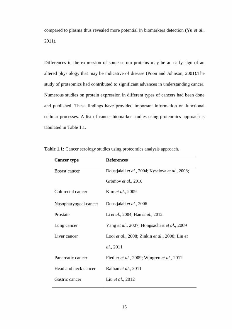
cellular processes. A list of cancer biomarker studies using proteomics approach is
tabulated in Table 1.1.
Table 1.1: Cancer serology studies using proteomics analysis approach.
Cancer type References
Breast cancer Doustjalali et al., 2004; Kyselova et al., 2008;
Gromov et al., 2010
Colorectal cancer Kim et al., 2009
Nasopharyngeal cancer Doustjalali et al., 2006
Prostate Li et al., 2004; Han et al., 2012
Lung cancer Yang et al., 2007; Hongsachart et al., 2009
Liver cancer Looi et al., 2008; Zinkin et al., 2008; Liu et
al., 2011
Pancreatic cancer Fiedler et al., 2009; Wingren et al., 2012
Head and neck cancer Ralhan et al., 2011
Gastric cancer Liu et al., 2012
16
1.7. IgM Antibody
The term antibodies were first used by Karl Landsteiner in 1900 which is a
translation from a German word “Antikörper” (Vollmers and Brändlein, 2005).
Immunoglobulin M (IgM) antibodies are present in the circulation of normal humans
and other mammalian species as part of the innate immunity and were conserved in
all animals (Parkin and Cohen, 2001; Marchalonis et al., 2002). Pentameric IgM
molecules made about 30% of the blood-circulating immunoglobulins in human. IgM
is initially secreted by B cells upon primary stimulation with antigen (Zouali, 2001;
Tchoudakova et al., 2009) and participates in natural defenses against foreign
pathogens as well as neoplastic cells and tumors (Brändlein et al., 2003).
In fact, autoantibodies against specific cancer antigens have been identified for
several types of tumors, including colon, breast, lung, ovary, prostate, and head and
neck. These antibodies have been found to recognize over expressed (e.g., Her2),
mutated (e.g., p53), or tissue-restricted (e.g., cancer-testis antigens) proteins, which
are produced by cancer cells and elicit immune responses (Lin et al., 2007).
Therefore, detection of such antibodies in patient sera can be exploited as a means of
cancer diagnosis. Indeed, the specificity and sensitivity of the antibody response to
low antigen levels make it an ideal screening or diagnostic tool for early
identification of cancer biomarkers in serum-based assays.
17
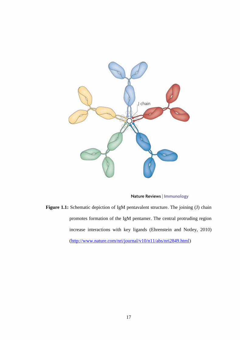
Figure 1.1: Schematic depiction of IgM pentavalent structure. The joining (J) chain
promotes formation of the IgM pentamer. The central protruding region
increase interactions with key ligands (Ehrenstein and Notley, 2010)
(http://www.nature.com/nri/journal/v10/n11/abs/nri2849.html)

18
1.8. Functional Annotation and Protein Interaction Analyses
DAVID (Database for Annotation, Visualization and Integrated Discovery)
bioinformatics resources are a high-throughput and integrated data-mining
environment which able to systematically extracting biological features or meaning
from large gene or protein lists (Huang et al., 2009). DAVID is uniquely
characterized with integrated and expanded back-end annotation database, advanced
modular enrichment algorithms and powerful exploratory ability (Sherman et al.,
2007; Alvord et al., 2007)
Without ever making direct contact, proteins can catalyze subsequent reactions in a
metabolic pathway, regulate each other transcriptionally or post-transcriptionally or
jointly contribute to larger, structural assemblies. Furthermore, together with direct
physical interactions, such indirect interactions constitute the larger superset of
‘functional protein-protein associations’ or ‘functional protein linkages’ (Eisenberg
et al., 2000). Protein-protein associations have proven to be a useful concept by
which to group and organize all protein-coding genes in a genome. The complete set
of associations can be assembled into a large network, which captures the current
knowledge on the functional modularity and interconnectivity in the cell. Protein
network information can aid in the interpretation of functional genomics data.
The STRING (Search Tool for the Retrieval of Interacting Genes) database has been
designed with the goal to assemble, evaluate and disseminate protein-protein
association information comprehensively. The STRING v9.1 in particular,
specializes in three ways. Firstly, it provides uniquely comprehensive coverage, with

19
more than one thousand organisms, five millions proteins and more than two hundred
millions interactions stored. It is also one of very few sites to hold experimental,
predicted and transferred interactions, together with interactions obtained through
text mining. It also includes a vast of accessory information, such as protein domains
and protein structures improving its day-to-day value for users.
20
1.9. Statement of the problem and rationale of the study
The advent of proteomic technologies have allowed the extensive fractionation of
proteins in biological specimens, analysis of peptides by MS and matching of mass
spectra to peptide sequences in human genome and protein databases (Omenn et al.,
2006). The dynamic nature of the circulatory system and its constituents reflects
diverse physiological or pathological states, and the serum-based biomarkers provide
noninvasive tests for screening as well as disease classification and monitoring for
cancer progression and regression (Hanash et al., 2008). Theoretically, each disease
may be uncovered and characterized by its unique biomarker. However, rather than
to see this biomarker as a single molecule, a biomarker should be regarded as a panel
of up- and down-regulated proteins or proteins with altered posttranslational
modifications, which differ in disease and normal state (Etzioni et al., 2003; Rifai et
al., 2006).
Even with advances in surgery, radiation and chemotherapy, oral cancer is one of the
lowest five-year survival rate among major cancer sites and this rate has not
significantly improved (Jou et al., 2010). In this present investigation, identification
of novel OSCC biomarkers in order to promote the development of specific
diagnostic tests for the early detection of oral cancer was aimed. The
immunoproteomic approach employed in this study allowed the detection of
immunogenic host-specific proteins in patient samples using pooled human
antibodies. Thus, host- or tumor-specific proteins represented markers for the
‘selected’ antibody. Proteomic analysis allows the characterization and quantification
of proteins and peptides in biological samples (Arnott and Emmert-Buck, 2010).
21
Therefore, a combined analytical platform involving two-dimensional gel
electrophoresis (2-DE) and mass spectrometry (MS) has been widely used for
biomarker identification (Srinivas et al., 2001; Kolch et al., 2005; Karpova et al.,
2010).
In the present study, unfractionated sera from OSCC patients and normal controls
were subjected to 2-DE and immunoblot for protein profiling, followed by
characterization of potential markers using MS. Notably, since the approach used
involved patient serum, which is complex and consists of various proteins, it can
potentially reflect numerous events occurring in vivo simultaneously (Yeng et al.,
2010). In addition to providing potential biomarkers for the early detection of oral
cancers, this study contributes to the understanding of naturally occurring antibodies
in cancer.
22
2.0. Objectives of the study
The aims of this study were as follows:
1) To develop the 2-DE serum protein profiles of patients with OSCC as well
as that from normal individuals
2) To identify serum proteins that are differentially expressed in the serum of
OSCC patients
3) To identify circulating immunogenic proteins and host specific proteins
eliciting humoral responses in OSCC patients using immunoproteomics
approach.
23
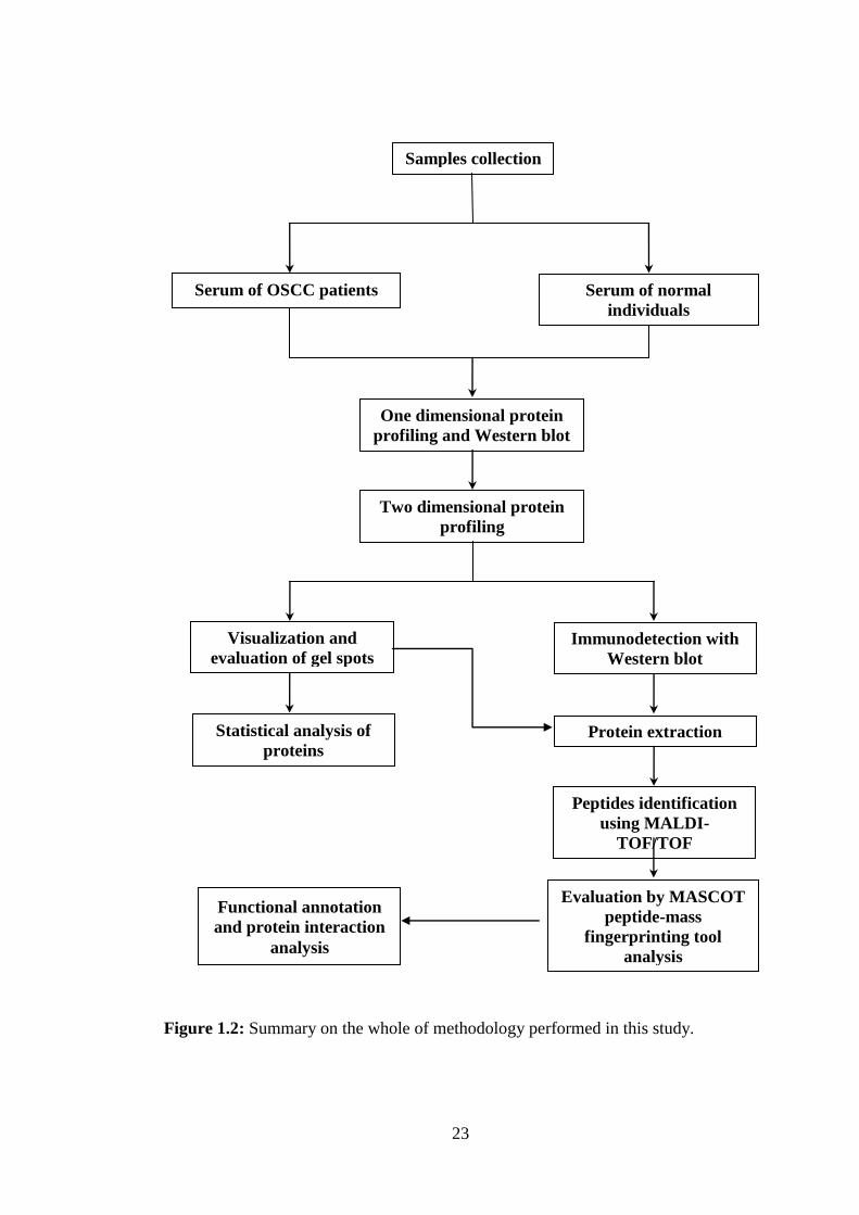
Figure 1.2: Summary on the whole of methodology performed in this study.
Samples collection
Serum of OSCC patients Serum of normal
individuals
One dimensional protein
profiling and Western blot
Two dimensional protein
profiling
Visualization and
evaluation of gel spots Immunodetection with
Western blot
Statistical analysis of
proteins Protein extraction
Peptides identification
using MALDI-
TOF/TOF
Evaluation by MASCOT
peptide-mass
fingerprinting tool
analysis
Functional annotation
and protein interaction
analysis
24
CHAPTER TWO
MATERIALS AND METHODOLOGY
2.1. Collection of Serum Samples
Based on clinical information and laboratory diagnosis for carcinogenesis, a total
of 25 serum samples of oral squamous cell carcinoma (OSCC) patients were
obtained from the Oral Cancer Research and Coordinating Centre (OCRCC),
University of Malaya, Kuala Lumpur and used in this study, Additionally, 25
serum samples were also collected from healthy individuals and used as control
group. The cancer group consisted of patients with early stages of oral squamous
cell carcinoma and without any history with other type of malignancy. The normal
healthy individuals referred to individuals that were determined not to have any
history with cancer. Samples obtained were with consent and approval granted by
both universities; the Medical Ethics Committee, Dental Faculty, University of
Malaya (UM) (Ref: DF OP0907/0050(P)) and Universiti Sains Malaysia (USM)
(Ref: USMKK/PPP/JEPeM [213.3.09]). The serum samples were stored in
aliquots of 10 µl and kept at -80°C until use.