functions of bard1 and associated proteins and its
TRANSCRIPT

Irmgard IrmingerIrmgard Irminger--FingerFinger
Biology of Aging Laboratory and Monitoring LaboratoryUniversity and University Hospitals of Geneva, Switzerland
Functions of BARD1 and associated proteins and its
application in cancer prognostics and treatment

Tumor suppressor BARD1Tumor suppressor BARD1patented:patented:
1.Use as vaccine1.Use as vaccine2. Use for cell killing2. Use for cell killing
3.Use as prognostic marker3.Use as prognostic marker
Irmgard IrmingerIrmgard Irminger--FingerFingerUniversity and University Hospitals GenevaUniversity and University Hospitals Geneva

Clinical application of BARD1: background and principlesThe “normal” functions of BARD1 BARD1 expression in tumors:
in breast and ovarian cancersin lung cancer
BARD1 expression the human cytotrophoblast:a non-malignant cells with an invasive phenotype
AND in amnion fluid-secreted by the cytotrophoblast?
Detection of BARD1 in serum for cancer screening?
Immunogenic BARD1 applied for tumor cell killing

Genes implicated in epithelial cancers: predisposition genes (tumor suppressor genes)
• p53 (lung, colon, breast, prostate)• BRCA1 (breast, ovary, prostate) • BRCA2 (breast, ovary)• BARD1 (breast, ovary, uterus,
– and other proliferative tissues)
Function: repair, apoptosis (cell death)

Old Age Cancers are derived from epithelial cells
BCC Basal cell carcinomaSCC Squamous cell carcinoma
DePinho, 2000

Accumulation of damage during aging
agingaging
environmentalinsult
apoptosis
long-term loss offunctional tissue(Alzheimer, heart
failure etc.)
decisionmaking
cancercancer
Accumulation of endogenous
damage
damaged cellsabnormal function
abnormal activationabnormal growth
damage
repairrepair
BARD1BARD1
BARD1BARD1
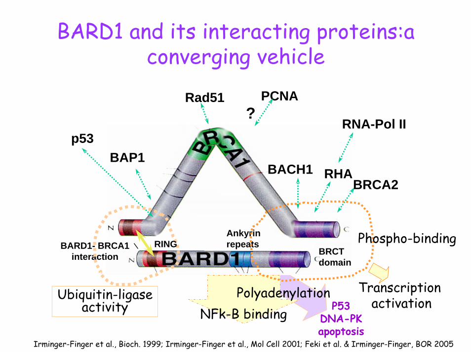
BARD1 and its interacting proteins:a converging vehicle
Irminger-Finger et al., Bioch. 1999; Irminger-Finger et al., Mol Cell 2001; Feki et al. & Irminger-Finger, BOR 2005
P53DNA-PKapoptosis
BARD1- BRCA1interaction
p53
Rad51
RNA-Pol II
RHA
PCNA?
BRCTdomain
Ankyrin repeats
Ubiquitin-ligaseactivity
BRCA2
BAP1BACH1
PolyadenylationNFk-B binding
Phospho-binding
Transcription activation
RING

BARD1 and BRCA1 mutations predisposing to breast cancer
HumanBARD1 RING ANK BRCT
Q513HC557S
N295SK312N
358D364G1765C
G1743CA1009T
A957G 1144del21
A1481G
G1592A
HumanBRCA1
RING BRCT
C>GC>G Y>AY>AMissense mutations


No one like BARD1
BARD1
BRCA1
Arabidopsis2 hypoth.p.
ArabidopsisCAC05430C. elegans3 hypoth. p.C. elegans6 hypoth. p.
C. elegansT15566
C. elegansT15564
RINGRING ANKANK BRCTBRCT
Joukov, Livingston, PNAS, 2001
Xenopus
BRCA1BARD1
Irminger-Finger and Leung, IJBCB 2002

transfection of BARD1
BARD1 induces apoptosis in a p53-mediatedpathway
B pcBARD1 pcBQ>HS P S P
p53
IP: α-BARD1
BARD1
non-treat. doxoS P S P
AIP: α -p53
BARD1
PCNA
IP: α -BARD1 non-treat. doxo
S P S P
p53
PCNA
p53+/+ p53-/-
BARD1 binds to p53BARD1 binds to p53
BARD1 stabilizes p53BARD1 stabilizes p53
Irminger-Finger et al., Mol Cell 2001

Mapping of the apoptotic regionpc
DNA3
BARD1
∆BR
CT∆
ANK
RING
∆RI
NG
ANK2
APORING ANKYRIN
BARD1BRCT
NLS131 NLS 408 NLS 693BARD1∆ RING∆ BRCT
ANK2RING
∆ ANK
Q564HQ564HC557SC557S
yes
yesyes
yes
yesno
noBARD1 Q564H
X?
Deletion mutants lacking the BRCA1 interaction domain are efficient in apoptosis induction and have increased protein stability

BARD1 is upregulated in response to cellular stress
DoxorubicinTAC-2 cells
On On the protein levelthe protein level
Western
On On the mRNA levelthe mRNA level
RT-PCR
UV - 1 4 - mindoxo - - - 10 µg
BARD1
p53
ES cells

Mechanism of apoptosis induction by BARD1
• Exogenous expression of BARD1 leads to apoptosis
• BARD1-induced apoptosis depends on functional p53
• Upregulation of BARD1 results in stabilization of p53 protein
• Repression/deletion of BARD1 results in apoptosis resistence

BARD1 deficient tumor cells: NuTu-19 resistent to apoptosis
BARD1
ANKRING BRCT
H-300PVC JH-3
0.8KB BARD1In1
In2 In3 In4 765 8 In9
In1In2 In3 In4
In7In6In5 In8 In9
InInIn In
2KB BARD1
C29
3T
H-300 JH3PVC
293T
NuTu-
1929
3TNuT
u-19
NuTu-
19BARD1
A
0.8 kb
2.2 kb2 kb
M p53 BARD1B
Feki et al., & Irminger-Finger, Oncogene 2005

BARD1 required for p53 phosphorylation
C
doxoNuTu-19TAC2
- ++-
p53
BARD1
p-p53
actin
Apoptotic treatment
HEK NuTu-19 BARD1
BARD1
p53
p-p53
- ++-
actin
BARD1 transfection

Exogenous expression of BARD1 can induce apoptosis in NuTu-19 cells
0
5
10
15
20
25
30
35
40
45
50
NuTu-19 NuTu-19 + Doxo
NuTu-19+ BARD1
% o
f apo
ptos
is
TAC-2 TAC-2+ Doxo
NuTu-19
TAC2
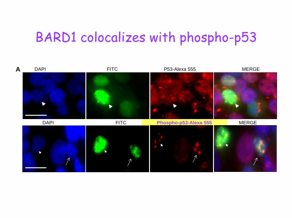
BARD1 colocalizes with phospho-p53
DAPI FITC Phospho-p53-Alexa 555 MERGE
A DAPI FITC P53-Alexa 555 MERGE

Mapping p53 binding regionIP p53
BARD1 ∆-RING ∆-HIND ∆-BRCT ANK RING EGFP
S P S P S P S P S P S P S P
G215CG176CG1743CA1009T
A957G
1144del21 A1481G
G1592A G2354A
apoapo
yes
yesyes
yes
noyes
100
80
60
100
0
p53p53bindingbinding
60
mBARD1
RING (aa 1-266) EGFP
ANK (aa 394-604) EGFP
∆ BRCT (aa 1-604) EGFP
∆ HIND (aa 394-765) EGFP
∆ RING (aa 137-765) EGFP
BARD1 (aa 1-765) EGFP
RING BRCT
NLS 131 NLS 408 NLS 693
ANK
Feki et al., and Irminger-Finger, Oncogene 2005

BARD1 induces apoptosis by catalyzing phosphorylation of p53
• BARD1 binds to p53 and to phosphorylated p53 • BARD1 binds to Ku-70, the catalytic subunit of
DNA-PK but not ATM• Exogenous expression of BARD1 in BARD1-
deficient cells induces p53 phosphorylation• Exogenous expression of BARD1 in BARD1
deficient cells restores apoptosis sensitivity

Co-localization of BRCA1 and BARD1 with repair proteins in “nuclear dots”
+BARD1
+BARD1
Jin, Baer, JBC 1997; Scully, Livingston, Cell 1997

pEGFP BARD1-EGFP ∆RING-EGFPA
....but not only “nuclear dots”
∆BRCT-EGFP ANK-EGFP RING-EGFP
RING-EGFP
0102030405060
EGFP-N1
BARD1-EGFP
RING-EGFP
∆-RING-EGFP
∆-HIND -EGFP
∆-BRCTEGFP
ANK-EGFP
%ap
opto
tic G
FP+
cells
B

BARD1 translocates to the cytoplasm during apoptosis
T=6 T=12
T=24 T=48
Jefford et al., & Irminger-Finger, Oncogene 2004

Intracellular localization of BARD1 upon apoptosis induction
non treated UV
BA
RD
1B
RC
A1

BARD1 is translocated to the cytoplasm during apoptosis
Ab: PVC
Total cell extract0 3 4 5 6 7 14 22 24 h
*
**
Nucl. extract
Ab: C20
Cytopl. extract
Ab: C20
0 3 4 5 6 7 14 22 24 h 0 3 4 5 6 7 14 22 24 h
p67
The C-terminal p67 is translocated to the cytoplasm and is immunogenic and anti-tumorigenec in an animal model of colon cancer (Gautier, Irminger-Finger et al., Cancer Res 2000).

Apoptotic function of BARD1 in vivo
• In morphogenesisspermatogenesis
• In homeostasisSpleen, bone
• In tumor suppression

Cytoplasmic localization of BARD1 in vivo
BARD1 N-19A BARD1 PVC
B BARD1 N-19

BARD1 upregulation in vivo by ischemia
Core region Penumbra
Left hemisphere
Left hemisphere
BRCA1 BARD1BRCA1

Apoptosis and (meiotic) repair during spermatogenesis
Bcl-xPMB4SCFIL-4LIFGas6bFGF
SCFBcl-xLtestosterone
LIFCNTF
PGCSpermatogonia Spermatocytes Spermatids Spermatozoa
Apoptosis Apoptosis Apoptosis Apoptosis
- +
TGFß Bax
+
Bax
- +-Bcl-xLtestosterone
Gonocytes
- -
BARD1?BARD1? Modified from:
Print, Bioassays, 2000

Schematic diagram of seminiferous tube
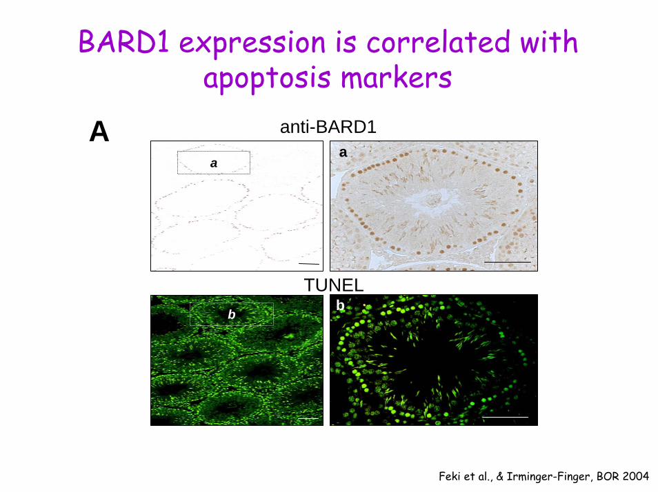
BARD1 expression is correlated with apoptosis markers
A
b
aa
b
anti-BARD1
TUNEL
Feki et al., & Irminger-Finger, BOR 2004

B activated caspase 3 anti-BARD1
N-19b
b
a
a
c d
d
N-19
JH3
c
e
ef
f

BARD1 and BRCA1 expression overlap partially
Ab
a
baBAR
D1
5x 20x 20x
B
c
c dd BR
CA1
5x 40x 40x
C
5x

Different BARD1 isoforms are expressed at different stages
A PPjrG PL SR
97 kD
C-2074 kD Western blot
97 kDJH-3
74 kD
97 kD
74 kDJH-2
B G P PjrL P SR
2300 bp2000 bp
RT-PC
R
BARD1
GAPDH

BARD1β lacks the RING finger
BARD1βIn1
In2 In3 In4In7In6In5 In8 In9
BARD1ANKRING BRCT
Q>HQ>H
N-19 C-20JH-2 JH-3PVC

BARD1β is an efficient apoptosis inducer
0102030405060708090
100
% c
elld
eath
CGR8 BARD1b BARD1bBRCA1
BARD1 BARD1BRCA1
Doxo
p53BARD1b BARD1
BRCA1BARD1bBRCA1
BARD1CGR8 Doxo

BARD1 expression associated with abortive spermatogenesis
Obstructive Azoospermia Sertoli cell SyndromeA
B d
12
3
ccryptorchidy
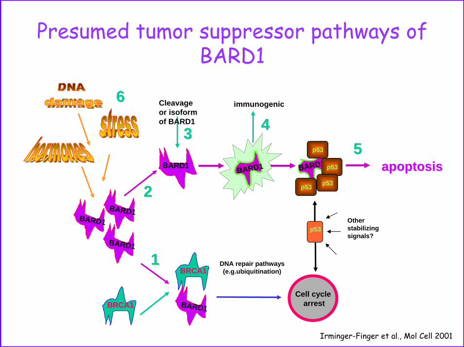
Presumed tumor suppressor pathways ofBARD1
DNA repair pathways(e.g.ubiquitination)
p53
Cell cyclearrest
Other stabilizingsignals?
BRCA1
11BARD1
BARD1BARD1
BRCA1
BARD1
22
BARD1
Cleavageor isoformof BARD1
33
Irminger-Finger et al., Mol Cell 2001
p53
BARD1 p53
p53p53
apoptosisapoptosis55
66 immunogenic
44
BARD1

Expected loss of BARD1 in tumorigenesis
Carcinogenic stresstranscription activation
apoptosisBARD1BARD1 p53p53
p53 turnover
PPser15ser15
- inhibition of degradation - direct stabilization
Carcinogenesis
-loss of BARD1 -loss of p53
BRCA1mutatedor deleted
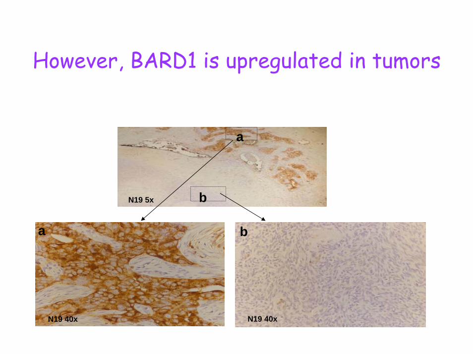
However, BARD1 is upregulated in tumors
N19 40x
b
N19 40x
a
N19 5x b
a

CC
CEn
CSe
CM
uCN19 C20
5x5x
5x5x
5x5x
5x5x
BARD1 expression is specific for tumor type
A

Expression of N- and C-terminal epitopes
40x
5x 5x
40x
N19 C20
5x
40x 40x
5x
N19 C20
0
10
20
30
40
50
60
70
80
90
100OB18 OB41OB42 OB15OB16 OB44OB43 OB11OB35 OB12OB38 OB39OB40 OB14OB13 OB36OB37 OB31OB33 OB29OB34 OB30OB32 OB2OB3 OB4OB9 OB17OB23 OB28OB24 OB25OB5 OB27OB22 OB26OB21 OB10OB19 OB20OB1 OB6OB7 OB8
CCC EnC SeC MuC
% o
fN19
pos
itive
cel
ls

Correlation of BARD1 expression with tumor grade
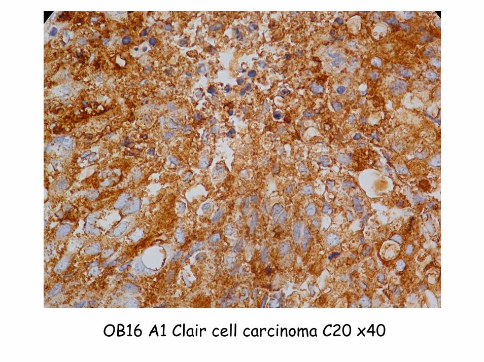
OB16 A1 Clair cell carcinoma C20 x40

BARD1 but not p53 is correlated with tumor type
40x 40x
40x40x
N19 p53A
010
20
3040
5060
7080
90
100
CCC EnC SeC MuC
N19C20p53
% o
fpos
itive
cel
ls
BCorrelation of BARD1 and p53 with histological type of
ovarian carcinoma
Figure 3

Correlation of BARD1 expression with tumor grade and stage
0
5
10
15
20
25
30
35
G1 G2 G3
N19C20p53
% o
fpos
itive
cel
ls
Correlation of BARD1 and p53 with differentiation in sporadic breast cancer
0
10
20
30
40
50
60
70
T1 T2 T3 T4
N19C20p53
% o
fpos
itive
cel
ls
Correlation of BARD1 and p53 with tumor size in sporadic breast cancer
D

No correlation of BARD1 and p53 expression in NSCLC
0102030405060708090
100
adenosquam
adenosqu large
6159 103566609 80408315 83411708 1274012748 11761659 85328664 1227811638 127566634 80759977 1276711601 11031531 83668930 103627987 901810319 66617723 865012111 98446699 81008981 114007993 1120311605 12500
Distribution of N19 expression in different pathological types
5x
N19 40x
5x
p53 40x
A B
% o
fN
19 p
o si ti
v e c
ells
Figure 5
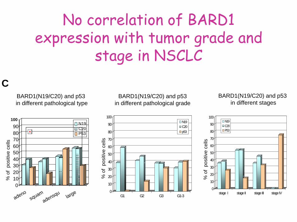
No correlation of BARD1 expression with tumor grade and
stage in NSCLC
BARD1(N19/C20) and p53in different pathological type
0
10
20
30
40
50
60
70
80
90
100
G1 G2 G3 G1-3
N19C20p53
0
10
20
30
40
50
60
70
80
90
100
stage I stage II stage III stage IV
N19C20P53
BARD1(N19/C20) and p53in different pathological grade
BARD1(N19/C20) and p53in different stages
C
% o
fp o
sitiv
e c e
lls
% o
fp o
sitiv
e c e
lls
% o
fp o
sitiv
e c e
lls
0102030405060708090
100
adenosquam
adensqularge
N19C20P53

A
C-terminus
1441-2252
N-terminus
74-1481
hGAPDH
OB C+ 8 8* 9 21 23 24 24* 25 33 33* 34 35 37 C-
Tumors express an aberrant form of BARD1
2252
74
1441
1481783
G1765C
G1743CA1009T
A957G 1144del21
A1481G
G1592A
Figure 6

BARD1 is transcriptionally altered in rat ovarian cancer cells
and in human cancers?
BARD12.2 kb
ANKRING BRCT
H-300 PVC JH-3
BARD1δ0.8 kb
8 In9
In1
In2
In3
In4
In7
In6
In5
In8 In
9
InIn
1In
2
804-826
1618-1641
BARD1γ2.0 kb
2252
74
1441
1481783
Figure 5A-C

BARD1 expression in human cancers
Elevated expression of BARD1 in cancerCytoplasmic localization of BARD1 in tumorsLoss of 5’region Expression is correlated with tumor type, grade and stage in breast and ovarian cancerNo correlation in NSCLCNo correlation of BARD1 and p53 expression
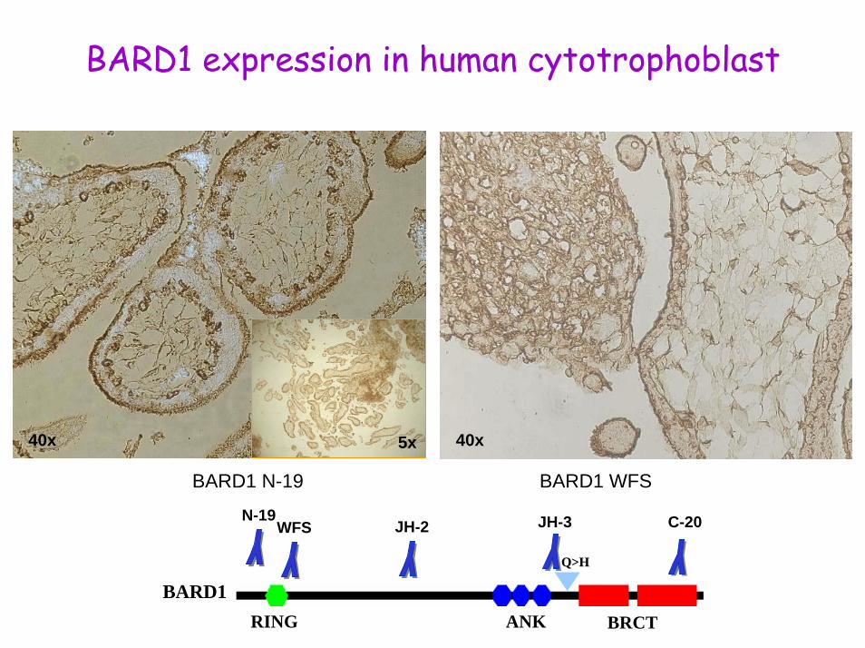
BARD1 expression in human cytotrophoblast
5x40x 40x
BARD1 N-19 BARD1 WFS
BARD1ANKRING BRCT
Q>HQ>H
N-19 C-20JH-2 JH-3WFS

BARD1 expression in placenta at 12 weeks of pregnancy
20x 40x
BARD1 N-19
Immunohistochemistry of human placenta at 12th weeks of pregnancy. BARD1 N-19 antibody was used on sections of human placentas of the first trimeter of pragnancy. Representative images at 12 weeks of pregnancy demonstrate BARD1 redistribution to the brush border membrane. This subcellular localization suggests that BARD1 might be secreted.

BARD1 expression is correlated with apoptoticmarkers
40x5x
Activated Caspase 3
Identification of potential apoptotic cells. Since BARD1 expression can induce apoptosis in vitro and in vivo, activating a p53-caspase pathway, we stained placenta sections with antibodies against acitvated caspase 3. A complete correlation of BARD1 expression and activated caspase 3 expression can be observed. However, whether the BARD1 expressing cells actually undergo apoptosis can not be determined by these experiments.

BARD1 expression in invasive cells

Only BARD1γ expressed in invasive cells
Ab: PVCAb: WFS
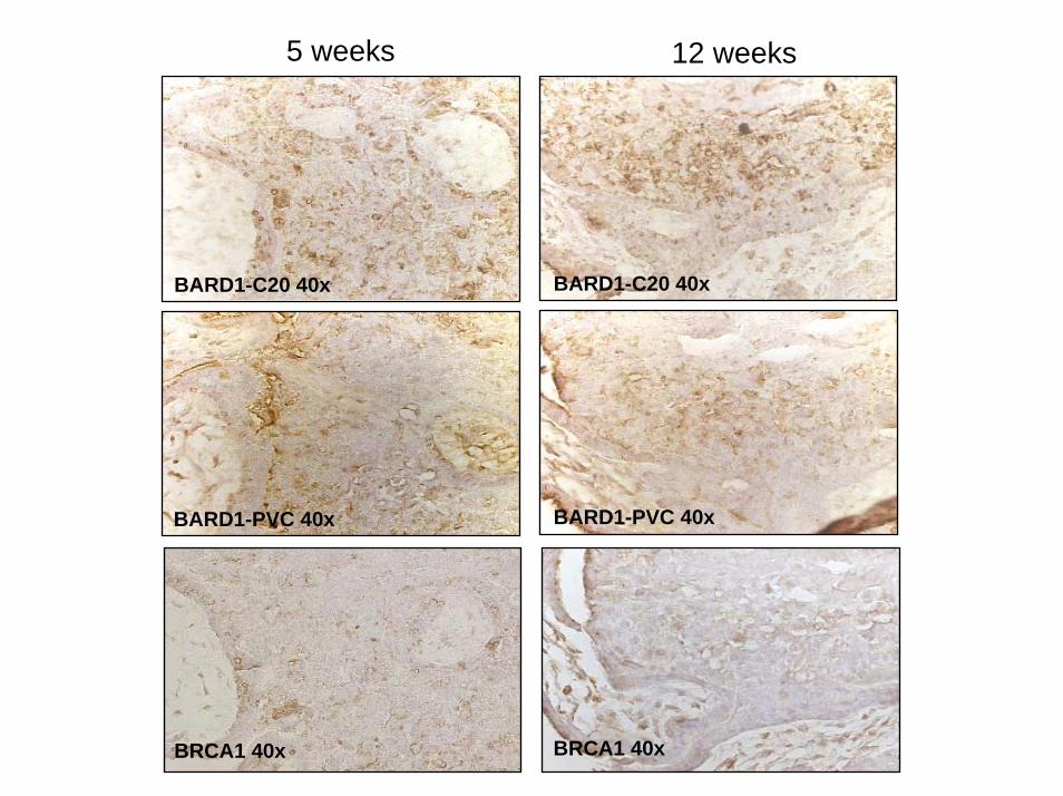
5 weeks 12 weeks
BARD1-C20 40xBARD1-C20 40x
BARD1-PVC 40xBARD1-PVC 40x
BRCA1 40x BRCA1 40x

Differentially spliced isoform of BARD1 in cytotrophoblast and placenta lack RING
finger
T P M CRT-PCR
BARD1
actin
2,3kb2kb
A
ANKRING BRCT
Q>HQ>H
JH-2 JH-3
BARD1γIn1
In2 In3 In4In7In6In5 In8 In9
BARD1
N-19 C-20PVC WFS
B

BARD1 is secreted from CTBs in cultureB
AR
D1
(UA
/mio
cells
/ml)
0
20
40
60
80
100
120
CTB (2) MCF-7 MDA-MB231CTB (1)
In cell lysatesA
CTB (2) MCF-7 MDA-MB231BA
RD
1 (U
A/m
ioce
lls/m
l)
0.01.02.03.04.05.06.07.08.09
In medium of cell culturesB
PMAdoxocontrol
0.01.02.03.04.05.06.07.08.09
0-24 h 24-28 h
In medium of CTBsC

Quantification of BARD1 by ELISA
Coating C 20 (1/200) Coating N19 (1/200)Tampon
Pool Liq. Amn. 1/4
Pool Liq. Amn. 1/20Pool Liq. Amn. 1/100MC CTB (conc 8x)
Serum PB 2.10.03 ndlSerum pool Femme enceinte ndlSerum AM 2.10.03 ndl
C20 biot1/1000
C20 biot1/1000
WFS 1/100
WFS 1/100
PVC 1/100
PVC 1/100

Conclusions• BARD1 has a novel cellular function in
CTBs and placenta– Regulated at different times of pregnancy– Regulated expression of different epitopes
• Cytotrophoblast is expressing full length and splice variant γ of BARD1
• BARD1 is secreted from CTBs• A function of BARD1
– in invasion?– in angiogenesis?

Animal cancer model for tumor cell killing
NuTu-19 ovarian cancer cells
After 5 weeks 100%
Metastases detection by photo medicine
A
GFPGFPretroviral vector
liver
diaphragma
digestive tube
B
visual light
omentum
FITC
peritoneum

Transduction of NuTu-19 cells with lentiviralvectors expressing BARD1 or GFP
Packaging plasmids: pCMV DR8.92, pCMV DR8.93 (core and enzymatic components of HIV-1)Envelop plasmid: pMDG (VSV-G protein)Transducing vectors: pLOX/TW-BARD1 or pLOX/TW-rTTAh
Ovarian cancer Model
GFPGFP--cancer cancer cellscells
Gene therapyfor cell killing byviral delivery of
Inducible BARD1
pCMV DR8.92, pCMV DR8.93pMDGpLox TW BARD1 or pLox/TW-rTTAh
293T cells
Transduced NuTu cellsexpressingGFP or
BARD1 and rTTAhproteins

Apoptosis induction by inducible BARD1
A BNuTu-19 cells
NuTu-19 cells transduced with
pLOX/TW-gfp lentiviral vector
NuTu-19 cells transduced with 50X concentrated pLOX/TW-gfp lentiviralvector
GFP expression
GFP expression
NuTu-19 cells
NuTu-19 cells transduced with
pLOX/TW-gfp lentiviral vector
NuTu-19 cells transduced with
pLOX/TW-gfp lentiviral vector
and induced with Doxycyclin (2µg/ml)
0
10
20
30
40
50
60
NuTu NuTu-PLOX/BARD1
+ 2.5 ugDoxycycline
+5ugDoxycycline
+10 ugDoxycycline
+15 ugDoxycycline
+20ug ofDoxycycline
C 50
0 1 ug/ml 2ug/ml 4ug/ml 8ug/ml 16ug/mlDoxycyclin
Death rate after GFP induction with Doxycyclin
Death rate after BARD1 induction with Doxycyclin 40
30
20
10
0

Diaphragm

Thymus

Omentum

Nutu-19-EGFP tumors in Fisher rat
Diaphragm Omentum Thymus

Presumed tumor suppressorpathways of BARD1
DNA repair pathways(e.g.ubiquitination)
BRCA1
apoptosisapoptosis
11
22BARD1
p53
Cell cyclearrest
Other stabilizingsignals?
BARD1
BARD1
BRCA1
BARD1
BARD1BARD1
Cleavageor isoformof BARD1
p53
immunogenic
BARD1
33
Irminger-Finger et al., Mol Cell 2001
p53
p53p53
4455
66

Viral BARD1 injection causes tumor shrinking
NuTu-19: epithelial ovarian cancer cells
2 weeks later
Fisher rat 344: immuno-competent
4 weeks later
Left RightBA
RD
1EG
FP
0.00
0.50
1.00
1.50
2.00
2.50
BIG in
jectio
n
Ctl righ
t
GFP injec
tion
Ctl righ
t
NuTu lef
tNuT
u right
L R L R L R
cm

BARD1 expression in tumors causes immune response
BARD1
40x
Nutu
GFP
NuTu and GFP
40x
NuTu and SNGFP
CD3
40x
40x

BARD1 for tumor cell killing
• Ovarian cancer model• BARD1 deficient tumor cells• Viral delivery of functional BARD1• Replace viral BARD1 by small
molecules• Use BARD1 in anti-tumor vaccination

Irminger-Finger labChantal GenetAurélie Caillon
Francoise PiottonLaura Ortolan
Stephan Stephan RyserRyser
Charles Edward Jefford
Igor Igor BondarevBondarev
FabriceCallabria
Philipe Philipe BerardiBerardi
MamadouHady Sow
Anis Feki
Visiting scientists
JianJian YuYu WuWu

Collaborations
Paul Bischof (Geneva)Geoff Laurent (London)Andrea Bianco (Naples)Jean Harb (Nantes)Maria-Adelaide Caligo (Pisa)Marie-Francoise Pelte (Geneva)Anne-ThereseVlastos (Geneva)
FundingFunding: : JnJJnJ, SNSF, , SNSF, EuroPrEuroPr, JT, GR , JT, GR