four plant defensins from an indigenous south african brassicaceae
TRANSCRIPT
RESEARCH ARTICLE Open Access
Four plant defensins from an indigenous SouthAfrican Brassicaceae species display divergentactivities against two test pathogens despite highsequence similarity in the encoding genesAbré de Beer and Melané A Vivier*
Abstract
Background: Plant defensins are an important component of the innate defence system of plants where theyform protective antimicrobial barriers between tissue types of plant organs as well as around seeds. These peptidesalso have other activities that are important for agricultural applications as well as the medical sector. Amongst thenumerous plant peptides isolated from a variety of plant species, a significant number of promising defensins havebeen isolated from Brassicaceae species. Here we report on the isolation and characterization of four defensinsfrom Heliophila coronopifolia, a native South African Brassicaceae species.
Results: Four defensin genes (Hc-AFP1-4) were isolated with a homology based PCR strategy. Analysis of thededuced amino acid sequences showed that the peptides were 72% similar and grouped closest to defensinsisolated from other Brassicaceae species. The Hc-AFP1 and 3 peptides shared high homology (94%) and formed aunique grouping in the Brassicaceae defensins, whereas Hc-AFP2 and 4 formed a second homology grouping withdefensins from Arabidopsis and Raphanus. Homology modelling showed that the few amino acids that differedbetween the four peptides had an effect on the surface properties of the defensins, specifically in the alpha-helixand the loop connecting the second and third beta-strands. These areas are implicated in determining differentialactivities of defensins. Comparing the activities after recombinant production of the peptides, Hc-AFP2 and 4 hadIC50 values of 5-20 μg ml-1 against two test pathogens, whereas Hc-AFP1 and 3 were less active. The activityagainst Botrytis cinerea was associated with membrane permeabilization, hyper-branching, biomass reduction andeven lytic activity. In contrast, only Hc-AFP2 and 4 caused membrane permeabilization and severe hyper-branchingagainst the wilting pathogen Fusarium solani, while Hc-AFP1 and 3 had a mild morphogenetic effect on thefungus, without any indication of membrane activity. The peptides have a tissue-specific expression pattern sincedifferential gene expression was observed in the native host. Hc-AFP1 and 3 expressed in mature leaves, stems andflowers, whereas Hc-AFP2 and 4 exclusively expressed in seedpods and seeds.
Conclusions: Two novel Brassicaceae defensin sequences were isolated amongst a group of four defensinencoding genes from the indigenous South African plant H. coronopifolia. All four peptides were active against twotest pathogens, but displayed differential activities and modes of action. The expression patterns of the peptideencoding genes suggest a role in protecting either vegetative or reproductive structures in the native host againstpathogen attack, or roles in unknown developmental and physiological processes in these tissues, as was shownwith other defensins.
* Correspondence: [email protected] for Wine Biotechnology, Department of Oenology and Viticulture,Faculty of AgriSciences, Stellenbosch University, Stellenbosch 7600, SouthAfrica
de Beer and Vivier BMC Research Notes 2011, 4:459http://www.biomedcentral.com/1756-0500/4/459
© 2011 Vivier et al; licensee BioMed Central Ltd. This is an open access article distributed under the terms of the Creative CommonsAttribution License (http://creativecommons.org/licenses/by/2.0), which permits unrestricted use, distribution, and reproduction inany medium, provided the original work is properly cited.
BackgroundPlants have developed complex defence systems to pro-tect them against a multitude of plant pathogens [1-8].These defence systems consists of an array of both che-mical and biochemical substances that protect the plantagainst colonization and subsequent spread of diseaseand can broadly be divided into the innate and activedefence responses [7,9-13]. The innate defenceresponses play an important role in establishing pre-formed barriers of defence to prevent colonization bypathogens. Antimicrobial peptides (AMPs) are animportant component of the innate defence response.They are small, mostly basic peptides that range in sizefrom 2-9 kDa and have been classified into nine groups.Plant defensins [10,14-21], thionins [22-27] and lipidtransfer proteins [28-34] are the best characterized ofthese nine groups.Plant defensins are small, basic, heat stable peptides
with a conserved tertiary structure that consists of a sin-gle a-helix and three anti-parallel b-strands [17,35-37].The defensin tertiary structure is internally stabilized bydisulphide bridges linking the a-helix to two of the b-strands to form a structure know as the cysteine stabi-lizing motif, a conserved motif identified in AMPs iso-lated from various prokaryotes and higher eukaryotes[38-41]. In addition to the cysteine stabilizing motif twoadditional conserved motives have been identified in theplant defensin structure, namely the a-core, encompass-ing the loop connecting the first b-strand and a-helixand the g-core containing the all important hairpin loopconnecting b-strand 2 and 3 (Lb2b3). Notwithstandingthis conserved tertiary structure, plant defensins sharevery little homology at amino acid level. It is howeverthis variability in primary amino acid sequence that con-tributes to the different biological functions that havebeen attributed to these peptides, where a single aminoacid can change the spectrum of activity exhibited byclosely related defensin peptides.The role of plant defensins in the preformed defence
of plants is well documented. They play an importantrole in the protection of germinating plant seeds, devel-oping seedlings and reproductive structures of plants[42-44] and have been isolated from roots [44-46], vege-tative tissues and reproductive structures such as flowersand fruits [45,47-55]. The majority of characterizedplant defensins show a constitutive pattern of expres-sion, with an induction in expression in response topathogen attack, wounding and some abiotic stresses[20,44-46]. Recently it was shown that pathogen-inducedexpression of Arabidopsis plant defensins is dependenton ENHANCED DISEASE RESISTANCE1 (EDR1),which interferes with the repressor function of MYC2allowing for defensin gene expression [56]. Some
defensins, however, show a strict tissue-specific anddevelopmentally regulated pattern of expression[47,50,54,57,58] which in some cases were linked to spe-cific biological functions other than plant defence, aswas demonstrated for the defensins from tomato andmaize that play a role during pollination [50,57].Plant defensins are best known for their antimicrobial
activity against a broad spectrum of plant pathogens thatinclude bacteria [59,60], yeast [61-64], oomycetes [65,66]and necrotrophic pathogens [47,61,64,65,67-71]. In addi-tion to these strong antimicrobial activities that estab-lished them as important agricultural biotechnologytargets, some members also show activities important formedical applications, including protease inhibitory activ-ity [23,72], anti cancer activity [61,73] and HIV inhibition[61,74-76]. Other agriculturally important activitiesinclude insecticidal activity [35,36,77,78], activity againstparasitic plants [79] and heavy metal tolerance [80].The isolation and characterization of a wide range of
defensin peptides are crucial for the continued develop-ment of economically and medically important products.Analysis of the sequenced plant genomes revealed thatdefensins are present as multigene families and are over-represented in the genomes of some plants species[46,81]. With the wealth of defensin nucleotide sequencesavailable, strategies of gene isolation coupled with recom-binant production are increasingly been used for thecharacterization of closely related plant defensin peptides.This work describes the successful isolation of four
plant defensin genes from the South African Brassicaceaespecies Heliophila coronopifolia. An isolation strategybased on the sequence homology that exists within thenucleotides encoding the signal peptides of defensinsfrom domesticated Brassicaceae species was used to iso-late four defensin sequences, of which two were shownto be novel for Brassicaceae defensins. Each of the defen-sin peptide was successfully purified through recombi-nant production in Escherichia coli and characterized fortheir activity and mode of action against two test patho-gens. These results as well as expression analysis in thehost showed that the four peptides have differentialexpression patterns in vegetative and reproductiveorgans, as well as differential activities and modes of inhi-bition under the conditions tested. In addition, the diver-gence in structural motifs and surface propertiesobserved for these peptides provide interest to studystructure-activity determinants in these peptides.
ResultsIsolation and in silico characterization of the Hc-AFPencoding sequencesPCR-based isolation of cDNA from H. coronopifolia tis-sues allowed for the isolation of four putative defensin
de Beer and Vivier BMC Research Notes 2011, 4:459http://www.biomedcentral.com/1756-0500/4/459
Page 2 of 19
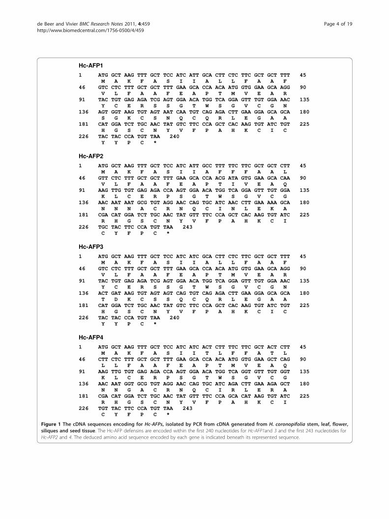
sequences ranging between 426 bp and 468 bp, contain-ing open reading frames of 240 and 243 bp, respectively.TBLASTN analysis of the nucleotide sequences showedhomology to sequences encoding for the super family ofplant antifungal peptides known as plant defensins. Theisolated gene sequences were thus termed H. coronopifo-lia antifungal peptide 1 to 4 (Hc-AFP 1 - 4) (Figure 1).Analysis of the deduced amino acid sequences showed
that Hc-AFP1 and 3 encode for 80 amino acid peptides,whereas Hc-AFP2 and 4 encode for 81 amino acid pep-tides (Figure 1). SignalP results showed that the first 30amino acids of each peptide encode for a signal peptidefollowed by a 50 amino acid mature peptide for Hc-AFP1 and 3 and a 51 amino acid mature peptide forHc-AFP2 and 4 (Table 1). The peptide parametersobtained from the Expasy-Compute pI/Mw tool (Table1) showed that the peptides had predicted mono-isoto-pic masses ranging between 5.48 and 5.73 kDa and arehighly basic with isoelectric points above 8.2.Alignment analysis of the deduced amino acid
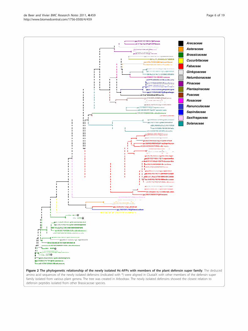
sequences revealed that the newly isolated H. coronopi-folia defensins shared the highest homology with defen-sins isolated from other members of the Brassicaceaefamily (Figure 2). Disulphide-bridge analyses conductedon the Hc-AFP peptides revealed that they share a disul-phide bridge pattern common to all plant defensins (Fig-ure 3). Further comparison of Hc-AFPs with membersof the Brassicaceae defensins (Figures 2 and 3) revealedthat Hc-AFP1 shared the closest homology to Hc-AFP3at 94% similarity and Rs-AFP3 from Raphanus sativa at82% similarity, whereas Hc-AFP2 showed the greatesthomology to the defensins isolated from Sinapsis albaand R. sativa (Rs-AFP2) at 98% similarity (Figures 2 and3). Hc-AFP4 was more closely related to PDF1.1 fromA. halleri, a defensin proposed to play a role in the zinctolerance of A. halleri.Analysis of homology models obtained for the differ-
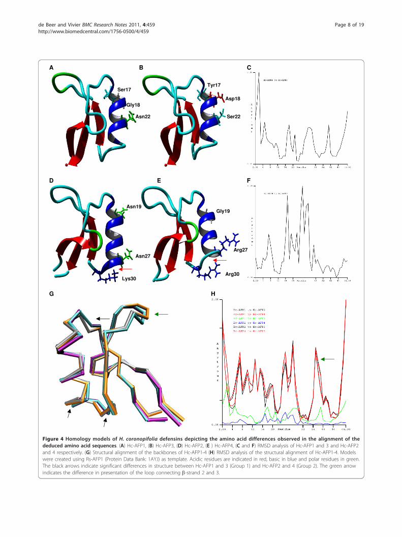
ent Hc-AFPs in combination with the alignment analysisof the Hc-AFPs showed that most of the amino acid dif-ferences occurred in the a-helical regions of the pep-tides. By plotting the amino acid differences betweenthe closely related Hc-AFP1 and 3 (94% similarity)where Ser17, Gly18 and Asn22 in Hc-AFP1 is replacedby Tyr17, Asp18 and Ser22 in Hc-AFP3 onto theirrespective models, it was observed that the change froma polar Gly18 to an acidic Aspartic18 residue in Hc-AFP3 resulted in a less polar a-helical region (Figure 4Aand 4B). Root mean square deviation (RMSD) compari-son between the structures of Hc-AFP1 and 3 revealedthat these differences, although occurring in the a-heli-cal region, caused a greater RMSD deviation in the N-and C-terminal ends of the peptide structure (Figure4C). Comparative analysis of the amino acids sequencesof Hc-AFP2 and 4 showed that they also share 94%
similarity, with Asn19 and 27 (numbering according toHc-AFP2) replaced with Gly19 and Arg27 and Lys30replaced with Arg30 in Hc-AFP4 (Figure 4D and 4E).Comparative analysis of the structural models of Hc-AFP2 and 4 revealed that these amino acid changes hadvery little effect on the overall structure of these pep-tides and only had a RMSD difference of 0.26 Å in thea-helical region of the peptides (Figure 4F), leading toan extended a-helix in Hc-AFP2 when compared to Hc-AFP4 (Figure 4D and 4E). These amino acid substitu-tions did however result in a difference of the predictedsurface properties between the Hc-AFP2 and 4 peptides.Hc-AFP4 is more basic and less hydrophilic in nature,whereas Hc-AFP2 is more polar in the regions sur-rounding the a-helix (Figure 4D and 4E).The amino acids encoding for the a-helical region of
Hc-AFP1 and 3 are unique when compared to defensinsisolated from the other Brassicaceae species. Structuralalignment of the backbones of the Hc-AFP1 - 4 modelsrevealed that these unique amino acids present in the a-helical region of Hc-AFP1 and 3 (designated Group 1)resulted in a difference in tertiary structure when com-pared to Hc-AFP2 and 4 (designated Group 2) (Figure4G). The a-helical regions of Group1 vs Group 2 had aRMSD value of more than 1.7 Å, and importantly, a sig-nificant difference of more than 1.6 Å was also observedin the Lb2b3 loop, which is encoded by amino acids 38to 41 (numbering according to Hc-AFP2) (Figure 4H).
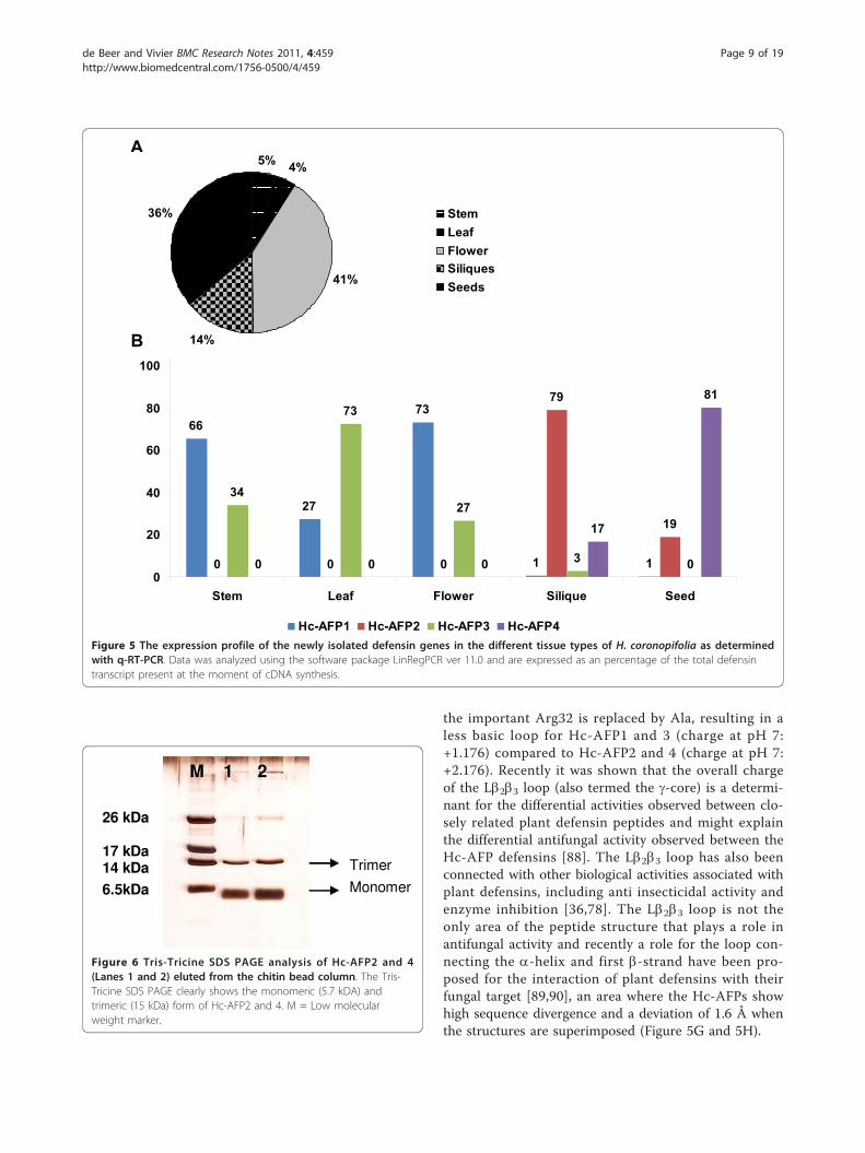
Expression analysis of the Hc-AFP encoding genesQuantitative RT-PCR (q-RT-PCR) analysis conducted onthe Heliophila defensin encoding genes revealed that thereproductive and storage organs of the H. coronopifoliaplant, which include the flowers, siliques and seeds, con-tributed to 91% of the observed defensin transcript pre-sent (Figure 5A). When considering the expressionpatterns of the individual peptides encoding genes, Hc-AFP1 and 3 showed expression in vegetative and repro-ductive tissues tested (leaves, stems, flowers), as well asvery low levels of expression in storage tissues (siliquesand seeds) (Figure 5B). The distribution of these tran-scripts within the tissue types differed however, withHc-AFP1 being the dominant transcript in stem andflower tissue contributing 66% and 73% respectively ofthe total defensin transcript present in these tissues. Hc-AFP3 was the dominant transcript in the leaf tissue con-tributing 73% of the observed defensin transcript pre-sent. In contrast Hc-AFP2 and 4 were the dominanttranscripts present in the storage organs of H. coronopi-folia and not expressed in leaves, stems or flowers (Fig-ure 5B). Hc-AFP2 was the dominant transcript in greensiliques contributing 79% of the total defensin transcriptpresent, but only contributed 19% of the total defensintranscript observed in mature seeds. Hc-AFP4 was
de Beer and Vivier BMC Research Notes 2011, 4:459http://www.biomedcentral.com/1756-0500/4/459
Page 3 of 19
Hc-AFP1
1 ATG GCT AAG TTT GCT TCC ATC ATT GCA CTT CTC TTC GCT GCT TTT 45 M A K F A S I I A L L F A A F 46 GTC CTC TTT GCT GCT TTT GAA GCA CCA ACA ATG GTG GAA GCA AGG 90 V L F A A F E A P T M V E A R 91 TAC TGT GAG AGA TCG AGT GGA ACA TGG TCA GGA GTT TGT GGA AAC 135 Y C E R S S G T W S G V C G N 136 AGT GGT AAG TGT AGT AAT CAA TGT CAG AGA CTT GAA GGA GCA GCA 180 S G K C S N Q C Q R L E G A A 181 CAT GGA TCT TGC AAC TAT GTC TTC CCA GCT CAC AAG TGT ATC TGT 225 H G S C N Y V F P A H K C I C 226 TAC TAC CCA TGT TAA 240 Y Y P C *
Hc-AFP2
1 ATG GCT AAG TTT GCT TCC ATC ATT GCC TTT TTC TTC GCT GCT CTT 45 M A K F A S I I A F F F A A L 46 GTT CTC TTT GCT GCT TTT GAA GCA CCA ACG ATA GTG GAA GCA CAA 90 V L F A A F E A P T I V E A Q 91 AAG TTG TGT GAG AGA CCA AGT GGA ACA TGG TCA GGA GTT TGT GGA 135 K L C E R P S G T W S G V C G 136 AAC AAT AAT GCG TGT AGG AAC CAG TGC ATC AAC CTT GAA AAA GCA 180 N N N A C R N Q C I N L E K A 181 CGA CAT GGA TCT TGC AAC TAT GTT TTC CCA GCT CAC AAG TGT ATC 225 R H G S C N Y V F P A H K C I 226 TGC TAC TTC CCA TGT TAA 243 C Y F P C *
Hc-AFP3
1 ATG GCT AAG TTT GCT TCC ATC ATC GCA CTT CTC TTC GCT GCT TTT 45 M A K F A S I I A L L F A A F 46 GTC CTC TTT GCT GCT TTT GAA GCA CCA ACA ATG GTG GAA GCA AGG 90 V L F A A F E A P T M V E A R 91 TAC TGT GAG AGA TCG AGT GGA ACA TGG TCA GGA GTT TGT GGA AAC 135 Y C E R S S G T W S G V C G N 136 ACT GAT AAG TGT AGT AGT CAG TGT CAG AGA CTT GAA GGA GCA GCA 180 T D K C S S Q C Q R L E G A A 181 CAT GGA TCT TGC AAC TAT GTC TTC CCA GCT CAC AAG TGT ATC TGT 225 H G S C N Y V F P A H K C I C 226 TAC TAC CCA TGT TAA 240 Y Y P C *
Hc-AFP4
1 ATG GCT AAG TTT GCT TCC ATC ATC ACT CTT TTC TTC GCT ACT CTT 45 M A K F A S I I T L F F A T L 46 CTT CTC TTT GCT GCT TTT GAA GCA CCA ACA ATG GTG GAA GCT CAG 90 L L F A A F E A P T M V E A Q 91 AAG TTG TGT GAG AGA CCA AGT GGA ACA TGG TCA GGT GTT TGT GGT 135 K L C E R P S G T W S G V C G 136 AAC AAT GGT GCG TGT AGG AAC CAG TGC ATC AGA CTT GAA AGA GCT 180 N N G A C R N Q C I R L E R A 181 CGA CAT GGA TCT TGC AAC TAT GTT TTC CCA GCA CAT AAG TGT ATC 225 R H G S C N Y V F P A H K C I 226 TGT TAC TTC CCA TGT TAA 243 C Y F P C *
Figure 1 The cDNA sequences encoding for Hc-AFPs, isolated by PCR from cDNA generated from H. coronopifolia stem, leaf, flower,siliques and seed tissue. The Hc-AFP defensins are encoded within the first 240 nucleotides for Hc-AFP1and 3 and the first 243 nucleotides forHc-AFP2 and 4. The deduced amino acid sequence encoded by each gene is indicated beneath its represented sequence.
de Beer and Vivier BMC Research Notes 2011, 4:459http://www.biomedcentral.com/1756-0500/4/459
Page 4 of 19
predominantly expressed in seeds and to a much lesserextent in green siliques (Figure 5B).
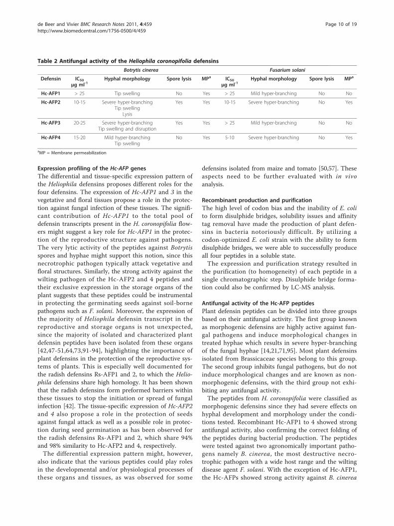
Bacterial production and purification of Hc-AFPsThe CBD-intein Hc-AFP fusions was successfully pro-duced in E. coli strain BL21DE3 Rosetta gami pLysS andwas visible as a 30 kDa band on a SDS PAGE gel (resultnot shown). The recombinant fusion proteins were suc-cessfully purified on a chitin bead column. On-columncleavage and peptide elution was confirmed with Tris-Tricine SDS PAGE analysis (Figure 6). The peptideswere correctly folded, displaying the expected trimericforms (15 kDa bands on the Tris-Tricine gel in Figure6) of the defensin peptides.Mass spectrometry analysis of the purified Hc-AFPs
revealed molecular masses (in Dalton) of 5471.25 forHc-AFP1, 5710.3 for Hc-AFP2, 5516.0 for Hc-AFP3 and5724.4 for Hc-AFP4 respectively, which correlates withtheir predicted mono-isotopic masses calculated withthe Expasy-Compute pI/Mw tool (Table 1) (-8 Dabecause of oxidized cysteines). This confirmed that thepurified defensins were derived from their respectivegenes in the bacterial expression vectors and indicatedthat the crucially important four disulphide bridgescommon to all plant defensins peptides formed.
Antifungal activity of the recombinant Hc-AFP peptidesThe four plant defensin peptides from H. coronopifoliashowed variable levels of activity against B. cinerea andF. solani in liquid plate assays (Table 2 and AdditionalFile 1 and 2).Hc-AFP2 was the most active of all the peptides tested
against B. cinerea with IC50 values ranging between 10-15 μg ml-1 and a similar IC50 against F. solani. Hc-AFP4inhibited B. cinerea with an IC50 value between 15-20μg ml-1, and strongly inhibited F. solani, having an IC50
value ranging between 5-10 μg ml-1 (Table 2 and Addi-tional File 2).Microscopical analysis conducted on B. cinerea hyphae
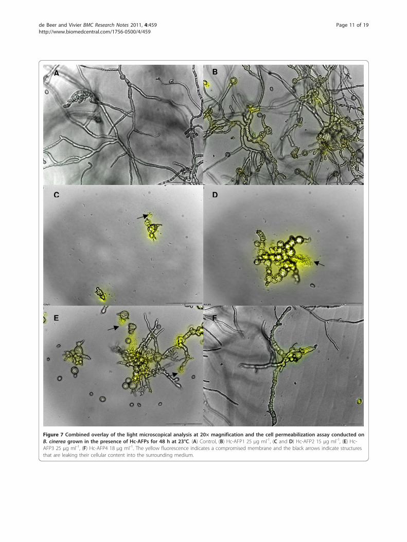
treated with the four defensin peptides revealed that allthe defensins induced changes in Botrytis hyphal mor-phology under the conditions tested. Compared to theuntreated control, hyper-branching, fungal tip swelling,increased granulation of hyphae and spores, as well ashyphal and spore disruption could be observed in thecultures treated with the peptides (Table 2 Figure 7 and
Additional File 3). In addition, Hc-AFP2 and 3 had asevere effect on spore and hyphae integrity, resulting indisintegration of the hyphae and spores, which could beobserved as leakage of the spore and hyphal cytoplasmiccontent into the surrounding environment. Moreover,assessment of propidium iodide assays revealed that theantifungal activity of all four Heliophila defensinsagainst B. cinerea were associated with an increase inmembrane permeabilization (Figure 7 and AdditionalFile 3).The peptides showed differential activity against F.
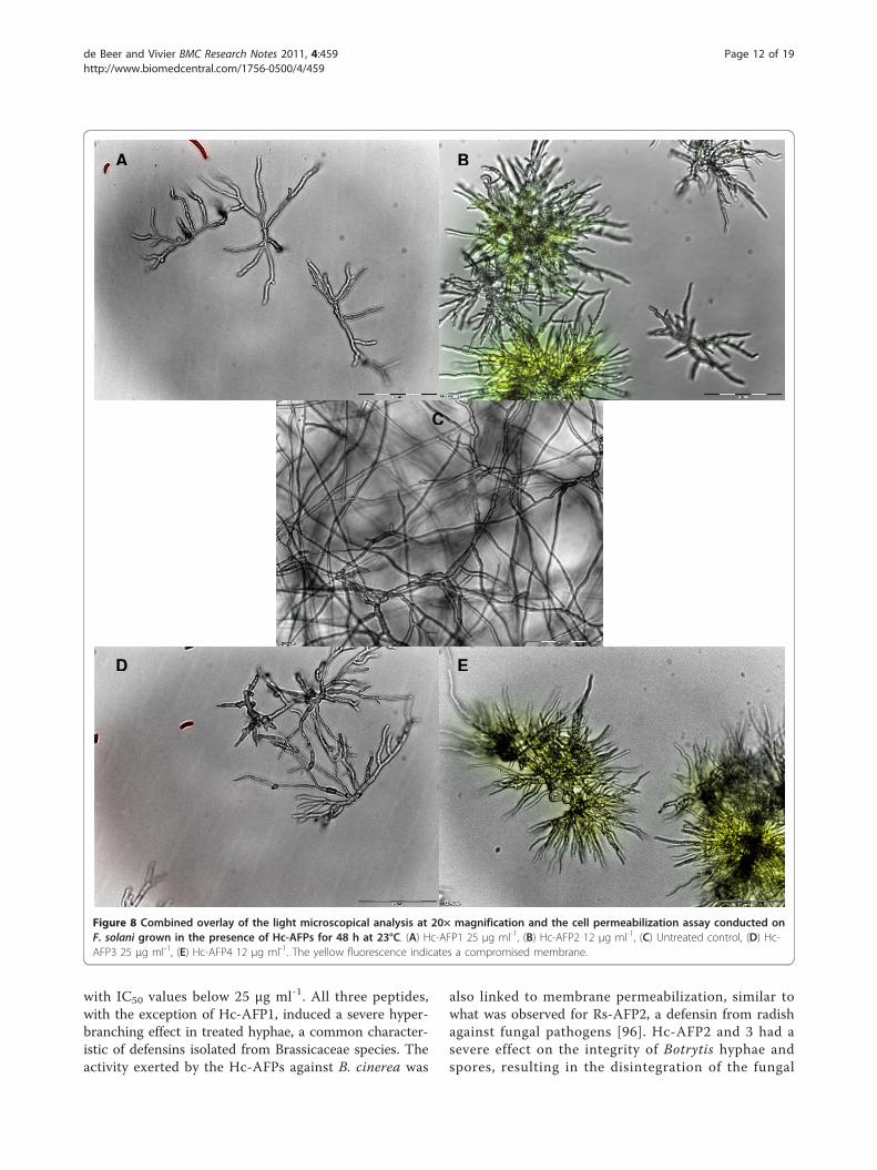
solani (Table 2 Figure 8 and Additional File 4). Hc-AFP2 and 4 caused severe hyper-branching, as well asmembrane permeabilization, whereas Hc-AFP1 and 3caused mild hyper-branching and no membrane disrup-tion against the wilting pathogen. Also, unlike theresults on Botrytis, no lysis was observed in F. solanispores and hyphae when treated with the four plantdefensins (Figure 8 and Additional File 4).
DiscussionPlant defensins isolated from Brassicaceae species haveespecially shown great promise in the fields of agricul-tural biotechnology and therapeutic drug design. Severalof these peptides have been overexpressed in crop spe-cies leading to disease resistant traits. The overexpres-sion of BrD1, wasabi defensin and Rs-AFP2 have led tothe engineering of disease resistant rice species[66,68,70,77], while the overexpression of AlfAFP1yielded disease resistant potatoes at field trail level[65,82]. The overexpression of wasabi defensin in toma-toes also showed resistance towards necrotrophic patho-gens [83]. Brassicaceae defensins are also used toevaluate the potential of defensin peptides in the designof new therapeutic drugs against human pathogenicyeast and fungi [62,63,84]. Moreover, since these defen-sins are well studied, they have been used as models tostudy the mechanisms of action of plant defensinsagainst their target organisms [16,17,24,85-87]. Of the449 defensin peptides listed in the protein database atthe NCBI, 379 peptides belong to the Brassicaceaefamily.Alignment analysis of the Brassicaceae defensin genes
in the NCBI database revealed a high level of similarity(72%) in the first 20 bp that encode the start of the sig-nal peptide (Additional File 5). By exploiting this
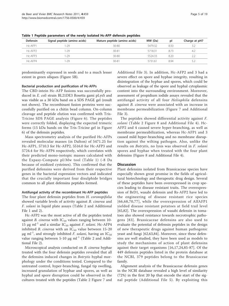
Table 1 Peptide parameters of the newly isolated Hc-AFP defensin peptides
Defensin Signal peptide (amino acids) Mature peptide (amino acids) MW (Da) pI Charge at pH7
Hc-AFP1 1-29 30-80 5479.32 8.50 3.2
Hc-AFP2 1-29 30-81 5718.31 8.73 4.2
Hc-AFP3 1-29 30-80 5524.33 8.20 2.2
Hc-AFP4 1-29 30-81 5731.61 8.94 5.2
de Beer and Vivier BMC Research Notes 2011, 4:459http://www.biomedcentral.com/1756-0500/4/459
Page 5 of 19
Arecaceae
Asteraceae
Brassicaceae
Cucurbitaceae
Ginkgoaceae
Fabaceae
Nelumbonaceae
Pinaceae
Plantaginaceae
Poaceae
Rosaceae
Solanaceae
Sapindaceae
Saxifragaceae
Ranunculaceae
Figure 2 The phylogenetic relationship of the newly isolated Hc-AFPs with members of the plant defensin super family. The deducedamino acid sequences of the newly isolated defensins (indicated with *) were aligned in ClustalX with other members of the defensin superfamily isolated from various plant genera. The tree was created in Arbodraw. The newly isolated defensins showed the closest relation todefensin peptides isolated from other Brassicaceae species.
de Beer and Vivier BMC Research Notes 2011, 4:459http://www.biomedcentral.com/1756-0500/4/459
Page 6 of 19
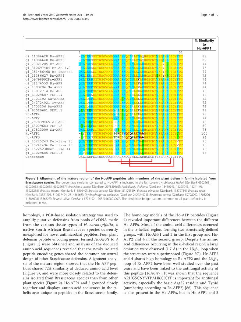
homology, a PCR-based isolation strategy was used toamplify putative defensins from pools of cDNA madefrom the various tissue types of H. coronopifolia, anative South African Brassicaceae species currentlyunexplored for novel antimicrobial peptides. Four plantdefensin peptide encoding genes, termed Hc-AFP1 to 4(Figure 1) were obtained and analysis of the deducedamino acid sequences revealed that the newly isolatedpeptide encoding genes shared the common structuraldesign of other Brassicaceae defensins. Alignment analy-sis of the mature region showed that the Hc-AFP pep-tides shared 72% similarity at deduced amino acid level(Figure 3), and were more closely related to the defen-sins isolated from Brassicaceae species than from otherplant species (Figure 2). Hc-AFP1 and 3 grouped closelytogether and displays amino acid sequences in the a-helix area unique to peptides in the Brassicaceae family.
The homology models of the Hc-AFP peptides (Figure4) revealed important differences between the differentHc-AFPs. Most of the amino acid differences occurredin the a-helical region, forming two structurally definedgroups, with Hc-AFP1 and 3 in the first group and Hc-AFP2 and 4 in the second group. Despite the aminoacid differences occurring in the a-helical region a largedeviation were observed (1.7 Å) in the Lb2b3 loop whenthe structures were superimposed (Figure 5G). Hc-AFP2and 4 shares high homology to Rs-AFP2 and the Lb2b3loop of Rs-AFP2 have been well studied over the pastyears and have been linked to the antifungal activity ofthis peptide [16,86,87]. It was shown that the sequenceARHGSCNYVFPAHKCICYF is important for antifungalactivity, especially the basic Arg32 residue and Tyr48(numbering according to Rs-AFP2) [86]. This sequenceis also present in the Hc-AFPs, but in Hc-AFP1 and 3
% Similarity to
Hc-AFP11 51
gi_11386628 Rs-AFP3 -KLCERSSGTWSGVCGNNNACKNQCIRLEGAQHGSCNYVFPAHKCICYFPCgi_11386640 Bn-AFP3 -KLCERSSGTWSGVCGNNNACKNQCIRLEGAQHGSCNYVFPAHKCICYFPCgi_23321205 Br-AFP QKLCERSSGTWSGVCGNNNACKNQCINLEGARHGSCNYVFPYHRCICYFPCgi_310697404 Br-AFP1.2 -KLCERSSGTWSGVCGNNNACKNQCINLEGARHGSCNYVFPYHRCICYFPCgi_281486468 Br insectR QKLCERSSGTWSGVCGNNNACKNQCINLEGARHGSCNYVFPYHRCICYFPCgi_11386627 Rs-AFP4 QKLCERSSGTWSGVCGNNNACKNQCINLEGARHGSCNYIFPYHRCICYFPCgi_59798992Rs-AFP1 QKLCERPSGTWSGVCGNNNACKNQCINLEKARHGSCNYVFPAHKCICYFPCgi_81176559 Bj-AFP QKLCERPSGTWSGVCGNNNACKNQCINLEKARHGSCNYVFPAHKCICYFPCgi_1703204 Sa-AFP1 QKLCERPSGTWSGVCGNNNACKNQCINLEKARHGSCNYVFPAHKCICYFPCgi_13872714 Bo-AFP QKLCERPSGTWSGVCGNNNACKNQCIRLEKARHGSCNYVFPAHKCICYFPCgi_63029687 PDF1.4 QKLCERASGTWSGVCGNNNACKNQCIRLEKARHGSCNYVFPAHKCICYFPCgi_1703192 Sa-AFP2a QKLCQRPSGTWSGVCGNNNACRNQCINLEKARHGSCNYVFPAHKCICYFPCgi_242724021 Ov-AFP QKLCQRPSGTWSGVCGNNNACKNQCINLEKARHGSCNYVFPAHKCICYFPCgi_1703206 Rs-AFP2 QKLCQRPSGTWSGVCGNNNACKNQCIRLEKARHGSCNYVFPAHKCICYFPCgi_63029681 PDF1.1 QRLCEKPSGTWSGVCGNNGACRNQCIRLEKARHGSCNYVFPAHKCICYFPCHc-AFP4 QKLCERPSGTWSGVCGNNGACRNQCIRLERARHGSCNYVFPAHKCICYFPCHc-AFP2 QKLCERPSGTWSGVCGNNNACRNQCINLEKARHGSCNYVFPAHKCICYFPCgi_297839465 Al-AFP QKLCEKPSGTWSGVCGNSGACKNQCINLEGARHGSCNYVFPAHKCICYFPCgi_63029683 PDF1.2 QRLCEKPSGTWSGVCGNSGACKNQCINLEGARHGSCNYVFPAHKCICYFPCgi_62823009 Sa-AFP QKLCERPSGTWSGVCGNSNSCKNQCINLEGARHGSCNYVFPAHKCICYFPCHc-AFP1 -RYCERSSGTWSGVCGNSGKCSNQCQRLEGAAHGSCNYVFPAHKCICYYPCHc-AFP3 -RYCERSSGTWSGVCGNTDKCSSQCQRLEGAAHGSCNYVFPAHKCICYYPCgi_15225243 Def-like 15 QKLCEKPSGTWSGVCGNSNACKNQCINLEGAKHGSCNYVFPAHKCICYVPCgi_15241496 Def-like 16 QKLCEKPSGTWSGVCGNSNACKNQCINLEGAKHGSCNYVFPAHKCICYVPCgi_15225238Def-like 14 QKLCEKPSGTWSGVCGNSNACKNQCINLEGAKHGSCNYVFPAHKCICYFPCgi_63029685 PDF1.3 QKFCEKPSGTWSGVCGNSNACKNQCINLEGAKHGSCNYVFPAHKCICYFPCConsensus QKLCERPSGTWSGVCGNNNACKNQCINLEGARHGSCNYVFPAHKCICYFPC
82827476747274747476767272747878747880781009476767676
Figure 3 Alignment of the mature region of the Hc-AFP peptides with members of the plant defensin family isolated fromBrassicaceae species. The percentage similarity compared to Hc-AFP1 is indicated in the last column. Arabidopsis halleri: [GenBank 63029681,63029683, 63029685, 63029687]; Arabidopsis lyrata: [GenBank 297839465]; Arabidopsis thaliana: [GenBank 18410943, 15225243, 15241496,15225238]; Brassica napus: [GenBank 11386640]; Brassica juncea: [GenBank 81176559]; Brassica oleracea: [Genbank 13872714]; Brassica rapa:[GenBank 23321205, 310697404, 281486468]; Orychophragmus violaceus: [GenBank 242724021]; Raphanus sativa: [GenBank 59798992, 1703206,1138662811386627]; Sinapsis alba: [GenBank 1703192, 170320462823009]. The disulphide bridge pattern, common to all plant defensins, isindicated in red.
de Beer and Vivier BMC Research Notes 2011, 4:459http://www.biomedcentral.com/1756-0500/4/459
Page 7 of 19
A B C
D E F
G H
Ser17
Gly18
Asn22 Ser22
Tyr17
Asp18
Asn19
Asn27
Lys30
Gly19
Arg27
Arg30
Figure 4 Homology models of H. coronopifolia defensins depicting the amino acid differences observed in the alignment of thededuced amino acid sequences. (A) Hc-AFP1, (B) Hc-AFP3, (D) Hc-AFP2, (E ) Hc-AFP4, (C and F) RMSD analysis of Hc-AFP1 and 3 and Hc-AFP2and 4 respectively. (G) Structural alignment of the backbones of Hc-AFP1-4 (H) RMSD analysis of the structural alignment of Hc-AFP1-4. Modelswere created using Rs-AFP1 (Protein Data Bank: 1AYJ) as template. Acidic residues are indicated in red, basic in blue and polar residues in green.The black arrows indicate significant differences in structure between Hc-AFP1 and 3 (Group 1) and Hc-AFP2 and 4 (Group 2). The green arrowindicates the difference in presentation of the loop connecting b-strand 2 and 3.
de Beer and Vivier BMC Research Notes 2011, 4:459http://www.biomedcentral.com/1756-0500/4/459
Page 8 of 19
the important Arg32 is replaced by Ala, resulting in aless basic loop for Hc-AFP1 and 3 (charge at pH 7:+1.176) compared to Hc-AFP2 and 4 (charge at pH 7:+2.176). Recently it was shown that the overall chargeof the Lb2b3 loop (also termed the g-core) is a determi-nant for the differential activities observed between clo-sely related plant defensin peptides and might explainthe differential antifungal activity observed between theHc-AFP defensins [88]. The Lb2b3 loop has also beenconnected with other biological activities associated withplant defensins, including anti insecticidal activity andenzyme inhibition [36,78]. The Lb2b3 loop is not theonly area of the peptide structure that plays a role inantifungal activity and recently a role for the loop con-necting the a-helix and first b-strand have been pro-posed for the interaction of plant defensins with theirfungal target [89,90], an area where the Hc-AFPs showhigh sequence divergence and a deviation of 1.6 Å whenthe structures are superimposed (Figure 5G and 5H).
66
27
73
1 10 0 0
79
19
34
73
27
3 00 0 0
17
81
0
20
40
60
80
100
120
140
Stem Leaf Flower Silique Seed
Hc-AFP1 Hc-AFP2 Hc-AFP3 Hc-AFP4
5% 4%
41%
14%
36% StemLeafFlowerSiliquesSeeds
A
B
Figure 5 The expression profile of the newly isolated defensin genes in the different tissue types of H. coronopifolia as determinedwith q-RT-PCR. Data was analyzed using the software package LinRegPCR ver 11.0 and are expressed as an percentage of the total defensintranscript present at the moment of cDNA synthesis.
M 1 2
26 kDa 17 kDa 14 kDa
6.5kDa
Trimer
Monomer
Figure 6 Tris-Tricine SDS PAGE analysis of Hc-AFP2 and 4(Lanes 1 and 2) eluted from the chitin bead column. The Tris-Tricine SDS PAGE clearly shows the monomeric (5.7 kDA) andtrimeric (15 kDa) form of Hc-AFP2 and 4. M = Low molecularweight marker.
de Beer and Vivier BMC Research Notes 2011, 4:459http://www.biomedcentral.com/1756-0500/4/459
Page 9 of 19
Expression profiling of the Hc-AFP genesThe differential and tissue-specific expression pattern ofthe Heliophila defensins proposes different roles for thefour defensins. The expression of Hc-AFP1 and 3 in thevegetative and floral tissues propose a role in the protec-tion against fungal infection of these tissues. The signifi-cant contribution of Hc-AFP1 to the total pool ofdefensin transcripts present in the H. coronopifolia flow-ers might suggest a key role for Hc-AFP1 in the protec-tion of the reproductive structure against pathogens.The very lytic activity of the peptides against Botrytisspores and hyphae might support this notion, since thisnecrotrophic pathogen typically attack vegetative andfloral structures. Similarly, the strong activity against thewilting pathogen of the Hc-AFP2 and 4 peptides andtheir exclusive expression in the storage organs of theplant suggests that these peptides could be instrumentalin protecting the germinating seeds against soil-bornepathogens such as F. solani. Moreover, the expression ofthe majority of Heliophila defensin transcript in thereproductive and storage organs is not unexpected,since the majority of isolated and characterized plantdefensin peptides have been isolated from these organs[42,47-51,64,73,91-94], highlighting the importance ofplant defensins in the protection of the reproductive sys-tems of plants. This is especially well documented forthe radish defensins Rs-AFP1 and 2, to which the Helio-phila defensins share high homology. It has been shownthat the radish defensins form preformed barriers withinthese tissues to stop the initiation or spread of fungalinfection [42]. The tissue-specific expression of Hc-AFP2and 4 also propose a role in the protection of seedsagainst fungal attack as well as a possible role in protec-tion during seed germination as has been observed forthe radish defensins Rs-AFP1 and 2, which share 94%and 98% similarity to Hc-AFP2 and 4, respectively.The differential expression pattern might, however,
also indicate that the various peptides could play rolesin the developmental and/or physiological processes ofthese organs and tissues, as was observed for some
defensins isolated from maize and tomato [50,57]. Theseaspects need to be further evaluated with in vivoanalysis.
Recombinant production and purificationThe high level of codon bias and the inability of E. colito form disulphide bridges, solubility issues and affinitytag removal have made the production of plant defen-sins in bacteria notoriously difficult. By utilizing acodon-optimized E. coli strain with the ability to formdisulphide bridges, we were able to successfully produceall four peptides in a soluble state.The expression and purification strategy resulted in
the purification (to homogeneity) of each peptide in asingle chromatographic step. Disulphide bridge forma-tion could also be confirmed by LC-MS analysis.
Antifungal activity of the Hc-AFP peptidesPlant defensin peptides can be divided into three groupsbased on their antifungal activity. The first group knownas morphogenic defensins are highly active against fun-gal pathogens and induce morphological changes intreated hyphae which results in severe hyper-branchingof the fungal hyphae [14,21,71,95]. Most plant defensinsisolated from Brassicaceae species belong to this group.The second group inhibits fungal pathogens, but do notinduce morphological changes and are known as non-morphogenic defensins, with the third group not exhi-biting any antifungal activity.The peptides from H. coronopifolia were classified as
morphogenic defensins since they had severe effects onhyphal development and morphology under the condi-tions tested. Recombinant Hc-AFP1 to 4 showed strongantifungal activity, also confirming the correct folding ofthe peptides during bacterial production. The peptideswere tested against two agronomically important patho-gens namely B. cinerea, the most destructive necro-trophic pathogen with a wide host range and the wiltingdisease agent F. solani. With the exception of Hc-AFP1,the Hc-AFPs showed strong activity against B. cinerea
Table 2 Antifungal activity of the Heliophila coronopifolia defensins
Botrytis cinerea Fusarium solani
Defensin IC50μg ml-1
Hyphal morphology Spore lysis MPa IC50μg ml-1
Hyphal morphology Spore lysis MPa
Hc-AFP1 > 25 Tip swelling No Yes > 25 Mild hyper-branching No No
Hc-AFP2 10-15 Severe hyper-branchingTip swelling
Lysis
Yes Yes 10-15 Severe hyper-branching No Yes
Hc-AFP3 20-25 Severe hyper-branchingTip swelling and disruption
Yes Yes > 25 Mild hyper-branching No No
Hc-AFP4 15-20 Mild hyper-branchingTip swelling
No Yes 5-10 Severe hyper-branching No Yes
aMP = Membrane permeabilization
de Beer and Vivier BMC Research Notes 2011, 4:459http://www.biomedcentral.com/1756-0500/4/459
Page 10 of 19
A B
C D
E F
Figure 7 Combined overlay of the light microscopical analysis at 20× magnification and the cell permeabilization assay conducted onB. cinerea grown in the presence of Hc-AFPs for 48 h at 23°C. (A) Control, (B) Hc-AFP1 25 μg ml-1, (C and D) Hc-AFP2 15 μg ml-1, (E) Hc-AFP3 25 μg ml-1, (F) Hc-AFP4 18 μg ml-1. The yellow fluorescence indicates a compromised membrane and the black arrows indicate structuresthat are leaking their cellular content into the surrounding medium.
de Beer and Vivier BMC Research Notes 2011, 4:459http://www.biomedcentral.com/1756-0500/4/459
Page 11 of 19
with IC50 values below 25 μg ml-1. All three peptides,with the exception of Hc-AFP1, induced a severe hyper-branching effect in treated hyphae, a common character-istic of defensins isolated from Brassicaceae species. Theactivity exerted by the Hc-AFPs against B. cinerea was
also linked to membrane permeabilization, similar towhat was observed for Rs-AFP2, a defensin from radishagainst fungal pathogens [96]. Hc-AFP2 and 3 had asevere effect on the integrity of Botrytis hyphae andspores, resulting in the disintegration of the fungal
A
C
D E
B
Figure 8 Combined overlay of the light microscopical analysis at 20× magnification and the cell permeabilization assay conducted onF. solani grown in the presence of Hc-AFPs for 48 h at 23°C. (A) Hc-AFP1 25 μg ml-1, (B) Hc-AFP2 12 μg ml-1, (C) Untreated control, (D) Hc-AFP3 25 μg ml-1, (E) Hc-AFP4 12 μg ml-1. The yellow fluorescence indicates a compromised membrane.
de Beer and Vivier BMC Research Notes 2011, 4:459http://www.biomedcentral.com/1756-0500/4/459
Page 12 of 19
membrane and leakage of the cytoplasmic content intothe surrounding environment. This lytic activity has notpreviously been described for Brassicaceae defensinsaccording to our knowledge. The differential activityagainst F. solani, where Hc-AFP1 and 3 show reducedactivity compared to Hc-AFP2 and 4, correlates wellwith their expression patterns. F. solani is a soil patho-gen and Hc-AFP2 and 4, which show expression only inthe storage organs, shows strong activity against thispathogen, strengthening the proposed role for thesepeptides during seed germination and seedling protec-tion against soil borne pathogens. The expression ofHc-AFP1 and 3 in the vegetative tissues might alsoexplain why they show more activity against pathogensevolved to infect vegetative tissues like the necrotrophicpathogen B. cinerea.The effects on Botrytis (positive membrane disruption,
severe morphological effects and even lytic activity) sug-gest that the activities could be orchestrated with themembrane being the primary target. However, recentevidence suggests that the cell wall also might play arole in membrane permeabilization [67] and that themembrane might actually be the secondary target. Inter-estingly, the Fusarium data indicated that Hc-AFP1 and3 did not affect the membranes of the pathogen, sinceno membrane permeabilization was observed. Thesedivergent activities of the Heliophilia peptides should bestudied further. Future work will focus on exploring thestructure-function relationships in the four peptides,and the implications on activity, specifically since thesefour peptides are highly homologous on amino acidsequence level, but display a few pointed changes in cer-tain important defensin motifs which might be under-pinning the observed variation in activities and mode-ofaction.
ConclusionsThe homology that resides within the signal peptides ofplant defensins belonging to the same plant family issignificant and allowed us to successfully implement aPCR-based method to isolate four Brassicaceae defen-sins. This strategy might be useful to isolate new defen-sin sequences from unsequenced plants speciesbelonging to the same plant family. Despite the highlevel of homology on sequence level that was observedfor the peptides, they were predicted to differ in theirstructural and surface properties, aspects that are knownto influence activity levels and range. These aspects, aswell as their observed divergent expression patterns,activities and modes of action against two test patho-gens, provide interest to explore the structure-activityrelationship of these peptides further.
MethodsMicrobial strains and plant materialEscherichia coli strain DH5a were used for all cloningexperiments, while E. coli strain BL21 Rosetta-gamipLysS DE3 (Novagen, Madison, WI, USA) were used forrecombinant protein production. Fusarium solani andBotrytis cinerea cultures were obtained from the Depart-ment of Plant Pathology (DPP), Stellenbosch Universityand maintained on potato dextrose agar at 25°C untilsporulation. Spores were harvested in dH2O and sporeconcentrations determined using a haemocytometer.Heliophila coronopifolia seeds were obtained from Sil-verhill seed company, South Africa. H. coronopifoliaplants were established in potting soil from seeds andmaintained under green-house conditions at 25°C.
Design of Primer SPDEF-5’The design of primer SPDEF-5 is based on the highlevel of homology that exists within the nucleotidesequences encoding for the signal peptides of plantdefensin peptides belonging to the plant family Brassica-ceae. Plant defensin encoding sequences isolated fromBrassicaceae species was identified in the Genbank data-base of the National Centre for Biotechnological infor-mation (NCBI). The first 50 nucleotides encoding forthe N-terminal signal peptide of the Brassicaceae plantdefensin peptides were selected and aligned in AlignX(Invitrogen, Carlsbad, USA) (Additional File 5). 72%similarity existed over the first 50 amino acids. The con-sensus sequence were identified and the first 20 nucleo-tides were used to design primer SPDEF-5’ (5’-ATGGCTAAGTTTGCTTCCATCAT-3’).
RNA isolation and cDNA synthesisTotal RNA was isolated from stem, leaf, flower, greensiliques and mature seeds of H. coronopifolia. The tissuewas ground to a fine powder in the presence of liquidnitrogen and total RNA was extracted from 200 mgpowdered tissue according to Chang et al [97]. TotalRNA was precipitated with 3 M LiCl and washed with70% (v/v) ethanol and dissolved in 26 μl RNase freewater. The samples were treated with DNaseI (RocheDiagnostics GmbH, Mannheim, Germany) to removegenomic DNA contamination in a 30 μl reaction. Sam-ple volumes was adjusted to 200 μl with RNase freewater and the DNaseI removed by extracting with anequal volume phenol/chloroform (50:50 v/v), followedby an equal volume chloroform to remove the phenol.The RNA was precipitated with a 1/10 volume 3 MNaOAc and 0.7 volumes isopropanol, washed with 70%(v/v) ethanol and dissolved in RNase free water. Firststrand cDNA was synthesized from 1 μg of total RNA
de Beer and Vivier BMC Research Notes 2011, 4:459http://www.biomedcentral.com/1756-0500/4/459
Page 13 of 19
using an anchored oligo dT23 primer (Sigma, St Louis,USA) and Superscript III (Invitrogen, Carlsbad, USA).cDNA synthesis was performed as described by themanufacturer.
Gene isolation and cloningThe coding regions of potential plant defensinsequences were PCR amplified from total stem, leaf,flower, silique and seed cDNA using primer set SPDEF-5’ and the anchored oligo dT23 primer (Sigma, St Louis,USA) used for cDNA synthesis. The PCR reaction wasperformed in a 50 μl reaction containing: 1× Expandbuffer with 1.5 mM MgCl2, 0.2 mM dNTPs, 200 nMSPDEF-5’ primer, 200 nM oligo dT23, 10 ng templateDNA and 1 U Expand high fidelity polymerase (RocheDiagnostics GmbH, Mannheim, Germany). The PCRprogram was as follows: 95°C for 5 min; followed by 30cycles of 95°C for 45 sec, 48°C for 30 sec and 72°C for45 sec. PCR products were cloned into pGEM-T easyvector (Promega Corporation, Madison, USA) and posi-tive clones were identified through restriction digestwith EcoRI. Plasmids containing inserts were confirmedby sequencing. Obtained sequences were analyzed withthe BLASTN algorithm http://blast.ncbi.nlm.nih.gov/Blast.cgi at the NCBI and clones containing open read-ing frames encoding for plant defensins were identifiedand termed pGEM-Hc1-4. The sequences were depos-ited to Genbank with the following accession numbers:JN203136 (Hc-AFP1), JN203137 (Hc-AFP2), JN203138(Hc-AFP3) and JN203139 (Hc-AFP4).
Bioinformatical analysis of the four H. coronopifoliadefensin sequencesThe deduced amino acid sequences of Hc-AFP1-4 wascreated in BioEdit [98] and analyzed with the Expasy-Compute pI/Mw tool http://web.expasy.org/compute_pi/to obtain the different peptide parameters and Biochem-istry online http://vitalonic.narod.ru/biochem to deter-mine the overall charge of the peptides and their Lb2b3-loops. The peptide structure of each peptide was evalu-ated for the presence of a signal peptide sequence withSignalP http://www.cbs.dtu.dk/services/SignalP/ and thepossible disulphide bridge pattern for each peptide wasdetermined using the web services DIpro http://down-load.igb.uci.edu/bridge.html.The deduced amino acid sequences encoding for the
mature plant defensin peptides were aligned against adiverse set of mature plant defensin sequences isolatedfrom various plant genera. All sequences were obtainedfrom the NCBI and alignment with the newly isolateddefensins was performed in ClustalX [99]. A graphicalrepresentation of the phylogenetic tree was created inArbodraw [100].
Homology models for each Hc-AFP peptide was cre-ated with the Bioinformatics toolkit at the Max PlanckInstitute for developmental biology http://toolkit.tuebin-gen.mpg.de/. The crystal structure of Rs-AFP1 (ProteinData Bank: 1AYJ) from radish was used as template.The models obtained were refined and analyzed withYASARA structure [101,102] and the FoldX plugin[103]. Models were visualized in Visual MolecularDynamics ver 1.8.4 and the final images rendered withPOV-Ray.
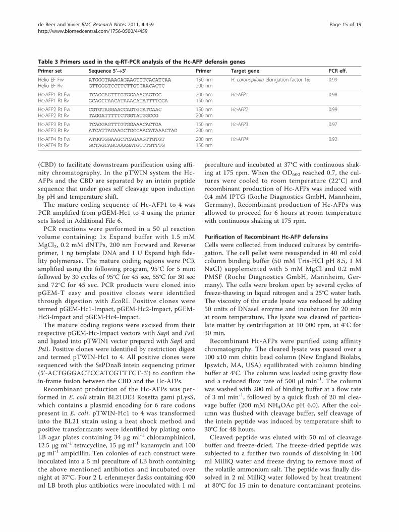
q-RT-PCR analysis of the Hc-AFP encoding genesAnalysis was conducted on each of the newly isolatedgenes using the primer sets listed in Table 3. Each pri-mer set was optimized to determine the optimal ratio ofthe forward and reverse primer in the primer set. ThePCR efficiency was determined by setting up a standardcurve prepared from the cDNA used to isolate therespective genes. The standard curve consisted of a 1/4to 1/1024 dilution of the respective cDNA template. q-RT-PCR reactions with different ratios of forward andreverse primers were performed with the KAPA SYBRFAST qPCR Kit (Kapa Biosystems, South Africa) in a 20μl reaction. All q-RT-PCR analysis were performed on aABI7500 Real-Time PCR System (Applied Biosystems,South Africa) with the following program: 95°C for 5min followed by 40 cycles of 95°C, 15 sec; 60°C, 32 sec.The 40 cycles was followed by a dissociation curve con-sisting of a ramp from 95°C to 60°C. The Ct valuesobtained were used as input data in the REST2009 soft-ware package [104] to calculate the PCR efficiency ofeach primer set. The optimized q-RT-PCR primer ratioswere used to evaluate the expression of Hc-AFP1-4 inthe different H. coronopifolia tissue types. The q-RT-PCR reactions were done as described above with eachreaction containing the optimized primer concentrationlisted in Table 3. The data obtained were analyzed inLinRegPCR v11.0 software package [105] to determinethe transcript levels for each gene present at the time ofcDNA synthesis in the various tissue types. Elongationfactor alpha (EFa) was used to standardize the expres-sion levels obtained between the different tissues. Thedata for the individual genes are expressed as a percen-tage of the total defensin transcript present in the tissue.
Recombinant production of Hc-AFPs in E. coliHc-AFPs were produced in E. coli by using theIMPACT system (New England Biolabs, Ipswich, MA,USA). The DNA regions encoding for mature Hc-AFPswas cloned into the pTWIN1 vector, which allows forexpression under control of the IPTG inducible T7 pro-moter. The cloning strategy allowed for a fusionbetween the Hc-AFPs and a chitin binding domain
de Beer and Vivier BMC Research Notes 2011, 4:459http://www.biomedcentral.com/1756-0500/4/459
Page 14 of 19
(CBD) to facilitate downstream purification using affi-nity chromatography. In the pTWIN system the Hc-AFPs and the CBD are separated by an intein peptidesequence that under goes self cleavage upon inductionby pH and temperature shift.The mature coding sequence of Hc-AFP1 to 4 was
PCR amplified from pGEM-Hc1 to 4 using the primersets listed in Additional File 6.PCR reactions were performed in a 50 μl reaction
volume containing: 1x Expand buffer with 1.5 mMMgCl2, 0.2 mM dNTPs, 200 nm Forward and Reverseprimer, 1 ng template DNA and 1 U Expand high fide-lity polymerase. The mature coding regions were PCRamplified using the following program, 95°C for 5 min;followed by 30 cycles of 95°C for 45 sec, 55°C for 30 secand 72°C for 45 sec. PCR products were cloned intopGEM-T easy and positive clones were identifiedthrough digestion with EcoRI. Positive clones weretermed pGEM-Hc1-Impact, pGEM-Hc2-Impact, pGEM-Hc3-Impact and pGEM-Hc4-Impact.The mature coding regions were excised from their
respective pGEM-Hc-Impact vectors with SapI and PstIand ligated into pTWIN1 vector prepared with SapI andPstI. Positive clones were identified by restriction digestand termed pTWIN-Hc1 to 4. All positive clones weresequenced with the SsPDnaB intein sequencing primer(5’-ACTGGGACTCCATCGTTTCT-3’) to confirm thein-frame fusion between the CBD and the Hc-AFPs.Recombinant production of the Hc-AFPs was per-
formed in E. coli strain BL21DE3 Rosetta gami pLysS,which contains a plasmid encoding for 6 rare codonspresent in E. coli. pTWIN-Hc1 to 4 was transformedinto the BL21 strain using a heat shock method andpositive transformants were identified by plating ontoLB agar plates containing 34 μg ml-1 chloramphinicol,12.5 μg ml-1 tetracycline, 15 μg ml-1 kanamycin and 100μg ml-1 ampicillin. Ten colonies of each construct wereinoculated into a 5 ml preculture of LB broth containingthe above mentioned antibiotics and incubated overnight at 37°C. Four 2 L erlenmeyer flasks containing 400ml LB broth plus antibiotics were inoculated with 1 ml
preculture and incubated at 37°C with continuous shak-ing at 175 rpm. When the OD600 reached 0.7, the cul-tures were cooled to room temperature (22°C) andrecombinant production of Hc-AFPs was induced with0.4 mM IPTG (Roche Diagnostics GmbH, Mannheim,Germany). Recombinant production of Hc-AFPs wasallowed to proceed for 6 hours at room temperaturewith continuous shaking at 175 rpm.
Purification of Recombinant Hc-AFP defensinsCells were collected from induced cultures by centrifu-gation. The cell pellet were resuspended in 40 ml coldcolumn binding buffer (50 mM Tris-HCl pH 8.5, 1 MNaCl) supplemented with 5 mM MgCl and 0.2 mMPMSF (Roche Diagnostics GmbH, Mannheim, Ger-many). The cells were broken open by several cycles offreeze-thawing in liquid nitrogen and a 25°C water bath.The viscosity of the crude lysate was reduced by adding50 units of DNaseI enzyme and incubation for 20 minat room temperature. The lysate was cleared of particu-late matter by centrifugation at 10 000 rpm, at 4°C for30 min.Recombinant Hc-AFPs were purified using affinity
chromatography. The cleared lysate was passed over a100 x10 mm chitin bead column (New England Biolabs,Ipswich, MA, USA) equilibrated with column bindingbuffer at 4°C. The column was loaded using gravity flowand a reduced flow rate of 500 μl min-1. The columnwas washed with 200 ml of binding buffer at a flow rateof 3 ml min-1, followed by a quick flush of 20 ml clea-vage buffer (200 mM NH4OAc pH 6.0). After the col-umn was flushed with cleavage buffer, self cleavage ofthe intein peptide was induced by temperature shift to30°C for 48 hours.Cleaved peptide was eluted with 50 ml of cleavage
buffer and freeze-dried. The freeze-dried peptide wassubjected to a further two rounds of dissolving in 100ml MilliQ water and freeze drying to remove most ofthe volatile ammonium salt. The peptide was finally dis-solved in 2 ml MilliQ water followed by heat treatmentat 80°C for 15 min to denature contaminant proteins.
Table 3 Primers used in the q-RT-PCR analysis of the Hc-AFP defensin genes
Primer set Sequence 5’®3’ Primer Target gene PCR eff.
Helio EF FwHelio EF Rv
ATGGGTAAAGAGAAGTTTCACATCAAGTTGGGTCCTTCTTGTCAACACTC
150 nm200 nm
H. coronopifolia elongation factor 1a 0.99
Hc-AFP1 Rt FwHc-AFP1 Rt Rv
TCAGGAGTTTGTGGAAACAGTGGGCAGCCAACATAAACATATTTTGGA
200 nm150 nm
Hc-AFP1 0.98
Hc-AFP2 Rt FwHc-AFP2 Rt Rv
CGTGTAGGAACCAGTGCATCAACTAGGATTTTTCTGGTATGGCCG
150 nm200 nm
Hc-AFP2 0.99
Hc-AFP3 Rt FwHc-AFP3 Rt Rv
TCAGGAGTTTGTGGAAACACTGAATCATTAGAAGCTGCCAACATAAACTAG
150 nm200 nm
Hc-AFP3 0.97
Hc-AFP4 Rt FwHc-AFP4 Rt Rv
ATGGTGGAAGCTCAGAAGTTGTGTGCTAGCAGCAAAGATGTTTGTTTG
200 nm150 nm
Hc-AFP4 0.92
de Beer and Vivier BMC Research Notes 2011, 4:459http://www.biomedcentral.com/1756-0500/4/459
Page 15 of 19
The sample was centrifuged at 12 000 rpm for 20 minand desalted on an Isolute C8 (EC) column (Biotage AB,Switzerland). The desalted peptide was eluted with 50%(v/v) acetonitrile and freeze-dried. Purified Hc-AFPs wasdissolved in MilliQ water at a final concentration of 1mg ml-1.
Analysis and identification of recombinant Hc-AFPdefensinsThe purity of eluted Hc-AFPs was evaluated by separat-ing 0.5 μg peptide on a 15% [w/v] Tris-Tricine gel [106];after separation the peptide bands were visualized by sil-ver staining.Purified Hc-AFPs were subjected to LC-MS analysis to
confirm that the plant defensins purified originated fromtheir respective gene constructs. 10 μl purified Hc-AFPpeptide was injected on a Waters Alliance 2690 Gradi-ent UPLC and separated on a Waters UPLC BEH C18column (2.1 × 50 mm, 1.7 μm) (Waters CorporationMilford MA, USA). The column was eluted with theprogram listed in Additional File 7. The eluted peak wassubmitted to MS analysis on a Waters API Q-TOFUltima with the following settings: Source, ESI+; Capil-lary voltage, 3.5 kV; Cone voltage, 35; RF1, 40; Source,100°C; Desolvation Temp, 350°C; Desolvation gas, 400 Lh-1 and Cone gas: 50 L h-1. The m/z ratios obtainedwere used to calculate the mono-isotopic mass of eachpeptide with all cysteine residues in an oxidized state.The mass obtained for each peptide was compared tothe predicted mono-isotopic mass for each peptide gen-erated with the Expasy-Compute pI/Mw tool (Table 1).
Antifungal activity of Hc-AFPsQuantitative antifungal activity of the Hc-AFPs wasassessed using a microspectrophotometric assay [107].The assay was performed in a 96-well microtiter plate(Bibby Sterilin Ltd, Stone, Staffs, UK), where each wellcontained 1000 fungal spores in 100 μl half strengthPotato Dextrose Broth (PDB) and purified Hc-AFPsconcentrations ranging from 5-25 μg ml-1. Control reac-tions contained no peptide. Plates were incubated in thedark at 23°C for 3 days, with microspectrophotometricreadings taken every 24 hours at A595. Hc-AFP defensinactivity was scored after 48 h and expressed in terms of% growth inhibition as described previously [107].Microscopical analysis was conducted on B. cinerea
grown in the presence of 25 μg ml-1 Hc-AFP1 and 3, 15μg ml-1 Hc-AFP2 and 18 μg ml-1 Hc-AFP4. F. solaniwas grown in the presence of 25 μg ml-1 Hc-AFP1 and3 and 12 μg ml-1 Hc-AFP2 and 4. Microscopical assayswere conducted in 200 μl reactions containing 1000 fun-gal spores in half strength PDB. After 48 h of growth at23°C, the samples were treated with Anexin V and
propidium iodide from an ApoAlert™ Annexin VApoptosis Kit (Clonetech, Takara Bio Inc, Japan) beforeimages were captured on an Olympus IX81 invertedmicroscope and analyzed with the CellIR® software(Olympus Soft Imaging Solutions GmbH). Fluorescentimages were captured with an intensity of 78% and anexposure time of 880 msec-1. Constant background sub-traction, with a setting of 10, was performed on all cap-tured images.
Additional material
Additional File 1: The antifungal activity of Hc-AFPs against B.cinerea after 48 h of growth at 23°C in the presence of 10-25 μgml-1 peptide. Growth was monitored by measuring the absorption at595 nm and compared to an untreated control. The data is presented as% growth inhibition as compared to the control reaction. (A) Hc-AFP1,(B) Hc-AFP2, (C) Hc-AFP3, (D) Hc-AFP4.
Additional File 2: The antifungal activity of Hc-AFPs against F.solani after 48 h of growth at 23°C in the presence of 10-25 μg ml-1
peptide. Growth was monitored by measuring the absorption at 595 nmand compared to an untreated control. The data is presented as %growth inhibition as compared to the control reaction. (A) Hc-AFP1, (B)Hc-AFP2, (C) Hc-AFP3, (D) Hc-AFP4.
Additional File 3: Combined overlay of the light microscopicalanalysis at 10× magnification and the cell permeabilization assayconducted on B. cinerea grown in the presence of Hc-AFPs for 48 hat 23°C. (A) Hc-AFP1 25 μg ml-1, (B) Hc-AFP2 15 μg ml-1, (C) Untreatedcontrol, (D) Hc-AFP3 25 μg ml-1, (E) Hc-AFP4 18 μg ml-1. The yellowindicates a compromised membrane and clearly shows the leakage ofthe cellular content into the surrounding medium.
Additional File 4: Combined overlay of the light microscopicalanalysis at 10× magnification and the cell permeabilization assayconducted on F. solani grown in the presence of Hc-AFPs for 48 hat 23°C. (A) Hc-AFP1 25 μg ml-1, (B) Hc-AFP2 12 μg ml-1, (C) Untreatedcontrol, (D) Hc-AFP3 25 μg ml-1, (E) Hc-AFP4 12 μg ml-1. The yellowindicates a compromised membrane.
Additional File 5: Alignment of the first 50 nucleotides of genesencoding for plant defensins belonging to the Brassicaceae family.The high level of homology within the region encoding for the signalpeptide was exploited to design primer SPDEF-5 (indicated in bold).
Additional File 6: Primers used in the construction of the bacterialexpression vectors.
Additional File 7: The elution program used on BEH C18 columnduring LC-MS analysis.
Acknowledgements and fundingWe would like to thank the following members of the Central AnalyticalFacility at Stellenbosch University, Dr M Stander for the LC-MS analysis andDr B Loos for the live cell imaging microscopy. Special thanks to Drs PRYoung and JP Moore for critical reading of the manuscript. The work wasfinancially supported by the National Research Foundation (NRF), the WineIndustry Network of Expertise and Technology (Winetech), the South AfricanTable Grape Industry (SATI) and the South African Technology and theHuman Resources for Industry Programme (THRIP).
Authors’ contributionsMAV supervised the work and helped with conceptual design andmanuscript preparation as well as final data analysis. AdB performedconceptual and experimental design and was responsible for all the researchprocedures, data analysis and writing the paper. Both authors read andapproved the final manuscript.
de Beer and Vivier BMC Research Notes 2011, 4:459http://www.biomedcentral.com/1756-0500/4/459
Page 16 of 19
Competing interestsThe authors declare that they have no competing interests.
Received: 12 September 2011 Accepted: 28 October 2011Published: 28 October 2011
References1. Dixon R, Harrison M, Lamb C: Early events in the activation of plant
defense responses. Annu Rev Phytopathol 1994, 32:479-501.2. Kuc J: Compounds from plants that regulate or participate in disease
resistance. Boioactive compounds from plants Wiley, Chichester (Ciba FoundSymp 154) 1990, 213-228.
3. Kuc J: Antifungal compounds in plants.Edited by: HN Nigg and D siegler.Phytochemical resources for medicine and agriculture Plenum Press, Newyork, NY; 1992:159-184.
4. Kuc J: Phytoalexins, stress metabolism and disease resistance in plants.Annu Rev Phytopathol 1995, 33:275-297.
5. Osbourn A: Saponins and plant defence - a soap story. TRENDS Plant Sci1996, 1:4-9.
6. Osbourn AE: Preformed Antimicrobial Compounds and Plant Defenseagainst Fungal Attack. Plant Cell 1996, 8(10):1821-1831.
7. Prost I, Dhondt S, Rothe G, Vicente J, Rodriguez MJ, Kift N, Carbonne F,Griffiths G, Esquerre-Tugaye MT, Rosahl S, et al: Evaluation of theantimicrobial activities of plant oxylipins supports their involvement indefense against pathogens. Plant Physiol 2005, 139(4):1902-1913.
8. van Loon LC, Rep M, Pieterse CM: Significance of inducible defense-related proteins in infected plants. Annu Rev Phytopathol 2006, 44:135-162.
9. da Cunha L, McFall AJ, Mackey D: Innate immunity in plants: a continuumof layered defenses. Microbes and Infection 2006, 8(5):1372-1381.
10. Lay FT, Anderson MA: Defensins–components of the innate immunesystem in plants. Curr Protein Pept Sci 2005, 6(1):85-101.
11. Jones DA, Takemoto D: Plant innate immunity - direct and indirectrecognition of general and specific pathogen-associated molecules. CurrOpin Immunol 2004, 16(1):48-62.
12. Flors C, Nonell S: Light and singlet oxygen in plant defense againstpathogens: phototoxic phenalenone phytoalexins. Acc Chem Res 2006,39(5):293-300.
13. Yamaguchi T, Minami E, Ueki J, Shibuya N: Elicitor-induced activation ofphospholipases plays an important role for the induction of defenseresponses in suspension-cultured rice cells. Plant Cell Physiol 2005,46(4):579-587.
14. Broekaert WF, Terras FR, Cammue BP, Osborn RW: Plant defensins: novelantimicrobial peptides as components of the host defense system. PlantPhysiol 1995, 108(4):1353-1358.
15. Cammue BP, De Bolle MF, Schoofs HM, Terras FR, Thevissen K, Osborn RW,Rees SB, Broekaert WF: Gene-encoded antimicrobial peptides from plants.1994, 186:91-106.
16. De Samblanx GW, Goderis IJ, Thevissen K, Raemaekers R, Fant F,Borremans F, Acland DP, Osborn RW, Patel S, Broekaert WF: Mutationalanalysis of a plant defensin from radish (Raphanus sativus L.) revealstwo adjacent sites important for antifungal activity. J Biol Chem 1997,272(2):1171-1179.
17. Fant F, Vranken W, Broekaert W, Borremans F: Determination of the three-dimensional solution structure of Raphanus sativus antifungal protein 1by 1H NMR. J Mol Biol 1998, 279(1):257-270.
18. Garcia-Olmedo F, Molina A, Alamillo JM, Rodriguez Palenzuela P: Plantdefense peptides. Biopolymers- 1998, 47(6):479-491.
19. Padovan L, Segat L, Tossi A, Antcheva N, Benko-Iseppon AM, Ederson AK,Brandao L, Calsa T, Crovella S: A plant-defensin from sugarcane(Saccharum spp.). Protein Pept Lett 2009, 16(4):430-436.
20. Padovan L, Segat L, Tossi A, Calsa T, Ederson AK, Brandao L, Guimaraes RL,Pandolfi V, Pestana-Calsa MC, Belarmino LC, et al: Characterization of anew defensin from cowpea (Vigna unguiculata (L.) Walp.). Protein PeptLett 2010, 17(3):297-304.
21. Thomma BP, Cammue BP, Thevissen K: Plant defensins. Planta 2002,216(2):193-202.
22. Castro MS, Fontes W: Plant defense and antimicrobial peptides. ProteinPept Lett 2005, 12(1):13-18.
23. Melo FR, Rigden DJ, Franco OL, Mello LV, Ary MB, Grossi de Sa MF, Bloch CJr: Inhibition of trypsin by cowpea thionin: characterization, molecularmodeling, and docking. Proteins 2002, 48(2):311-319.
24. Thevissen K, Ghazi A, De Samblanx GW, Brownlee C, Osborn RW,Broekaert WF: Fungal membrane responses induced by plant defensinsand thionins. J Biol Chem 1996, 271(25):15018-15025.
25. Florack DEA, Stiekema WJ: Thionins: properties, possible biological rolesand mechanisms of action. Plant Mol Biol 1994, 26(1):25-37.
26. Bohlmann H, Apel K: Thionins. Annu Rev Plant Physiol Plant Mol Biol 1991,42:227-240.
27. Reimann-Philipp U, Schrader G, Martinoia E, Barkholt V, Apel K: Intracellularthionins of barley. A second group of leaf thionins closely related to butdistinct from cell wall-bound thionins. J Biol Chem 1989,264(15):8978-8984.
28. Yokoyama S, Kato K, Koba A, Minami Y, Watanabe K, Yagi F: Purification,characterization, and sequencing of antimicrobial peptides, Cy-AMP1,Cy-AMP2, and Cy-AMP3, from the Cycad (Cycas revoluta) seeds. Peptides2008, 29(12):2110-2117.
29. Chou M-X, Wei X-Y, Chen D-S, Zhou J-C: Thirteen nodule-specific ornodule-enhanced genes encoding products homologous to cysteinecluster proteins or plant lipid transfer proteins are identified inAstragalus sinicus L. by suppressive subtractive hybridization. J Exp Bot2006, 57(11):2673-2685.
30. Wijaya R, Neumann GM, Condron R, Hughes AB, Polya GM: Defenseproteins from seed of Cassia fistula include a lipid transfer proteinhomologue and a protease inhibitory plant defensin. Plant Sci 2000,159(2):243-255.
31. Charvolin D, Douliez J, Marion D, Cohen-Addad C, Pebay-Peyroula E: Thecrystal structure of a wheat nonspecific lipid transfer protein (ns-LTP1)complexed with two molecules of phospholipid at 2.1 A resolution. Eur JBiochem 1999, 264:562-568.
32. Kader J-C: Lipid-transfer proteins in plants. Annu Rev Plant Physiol PlantMol Biol 1996, 47:627-654.
33. Molina A, Segura A, Garcia-Olmedo F: Lipid transfer proteins (nsLTPs) frombarley and maize leaves are potent inhibitors of bacterial and fungalplant pathogens. FEBS Lett 1993, 316(2):119-122.
34. Wirtz K, Gadella T Jr: Properties and modes of action of specific and non-specific phospholipid transfer proteins. Experentia 1990, 46:592-599.
35. Shiau YS, Horng SB, Chen CS, Huang PT, Lin C, Hsueh YC, Lou KL: Structuralanalysis of the unique insecticidal activity of novel mungbean defensinVrD1 reveals possibility of homoplasy evolution between plantdefensins and scorpion neurotoxins. J Mol Recognit 2006, 19:441-450.
36. Liu YJ, Cheng CS, Lai SM, Hsu MP, Chen CS, Lyu PC: Solution structure ofthe plant defensin VrD1 from mung bean and its possible role ininsecticidal activity against bruchids. Proteins 2006, 63(4):777-786.
37. Lay FT, Schirra HJ, Scanlon MJ, Anderson MA, Craik DJ: The three-dimensional solution structure of NaD1, a new floral defensin fromNicotiana alata and its application to a homology model of the cropdefense protein alfAFP. J Mol Biol 2003, 325(1):175-188.
38. Yang YF, Cheng KC, Tsai PH, Liu CC, Lee TR, Lyu PC: Alanine substitutionsof noncysteine residues in the cysteine-stabilized alphabeta motif.Protein Sci 2009, 18(7):1498-1506.
39. Zhu S, Gao B, Tytgat J: Phylogenetic distribution, functional epitopes andevolution of the CSab superfamily. Cell Mol Life Sci 2005, 62:2257-2269.
40. Tamaoki H, Miura R, Kusunoki M, Kyogoku Y, Kobayashi Y, Moroder L:Folding motifs induced and stabilized by distinct cystine frameworks.Prot Eng 1998, 11:649-659.
41. Kobayashi Y, Sato A, Takashima H, Tamaoki H, Nishimura S, Kyogoku Y,Ikenaka K, Kondo I, Mikoshiba K, Hojo H, et al: A new alpha -helical motifin membrane active peptides. Neurochem Internat 1991, 18:523-534.
42. Terras FRG, Eggermont K, Kovaleva V, Raikhel NV, Osborn RW, Kester A,Rees SB, Torrekens S, Leuven Fv, Vanderleyden J, et al: Small cysteine-richantifungal proteins from radish: their role in host defense. Plant Cell1995, 7(5):573-588.
43. Terras FR, Torrekens S, Van Leuven F, Osborn RW, Vanderleyden J,Cammue BP, Broekaert WF: A new family of basic cysteine-rich plantantifungal proteins from Brassicaceae species. FEBS Lett 1993,316(3):233-240.
44. Kovalchuk N, Li M, Wittek F, Reid N, Singh R, Shirley N, Ismagul A, Eliby S,Johnson A, Milligan AS, et al: Defensin promoters as potential tools forengineering disease resistance in cereal grains. Plant Biotechnol J 2010,8(1):47-64.
45. Bahramnejad B, Erickson LR, Atnaseo C, Goodwin PH: Differentialexpression of eight defensin genes of N. benthamiana following biotic
de Beer and Vivier BMC Research Notes 2011, 4:459http://www.biomedcentral.com/1756-0500/4/459
Page 17 of 19
stress, wounding, ethylene, and benzothiadiazole treatments. Plant CellRep 2009, 28(4):703-717.
46. Hanks JN, Snyder AK, Graham MA, Shah RK, Blaylock LA, Harrison MJ,Shah DM: Defensin gene family in Medicago truncatula: structure,expression and induction by signal molecules. Plant Mol Biol 2005,58(3):385-399.
47. de Beer A, Vivier MA: Vv-AMP1, a ripening induced peptide from Vitisvinifera shows strong antifungal activity. BMC Plant Biol 2008, 8:75.
48. Meyer B, Houlne G, Pozueta-Romero J, Schantz ML, Schantz R: Fruit-specificexpression of a defensin-type gene family in bell pepper. Upregulationduring ripening and upon wounding. Plant Physiol 1996, 112(2):615-622.
49. Oh BJ, Ko MK, Kostenyuk I, Shin B, Kim KS: Coexpression of a defensingene and a thionin-like via different signal transduction pathways inpepper and Colletotrichum gloeosporioides interactions. Plant Mol Biol1999, 41(3):313-319.
50. Stotz HU, Spence B, Wang Y: A defensin from tomato with dual functionin defense and development. Plant Mol Biol 2009, 71:(1-2):131-143.
51. Lay FT, Brugliera F, Anderson MA: Isolation and properties of floraldefensins from ornamental tobacco and petunia. Plant Physiol 2003,131(3):1283-1293.
52. Janssen BJ, Schirra HJ, Lay FT, Anderson MA, Craik DJ: Structure of Petuniahybrida defensin 1, a novel plant defensin with five disulfide bonds.Biochemistry 2003, 42(27):8214-8222.
53. Park HC, Kang YH, Chun HJ, Koo JC, Cheong YH, Kim CY, Kim MC,Chung WS, Kim JC, Yoo JH, et al: Characterization of a stamen-specificcDNA encoding a novel plant defensin in Chinese cabbage. Plant MolBiol 2002, 50(1):59-69.
54. Urdangarin MC, Norero NS, Broekaert WF, de lCL: A defensin geneexpressed in sunflower inflorescence. Plant Physiology and Biochemistry2000, 38(3):253-258.
55. Karunanandaa B, Singh A, Kao TH: Characterization of a predominantlypistil-expressed gene encoding a gamma-thionin-like protein of Petuniainflata. Plant Mol Biol 1994, 26(1):459-464.
56. Hiruma K, Nishiuchi T, Kato T, Bednarek P, Okuno T, Schulze-Lefert P,Takano Y: Arabidopsis ENHANCED DISEASE RESISTANCE 1 is required forpathogen-induced expression of plant defensins in nonhost resistanceand acts through interference of MYC2-mediated repressor function.Plant J 2011.
57. Amien S, Kliwer I, Márton ML, Debener T, Geiger D, Becker D, Dresselhaus T:Defensin-Like ZmES4 Mediates Pollen Tube Burst in Maize via Openingof the Potassium Channel KZM1. PLoS Biol 2010, 8(6):e1000388.
58. Nielsen ME, Lok F, Nielsen HB: Distinct developmental defense activationsin barley embryos identified by transcriptome profiling. Plant Mol Biol2006, 61(4-5):589-601.
59. Franco OL, Murad AM, Leite JR, Mendes PAM, Prates MV, Bloch C:Identification of a cowpea gamma-thionin with bactericidal activity. FEBSJ 2006, 273(15):3489-3497.
60. Segura A, Moreno M, Molina A, Garcia-Olmedo F: Novel defensinsubfamily from spinach (Spinacia oleracea). FEBS Lett 1998, 435(2-3):159-162.
61. Lin P, Wong JH, Ng TB: A defensin with highly potent antipathogenicactivities from the seeds of purple pole bean. Biosci Rep 2010,30(2):101-109.
62. Aerts AM, Carmona-Gutierrez D, Lefevre S, Govaert G, Francois IE, Madeo F,Santos R, Cammue BP, Thevissen K: The antifungal plant defensin RsAFP2from radish induces apoptosis in a metacaspase independent way inCandida albicans. FEBS Lett 2009, 583(15):2513-2516.
63. Tavares PM, Thevissen K, Cammue BP, Francois IE, Barreto-Bergter E,Taborda CP, Marques AF, Rodrigues ML, Nimrichter L: In vitro activity ofthe antifungal plant defensin RsAFP2 against Candida isolates and its invivo efficacy in prophylactic murine models of candidiasis. AntimicrobAgents Chemother 2008, 52(12):4522-4525.
64. Games PD, Dos Santos IS, Mello EO, Diz MS, Carvalho AO, de Souza-Filho GA, Da Cunha M, Vasconcelos IM, Ferreira Bdos S, Gomes VM:Isolation, characterization and cloning of a cDNA encoding a newantifungal defensin from Phaseolus vulgaris L. seeds. Peptides 2008,29(12):2090-2100.
65. Portieles R, Ayra C, Gonzalez E, Gallo A, Rodriguez R, Chacón O, López Y,Rodriguez M, Castillo J, Pujol M, et al: NmDef02, a novel antimicrobialgene isolated from Nicotiana megalosiphon confers high-level pathogen
resistance under greenhouse and field conditions. Plant Biotechnol J2010, 8(6):678-690.
66. Kanzaki H, Nirasawa S, Saitoh H, Ito M, Nishihara M, Terauchi R, Nakamura I:Overexpression of the wasabi defensin gene confers enhancedresistance to blast fungus (Magnaporthe grisea) in transgenic rice. TheorAppl Genet 2002, 105(6-7):809-814.
67. van der Weerden NL, Hancock REW, Anderson MA: Permeabilization ofFungal Hyphae by the Plant Defensin NaD1 Occurs through a Cell Wall-dependent Process. J Biol Chem 2010, 285(48):37513-37520.
68. Jha S, Chattoo BB: Expression of a plant defensin in rice confersresistance to fungal phytopathogens. Transgenic Res 2010, 19(3):373-384.
69. Kovaleva V, Kiyamova R, Cramer R, Krynytskyy H, Gout I, Filonenko V,Gout R: Purification and molecular cloning of antimicrobial peptidesfrom Scots pine seedlings. Peptides 2009, 30(12):2136-2143.
70. Jha S, Tank HG, Prasad BD, Chattoo BB: Expression of Dm-AMP1 in riceconfers resistance to Magnaporthe oryzae and Rhizoctonia solani.Transgenic Res 2009, 18(1):59-69.
71. Terras F, Schoofs H, De Bolle M, Van Leuven F, Rees S, Vanderleyden J,Cammue B, Broekaert W: Analysis of two novel classes of plant antifungalproteins from radish (Raphanus sativus L) seeds. Journal Biol Chem 1992,267(22):15301-15309.
72. Bloch C Jr, Richardson M: A new family of small (5 kD) protein inhibitorsof insect alpha-amylase from seeds of sorghum (Sorghum bicolor (L.)Moench) have sequence homologies with wheat gamma-purothionins.FEBS Lett 1991, 279:101-104.
73. Leung EH, Wong JH, Ng TB: Concurrent purification of two defenseproteins from French bean seeds: a defensin-like antifungal peptide anda hemagglutinin. J Pept Sci 2008, 14(3):349-353.
74. Ngai PH, Ng TB: Phaseococcin, an antifungal protein withantiproliferative and anti-HIV-1 reverse transcriptase activities from smallscarlet runner beans. Biochem Cell Biol 2005, 83(2):212-220.
75. Wong JH, Ng TB: Gymnin, a potent defensin-like antifungal peptide fromthe Yunnan bean (Gymnocladus chinensis Baill). Peptides 2003,24(7):963-968.
76. Wong JH, Ng TB: Sesquin, a potent defensin-like antimicrobial peptidefrom ground beans with inhibitory activities toward tumor cells andHIV-1 reverse transcriptase. Peptides 2005, 26(7):1120-1126.
77. Choi MS, Kim YH, Park HM, Seo BY, Jung JK, Kim ST, Kim MC, Shin DB,Yun HT, Choi IS, et al: Expression of BrD1, a plant defensin from Brassicarapa, confers resistance against brown planthopper (Nilaparvata lugens)in transgenic rices. Mol Cells 2009, 28(2):131-137.
78. Pelegrini PB, Lay FT, Murad AM, Anderson MA, Franco OL: Novel insightson the mechanism of action of alpha-amylase inhibitors from the plantdefensin family. Proteins 2008, 73(3):719-729.
79. de Zélicourt A, Letousey P, Thoiron S, Campion C, Simoneau P, Elmorjani K,Marion D, Simier P, Delavault P: Ha-DEF1, a sunflower defensin, inducescell death in Orobanche parasitic plants. Planta 2007, 226(3):592-600.
80. Mirouze M, Sels J, Richard O, Czernic P, Loubet S, Jacquier A, Francois IEJA,Cammue BPA, Lebrun M, Berthomieu P, et al: A putative novel role forplant defensins: a defensin from the zinc hyper-accumulating plant,Arabidopsis halleri, confers zinc tolerance. Plant J 2006, 47(3):329-342.
81. Silverstein KA, Graham MA, Paape TD, VandenBosch KA: Genomeorganization of more than 300 defensin-like genes in Arabidopsis. PlantPhysiol 2005, 138(2):600-610.
82. Gao AG, Hakimi SM, Mittanck CA, Wu Y, Woerner BM, Stark DM, Shah DM,Liang J, Rommens CM: Fungal pathogen protection in potato byexpression of a plant defensin peptide. Nat Biotechnol 2000,18(12):1307-1310.
83. Khan RS, Nakamura I, Mii M: Development of disease-resistant marker-freetomato by R/RS site-specific recombination. Plant Cell Rep 2011,30(6):1041-1053.
84. Thomma BP, Cammue BP, Thevissen K: Mode of action of plant defensinssuggests therapeutic potential. Curr Drug Targets Infect Disord 2003,3(1):1-8.
85. Terras FR, Eggermont K, Kovaleva V, Raikhel NV, Osborn RW, Kester A,Rees SB, Torrekens S, Van Leuven F, Vanderleyden J, et al: Small cysteine-rich antifungal proteins from radish: their role in host defense. Plant Cell1995, 7(5):573-588.
86. Schaaper WM, Posthuma GA, Plasman HH, Sijtsma L, Fant F, Borremans FA,Thevissen K, Broekaert WF, Meloen RH, van Amerongen A: Syntheticpeptides derived from the beta2-beta3 loop of Raphanus sativus
de Beer and Vivier BMC Research Notes 2011, 4:459http://www.biomedcentral.com/1756-0500/4/459
Page 18 of 19
antifungal protein 2 that mimic the active site. J Pept Res 2001,57(5):409-418.
87. De Samblanx GW, Fernandez del Carmen A, Sijtsma L, Plasman HH,Schaaper WM, Posthuma GA, Fant F, Meloen RH, Broekaert WF, vanAmerongen A: Antifungal activity of synthetic 15-mer peptides based onthe Rs-AFP2 (Raphanus sativus antifungal protein 2) sequence. Pept Res1996, 9(6):262-268.
88. Sagaram US, Pandurangi R, Kaur J, Smith TJ, Shah DM: Structure-activitydeterminants in antifungal plant defensins MsDef1 and MtDef4 withdifferent modes of action against Fusarium graminearum. PLoS One2011, 6(4):e18550.
89. de Paula VS, Razzera G, Barreto-Bergter E, Almeida FC, Valente AP: Portrayalof complex dynamic properties of sugarcane defensin 5 by NMR:multiple motions associated with membrane interaction. Structure 2011,19(1):26-36.
90. de Medeiros LN, Angeli R, Sarzedas CG, Barreto-Bergter E, Valente AP,Kurtenbach E, Almeida FCL: Backbone dynamics of the antifungal Psd1pea defensin and its correlation with membrane interaction by NMRspectroscopy. Biochim et Biophys Acta (BBA) - Biomembranes 2010,1798(2):105-113.
91. Kovaleva V, Krynytskyy H, Gout I, Gout R: Recombinant expression, affinitypurification and functional characterization of Scots pine defensin 1.Appl Microbiol Biotechnol 2010, 1-9.
92. Dos Santos IS, Carvalho Ade O, de Souza-Filho GA, do Nascimento VV,Machado OL, Gomes VM: Purification of a defensin isolated from Vignaunguiculata seeds, its functional expression in Escherichia coli, andassessment of its insect alpha-amylase inhibitory activity. Protein ExprPurif 2010, 71(1):8-15.
93. Finkina EI, Shramova EI, Tagaev AA, Ovchinnikova TV: A novel defensinfrom the lentil Lens culinaris seeds. Biochem Biophys Res Commun 2008,371(4):860-865.
94. Chen KC, Lin CY, Kuan CC, Sung HY, Chen CS: A novel defensin encodedby a mungbean cDNA exhibits insecticidal activity against bruchid. JAgric Food Chem 2002, 50(25):7258-7263.
95. Garcia-Olmedo F, Molina A, Alamillo JM, Rodriguez-Palenzuela P: Plantdefense peptides. Biopolymers 1998, 47(6):479-491.
96. Thevissen K, Warnecke DC, Francois IE, Leipelt M, Heinz E, Ott C,Zahringer U, Thomma BP, Ferket KK, Cammue BP: Defensins from insectsand plants interact with fungal glucosylceramides. J Biol Chem 2004,279(6):3900-3905.
97. Chang S, Puryear J, Cairney J: A Simple and Efficient Method for IsolatingRNA from Pine Trees. Plant Mol Biol Report 1993, 11:113-116.
98. Hall T: BioEdit: a user-friendly biological sequence alignment editor andanalysis program for Windows 95/98/NT. 41. Nucl Acids Symp Ser 1999,41:95-98.
99. Thompson JD, Gibson TJ, Plewniak F, Jeanmougin F, Higgins DG: TheClustalX windows interface: flexible strategies for multiple sequencealignment aided by quality analysis tools. Nucl Acids Res 1997,25:4876-4882.
100. Canutescu A, Dunbrack R Jr: Arbodraw 2006.101. Krieger E, Koraimann G, Vriend G: Increasing the precision of comparative
models with YASARA NOVA - a self-parameterizing force field. Proteins2002, 47:393-402.
102. Krieger E, Darden T, Nabuurs S, Finkelstein A, Vriend G: Making optimal useof empirical energy functions: force field parameterization in crystalspace. Proteins 2004, 57:678-683.
103. Schymkowitz JWH, Rousseau F, Martins IC, Ferkinghoff-Borg J, Stricher F,Serrano L: Prediction of water and metal binding sites and their affinitiesby using the Fold-X force field. Proc NatAcad Sci USA 2005,102(29):10147-10152.
104. Pfaffl MW, Horgan GW, Dempfle L: Relative expression software tool(REST©) for group-wise comparison and statistical analysis of relativeexpression results in real-time PCR. Nucl Acids Res 2002, 30:1-10.
105. Ruijter JM, Ramakers C, Hoogaars WMH, Karlen Y, Bakker O, van denHoff MJB, Moorman AFM: Amplification efficiency: linking baseline andbias in the analysis of quantitative PCR data. Nucl Acids Res 2009, 37(6):e45.
106. Schagger H, G VJ: Tricine-sodium dodecyl sulfate-polyacrylamide gelelectrophoresis for the separation of proteins in the range from 1 to100 kDa. Anal Biochem 1987, 166:368-379.
107. Broekaert W, Terras F, Cammue B, Vandereyden J: An automatedquantitive assay for fungal growth inhibition. FEMS Microbiol Lett 1990,69:55-60.
doi:10.1186/1756-0500-4-459Cite this article as: de Beer and Vivier: Four plant defensins from anindigenous South African Brassicaceae species display divergentactivities against two test pathogens despite high sequence similarityin the encoding genes. BMC Research Notes 2011 4:459.
Submit your next manuscript to BioMed Centraland take full advantage of:
• Convenient online submission
• Thorough peer review
• No space constraints or color figure charges
• Immediate publication on acceptance
• Inclusion in PubMed, CAS, Scopus and Google Scholar
• Research which is freely available for redistribution
Submit your manuscript at www.biomedcentral.com/submit
de Beer and Vivier BMC Research Notes 2011, 4:459http://www.biomedcentral.com/1756-0500/4/459
Page 19 of 19