for arousal promotion and maintenance paraventricular
TRANSCRIPT

Page 1/25
Paraventricular hypothalamic nucleus are essentialfor arousal promotion and maintenanceZhi-Li Huang ( [email protected] )
Department of Pharmacology, School of Basic Medical Sciences; State Key Laboratory of MedicalNeurobiology and MOE Frontiers Center for Brain Science, Fudan University, Shanghai
https://orcid.org/0000-0001-9359-1150Chang-Rui Chen
Department of Pharmacology, School of Basic Medical Sciences; State Key Laboratory of MedicalNeurobiology and MOE Frontiers Center for Brain Science, Fudan University, ShanghaiYu-Heng Zhong
Department of Pharmacology, School of Basic Medical Sciences; State Key Laboratory of MedicalNeurobiology and MOE Frontiers Center for Brain Science, Fudan University, ShanghaiShan Jiang
Department of Pharmacology, School of Basic Medical Sciences; State Key Laboratory of MedicalNeurobiology and MOE Frontiers Center for Brain Science, Fudan University, ShanghaiWei Xu
Department of Pharmacology, School of Basic Medical Sciences; State Key Laboratory of MedicalNeurobiology and MOE Frontiers Center for Brain Science, Fudan University, ShanghaiZan Wang
Department of Neurology, The First hospital of Jilin University, ChangchunLei Xiao
Fudan UniversityWei-Min Qu
School of Basic Medical Sciences, State Key Laboratory of Medical Neurobiology and MOE FrontiersCenter for Brain Science, Institutes of Brain Science, Fudan University https://orcid.org/0000-0002-0251-5214
Article
Keywords: circadian rhythm, wakefulness, sleep
Posted Date: January 27th, 2021
DOI: https://doi.org/10.21203/rs.3.rs-140209/v1

Page 2/25
License: This work is licensed under a Creative Commons Attribution 4.0 International License. Read Full License

Page 3/25
AbstractAdequate wakefulness is fundamental for proper daytime functioning. Clinical observations indicate thatthe paramedian region of the hypothalamus is a critical node for controlling wakefulness. However, thespeci�c nucleus and neural circuitry for this function remain unknown. Here, we found that inhibition ofPVHvglut2 neurons induced 3-h increase of NREM sleep. Chemogenetic activation of PVHvglut2 neuronspotently induced 9-h wakefulness, and PVHCRH neuronal activation also exerted wakefulness.Photostimulation of PVHvglut2→parabrachial complex/ventral lateral septum circuits immediately drovetransitions from NREM to wakefulness. Furthermore, using in vivo �ber photometry or multichannelelectrophysiological recordings in mice, we �nd arousal-dependent increases in population activity ofPVHvglut2 neurons. Most importantly, ablation of PVHvglut2 neurons dramatically led mice to hypersomnia-like behaviors. These results demonstrate that PVHvglut2 neurons are essential for physiologic arousal inthe hypothalamus.
IntroductionHypersomnia is characterized by an irresistible need for sleep and an inability to stay awake during majorwaking episodes, which results in reduced function and overall worse quality of life and even inducesmental diseases, highlighting its public health importance1. However, few dysfunctional wake-promotingnuclei have been identi�ed to induce hypersomnia. Therefore, further identi�cation of key nuclei andneural circuitry for promoting wakefulness represents a common goal for clinicians and researchers.
In the last 100 years, more than 15 wake-promoting nuclei have been identi�ed. Von Economo �rstproposed a wake-active and promoting region located in the posterior hypothalamus from observationsof marked somnolence in patients with epidemic encephalitis lethargic 2. Furthermore, Moruzzi et al. andother studies have revealed that a brainstem ascending reticular activating system (ARAS) is responsiblefor wakefulness 3–5. However, cell-body-speci�c ablation or inhibition of components of the ARAS—including the laterodorsal tegmentum (LDT), basal forebrain (BF), pedunculopontine tegmental nucleus(PPT) cholinergic neurons, dorsal raphe nucleus (DRN) serotonergic neurons, and locus coeruleus (LC)noradrenergic neurons—yields limited alterations in sleep 6–8. Additionally, the lateral hypothalamic area(LH), parabrachial complex (PB), tuberomammilary nucleus (TMN), paraventricular nucleus of thethalamus (PVT), ventral tegmental area (VTA), and supramammillary nucleus (SUM) have also beendemonstrated to be involved in arousal regulation 6,9−13. However, among these wake-promoting nuclei,only LH orexinergic and PB glutamatergic neurons have been shown to be related to hypersomnia.Dysfunction of orexinergic neurons in the LH results in narcolepsy and sleep fragmentation 6,12,14,15; PBglutamatergic neurons are considered to serve as a hub, as they receive afferent chemosensoryinformation and play a role in triggering hypercapnia-induced arousal in obstructive sleep apnea (OSA),whereas ablation of PB glutamatergic neurons decreases hypercapnia-induced arousal 16–18. The furtheramazing research found that ablation of LH orexinergic neurons and lateral parabrachial nucleus (L-PBN)glutamatergic neurons has little effect on sleep under baseline conditions, and deletion of vesicular

Page 4/25
glutamate transporter 2 (vglut2) from the medial PB (MPB) causes only a modest (approximately 20%)reduction in wakefulness 8,14. Clinically, patients with Parkinson’s disease (PD), Alzheimer’s disease (AD),Kleine-Levin Syndrome, and idiopathic hypersomnia (IH), in which LH orexinergic and PB glutamatergicneurons are thought to function normally, still show hypersomnolence 19. Collectively, these resultssuggest that the key hypersomnolence control nucleus remains unidenti�ed.
More than 90% of the PVH consists of glutamatergic neurons, whereas GABAergic neurons are morescarcely represented 20–22. PVHvglut2 neurons co-express corticotropin-releasing hormone (PVHCRH) 23,arginine vasopressin (PVHAVP) 24,25. In the present study, we used cutting-edge techniques in transgenicmice to elucidate that activation of PVHvglut2, PVHCRH neurons induced wakefulness. Conversely, ablationor suppression of PVHvglut2 neurons caused hypersomnia-like behaviors. Taken together, our �ndingsindicate that the PVH is essential for wakefulness.
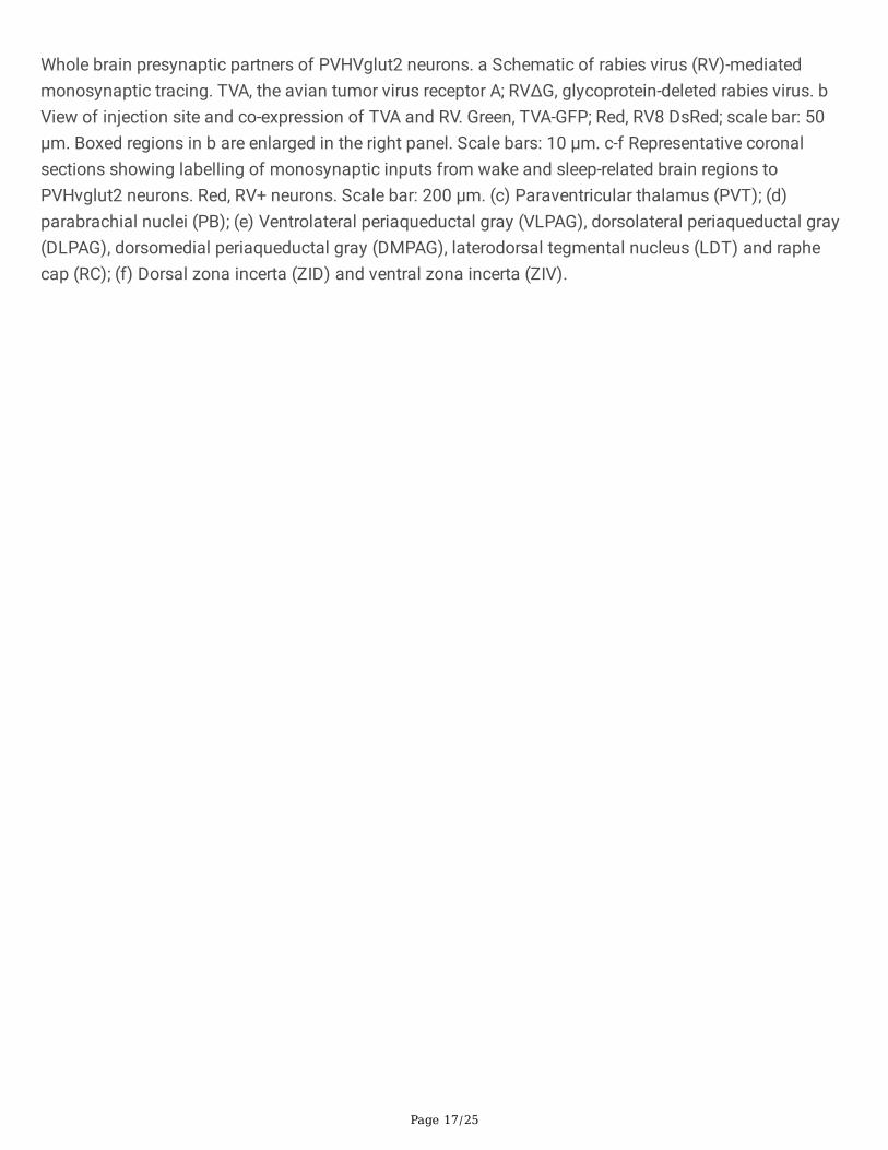
ResultsPVH receives direct inputs from the PVT and PB. Considering that the homologous area of the primateposterior hypothalamus in rodents is around the PVH area, which contains mainly glutamatergic neurons,we examined the role of PVHvglut2 neurons in the regulation of wakefulness. We used Cre-dependentrabies virus–mediated monosynaptic retrograde tracing in Vglut2-Cre mice (Fig. 1a, b) and found thatPVHvglut2 neurons received direct inputs from the PVT, PB, ZI and VLPAG (Fig. 1c-f), which are involved insleep-wake control 9, 15–17,25,26, suggestion that the PVH might act as a key central node for sleep-wakeregulation.
PVH vglut2 neurons are preferentially active during wakefulness. We next performed in-vivo �berphotometry to investigate the real-time activity of PVHvglut2 neurons across spontaneous sleep–wakecycles in freely moving mice. The recording mode for �ber photometery and the expression of the Cre-dependent AAVs expressing the �uorescent calcium indicator, GCaMP6f (AAV-EF1α-DIO-GCaMP6f), in thePVH of Vglut2-Cre mice are shown in Fig. 2a, b. PVHvglut2 neuronal activities during wakefulness weresigni�cantly higher than those during NREM sleep (Fig. 2c–e).
We next performed in-vivo multichannel electrophysiological recordings to monitor the spike �ring ofindividual PVH neurons in freely behaving mice (Fig. 2f). PVHvglut2 neurons exhibited a higher �ring rateduring wakefulness than during sleep (Fig. 2g, h). The PVHvglut2 neuronal �ring rate gradually decreasedbefore sleep onset and increased during transitions from sleep to wakefulness (Fig. 2i–k). At the onset ofbehavioral arousal from NREM sleep, the mean �ring rate reached 13.5 Hz (Fig. 2i). Collectively, theseelectrophysiological results clearly indicate a mechanistic framework for the activity-dependentparticipation of PVH neurons in the regulation of sleep and wakefulness.
Chemogenetic activation of PVH vglut2 and PVHCRH neurons signi�cantly increases wakefulness. Next, weinvestigated the activation effect of PVHvglut2 neurons in freely moving mice on wakefulness regulation
Page 5/25
by injecting adeno-associated virus (AAV)-EF1α-double-�oxed inverse-orientation (DIO)-hM3D(Gq)-mCherry into the PVH, respectively (Fig. 3a). At the beginning of the light phase (zeitgeber time 3 [ZT3];9:00), chemogenetic activation of PVHvglut2 neurons caused a potent increase in wakefulness lastingapproximately 9 h and concomitantly decreased both NREM and REM sleep (Fig. 3b). CNO administration(3 mg/kg) resulted in a 139.97% increase in total wakefulness, as well as 81.63% and 94.51% reduction inNREM and REM sleep, respectively, during the 9-h post-injection period (Supplementary Fig. 1b).Compared with vehicle injection, chemogenetic activation of PVHvglut2 neurons signi�cantly increasedelectroencephalographic (EEG) low delta power (0.25–1.00 Hz) and decreased high delta power (1.25–4.75 Hz) (Supplementary Fig. 1k). No sleep rebound followed the long-lasting wakefulness, as indicatedby no change in the time spent in NREM sleep during the following dark period (19:00–07:00;Supplementary Fig. 1j). Besides, there is no signi�cant difference in the EEG power density of NREM sleepduring the day (7:00–18:00) before/after the day of CNO injection (Supplementary Fig. 1l). Similarly, CNOinjection during the dark period also signi�cantly increased wakefulness and induced high levels ofarousal (Supplementary Fig. 2a, b), further demonstrating that activation of PVHvglut2 neurons prolongedarousal even during the dark (active) period.
Next, we further explored arousal-promoting roles of subtype neurons of PVHvglut2 neurons (PVHCRH andPVHAVP neurons) and found that chemogenetic activation of PVHCRH neurons caused a potent increasein wakefulness lasting approximately 3 h and concomitantly decreased both NREM and REM sleep(Fig. 3d). CNO administration (3 mg/kg) induced a 75.7% increase in wakefulness and a 67.7%, 46%reduction in NREM and REM sleep during 3-h post-injection period (Supplementary Fig. 1e), whereaschemogenetic activation of PVHAVP neurons had no effect on the amount of wakefulness ((Fig. 3f andSupplementary Fig. 1h).
Optogenetic activation of PVH vglut2 neurons initiates wakefulness. Compared with the temporal precisionof chemogenetic activation, optogenetic manipulations can achieve millisecond-scale control of neuronalactivity. Therefore, we next employed optogenetic methods to elucidate the causal role of the PVH incontrolling wakefulness. We stereotaxically injected AAVs expressing channelrhodopsin-2 (AAV-DIO-ChR2-mCherry) into the PVH (Fig. 4a). Functional expression of ChR2 was veri�ed by in-vitro electrophysiology(Fig. 4b). Next, we applied optical blue-light stimulation (10 ms, 20 Hz, 20–30 mW/mm2) after the onsetof stable NREM or REM sleep during the light phase (Fig. 4c). Optical stimulation of PVHvglut2 neuronsduring NREM sleep reliably induced transitions to wakefulness in a frequency-dependent manner(Fig. 4d). Analysis of the probability of transitions between each pair of sleep-wake states showed thatoptical stimulation signi�cantly enhanced the probability of wakefulness, along with a complementarydecrease in the probability of NREM or REM sleep (Fig. 4e). To test whether these neurons alsocontributed to the maintenance of wakefulness, photostimulation was given for 1 h during the lightperiod (09:00–10:00). Sustained activation of PVHvglut2 neurons via semi-chronic optical stimulation (10-ms blue-light pulses at 20 Hz for 25 s, every 60 s for 1 h) signi�cantly increased the amount ofwakefulness in ChR2-mCherry mice compared with that of the baseline control between 09:00 and 10:00(12.3 ± 1.8 min at baseline vs. 48.6 ± 1.5 min after stimulation, n = 5; Fig. 4f). These �ndings demonstrate

Page 6/25
that optogenetic activation of PVHvglut2 neurons potently enhanced both the initiation and maintenanceof wakefulness.
PVH vglut2 neurons promote wakefulness via PB and LSv connections. We next sought to determine thedownstream targets by which PVHvglut2 neurons promote wakefulness. Speci�cally, AAV-hSyn-DIO-ChR2-mCherry or AAV-hSyn-DIO-mCherry constructs were injected into the PVH of Vglut2-Cre mice. We foundthat PVHvglut2 neurons mainly projected to two neuroanatomical sites: the PB (Fig. 5a) and LSv (Fig. 5e).To identify the neuronal circuits mediating the wake-promoting effect of PVHvglut2 neurons, ChR2 wasexpressed in the PVH with optic �bers targeting terminals in the PB or LSv (Fig. 5a, e). Optogeneticstimulation (10-ms pulses at 10 Hz for 2 s) of the ChR2-expressing PVH terminals evoked excitatorypostsynaptic currents (EPSCs) in most of the patch-recorded PB (n = 6 cells, Fig. 5b) or LSv neurons (n = 8cells, Fig. 5f). Moreover, 20-Hz stimulation of the bilateral PB or LSv induced a shorter transition fromNREM sleep to wakefulness (latency for PB: 1.0 ± 0.8 s, latency for LSv: 1.2 ± 0.9 s) compared with that inthe control (Fig. 5c, g). Analysis of the probability of transitions between each pair of sleep-wake statesshowed that optical stimulation signi�cantly enhanced the probability of wakefulness, along with acomplementary decrease in the probabilities of NREM and REM sleep (Fig. 5d, h). These resultsdemonstrate that PVH→PB and PVH→LSv circuits mediated the wakefulness-controlling effect ofPVHvglut2 neurons.
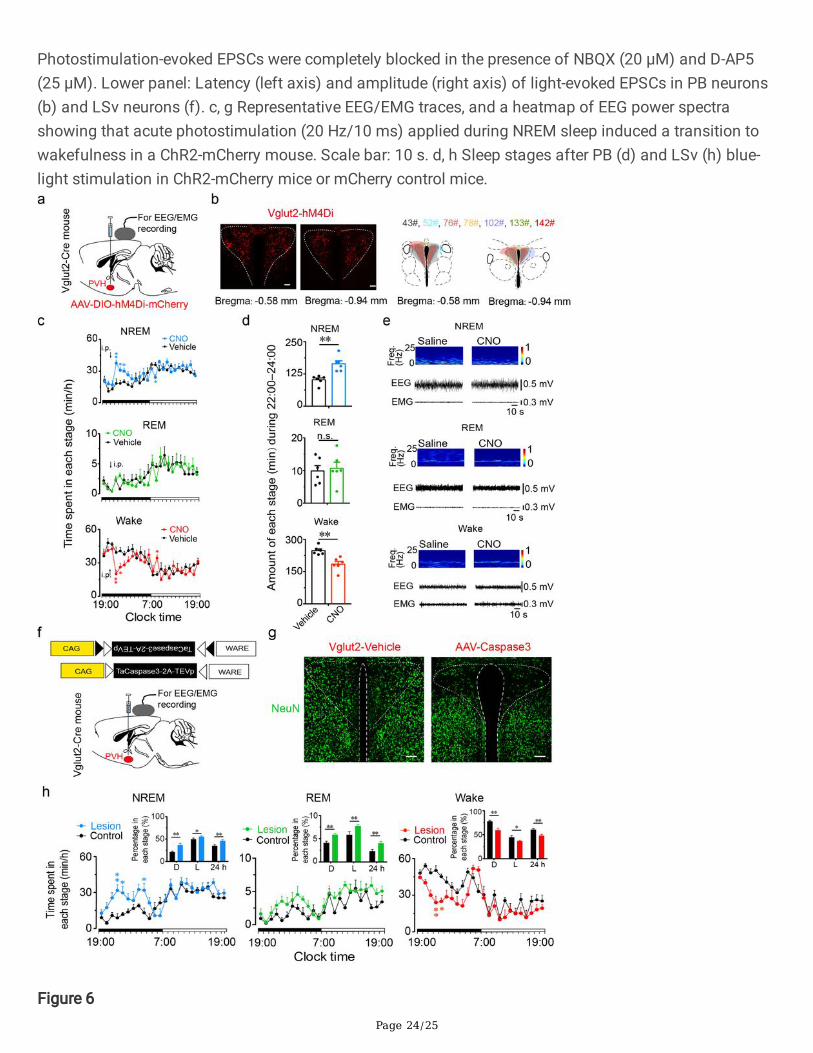
PVH vglut2 neurons are necessary for the control of natural wakefulness. To determine whether PVHvglut2
neurons are necessary for natural wakefulness, we inhibited or ablated PVHvglut2 neurons with two typesof AAV constructs encoding engineered Gi-coupled hM4D receptor (AAV-EF1α-DIO-hM4D(Gi)-mCherry) orcaspase3 (CAG�ex-taCasp3-TEVp-AAV), respectively (Fig. 6a, f). Chemogenetic inhibition of PVHvglut2
neurons decreased wakefulness during the 3 h following administration of CNO compared with that ofvehicle (Fig. 6c). At the beginning of the dark phase (ZT15; 21:00), CNO injection induced a signi�cantreduction in wakefulness for approximately 2 h and resulted in a 64.0% increase in NREM sleep duringthe 5 h post-injection period, which was accompanied by a 26.0% decrease in wakefulness (Fig. 3d). Inaddition, the EEG power spectrum for each state were not affected by CNO injection (Fig. 3e). Next, inorder to explore the role of subtype neurons of PVHvglut2 neurons (PVHCRH, PVHAVP neurons) in controllingsleep, we inhibited these two types of neurons with AAV constructs encoding engineered Gi-coupledhM4D receptor (AAV-EF1α-DIO-hM4D(Gi)-mCherry), respectively (Supplementary Fig. 3a, e). There was nosigni�cant sleep increase found in CRH-Cre mice or AVP-Cre mice (Supplementary Fig. 3b, f), and the EEGpower spectrum for each state were not affected by CNO injection (Supplementary Fig. 3d, h). Theseresults suggested that the PVHvglut2 neurons might act as a critical role in regulating sleep.
To further assess the functional importance of PVHvglut2 neurons controlling natural wakefulness, wespeci�cally ablated these neurons by bilaterally microinjecting AAV-EF1a-DIO-taCasp3-TEVp into the PVHregion of Vglut2-Cre mice. This construct expressed a designer pro-caspase-3 (pro-taCasp3) in the PVH,the activation of which causes apoptosis (Fig. 6f, g). Compared with that of the control group, mice thatunderwent PVHvglut2 neuronal ablation showed a 28.6% decrease in the amount of wakefulness and a

Page 7/25
74.7% increase in the amount of NREM sleep during the dark period. Similarly, ablation of PVHvglut2
neurons induced a 20.8% reduction in wakefulness and 30.6% increase in NREM sleep across an entire24-h light/dark cycle (Fig. 6h). These results indicate that PVHvglut2 neurons are necessary for wakeregulation under physiological conditions, and that dysfunction of these neurons may inducehypersomnia.
DiscussionAdequate wakefulness is essential for life and survival. In the present study, we identi�ed the PVH as acritical hypothalamic nucleus for the regulation of wakefulness. In previous study, 15% reduction inbaseline wakefulness is considered signi�cant 6,26. Lu et al have reported that lesion of the PPT and theventral sublaterodorsal nucleus (vSLD) results in a 20–30% reduction in baseline wakefulness 6.However, bidirectional chemogenetic manipulations that inhibit the PPT or activate SLD neurons havebeen shown to have little in�uence on baseline sleep 27,28. In our present study, three patients with lesionsmostly around the PVH showed hypersomnolence lasting above 20 h per day. PSG recordings from thesepatients showed that stage-two NREM was strikingly dominant, indicating that these patients slept stablyand were not easily awakened. Importantly, we found that following recovery from injury around the PVHin one of these patients, the proportion of stage-two NREM sleep decreased, and this patient wasconcomitantly better able to stay awake. Furthermore, ablation of PVHvglut2 neurons in mice induced a30.6% reduction in wakefulness across the 24-h light/dark cycle, highlighting the signi�cance of PVHvglut2
neurons in maintaining wakefulness and preventing hypersomnia. Besides, in our murine experiments, nosleep rebound was seen after PVHvglut2-activation-induced enhancement of wakefulness. This �nding isin accordance with previous studies using chemogenetics to speci�cally activate wake-promotingneuronal populations 9,28−30 and indicates that chemogenetic activation of wake-promoting neuronalpopulations does not enhance the homeostatic drive for sleep. Taken together, our present �ndingsprovide evidence of the su�cient and necessary wake-promoting action of PVHvglut2 neurons inpreventing hypersomnia.
The PVH is composed of abundant, diverse, and functionally distinct groups of neuroendocrine neurons,including CRH, AVP neurons 23–25,31−33. The PVH is estimated to consist of approximately 56,000neurons in humans 34, of which 21,000 neurons express AVP, and 2,000 neurons express CRH 35–37. Over90% of PVHCRH neurons express vglut2 mRNA 22. Morphological analysis has revealed that 50% ofPVHCRH neurons colocalize with PVHAVP neurons, which regulate stress, fear, and immune responses, aswell as neuroendocrine and autonomic functions 23,25,33,38,39. There is mounting evidence that exposureto various stressors induces CRH release into the peripheral circulation 40. Considering that PVHCRH
neurons are actively involved in stress-related behaviors, our results suggest that PVHCRH neurons play animportant role in stress-related insomnia.
Page 8/25
The PVH has distinct input and output connections that participate in various brain functions. PVHvglut2
neurons are connected to the BF 41, and PVT 42, and project to the nucleus of the solitary tract (NTS) 43,lateral parabrachial nucleus (L-PBN) 16,17, pre-locus coeruleus (pLC) 31 and ventral lateral septum (LSv)33. Our present �ndings identi�ed two speci�c neural circuits, among which the PVHvglut2→PB/LSv circuitrather than the PVH vglut2→NTS circuit was required for the maintenance of physiological wakefulness.Glutamatergic neurons in the L-PB are necessary for arousal in response to CO2 18; therefore, the
PVHvglut2→L-PB circuit may be involved in OSA. Xu et al reported that photostimulation ofPVHvglut2→LSv projections mediates stress-related self-grooming and fear-like jumping behaviors 33.Thus, the PVH orchestrates sleep/wake states related to stress and chemicals through different circuits.
In conclusion, our results indicate that the PVH is a key arousal-controlling nucleus in which PVHvglut2,PVHCRH neurons are critical for wakefulness.
MethodsAnimals. Vglut2-Cre mice were obtained from Jackson Laboratory (Bar Harbor, Maine, USA). CRH-Cremice were obtained from the Shanghai Model Organisms Center. AVP-Cre mice were generously providedby Ying-Xu. Mice were housed in a soundproof room at an ambient temperature of 24 ± 0.5 °C, with arelative humidity of 60 ± 2%. A 12-h light/dark cycle (100 Lux, light on at 07:00) was automaticallycontrolled47. Food and water were available ad libitum. Male heterozygous mice at 6–8 weeks of agewere used for all experiments. All animal experiments were approved by the Medical Experimental AnimalAdministrative Committee of Shanghai. All experimental procedures involving animals were approved bythe Animal Experiment and Use Committee of Fudan University (20150119–067).
Preparation of viral vectors. The AAVs of serotype rh10 for AAV-hSyn-DIO-hM3Dq-mCherry, AAV-hSyn-DIO-hM4Di-mCherry, AAV-hSyn-DIO-ChR2-mCherry, AAV-hSyn-DIO-mCherry, and AAV-CAG-FLEX-taCasp3-TEVpwere used. AAV vectors were packaged into serotype 2/9 vectors, which consisted of AAV2 ITR genomescoupled with AAV9 serotype capsid proteins. The �nal viral concentrations of the transgenes were in therange of 1–5 × 10¹² viral particles/mL.
Surgery and injection of viral vectors. All mice were anesthetized with chloral hydrate (360 mg/kg, i.p.) forsurgical procedures and were placed in a stereotaxic apparatus (RWD, Shenzhen, China). The skin abovethe skull was cut, a burr hole was made, and a small craniotomy was performed above the PVH. AAVconstructs were slowly injected (30 nL/min) into the bilateral PVH (70 nL for each position; AP = -0.5 mm;ML = ± 0.2 mm; DV = -4.2 mm) for PSG recordings and brain-slice electrophysiology, or were unilaterallyinjected into the PVH for neuronal tracing. The glass pipette was left in the brain for an additional 10 minfollowing injections and was then slowly withdrawn. All mice were implanted with electrodes for EEG andEMG recordings that were used for in-vivo tests at four weeks after injections under anesthesia of chloralhydrate (intraperitoneal, 360 mg/kg). The implant consisted of two stainless steel screws (1 mm indiameter), and EEG electrodes were inserted through the skull (+ 1.5 mm anteroposterior; -2.0 mm

Page 9/25
mediolateral from bregma or lambda), while two �exible silver wires were inserted into the neck muscles.The electrodes were attached to a mini-connector and were �xed to the skull with dental cement. Thescalp wound was sutured, and the mouse was when kept in a warm environment until it resumed normalactivity.
Polysomnographic recordings and analysis. After a 2–3-week recovery period, each mouse wasindividually housed in a recording chamber and habituated to the recording cable for 2–3 days beforeelectrophysiological recordings. Simultaneous EEG/EMG recordings were carried out with a slip ring sothat movement of the mice would not be restricted. For experiments using designer receptors exclusivelyactivated by designer drugs (DREADDs), the recordings started at 07:00 (i.e., at the beginning of the lightperiod), and each mouse received either vehicle or CNO (3 mg/kg, C2041, LKT) treatment for twoconsecutive days at 09:00 (inactive period) or 21:00 (active period). As previously described10,48,EEG/EMG signals were ampli�ed and �ltered (0.5–30 Hz for EEG, 40–200 Hz for EMG), and were thendigitized at 128 Hz and recorded with SleepSign software (Kissei Comtec, Nagano, Japan). Sleep–wakestates were automatically classi�ed into 4-s epochs as follows: wakefulness was considered to havedesynchronized EEG and high levels of EMG activity, NREM sleep was considered to have synchronized,high-amplitude, low-frequency (0.5–4 Hz) EEG signals in the absence of motor activity; and REM sleepwas considered to have pronounced theta-like (4–9 Hz) EEG activity and muscle atonia. All scoring wasautomated based on EEG and EMG waveforms in 4-s epochs for both chemogenetic and optogeneticstudies.
Optogenetic stimulation. Before the testing day, mice were given one day to adapt to optical �ber cables(0.8-m long, 200-µm diameter; RWD) that were placed inside the implanted �ber cannulae. On the testingday, 473-nm laser pulses (10 ms, 20 Hz) were delivered via an optic cable (Newton Inc., Hangzhou, China)using a pulse generator. Light pulse trains were generated via a stimulator (SEN-7103, Nihon Kohden,Japan) and delivered through an isolator (ss- 102J, Nihon Kohden). For acute photostimulation, eachstimulation epoch was applied at 20 s after identifying a stable NREM or REM sleep event via real-timeonline EEG/EMG analysis. Light pulse trains (5-ms pulses of various frequencies and durations) wereprogrammed and conducted during the inactive period. For chronic photostimulation, programmed lightpulse trains (5-ms pulses at 20 Hz for 10 s and at 30-s intervals for 1 h) were used. The 473-nm laserstimulation was performed from 09:00 to 10:00. Baseline EEG/EMG recordings were acquired at thesame time of day on the previous day prior to laser stimulation. Sleep–wake cycle parameters (e.g.,durations of NREM sleep, REM sleep, and wakefulness, as well as sleep–wake transitions) were scoredover an entire hour for each mouse. After receiving photostimulation, mice were sacri�ced at 30 min afterthe �nal stimulation for subsequent c-Fos staining.
In-vitro electrophysiological recordings. At 3–4 weeks after AAV-ChR2 injections, Vglut2-Cre mice wereanesthetized and transcardially perfused with ice-cold slicing buffer containing the following (in mM):213 sucrose, 26 NaHCO3, 10 glucose, 0.1 CaCl2, 3 MgSO4, 2.5 KCl, 1.25 NaH2PO4, 2 sodium pyruvate, and0.4 ascorbic acid. The buffer was saturated with 95% O2 and 5% CO2. Brains were then rapidly removed,and acute coronal slices (300 µm) containing the PVH were cut using a vibratome (Leica VT 1200S,

Page 10/25
Nussloch, Germany). Next, slices were transferred to a holding chamber containing normal recordingarti�cial cerebrospinal �uid (aCSF) containing the following (in mM): 119 NaCl, 26 NaHCO3, 25 glucose,2.5 KCl, 2CaCl2, 1.25 NaH2PO4, and 1.0 MgSO4. After being transferred, splices were allowed to recoverfor 30 min at 32 °C. Then, slices were maintained at room temperature for at least 30 min beforerecordings. During recordings, slices were transferred to and submerged in a recording chamber in whichoxygenated aCSF was continuously perfused.
Expression of ChR2 was con�rmed by visualization of mCherry �uorescence in PVH neuronal somataand axonal terminals. Neurons were identi�ed and visualized with an upright microscope (Olympus,Japan) equipped with differential contrast optics, including a 40 × water-immersion objective lens(BX51WI, Olympus). Images were detected with an infrared-sensitive CCD camera (OptiMOS, Q-imaging).Patch-clamp recordings were performed with capillary glass pipettes �lled with an intrapipette solutioncontaining the following (in mM): 130 potassium gluconate, 10 KCl, 10 Hepes, 0.5 EGTA, 4 ATP-Mg, 0.5GTP-Na, and 10 phosphocreatine, adjusting to a pH of 7.2–7.4 with KOH.
Whole-cell patch-clamp recordings were obtained using a MultiClamp 700B ampli�er (Molecular Device,Union City, CA, USA) and a Digidata 1440A A/D converter (Molecular Device). Signals were sampled at10 kHz and �ltered at 2 kHz. Data were acquired and analyzed using pClamp 10.3 software (pClamp,Molecular Devices). ChR2 stimulation was evoked using 470-nm light. In voltage-clamp experiments, theholding potential was − 70 mV. When needed, 20 µM of 2,3-dihydroxy 6-nitro-7-sulfamoyl-benzoquinoxaline-2,3-dione (NBQX, 1044, Tocris Bioscience, UK), 25 µM of d-(-)-2-amino-5-phosphonopentanoic acid (D-AP5, 0106, Tocris Bioscience, UK), and 10 µM of SR95531 (SR, ab144487,Abcam Biochemicals, UK) were added to block N-methyl-D-aspartic acid receptor (NMDA), α-Amino-3-hydroxy-5-methyl-4-isoxazolepropionic acid (AMPA), and gamma aminobutyric acid A (GABAA) receptors,respectively.
Fiber photometry. Following AAV-EF1α-DIO-GCaMP6f injections, an optical �ber (125-µm outer diameter,0.37 numerical aperture; Newdoon, Shanghai) was placed in a ceramic ferrule and was inserted towardthe PVH. Fiber photometry49 uses the same �ber to both excite and record from GCaMP in real time. Aftersurgery, mice were individually housed for at least 10 days to recover. Fluorescent signals were acquiredwith a laser beam passed through a 488-nm excitation laser (OBIS 488LS; Coherent), re�ected off adichroic mirror (MD498; Thorlabs), focused by an objective lens (Olympus), and coupled through a �bercollimation package (F240FC-A, Thorlabs) into a patch cable connected to the ferrule of an upright optic�ber implanted in the mouse via a ceramic sleeve (125 µm O.D.; Newdoon, Shanghai). GCaMP6�uorescence was bandpass �ltered (MF525–39, Thorlabs) and collected by a photomultiplier tube(R3896, Hamamatsu). An ampli�er (C7319, Hamamatsu) was used to convert the photomultiplier-tubecurrent output to voltage signals, which were further �ltered through a low-pass �lter (40-Hz cut-off;Brownlee 440). The photometry voltage traces were down-sampled using interpolation to match theEEG/EMG sampling rate of 512 Hz via a Power1401 digitizer and Spike2 software (CED, Cambridge, UK).
Page 11/25
Photometry data were exported to MATLAB R2018b mat �les from Spike2 for further analysis. Wesegmented the value of the �uorescent change (ΔF/F) by calculating (F – F0)/F0, where F0 is the baselineof the �uorescent signal. We recorded data for 3–5 h per mouse for the analysis of sleep–waketransitions to calculate the averaged calcium signal of ΔF/F during all times of vigilant states. Foranalyzing state transitions, we determined each sleep-wake transition and calculated ΔF/F in a ± 40-swindow around that time point.
Firing rate analysis. Electrophysiological data were �ltered with a band-pass �lter (300–6,000 Hz) toobtain neuronal spikes. Single-unit activities were sorted according to a threshold and shape detectorusing principal component analysis via O�ine Sorter software (Plexon Co, USA). The �rst two principalcomponents of each spike on the two-dimensional plot of detected spike events were extracted.Waveforms with similar principal components were clustered via a K-means sorting method. The isolatedcluster was considered as a single unit recorded from the same neuron. Spikes with inter-spike intervals < 2 ms were discarded. Cross-correlation histograms were used to eliminate cross-channel artifacts.NeuroExplorer software (version 5.0) was used for producing �ring-rate rastergrams, and Prism (version7.0) was used for producing �ring-rate histograms.
Histology and immunohistochemistry. For dual immunostaining with c-Fos and mCherry, mice weredeeply anesthetized with chloral hydrate (400 mg/kg) and were perfused with phosphate-buffered saline(PBS) followed by 4% PFA in 0.1-M phosphate buffer. The brain was then dissected and �xed in 4% PFAat 4 °C overnight. Fixed samples were sectioned into 30-µm coronal sections using a freezing microtome(CM1950, Leica, Germany). For immunohistochemistry, the �oating sections were washed in PBS andwere then incubated in the following primary antibodies in PBS containing 0.3% Triton X-100 (PBST) at4 °C: anti-rabbit c-Fos (1:10000 for 48 h); primary antibody (Millipore); and anti-mouse NeuN (1:1000 for12 h; MAB377, Millipore). Primary antibodies were washed �ve times with PBS before incubation withsecondary antibodies at room temperature for 2 h (Alexa 488, 1:1000; abcam). Finally, the sections weremounted on glass slides, dried, dehydrated, and cover-slipped. Fluorescent images were collected with aconfocal microscope (Nikon AIR-MP).
Statistical analysis. All data are expressed as the mean ± standard error of the mean (SEM). Sample sizeswere chosen based on previous studies 48,50. Two-way repeated-measures analysis of variance (ANOVA)was used to perform group comparisons with multiple measurements. Paired and unpaired t tests wereused for single-value comparisons. One-way ANOVA was used to compare more than two groups,followed by post-hoc Tukey tests for multiple pairwise comparisons. Prism 7.0 (GraphPad Software, SanDiego, CA, USA) was used for all statistical analyses. A two-tailed P < 0.05 was considered statisticallysigni�cant.
DeclarationsData availability
Page 12/25
The data supporting the �ndings of this study are available within the article and its SupplementaryInformation.
Competing Interests
The authors declare no competing interests.
Author contributions
Chang-rui Chen, Zhi-Li Huang conceived and designed the experiments. Yu-Heng Zhong and Shan Jiangperformed the experiments and analyzed the data. Wei Xu performed patch-clamp electrophysiology. LeiXiao performed multichannel electrophysiological recordings. Zan Wang collected the clinical data.Chang-Rui Chen, Yu-Heng Zhong, Shan Jiang wrote the manuscript, and all of the authors helped with therevision of the manuscript.
Acknowledgements
This study was supported in part by the National Natural Science Foundation of China (81671317,31530035, 81420108015, 31671099, 31871072, 31571103, 81701305, 81970727 and 31900738), theNational Basic Research Program of China (2015CB856401), Shanghai Pujiang Program (19PJ1401800)and ZJLab, Program for Shanghai Outstanding Academic Leaders (to Zhi-Li Huang). We are grateful toHui Dong, Ze-Ka Chen, Ya-Nan Zhao, Peng-Fei Xu, Ming-Jie Yang, Fan Yang, and An-Shu Chen fortechnical assistance.
References1 Mahowald, M. W. & Schenck, C. H. Insights from studying human sleep disorders. Nature 437,1279-1285, doi:10.1038/nature04287 (2005).
2 Saper, C. B., Chou, T. C. & Scammell, T. E. The sleep switch: hypothalamic control of sleep andwakefulness. Trends Neurosci 24, 726-731, doi:10.1016/s0166-2236(00)02002-6 (2001).
3 Moruzzi, G. & Magoun, H. W. Brain stem reticular formation and activation of the EEG.Electroencephalogr Clin Neurophysiol 1, 455-473 (1949).
4 Xu, M. et al. Basal forebrain circuit for sleep-wake control. Nat Neurosci 18, 1641-1647,doi:10.1038/nn.4143 (2015).
5 Carter, M. E. et al. Tuning arousal with optogenetic modulation of locus coeruleus neurons. NatNeurosci 13, 1526-1533, doi:10.1038/nn.2682 (2010).
6 Lu, J., Sherman, D., Devor, M. & Saper, C. B. A putative �ip-�op switch for control of REM sleep.Nature 441, 589-594, doi:10.1038/nature04767 (2006).

Page 13/25
7 Chen, L. et al. Basal Forebrain Cholinergic Neurons Primarily Contribute to Inhibition ofElectroencephalogram Delta Activity, Rather Than Inducing Behavioral Wakefulness in Mice.Neuropsychopharmacology 41, 2133-2146, doi:10.1038/npp.2016.13 (2016).
8 Fuller, P. M., Sherman, D., Pedersen, N. P., Saper, C. B. & Lu, J. Reassessment of the structural basisof the ascending arousal system. J Comp Neurol 519, 933-956, doi:10.1002/cne.22559 (2011).
9 Pedersen, N. P. et al. Supramammillary glutamate neurons are a key node of the arousal system.Nat Commun 8, 1405, doi:10.1038/s41467-017-01004-6 (2017).
10 Ren, S. et al. The paraventricular thalamus is a critical thalamic area for wakefulness. Science 362,429-434, doi:10.1126/science.aat2512 (2018).
11 Eban-Rothschild, A., Rothschild, G., Giardino, W. J., Jones, J. R. & de Lecea, L. VTA dopaminergicneurons regulate ethologically relevant sleep-wake behaviors. Nat Neurosci 19, 1356-1366,doi:10.1038/nn.4377 (2016).
12 Chemelli, R. M. et al. Narcolepsy in orexin knockout mice: molecular genetics of sleep regulation.Cell 98, 437-451, doi:10.1016/s0092-8674(00)81973-x (1999).
13 Sherin, J. E., Shiromani, P. J., McCarley, R. W. & Saper, C. B. Activation of ventrolateral preopticneurons during sleep. Science 271, 216-219, doi:10.1126/science.271.5246.216 (1996).
14 Gerashchenko, D., Blanco-Centurion, C., Greco, M. A. & Shiromani, P. J. Effects of lateralhypothalamic lesion with the neurotoxin hypocretin-2-saporin on sleep in Long-Evans rats. Neuroscience116, 223-235, doi:10.1016/s0306-4522(02)00575-4 (2003).
15 Adamantidis, A. R., Zhang, F., Aravanis, A. M., Deisseroth, K. & de Lecea, L. Neural substrates ofawakening probed with optogenetic control of hypocretin neurons. Nature 450, 420-424,doi:10.1038/nature06310 (2007).
16 Guyenet, P. G. & Bayliss, D. A. Neural Control of Breathing and CO2 Homeostasis. Neuron 87, 946-961, doi:10.1016/j.neuron.2015.08.001 (2015).
17 Yokota, S., Kaur, S., VanderHorst, V. G., Saper, C. B. & Chamberlin, N. L. Respiratory-related outputsof glutamatergic, hypercapnia-responsive parabrachial neurons in mice. J Comp Neurol 523, 907-920,doi:10.1002/cne.23720 (2015).
18 Kaur, S. et al. Glutamatergic signaling from the parabrachial nucleus plays a critical role inhypercapnic arousal. J Neurosci 33, 7627-7640, doi:10.1523/jneurosci.0173-13.2013 (2013).
19 Bollu, P. C., Manjamalai, S., Thakkar, M. & Sahota, P. Hypersomnia. Mo Med 115, 85-91 (2018).

Page 14/25
20 Vong, L. et al. Leptin action on GABAergic neurons prevents obesity and reduces inhibitory tone toPOMC neurons. Neuron 71, 142-154, doi:10.1016/j.neuron.2011.05.028 (2011).
21 Xu, Y. et al. Glutamate mediates the function of melanocortin receptor 4 on Sim1 neurons in bodyweight regulation. Cell Metab 18, 860-870, doi:10.1016/j.cmet.2013.11.003 (2013).
22 Ziegler, D. R., Cullinan, W. E. & Herman, J. P. Organization and regulation of paraventricular nucleusglutamate signaling systems: N-methyl-D-aspartate receptors. J Comp Neurol 484, 43-56,doi:10.1002/cne.20445 (2005).
23 Zhang, X. et al. Brain control of humoral immune responses amenable to behavioural modulation.Nature 581, 204-208, doi:10.1038/s41586-020-2235-7 (2020).
24 Sterley, T. L. et al. Social transmission and buffering of synaptic changes after stress. NatNeurosci 21, 393-403, doi:10.1038/s41593-017-0044-6 (2018).
25 Holmes, M. C., Antoni, F. A., Aguilera, G. & Catt, K. J. Magnocellular axons in passage through themedian eminence release vasopressin. Nature 319, 326-329, doi:10.1038/319326a0 (1986).
26 Lu, J., Jhou, T. C. & Saper, C. B. Identi�cation of wake-active dopaminergic neurons in the ventralperiaqueductal gray matter. J Neurosci 26, 193-202, doi:10.1523/jneurosci.2244-05.2006 (2006).
27 Kroeger, D. et al. Cholinergic, Glutamatergic, and GABAergic Neurons of the PedunculopontineTegmental Nucleus Have Distinct Effects on Sleep/Wake Behavior in Mice. J Neurosci 37, 1352-1366,doi:10.1523/jneurosci.1405-16.2016 (2017).
28 Erickson, E. T. M., Ferrari, L. L., Gompf, H. S. & Anaclet, C. Differential Role of PontomedullaryGlutamatergic Neuronal Populations in Sleep-Wake Control. Front Neurosci 13, 755,doi:10.3389/fnins.2019.00755 (2019).
29 Anaclet, C. et al. Basal forebrain control of wakefulness and cortical rhythms. Nat Commun 6,8744, doi:10.1038/ncomms9744 (2015).
30 Venner, A., Anaclet, C., Broadhurst, R. Y., Saper, C. B. & Fuller, P. M. A Novel Population of Wake-Promoting GABAergic Neurons in the Ventral Lateral Hypothalamus. Curr Biol 26, 2137-2143,doi:10.1016/j.cub.2016.05.078 (2016).
31 Li, M. M. et al. The Paraventricular Hypothalamus Regulates Satiety and Prevents Obesity via TwoGenetically Distinct Circuits. Neuron 102, 653-667 e656, doi:10.1016/j.neuron.2019.02.028 (2019).
32 Hung, L. W. et al. Gating of social reward by oxytocin in the ventral tegmental area. Science 357,1406-1411, doi:10.1126/science.aan4994 (2017).

Page 15/25
33 Xu, Y. et al. Identi�cation of a neurocircuit underlying regulation of feeding by stress-relatedemotional responses. Nat Commun 10, 3446, doi:10.1038/s41467-019-11399-z (2019).
34 Morton, A. A quantitative analysis of the normal neuron population of the hypothalamicmagnocellular nuclei in man and of their projections to the neurohypophysis. J Comp Neurol 136, 143-157, doi:10.1002/cne.901360203 (1969).
35 Wierda, M. et al. Oxytocin cell number in the human paraventricular nucleus remains constant withaging and in Alzheimer's disease. Neurobiol Aging 12, 511-516, doi:10.1016/0197-4580(91)90081-t(1991).
36 Purba, J. S., Hofman, M. A., Portegies, P., Troost, D. & Swaab, D. F. Decreased number of oxytocinneurons in the paraventricular nucleus of the human hypothalamus in AIDS. Brain 116 ( Pt 4), 795-809,doi:10.1093/brain/116.4.795 (1993).
37 Purba, J. S. et al. Increased number of corticotropin-releasing hormone expressing neurons in thehypothalamic paraventricular nucleus of patients with multiple sclerosis. Neuroendocrinology 62, 62-70,doi:10.1159/000126989 (1995).
38 Kondoh, K. et al. A speci�c area of olfactory cortex involved in stress hormone responses topredator odours. Nature 532, 103-106, doi:10.1038/nature17156 (2016).
39 Mezey, E., Kiss, J. Z., Skirboll, L. R., Goldstein, M. & Axelrod, J. Increase of corticotropin-releasingfactor staining in rat paraventricular nucleus neurones by depletion of hypothalamic adrenaline. Nature310, 140-141, doi:10.1038/310140a0 (1984).
40 Winter, J. & Jurek, B. The interplay between oxytocin and the CRF system: regulation of the stressresponse. Cell Tissue Res 375, 85-91, doi:10.1007/s00441-018-2866-2 (2019).
41 Agostinelli, L. J., Geerling, J. C. & Scammell, T. E. Basal forebrain subcortical projections. BrainStruct Funct 224, 1097-1117, doi:10.1007/s00429-018-01820-6 (2019).
42 Yuan, Y. et al. Reward Inhibits Paraventricular CRH Neurons to Relieve Stress. Curr Biol 29, 1243-1251 e1244, doi:10.1016/j.cub.2019.02.048 (2019).
43 Gasparini, S., Howland, J. M., Thatcher, A. J. & Geerling, J. C. Central afferents to the nucleus of thesolitary tract in rats and mice. J Comp Neurol 528, 2708-2728, doi:10.1002/cne.24927 (2020).
44 Baloyannis, S. J., Mavroudis, I., Mitilineos, D., Baloyannis, I. S. & Costa, V. G. The hypothalamus inAlzheimer's disease: a Golgi and electron microscope study. Am J Alzheimers Dis Other Demen 30, 478-487, doi:10.1177/1533317514556876 (2015).
45 Purba, J. S., Hofman, M. A. & Swaab, D. F. Decreased number of oxytocin-immunoreactive neuronsin the paraventricular nucleus of the hypothalamus in Parkinson's disease. Neurology 44, 84-89,

Page 16/25
doi:10.1212/wnl.44.1.84 (1994).
46 Manaye, K. F. et al. Selective neuron loss in the paraventricular nucleus of hypothalamus inpatients suffering from major depression and bipolar disorder. J Neuropathol Exp Neurol 64, 224-229,doi:10.1093/jnen/64.3.224 (2005).
47 Zhang, Z., Wang, H. J., Wang, D. R., Qu, W. M. & Huang, Z. L. Red light at intensities above 10 lxalters sleep-wake behavior in mice. Light Sci Appl 6, e16231, doi:10.1038/lsa.2016.231 (2017).
48 Luo, Y. J. et al. Nucleus accumbens controls wakefulness by a subpopulation of neuronsexpressing dopamine D(1) receptors. Nat Commun 9, 1576, doi:10.1038/s41467-018-03889-3 (2018).
49 Li, J. et al. Primary Auditory Cortex is Required for Anticipatory Motor Response. Cereb Cortex 27,3254-3271, doi:10.1093/cercor/bhx079 (2017).
50 Zhang, Z. et al. Superior Colliculus GABAergic Neurons Are Essential for Acute Dark Induction ofWakefulness in Mice. Curr Biol 29, 637-644.e633, doi:10.1016/j.cub.2018.12.031 (2019).
Figures
Figure 1
Page 17/25
Whole brain presynaptic partners of PVHVglut2 neurons. a Schematic of rabies virus (RV)-mediatedmonosynaptic tracing. TVA, the avian tumor virus receptor A; RVΔG, glycoprotein-deleted rabies virus. bView of injection site and co-expression of TVA and RV. Green, TVA-GFP; Red, RV8 DsRed; scale bar: 50μm. Boxed regions in b are enlarged in the right panel. Scale bars: 10 μm. c-f Representative coronalsections showing labelling of monosynaptic inputs from wake and sleep-related brain regions toPVHvglut2 neurons. Red, RV+ neurons. Scale bar: 200 μm. (c) Paraventricular thalamus (PVT); (d)parabrachial nuclei (PB); (e) Ventrolateral periaqueductal gray (VLPAG), dorsolateral periaqueductal gray(DLPAG), dorsomedial periaqueductal gray (DMPAG), laterodorsal tegmental nucleus (LDT) and raphecap (RC); (f) Dorsal zona incerta (ZID) and ventral zona incerta (ZIV).

Page 18/25
Figure 2
PVHvglut2 neurons are preferentially active during wakefulness. a Schematic of the �ber photometrysetup and in-vivo recording con�guration (DM dichroic mirror, PMT photomultiplier tube). b Unilateral viraltargeting of AAV-EF1α-DIO-GCaMP6f into the PVH, in which the tip of the �ber optic is above the PVH.Scale bar: 200 μm. c Representative �uorescent traces, relative EEG power, and EEG/EMG traces acrossspontaneous sleep–wake states. ΔF/F represents the change in �uorescence from the median of the

Page 19/25
entire time series. d Fluorescence (mean ± SEM) during wakefulness, NREM sleep, and REM sleep fromthree mice; the �uorescent signal was the highest during wakefulness, intermediate during REM, and thelowest during NREM sleep (n = 7 from 3 mice, one-way ANOVA followed by Tukey’s post-hoc tests; F6,12 =2.94, P < 0.001; P [wake vs NREM] < 0.001, P [wake vs REM] < 0.001, P [NREM vs REM] = 0.013). eFluorescent signals aligned to sleep–wake transitions. Upper panel, Individual transitions with color-coded �uorescent intensities (NREM to wake, n = 54; wake to NREM, n = 45; REM to wake, n = 44). fSchematic con�guration of in-vivo multichannel electrophysiological recordings. Upper panel: A brainslice from a mouse with electrodes implanted in the PVH. White arrow indicates the electrode track. Scalebar: 200 μm. Lower panel: Waveforms from a recorded PVH neuron. g EEG/EMG and PVH multi-unitrecording traces during wakefulness, NREM sleep, and REM sleep. h Average �ring rates of PVH neuronsduring each state (n= 8 cells from 3 mice, one-way RM ANOVA followed by LSD post hoc tests; F2,14=12.51, P [NREM vs wake] < 0.001, P [wake vs NREM] < 0.01, P [NREM vs REM] = 0.613). i-k Firing rates ofPVH neurons during state transitions: wake-to-NREM (i), NREM-to-wake (j), and REM-to-wake transitions(k). Top: Example rastergrams of a PVH neuron during �ve trials of different state transitions. Bottom left:Average �ring rate during the state-transition period. Bottom right: Average �ring rate during 8 s beforeand after state transitions (P [wake–NREM] < 0.01, P [wake–NREM] < 0.01, P [REM–wake] < 0.01, paired ttest). Data represent the mean ± SEM (*P < 0.05, **P < 0.01).

Page 20/25
Figure 3
Chemogenetic activation of PVHvglut2, PVHCRH neurons rather than PVHAVP neurons during the lightphase increases wakefulness. a, c and e Expression of AAV injection site in the PVH of Vglut2-Cre mice(a), CRH-Cre mice (c) and AVP-Cre mice (e). b, d and f Time-course changes in wakefulness, NREM sleep,and REM sleep after administration of vehicle or CNO in mice expressing hM3Dq in PVHvglut2 neurons(b, n = 7, repeated-measures ANOVA; F1,12 = 87.09 [wake], 63.61 [NREM], 612.30 [REM]; P < 0.001 [wake],P < 0.001 [NREM], P < 0.001 [REM]), PVHCRH neurons (d, n = 7, repeated-measures ANOVA; F1, 12= 0.06[wake], 0.01 [NREM], 0.83 [REM]; P < 0.05 [wake], P < 0.05 [NREM], P < 0.05 [REM]) and PVHAVP neurons(f, n = 8, repeated-measures ANOVA; F1, 14= 0.18 [wake], 1.03 [NREM], 1.94 [REM]; P = 0.09 [wake], 0.09[NREM], 0.21 [REM]).

Page 21/25
Figure 4
Optogenetic activation of PVHvglut2 neurons induces a rapid transition from NREM sleep towakefulness. a Left: Schematic of optogenetic manipulation of PVHvglut2 neurons and EEG/EMGrecordings. Right: ChR2-mCherry expression and location of optical �ber in the PVH. Scale bar: 200 μm. bExample traces (left) and �delity of action potential �ring (right) of ChR2-expressing PVH neurons evokedby 473-nm light stimulation with different frequencies. c Representative EEG/EMG traces, and heatmap

Page 22/25
of EEG power spectra showing that acute photostimulation (20 Hz/10 ms) applied during NREM sleepinduced a transition to wakefulness in a ChR2-mCherry mouse. Scale bar: 10 s. d Latencies of transitionsfrom NREM sleep to wakefulness after photostimulation at different frequencies (n = 5, unpaired t test; 1Hz, t8 =1.4, P = 0.19; 5 Hz, t8 = 10.29, P < 0.01; 10 Hz, t8 =13.3, P < 0.01; 20 Hz, t8 =14.04, P < 0.01). eSleep stage after blue-light stimulation in a PVH-vglut2-ChR2 mouse or PVH-vglut2-mCherry mouse.Percentages of NREM, REM, and wakefulness during short-stimulation experiments. f Time course duringsemi-chronic optogenetic experiments (20 Hz/10 ms, 25-s on /35-s off). The blue column indicates thephotostimulation period of the stimulation group (n = 5, repeated-measures ANOVA; F1,8 = 59.37 (wake),18.20 (REM), 103.30 (NREM); P < 0.001 [wake], P =0.003 [REM], P <0.001 [NREM]). Data represent themean ± SEM (*P < 0.05, **P < 0.01).

Page 23/25
Figure 5
PVHvglut2 neurons control arousal through PB and LSv pathways. a, e Left: Schematic diagram showingthe location of the optic �ber in the PB and LSv, and EEG/EMG recordings of a Vglut2-Cre mouse injectedwith AAV-ChR2-mCherry or AAV-mCherry in the PVH. Right: This brain section was stained againstmCherry to con�rm that ChR2 protein was expressed in the PVH. Scale bar: 200 μm. b, f Upper-left panel:Photostimulation-evoked EPSCs in PB neurons (b) and LSv neurons (f). Upper-right panel:
Page 24/25
Photostimulation-evoked EPSCs were completely blocked in the presence of NBQX (20 μM) and D-AP5(25 μM). Lower panel: Latency (left axis) and amplitude (right axis) of light-evoked EPSCs in PB neurons(b) and LSv neurons (f). c, g Representative EEG/EMG traces, and a heatmap of EEG power spectrashowing that acute photostimulation (20 Hz/10 ms) applied during NREM sleep induced a transition towakefulness in a ChR2-mCherry mouse. Scale bar: 10 s. d, h Sleep stages after PB (d) and LSv (h) blue-light stimulation in ChR2-mCherry mice or mCherry control mice.
Figure 6

Page 25/25
PVHvglut2 neurons are required for wakefulness. a Expression of AAV injection site in the PVH of Vglut2-Cre mice. b Location of hM4Di expression in the PVHvglut2 neurons. Scale bars: 200 μm. c Time-coursechanges in NREM sleep, wakefulness, and REM sleep after administration of vehicle or CNO in miceexpressing hM4Di in PVHvglut2 neurons (n = 6, repeated-measures ANOVA; F1,10= 21.95 [wake], 7.68[NREM], 29.23 [REM]). d Total time spent in each stage after vehicle or CNO injection (n = 6, paired t test).e Representative EEG power spectra and EEG-EMG traces of wakefulness, NREM and REM sleep afterinjection in Vglut2-Cre mouse. f Expression of an AAV injection site in the PVH of Vglut2-IRES-cre mice. gImage showing NeuN (neuron-speci�c nuclear protein) staining from a control mouse (left) and a mousewith a PVH lesion (right). Scale bars: 200 μm. h Time spent in each stage across the 24-h sleep-wakecycle. L, light phase; D, dark phase. (control group, n = 6; lesion group, n = 7, repeated-measures ANOVA;F1,10= 2.88 [wake], 2.90 [NREM], 0.06 [REM]; Inset: unpaired t test; NREM, dark phase, t11=3.27, P<0.01,light phase, t11=2.32, P=0.04, 24 h, t11=3.27, p<0.01; REM, dark phase, t11=2.94, P=0.01; light phase,t11=2.88, P=0.05; 24 h, t11=4.36, p<0.01; Wake, dark phase, t11=3.61, P<0.01; light phase, t11=2.24,P=0.05; 24 h, t11=3.56, p<0.01 ). Data represent the mean ± SEM (*P < 0.05, **P < 0.01).
Supplementary Files
This is a list of supplementary �les associated with this preprint. Click to download.
SupplementaryMaterials.docx