
The
Jour
nal o
f Exp
erim
enta
l Bio
logy
– A
CC
EPTE
D A
UTH
OR
MA
NU
SCR
IPT
© 2013. Published by The Company of Biologists Ltd
Null point of discrimination in crustacean polarisation vision 1
Martin J. How1,2,3, John Christy3, Nicholas W. Roberts1 and N. Justin Marshall2 2
1 – School of Biological Sciences, Woodland Road, University of Bristol, Bristol, BS8 1UG, UK 3
2 - Queensland Brain Institute, University of Queensland, St Lucia, QLD 4072, Australia 4
3 - Smithsonian Tropical Research Institute, Panama City, Republic of Panama 5
6
7
Abstract 8
The polarisation of light is used by many species of cephalopods and crustaceans to discriminate 9
objects or to communicate. Most visual systems with this ability, such as that of the fiddler crab, 10
include receptors with photopigments that are oriented horizontally and vertically relative to the 11
outside world. Photoreceptors in such an orthogonal array are maximally sensitive to polarised light 12
with the same fixed e-vector orientation. Using opponent neural connections, this two-channel 13
system may produce a single value of polarisation contrast and, consequently, it may suffer from 14
null points of discrimination. Stomatopod crustaceans use a different system for polarisation vision, 15
comprising at least four types of polarisation-sensitive photoreceptor arranged at 0°, 45°, 90° and 16
135° relative to each other, in conjunction with extensive rotational eye movements. This 17
anatomical arrangement should not suffer from equivalent null points of discrimination. To test 18
whether these two systems were vulnerable to null points, we presented the fiddler crab Uca 19
heteropleura and the stomatopod Haptosquilla trispinosa with polarised looming stimuli on a 20
modified LCD monitor. The fiddler crab was less sensitive to differences in the degree of polarised 21
light when the e-vector was at -45°, than when the e-vector was horizontal. In comparison, 22
stomatopods showed no difference in sensitivity between the two stimulus types. The results 23
suggest that fiddler crabs suffer from a null point of sensitivity, while stomatopods do not. 24
25
Keywords: polarisation distance, fiddler crab, mantis shrimp, discrimination threshold 26
http://jeb.biologists.org/lookup/doi/10.1242/jeb.103457Access the most recent version at J Exp Biol Advance Online Articles. First posted online on 15 April 2014 as doi:10.1242/jeb.103457

The
Jour
nal o
f Exp
erim
enta
l Bio
logy
– A
CC
EPTE
D A
UTH
OR
MA
NU
SCR
IPT
Introduction 27
The polarisation of light provides an independent channel of visual information that some animals 28
use to discriminate objects or for communication (Shashar et al., 1998; Wehner and Labhart, 2006; 29
Glantz, 2007; Chiou et al., 2011; How et al., 2012; Temple et al., 2012; Marshall et al., 2014b). Visual 30
systems that detect and identify objects using polarised light rely on comparative processing of the 31
signals generated by polarisation-sensitive photoreceptors distributed across parts of the visual field. 32
This is a process somewhat analogous to colour vision and adds to the well-known uses of the 33
polarisation of light for navigation, orientation and habitat localisation (Wehner, 1976; Schwind, 34
1983; Labhart and Meyer, 1999). Animals may use this channel to improve the detection of objects 35
through veiling light or glare (Schechner and Karpel, 2005; Alkaladi et al., 2013), to enhance the 36
contrast of transparent prey (Shashar et al., 1998; Tuthill and Johnsen, 2006; but see Johnsen et al., 37
2011), to break camouflage (Shashar et al., 2000; Jordan et al., 2012; Temple et al., 2012) or to allow 38
the detection of polarised body patterns for communication (Shashar et al., 1996; Marshall et al., 39
1999). 40
Object-based polarisation vision systems that have so far been studied are restricted to 41
cephalopods and crustaceans. In these examples, the photoreceptors that act as polarised light 42
detectors in the eye are aligned horizontally and vertically (Tasaki and Karita, 1966; Shaw, 1969; 43
Waterman, 1981) forming two channels of polarisation sensitivity. Furthermore, these animals 44
maintain precise alignment of their eyes relative to the visual world around them (Zeil et al., 1986; 45
Talbot and Marshall, 2011), thus ensuring that the polarisation-sensitive receptors remain horizontal 46
and vertical relative to the visual scene. Stomatopods are an exception. These crustaceans possess 47
an anatomically complex array of polarisation receptors with e-vector sensitivities arranged at 0°, 48
45°, 90° and 135° relative to each other (Marshall et al., 1991; Kleinlogel et al., 2003). They also have 49
receptors in rows 5 and 6 of the mid-band of the eye that are sensitive to left and right-handed 50
circularly polarised light (Chiou et al., 2008; Roberts et al., 2009). In addition to this anatomical 51
complexity, these animals regularly rotate their eyes around the axis of view (Cronin et al., 1988; 52
Land et al., 1990), thereby altering the orientation of the polarisation detectors relative to the 53
outside world. Such a system is likely, therefore, to have very different properties from the more 54
common two-channel polarisation vision. 55
An understanding of the underlying anatomy of polarisation vision opens the possibility for 56
modelling an animal’s sensitivity to polarised light cues. One approach, inspired by analogous 57
systems in colour vision (Vorobyev and Osorio, 1998), is to simulate the proposed neural 58
architecture of early visual processing. For simple systems, two orthogonally oriented polarisation 59

The
Jour
nal o
f Exp
erim
enta
l Bio
logy
– A
CC
EPTE
D A
UTH
OR
MA
NU
SCR
IPT
channels are assumed to have opponent connections (similar to dichromatic colour vision models) 60
and produce a single level of neural activity (Bernard and Wehner, 1977). Subsequent comparison of 61
this activity between two different parts of the visual field (one viewing an object and one viewing 62
the background) results in a measure of polarisation contrast, termed ‘polarisation distance’(How 63
and Marshall, 2014). Presumably this output is modulated at some stage by intensity variations, so 64
that intensity signals and polarisation signals are fused into a single measure of contrast; however 65
this falls beyond the scope of the current paper. Such models allow us to predict how well animals 66
will perform in a discrimination task, assuming that they are using a given neurosensory apparatus. 67
Previous studies using this modelling approach identified the problem of potential ambiguities 68
and null points in two-channel polarisation vision systems (Bernard and Wehner, 1977; How and 69
Marshall, 2014). For example, polarised light cues with e-vector axes oriented at +45° or -45° 70
stimulate horizontal and vertical polarisation receptors equally, and so are indistinguishable from 71
each other and from unpolarised light of the same intensity. This is widely accepted as a problem for 72
two-channel polarisation vision, yet, with the exception of some associative learning experiments 73
with cephalopods (Moody and Parriss, 1961; Shashar and Cronin, 1996), this has not been 74
investigated in detail at the behavioural level. To this end, we used a modified liquid crystal display 75
(LCD) monitor (Glantz and Schroeter, 2006; Pignatelli et al., 2011; How et al., 2012; Temple et al., 76
2012) to present polarised looming stimuli to the two-channel polarisation vision system of the 77
fiddler crab Uca heteropleura (Smith, 1870) and to the dynamic, multi-channelled polarisation vision 78
system of the stomatopod Haptosquilla trispinosa (Dana, 1852). These looming stimuli differed in 79
the degree of polarisation, while the overall light intensity remained constant. We compared the 80
response probability of these two species to stimuli that fell, either within, or outside a predicted 81
null point of discrimination for a two-channel polarisation vision system. 82
83
Results 84
Fiddler crabs U. heteropleura responded to polarised looming stimuli in a number of ways similar to 85
those reported for U. vomeris (How et al., 2012), including cessation of all movement (freeze), 86
drawing of the legs or claw closer to the body (tuck), a jerk or twitch, and a sprint. A small number 87
of crabs responded to the zero contrast stimulus (Fig. 1a, dotted line) as a result of coincidental 88
changes in behaviour during stimulus presentation. These therefore estimate the probability of false 89
positive responses during the experiment. A significant response was recorded for crabs viewing the 90
horizontally polarised stimuli down to differences in degree as low as 8% (linear mixed effects model 91
The
Jour
nal o
f Exp
erim
enta
l Bio
logy
– A
CC
EPTE
D A
UTH
OR
MA
NU
SCR
IPT
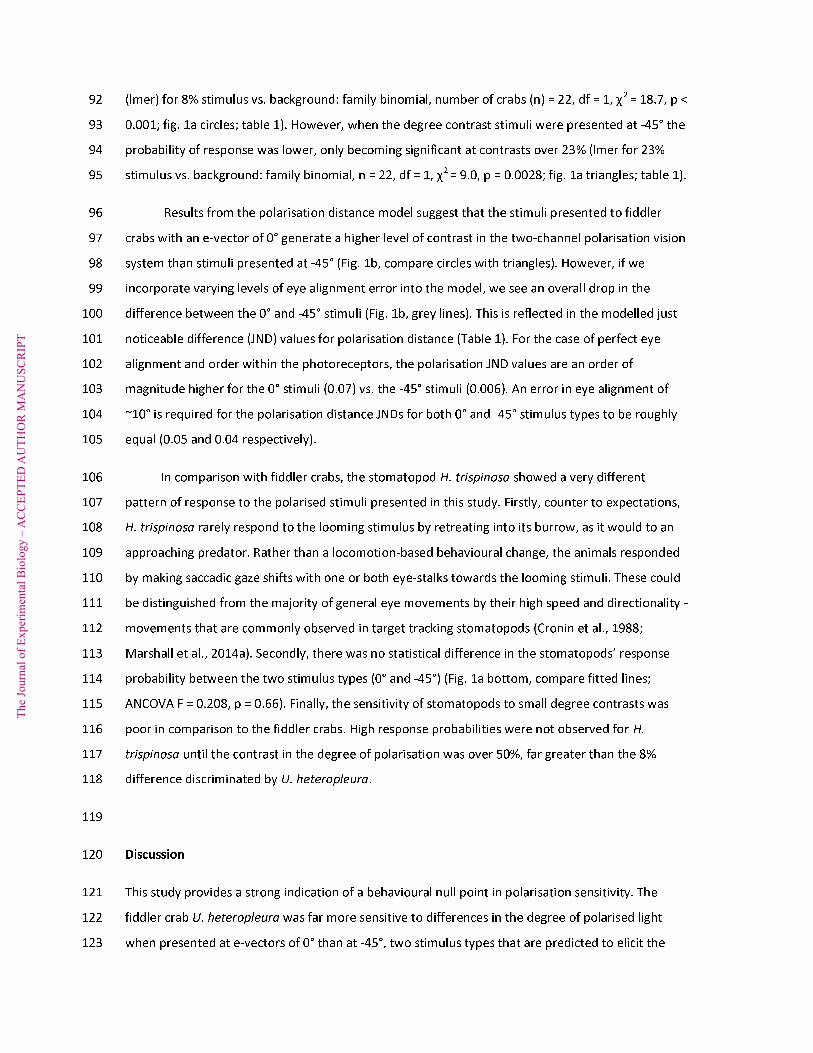
(lmer) for 8% stimulus vs. background: family binomial, number of crabs (n) = 22, df = 1, χ2 = 18.7, p < 92
0.001; fig. 1a circles; table 1). However, when the degree contrast stimuli were presented at -45° the 93
probability of response was lower, only becoming significant at contrasts over 23% (lmer for 23% 94
stimulus vs. background: family binomial, n = 22, df = 1, χ2 = 9.0, p = 0.0028; fig. 1a triangles; table 1). 95
Results from the polarisation distance model suggest that the stimuli presented to fiddler 96
crabs with an e-vector of 0° generate a higher level of contrast in the two-channel polarisation vision 97
system than stimuli presented at -45° (Fig. 1b, compare circles with triangles). However, if we 98
incorporate varying levels of eye alignment error into the model, we see an overall drop in the 99
difference between the 0° and -45° stimuli (Fig. 1b, grey lines). This is reflected in the modelled just 100
noticeable difference (JND) values for polarisation distance (Table 1). For the case of perfect eye 101
alignment and order within the photoreceptors, the polarisation JND values are an order of 102
magnitude higher for the 0° stimuli (0.07) vs. the -45° stimuli (0.006). An error in eye alignment of 103
~10° is required for the polarisation distance JNDs for both 0° and -45° stimulus types to be roughly 104
equal (0.05 and 0.04 respectively). 105
In comparison with fiddler crabs, the stomatopod H. trispinosa showed a very different 106
pattern of response to the polarised stimuli presented in this study. Firstly, counter to expectations, 107
H. trispinosa rarely respond to the looming stimulus by retreating into its burrow, as it would to an 108
approaching predator. Rather than a locomotion-based behavioural change, the animals responded 109
by making saccadic gaze shifts with one or both eye-stalks towards the looming stimuli. These could 110
be distinguished from the majority of general eye movements by their high speed and directionality - 111
movements that are commonly observed in target tracking stomatopods (Cronin et al., 1988; 112
Marshall et al., 2014a). Secondly, there was no statistical difference in the stomatopods’ response 113
probability between the two stimulus types (0° and -45°) (Fig. 1a bottom, compare fitted lines; 114
ANCOVA F = 0.208, p = 0.66). Finally, the sensitivity of stomatopods to small degree contrasts was 115
poor in comparison to the fiddler crabs. High response probabilities were not observed for H. 116
trispinosa until the contrast in the degree of polarisation was over 50%, far greater than the 8% 117
difference discriminated by U. heteropleura. 118
119
Discussion 120
This study provides a strong indication of a behavioural null point in polarisation sensitivity. The 121
fiddler crab U. heteropleura was far more sensitive to differences in the degree of polarised light 122
when presented at e-vectors of 0° than at -45°, two stimulus types that are predicted to elicit the 123

The
Jour
nal o
f Exp
erim
enta
l Bio
logy
– A
CC
EPTE
D A
UTH
OR
MA
NU
SCR
IPT
maximal and minimal amount of visual contrast respectively. The level of diminished response at -124
45°, compared with a complete null response, is entirely consistent with a degree of misalignment 125
between or across the eyes. In comparison, the stomatopod H. trispinosa - with its multi-channel 126
and rotationally-dynamic polarisation vision system - did not show evidence for a null point of 127
discrimination at an e-vector angle of -45°. These results also provide evidence that crustaceans 128
respond, not just to differences in the e-vector orientation of polarised light (Marshall et al., 1999; 129
Glantz and Schroeter, 2007; How et al., 2012), but also to small differences in the degree of 130
polarised light. 131
What might be the functional consequences of this polarisation null point for fiddler crabs in 132
their natural environment? At present it is unknown exactly how the polarisation and intensity 133
channels are integrated so it is difficult to interpret the full functional relevance of their polarisation 134
sensitivity. In related crustaceans the polarisation system is thought to act as a contrast enhancer for 135
the intensity channel, and possibly also related to movement detection (Glantz, 2001; Glantz and 136
Schroeter, 2006), so it is in this context that we frame our discussion. One of the main polarisation 137
cues in the fiddler crab’s mudflat environment is likely to be found in the area of damp mudflat in 138
the direction of the sun. Here, most reflected light will be roughly horizontally polarised (Zeil and 139
Hofmann, 2001; How and Marshall, 2014). The horizontal/vertical receptor organisation of the 140
fiddler crab could therefore be adapted for detecting small changes in the degree of polarisation in 141
this region. This could be used for enhancing the contrast of conspecific crabs against the mudflat 142
surface, and could therefore be thought of as a ‘matched filter’ for this environment (Wehner, 1987; 143
Zeil and Hofmann, 2001; Alkaladi et al., 2013; How and Marshall, 2014). However, there are several 144
sources of non-horizontally polarised cues. For example, the sky polarisation field spans the entire 145
celestial hemisphere (although it will often be obscured by clouds or large objects on the horizon) 146
and the e-vectors of this polarised light are oriented in parallel to a circular band 90° from the sun’s 147
position (Labhart and Meyer, 2002). Therefore, for certain solar positions the polarised light field will 148
be oriented at ±45° across large parts of the sky. This could have an impact on the use of polarised 149
light for enhancing the contrast of airborne predators in these parts of the sky. However, it is unclear 150
whether or not polarised light plays a role in predator surveillance in the wild. There have also been 151
suggestions that fiddler crabs may use their acute polarisation vision for detecting subtle variations 152
in the cuticle surface of conspecifics (Zeil and Hofmann, 2001). Light reflected from damp crab 153
cuticle is likely to occupy a whole range of e-vector angles, many of which may fall at or near the null 154
point of discrimination. 155

The
Jour
nal o
f Exp
erim
enta
l Bio
logy
– A
CC
EPTE
D A
UTH
OR
MA
NU
SCR
IPT
In conjunction with previous modelling attempts (How and Marshall, 2014), our current 156
results imply that the fiddler crab’s polarisation vision system is most sensitive to small differences in 157
the degree of polarised light when the e-vector angle is close to horizontal. This suggests that the 158
horizontal/vertical arrangement of the polarisation receptors is optimally adapted for discriminating 159
small differences in the degree of polarised light (caused, for example, by a conspecific crab) against 160
the horizontally polarised mudflat background (Zeil and Hofmann, 2001; How and Marshall, 2014). 161
However, there is another related explanation for this kind of arrangement. The fiddler crab U. 162
vomeris was recently found to have differences in the anatomical arrangement between the two 163
polarised light channels suggesting that, in the equatorial region of the eye, each ommatidial unit is 164
more sensitive to vertically polarised light than to horizontally polarised light (Alkaladi et al., 2013). 165
This would have the consequence of screening out the horizontally polarised glare reflected from 166
the mudflat, analogous to the effect of wearing polaroid sunglasses. This asymmetrical sensitivity to 167
vertically polarised light is only possible if the receptors have a horizontal/vertical arrangement 168
relative to the outside world. It must, however, be noted that this anatomical organisation would 169
not affect the location of the polarisation null point, as long as the sensitivity maxima remain 170
horizontally and vertically oriented. 171
An alternative explanation that must be addressed is that the fiddler crabs may be responding 172
differently to the two stimulus types according to how realistic they seem. The horizontal stimulus 173
set simulates a polarisation degree contrast viewed against a horizontally polarised background, a 174
situation that may be common in the fiddler crabs’ mudflat environment. It may be argued that the -175
45° stimulus set represents a less natural situation (although see discussion above), leading to a 176
reduced response from the fiddler crabs. This hypothesis leads to the prediction that vertically 177
oriented stimuli (even more unusual in the fiddler crab’s environment) should elicit even lower 178
response probabilities than the -45° set. We present preliminary data from a reduced sample (n = 6 179
crabs) that provide a good indication that this is not the case (Supp. fig. 1). Fiddler crabs presented 180
with vertically oriented degree contrasts responded with a similar probability as they did to 181
horizontally oriented stimuli. 182
As a final note on the experimental methods used for the fiddler crab part of this study, we 183
recorded an interesting discrepancy between our behavioural results and the predictions of the 184
polarisation distance model. Our results initially suggested that fiddler crabs were more sensitive to 185
polarised objects at the -45° null point than the polarisation distance model predicts. The 186
polarisation distance JND values modelled for these -45° stimuli were 10 times lower than for the 0° 187
stimuli (0.006 and 0.07 respectively, table 1) implying that the fiddler crabs should not have been 188

The
Jour
nal o
f Exp
erim
enta
l Bio
logy
– A
CC
EPTE
D A
UTH
OR
MA
NU
SCR
IPT
able to detect these stimuli. However, when we took into account the possible effect of slight 189
misalignment between the crab eyes and the monitor (Fig. 1b, grey lines) this discrepancy could 190
largely be explained. An alignment error of ~10° resulted in modelled polarisation distance values 191
that were approximately equal for 0° and -45° JND points ( 0.05 and 0.04 respectively, table 1), and 192
this level of misalignment was certainly possible in the experimental apparatus. In their natural 193
environment, fiddler crabs use the horizon as a visual cue to maintain the alignment of their 194
eyestalks (Zeil et al., 1986). In our experiment, the false horizon presented to the test animal was 195
only displayed on three of the four surrounding monitors and the close distance of these monitors 196
(220 mm) suggests that small misplacements of the virtual horizon could have large effects on its 197
perceived location. Future research of this kind should take extra precautions to reduce these kinds 198
of alignment errors. 199
A second explanation, which cannot be discounted, for this higher-than-expected sensitivity to 200
stimuli at -45° is that the fiddler crab may use some form of local comparison between imperfectly 201
oriented polarisation receptors in adjacent regions of the eye to enhance contrast to such cues. The 202
current polarisation distance model only attempts to represent visual input from two single-sets of 203
opponent receptors, and so may miss any effects of regional processing across the eye (Glantz and 204
Schroeter, 2007). Future anatomical, electrophysiological and behavioural studies will be needed to 205
investigate this fully. 206
In comparison to fiddler crabs, the stomatopod H. trispinosa showed no evidence of a null 207
point in polarisation sensitivity at an e-vector of -45°: These animals responded with equal 208
probability to polarised stimuli at 0° and at -45°. This is not surprising given that anatomical evidence 209
shows that these animals have a complex polarisation anatomy. Although these species do possess 210
two-channel polarisation vision systems across the majority of the eye, the receptors are oriented 211
differently in distinct regions. Firstly, orthogonal receptors in the eye’s dorsal hemisphere tend to be 212
shifted in orientation by 45° from orthogonal receptors in the ventral hemisphere, providing two 213
pairs of differently oriented polarisation channels in regions with overlapping visual fields (Marshall 214
et al., 1991). Secondly, these species make frequent eye rotations around the axis of view, thus 215
altering the orientation of the polarisation receptors relative to the outside world (Land et al., 1990). 216
Thirdly, stomatopods do not make maintain a stable body orientation as they crawl or swim in their 217
complex three-dimensional habitat, and changes in body position do not seem to be compensated 218
by adjustments in eye alignment (as is the case for fiddler crabs). For example, H. trispinosa will 219
often rest in a tilted, or occasionally upside-down, position in the entrance of the burrow, thus 220
affecting the orientation of their polarisation vision system relative to the outside world. 221

The
Jour
nal o
f Exp
erim
enta
l Bio
logy
– A
CC
EPTE
D A
UTH
OR
MA
NU
SCR
IPT
There are several proposed explanations for the extreme mobility of stomatopod eyes, 222
including their use for target acquisition and tracking, and for scan movements associated with the 223
colour and polarisation sensitivity in the mid-band region (Cronin et al., 1988; Land et al., 1990; 224
Thoen et al., 2014; Marshall et al., 2014a). Such movements have strong implications for their 225
polarisation vision system, which, as a result, does not appear to suffer from the null points 226
experienced by stabilised two-channel receptor arrangements. This could be of particular relevance 227
for signalling and communication in stomatopods. Many species (including H. trispinosa) use linearly 228
polarised body patterns (Chiou et al., 2011) and, given that stomatopods are highly mobile animals, 229
these patterns could potentially be viewed from a variety of orientations. A rotatable multi-channel 230
polarisation system is likely to be more efficient at detecting these communication signals than fixed 231
two-channel arrays, which may miss those signals falling in or near the null point of discrimination. 232
Eye anatomy and movement may therefore have evolved, in part, to allow the detection of linearly 233
polarised signals regardless of their orientation. There is also the tantalising possibility that 234
stomatopods may use eye rotations around the visual axis as a method of gain control, for 235
optimising the polarisation contrast between an object of interest and the background. However, 236
this has yet to be demonstrated. 237
Finally, it is interesting to note that fiddler crabs and stomatopods differed markedly in their 238
overall sensitivity to the polarised light cues. Fiddler crabs showed clear responses to differences in 239
the degree of polarised light as low as 8%. In comparison, stomatopods only responded well to 240
stimuli with contrasts greater than 50%. Also, the two different species differed clearly in the types 241
of responses that these stimuli elicited. Fiddler crabs exhibited clear anti-predator behaviour, 242
including sprints away from the stimulus, freezing and/or tucking in the limbs. Stomatopods, on the 243
other hand, simply glanced at the looming stimulus as if it was a prey object of interest. The 244
difference in sensitivity could be explained by the apparent difference in behavioural context 245
between fiddler crabs and stomatopods. The former were clearly attempting to avoid predation and 246
so were highly motivated to respond to looming stimuli, while the latter would simply have been 247
looking for food items from the safety of the home burrow. Another possibility is that the results 248
demonstrate an actual difference in the behavioural sensitivity of these two animal groups to 249
polarised cues. Fiddler crabs are known to respond to very weak intensity cues in the dorsal part of 250
the visual field, corresponding to the retinal position of small avian predators such as terns (Smolka 251
and Hemmi, 2009; Smolka et al., 2011; Smolka et al., 2012). Given that polarisation is likely to act as 252
a contrast enhancer for the intensity channel it would seem logical that these animals should also 253
respond well to small differences in polarised light. On the other hand, stomatopods may use their 254
polarisation sensitivity primarily for detecting communication signals on the cuticle of conspecifics, 255

The
Jour
nal o
f Exp
erim
enta
l Bio
logy
– A
CC
EPTE
D A
UTH
OR
MA
NU
SCR
IPT
body patterns which tend to be very strongly polarised. Perhaps the relatively low polarisation 256
sensitivity observed in the stomatopod is a consequence of some level of filtering of the polarisation 257
information, a process that may be important for simplifying subsequent processing steps necessary 258
for this unique scan vision system. Similar information reduction steps also seem to occur early in 259
the stomatopod colour vision system (Thoen et al., 2014). 260
261
Materials and Methods 262
Male fiddler crabs (n=22) of the species U. heteropleura were collected from the mudflats at the 263
Pacific entrance to the Panama Canal (8°56’56.3”N; 79°34’24.3”W) in April 2013 and were 264
transported to an outdoor flow-through aquarium facility at the Naos Island Laboratories of the 265
Smithsonian Tropical Research Institute. The crabs were housed in clear perforated plastic 266
containers for a period of 1-5 days and were fed daily with fish flake (TetraMin, Tetra, Germany). 267
Individual crabs were presented with polarised light stimuli using similar methods to those used 268
previously (How et al., 2012). A wire hinge was attached to the animal’s carapace with cyano-269
acrylate glue, and connected to a metal hanger, which tethered the animal on top of a 200 mm 270
diameter Styrofoam ball (Fig. 2a, treadmill). The treadmill was suspended on a cushion of air 271
provided by a hairdryer (Venezia 2400, VS Sassoon, P&G and Conair) allowing the crab to walk freely 272
in any direction (causing the treadmill to rotate beneath it). The treadmill was surrounded by four 273
LCD monitors (1905fp, Dell, Round Rock, USA) three of which were unmodified and displayed a 274
white sky background and a dark brown floor with a flat horizon, level with the top of the treadmill. 275
The fourth monitor had the front polarising filter removed so that the monitor output could vary in 276
polarised light only. This LCD monitor uses an in-plane switching architecture; as the greyscale level 277
addressed to the monitor varies from black (U8 = 0 [on 8 bit scale]), through to grey (U8 = 200), the 278
degree of linearly polarised light declines, while intensity, e-vector angle (0°) and ellipticity (~0%) 279
remain constant (Fig. 3a). This allowed us to present the animals with a range of dynamic visual 280
stimuli varying only in the degree of polarised light (Fig. 3a-c). It must be noted that unmodified LCD 281
monitors also generate polarised light. In the case of the three Dell monitors used in this experiment 282
this was 100% polarised and oriented in a vertical direction, but remained unchanged for the 283
duration of the experiment and so had no influence on the fiddler crabs’ response. 284
Adult male and female stomatopods of the species H. trispinosa were collected from Lizard 285
Island lagoon on the Great Barrier Reef, Australia (14°40'40.8"S; 145°26'48.1"E) in July 2013. Animals 286
were housed separately in artificial burrows under natural illumination in a flow-through aquarium 287

The
Jour
nal o
f Exp
erim
enta
l Bio
logy
– A
CC
EPTE
D A
UTH
OR
MA
NU
SCR
IPT
system at the Lizard Island Research Station. Individuals were transferred to a 250 x 150 x 200 mm 288
(length x width x height) partition within a test aquarium, and the animal’s burrow was placed 289
centrally facing the small glass front of the partition (fig. 2b). The aquarium was constructed of non-290
tempered glass, and did not affect the transmission of polarised light in any way. Animals were 291
allowed to acclimatise for >10 minutes. Then, prior to testing, a polarised LCD monitor (1905fp, Dell, 292
Round Rock, USA – same as above) was placed against the front glass panel. 293
To test whether the threshold response of crabs and stomatopods to the degree of polarised 294
light depended on its e-vector axis, we presented the animals with a range of stimuli on the monitor 295
in the standard position or with the monitor rotated by -45° (Fig. 3b). These are hereafter referred to 296
as the 0° and -45° stimulus sets. We use the term ‘degree’ to refer to the proportion of polarised 297
light, and ‘degree contrast’ as the difference in the amount of polarised light between stimulus and 298
background. In this manuscript we use the percent scale to represent both of these measures, 299
varying from 0 to 100%. For fiddler crabs, each animal was presented with each stimulus condition 300
twice, and monitor orientation was alternated so that half the animals received the 0° stimulus set 301
first, and the other half received the -45° stimulus set first. The stimulus consisted of a looming circle 302
that expanded over 1 second from 0 to 145 mm diameter at a distance of 220 mm from the crab 303
(thus occupying 36.5° of the visual field when fully expanded). The contrast in the degree of 304
polarised light between the looming stimulus and the background was set to the following values, 305
which were presented in a fully randomised order: 0%, 3.2%, 8.0%, 13.0%, 18.3%, 23.0%, 48.1%, or 306
82.9% (note that the last stimulus contrast also varied in e-vector angle; fig. 3a,c). 307
The procedure for the stomatopod experiment was similar, differing only in the following 308
ways: Because these animals were unrestrained, there were frequent instances of the stomatopod 309
being in an incorrect position for viewing the stimulus (e.g. tucked inside the burrow). Therefore, the 310
0° and -45° stimuli were presented to a different set of animals to maintain statistical independence 311
between the treatments and for ease of analysis. A single stimulus set was presented to each of 56 312
different individuals twice, so that half viewed two presentations of the 0° stimulus set and half 313
viewed two presentations of the -45° set. This proceeded in alternating order to avoid any diurnal or 314
long term temporal changes in stomatopod behaviour. The actual stimulus was similar to that 315
presented to the fiddler crabs, consisting of an 80 mm loom expanding over 1 second, viewed at a 316
distance of 125 mm (thus occupying a visual angle of 35.5°). The exact values of degree contrast 317
presented at random to the stomatopod also differed slightly from those chosen for the fiddler crab 318
experiment: 0%, 10%, 20%, 30%, 40%, 50%, 60%, or 70%. 319

The
Jour
nal o
f Exp
erim
enta
l Bio
logy
– A
CC
EPTE
D A
UTH
OR
MA
NU
SCR
IPT
All stimuli were generated in Matlab (Mathworks, 2012) and responses were filmed either 320
with a webcam (model C210, Logitech, Romanel-sur-Morges, Switzerland) or digital video camera 321
(HDR-SR11E, Sony Corp, Tokyo, Japan). The responses of animals to the looming stimuli were 322
subsequently scored from the digital video recordings by visually detecting changes in the animal’s 323
behaviour. This was undertaken in a fully blind process, in which all videos were randomised before 324
analysis. Animals that responded to either of the repeat stimuli were scored 1, and those that 325
responded to neither were scored 0. The score was then averaged across all animals to produce a 326
response probability value for each stimulus/background condition. Because some of the scored 327
behaviours occurred independently of the stimulus, we faced the potential problem of recording 328
false positive responses. To overcome this, we used the response probability to the zero contrast 329
stimuli as an estimate of the background probability of the behaviour. 330
For the fiddler crab results a linear mixed model approach was used to determine which data 331
points differed significantly from background levels. This was implemented in R (v.3.0.1, CRAN May 332
2013) using the ‘lmer’ function from the lme4 package. The model included animal identity as a 333
random factor to account for any variance and possible biases due to response differences between 334
individuals. We used the link function ‘logit’ and family ‘binomial’, and the significance of each 335
stimulus response was determined by comparing the model that included the background level of 336
response against the model without the background level of response. For the stomatopod results, 337
an analysis of covariance (ANCOVA) was performed to test for any effect of stimulus orientation on 338
the response probability of the stomatopods. This method was used rather than the mixed model 339
approach due to the statistical independence of the two treatments, and was implemented in R 340
using the ‘lm’ function. 341
For each fiddler crab stimulus condition an estimate of the level of contrast available to a two-342
channel polarisation vision system was calculated using the How-Marshall polarisation distance 343
model (How and Marshall, 2014). Briefly, the model assumes that the animal uses a two-channel 344
polarisation vision system with receptors organised horizontally and vertically relative to the outside 345
world. These receptors then have opponent connections to a simulated visual interneuron, and the 346
relative levels of activity between interneurons viewing the stimulus and the background are 347
compared to produce a single estimate of contrast (see How and Marshall, 2014 for details). For this 348
specific case we used a polarisation sensitivity (Sp) of 10 for the model. However, the results would 349
equally apply for lower Sp values. 350
351

The
Jour
nal o
f Exp
erim
enta
l Bio
logy
– A
CC
EPTE
D A
UTH
OR
MA
NU
SCR
IPT
Acknowledgements 352
Thanks to Kecia Kerr for her help collecting fiddler crabs; to the Smithsonian Tropical Research 353
Institute in Panama and to Lizard Island Research Station in Australia for their logistical support; the 354
following authorities in Panama for research permits and access to the study site: Autoridad de 355
Recursos Acuáticos de Panamá (ARAP); Unidad Administrativa de Bienes Revertidos del Ministerio de 356
Economía y Finanzas, el Servicio Nacional Aeronaval and the Autoridad del Canal de Panama (ACP); 357
and to Shelby Temple for helpful comments on the manuscript. 358
359
Funding 360
MJH, NJM and NWR supported by the US Air Force Office of Scientific Research (Grant # FA8655-12-361
1-2112) and the Asian and European Offices of Aerospace Research and Development. MJH also 362
supported by a Queensland-Smithsonian Fellowship award. 363
364
References 365
Alkaladi, A., How, M. and Zeil, J. (2013). Systematic variations in microvilli banding patterns along 366
fiddler crab rhabdoms. J. Comp. Physiol. A 199, 99-113. 367
Bernard, G. D. and Wehner, R. (1977). Functional similarities between polarization vision and color 368
vision. Vision Res. 17, 1019-1028. 369
Chiou, T.-H., Marshall, N. J., Caldwell, R. L. and Cronin, T. W. (2011). Changes in light-reflecting 370
properties of signalling appendages alter mate choice behaviour in a stomatopod crustacean 371
Haptosquilla trispinosa. Mar. Freshw. Behav. Phy. 44, 1-11. 372
Chiou, T. H., Kleinlogel, S., Cronin, T., Caldwell, R., Loeffler, B., Siddiqi, A., Goldizen, A. and 373
Marshall, J. (2008). Circular polarization vision in a stomatopod crustacean. Curr. Biol. 18, 429-374
434. 375
Cronin, T. W., Nair, J. N., Doyle, R. D. and Caldwell, R. L. (1988). Ocular Tracking of Rapidly Moving 376
Visual Targets by Stomatopod Crustaceans. J. Exp. Biol. 138, 155-179. 377
Dana, J. D. (1852). Crustacea, Part 1. In United States exploring expedition during the years 1838, 378
1839, 1840, 1841, 1842, under the command of Charles Wilkes, U.S.N., pp. 1-685. 379
Glantz, R. and Schroeter, J. (2006). Polarization contrast and motion detection. J. Comp. Physiol. A 380
192, 905-914. 381
Glantz, R. M. (2001). Polarization analysis in the crayfish visual system. J. Exp. Biol. 204, 2383-2390. 382

The
Jour
nal o
f Exp
erim
enta
l Bio
logy
– A
CC
EPTE
D A
UTH
OR
MA
NU
SCR
IPT
Glantz, R. M. (2007). The distribution of polarization sensitivity in the crayfish retinula. J. Comp. 383
Physiol. A 193, 893-901. 384
Glantz, R. M. and Schroeter, J. P. (2007). Orientation by polarized light in the crayfish dorsal light 385
reflex: behavioral and neurophysiological studies. J. Comp. Physiol. A 193, 371-384. 386
How, M. J., Pignatelli, V., Temple, S. E., Marshall, N. J. and Hemmi, J. M. (2012). High e-vector 387
acuity in the polarisation vision system of the fiddler crab Uca vomeris. J. Exp. Biol. 215, 2128-388
2134. 389
How, M. J. and Marshall, N. J. (2014). Polarization distance: a framework for modelling object 390
detection by polarization vision systems. Proc. R. Soc. B 281. 391
Johnsen, S., Marshall, N. J. and Widder, E. A. (2011). Polarization sensitivity as a contrast enhancer 392
in pelagic predators: lessons from in situ polarization imaging of transparent zooplankton. 393
Philos. Trans. R. Soc. Lond. B 366, 655-670. 394
Jordan, T. M., Partridge, J. C. and Roberts, N. W. (2012). Non-polarizing broadband multilayer 395
reflectors in fish. Nat. Photon. 6, 759-763. 396
Kleinlogel, S., Marshall, N. J., Horwood, J. M. and Land, M. F. (2003). Neuroarchitecture of the 397
color and polarization vision system of the stomatopod haptosquilla. J. Comp. Neurol. 467, 398
326-342. 399
Labhart, T. and Meyer, E. P. (1999). Detectors for polarized skylight in insects: a survey of 400
ommatidial specializations in the dorsal rim area of the compound eye. Microsc. Res. Tech. 47, 401
368-379. 402
Labhart, T. and Meyer, E. P. (2002). Neural mechanisms in insect navigation: polarization compass 403
and odometer. Curr. Opin. Neurobiol. 12, 707-714. 404
Land, M. F., Marshall, J. N., Brownless, D. and Cronin, T. W. (1990). The eye-movements of the 405
mantis shrimp Odontodactylus scyllarus (Crustacea, Stomatopoda). J. Comp. Physiol. A 167, 406
155-166. 407
Marshall, J., Cronin, T. W., Shashar, N. and Land, M. (1999). Behavioural evidence for polarisation 408
vision in stomatopods reveals a potential channel for communication. Curr. Biol. 9, 755-758. 409
Marshall, N. J., Land, M. F., King, C. A. and Cronin, T. W. (1991). The Compound Eyes of Mantis 410
Shrimps (Crustacea, Hoplocarida, Stomatopoda) .1. Compound Eye Structure - the Detection 411
of Polarized-Light. Philos. Trans. R. Soc. Lond. B 334, 33-56. 412
Marshall, N. J., Land, M. F. and Cronin, T. W. (2014a). Shrimps that pay attention: saccadic eye 413
movements in stomatopod crustaceans. Philosophical Transactions of the Royal Society B 369. 414
Marshall, N. J., Roberts, N. W. and Cronin, T. W. (2014b). Polarisation signals. In Polarisation 415
Vision (ed. G. Horvath), pp. In Press. New York: Springer. 416

The
Jour
nal o
f Exp
erim
enta
l Bio
logy
– A
CC
EPTE
D A
UTH
OR
MA
NU
SCR
IPT
Mathworks. (2012). Matlab R2012b. Natick, MA, USA. 417
Moody, M. F. and Parriss, J. R. (1961). The discrimination of polarized light by Octopus: a 418
behavioural and morphological study. Z. Vergl. Physiol. 44, 268-291. 419
Pignatelli, V., Temple, S. E., Chiou, T.-H., Roberts, N. W., Collin, S. P. and Marshall, N. J. (2011). 420
Behavioural relevance of polarization sensitivity as a target detection mechanism in 421
cephalopods and fishes. Philos. Trans. R. Soc. Lond. B 366, 734-741. 422
Roberts, N. W., Chiou, T. H., Marshall, N. J. and Cronin, T. W. (2009). A biological quarter-wave 423
retarder with excellent achromaticity in the visible wavelength region. Nat Photon 3, 641-644. 424
Schechner, Y. Y. and Karpel, N. (2005). Recovery of underwater visibility and structure by 425
polarization analysis. IEEE Journal of Oceanic Engineering 30, 570-587. 426
Schwind, R. (1983). A polarization-sensitive response of the flying water bug Notonecta glauca to 427
UV light. J. Comp. Physiol. A 150, 87-91. 428
Shashar, N. and Cronin, T. W. (1996). Polarization contrast vision in Octopus. J. Exp. Biol. 199, 999-429
1004. 430
Shashar, N., Rutledge, P. S. and Cronin, T. W. (1996). Polarization vision in cuttlefish - a concealed 431
communication channel? J. Exp. Biol. 199, 2077-2084. 432
Shashar, N., Hanlon, R. T. and Petz, A. d. (1998). Polarization vision helps detect transparent prey. 433
Nature 393, 222-223. 434
Shashar, N., Hagan, R., Boal, J. G. and Hanlon, R. T. (2000). Cuttlefish use polarization sensitivity in 435
predation on silvery fish. Vision Res. 40, 71-75. 436
Shaw, S. R. (1969). Sense-cell structure and interspecies comparisons of polarized-light absorption 437
in arthropod compound eyes. Vision Res. 9, 1031-1040. 438
Smith, S. I. (1870). Notes on American crustacea. No. 1. Ocypodoidea. Trans. Conn. Acad. Arts Sci. 439
2, 113-176. 440
Smolka, J. and Hemmi, J. M. (2009). Topography of vision and behaviour. J. Exp. Biol. 212, 3522-441
3532. 442
Smolka, J., Zeil, J. and Hemmi, J. M. (2011). Natural visual cues eliciting predator avoidance in 443
fiddler crabs. Proc. R. Soc. B 278, 3584-3592. 444
Smolka, J., Raderschall, C. A. and Hemmi, J. M. (2012). Flicker is part of a multi-cue response 445
criterion in fiddler crab predator avoidance. J. Exp. Biol. 446
Talbot, C. M. and Marshall, J. N. (2011). The retinal topography of three species of coleoid 447
cephalopod: significance for perception of polarized light. Philos. Trans. R. Soc. Lond. B 366, 448
724-733. 449

The
Jour
nal o
f Exp
erim
enta
l Bio
logy
– A
CC
EPTE
D A
UTH
OR
MA
NU
SCR
IPT
Tasaki, K. and Karita, K. (1966). Intraretinal Discrimination of Horizontal and Vertical Planes of 450
Polarized Light by Octopus. Nature 209, 934-935. 451
Temple, S. E., Pignatelli, V., Cook, T., How, M. J., Chiou, T.-H., Roberts, N. W. and Marshall, N. J. 452
(2012). High-resolution polarisation vision in a cuttlefish. Curr. Biol. 22, R121-R122. 453
Thoen, H. H., How, M. J., Chiou, T.-H. and Marshall, N. J. (2014). A different form of colour vision 454
in mantis shrimps. Science 343, 411-413. 455
Tuthill, J. C. and Johnsen, S. (2006). Polarization sensitivity in the red swamp crayfish Procambarus 456
clarkii enhances the detection of moving transparent objects. J. Exp. Biol. 209, 1612-1616. 457
Vorobyev, M. and Osorio, D. (1998). Receptor noise as a determinant of colour thresholds. Proc. R. 458
Soc. Lond. B 265, 351-358. 459
Waterman, T. H. (1981). Polarization sensitivity. In Handbook of Sensory Physiology 7/6B vol. 7 (ed. 460
H. Autrum), pp. 281-469. New York: Springer. 461
Wehner, R. (1976). Polarized-light navigation by insects. Sci. Am. 235, 106-115. 462
Wehner, R. (1987). ‘Matched filters’ — neural models of the external world. J. Comp. Physiol. A 463
161, 511-531. 464
Wehner, R. and Labhart, T. (2006). Polarisation vision. In Invertebrate Vision (eds. E. Warrant and 465
D.-E. Nilsson), pp. 291-348. Cambridge: Cambridge University Press. 466
Zeil, J., Nalbach, G. and Nalbach, H.-O. (1986). Eyes, eye stalks and the visual world of semi-467
terrestrial crabs. J. Comp. Physiol. A 159, 801-811. 468
Zeil, J. and Hofmann, M. (2001). Signals from 'crabworld': cuticular reflections in a fiddler crab 469
colony. J. Exp. Biol. 204, 2561-2569. 470

The
Jour
nal o
f Exp
erim
enta
l Bio
logy
– A
CC
EPTE
D A
UTH
OR
MA
NU
SCR
IPT
Table 471
Table 1. Just noticeable difference (JND) values for the two stimulus orientation conditions (0° and -472
45°) presented as recorded degree contrast and calculated polarisation distance. The 5°, 10° and 15° 473
error values represent the calculated polarisation distance for incremental errors in eyestalk 474
misalignment. 475
JND value 0° stimuli -45° stimuli
Degree contrast 8% 23%
Polarisation distance 0.07 0.006
Polarisation distance (5° error) 0.06 0.02
Polarisation distance (10° error) 0.05 0.04
Polarisation distance (15° error) 0.04 0.06
476
477
478
Figures 479
Figure 1. Response probability to polarised stimuli. a) Response probability of the fiddler crab U. 480
heteropleura (top) and the stomatopod H. trispinosa (bottom) to horizontal (circles) and -45° 481
(triangles) stimulus e-vector orientations (see insert bottom right). Curves represent the fitted 482
hyperbolic tangent (top) or straight line (bottom) for each set of data. Both fitted lines were 483
generated using the Matlab curve fitting toolbox. Grey shading represents the 95% confidence 484
interval for the fitted curves. Background dotted line indicates the probability of false positive 485
responses. b) Polarisation distance modelled for the fiddler crab stimuli and for each monitor 486
orientation. Grey lines indicate the effect of incremental levels of crab eye misalignment relative to 487
the monitor (5°, 10° and 15°). 488
489
Figure 2. Experimental apparatus for the presentation of polarisation stimuli. (a) Fiddler crab 490
apparatus (according to How et al., 2012): The animal was tethered above a treadmill and stimuli 491
were presented on a modified LCD monitor (left). (b) Stomatopod apparatus: Unrestrained animals 492
occupying a burrow within an aquarium partition were presented with stimuli on a modified LCD 493
monitor pressed against the wall of the aquarium (left). 494

The
Jour
nal o
f Exp
erim
enta
l Bio
logy
– A
CC
EPTE
D A
UTH
OR
MA
NU
SCR
IPT
495
Figure 3. Polarisation properties of the modified LCD monitor. (a) Degree of polarised light (solid 496
line, y-axis left) changed gradually with greyscale value addressed to the monitor (x-axis), while e-497
vector angle (dashed line, y-axis right) and ellipticity (dotted line, y-axis left) remained constant (at 498
~0° and ~0% respectively) between 0 and 200 U8 value of the greyscale range. White circles 499
represent the exact stimulus set used in the fiddler crab experiment. (b) Representation of monitor 500
output in Stokes coordinates. Note that the z-axis (Stokes parameter S3 - ellipticity) remained close 501
to zero so a 2D projection in the equatorial plane of the Poincaré sphere captured the relevant 502
information. Solid line = full range of monitor output; white circles = fiddler crab stimuli. (c) 503
Illustration of the polarisation properties of all stimuli used in the fiddler crab experiment and the 504
background against which they were presented. The relative length and angle of the black arrows 505
represent the degree and angle of polarised light respectively. These are illustrated for the two 506
stimulus sets: horizontal (0°; left) and diagonal (-45°; right). The stimuli presented to the 507
stomatopods had similar polarisation properties, varying only in the degree values chosen. Left hand 508
column in the table provides the actual degree of polarisation value. 509

The
Jour
nal o
f Exp
erim
enta
l Bio
logy
– A
CC
EPTE
D A
UTH
OR
MA
NU
SCR
IPT

The
Jour
nal o
f Exp
erim
enta
l Bio
logy
– A
CC
EPTE
D A
UTH
OR
MA
NU
SCR
IPT

The
Jour
nal o
f Exp
erim
enta
l Bio
logy
– A
CC
EPTE
D A
UTH
OR
MA
NU
SCR
IPT


