fibrosing mediastinitis series medicine2011... · fibrosing mediastinitis clinical presentation,...
TRANSCRIPT
Fibrosing MediastinitisClinical Presentation, Therapeutic Outcomes, and Adaptive
Immune Response
Tobias Peikert, MD, Thomas V. Colby, MD, David E. Midthun, MD, Peter C. Pairolero, MD, Eric S. Edell, MD,Darrell R. Schroeder, MS, and Ulrich Specks, MD
Abstract: Fibrosing mediastinitis (FM) is a rare disorder characterizedby the invasive proliferation of fibrous tissue within the mediastinum.FM frequently results in the compression of vital mediastinal structuresand has been associated with substantial morbidity and mortality. Itspathogenesis remains unknown. However, in North America most casesare thought to represent an immune-mediated hypersensitivity responseto Histoplasma capsulatum infection.
To characterize the clinical disease spectrum, natural disease pro-gression, responses to therapy, and overall survival, we retrospectivelyanalyzed all 80 consecutive patients with a diagnosis of FM evaluated atMayo Clinic, Rochester, MN, from 1998 to 2007. Furthermore, wecharacterized the adaptive immune response in 15 representative patientsby immunohistochemistry.
The majority of patients presented with nonspecific respiratory symp-toms due to the compression of mediastinal broncho-vascular structures.Chest radiographic imaging most frequently revealed localized, invasive,and frequently calcified right-sided mediastinal masses. Most patients hadradiographic or serologic evidence of previous histoplasmosis.
In contrast to earlier reports summarizing previously reported FMcases, the clinical course of our patients appeared to be more benign andless progressive. The overall survival was similar to that of age-matchedcontrols. There were only 5 deaths, 2 of which were attributed to FM.These differences may reflect publication bias associated with the pref-erential reporting of more severely affected FM patients in the medicalliterature, as well as the more inclusive case definition used in ourconsecutive case series.
Surgical and nonsurgical interventions effectively relieved symptomscaused by the compression of mediastinal vascular structures in thesecarefully selected patients. In contrast, antifungal and antiinflammatoryagents appeared ineffective. Histologic examination and immunostainingrevealed mixed inflammatory infiltrates consistent with a fibroin-flammatory tissue response in these histoplasmosis-associated FM cases.The immune cell infiltrates included large numbers of CD20-positive Blymphocytes. As B lymphocytes may contribute to the pathogenesis ofthe disease, therapeutic B-cell depletion should be investigated as atherapeutic strategy for FM.
(Medicine 2011;90: 412Y423)
Abbreviations: CT = computed tomography, FM = fibrosingmediastinitis, PET = positron emission tomography, SVC = superiorvena cava.
INTRODUCTION
F ibrosing mediastinitis (FM) is a rare disorder characterizedby the proliferation of locally invasive fibrous tissue within
the mediastinum.11,19 Its etiology remains unknown. FM mayrepresent a clinical-pathologic syndrome rather than a singledisease.29 A number of triggers with variable geographic fre-quencies have been associated with FM, including fungal in-fections, tuberculosis, and sarcoidosis.12,29 In the absence of aspecific identifiable cause, FM is categorized as idiopathic.29
In North America, FM is most commonly associated withHistoplasma capsulatum infections and considered to representan immune-mediated hypersensitivity reaction to this fungalorganism.23,33,39 However, these abnormal host responses areexceedingly rare. Only 3 of 100,000 patients with histoplasmosisdeveloped FM during an outbreak of this fungal infection inIndianapolis between 1978 and 1979.40 The precise pathogeneticmechanisms of FM remain unknown. The exuberant mediastinalfibrosis has been attributed to chronic inflammation in responseto various antigens.11 The reported association of FM with 1 ofthe MHC class I antigen presentation molecules, HLA-A2,supports the involvement of host-specific immune factors.27
In the absence of universally accepted diagnostic criteria,the clinical diagnosis of FM remains challenging. Chest radio-graphic findings frequently help to distinguish between FM andmediastinal malignancies such as lymphomas. Although tissuesampling is generally not required to establish a diagnosis ofFM at experienced referral centers, diagnostic uncertainty byprimary treating physicians often leads to surgical biopsies inthese patients.
Clinical, radiographic, and histopathologic criteria havebeen proposed for FM.11,23 Once malignancy has been excluded,FM needs to be distinguished from other nonmalignant pro-cesses, such as mediastinal necrotizing granulomatous lymph-adenitis (also called mediastinal granuloma).11
The current understanding of FM is derived from singlecase reports, small case series, and synopses of cases re-ported in the literature.3,4,7,9Y11,23,24,26,28,29,32,33,35,36,39 Manyof these reports do not clearly distinguish between FM andmediastinal necrotizing granulomatous lymphadenitis (medi-astinal granuloma).4,10,32,36
In North America most cases of mediastinal necrotizinggranulomatous lymphadenitis are also attributed to infectionwith H. capsulatum. Most of these patients are asymptomatic,and the diagnosis is made when radiographic abnormalities arediscovered incidentally. The long-term prognosis of mediastinalnecrotizing granulomatous lymphadenitis is excellent.11
412 www.md-journal.com Medicine & Volume 90, Number 6, November 2011
From Division of Pulmonary and Critical Care Medicine (TP, DEM, ESE, US);Emeritus staff, Division of General Thoracic Surgery (PCP); Division of Bio-statistics (DRS), Mayo Clinic, Rochester, Minnesota; and Department of Lab-oratory Medicine and Pathology (TVC), Mayo Clinic Scottsdale, Scottsdale,Arizona.Funding and conflicts of interest: Immunohistochemistry studies described in
this manuscript were supported by an investigator-initiated grant fromGenentech, Inc. None of the authors had any conflicts of interest inregards to the content of this manuscript.
Reprints: Ulrich Specks, MD, Division of Pulmonary and Critical CareMedicine, Department of Internal Medicine, Mayo Clinic College ofMedicine, 200 First Street SW, Gonda 18 South, Rochester, MN 55901(e-mail: Specks.Ulrich)mayo.edu).
Copyright * 2011 by Lippincott Williams & WilkinsISSN: 0025-7974DOI: 10.1097/MD.0b013e318237c8e6
Copyright © 2011 Lippincott Williams & Wilkins. Unauthorized reproduction of this article is prohibited.
In contrast, compression or occlusion of vital mediastinalstructures, including the tracheobronchial tree, pulmonary ar-teries and pulmonary veins, the superior vena cava (SVC), orthe esophagus, caused by FM has been associated withhigh morbidity and mortality.7,23,24,33,39 The optimal therapeu-tic approach to FM remains controversial. Isolated case re-ports have suggested that some patients with FM may benefitfrom antiinflammatory, antifungal, or antifibrotic medica-tions.15Y17,21,31,33,39 However, larger case series have not con-firmed these observations.23 Similarly the reported results ofsurgical and nonsurgical interventions to relieve the compressionof mediastinal structures range from very successful proceduresto therapeutic failures associated with high peri- and postoper-ative mortality in excess of 30%.4Y7,10,13,18,23,24,30,34,36,38,39
We conducted the present review of 80 consecutive patientswith FM evaluated and treated at Mayo Clinic, Rochester, MN,between 1998 and 2007 to analyze the clinical presentation,radiographic and pathologic findings, therapeutic interventions,and clinical outcomes of the disease. In addition, we characterizethe adaptive immune response within FM lesions using 15 rep-resentative FM tissue biopsy specimens.
PATIENTS AND METHODS
Patients in the Clinical Case SeriesThe Institutional Review Board approved this study. The
Mayo Clinic electronic medical record database was queriedusing the terms fibrosing mediastinitis, sclerosing mediastinitis,and mediastinal fibrosis to identify patients diagnosed with FMduring the 10 years from January 1, 1998, to December 31, 2007.The medical records of all identified cases were reviewed, andall patients meeting our clinical case definition for FM wereincluded. Data regarding their clinical presentation, diagnosticevaluation, treatment response, and long-term outcomes wereextracted from their medical records.
DefinitionsClinical Definition of FM
Patients included in this study had chest radiographic evi-dence of an infiltrative mediastinal process and associated vas-cular, airway, or esophageal compression (n = 78), or, in theabsence of any compromised mediastinal structures, histologicfeatures of FM (n = 2) as described below. Patients with medi-astinal malignancies and/or prior mediastinal radiation therapywere excluded.
Histologic Case Definition of FMThe histopathologic diagnosis of FM is based on the find-
ing of a predominance of extensive pauci-cellular fibrous tissue(keloid scar tissue) infiltrating and obliterating adipose tissue withor without patchy infiltration of mononuclear cells in the absenceof malignancy. All diagnoses of FM were based on this diagnosisprovided in the Mayo Clinic clinical pathology reports doc-umenting the review of the pathology specimens by a pathologistat our institution. The 15 cases evaluated by immunostainingwere reviewed and confirmed by 1 of the authors (TVC). Becausemost of the biopsy specimens had been obtained elsewhere, acentral re-review of all samples was not feasible.
Histoplasma capsulatum InfectionA conclusive diagnosis of infection was assumed in
the presence of a positive fungal stain (Grocott methenaminesilver) or culture of the biopsy tissue and/or serologic titer Q1:32and/or presence of an M or H band by complement fixation/
immunodiffusion. A suggestive diagnosis was defined as a sero-logic titer 91:8 and/or radiographic features (pulmonary, splenic,and/or hepatic granulomas) suggestive of previous granulomatousinfection.
Outcome AssessmentsTherapeutic Response
All patients with more than 3 months of follow-up wereincluded in the analysis of therapeutic benefit. According tothe Mayo Clinic standard of care, all chest computed tomogra-phy (CT) scans were interpreted by chest radiologists unawareof clinical treatment information for the patient. This clinicalinterpretation routinely includes comparing the current exami-nation to previous studies. Data regarding radiographic responseor progression were collected from these original radiologyreports. In addition, all serial CT images were reviewed againby 1 of the coinvestigators (TP) prior to and independent of thetabulation of therapeutic interventions.
MortalityTo evaluate the mortality, we conducted a search of the
Social Security Administration death master file using the in-ternet site Ancestry.com (http://Ancestry.com; Ancestry.comInc, Provo, UT). In addition, we searched the comprehensivecommercial database Accurint (http://accurint.com; LexisNexis,Dayton, OH). Based on the Accurint database, the last confirmeddate to be alive was defined for all patients 6 months prior to theday of the search, July 1, 2007.
Patients With Tissue Specimens Obtainedat Mayo Clinic
A search of the Mayo Clinic pathology database between1985 and 2006 identified all biopsy specimens with a clinicalhistopathologic diagnosis of FM. The medical records and pa-thology slides from these patients were reviewed (n = 15).
ImmunostainingImmunostaining was performed using a DAKO autostain-
ing system (DAKO, Carpinteria, CA). Consecutive sections offormalin-fixed, paraffin-embedded tissues were stained usingantibodies against CD3, CD8, CD20, CD138, and S100 (allDAKO). All histopathologic specimens from these 15 patientswith FM were evaluated for the presence and distribution ofCD3+ and CD8+ T cells, CD20+ B cells, CD138+ plasma cells,and S100+ dendritic cells. After scanning the entire slide at lowpower in all cases, inflammatory infiltrates, CD8+ T cells,CD20+ B cells, CD138+ plasma cells, and S100+ dendritic cellswere semiquantitatively scored as 0 = absent/rare, 1 = moderate,or 2 = frequent. The distribution of these cells was also recorded.
All antibodies used were developed for use in fixed andparaffin-embedded tissues. The staining protocols used weredeveloped in the Clinical Laboratory Improvement Amendments(CLIA)-certified Mayo Clinic pathology laboratory and per-formed in a research core facility. Representative tissue sectionswere identified by an expert pulmonary pathologist (TVC).
The immunohistochemistry studies were supported by anIST (investigator sponsored trial) from Genentech, Inc. (SouthSan Francisco, CA). Genentech played no role in designing thestudy; collecting, analyzing, interpreting the data; or drafting themanuscript. The final manuscript was reviewed and approved byGenentech.
Statistical AnalysisWe analyzed patient data using GraphPad Prism 5.0
(La Jolla, CA). Continuous variables were analyzed using the
Medicine & Volume 90, Number 6, November 2011 Fibrosing Mediastinitis
* 2011 Lippincott Williams & Wilkins www.md-journal.com 413
Copyright © 2011 Lippincott Williams & Wilkins. Unauthorized reproduction of this article is prohibited.
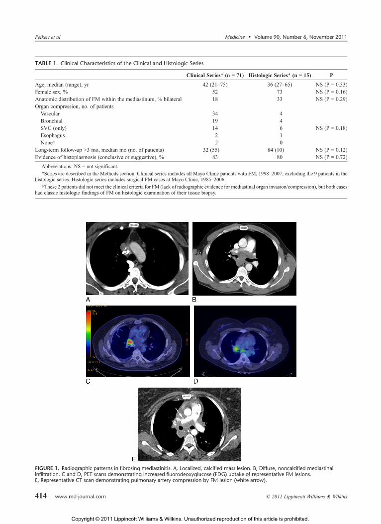
TABLE 1. Clinical Characteristics of the Clinical and Histologic Series
Clinical Series* (n = 71) Histologic Series* (n = 15) P
Age, median (range), yr 42 (21Y75) 36 (27Y65) NS (P = 0.33)Female sex, % 52 73 NS (P = 0.16)Anatomic distribution of FM within the mediastinum, % bilateral 18 33 NS (P = 0.29)Organ compression, no. of patientsVascular 34 4Bronchial 19 4SVC (only) 14 6 NS (P = 0.18)Esophagus 2 1None† 2 0
Long-term follow-up 93 mo, median mo (no. of patients) 32 (55) 84 (10) NS (P = 0.12)Evidence of histoplasmosis (conclusive or suggestive), % 83 80 NS (P = 0.72)
Abbreviations: NS = not significant.*Series are described in the Methods section. Clinical series includes all Mayo Clinic patients with FM, 1998Y2007, excluding the 9 patients in the
histologic series. Histologic series includes surgical FM cases at Mayo Clinic, 1985Y2006.†These 2 patients did not meet the clinical criteria for FM (lack of radiographic evidence for mediastinal organ invasion/compression), but both cases
had classic histologic findings of FM on histologic examination of their tissue biopsy.
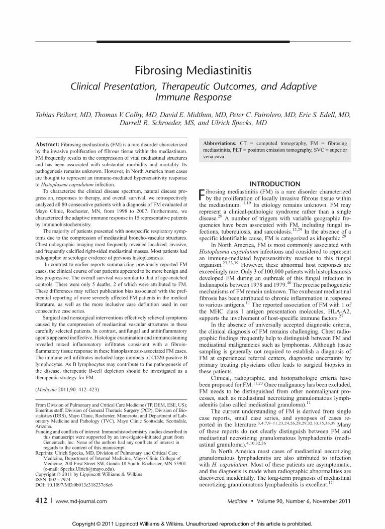
FIGURE 1. Radiographic patterns in fibrosing mediastinitis. A, Localized, calcified mass lesion. B, Diffuse, noncalcified mediastinalinfiltration. C and D, PET scans demonstrating increased fluorodeoxyglucose (FDG) uptake of representative FM lesions.E, Representative CT scan demonstrating pulmonary artery compression by FM lesion (white arrow).
Peikert et al Medicine & Volume 90, Number 6, November 2011
414 www.md-journal.com * 2011 Lippincott Williams & Wilkins
Copyright © 2011 Lippincott Williams & Wilkins. Unauthorized reproduction of this article is prohibited.

Mann-Whitney test and categorical datawere compared by eitherthe Fisher exact test or the chi-square test. Survival curves wereconstructed using the Kaplan-Meier method.
RESULTS
PatientsA search and review of the Mayo Clinic electronic records
between 1998 and 2007 identified 80 patients with FM (con-sidered the ‘‘clinical series’’). Paraffin-embedded biopsy speci-mens were available for 9 of these patients, and a search of theMayo Clinic pathology database identified biopsy specimensfrom 6 additional patients with FM obtained between 1985 and1997. The clinical characteristics of these 15 patients (consid-ered the ‘‘histologic series’’) were compared to the remaining 71patients of the clinical series (Table 1). There were no significantdifferences in clinical, radiographic, or histologic characteristicsor survival between the 2 groups. Therefore, we concluded thatthe cases in the smaller histologic series are representative of thelarger clinical series, and we used these specimens to charac-terize the adaptive immune response by immunostaining.
Demographics and Presenting SymptomsEighty patients with FM were identified between 1998 and
2007. The median age was 42 years (range, 21Y75 yr). Fifty-fourpercent were women, and 94% resided in endemic areas forH. capsulatum at the time of evaluation at Mayo Clinic.
Only 4 patients (5%) were asymptomatic at the time ofpresentation. Reported symptoms included respiratory com-plaints such as dyspnea on exertion (n = 39, 47%), cough (n = 17,21%), facial swelling, head fullness, or headaches consistentwith SVC syndrome (n = 17, 21%), chest pain (n = 16, 20%), andhemoptysis (n = 16, 20%). Twenty-seven patients (34%) pre-sented with multiple symptoms.
Chest ImagingChest CT scans were available for review in 74 patients
(93%). Focal mediastinal abnormalities were identified in 70 ofthe 74 patients (95%) (Figure 1A). Diffuse mediastinal infiltra-tion was found in only 4 patients (5%) (Figure 1B). Radiographicabnormalities localized to the right side in 53 (72%) and to theleft in 4 patients (5%); bilateral lesions were found in 17 patients(23%). Calcifications were detected in 54 patients (73%).
Positron emission tomography (PET) scans were obtainedin 7 patients (9%). Clinical indications included suspicion formalignancy (for example, lymphoma) at the time of diagnosis(n = 5) or concerns about malignancy raised by a new lung lesionduring FM follow-up. All PET scans showed increased meta-bolic activity within the FM-associated mediastinal lesions.PET images were routinely interpreted as highly suspicious
for malignancy. Subsequent surgical biopsies (n = 5) revealedFM in 3 cases and granulomatous inflammation in 2 cases.Both patients with granulomatous inflammation had a typicalclinical presentation of FM characterized by large airway com-pression. PET images of 2 representative cases are shown inFigure 1C and 1D.
Compression of Mediastinal StructuresAnatomic structures within the mediastinum were com-
pressed or obstructed in 78 patients (98%), and more than 2mediastinal structures were affected in 28 (35%) (Figure 2).
Histopathologic FindingsSurgical resections with diagnostic (n = 29) or therapeutic
(n = 14) intent, or autopsy (n = 1) were performed in 44 patients(55%), and tissue for histologic examination was obtained in43 of 44 cases (98%). A histologic diagnosis of FM was estab-lished in 34 of 43 patients (79%). In 6 of the remaining 9 spe-cimens only excessive fibrosis, chronic mixed inflammatoryinfiltrates, and granulomatous inflammation were described, butno diagnosis of FM was specified in the pathology report. Theremaining 3 cases demonstrated only granulomatous inflam-mation. All 9 cases met the clinical diagnostic criteria for FM. In8 of these cases compression of the large airway, pulmonaryartery, pulmonary vein, or SVCVor a combination thereofVwas detected. One individual had isolated compression of theesophagus.
EtiologyAntibodies against H. capsulatum were measured by
enzyme-linked immunosorbent assay, immunodiffusion, and/orcomplement fixation in 58 of 80 patients (73%). Antibody
FIGURE 2. Radiographic compression of mediastinal structuresin FM (n = 78 patients).
TABLE 2. Therapeutic Interventions for Mayo Clinic FMPatients, 1998Y2007 (n = 80 Patients)
Medical therapyAntifungal therapy
Itraconazole 28Other antifungals 3
Antiinflammatory therapyPrednisone 5Tamoxifen 2Other agents 2
Nonsurgical procedural interventionsSVC balloon angioplasty/stent 10*Endobronchial balloon dilatation/stent 2Pulmonary artery balloon angioplasty/stent 3*Pulmonary vein balloon angioplasty 1
Surgical proceduresDebulking/decompression procedures 4SVC bypass procedures
Spiral vein grafts 3PTFE graft 2Right ventricle to pulmonary artery conduits 2
Pulmonary resection for uncontrollable hemoptysisLobectomy 4Pneumonectomy 1
Pulmonary vein reconstruction 1
Abbreviations: PTFE = polytetrafluoroethylene.*One patient underwent balloon angioplasty and stent placement of
both the pulmonary artery and SVC.
Medicine & Volume 90, Number 6, November 2011 Fibrosing Mediastinitis
* 2011 Lippincott Williams & Wilkins www.md-journal.com 415
Copyright © 2011 Lippincott Williams & Wilkins. Unauthorized reproduction of this article is prohibited.
titers established a conclusive diagnosis of histoplasmosis in17 of 58 patients (29%). Furthermore, yeast forms typical ofH. capsulatum were seen on Grocott methenamine silver stainsin 11 of 43 tissue specimens (26%), and these organisms werealways found in the necrotic areas of old necrotizing granulomas.However, fungal tissue cultures remained negative when per-formed (n = 5). Taken together, a conclusive diagnosis of histo-plasmosis was established in 22 of 68 patients (32%) who hadeither serologic tests or tissue stains for the organism.
Twenty-one additional patients had antihistoplasma anti-body titers and radiographic findings suggestive of histoplas-mosis. Radiographic evidence of a prior granulomatous infectionwas detectable in 46 of 74 patients (62%) by chest CT, and basedon these radiographic findings another 24 patients were classi-fied as having a suggestive diagnosis of prior histoplasmosis.Therefore, overall serologic and/or radiographic evidence ofhistoplasmosis was detected in 67 of 80 patients (84%). None ofthe 80 patient with FM had a history of granulomatous med-iastinitis progressing to FM.
Two of the 13 patients without evidence of previous his-toplasmosis had radiographic findings of diffuse mediastinalinfiltration. One of these 2 individuals and another patient hadassociated retroperitoneal fibrosis, both features typically detectedin patients with idiopathic FM. No other possible etiologic factorswere identified.
TreatmentTherapeutic interventions were recommended for 54
patients, and observation was advised in 26. The specific med-ical, interventional, and surgical treatments used are outlined in
Table 2. Primary therapeutic interventions consisted of antifun-gal and antiinflammatory drug therapy (n = 23 patients), non-surgical procedural interventions (n = 6 patients) and surgicalprocedures (n = 14 patients). In 11 patients nonsurgical inter-ventions (9 patients) and surgical procedures (2 patients) werecombined with medical therapies.
Observation alone was recommended for patients withoutsymptoms, or those with mild and nonspecific symptoms thatcould not be attributed clearly to the compressed mediastinalstructures. Medical therapy consisted predominantly of antifungalmedications in patients with serologic evidence of histoplasmosis.
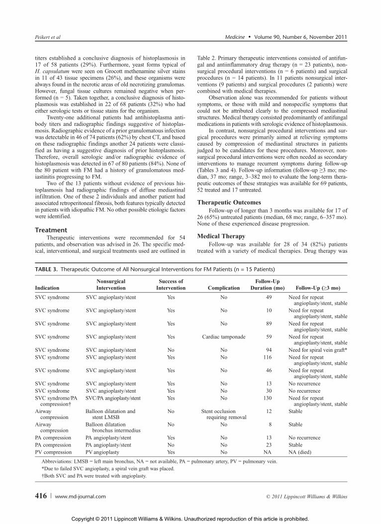
In contrast, nonsurgical procedural interventions and sur-gical procedures were primarily aimed at relieving symptomscaused by compression of mediastinal structures in patientsjudged to be candidates for these procedures. Moreover, non-surgical procedural interventions were often needed as secondaryinterventions to manage recurrent symptoms during follow-up(Tables 3 and 4). Follow-up information (follow-up Q3 mo; me-dian, 37 mo; range, 3Y382 mo) to evaluate the long-term thera-peutic outcomes of these strategies was available for 69 patients,52 treated and 17 untreated.
Therapeutic OutcomesFollow-up of longer than 3 months was available for 17 of
26 (65%) untreated patients (median, 68 mo; range, 6Y357 mo).None of these experienced disease progression.
Medical TherapyFollow-up was available for 28 of 34 (82%) patients
treated with a variety of medical therapies. Drug therapy was
TABLE 3. Therapeutic Outcome of All Nonsurgical Interventions for FM Patients (n = 15 Patients)
IndicationNonsurgicalIntervention
Success ofIntervention Complication
Follow-UpDuration (mo) Follow-Up (Q3 mo)
SVC syndrome SVC angioplasty/stent Yes No 49 Need for repeatangioplasty/stent, stable
SVC syndrome SVC angioplasty/stent Yes No 10 Need for repeatangioplasty/stent, stable
SVC syndrome SVC angioplasty/stent Yes No 89 Need for repeatangioplasty/stent, stable
SVC syndrome SVC angioplasty/stent Yes Cardiac tamponade 59 Need for repeatangioplasty/stent, stable
SVC syndrome SVC angioplasty/stent No No 94 Need for spiral vein graft*SVC syndrome SVC angioplasty/stent Yes No 116 Need for repeat
angioplasty/stent, stableSVC syndrome SVC angioplasty/stent Yes No 46 Need for repeat
angioplasty/stent, stableSVC syndrome SVC angioplasty/stent Yes No 13 No recurrenceSVC syndrome SVC angioplasty/stent Yes No 30 No recurrenceSVC syndrome/PAcompression†
SVC/PA angioplasty/stent Yes No 130 Need for repeatangioplasty/stent, stable
Airwaycompression
Balloon dilatation andstent LMSB
No Stent occlusionrequiring removal
12 Stable
Airwaycompression
Balloon dilatationbronchus intermedius
No No 8 Stable
PA compression PA angioplasty/stent Yes No 13 No recurrencePA compression PA angioplasty/stent No No 23 StablePV compression PV angioplasty Yes No NA NA (died)
Abbreviations: LMSB = left main bronchus, NA = not available, PA = pulmonary artery, PV = pulmonary vein.*Due to failed SVC angioplasty, a spiral vein graft was placed.†Both SVC and PA were treated with angioplasty.
Peikert et al Medicine & Volume 90, Number 6, November 2011
416 www.md-journal.com * 2011 Lippincott Williams & Wilkins
Copyright © 2011 Lippincott Williams & Wilkins. Unauthorized reproduction of this article is prohibited.
administered for a median duration of 8 months (range, 3Y129 mo). No patient achieved complete remission, and only aminority (5 of 28, 18%) experienced partial radiographic and/orsymptomatic responses. Itraconazole therapy resulted in theradiographically detected decrease in size of the mediastinallesions in 3 patients, and 1 of these also reported improveddyspnea. A fourth individual experienced a reduction in chestpain following therapy with itraconazole. The clinical sig-nificance of the 2 isolated radiographic improvements remainsunclear.
Only 1 of 6 patients treated with antiinflammatory therapiesbenefited from this intervention (improved symptoms and de-creased radiographic infiltration). This patient was 1 of the 3cases with either diffuse mediastinal involvement or retroperito-neal fibrosis (idiopathic FM). The patient’s disease subsequentlydeteriorated despite therapy, and he died as a consequence ofprogressive pulmonary artery compression and pulmonary hy-pertension. In 2 patients treated medically (1 with itraconazole, 1with prednisone), radiographic disease progression was detectedduring follow-up. In the remaining 21 patients managedmedically,FM remained stable.
Medical therapies were generally well tolerated; however,1 patient discontinued itraconazole because of hives, and anotherpatient was unable to tolerate tamoxifen.
Nonsurgical Procedural InterventionsFifteen patients were managed with primary nonsurgical
procedures. Endovascular interventions effectively relievedsymptoms caused by compression of the SVC, pulmonary artery,and pulmonary vein by FM-associated fibrous tissue (seeTable 3; Figure 1E). In contrast, the 2 patients treated withbronchoscopic interventions to restore airway patency did notexperience symptomatic relief. The nonsurgical procedures weregenerally safe, and there were no procedure-related deaths.Cardiac tamponade occurred in 1 patient following SVC stent-ing, but this complication did not have any lasting consequences.
Long-term follow-up information was available for 14patients (median, 38 mo; range, 8Y130 mo). The majority ofpatients treated with endovascular procedures experienced stentre-stenosis with recurrent symptoms and required repeatedinterventions at 6Y12 months intervals (see Table 3).
Surgical TherapyThe operations performed in the 17 patients managed sur-
gically (primary surgery in 16 and failed stenting in 1 patient) areoutlined in Tables 4 and 5. Despite being technically challeng-ing, all but 1 procedure successfully relieved the symptomscaused by the obstruction of various mediastinal structures. The
TABLE 4. Therapeutic Outcome of All Surgical Procedures for FM Patients (n = 17 Patients)
Surgical Indication Surgical ProcedureSurgicalSuccess
SurgicalComplication
Follow-UpDuration (mo) Follow-Up (Q3 mo)
SVC syndrome Spiral vein graft Yes No 46 Need for repeatangioplasty/stent, stable
SVC syndrome Spiral vein graft Yes No 94 Need for repeatangioplasty/stent, stable
SVC syndrome Spiral vein graft Yes Pericarditis 382 No recurrenceSVC syndrome PTFE graft Yes No 51 No recurrenceSVC syndrome PTFE graft Yes No NA NAHemoptysis Lobectomy Yes No 208 Stable, recurrence with need for
bronchial artery embolization4 yr later
Hemoptysis Lobectomy Yes Stroke with minimalresidual defect,respiratory failure
18 No recurrence
Hemoptysis Lobectomy Yes Empyema 70 No recurrenceHemoptysis Pneumonectomy Yes No NA NAAirway compression Surgical decompression BI Yes No 24 No recurrenceAirway compression Surgical decompression
RMSBYes No NA NA
Airway compression Bronchoplasty LMSB No Unscheduledpneumonectomy
162 RMSB stent
PA compression Bypass Yes Tricuspidregurgitation
83 No recurrence
PA compression Bypass Yes No 106 No recurrencePV compression Surgical decompression PV Yes No 145 Need for repeat angioplasty/stent,
stable. Died of non-small celllung cancer.
PV compression Surgical decompression andenlargement PV
Yes No NA NA
Esophagealcompression
Surgical decompressionesophagus
Yes No NA NA
Abbreviations: See previous tables. BI = bronchus intermedius, RMSB = right main bronchus.
Medicine & Volume 90, Number 6, November 2011 Fibrosing Mediastinitis
* 2011 Lippincott Williams & Wilkins www.md-journal.com 417
Copyright © 2011 Lippincott Williams & Wilkins. Unauthorized reproduction of this article is prohibited.
unsuccessful operation, a failed bronchoplasty of the left main-stem bronchus, required an unscheduled left pneumonectomy.There were 4 surgical complications and no peri- and post-operative deaths (see Table 4). The only peri- and postoperativecomplication of significance was 1 patient suffering minimalneurologic deficit (mild weakness) following a perioperativestroke.
Long-term follow-up information (median, 89 mo; range,18Y382 mo) was available for 12 of 17 patients (71%) in whomthe surgical intervention had a therapeutic intent. The benefitsof the surgical interventions were only temporary in 5 of the12 patients (42%), and they required subsequent procedures forrecurrent symptoms: balloon angioplasty/stenting for vasculargraft obstruction (n = 3 patients), angiographic embolization for
hemoptysis (n = 1 patient), and endobronchial stent placementfor airway torsion (n = 1 patient) (see Table 4).
SurvivalTo determine the survival endpoint for our patient cohort,
we searched the Social Security Death Index, and the commer-cial Accurint database was searched in addition to the availableclinical follow-up. Using this approach the median follow-upwas 68 months (range, 0Y401 mo). Five deaths were identified.Two of these were the result of FM. One patient died shortly afterthe initial presentation from the hemodynamic consequences ofsevere pulmonary venous obstruction, despite temporary im-provement after angioplasty. The second fatality was a patientwith idiopathic FM and associated retroperitoneal fibrosis. He
TABLE 5. Clinical Characteristics of FM Patients in the Histologic Series, Mayo Clinic, 1985Y2006 (n = 15 Patients)
Patient Age/Sex (yr) Organ Involvement Chest Radiography Histoplasmosis Treatment Outcome
1 25/M SVC Right mediastinal mass Suggestive Surgical NA2 32/M PV Left hilar mass Conclusive Surgical and antifungal NA3 65/F Bronchial tree Right mediastinal mass Conclusive Surgical Stable, 29 mo4 51/F Carotid artery, internal
jugular veinLeft cervical and righthilar mass
Conclusive Surgical NA
5 31/F SVC Right mediastinal mass Suggestive Surgical Stable, 24 mo6 27/F PA Right mediastinal mass Conclusive Surgical and antifungal NA7 27/F PV Diffuse mediastinal
infiltrationConclusive Interventional Died
8 48/F Bronchial tree Left mediastinal mass Suggestive Surgical Stable, 113 mo9 35/M PA and bronchial tree Bilateral hilar masses Conclusive Surgical NA10 43/F SVC Right mediastinal mass NA Surgical Stable, 207 mo11 44/M Chest wall Anterior mediastinal mass NA Surgical Stable, 224 mo12 27/F Bronchial tree Right mediastinal mass Conclusive Surgical and antifungal Stable, 24 mo13 59/F SVC Right mediastinal mass Conclusive Surgical Stable, 112 mo14 36/F SVC Right mediastinal mass Conclusive Surgical Stable, 95 mo15 58/F SVC Right mediastinal mass NA Surgical and antifungal Stable, 44 mo
Abbreviations: See previous tables.
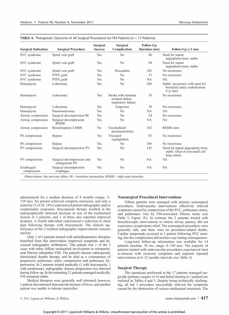
FIGURE 3. Overall survival of FM patients evaluated at Mayo Clinic Rochester, MN, 1998Y2007 compared to the survival ofage-matched controls (expected mortality) (Kaplan-Meier curve).
Peikert et al Medicine & Volume 90, Number 6, November 2011
418 www.md-journal.com * 2011 Lippincott Williams & Wilkins
Copyright © 2011 Lippincott Williams & Wilkins. Unauthorized reproduction of this article is prohibited.
initially responded to immunosuppressive therapy but eventuallydied 10 years after the diagnosis from pulmonary hypertensioncaused by progressive pulmonary artery compression. We notethat both of these patients had bilateral mediastinal involvement.Two additional patients died of non-small cell lung cancer, whichdeveloped 12 and 29 years after the diagnosis of FM, respec-tively. One additional patient died of an unknown cause. This
patient originally presented with right-sided bronchial com-pression and broncholithiasis, for which he underwent bi-lobectomy. The death occurred 8 years following his diagnosisand surgical intervention.
Overall, the Kaplan-Meier analysis did not reveal a differ-ence between the observed and predicted mortalities (Figure 3).However, the small number of deaths (n = 5) precludes a
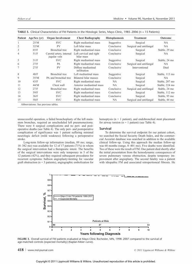
TABLE 6. Histologic and Immunohistochemistry Staining of FM Tissue Samples (n = 15 Patients)
Patient Inflammation CD20 Pattern CD8 CD138 S100
1 ++ ++ Peripheral rim + ++ 02 ++ ++ Peripheral rim, follicles + ++ +3 ++ + Follicles + + +4 + ++ Follicles + + +5 ++ ++ Peripheral rim ++ + ++6 + + Follicles 0 + 07 ++ ++ Peripheral rim, follicles, sheets + 0 08 + + Follicles + + 09 + + Peripheral rim, follicles 0 0 010 ++ ++ Peripheral rim + + +11 + + Follicles + 0 +12 + + Follicles 0 + 013 + ++ Peripheral rim, sheets ++ 0 014 ++ ++ Peripheral rim, follicles + + 015 0 + Follicles 0 0 0
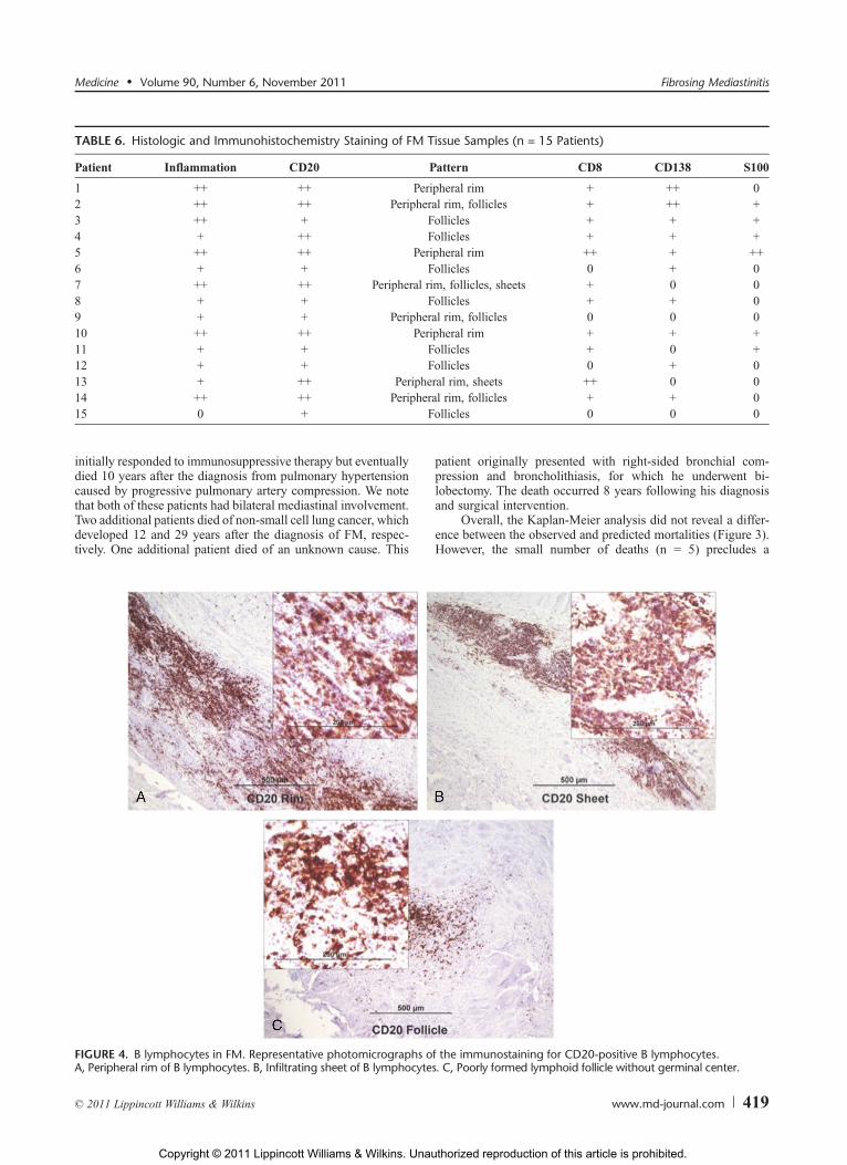
FIGURE 4. B lymphocytes in FM. Representative photomicrographs of the immunostaining for CD20-positive B lymphocytes.A, Peripheral rim of B lymphocytes. B, Infiltrating sheet of B lymphocytes. C, Poorly formed lymphoid follicle without germinal center.
Medicine & Volume 90, Number 6, November 2011 Fibrosing Mediastinitis
* 2011 Lippincott Williams & Wilkins www.md-journal.com 419
Copyright © 2011 Lippincott Williams & Wilkins. Unauthorized reproduction of this article is prohibited.
meaningful statistical analysis of the Kaplan-Meier curves. It isnoteworthy that both FM-related fatalities occurred in patientswith bilateral mediastinal involvement, suggesting that suchindividuals are perhaps at a higher risk for adverse outcomes.
Characterization of the Adaptive ImmuneResponse
Paraffin-embedded tissue samples were available for 15 FMcases evaluated at Mayo Clinic between 1985 and 2006 (thehistologic series). Based on their clinical characteristics andtherapeutic outcomes, these patients were representative of ourlarger clinical cohort (see Table 1). The clinical characteristics,treatments, and therapeutic outcomes are summarized in Table 5.
Detailed results of the histologic and immunohistochemicalanalysis are listed in Table 6. Mixed, lymphocytic inflammatoryinfiltrates were almost universally present (14 of 15 patients).CD20-positive B lymphocytes accounted for a large proportionof these cells and were seen in all cases examined. These cellseither formed a peripheral rim surrounding the fibrotic lesion orclustered within infiltrating sheets of cells (Figure 4A and 4B).Alternatively, they formed poorly structured lymphoid follicleswithout germinal centers (Figure 4C). These distribution pat-terns varied between patients but were simultaneously detectedin the same cases. CD3- and CD8-positive lymphocytes werealso frequently detected (11 of 15 patients), but their distribu-tion was distinct from the CD20-positive B lymphocytes (seeTable 6). T lymphocytes predominately infiltrated into fibrotic
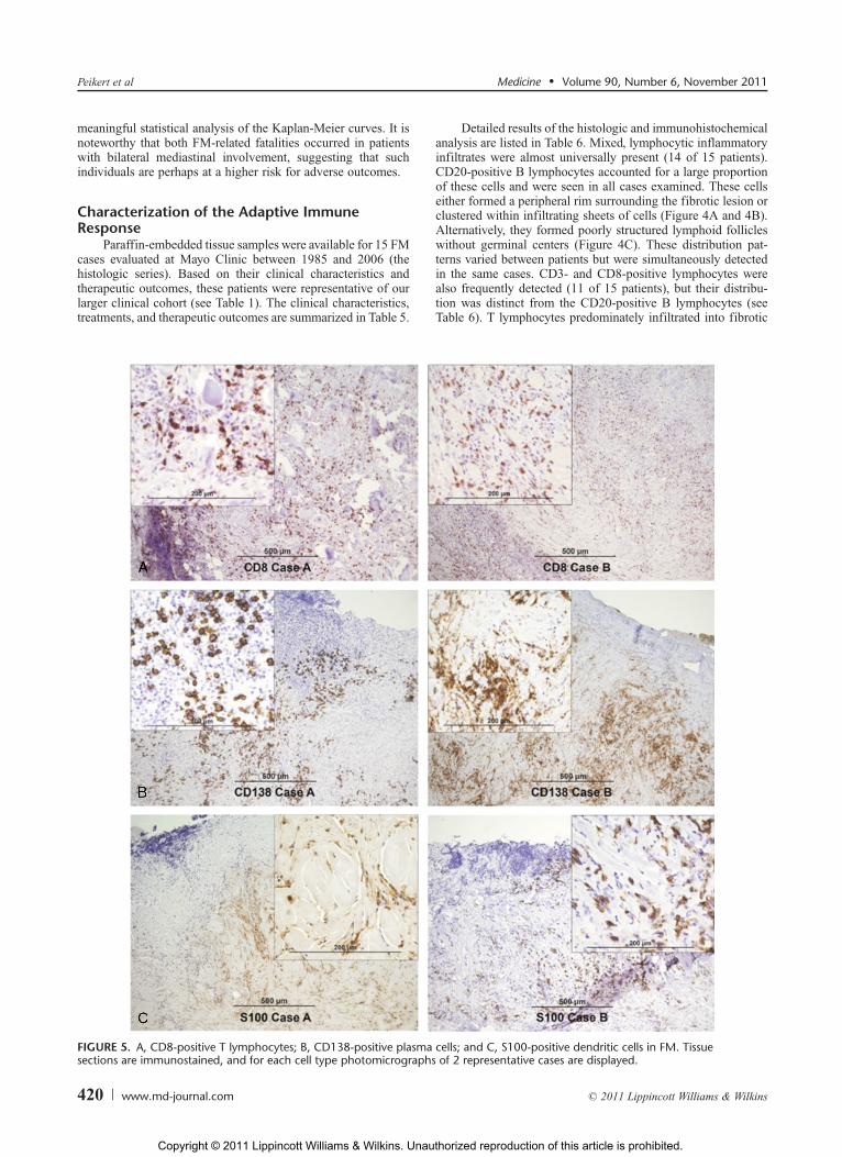
FIGURE 5. A, CD8-positive T lymphocytes; B, CD138-positive plasma cells; and C, S100-positive dendritic cells in FM. Tissuesections are immunostained, and for each cell type photomicrographs of 2 representative cases are displayed.
Peikert et al Medicine & Volume 90, Number 6, November 2011
420 www.md-journal.com * 2011 Lippincott Williams & Wilkins
Copyright © 2011 Lippincott Williams & Wilkins. Unauthorized reproduction of this article is prohibited.
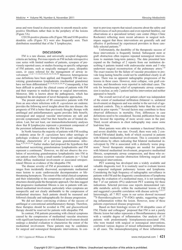
areas and were found in close proximity to smooth muscle actin-positive fibroblasts rather than in the periphery of the lesions(Figure 5A).
CD138-positive plasma cells (Figure 5B) and S100-positivedendritic cells (Figure 5C) were also frequently seen. Theirdistribution resembled that of the T lymphocytes.
DISCUSSIONFM is a rare disease, and universally accepted diagnostic
criteria are lacking. Previous reports on FM include retrospectivecase series with limited numbers of patients, synopses of pre-viously reported cases, or studies focusing on specific subgroupsof FM patients, such as surgically managed cases, idiopathiccases, or cases referred for the management of airway in-volvement.1,3,4,7Y11,14,23,24,26,32,33,36,39 Moreover, heterogeneouscase definitions have been applied, and frequently FM and nec-rotizing granulomatous lymphadenitis (mediastinal granuloma)have not been differentiated.4,7,9,10,24,32,36,39 Consequently, it hasbeen difficult to predict the clinical course of patients with FMand their response to medical therapy or surgical interventions.Moreover, little is known about the pathogenesis, which hashampered the development of novel therapeutic approaches.
The present analysis of 80 consecutive patients with FMfrom an area where infections with H. capsulatum are endemicprovides the following novel insights about this rare disease: theprognosis of FM is better than previously thought; therapy withantifungal and antiinflammatory agents provides little benefit;nonsurgical and surgical vascular interventions are safe andprovide symptomatic relief but their benefits are of limited du-ration. Last, but not least, our immunohistochemistry analysesshow that B lymphocytes represent a prominent component ofthe persistent inflammation within the FM lesions.
In North America the majority of patients with FM residingin endemic areas for H. capsulatum have either serologic orpathologic evidence of prior histoplasmosis and radiographicfindings characteristic of a previous granulomatous infec-tion.4,23,29,33,39 Earlier studies had proposed the hypothesis thatmediastinal necrotizing granulomatous lymphadenitis and FMrepresent a continuum.4 However, we did not observe the evo-lution of necrotizing granulomatous lymphadenitis into FM inour patient cohort. Only a small number of patients (n = 3) hadeither diffuse mediastinal involvement or associated retroperi-toneal fibrosis as evidence of the idiopathic FM.
The disease severity of FM at presentation is variable andranges from incidentally discovered, asymptomatic mediastinalmass lesions to acute cardiovascular decompensation or life-threatening hemoptysis. The extent of the initial clinical symptomsand their relationship to the compromise of specific mediastinalstructures should guide the therapeutic approach. Our data suggestthat progressive mediastinal fibrosis is rare in patients with uni-lateral mediastinal involvement, particularly when symptoms arenonspecific and not clearly attributable to the compression ofmediastinal structures. Such patients should be reassured andfollowed clinically and radiographically for disease progression.
We did not detect convincing evidence of the success ofantifungal or conventional antiinflammatory therapy. Therefore,these therapies should be avoided in FM, and glucocorticoidtherapy should be reserved for patients with idiopathic FM.
In contrast, FM patients presenting with clinical symptomscaused by the compression of mediastinal vascular structureswith significant hemoptysis or with bilateral involvement shouldundergo a detailed evaluation to characterize the extent of themediastinal involvement. These patients may be candidatesfor surgical and nonsurgical therapeutic interventions. In con-
trast to previous reports that raised concerns about the safety andeffectiveness of such procedures and even reported fatalities, ourobservations at a specialized tertiary care center (Mayo ClinicRochester) reflecting more recent advances in surgical tech-niques suggest that these interventions are generally safe andeffective if performed by experienced providers in these care-fully selected patients.23
Unfortunately, the durability of the therapeutic success ofthese interventions is frequently limited. Subsequent stent orgraft obstruction often requires repeated nonsurgical interven-tions to maintain long-term patency. The data presented hereexpand on the findings of 2 reports from our institution de-scribing 6 patients treated with endovascular stents and 5 whounderwent surgical reconstruction of the pulmonary artery.1,8
The exact reasons for the failure of vascular procedures to pro-vide long-lasting benefits could not be established clearly in allcases. There was no apparent radiographic progression of thelesions in these cases. However, stent collapse, vein graft con-traction, and thrombosis were reported in individual cases. Therole for bronchoscopic relief of symptomatic airway compres-sion is unclear, as only 2 patient had this intervention and neitherappeared to benefit.
The overall survival of our patient cohort was independentof the extent of mediastinal infiltration (bilateral vs. unilateralinvolvement) at diagnosis and was similar to the survival of age-matched controls. This is substantially better than the survivalstated in prior reports.23 Several factors may contribute to theseobserved differences in mortality. First, differences in casedefinitions need to be considered. Second, publication bias mayhave favored the reporting of more severe cases in the past.Third, recent advances in chest radiography may cause somelead-time bias.
Most patients were able to maintain a functional lifestyle,and severe disability was rare. Overall, there were only 2 con-firmed FM-related deaths, both of which occurred in patientswith bilateral mediastinal involvement. This is consistent withobservations by others indicating that bilateral mediastinal in-volvement by FM is associated with a distinctly worse prog-nosis.3 Novel therapeutic strategies are needed for patientswith bilateral mediastinal involvement, progressive mediastinalfibrosis, symptomatic airway compression, and those who ex-perience recurrent vascular obstruction following surgical andnonsurgical interventions.
PET scanning has developed into a widely available andvaluable diagnostic tool. It is routinely used to evaluate knownor suspected malignant pulmonary lesions and lymphoma.Considering the high frequency of radiographic surveillance inpatients with FM and the diagnostic considerations of lymphomaduring the evaluation of a mediastinal mass, it is not surprisingthat 7 of our patients (9%) underwent PET scanning for theseindications. Selected previous case reports demonstrated vari-able metabolic activity within the mediastinal lesions of FMand suggested a possible correlation with disease activity.2,22,37
Interestingly, all 7 PET scans in our series showed increased18F-fluorodeoxyglucose (18F-FDG) uptake, suggesting ongo-ing inflammation within the lesion. However, none of thesepatients experienced disease progression.
Based on their histologic review of 30 idiopathic cases ofFM, Flieder and colleagues9 proposed that FM is not a purelyfibrotic lesion but rather represents a fibroinflammatory diseasewith a variable degree of inflammation. Our analysis of 15patients with predominantly histoplasmosis-associated FM,who were treated with surgical or nonsurgical interventions,confirmed various degrees of mixed inflammatory infiltrationin all cases. The immunophenotyping of these inflammatory
Medicine & Volume 90, Number 6, November 2011 Fibrosing Mediastinitis
* 2011 Lippincott Williams & Wilkins www.md-journal.com 421
Copyright © 2011 Lippincott Williams & Wilkins. Unauthorized reproduction of this article is prohibited.
infiltrates identified a large number of CD20-positive B lym-phocytes. These B cells either accumulate in the periphery orcluster in sheets or poorly formed lymphoid follicles withinthe fibrous tissue. We propose that these infiltrating B lym-phocytes play a pathogenic role in FM, and consequently, rep-resent a rational therapeutic target. In the absence of anyeffective medical therapy to date, depletion of B lymphocytesmay be a potential therapeutic option for those patients with FMwho have a poor prognosis (bilateral mediastinal or progressiveinvolvement), suffer symptomatic airway compression, or haverecurrent symptoms attributable to obstruction of mediastinalstructures.
Further support for such an approach comes from murinemodels of bleomycin-induced fibrosis as B lymphocytes, spe-cifically CD19 expression, were shown to be required for thedevelopment of fibrosis.20 This is consistent with reports dem-onstrating effective depletion of B lymphocytes within fibroticskin lesions and initial therapeutic success of rituximab inscleroderma.25,41
The current study is limited by its retrospective design andincomplete long-term follow-up information. Although difficultto perform, prospective studies to confirm and further charac-terize the disease spectrum, natural history, and outcome of FMare needed. Furthermore, since the disease spectrum and prog-nosis may be affected by the diagnostic criteria used, it is crucialto standardize the case definitions. Our diagnostic criteriaclosely resemble the case definitions of FM used by the majorityof authors. In addition to clinical and radiographic criteria,our case definition includes information obtained from biopsies.Although the histologic pattern of FM is nonspecific, it is highlycharacteristic and distinctly different from other processeswithin the mediastinum, such as mediastinal necrotizing gran-ulomatous lymphadenitis associated with histoplasma infec-tion.4,9,26,32,33,36,39 In contrast to the largest FM case seriesreported previously, we decided not to limit our inclusion criteriato the compression of pulmonary arterial, venous, or bronchialstructures, but also included cases with invasive mediastinalmass lesions with isolated SVC or esophageal compression, aswell as cases with an invasive mediastinal mass and excessivehistologic deposition of fibrous tissue and lack of mediastinalcompromise.23 Even if we excluded the 18 patients with isolatedSVC or esophageal compression lacking mediastinal compres-sion, the surgical success rates as well as the overall survivalremain unchanged. Moreover, if the exclusion criteria (definitionof mediastinal granuloma) provided by Loyd et al23 were appliedto the current patient cohort, only 5 patients would have to beexcluded. These patients were either asymptomatic (n = 4) orhad no organ compression (n = 1). Nevertheless, tissue biopsiesand chest-radiographic findings clearly supported a diagnosisof FM in these patients.
This suggests that the observed differences between thecurrent study and previous reports are more likely to be theresult of differences in study design than in case definitions.Our cohort of consecutive patients is more likely to reflect thenatural disease spectrum than a synopsis of all previouslyreported cases, which is likely to be confounded by publicationbias toward more severe cases with worse outcomes.23
In conclusion, most cases of FM can be diagnosed based onclinical and radiographic criteria. Tissue biopsies are neededonly if diagnostic uncertainty remains in asymptomatic patientspresenting without compression of mediastinal structures. Theanalysis of this large cohort of consecutive patients with FMindicates that the clinical course of FM overall appears to bemore favorable than previously reported, and patients are lesslikely to progress. In contrast to mostly ineffective medical
approaches, surgical and nonsurgical interventions can effec-tively relieve the symptoms caused by compression of vascularmediastinal structures. The overall survival of FM is similar toage-matched controls. PET scanning and immunophenotypingsuggest that FM from histoplasmosis is associated with persis-tent inflammation characterized by large numbers of CD20-positive B lymphocytes. These B lymphocytes may contribute tothe pathogenesis of the disease. Therapeutic B-cell depletionshould be investigated as a potential therapeutic strategy forselected patients with FM, particularly those with progressivedisease, recurrent compression of mediastinal structures, or theidiopathic variant of the disease.
REFERENCES1. Brown ML, Cedeno AR, Edell ES, Hagler DJ, Schaff HV. Operative
strategies for pulmonary artery occlusion secondary to mediastinalfibrosis. Ann Thorac Surg. 2009;88:233Y237.
2. Chong S, Kim TS, Kim BT, Cho EY. Fibrosing mediastinitis mimickingmalignancy at CT: negative FDG uptake in integrated FDG PET/CTimaging. Eur Radiol. 2007;17:1644Y1646.
3. Davis AM, Pierson RN, Loyd JE. Mediastinal fibrosis. Semin RespirInfect. 2001;16:119Y130.
4. Dines DE, PayneWS, Bernatz PE, Pairolero PC. Mediastinal granulomaand fibrosing mediastinitis. Chest. 1979;75:320Y324.
5. Dodds GA III, Harrison JK, O’Laughlin MP, Wilson JS, Kisslo KB,Bashore TM. Relief of superior vena cava syndrome due to fibrosingmediastinitis using the Palmaz stent. Chest. 1994;106:315Y318.
6. Doyle TP, Loyd JE, Robbins IM. Percutaneous pulmonary arteryand vein stenting: a novel treatment for mediastinal fibrosis.Am J Respir Crit Care Med. 2001;164:657Y660.
7. Dunn EJ, Ulicny KS Jr, Wright CB, Gottesman L. Surgical implicationsof sclerosing mediastinitis. A report of six cases and review of theliterature. Chest. 1990;97:338Y346.
8. Ferguson ME, Cabalka AK, Cetta F, Hagler DJ. Results of intravascularstent placement for fibrosing mediastinitis. Congenit Heart Dis.2010;5:124Y133.
9. Flieder DB, Suster S, Moran CA. Idiopathic fibroinflammatory(fibrosing/sclerosing) lesions of the mediastinum: a study of 30 caseswith emphasis on morphologic heterogeneity. Mod Pathol.1999;12:257Y264.
10. Garrett HE Jr, Roper CL. Surgical intervention in histoplasmosis.Ann Thorac Surg. 1986;42:711Y722.
11. Goodwin RA, Nickell JA, Des Prez RM. Mediastinal fibrosiscomplicating healed primary histoplasmosis and tuberculosis.Medicine (Baltimore). 1972;51:227Y246.
12. Graham JR, Suby HI, LeCompte PR, Sadowsky NL. Fibrotic disordersassociated with methysergide therapy for headache. N Engl J Med.1966;274:359Y368.
13. Guerrero A, Hoffer EK, Hudson L, Schuler P, Karmy-Jones R.Treatment of pulmonary artery compression due to fibrous mediastinitiswith endovascular stent placement. Chest. 2001;119:966Y968.
14. Hammoud ZT, Rose AS, Hage CA, Knox KS, Rieger K, Kesler KA.Surgical management of pulmonary and mediastinal sequelae ofhistoplasmosis: a challenging spectrum. Ann Thorac Surg.2009;88:399Y403.
15. Ichimura H, Ishikawa S, Yamamoto T, Onizuka M, Inadome Y,Noguchi M, Sakakibara Y. Effectiveness of steroid treatmentfor hoarseness caused by idiopathic fibrosing mediastinitis:report of a case. Surg Today. 2006;36:382Y384.
16. Ikeda K, Nomori H, Mori T, Kobayashi H, Iwatani K, Yoshimoto K,Yoshioka M. Successful steroid treatment for fibrosing mediastinitisand sclerosing cervicitis. Ann Thorac Surg. 2007;83:1199Y1201.
Peikert et al Medicine & Volume 90, Number 6, November 2011
422 www.md-journal.com * 2011 Lippincott Williams & Wilkins
Copyright © 2011 Lippincott Williams & Wilkins. Unauthorized reproduction of this article is prohibited.
17. Inoue M, Nose N, Nishikawa H, Takahashi M, Zen Y, Kawaguchi M.Successful treatment of sclerosing mediastinitis with a high serumIgG4 level. Gen Thorac Cardiovasc Surg. 2007;55:431Y433.
18. Kandzari DE, Warner JJ, O’Laughlin MP, Harrison JK. Percutaneousstenting of right pulmonary artery stenosis in fibrosing mediastinitis.Catheter Cardiovasc Interv. 2000;49:321Y324.
19. Katzenstein AA, Askin FB, Livolsi VA. Katzenstein and Askin’sSurgical Pathology of Non-Neoplastic Lung Disease. 3rd ed.Philadelephia: WB Saunders; 1997.
20. Lafyatis R, Kissin E, York M, Farina G, Viger K, Fritzler MJ,Merkel PA, Simms RW. B cell depletion with rituximab in patientswith diffuse cutaneous systemic sclerosis. Arthritis Rheum.2009;60:578Y583.
21. Lal C, Weiman D, Eltorky M, Pugazhenthi M. Complete resolution offibrosing mediastinitis with corticosteroid therapy. South Med J.2005;98:749Y750.
22. Lee KY, Yi JG, Park JH, Kim YJ, So Y, Kim JS. Fibrosing mediastinitismanifesting as thoracic prevertebral thin band-like mass on MRI andPET-CT. Br J Radiol. 2007;80:e141Ye144.
23. Loyd JE, Tillman BF, Atkinson JB, Des Prez RM. Mediastinalfibrosis complicating histoplasmosis. Medicine (Baltimore).1988;67:295Y310.
24. Mathisen DJ, Grillo HC. Clinical manifestation of mediastinalfibrosis and histoplasmosis. Ann Thorac Surg. 1992;54:1053Y1057.
25. McGonagle D, Tan AL, Madden J, Rawstron AC, Rehman A, Emery P,Thomas S. Successful treatment of resistant scleroderma-associatedinterstitial lung disease with rituximab. Rheumatology (Oxford).2008;47:552Y553.
26. Mole TM, Glover J, Sheppard MN. Sclerosing mediastinitis: a reporton 18 cases. Thorax. 1995;50:280Y283.
27. Peebles RS, Carpenter CT, Dupont WD, Loyd JE. Mediastinalfibrosis is associated with human leukocyte antigen-A2.Chest. 2000;117:482Y485.
28. Peikert T, Goetze S, Ghaffari S. A rare cause of coronary obstructionand angina pectoris. Heart. 2003;89:1120.
29. Rossi SE, McAdams HP, Rosado-de-Christenson ML, Franks TJ,Galvin JR. Fibrosing mediastinitis. Radiographics. 2001;21:737Y757.
30. Satpathy R, Aguila V, Mohiuddin SM, Khan IA. Fibrosing mediastinitispresenting as pulmonary stenosis: stenting works. Int J Cardiol.2007;118:e85Ye86.
31. Savelli BA, Parshley M, Morganroth ML. Successful treatment ofsclerosing cervicitis and fibrosing mediastinitis with tamoxifen.Chest. 1997;111:1137Y1140.
32. Schowengerdt CG, Suyemoto R, Main FB. Granulomatous andfibrous mediastinitis. A review and analysis of 180 cases.J Thorac Cardiovasc Surg. 1969;57:365Y379.
33. Sherrick AD, Brown LR, Harms GF, Myers JL. The radiographicfindings of fibrosing mediastinitis. Chest. 1994;106:484Y489.
34. Smith SJ, Vyborny CJ, Hines JL. Re: chronic superior vena cavaocclusion related to fibrosing mediastinitis treated with self-expandingshunts. Cardiovasc Intervent Radiol. 1997;20:161Y162.
35. Straus SE, Jacobson ES. The spectrum of histoplasmosis in a generalhospital: a review of 55 cases diagnosed at Barnes Hospital between1966 and 1977. Am J Med Sci. 1980;279:147Y158.
36. Strimlan CV, Dines DE, Payne WS. Mediastinal granuloma.Mayo Clin Proc. 1975;50:702Y705.
37. Takalkar AM, Bruno GL, Makanjoula AJ, El-Haddad G, Lilien DL,Payne DK. A potential role for F-18 FDG PET/CT in evaluationand management of fibrosing mediastinitis. Clin Nucl Med.2007;32:703Y706.
38. Thiessen R, Matzinger FR, Seely J, Aina R, Macleod P. Fibrosingmediastinitis: successful stenting of the pulmonary artery.Can Respir J. 2008;15:41Y44.
39. Urschel HC Jr, Razzuk MA, Netto GJ, Disiere J, Chung SY.Sclerosing mediastinitis: improved management with histoplasmosistiter and ketoconazole. Ann Thorac Surg. 1990;50:215Y221.
40. Wheat LJ, Slama TG, Eitzen HE, Kohler RB, French ML,Biesecker JL. A large urban outbreak of histoplasmosis: clinicalfeatures. Ann Intern Med. 1981;94:331Y337.
41. Yoshizaki A, Iwata Y, Komura K, Ogawa F, Hara T, Muroi E,Takenaka M, Shimizu K, Hasegawa M, Fujimoto M, Tedder TF,Sato S. CD19 regulates skin and lung fibrosis via Toll-like receptorsignaling in a model of bleomycin-induced scleroderma. Am J Pathol.2008;172:1650Y1663.
Medicine & Volume 90, Number 6, November 2011 Fibrosing Mediastinitis
* 2011 Lippincott Williams & Wilkins www.md-journal.com 423
Copyright © 2011 Lippincott Williams & Wilkins. Unauthorized reproduction of this article is prohibited.