descending necrotizing mediastinitis mimicking acute … · descending necrotizing mediastinitis...
TRANSCRIPT

Descending Necrotizing Mediastinitis Mimicking
Acute ST Segment Elevation Myocardial Infarction
Hung-Yu Chang,1 Yung-Nien Yang,1 Wei-Hsian Yin1 and Mason-Shing Young2
Descending necrotizing mediastinitis (DNM) is a rare but lethal disease. Despite prompt use of broad-spectrum
antibiotics and proper surgical drainage, the mortality is still high. High vigilance for disease presentation is helpful
to early diagnosis. A 59-year-old female with a history of hypertension, diabetes and dyslipidemia came to our
emergency room. Her initial presentation of chest pain, elevated troponin-I level and ST segment elevation in
electrocardiogram (ECG) mimicked acute ST segment elevation myocardial infarction. However, DNM was
suspected by her atypical clinical presentation and imaging studies. The ST segment elevation in ECG resolved
immediately after pigtail catheter drainage. Finally, DNM was proved by right thoracotomy with mediastinal
exploration. Klebsiella pneumoniae was isolated from pus and necrotic tissue.
Key Words: Coronary artery disease � Descending necrotizing mediastinitis � Myocardial infarction
INTRODUCTION
Descending necrotizing mediastinitis (DNM) is a
rare but lethal disease. This disease results from odon-
togenic, pharyngeal, or cervical infection extending
downward into the mediastinum via pretracheal, para-
pharyngeal and retrovisceral spaces of the neck. The in-
fection is often polymicrobial in etiology, with gas-pro-
ducing organisms. Despite prompt use of broad-spec-
trum antibiotics and proper surgical drainage, the mortal-
ity is still high. We report a case of descending necro-
tizing mediastinitis with initial presentation of chest
pain, elevated troponin-I level and ST segment elevation
in electrocardiogram (ECG), mimicking acute ST seg-
ment elevation myocardial infarction, so we couldn’t
recognize it at the first time.
CASE REPORT
A 59-year-old woman presented at the emergency
room of our institution with progressive, substernal dull
chest pain for three days and mild drowsiness for one
day. Within the previous two weeks, she had experi-
enced non-productive cough and snuffle. She had his-
tory of hypertension, type 2 diabetes and dyslipidemia,
but she denied smoking. On physical examination, she
was afebrile. Her heart rate was 108 beats per minute,
blood pressure was 100/64 mmHg and respiratory rate
was 24 breaths per minute. She had regular and rapid
heart rhythm, S4 gallop without audible murmur or
pericardial friction rub. She had clear breathing sounds,
no jugular vein distention or peripheral edema. Chest ra-
diography demonstrated cardiomegaly, slight widening
of the superior mediastinum and clear lungs field. The
ECG showed sinus rhythm with complete right bundle
branch block and 1- to 2-mm ST segment elevation in
leads III, aVF, and V3-V6 (Figure 1A). The results of
laboratory studies were as follows: troponin-I 2.5 ng/dL,
creatine kinase (CK) 133 U/L, CK-MB isoenzyme 12
U/L, glucose 569 mg/dL, blood urea nitrogen 34 mg/dL,
creatinine 1.3 mg/dL, sodium 126 mmol/L, potassium
123 Acta Cardiol Sin 2010;26:123�6
DNM Mimicking Acute STEMICase Report Acta Cardiol Sin 2010;26:123�6
Received: April 19, 2009 Accepted: July 14, 20091Division of Cardiology, 2Department of Internal Medicine, Cheng-
Hsin General Hospital, Taipei, Taiwan.
Address correspondence and reprint requests to: Dr. Yung-Nien
Yang, Division of Cardiology, Cheng-Hsin Rehabilitation Medical
Center, No. 45, Cheng-Hsin street, Pei-Tou, Taipei 112, Taiwan.
Tel: 886-2-2826-4400 ext. 2040; Fax: 886-2-2826-1242; E-mail:

4.4 mmol/L, white blood count 12,900/uL with 88% seg-
mented neutrophils, hemoglobin 15.4 g/dL and platelets
207,000/uL. Arterial blood gas sampling showed meta-
bolic acidosis (pH 7.34, HCO3 14 mmol/L, PaCO2 27
mmHg, PaO2 110mmHg), and ketone body test was
strongly positive. Bedside echocardiography performed
in the emergency department revealed mildly general-
ized hypokinesis of the left ventricle, with an ejection
fraction of 45%. There was no evidence of aortic dissec-
tion intima flap, pericardial thickening or effusion. Un-
der the impression of acute myocardial infarction and
type 2 diabetes with impending ketoacidosis, the patient
was treated as acute coronary syndrome with intrave-
nous nitroglycerin, subcutaneous low-molecular heparin,
oral aspirin, clopidogrel, carvedilol and intravenous in-
sulin pump, then transferred to the intensive care unit.
By the second day of hospitalization, blood glucose level
was under control. The CK and CK-MB levels remained
within normal limits. However, the patient’s conscious-
ness remained drowsy and her body temperature ele-
vated over 38 �C, producing suspicion of uncertain
source of infection. On the other hand, she also experi-
enced intractable cough. Hoarseness and dysphonia had
been noted for days, which led us to perform a lateral
view chest X-ray, showing subtle air bubbles over the
anterior superior mediastinum (Figure 2A). Contrast-en-
hanced cervicothoracic computed tomography revealed
diffuse inflammation with air intermixed tissue in para-
tracheal, pre-cardiac, and bilateral para-vertebral region,
indicating descending necrotizing mediastinitis (Figures
2B-D). Tracing back for the possible cause of this dis-
ease, the patient stated that she had undergone dental
procedures many times in the recent six months for full-
mouth dental bridges. Coronary angiography was per-
Acta Cardiol Sin 2010;26:123�6 124
Hung-Yu Chang et al.
Figure 1. (A) ECG done in emergency room shows sinus rhythm with complete right bundle branch block and 1- to 2-mm ST segment elevation in
leads III, aVF, and V3-V6 (V4 and V6 leads were reversed incautiously). (B) ECG done immediately after pigtail catheter drainage on the seventh day
of hospitalization shows resolution of ST segment elevation.
A
B
formed on the third day of hospitalization. Although tri-
ple-vessel disease was seen, no culprit lesion could sup-
port the diagnosis of acute myocardial infarction. Empir-
ical antibiotic therapy of piperacillin, tazobactam and
levofloxacin were started. Two pigtail catheters were in-
serted into the mediastinal space and yellowish pus ma-
terial was drained. Although we initially thought about
acute myocardial infarction, ECG did not disclose evo-
lutional change. After pigtail catheter drainage, ECG
showed resolution of ST segment elevation immediately
(Figure 1B), which suggested pericardial involvement of
DNM. After one week of antibiotic treatment and pigtail
catheter drainage, she received right thoracotomy with
mediastinal exploration and drainage. Klebsiella pneu-
moniae was isolated from pus and necrotic tissue. The
patient’s post-operative course was uneventful.
DISCUSSION
Acute mediastinitis is an uncommon disease. The
majority of mediastinitis results from esophageal perfo-
ration or infection following median sternotomy. Rarely,
odontogenic, pharyngeal, or cervical infection may ex-
tend downward into the mediastinum via pretracheal,
parapharyngeal and retrovisceral spaces of the neck, de-
scribed as DNM, the most lethal form of mediastinitis.
In 1938, Pearse reported the first series of DNM; overall
mortality rate was 55% (86% in patients treated medi-
cally and 35% in patients treated surgically).1 Criteria
for the diagnosis of DNM were established in 1983 by
Estrera.2 They include (1) clinical manifestation of sep-
sis, (2) characteristic roentgenographic features, (3) do-
cumentation of the necrotizing mediastinal infection at
operation or at postmortem examination, and (4) estab-
lishment of the relationship of oropharyngeal infection
and the mediastinal process. Endo classified DNM into
three groups.3 Type 1 DNM is localized to the upper
mediastinal space above the carina. Type 2A DNM ex-
tends into the lower anterior mediastinum and type 2B
DNM extends into both the anterior and posterior lower
mediastinum. Bacteriologically, DNM is most frequently
a polymicrobial process, with anaerobes playing a major
role.2 Because of its rapid development and severe com-
plication such as purulent pericarditis, abscess or tho-
racic empyema leading to multiple organ dysfunction,
the mortality is reported to be 40-50%, even with the
proper use of computed tomography scanning, aggres-
sive drainage, and antibiotic treatment.4,5 Delay of diag-
nosis and delayed or inappropriate drainage of the me-
diastinum are the main causes for the high mortality in
this life-threatening condition.6
To date, DNM mimicking acute ST segment eleva-
tion myocardial infarction has rarely been reported.
With our patient’s presentation of dull chest pain, ele-
vated troponin-I level and ECG change, acute myocar-
dial infarction was initially impressed, but clinical pre-
sentation and persistent ST-T elevation without evolu-
tion made this diagnosis doubtful. These findings could
be explained by pericardial involvement of DNM. Ho-
arseness and dysphonia indicated paratracheal region or
recurrent laryngeal nerve involvement. As demon-
strated by this DNM mimicking acute myocardial in-
farction case, it is very important to organize clinical
data explaining any other possibilities of myocardial
infarction, and to not consider only ECG or serum
troponin-I in diagnosis.
125 Acta Cardiol Sin 2010;26:123�6
DNM Mimicking Acute STEMI
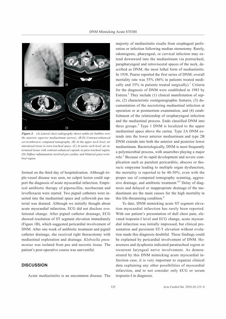
Figure 2. (A) Lateral chest radiography shows subtle air bubbles over
the anterior superior mediastinum (arrow). (B-D) Contrast-enhanced
cervicothoracic computed tomography. (B) At the upper neck level, air
intermixed tissue in retro-tracheal space. (C) At aortic arch level, air in-
termixed tissue with contrast-enhanced capsule in para-tracheal region.
(D) Diffuse inflammation involved pre-cardiac and bilateral para-verte-
bral region.
A B
C D

REFERENCES
1. Pearse HE Jr. Mediastinitis following cervical suppuration. Ann
Surg 1938;107:588-611
2. Estrera AS, Lanay MJ, Grisham JM. Descending necrotizing
mediastinitis. Surg Gynecol Obstet 1983;157:545-52.
3. Endo S, Murayama F, Hasegawa T, et al. Guideline of surgical
management based on diffusion of descending necrotizing me-
diastinitis. Jpn J Thoracic Cardiovasc Surg 1999;47:14-9.
4. Bulut M, Balcy V, Akose S, et al. Fatal descending necrotizing
mediastinitis. Emerg Med J 2004;21:122-3.
5. Choudhary N, Agrawal S, Rai AK. Descending necrotizing me-
diastinitis: trends in developing countries. Ear Nose Throat J
2005;54:246-8.
6. Charles-Henri, Marty-Ané, Jean-Philippe, et al. Management of
descending necrotizing mediastinitis: an aggressive treatment for
an aggressive disease. Ann Thorac Surg 1999;68:212-7.
Acta Cardiol Sin 2010;26:123�6 126
Hung-Yu Chang et al.