faculty of resource science and technology … of cellulose fibers from...isolation of cellulose...
TRANSCRIPT

ISOLATION OF CELLULOSE FIBERS FROM BANANA LEAVES
Premanarayani Menon D/O Narayanan Nair
Bachelor of Science with Honours
(Resource Chemistry)
2013
Faculty of Resource Science and Technology

Isolation of Cellulose Fibers from Banana Leaves
Premanarayani Menon D/O Narayanan Nair (27964)
A final report submitted in the fulfillment of the requirement for the degree of
Bachelor of Science with Honours
(Resource Chemistry)
Supervisor: Dr. Chin Suk Fun
Co-Supervisor: Associate Professor Dr. Pang Suh Cem
Resource Chemistry
Department of Chemistry
Faculty of Resource Science and Technology
Universiti Malaysia Sarawak
2013
I
Acknowledgment
First and foremost I would like to express my gratitude to my supervisor Dr. Chin Suk Fun for
her supervision, support and insightful comments that have helped me to improve the quality
of this project. Without her help, I would not be able to experience a remarkable progress in
my final thesis. I really appreciate her effort in assisting me to accomplish this project
successfully.
I would like to thank the Department of Chemistry, Universiti Malaysia Sarawak for giving
me the opportunity to fulfill my Final Year Project. I am thankful for all the materials,
equipment, instruments and other facilities which were provided in order to complete my
project.
I would like to appreciate Physical Chemistry lab group members especially Ms. Ain and Ms.
Fiona for their assistance and support throughout the project. I am grateful for the valuable
experience, knowledge and laboratory skills that I have attained throughout this project.
Finally, I would like to thank my friends and family for their effort and guidance for the
project development. We have exchanged knowledge and helped each other in order to finish
our task. Their encouragement had given me the motivation to complete this project. I hope
God bless all those who have helped me in the process of completing my final year project.

II
Declaration
I declare that this thesis entitled “Isolation of Cellulose Fibers from Banana Leaves” is the
result of my own research except as cited in the references. The thesis has not been accepted
for any degree and is not concurrently submitted in candidature of any other degree.
Signature ……………………………………
Name …………………………………….
Date ……………………………………..

III
Table of Content
Acknowledgement ....................................................................................................................... I
Declaration.................................................................................................................................. II
Table of contents .......................................................................................................................III
List of Abbreviations .................................................................................................................. V
List of Tables ............................................................................................................................ VI
List of Figures .......................................................................................................................... VII
Abstract ........................................................................................................................................1
1.0 Introduction ...........................................................................................................................2
1.1 Problem Statement .....................................................................................................4
1.2 Objectives ..................................................................................................................5
1.3 Hypothesis .................................................................................................................5
2.0 Literature Review ..................................................................................................................6
2.1 Cellulose ....................................................................................................................6

IV
2.2 Ultrasonic technique for isolation of cellulose ..........................................................7
2.3 Morphology of the chemical-purified cellulose ........................................................9
2.4 Influence of acid hydrolysis reaction.......................................................................10
2.5 Mixed Acid Treatment ............................................................................................12
2.6 Conventional process of isolation of cellulose ........................................................13
2.7 Isolation of cellulose from grape skin .....................................................................14
2.8 Isolation of cellulose from palm kernel cake...........................................................15
2.9 Isolation of α-cellulose from wholewood material ..................................................17
3.0 Materials and Methods ........................................................................................................20
3.1 Materials ..................................................................................................................20
3.2 Isolation of cellulose ................................................................................................20
4.0 Results and Discussion ........................................................................................................22
4.1 Treatment of sample with acidified sodium chlorite ...............................................22
4.1.1 Chemical composition analysis with FTIR .............................................23
4.1.2 Surface morphology analysis....................................................................29
4.2 Treatment of delignified sample with sodium hydroxide .......................................32
4.2.1 Chemical composition analysis with FTIR ..............................................33
4.2.2 Surface morphology analysis with FTIR ..................................................36
4.3 Effect of ultrasonication time on the morphology of isolated cellulose ..................42
5.0 Conclusion ...........................................................................................................................46
6.0 Reference .............................................................................................................................48
V
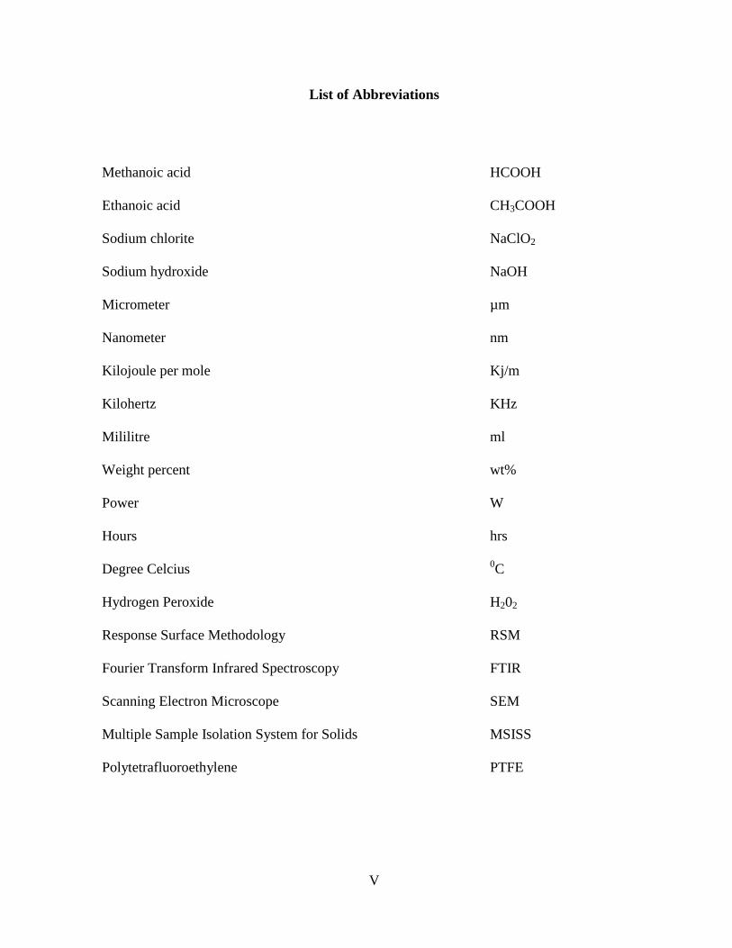
List of Abbreviations
Methanoic acid HCOOH
Ethanoic acid CH3COOH
Sodium chlorite NaClO2
Sodium hydroxide NaOH
Micrometer µm
Nanometer nm
Kilojoule per mole Kj/m
Kilohertz KHz
Mililitre ml
Weight percent wt%
Power W
Hours hrs
Degree Celcius 0C
Hydrogen Peroxide H202
Response Surface Methodology RSM
Fourier Transform Infrared Spectroscopy FTIR
Scanning Electron Microscope SEM
Multiple Sample Isolation System for Solids MSISS
Polytetrafluoroethylene PTFE
VI
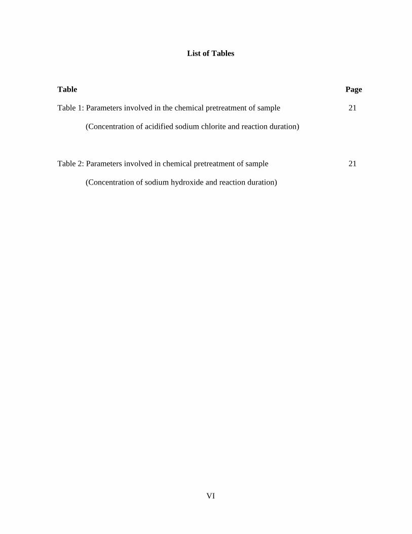
List of Tables
Table Page
Table 1: Parameters involved in the chemical pretreatment of sample 21
(Concentration of acidified sodium chlorite and reaction duration)
Table 2: Parameters involved in chemical pretreatment of sample 21
(Concentration of sodium hydroxide and reaction duration)

VII
List of Figures
Figure Page
Figure 1: Procedure for individualizing cellulose nanofibers 8
Figure 2: Structure of hemicellulose 14
Figure 3: Structure of S, H and G-lignin 15
Figure 4: Structure of Cellulose 16
Figure 5: Structure of Multiple Sample Isolation System For Solids (MSISS) 18
Figure 6a: FTIR spectra of the untreated sample 24
Figure 6b: FTIR spectra of the sample treated with 2 wt% of NaClO2 for 1hr 24
Figure 6c: FTIR spectra of the sample treated with 2 wt% of NaClO2 for 3hrs 24
Figure 7a: FTIR spectra of the untreated sample 25
Figure 7b: FTIR spectra of the sample treated with 2 wt% of NaClO2 for 5hrs 25

VIII
Figure 7c: FTIR spectra of the sample treated with 2 wt% of NaClO2 for 7hrs 25
Figure 8a: FTIR spectra of the untreated sample 26
Figure 8b: FTIR spectra of the sample treated for 1hr with 2 wt% of NaClO2 26
Figure 8c: FTIR spectra of the sample treated for 1hr with 4 wt% of NaClO2 26
Figure 9a: FTIR spectra of the untreated sample 27
Figure 9b: FTIR spectra of the sample treated for 1hr with 6 wt% of NaClO2 27
Figure 9c: FTIR spectra of the sample treated for 1hr with 8 wt% of NaClO2 27
Figure 9d: FTIR spectra of the sample treated for 1hr with 10 wt% of NaClO2 27
Figure 10a: FTIR spectra of the untreated sample 28
Figure 10b: FTIR spectra of the sample treated with 10 wt% of NaClO2 for 1hr 28
Figure 10c: FTIR spectra of the sample treated with 10 wt% of NaClO2 for 3hrs 28
Figure 11a: SEM images of sample treated with 2 wt% of NaClO2 for 1hr 29
IX
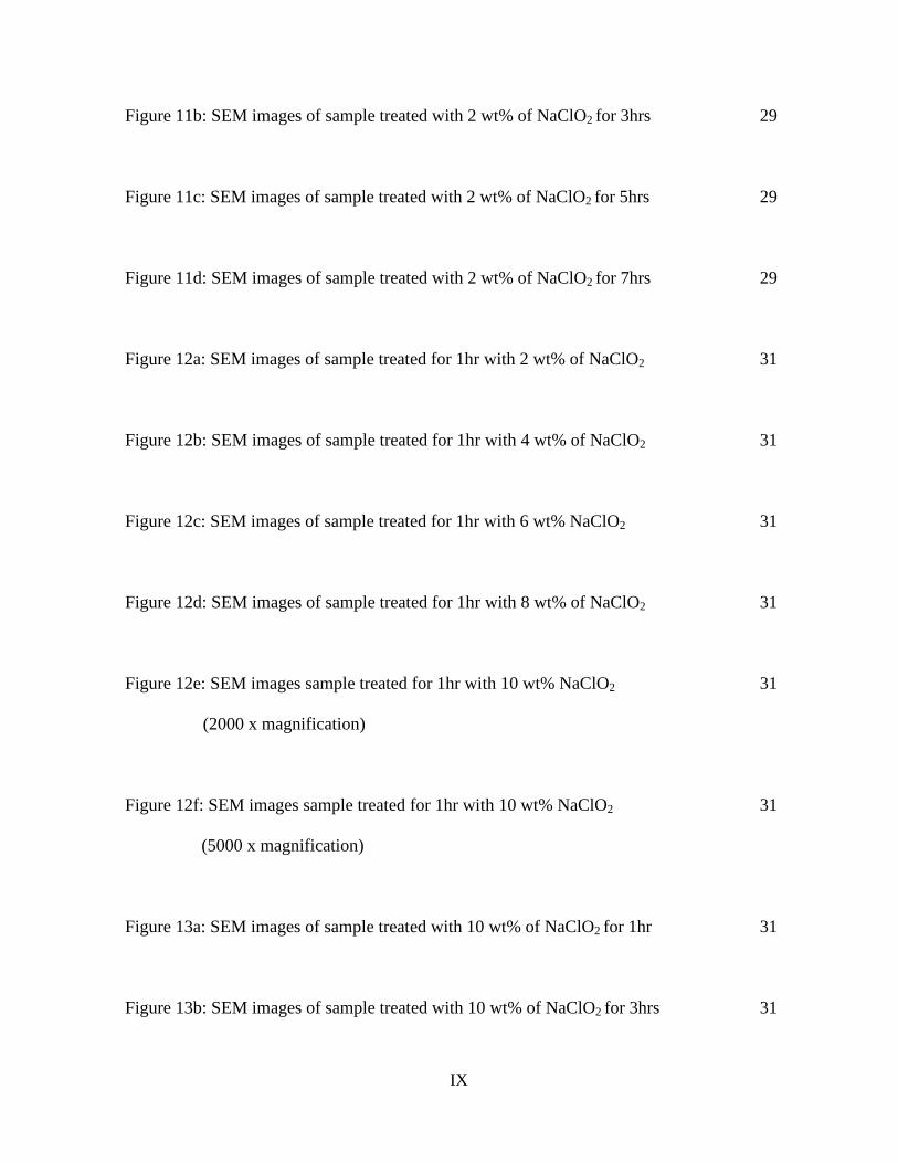
Figure 11b: SEM images of sample treated with 2 wt% of NaClO2 for 3hrs 29
Figure 11c: SEM images of sample treated with 2 wt% of NaClO2 for 5hrs 29
Figure 11d: SEM images of sample treated with 2 wt% of NaClO2 for 7hrs 29
Figure 12a: SEM images of sample treated for 1hr with 2 wt% of NaClO2 31
Figure 12b: SEM images of sample treated for 1hr with 4 wt% of NaClO2 31
Figure 12c: SEM images of sample treated for 1hr with 6 wt% NaClO2 31
Figure 12d: SEM images of sample treated for 1hr with 8 wt% of NaClO2 31
Figure 12e: SEM images sample treated for 1hr with 10 wt% NaClO2 31
(2000 x magnification)
Figure 12f: SEM images sample treated for 1hr with 10 wt% NaClO2 31
(5000 x magnification)
Figure 13a: SEM images of sample treated with 10 wt% of NaClO2 for 1hr 31
Figure 13b: SEM images of sample treated with 10 wt% of NaClO2 for 3hrs 31
X
Figure 14a: FTIR spectra of the untreated sample 33
Figure 14b: FTIR spectra of the sample treated with 2 wt% of NaOH for 1hr 33
Figure 14c: FTIR spectra of the sample treated with 2 wt% of NaOH for 3hrs 33
Figure 14d: FTIR spectra of the sample treated with 2 wt% of NaOH for 5hrs 33
Figure 14e: FTIR spectra of the sample treated with 2 wt% of NaOH for 7hrs 33
Figure 15a: FTIR spectra of the untreated sample 34
Figure 15b: FTIR spectra of the sample treated for 1hr with 2 wt% of NaOH 34
Figure 15c: FTIR spectra of the sample treated for 1hr with 4 wt% of NaOH 34
Figure 15d: FTIR spectra of the sample treated for 1hr with 6 wt% of NaOH 34
Figure 16a: FTIR spectra of the untreated sample 35
Figure 16b: FTIR spectra of the sample treated with 8 wt% of NaOH for 1hr 35
Figure 16c: FTIR spectra of the sample treated with 10 wt% of NaOH for 1hr 35

XI
Figure 16d: FTIR spectra of the sample treated with 10 wt% of NaOH for 3hrs 35
Figure 17a: SEM images of sample before NaOH treatment 36
Figure 17b: SEM images of sample after NaOH treatment 36
Figure 18a: SEM images of sample treated with 2 wt% of NaOH for 1hr 37
(800 x magnifications)
Figure 18b: SEM images of sample treated with 2 wt% of NaOH for 1hr 37
(2000 x magnifications)
Figure 19a: SEM images of sample treated with 2 wt% of NaOH for 3hrs 37
(800 x magnifications)
Figure 19b: SEM images of sample treated with 2 wt% of NaOH for 3hrs 37
(2000 x magnification)
Figure 20a: SEM images of sample treated with 2 wt% of NaOH for 5hrs 38
(800 x magnifications)
Figure 20b: SEM images of sample treated with 2 wt% of NaOH for 5hrs 38
(2000 x magnifications)

XII
Figure 21a: SEM images of sample treated with 2 wt% of NaOH for 7hrs 38
(800 x magnifications)
Figure 21b: SEM images of sample treated with 2 wt% of NaOH for 7hrs 38
(2000 x magnification)
Figure 22a: SEM images of sample treated with 4 wt% of NaOH for 1hr 39
(800 x magnifications)
Figure 22b: SEM images of sample treated with 4 wt% of NaOH for 1hr 39
(2000 x magnification)
Figure 23a: SEM images of sample treated with 6 wt% NaOH for 1hr 39
(800 x magnifications)
Figure 23b: SEM images of sample treated with 6 wt% NaOH for 1hr 39
(2000 x magnification)
Figure 24a: SEM images of sample treated with 8 wt% NaOH for 1hr 40
(800 x magnifications)
Figure 24b: SEM images of sample treated with 8 wt% NaOH for 1hr 40
(2000 x magnification)

XIII
Figure 25a: SEM images of sample treated with 10 wt% of NaOH for 1hr 40
(800 x magnifications)
Figure 25a: SEM images of sample treated with 10 wt% of NaOH for 1hr 40
(2000 x magnification)
Figure 26a: SEM images of sample treated with 10 wt% of NaOH for 3hrs 41
(800 x magnifications)
Figure 26a: SEM images of sample treated with 10 wt% of NaOH for 3hrs 41
(2000 x magnification)
Figure 27a: Morphology of chemically isolated cellulose after ultrasonication 43
of 30 seconds
Figure 27b: Morphology of chemically isolated cellulose after ultrasonication 43
of 60 seconds
Figure 27c: Morphology of chemically isolated cellulose after ultrasonication 43
of 90 seconds
Figure 27d: Morphology of chemically isolated cellulose after ultrasonication 43
of 120 seconds

XIV
Figure 27e: Morphology of chemically isolated cellulose after ultrasonication 43
of 150 seconds
Figure 27f: Morphology of chemically isolated cellulose after ultrasonication 43
of 180 seconds
Figure 27g: Morphology of chemically isolated cellulose after ultrasonication 43
of 210 seconds
Figure 27h: Morphology of chemically isolated cellulose after ultrasonication 43
of 240 seconds
Figure 28: Effect of ultrasonication on the average diameter of isolated 45
Cellulose fiber

1
Isolation of Cellulose Fibers from Banana Leaves
N. Premanarayani Menon
Resource Chemistry Programme
Faculty of Resource Science and Technology
Universiti Malaysia Sarawak
ABSTRACT
Banana leaves are one of the abundant agricultural wastes from banana plants cultivation.
Thus, isolation of cellulose from banana leaves aims to reduce the waste production.
Celluloses have received tremendous attention due to its potential application as reinforcing
agent. Cellulose fibers from banana leaves have been isolated and characterized by FTIR and
SEM. Banana fibers were treated with acidified sodium chlorite (NaClO2) and sodium
hydroxide (NaOH) to remove lignin and hemicellulose respectively. The chemical treatment
was subjected under different conditions, to study the effects of concentration, reaction
duration on the removal of lignin and hemicellulose as well as on the surface morphology of
the resultant cellulose fibers. Treatment of sample with 10 wt% of NaClO2 for 3 hours and 10
wt% of NaOH for 3 hours is the optimum conditions obtained from the study. Furthermore,
the morphology of the isolated cellulose was studied across different ultrasonication time. The
increase in ultrasonication time from 30 seconds to 240 seconds has resulted in the reduction
of the diameter of cellulose fibers.
Keywords: Banana leaves, isolation, cellulose fiber.
ABSTRAK
Daun pisang merupakan salah satu sisa bahan buangan pertanian selepas penanaman pokok
pisang. Justeru, pengekstrakan selulosa daripada daun pisang bertujuan untuk
mengurangkan sisa-sisa bahan buangan. Kelebihan sellulosa terfokus dalam aplikasi
sellulosa sebagai penguat sellulosa. Gentian selulosa daripada daun pisang telah diekstrak
dan dicirikan oleh FTIR dan SEM. Gentian daun pisang telah dirawat dengan natrium klorida
(NaClO2) dan natrium hidroksida (NaOH) untuk membuang lignin dan hemiselulosa masing-
masingnya. Rawatan kimia telah dijalankan dalam keadaan yang berbeza, untuk mengkaji
kesan kepekatan, masa tindak balas terhadap penyingkiran lignin dan hemiselulosa serta
perubahan terhadap morfologi permukaan gentian selulosa. Rawatan sampel dengan 10 %
NaClO2 selama 3 jam dan 10 % NaOH selama 3 jam merupakan keadaan optimum yang
diperolehi daripada kajian ini. Tambahan pula, kesan perbezaan masa ultrasonikasi terhadap
morfologi selulosa telah dikaji. Didapati bahawa diameter gentian selulosa telah menurun
apabila masa ultrasonikasi meningkat dari 30 saat kepada 240 saat.
Kata Kunci: Daun pisang, gentian selulosa, pengekstrakan.

2
1.0 Introduction
Isolation of Cellulose from agricultural wastes has gained a lot of attention from the scientists
and researchers due to its high demand in paper industries, food industries, pharmaceutical
and cosmetics. Agricultural by products such as banana leaves considered as an alternative
source for the production of cellulose. In tropical and sub-tropical region, bananas are
produced in large amount (Aziz et al., 2011). Banana leaves are used for wrapping food and
serving food (Mohapatra et al., 2010). However, after that the leaves are thrown away and this
generates agriculture wastes. Accumulation of agriculture waste has affected the environment.
Cellulose is an organic compound and it is a linear polysaccharide that is commonly found as
main constituent of primary cell wall of green plants (Saxena & Brown, 2008). According to
Habibi et al. (2008), cellulose represents about 1.5 x 10¹²
tons of the total biomass produced
in nature in a year. Thus, cellulose can be considered as an unlimited source and it can be used
to manufacture eco-friendly products. Cellulose is restored by nature through photosynthesis.
However, it is depleted in the form of pre-harvest and post-harvest agricultural losses and
wastes of food processing industry. In developing countries, like India, a major portion of the
cellulose generated remains unutilized and goes for wastes, namely in the forms of waste from
forest resources, wood, paper, and board after human uses, pulp, textile, agriculture, and food
industries (Elanthikkal et al., 2010)
In recent studies, many researches have been done in the aspect of characterization of
cellulose, hemicelluloses and lignin. Besides that, studies were carried on their usage, sources
3
and composition of cellulosic and lignocellulosic wastes of agriculture and food industry
(Nada et al., 2010). Furthermore, conversion of cellulose into value-added products of food
and medical application and constraints in their conversions were reviewed in details. For
example, microcrystalline cellulose is expected to act as water retainer, suspension stabilizer,
or as a reinforcing agent for products such as medical tablet (Nuruddin et al., 2011). The
extending property of cellulose which includes high stiffness and high degree of crystalline
makes it ideal as reinforcing agent (Bhattacharya et al., 2008).
The research covers the production of cellulose for various purposes which includes
reinforcement in composite and pulps for producing paper. Besides that, these cellulose
wastes have an enormous potential to be utilized for the production cellulosic material such as
cellulose biofilms and cellulose nanoparticles. Cellulose can be used as a raw material for
production of commodity product in industries (Gupta et al., 2012). Besides that, it has the
potential to produce derivative products such as rayon (Shin et al., 2012).
This study has practical significances. A proper method is developed to separate the cellulose
from the mixture of banana fibers. Methods are developed and the parameters are studied in
order to improve the quality of the results obtained. The minimum concentration of NaClO2
and NaOH at a fixed reaction duration required for a complete removal of lignin and
hemicellulose was determined respectively. On the other hand, the minimum reaction duration
of NaClO2 and NaOH treatment at a fixed concentration was also determined respectively.
Finally, the reaction condition that reveals the complete removal of hemicellulose and lignin
and better separation of cellulose fibers was chosen as the optimum condition for isolation of
4
cellulose from banana leaves. Furthermore the effect of ultrasonication time on the
morphology of the isolated cellulose was investigated.
1.1 Problem Statement
1. Accumulation of agriculture wastes including banana leaves pose potential environmental
problems.
2. Cellulosic fibers from banana leaves have potential use and can be applied in various
fields.
3. The banana fibers consist of a mixture of components. It is required to remove other
components such as lignin and hemicelluloses in order to obtain cellulose fibers.
4. Surface morphology and chemical composition of cellulose are affected by various
parameters involved in pretreatment and isolation process.

5
1.2 Objectives
The objectives of this study are:
(i) To isolate cellulose from banana leaves.
(ii) To study the effects of the parameters such as the duration of chemical treatment,
concentration of chemicals on the chemical composition and surface morphology of
cellulose.
(iii) To study the effect of ultrasonication time on the surface morphology of isolated
cellulose.
1.3 Hypothesis
The hypothesis of this study is that the lignin and hemicellulose from the mixtures are
removed completely and more individualized cellulose can be obtained by optimizing the
reaction conditions.

6
2.0 Literature Review
2.1 Cellulose
Cellulose is a type of natural fiber. Cellulosic fibers that are produced by plants are referred as
natural fibers. Natural fibers have many advantages which includes low cost, low density,
high toughness, acceptable specific strength properties and biodegradability (Lee et al., 2007).
The composition of natural fibers includes cellulose, lignin and hemicelluloses. Others
components such as pectin, pigments and extractives can be found in lower quantities. The
rigid microfibrillated cellulose is embedded in a soft matrix mainly composed of lignin and
hemicelluloses. It is abundantly found in wood or other lignocellulosic plant (Wang et al.,
2010).
Cellulose can only be dissolved in certain specific complexes and high concentrated acid and
base due to its degree of polymerization and partial crystalline structure. Therefore it does not
easily dissolve in common solvents (Klemm et al., 2005). Cellulose is a linear polymer of
poly-β (1 4) D-glucose (Zuluaga et al., 2007). It is composed of both amorphous and
crystalline region. It has secondary OH at C-2, secondary OH at C-3 and primary OH at C-6
position (Kadla & Gilbert, 2000). Besides that, cellulose can form strong intermolecular and
intramolecular hydrogen bond. Thus it does not melt before thermal degradation (Oh et al.,
2005a). Cellulose also acts as a structural polymer.

7
In order to fulfill the demand of industrial use, cellulose is often modified in terms of its
physical and chemical properties as well as enzymatic means. Cellulose exhibit changes in its
solubility, properties and behavior of the polymer. The chemical modification that takes place
is based on the free hydroxyl group in the glucopyranose units (Oh et al., 2005b). This results
in the formation of cellulose derivatives. One of the industrial applications includes the
production of rayon through viscose process where cellulose is converted to sodium cellulose
by treatment with sodium hydroxide (Oh et al., 2005b).
2.2 Ultrasonic technique for isolation of cellulose
Cellulose nanofibers were isolated from wood by using high intensity ultrasonification
combined with chemical pretreatments (Chen et al., 2010). Matrix substances such as
hemicellulose and lignin are mixed together with the cellulose. Thus, chemical methods were
used to separate the matrix from cellulose nanofibers. After the chemical treatment, the
compositional changes were revealed through FTIR spectroscopic analyses. The prominent
peak at 1731 cm-1
represents the acetyl and uronic ester groups of the hemicelluloses or the
ester linkage of carboxylic group of the ferulic and p-coumeric acids of lignin and/or
hemicelluloses (Chen et al., 2010). On the other hand, the peak at 1513 cm-1
represents the
carbon-carbon double bond stretch of the aromatic rings of lignin (Elanthikkal et al., 2010).
The absence of these peaks in the treated fibers indicates the effective removal of lignin and
hemicelluloses (Elanthikkal et al., 2010).

8
The ultrasonic treatment was carried out with an output power of 400, 800, 1000 and 1200W
to isolate the nanofibers. Increase in ultrasonic power from 400W to 1200W was accompanied
by the increase in dispersion of the nanofibers suspension (Chen et al., 2010). Ultrasonic
energy was transferred to cellulose through a process called cavitation. Cavitation refers to the
formation, growth, and violent collapse of cavities in water. The energy transferred is called as
sonochemistry which is approximately 10-100 kJ/mol. The energy transferred is within the
hydrogen bond energy scale. The impact of ultrasonic energy disintegrates the micron sized
cellulose fibers into nanofibers. The nanofibers obtained were with wide distribution in width
due to the complicated multilayered structure of plant fibers and the intermolecular hydrogen
bond (Chen et al., 2010). Ultrasonic technique produced cellulose with higher surface area.
Finally, colloidal suspensions were produced (Chen et al., 2010) (Figure 1).
Figure 1: Procedure for individualizing cellulose nanofibers (Chen et al., 2010)