expression of the netrin-1 receptor, deleted in colorectal cancer (dcc), is largely confined to...
TRANSCRIPT

Expression of the Netrin-1 Receptor,Deleted in Colorectal Cancer (DCC), IsLargely Confined to Projecting Neurons
in the Developing Forebrain
TIANZHI SHU,1 KIMBERLY M. VALENTINO,1 CLARE SEAMAN,2 HELEN M. COOPER,2
AND LINDA J. RICHARDS1*1University of Maryland School of Medicine, Department of Anatomy and Neurobiology
and the Program in Neuroscience, Baltimore, Maryland 212012Ludwig Institute for Cancer Research, Royal Melbourne Hospital, Parkville,
Victoria 3050, Australia
ABSTRACTAxon guidance mechanisms are crucial to the development of an integrated nervous
system. One family of molecules that may be important in establishing axonal connectivity inmammals is the Netrins, and their putative receptors DCC (deleted in colorectal cancer),Neogenin, and Unc-5. Knockout and mutational analyses of some of these genes have shownthat they are critically involved in the development of several specific pathways in thedeveloping brain. However, previous expression analyses of these genes have largely beenconfined to the developing spinal cord. In the present study, we analyzed the expression ofDCC in the developing mouse forebrain. We found that DCC protein is expressed in specific axonalpopulations projecting from the developing olfactory bulb, neocortex, hippocampus, and epithalamus/habenular complex. In the developing olfactory bulb and neocortex, DCC expression is particularlyevident during the targeting phase of axon outgrowth and is then rapidly downregulated. Aspredicted from the knockout and mutational analyses of this gene, DCC is expressed in axonalcommissures, in particular the corpus callosum, hippocampal commissure, and the anteriorcommissure. In addition, we found that DCC is expressed in the habenular commissure, thefasciculus retroflexus, and the stria medularis. Therefore, this analysis implicates a functionfor DCC in additional axonal guidance systems not predicted from the knockout andmutational analyses. J. Comp. Neurol. 416:201–212, 2000. r 2000 Wiley-Liss, Inc.
Indexing terms: olfactory bulb; cortex; hippocampus; habenula; mouse; axon guidance
Precise development of neuronal connectivity is notrigidly predetermined and ‘‘hard-wired’’ but is profoundlyinfluenced by the environment (Frost and Metin, 1985; Suret al., 1988; O’Leary and Stanfield, 1989; Schlaggar andO’Leary, 1991). As an axon grows toward its target duringdevelopment, its growth cone senses molecules within theenvironment that it uses to determine the correct path ofgrowth (Garrity and Zipursky, 1995). These molecular cuesmay be soluble and freely diffusing, bound to cellularmembranes, or bound to the extracellular environment.Along their way axons use intermediate ‘‘guideposts’’ tofind the final target (Tessier-Lavigne et al., 1988). Thus,axons may follow gradients of molecules released by thetarget itself or the intermediate guideposts. Such mol-ecules may deliver attractive (Placzek et al., 1990), repul-sive (Pini, 1993; Luo et al., 1993), or suppressive (Wang etal., 1996) cues to the growth cone.
Some molecules can act as either attractive or repulsivecues for axons (Hedgecock et al., 1990) depending on theexpression of separate receptors for attraction or repulsionin the responding cells (Chan et al., 1996). The guidancemolecule Netrin-1 can mediate both attraction and repul-sion. Netrin-1 was first purified by its ability to attractsensory commissural neurons in the spinal cord (Serafiniet al., 1994), but it also repels motor axons (Colamarino
Grant sponsor: National Institutes of Health; Grant number: NINDSNS37792; Grant sponsor: University of Maryland School of Medicine.
*Correspondence to: Linda J. Richards, University of Maryland School ofMedicine, Department of Anatomy and Neurobiology, and the Program inNeuroscience, 685 West Baltimore St, Baltimore, MD, 21201.E-mail: [email protected]
Received 10 June 1999; Revised 21 September 1999; Accepted 21September 1999
THE JOURNAL OF COMPARATIVE NEUROLOGY 416:201–212 (2000)
r 2000 WILEY-LISS, INC.

and Tessier-Lavigne, 1995; Varela-Echavarria et al., 1997).The Netrins belong to a phylogenetically conserved familyof long-range guidance molecules with activities in C.elegans (UNC-6; Ishii et al., 1992), Drosophila (Netrin-Aand Netrin-B; Harris et al., 1996; Mitchell et al., 1996),and vertebrates (Kennedy et al., 1994; Serafini et al., 1994;Colamarino and Tessier-Lavigne, 1995; Shirasaki et al.,1996; Richards et al., 1997; Varela-Echavarria et al.,1997).
Netrin-1 is either repulsive or attractive for axonsdepending on the neuronal population. This bifunctional-ity correlates with the activity of the C. elegans homologueof Netrin-1, Unc-6, which has also been shown to mediateboth attractive and repulsive responses (Hedgecock et al.,1990). Unc-6 is required for the dorsal migration of meso-dermal cells and for the guidance of circumferential axons(Hedgecock et al., 1990). Two known receptors for Unc-6are Unc-40 and Unc-5. Genetic experiments have shownthat axons are attracted to Unc-6 if they express theUnc-40 receptor but are repelled from Unc-6 if theyexpress the Unc-5 receptor. The mammalian homologuesof Unc-40 and Unc-5 have now been cloned. Neogenin(Vielmetter et al., 1994) and deleted in colorectal carci-noma (DCC; Keino-Masu et al., 1996) are homologous to C.elegans Unc-40, and Unc-5H1, Unc-5H2 (Leonardo et al.,1997) and rostral cerebellar malformation (rcm; Ackermanet al., 1997) are homologous to C. elegans Unc-5 (Lenug-Hagesteijn et al., 1992).
DCC is a transmembrane protein belonging to theimmunoglobulin superfamily of cell adhesion molecules(Walsh and Doherty, 1997) and has been shown to bindNetrin-1 in vitro (Keino-Masu et al., 1996). DCC wasoriginally cloned as a tumor supressor gene in colorectalcarcinomas (Fearon et al., 1990). In the developing ner-vous system, DCC has been evolutionarily conserved withhomologues cloned in Drosophila (Kolodziej et al., 1996),C. elegans (Chan et al., 1996), Xenopus (Pierceall et al.,1994), rodents (Cooper et al., 1995; Keino-Masu et al.,1996), and humans (Fearon et al., 1990; Nigro et al., 1991).
In the mammalian brain, Netrin-1 mutants and DCCknockout mice do not develop a corpus callosum or ahippocampal commissure and have a greatly reduced orabsent anterior commissure (Serafini et al., 1996; Fazeli etal., 1997). However, the mechanism for these pathfindingdefects in the brain is not known. In the developingforebrain, Netrin-1 has been shown to attract laterallydirected cortical axons in vitro (Metin et al., 1997; Rich-ards et al., 1997), indicating that these molecules areimportant for additional axonal guidance systems in theforebrain other than commissural axon guidance. Al-though there has been some expression analysis of DCCand Netrin mRNA in the developing nervous system(Cooper et al., 1995; Serafini et al., 1996; Gad et al., 1997,1999; Livesey and Hunt, 1997) and some protein expres-sion in the spinal cord (MacLennan et al., 1997), there hasbeen no detailed analysis of the protein expression ofeither of these genes in the developing forebrain (apartfrom the developing optic nerve; Deinier et al., 1997).
To investigate the role of DCC in the development of theforebrain, we performed a detailed analysis of DCC proteinexpression in the telencephalon and diencephalon (andfollowing some of these projections to the mesencephalon).We found that such an analysis implicates DCC in avariety of specific axonal guidance systems in the forebrainin addition to those previously described.
MATERIALS AND METHODS
Animals
Timed-pregnant female CD1 mice were obtained fromCharles River Laboratories (Wilmington, MA). Mice werehoused at the University of Maryland School of Medicineanimal facility. Embryos were staged based on a plug dateof embryonic day (E) 0 and were used between E12 andpostnatal day (P) 0. Procedures involving animals wereapproved by the animal care and use committee at theUniversity of Maryland, Baltimore.
Fixation and immunohistochemistry
On the required day of gestation, pregnant dams wereanesthetized with sodium pentobarbital (Nembutal, Ab-bott Laboratories, North Chicago, IL) at 0.07 mg/g bodyweight and placed on a warming pad during surgery tomaintain their body temperature. Once deeply anesthe-tized, the mother’s abdomen was opened to expose theuterus. Pups were removed sequentially and placed onice until deeply anesthetized before perfusion fixationwith either saline, followed by 4% paraformaldehyde/2.5%Acrolein (Polysciences Inc., Warrington, PA) for pups olderthan E15, or immersed in 4% paraformaldehyde/2.5%Acrolein for younger pups. After perfusion or immersionfixation, the skulls were opened, and the heads weretransferred to 4% paraformaldehyde and stored at 4°Cuntil cutting. Deeply anesthetized dams were killed afterthe pups were removed by a lethal injection of sodiumpentobarbital.
All sections were cut at 45 µm on a Vibratome (Leica,Deerfield, IL) and embedded in 3% agarose (Noble Agar,Difco, Detroit, MI). Vibratome sections were placed inphosphate buffered saline (PBS; pH 7.4) and washedbefore immunostaining. Sections were immunostainedfree floating but before the staining procedure were incu-bated in 1% sodium borohydride (Sigma, St. Louis, MO) for20 minutes to remove the Acrolein and washed 3 3 10minutes with PBS. The blocking step was done in 0.2%Triton X-100 (Sigma) and 2% normal goat serum (VectorLaboratories, Burlingame, CA) in PBS for 2 hours. Thesections were then incubated overnight in anti-DCC anti-body 2744 (rabbit polyclonal) at a concentration of 1:30,000diluted in the blocking solution. Sections were washed 3 320 minutes with PBS, incubated for 1 hour with secondaryantibody (biotinylated goat anti-rabbit; Vector Laborato-ries) diluted 1:600 in 0.2% Triton X-100 in PBS, andwashed another 3 3 20 minutes. Sections were incubatedfor 1 hour with AB solution (Vectastain elite kit, VectorLaboratories) made up 1 hour before at 1:500 dilution inPBS with 0.2% Triton X-100 and washed 3 3 10 minutes inPBS. The chromogen solution consisted of 1.25 g of nickelsulfate (Sigma) dissolved in 50 ml of 0.175 M sodiumacetate, followed by one 10-mg tablet of 3,38-diaminobenzi-dine tetrahydrochloride (DAB; Sigma) dissolved in thesame solution. The nickel-DAB solution was filtered, and 5µl of 30% hydrogen peroxide (Sigma) was added just priorto immersing the sections. The chromogen reaction wasterminated by placing the sections in PBS. For doublestaining with neurofilament and DCC, sections wereblocked for 2 hours, and then a polyclonal neurofilamentantibody (used at 1:50,000; Chemicon, Temecula, CA) wasapplied to the sections overnight. The sections were thenwashed and incubated in AB solution, as described above.The chromogen used was DAB dissolved in Tris buffered
202 T. SHU ET AL.

saline (pH 7.2) without nickel sulfate. Procedures for thesecond day of DCC staining were identical to those de-scribed above. Sections were mounted on 2% gelatin-coated glass slides, allowed to air dry, and dehydratedthrough a series of alcohols before being coverslipped withDPX (Sigma) mounting medium.
Microscopy and productionof photomicrographs
Mounted sections were analyzed with an upright lightmicroscope (Leica). Images for figures were scanned with aPowerPhase digital camera (Phase-one, Coppenhagen, Den-mark) directly into Adobe Photoshop software (version 4.0)running on a Power Computing Power Center Pro 210computer. In some cases, alterations of the brightnessand/or contrast were then made to the images. Imageswere then labeled, and collages were made for each figurein Adobe Photoshop. Figures were printed on qualityphotographic paper with a high-resolution color printer(Fujix Pictrography 3000).
RESULTS
Immunohistochemistry of DCC protein expression wasobserved only on axonal processes in most cases, inparticular those forming large fasciculated axon tracts.Staining of neuronal cell bodies was rarely observed withthis antibody.
Expression of DCC protein in thedeveloping olfactory system
Previous studies of DCC mRNA have shown that theDCC message is expressed in the granule cells of theolfactory bulbs at E18 in the mouse (Gad et al., 1997).Staining with the DCC antibody shows that DCC proteinis initially expressed on the axons of mitral cells at E14(Fig. 1A) but is not expressed in the granule cell layer untilE17 (Fig. 1B). Staining within the granule cell layer prob-ably represents the dendrites of granule cells that ramifywithin the granule cell layer (white arrows in Fig. 1C) be-cause these cells do not have axons (Shipley et al., 1995).In addition to the mitral cell layer, DCC is expressed on theprocesses of cells within an outer ring of the bulb that mayrepresent tufted cell axons (arrow in Fig. 1B). Tufted cellaxons project to the anterior olfactory nucleus (AON) andthe central olfactory cortex (Shipley et al., 1995). Somediffuse DCC staining is present within the AON and oncentrifugal fibers in the central olfactory cortex (data notshown), indicating that tufted cell axons may be labeledwith DCC. The mitral cells and the tufted cells togetherconstitute the major efferent projecting cell types of theolfactory bulb (Shipley et al., 1995). DCC is expressed onprojecting axon bundles as they fasciculate and leave theolfactory bulb (Fig. 1C) to form the lateral olfactory tract(Fig. 1D,E). At E17, DCC is highly expressed in the lateralolfactory tract on efferently projecting axons from theolfactory bulbs (Fig. 1D) to the piriform cortex (Fig. 1E).However, by E18, the expression in the lateral olfactorytract and the bulb begins to decline (data not shown),indicating that, at least for mitral and tufted cell axons,DCC may only be expressed during the targeting phase oftheir growth (between E13 and E17 in the rat; Marchandand Belanger, 1991; Lopez-Mascaraque et al., 1996). Inaddition, we observed some DCC labeling within theolfactory tubercle (arrow in Fig. 2F), which receives inputs
from the olfactory bulb via mitral cells but does not send areciprocal projection back to the olfactory bulb (Shipley etal., 1995). Therefore, this staining probably corresponds tothe mitral and tufted cell inputs rather than to an expres-sion within the tubercle itself. DCC was not observed inthe olfactory nerve (data not shown), but mRNA expres-sion for DCC has been reported in deep layers of theolfactory epithelium that contain mesodermal and glialcells (Livesey and Hunt, 1997).
DCC is expressed on neocortical axonsduring the targeting phase of growth
In the developing neocortex, DCC expression appears tobe tightly regulated and restricted to axons as they areprojecting. DCC mRNA is first expressed at E11.5 in thedeveloping preplate, where its expression is confined toearly postmitotic neurons (Gad et al., 1997). From E13,DCC protein is expressed on the axons of neurons project-ing within the intermediate zone and on axons within themarginal zone but is not expressed within the corticalplate or ventricular zone (Fig. 2). DCC expression withinthe marginal zone corresponds to that noted in previousreports showing DCC mRNA in neurons within layer 1,which are likely to be Cajal–Retzius cells (Keino-Masu etal., 1996).
The first axons to project out of the neocortex are thesubplate axons that pioneer the internal capsule (McCon-nell et al., 1989; DeCarlos and O’Leary, 1992). Although wefailed to detect DCC protein expression at E12, mRNA forDCC is expressed as early as E11.5 in mouse neocortex(Gad et al., 1997). However, DCC protein is highly ex-pressed by E13 (Fig. 2A) and E14 (Fig. 2B,C), probablyrepresenting both the subplate pioneers and some axons oflayers 5 and 6. Because DCC is not expressed on dorsalthalamic afferents projecting into the neocortex, DCCexpression within the internal capsule appears to bespecific for cortical efferent axons within this region. It hasbeen previously shown that the lateral projection from theneocortex extends at E14–E15 in rats, approximately 2days before the medial projection at E16–E17 (DeCarlosand O’Leary, 1992; Koester and O’Leary, 1994; Richards etal., 1997). Our results show that DCC expression reflects asimilar developmental difference in directional targetingin mouse. Initially DCC is highly expressed on the lateralprojection as it enters the internal capsule (Fig. 2C);however, by E16, DCC expression is downregulated onthese axons, as reflected by a decrease in staining withinthe internal capsule (Fig. 2E). At the same time, DCCexpression is maintained on medially projecting neuronsboth before and during their targeting phase of growth(compare Fig. 2C with 2E).
Medially projecting neocortical afferents cross the mid-line at E16.4 in mouse (Ozaki and Whalsten, 1998). At thisstage of development, DCC is expressed on these axons asthey grow toward and cross the midline (Fig. 2E). At E17,DCC expression at the midline is observed in both a dorsalregion of the corpus callosum and a ventral region. Thedorsal region (small black arrow in Fig. 2F) probablycorresponds to callosally projecting cortical axons becausethis staining is continuous with the projections from eachhemisphere. The ventral staining (white arrow in Fig. 2F)may correspond to a group of axons that defasciculatesfrom the main bundle of axons at the midline, or it may bestaining the glial sling, a midline glial ‘‘bridge’’ thatdirectly underlies the corpus callosum at this stage of
DCC EXPRESSION IN PROJECTING FOREBRAIN NEURONS 203

Fig. 1. Deleted in colorectal cancer (DCC) expression in theolfactory system. A: In the olfactory bulb, DCC is expressed within themitral cell axons (arrowhead in A,B) by embryonic day 14 (E14; theearliest age examined). B: By E17, DCC is also expressed in the tuftedcell axons (arrow) and in the granule cell layer. C, D: Mitral and tuftedcell axons expressing DCC at E17 form fasciculated bundles (arrow-
head in C) as they exit the olfactory bulbs and form the lateralolfactory tracts (lot). E: DCC is expressed at E17 within the lot alongthe entire length of the tract toward the olfactory cortex (E is anenlargement of the boxed area in the inset). A–C and E are coronalsections, and D is a horizontal section. Scale bars 5 300 µm in A,B, 30µm in C,E, 650 µm in D.
Fig. 2. Deleted in colorectal cancer (DCC) expression in thedeveloping cortex. A: In the developing cortex, DCC is expressed atembryonic day 13 (E13; the earliest age detected by immunohistochem-istry) in the intermediate zone of the neocortex and within thecingulate cortex. B–D: As the lateral neocortical projection forms, DCCis highly expressed at E14 (B,C) and E15 (D) on axons within theinternal capsule (ic; C is a higher power view of B) and in the dorsallateral regions of the septum (arrowheads in D). E: However, by E16,DCC begins to be downregulated in the internal capsule in morerostral regions initially while still being highly expressed in medialaspects of the neocortex and the cingulate cortex. F: At E17, DCC ismost highly expressed on the medially projecting cortical axonsforming the corpus callosum (small black arrow). At the ventralmidline, a second band of expression is seen that may correspond toaxonal labeling or labeling of the glial sling (white arrow). At E17,
DCC is still highly expressed in the dorsal–lateral regions of theseptum (black arrowheads) and in the olfactory tubercle (large blackarrow). In more caudal regions such as this, DCC is still expressed inthe internal capsule, although this expression is also declining. G: ByE18, DCC expression has been largely downregulated and onlyremains on the most dorsal axons of the corpus callosum (cc) and is nolonger expressed in the internal capsule. H: In the sagittal view atE17, it is evident that DCC expression declines after laterally project-ing cortical axons have grown through the internal capsule (ic) and isonly visible in the rostral regions of the corticospinal tract (cst). It isnot possible to see axons entering the thalamus after passing throughthe internal capsule. A–G are coronal sections, and H is a sagittalsection. Scale bars in A,C,H 5 400 µm, in G 5 500 µm for B, 400 µm forD,G, 450 µm for F, in E 5 650 µm.
204 T. SHU ET AL.

Figure 2

development (Silver et al., 1982). Callosal axons cross themidline in a temporally and regionally specific manner,with lateral axons crossing first in the ventral region of thecorpus callosum (CC) and medial axons crossing last in thedorsal region of the CC (Ozaki and Wahlsten, 1992). This isalso reflected by the regulation of DCC expression. By E18,DCC expression is downregulated on all other axons and isonly present in the dorsal region of the CC (Fig. 2G).
To determine whether DCC expression could be seen inmore caudal regions of the corticospinal tract, we cutsections in the sagittal plane of embryos. We found that,after passing through the region of the internal capsule, asmall amount of expression was observed in the rostralregion of the corticospinal tract (Fig. 2H) but was not foundalong the entire tract. These same axons were observed incoronal sections in more caudal regions of the brainmaking up the cerebral peduncles (Fig. 6C). In addition,we did not see DCC expression on cortical axons enteringthe thalamus (Fig. 2H), even at a range of antibodyconcentrations (data not shown), indicating that DCC wasnot even expressed at low levels by these axons.
DCC expression in the hippocampusand the major midline commissures
In horizontal sections of the developing forebrain, DCCexpression is most evident in the major midline commis-sures. DCC is expressed in the hippocampal commissure(Figs. 3E, 4C,D), the CC (Figs. 2F,G, 3E), and the anteriorcommissure (Fig. 4E,F). Sections alternately labeled withDCC alone (Fig. 3A,C,E) or neurofilament and DCC(Fig. 3B,D,F) show that DCC is expressed before neurofila-ment in some projecting neurons of the hippocampal andcallosal commissures. Figure 3C,D illustrates the reduc-tion in DCC expression within the internal capsule at E16(Fig. 3C), despite the high expression of neurofilament inthe same axons (Fig. 3D).
In the developing hippocampus, DCC is expressed byE13.5 in the hippocampal primordia (Gad et al., 1997) andis clearly evident in the axonal layer by E14 (Fig. 4A) but isnot seen in the cell bodies of these cells. DCC is expressedin the fiber layer underlying all regions of the hippocam-pus from the dentate gyrus and in regions CA1–CA3.Given that these axons project toward and fasciculate toform the fimbria, it appears that these are extrinsicallyprojecting axons (Fig. 4B). DCC continues to be expressedalong the entire hippocampal projection toward the mid-line within the fornix and within both the dorsal andventral hippocampal commissures (Fig. 4C,D). The hippo-campal commissure forms approximately 1 day earlierthan the CC but continues to form until birth (Wahlsten,1981; Valentino and Jones, 1982; Super and Soriano,1994). DCC is also expressed on these hippocampal axonsas they first begin to cross the midline at E14–E15 (datanot shown) and during this extended targeting phase ofgrowth.
The anterior commissure is the first major commissureto cross the rostral midline in the mouse at E14 but doesnot have an extended targeting phase of growth (Silver etal., 1982; Wahlsten, 1981). Coincident with this, DCC isexpressed within the axons of the anterior commissure atE14 (Fig. 4E) and E15 (Fig. 4F) but is completely downregu-lated by E16 (data not shown).
These results in the CC, hippocampal commissure, andthe anterior commissure and those described within theneocortical and olfactory bulb projections suggest that
DCC is only expressed on these long-range efferent axonswhile they are growing toward their targets. Upon reach-ing their targets, DCC is then apparently rapidly downregu-lated.
DCC expression in the septum
Septal defects have been shown to be associated withagenesis of the CC (Wahlsten and Bulman-Fleming, 1994),a defect seen in both the DCC (Fazeli et al., 1997) andNetrin-1 (Serafini et al., 1996) mutant mice. The ventralseptum is a region of high Netrin-1 mRNA expression(K.M. Valentino and L.J. Richards, unpublished observa-tion). Therefore, we wanted to determine where DCC wasexpressed within this region. The most striking region ofDCC expression in the septum is bilaterally in the ventro-medial aspects of the lateral ventricles, corresponding tothe ventricular zone of the septum (arrowheads in Fig.2E,F and sep in Fig. 3C). In addition, fibers of theperforant pathway, which run rostrocaudally within theseptum, parallel to, and just lateral to the midline, are alsolabeled with DCC (Fig. 2F).
DCC expression in the diencephalon
The highest DCC expression in the diencephalon is inthe epithalamus/habenula complex (Fig. 5). DCC expres-sion was evident from E13 and was highly expressed byE14 (Fig. 5A,B) in this region. DCC is expressed in bothmedially projecting axons in the habenula commissure(hbc in Fig. 5D) and ventrally projecting axons running inthe lateral region of the diencephalon close to the pialsurface, called the stria medularis (sm in Fig. 5C), whichextend down to the hypothalamus. In addition, DCC isexpressed in axons running rostrocaudally through thehabenula, seen in cross-section in Figures 5B,D and 6C,E.These axons form two fasciculated bundles of axons calledthe fasciculus retroflexus (fr; also known as the habenulo-interpeduncular tract) that project to the ventral tegmen-tal area (vt), as seen in sagittal sections (Fig. 6A,B,D).DCC-expressing axons are also seen within the proximalregion of the mammilotegmental tract (mtg in Fig. 6D).
In the dorsal thalamus, DCC is mostly expressed inmedial (or limbic) regions of dorsal thalamus, particularlyin the anterior medial thalamus and periventricular tha-lamic nucleus (Fig. 5E), and in the inferior thalamicradiation (itr; arrowheads in Fig. 6E,F). DCC is alsoexpressed in a band of axons that extend mediolaterallybetween the dorsal and ventral thalamus in the region ofthe external medullary lamina (not shown).
In the ventral aspect of the diencephalon, DCC is highlyexpressed in the optic chiasm (Fig. 5F) and bilaterally inthe rostromedial region of the suprachiasmatic nucleus(Fig. 5F). We have also shown DCC protein expressionwithin the developing retina and optic nerve (T. Shu andL.J. Richards, unpublished observations); therefore, theexpression of DCC within the optic chiasm is consistentwith this finding and suggests that DCC may play a role inaxon targeting of the optic nerve in regions distal to theretina.
In more caudal sections, DCC is expressed within thenigrostriatal pathway (nsp; Fig. 6C,E,F), corresponding toprevious reports of DCC mRNA expression within thesubstantia nigra (Livesey and Hunt, 1996; Gad et al.,1997).
206 T. SHU ET AL.
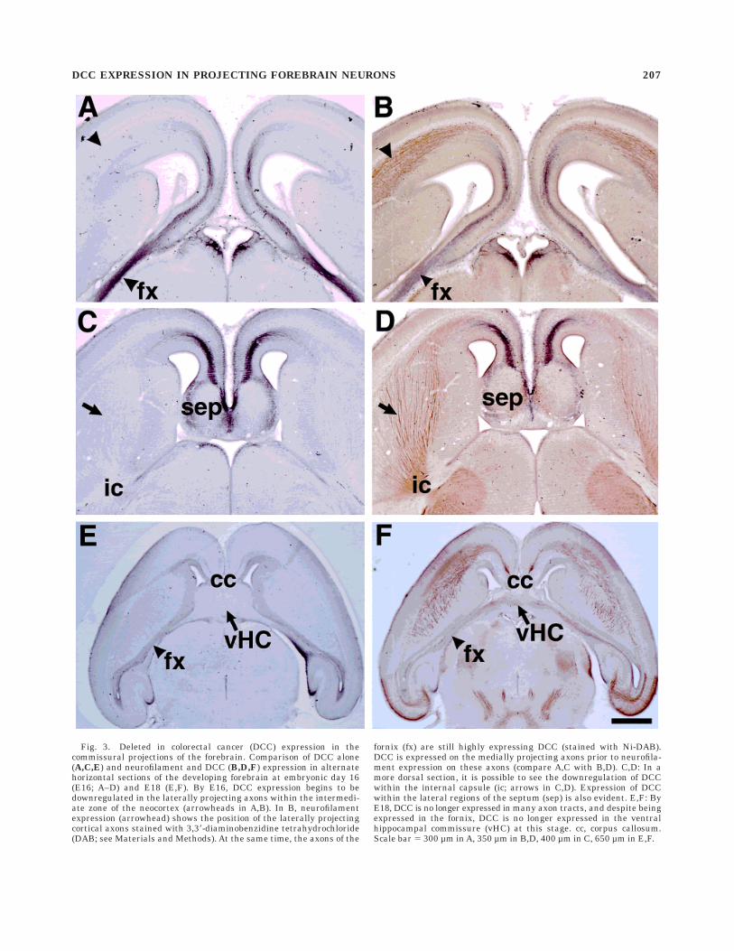
Fig. 3. Deleted in colorectal cancer (DCC) expression in thecommissural projections of the forebrain. Comparison of DCC alone(A,C,E) and neurofilament and DCC (B,D,F) expression in alternatehorizontal sections of the developing forebrain at embryonic day 16(E16; A–D) and E18 (E,F). By E16, DCC expression begins to bedownregulated in the laterally projecting axons within the intermedi-ate zone of the neocortex (arrowheads in A,B). In B, neurofilamentexpression (arrowhead) shows the position of the laterally projectingcortical axons stained with 3,38-diaminobenzidine tetrahydrochloride(DAB; see Materials and Methods). At the same time, the axons of the
fornix (fx) are still highly expressing DCC (stained with Ni-DAB).DCC is expressed on the medially projecting axons prior to neurofila-ment expression on these axons (compare A,C with B,D). C,D: In amore dorsal section, it is possible to see the downregulation of DCCwithin the internal capsule (ic; arrows in C,D). Expression of DCCwithin the lateral regions of the septum (sep) is also evident. E,F: ByE18, DCC is no longer expressed in many axon tracts, and despite beingexpressed in the fornix, DCC is no longer expressed in the ventralhippocampal commissure (vHC) at this stage. cc, corpus callosum.Scale bar 5 300 µm in A, 350 µm in B,D, 400 µm in C, 650 µm in E,F.
DCC EXPRESSION IN PROJECTING FOREBRAIN NEURONS 207

DISCUSSION
The present findings demonstrate that the expression ofDCC protein is dynamically regulated by a number ofdifferent projecting populations of axons in the developingforebrain. In addition, axons expressing DCC tend to belong-range projecting axons (some exceptions include thegranule cells of the olfactory bulb that do not have axons;
Shipley et al., 1995). Because DCC immunohistochemistryis largely confined to axonal membranes, DCC protein islocated predominantly in large fiber tracts such as thelateral olfactory tract, the internal capsule, the corpuscallosum, the anterior commissure, the fimbria/fornix, thefasciculus retroflexus, and the stria medularis. Given thehigh expression of DCC on midline commissural axons, our
Fig. 4. Deleted in colorectal cancer (DCC) expression in thedeveloping hippocampus and anterior commissure. A,B: Coronalsections through the developing hippocampus at embryonic day 14(E14; A) and E17 (B) show that DCC is expressed exclusively in theaxon layer, particularly in those axons leaving the hippocampus viathe fimbria (fb). C,D: DCC is expressed on both the ventral (vHC; C)and dorsal (dHC; D) regions of the hippocampal commissure and the
fornix (fx). (C is a horizontal section at E17, and D is a coronal sectionat E18.) E,F: DCC is expressed in the anterior commissure (ac) asearly as E14 (the earliest age examined) and is still expressed at E15(F; E,F are coronal sections). DCC is no longer expressed in theanterior commissure at E16 (not shown). dg, dentate gyrus; cc, corpuscallosum. Scale bars in A 5 150 µm, in C 5 200 µm for B, 550 µm in C,in D 5 600 µm, in E 5 300 µm for E,F.
208 T. SHU ET AL.

Fig. 5. Deleted in colorectal cancer (DCC) expression in thedeveloping diencephalon. A–B: From embryonic day (E14; the earliestage examined), DCC is highly expressed within the epithalamus/habenula complex. B is a high power view of A. Particularly evidentare the large fasciculated axon bundles making up the fasciculusretroflexus (fr; B). C: In more rostral regions of the diencephalon atE17, DCC-expressing axons are found within the stria medularis (sm).D: At E17, DCC continues to be highly expressed within the fasciculusretroflexus and in the habenula commissure (hbc). E: In rostral
regions of the diencephalon at E16, there is diffuse expression of DCCwithin the periventricular thalamic nucleus. F: High power view of theboxed region of the inset in F. In the ventral diencephalon (shown hereat E17), DCC is expressed within the optic chiasm (oc) and within themost rostral, medial, and ventral regions of the suprachiasmaticnucleus (scn). This region may correspond to axons coming directlyfrom the retina making up the retinal hypothalamic pathway. A–F arecoronal sections. Scale bars in D 5 600 µm for A, 200 µm for B–D, 250µm for E, in F 5 100 µm.
DCC EXPRESSION IN PROJECTING FOREBRAIN NEURONS 209

results are consistent with previous findings indicating arole for DCC in the development of commissural pathways.However, our results also indicate that the role of DCC inguiding axons may be more generalized to large fibertracts containing long-range projecting axons (of whichthese commissural pathways comprise a subset). Theseresults suggest that DCC may be involved in the develop-ment of more axon tracts than previously described byDCC gene knockout analysis (Fazeli et al., 1997).
During neocortical development, the lateral axonal pro-jection through the internal capsule is pioneered at E14 inthe rat by subplate axons (DeCarlos and O’Leary, 1992).However, the medial axonal projection through the corpuscallosum is not pioneered until at least 2 days later in therat (Koester and O’Leary, 1994). In previous studies, wehave shown that this initial laterally directed outgrowthmay occur in response to an attractive gradient of Netrin-1(Richards et al., 1997), which is expressed within theinternal capsule (Serafini et al., 1996). In this study, weshow two new findings with regard to the development ofthe efferent neocortical axon projections. First, the Netrin-1-induced attraction of laterally directed neocortical axonsmay be mediated by DCC expression on these axonsduring their growth through this region. Second, theexpression of DCC on cortical axons reflects this develop-mental difference between the laterally and mediallyprojecting populations. From at least E13, DCC is ex-pressed throughout the intermediate zone and is highlyexpressed on laterally directed cortical axons within theinternal capsule. However, by E16, DCC expression in the
internal capsule begins to decrease, whereas expression onmedially projecting neurons remains high as these axonscross the midline and form the corpus callosum. Therefore,DCC is downregulated on the lateral projection first,which is consistent with the idea that DCC is only ex-pressed while axons are growing toward their targets (inthis case, an intermediate target such as the internalcapsule). Further analysis of this pathway in the DCCknockout mouse is required to determine the role of DCCin the development of the lateral cortical projection throughthe internal capsule.
DCC is regulated by mammalian homologues of theseven in absentia (Siahs) gene (Hu et al., 1997). In thispathway, Siahs regulates DCC expression by degradingthe protein via the ubiquitin–proteasome pathway, whichtargets proteins for rapid degradation. One function of thisrapid downregulation in DCC protein may be to inhibitDCC-induced cell-death pathways involving caspases(Mehlen et al., 1998) once the DCC ligand is removed. Ourresults show that, once cortical axons leave the region ofthe internal capsule, which expresses Netrin-1, DCC pro-tein is no longer expressed (in axons entering the thala-mus) or is greatly downregulated (in the corticospinaltract) on the distal part of the axon that grows beyond theinternal capsule. Soon after this time, DCC expression isdownregulated on the whole axon. This suggests a local-ized regulation of DCC expression in the distal part of theaxon initially, possibly by Siahs, followed by completedownregulation of DCC in the entire axon. This result alsoindicates that the DCC/Netrin-1 interaction may only be
Fig. 6. Projections between the forebrain and midbrain that ex-press Deleted in colorectal cancer (DCC). A,D: In the sagittal view, thecaudal region of the fasciuclus retroflexus (fr) is clearly evidententering the ventral tegmentum (vt). D is a higher power view of theboxed region shown in A. Also shown in D is some low-level DCCstaining within the mammilotegmental tract (mtg), although thisstaining was seen only within the most proximal regions of the axons.B: Rostral view of the fasciculus retroflexus leaving the habenula (hb)in sagittal section. C: Coronal section within the caudal diencephalonshows three major tracts: the fasciculus retroflexus (fr), the cerebral
peduncles (cp), and the nigrostriatal pathway (nsp). E–F: In morerostral regions of the diencephalon, DCC is seen in both the habenulaand fasciculus retroflexus, as previously described, and in axonswithin the inferior thalamic radiation (itr) and the nigrostriatalpathway (nsp). Labeling within the nigrostriatal pathway was seenonly within these regions and not within the entire nigrostriatalpathway. A, B, and D are sagittal sections, and C, E, and F are coronalsections. All sections are from embryonic day 17 brains. Scale bar 5350 µm for A, 60 µm for B,D, 600 µm for C, 500 µm for E, 125 µm for F.
210 T. SHU ET AL.

involved in guiding cortical axons to this intermediatetarget and is not required for growth to the final target orthe recognition of the final target of the axon.
In further support of a role for DCC in guiding axons tointermediate targets, DCC is highly expressed on many ofthe major commissural axons within the brain, includingthe corpus callosum, hippocampal commissure, and thehabenula commissure. All these midline structures may beconsidered to be ‘‘intermediate’’ targets of these axons andimplicate DCC in a more general role of commissuralformation in the brain and in the spinal cord, as previouslydescribed (Keino-Masu et al., 1996). This hypothesis hasbeen supported by the loss of these commissural pathwaysin the DCC knockout mouse (Fazeli et al., 1997). However,one major difference exists between our findings of DCCprotein expression and the phenotype observed in theNetrin-1 and DCC mutant mice. DCC is highly expressedin the habenula commissure, but this structure is presentin both mutants. The reason for this difference in findingsis unclear. One reason may be that Neogenin may substi-tute for DCC in the development of the habenula commis-sure in the DCC knockout, or that DCC may be involved ineither habenula axon guidance or other developmentalmechanisms apart from guiding axons across the commis-sure. Because the development of the entire projection ofthese axons has not been extensively studied in the DCCknockout, we cannot rule out this possibility. Similarly, thelateral olfactory tract has not been studied in the DCCknockout and, therefore, may also show defects not previ-ously described in either the Netrin-1 or DCC knockout.
In the anterior commissure, DCC is expressed only untilE15 and is completely downregulated by E16. This resultalso indicates that DCC expression is only present duringthe targeting phase of axon outgrowth.
Previous studies have shown the mRNA for DCC to bepresent by E11.5 in the developing cortex (Gad et al.,1997). We have found that DCC protein expression is notdetectable with this antibody until E13 and is expressedwithin the intermediate zone and not within the cell bodiesof these neurons. In addition, we found that DCC proteinexpression also remains within these axons 1–2 days afterthe mRNA is no longer detectable. These findings indicateeither that DCC mRNA and protein levels may be indepen-dently regulated or that a threshold amount of DCCprotein must be expressed before we can detect it with thisantibody. Further expression analysis with antibodiesdirected toward different regions of the DCC protein willbe required to investigate this possibility.
We have shown that DCC is expressed in particularpopulations of projecting axons within the developingforebrain. Although some expression analysis of Netrin-1mRNA has been performed within the developing fore-brain (Livesey and Hunt, 1997), there has not been adetailed analysis performed of Netrin-1 protein expressionwithin this region. Given that Netrin-1 may diffuse awayfrom its site of synthesis, a detailed analysis of proteinexpression may be able to reconcile the expression ofNetrin-1 within targeting regions of DCC-expressing axons.At present, there are some inconsistencies with the knownmRNA expression patterns of Netrin-1 and DCC proteinexpression. For example, Netrin-1 is not expressed withinthe cortical plate, hippocampus, or the developing olfac-tory bulb, but DCC protein is highly expressed on axons ofthese regions. Although this may be resolved with betterantibodies to Netrin-1, an alternative explanation is that
there are additional ligands for DCC that may be ex-pressed along the pathways of these axons.
ACKNOWLEDGMENTS
We thank Dr. Gloria Hoffman and Dr. Michael Shipleyfor helpful discussions on the manuscript. This work wassupported by a grant from the National Institutes ofHealth (NINDS) NS37792 to L.J.R., and by an intramuralgrant from the University of Maryland, Baltimore, Schoolof Medicine.
LITERATURE CITED
Ackerman SL, Kozak LP, Przyborski SA, Rund LA, Boyer BB, Knowles BB.1997. The mouse rostral cerebellar malformation gene encodes anUNC-5-like protein. Nature 386:838–842.
Chan SS, Zheng H, Su MW, Wilk R, Killeen MT, Hedgecock EM, Culotti JG.1996. UNC-40, a C. elegans homolog of DCC (deleted in colorectalcancer), is required in motile cells responding to UNC-6 Netrin cues.Cell 87:187–195.
Colamarino SA, Tessier-Lavigne M. 1995. The axonal chemoattractantNetrin-1 is also a chemorepellent for trochlear motor axons. Cell81:621–629.
Cooper HM, Armes P, Britto J, Gad J, Wilks AF. 1995. Cloning of the mousehomologue of the deleted in colorectal cancer gene (mDCC) and itsexpression in the developing mouse embryo. Oncogene 11:2243–2254.
DeCarlos JA, O’Leary DDM. 1992. Growth and targeting of subplate axonsand establishment of major cortical pathways. J Neurosci 12:1194–1211.
Deiner MS, Kennedy TE, Fazeli A, Serafini T, Tessier-Lavigne M, StretavanDW. 1997. Netrin-1 and DCC mediate guidance locally at the optic disc:loss of function leads to optic nerve hypoplasia. Neuron 19:575–589.
Fazeli A, Dickinson SL, Hermiston ML, Tighe RV, Steen RG, Small CG,Stoeckli ET, Keino-Masu K, Masu M, Rayburn H, Simons J, BronsonRT, Gordon JI, Tessier-Lavigne M, Weinberg RA. 1997. Phenotype ofmice lacking functional deleted in colorectal cancer (DCC) gene. Nature386:796–804.
Fearon ER, Cho KR, Nigro JM, Kern SE, Simons JW, Ruppert JM,Hamilton SR, Preisinger AC, Thomas G, Kinzler KW, Vogelstein B.1990. Identification of a chromosome 18q gene that is altered incolorectal cancers. Science 247:49–56.
Frost DQ, Metin C. 1985. Induction of functional retinal projections to thesomatosensory system. Nature 317:162–164.
Gad JM, Keeling SL, Wilks AF, Tan SS, Cooper HM. 1997. The expressionpatterns of guidance receptors, DCC and Neogenin, are spatially andtemporally distinct throughout mouse embryogenesis. Dev Biol 192:258–273.
Garrity PA, Zipursky SL. 1995. Neuronal target recognition. Cell 83:177–185.
Harris R, Sabatelli LM, Seeger MA. 1996. Guidance cues at the DrosophilaCNS midline: identification and characterization of two DrosophilaNetrin/UNC-6 homologs. Neuron 17:217–228.
Hedgecock EM, Culotti JG, Hall DH. 1990. The unc-5, unc-6 and unc-40genes guide circumferential migrations of pioneer axons and mesoder-mal cells on the epidermis in C. elegans. Neuron 2:61–85.
Hu G, Zhang S, Vidal M, La Baer J, Xu T, Fearon ER. 1997. Mammalianhomologs of seven in absentia regulate DCC via the ubiqitin-proteasomepathway. Genes Dev 11:2701–2714.
Ishii N, Wadsworth WG, Stern BD, Culotti JG, Hedgecock EM. 1992.UNC-6, a laminin-related protein, guides cell and pioneer axon migra-tions in C. elegans. Neuron 9:873–881.
Keino-Masu K, Masu M, Hinck L, Leonardo ED, Chan SS Culotti JG,Tessier-Lavigne M. 1996. Deleted in colorectal cancer (DCC) encodes aNetrin receptor. Cell 87:175–185.
Kennedy TE, Serafini T, de la Torre JR, Tessier-Lavigne M. 1994. Netrinsare diffusible chemotropic factors for commissural axons in the embry-onic spinal cord. Cell 78:425–435.
Koester SE, O’Leary DDM. 1994. Axons of early generated neurons incingulate cortex pioneer the corpus callosum. J Neurosci 14:6608–6620.
Kolodziej PA, Timpe LC, Mitchell KJ, Fried SR, Goodman CS, Jan LY, JanYN. 1996. Frazzled encodes a Drosophila member of the DCC immuno-globulin subfamily and is required for CNS and motor axon guidance.Cell 87:197–204.
DCC EXPRESSION IN PROJECTING FOREBRAIN NEURONS 211
Leonardo ED, Hinck L, Masu M, Keino-Masu K, Ackerman SL, Tessier-Lavigne M. 1997. Vertebrate homologues of C. elegans UNC-5 arecandidate Netrin receptors. Nature 386:833–838.
Leung-Hagesteijn C, Spence AM, Stern BD, Zhou Y, Su MW, HedgecockEM, Culotti JG. 1992. UNC-5, a transmembrane protein with immuno-globulin and thrombospondin type 1 domains, guides cell and pioneeraxon migrations in C. elegans. Cell 71:289–299.
Livesey FJ, Hunt SP. 1997. Netrin and Netrin receptor expression in theembryonic mammalian nervous system suggests roles in retinal, stria-tal, nigral, and cerebellar development. MCN 8:417–429.
Lopez-Mascaraque L, De Carlos JA, Valverde F. 1996. Early onset of the ratolfactory bulb projections. Neuroscience 70:255–266.
Luo Y, Raible D, Raper JA. 1993. Collapsin: a protein in brain that inducesthe collapse and paralysis of neuronal growth cones. Cell 75:217–227.
MacLennan AJ, McLaurin DL, Marks L, Vinson EN, Pfeifer M, Szulc SV,Heaton MB, Lee N. 1997. Immunohistochemical localization of Netrin-1in the embryonic chick nervous system. J Neurosci 17:5466–5479.
Marchand R, Belanger MC. 1991. Ontogenesis of the axonal circuitryassociated with the olfactory system of the rat embryo. Neurosci Lett129:285–290.
McConnell SK, Ghosh A, Shatz CJ. 1989. Subplate neurons pioneer the firstaxon pathway from the cerebral cortex. Science 245:978–982.
Mehlen P, Rabizadeh S, Snipas SJ, Assa-Munt N, Salvesen GS, BredesenDE. 1998. The DCC gene product induces apoptosis by a mechanismrequiring receptor proteolysis. Nature 395:801–804.
Metin C, Deleglise D, Serafini T, Kennedy TE, Tessier-Lavigne M. 1997. Arole for Netrin-1 in the guidance of cortical afferents. Development124:5063–5074.
Mitchell KJ, Doyle JL, Serafini T, Kennedy TE, Tessier-Lavigne M,Goodman CS, Dickson BJ. 1996. Genetic analysis of Netrin genes inDrosophila: Netrins guide CNS commissural axons and peripheralmotor axons. Neuron 17:203–215.
Nigro JM, Cho KR, Fearon ER, Kern SE, Ruppert JM, Oliner JD, KinzlerKW, Vogelstein B. 1991. Scrambled exons. Cell 64:607–613.
O’Leary DDM, Stanfield BB. 1989. Selective elimination of axons extendedby developing cortical neurons is dependent on regional locale: experi-ments utilizing fetal cortical transplants. J Neurosci 9:2230–2246.
Ozaki HS, Wahlsten D. 1992. Prenatal formation of the normal mousecorpus callosum: a quatitative study with carbocyanine dyes. J CompNeurol 323:81–90.
Ozaki HS, Wahlsten D. 1998. Timing and origin of the first cortical axons toproject through the corpus callosum and the subsequent emergence ofcallosal projection cells in mouse. J Comp Neurol 400:197–206.
Pierceall WE, Reale MA, Candia AF, Wright CVE, Cho KR, Fearon ER.1994. Expression of a homologue of the deleted in colorectal cancer(DCC) gene in the nervous system of developing Xenopus embryos. DevBiol 166:654–665.
Pini A. 1993. Chemorepulsion of axons in the developing mammaliancentral nervous system. Science 261:95–98.
Placzek M, Tessier-Lavigne M, Jessell T, Dodd J. 1990. Orientation ofcommissural axons in vitro in response to a floor plate-derived chemoat-tractant. Development 110:19–30.
Richards LJ, Koester SE, Tuttle R, O’Leary DDM. 1997. Directed growth ofearly cortical axons is influenced by a chemoattractant released from anintermediate target. J Neurosci 17:2445–2458.
Schlaggar BL, O’Leary DDM. 1991. Potential of visual cortex to develop anarray of functional units unique to somatosensory cortex. Science252:1556–1560.
Serafini T, Kennedy TE, Galko MJ, Mirzayan C, Jessell TM, Tessier-Lavigne M. 1994. The Netrins define a family of axon outgrowth-promoting proteins homologous to C. elegans UNC-6. Cell 78:409–424.
Serafini T, Colamaino SA, Leonardo ED, Wang H, Beddington R, SkarnesWC, Tessier-Lavigne M. 1996. Netrin-1 is required for commissuralaxon guidance in the developing vertebrate nervous system. Cell87:1001–1014.
Shipley MT, McLean JH, Ennis M. 1995. Olfactory System. In: Paxinos G,editor. The rat nervous system. San Diego: Academic Press. p 899–926.
Shirasaki R, Mirzayan C, Tessier-Lavigne M, Murakami F. 1996. Guidanceof circumferentially growing axons by Netrin-dependent and -indepen-dent floor plate chemotropism in the vertebrate brain. Neuron 17:1079–1088.
Silver J, Lorenz SE, Wahlsten D, Coughlin J. 1982. Axonal guidance duringdevelopment of the great cerebral commissures: descriptive and experi-mental studies, in vivo, on the role of preformed glial pathways. J CompNeurol 210:10–29.
Super H, Soriano E. 1994. The organization of the embryonic and earlypostnatal murine hippocampus. II. Development of entorhinal, commis-sural, and septal connections studied with the lipophilic tracer DiI. JComp Neurol 344:101–120.
Sur M, Garraghty PE, Roe AW. 1988. Experimentally induced visualprojections into auditory thalamus and cortex. Science 242:1437–1441.
Tessier-Lavigne M, Placzek M, Lumsden AG, Dodd J, Jessell TM. 1988.Chemotropic guidance of developing axons in the mammalian centralnervous system. Nature 336:775–778.
Valentino KL, Jones EG. 1982. The early formation of the corpus callosum:a light and electron microscopic study in foetal and neonatal rats. JNeurocytol 11:583–609.
Varela-Echavarria A, Tucker A, Puschel AW, Guthrie S. 1997. Motor axonsubpopulations respond differentially to the chemorepellents Netrin-1and semaphorin D. Neuron 18:193–207.
Vielmetter J, Kayyem JF, Roman JM, Dreyer WJ. 1994. Neogenin, an aviancell surface protein expressed during neuronal differentiation, is closelyrelated to the human tumor suppressor molecule deleted in colorectalcancer. J Cell Biol 127:2009–2020.
Wahlsten D. 1981. Prenatal schedule of appearance of mouse braincommissures. Dev Brain Res 1:461–473.
Wahlsten D, Bulman-Fleming B. 1994. Retarded growth of the medialseptum: a major gene effect in acallosal mice. Dev Brain Res 77:203–214.
Walsh FS, Doherty P. 1997. Neural cell adhesion molecules of the immuno-globulin superfamily: role in axon growth and guidance. Annu Rev CellDev Biol 13:425–456.
Wang LC, Rachel RA, Marcus RC, Mason CA. 1996. Chemosuppression ofretinal axon growth by the mouse optic chiasm. Neuron 17:849–862.
212 T. SHU ET AL.