ethanolicextractofpropolisaugmentstrail-induced...
TRANSCRIPT
Hindawi Publishing CorporationEvidence-Based Complementary and Alternative MedicineVolume 2011, Article ID 535172, 11 pagesdoi:10.1093/ecam/nep180
Original Article
Ethanolic Extract of Propolis Augments TRAIL-InducedApoptotic Death in Prostate Cancer Cells
Ewelina Szliszka,1 Zenon P. Czuba,1 Joanna Bronikowska,1 Anna Mertas,1 Andrzej Paradysz,2
and Wojciech Krol1
1 Chair and Department of Microbiology and Immunology, Jordana 19, 41808 Zabrze, Poland2 Chair and Department of Urology, 3-go Maja 13, 41 800 Zabrze, Medical University of Silesia in Katowice, Poland
Correspondence should be addressed to Wojciech Krol, [email protected]
Received 28 February 2009; Accepted 6 October 2009
Copyright © 2011 Ewelina Szliszka et al. This is an open access article distributed under the Creative Commons AttributionLicense, which permits unrestricted use, distribution, and reproduction in any medium, provided the original work is properlycited.
Prostate cancer is a commonly diagnosed cancer in men. The ethanolic extract of propolis (EEP) and its phenolic compoundspossess immunomodulatory, chemopreventive and antitumor effects. Tumor necrosis factor-related apoptosis-inducing ligand(TRAIL/APO2L) is a naturally occurring anticancer agent that preferentially induces apoptosis in cancer cells and is not toxicto normal cells. We examined the cytotoxic and apoptotic effects of EEP and phenolic compounds isolated from propolis incombination with TRAIL on two prostate cancer cell lines, hormone-sensitivity LNCaP and hormone-refractory DU145. Thecytotoxicity was evaluated by MTT and LDH assays. The apoptosis was determined using flow cytometry with annexin V-FITC/propidium iodide. The prostate cancer cell lines were proved to be resistant to TRAIL-induced apoptosis. Our studydemonstrated that EEP and its components significantly sensitize to TRAIL-induced death in prostate cancer cells. The percentageof the apoptotic cells after cotreatment with 50 μg mL−1 EEP and 100 ng mL−1 TRAIL increased to 74.9± 0.7% for LNCaP and 57.4± 0.7% for DU145 cells. The strongest cytotoxic effect on LNCaP cells was exhibited by apigenin, kaempferid, galangin and caffeicacid phenylethyl ester (CAPE) in combination with TRAIL (53.51 ± 0.68–66.06 ± 0.62% death cells). In this work, we showedthat EEP markedly augmented TRAIL-mediated apoptosis in prostate cancer cells and suggested the significant role of propolis inchemoprevention of prostate cancer.
1. Introduction
Prostate cancer is a commonly diagnosed cancer in men, andit is the second leading cause of death due to cancer in menin the European Union and in the USA. The rate of prostatecancer among all new cancer cases has been estimatedat 12% in the EU and 29% in the USA. The molecularmechanisms responsible for the initiation and progressionof prostate cancer have not been elucidated, and the onlyestablished risk factors for this disease include age, ethnicgroup, diet and hereditary susceptibility [1]. Prostate cancerbehavior is mostly unpredictable; however, its longer time ofprogression to malignancy and metastasis provides broaderpossibilities for its managements, including the suitabilityfor chemopreventive intervention. Chemoprevention is arapidly growing area of uro-oncology, which focuses onprevention of prostate cancer using naturally occurring orsynthetic agents [2, 3]. Many plant and animal extracts show
various biological activities, such as immunopotentiatingand antitumor properties [4–6].
Propolis (bee glue) is a resinous hive product collectedby honey bees from many plant sources. Propolis usu-ally contains a variety of different chemical compounds,including phenolic acids or their esters, flavonoids (flavones,flavanones, flavonols, dihydroflavonols and chalcones), ter-penes, aromatic aldehydes and alcohols, fatty acids, stilbenesand β-steroids [7, 8]. Propolis cannot be used in its crudeform, and so it must be purified by extraction to remove theinert material and preserve the polyphenolic fraction. Theethanolic extract of propolis (EEP) has attracted researchers’interest in the last decades because of its biological and phar-macological properties, such as immunomodulatory andanticancer effects [9–11]. Several mechanisms contributeto the overall cancer preventive and antitumor propertiesof propolis and its phenolic components. Further studydemonstrated that flavonoids, phenolic acids, as well as EEP
2 Evidence-Based Complementary and Alternative Medicine
inhibit the cancer cells proliferation and tumor growth,induce cell-cycle arrest and apoptosis [10–14].
The target of much research has been on discoveryof natural and synthetic compounds that can be used inthe prevention of cancer. Epidemiological and preclinicalevidence suggest that polyphenols isolated from propolispossess cancer chemopreventive properties [12]. Due to thefact that propolis is a rich source of plant phenolics andpolyphenolics, it can be used as a dietary supplement inprostate cancer prevention.
The role of host immune functions has become increas-ingly important in our understanding of the mechanismsinvolved in cancer prevention. EEP stimulated nonspecificimmunity, activated humoral immunity, and enhanced cell-mediated immunity [10, 15]. The increase of the hostimmune defence by propolis against tumor cells suggests thatimmunomodulatory effects of EEP may be involved in cancerchemoprevention.
Tumor necrosis factor-related apoptosis inducing ligand(TRAIL), a member of TNF superfamily, selectively inducesapoptosis in cancer cells with no toxicity against normal tis-sues. Soluble, or expressed on lymphocytes T, macrophagesand NK cells molecules, TRAIL plays an important role inimmune surveillance and defence mechanisms against tumorcells. The cytotoxic effector functions of those immune cellsare important for enabling the immune system to copeefficiently with malignancy. TRAIL induces programed deathin various cancer cells through its interaction with the deathreceptor TRAIL-R1 and/or TRAIL-R2 [16].
However, some tumor cells are resistant to TRAIL-mediated cytotoxicity. The decreased expression of deathreceptors TRAIL-R1 and TRAIL-R2 or increased expressionof antiapoptotic protein in cancer cells are involved inTRAIL-resistance. We and others have shown that TRAIL-resistant prostate cancer cells can be sensitized by chemother-apeutic agents, ionizing radiation, or dietary polyphenols[17–19].
In this work, we investigated the apoptotic and/orcytotoxic effect of EEP and some of its phenolic derivativesin combination with TRAIL on prostate cancer cells. Weshowed for the first time that EEP sensitizes prostate cancercells to TRAIL-induced apoptosis. Our results indicatedthat EEP markedly augments TRAIL-mediated apoptosisin hormone-sensitivity LNCaP and hormone-refractoryDU145 prostate cancer cells. The TRAIL-mediated cytotoxicand apoptotic pathways may be a target of the chemopre-ventive agents in prostate cancer cells, and the overcome ofTRAIL-resistance by propolis and its phenolic componentsmay be one of the mechanisms responsible for their cancerpreventive effects.
2. Methods
2.1. Propolis Sample and EEP. Propolis was collected manu-ally from beehives located in southern Poland (The Carpathi-ans, Nowy Sacz region) and kept desiccated pending itsprocessing. It was extracted in 95% (v/v) ethyl alcohol, ina hermetically closed glass vessel for 4 days at 37◦C, under
occasional shaking. The ethanolic extract was then filteredthrough a Whatman filter paper no 4 and evaporated in arotary evaporator, under reduced pressure at 60◦C. The samecollection and extraction procedures were used throughoutall our laboratory studies [9]. EEP was dissolved in DMSO(50 mg mL−1), and the final concentration of DMSO in theculture medium was controlled at 0.1% (v/v).
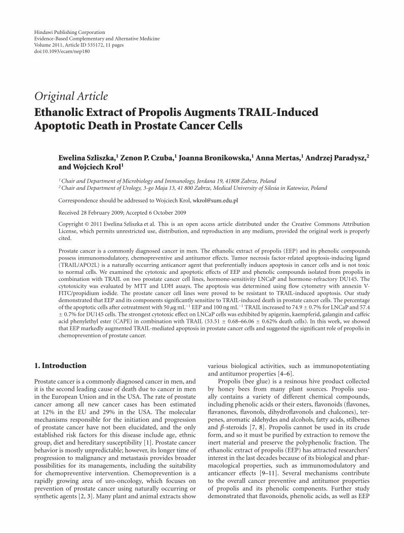
2.2. Flavonoids and Phenolic Acids. Propolis samples fromvarious geographical areas contain different compounds.The major active components of propolis from Poland areflavonoids and phenolic acids or their esters [7]. All testedcompounds were detected in our sample of EEP as describedpreviously [9]. Table 1 presents the structures of compoundsfound in the tested sample of EEP. Chrysin, apigenin,acacetin, galangin, kaempferol, kaempferid, quercetin, cin-nanic acid, o-coumaric acid, m-coumaric acid, p-coumaricacid, caffeic acid and caffeic acid phenylethyl ester (CAPE)were purchased from Carl Roth GmbH (Karlsruhe, Ger-many) and Sigma Chemical Company (St Louis, MO, USA).The reagents were dissolved in DMSO (flavonoids and phe-nolic acids—50 mM) and the final concentration of DMSOin the culture medium was controlled at 0.1% (v/v). The finalconcentration of flavonoids and phenolic acids was 50 μM(chrysin, 12.7 μg mL−1; apigenin, 13.5 μg mL−1; acacetin,14.2 μg mL−1; galangin, 13.5 μg mL−1; kaempferol, 14.3 μgmL−1; kaempferid, 15.0 μg mL−1; quercetin, 15.1 μg mL−1;cinnanic acid, 7.4 μg mL−1; o-coumaric acid, 8.2 μg mL−1; m-coumaric acid, 8.2 μg mL−1; p-coumaric acid, 8.2 μg mL−1;caffeic acid, 9.0 μg mL−1; CAPE, 14.2 μg mL−1).
2.3. TRAIL. Recombinant human TRAIL was purchasedfrom PeproTech (Rocky Hill, NJ, USA).
2.4. Prostate Cancer Cells Culture. The experiments were per-formed on two human prostate cancer cell lines: hormone-sensitivity LNCaP cells and hormone-refractory DU145 cells(DSMZ—German Collection of Microorganisms and CellCultures, Braunschweig, Germany). The cells were grownin monolayer cultures in RPMI 1640 medium containing10% fetal bovine serum, 4 mM l-glutamine, 100 U mL−1
penicillin and 100 μg mL−1 streptomycin and incubated at37◦C in atmosphere containing 5% CO2 [19]. Reagents forcells culture were purchased from PAA The Cell CultureCompany (Pasching, Austria).
2.5. Cytotoxicity Assay. The cytotoxicity was measured by 3-[4, 5-dimethylthiazol-2-yl]-2,5 diphenyltetrazolium (MTT)assay as described [19]. The LNCaP cells (2 × 10⁵ mL−1)and DU145 (1 × 10⁵ mL−1) were seeded 48–24 h beforethe experiments onto a 96-well plate. Various combina-tions of EEP (5–50 ng mL−1) with or without TRAIL (50–200 ng mL−1), flavonoids (50 μM) with or without TRAIL(100 ng mL−1), and phenolic acids (50 μM) with or withoutTRAIL (100 ng mL−1) were added to the cells, and, after 48 h,the medium was removed, and 20 μL of a MTT solutionprepared at 5 mg mL−1 (Sigma Chemical Company, MO,USA) were added to each well for 4 h. The resulting crystals
Evidence-Based Complementary and Alternative Medicine 3
Table 1: Chemical structure of the phenolic compounds used in this study.
CH=CH-COOH
R1
R2
R3
R1 R2 R3
Cinnamic acid H H H
o-Coumaric acid OH H H
m-Coumaric acid H OH H
p-Coumaric acid H H OH
Caffeic acid H OH OH
Caffeic acid phenylethyl esterOH
OH
O
O
O
O
R4
R5
R6
R1
R2
R3
R1 R2 R3 R4 R5 R6
Chrysin H OH H OH H H
Apigenin H OH H OH OH H
Acacetin H OH H OH OCH3 H
Galangin OH OH H OH H H
Kaempferol OH OH H OH OH H
Kaempferid OH OH H OH OCH3 H
Quercetin OH OH H OH OH OH
were dissolved in DMSO. Controls included native cells andmedium alone. The spectrophotometric absorbance of eachwell was measured using a microplate reader (ELx 800,Bio-Tek Instruments, Winooski, VT, USA) at 550 nm. Thepercent cytotoxicity was calculated by the formula: percent
cytotoxicity (cell death) = (1 − [absorbance of experimentalwells/absorbance of control wells]) × 100%.
2.6. Lactate Dehydrogenase Release Assay. Lactate dehydro-genase (LDH) is a stable cytosolic enzyme that is released
4 Evidence-Based Complementary and Alternative Medicine
25
20
15
10
5
00 255 50
Concentration of EEP (µg/mL)
Cyt
otox
icit
y(%
)
(a)
25
20
15
10
5
00 255 50
Concentration of EEP (µg/mL)
Cyt
otox
icit
y(%
)
(b)
25
20
15
10
5
00 255 50
Concentration of EEP (µg/mL)
Apo
ptot
icce
lls(%
)(a
nn
exin
V)
(c)
25
20
15
10
5
00 255 50
Concentration of EEP (µg/mL)
Apo
ptot
icce
lls(%
)(a
nn
exin
V)
(d)
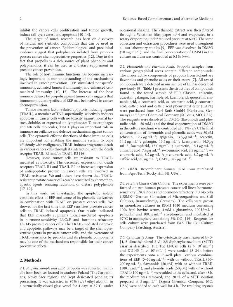
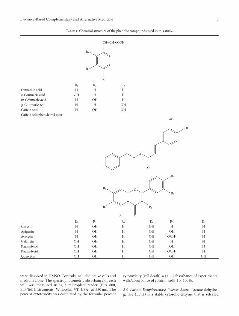
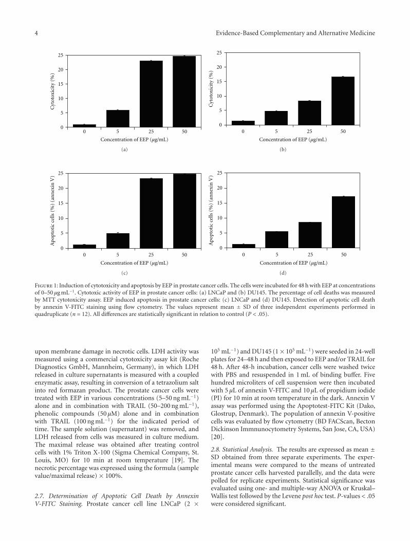
Figure 1: Induction of cytotoxicity and apoptosis by EEP in prostate cancer cells. The cells were incubated for 48 h with EEP at concentrationsof 0–50 μg mL−1. Cytotoxic activity of EEP in prostate cancer cells: (a) LNCaP and (b) DU145. The percentage of cell deaths was measuredby MTT cytotoxicity assay. EEP induced apoptosis in prostate cancer cells: (c) LNCaP and (d) DU145. Detection of apoptotic cell deathby annexin V-FITC staining using flow cytometry. The values represent mean ± SD of three independent experiments performed inquadruplicate (n = 12). All differences are statistically significant in relation to control (P < .05).
upon membrane damage in necrotic cells. LDH activity wasmeasured using a commercial cytotoxicity assay kit (RocheDiagnostics GmbH, Mannheim, Germany), in which LDHreleased in culture supernatants is measured with a coupledenzymatic assay, resulting in conversion of a tetrazolium saltinto red formazan product. The prostate cancer cells weretreated with EEP in various concentrations (5–50 ng mL−1)alone and in combination with TRAIL (50–200 ng mL−1),phenolic compounds (50 μM) alone and in combinationwith TRAIL (100 ng mL−1) for the indicated period oftime. The sample solution (supernatant) was removed, andLDH released from cells was measured in culture medium.The maximal release was obtained after treating controlcells with 1% Triton X-100 (Sigma Chemical Company, St.Louis, MO) for 10 min at room temperature [19]. Thenecrotic percentage was expressed using the formula (samplevalue/maximal release) × 100%.
2.7. Determination of Apoptotic Cell Death by AnnexinV-FITC Staining. Prostate cancer cell line LNCaP (2 ×
105 mL−1) and DU145 (1× 10⁵ mL−1) were seeded in 24-wellplates for 24–48 h and then exposed to EEP and/or TRAIL for48 h. After 48-h incubation, cancer cells were washed twicewith PBS and resuspended in 1 mL of binding buffer. Fivehundred microliters of cell suspension were then incubatedwith 5 μL of annexin V-FITC and 10 μL of propidium iodide(PI) for 10 min at room temperature in the dark. Annexin Vassay was performed using the Apoptotest-FITC Kit (Dako,Glostrup, Denmark). The population of annexin V-positivecells was evaluated by flow cytometry (BD FACScan, BectonDickinson Immnunocytometry Systems, San Jose, CA, USA)[20].
2.8. Statistical Analysis. The results are expressed as mean ±SD obtained from three separate experiments. The exper-imental means were compared to the means of untreatedprostate cancer cells harvested parallelly, and the data werepolled for replicate experiments. Statistical significance wasevaluated using one- and multiple-way ANOVA or Kruskal–Wallis test followed by the Levene post hoc test. P-values < .05were considered significant.

Evidence-Based Complementary and Alternative Medicine 5
25
20
15
10
5
00 50 100 200
Concentration of TRIAL (ng/mL)
Cyt
otox
icit
y(%
)
(a)
25
20
15
10
5
00 50 100 200
Concentration of TRIAL (ng/mL)
Cyt
otox
icit
y(%
)
(b)
Apo
ptot
icce
lls(%
)(a
nn
exin
V)
25
20
15
10
5
00 50 100 200
Concentration of TRIAL (ng/mL)
(c)
Apo
ptot
icce
lls(%
)(a
nn
exin
V)
25
20
15
10
5
00 50 100 200
Concentration of TRIAL (ng/mL)
(d)
Figure 2: Induction of cytotoxicity and apoptosis by TRAIL in prostate cancer cells. The cells were incubated for 48 h with TRAIL atconcentrations of 0–200 ng mL−1. Cytotoxic activity of TRAIL in prostate cancer cells: (a) LNCaP and (b) DU145. The percentage of celldeaths was measured by MTT cytotoxicity assay. TRAIL induced apoptosis in prostate cancer cells: (c) LNCaP and (d) DU145. Detection ofapoptotic cell death by annexin V-FITC staining using flow cytometry. The values represent mean ± SD of three independent experimentsperformed in quadruplicate (n = 12). All differences are statistically significant in relation to control (P < .05).
3. Results
3.1. Induction of Cytotoxicity and Apoptosis by Studied Agents
on Prostate Cancer Cells
3.1.1. EEP. EEP inhibited growth and induced apoptosis inprostate cancer cells in a dose-dependent manner. The cyto-toxic and apoptotic effects of EEP on hormone-sensitivityLNCaP and hormone-refractory DU145 prostate cancer cellsare given in Figure 1. The cells were incubated with 5–50 μg mL−1 EEP for 48 h. The rate of cytotoxicity upontreatment cancer cells with 5, 25 and 50 μg mL−1 EEP was5.96 ± 0.61, 23.08 ± 0.78 and 24.83 ± 0.59% for LNCaPcells and 4.75 ± 0.67, 8.20 ± 1.12 and 16.63 ± 0.77% forDU145 when compared with untreated control, respectively.The annexin V assay revealed apoptotic prostate cancer cellsexposed to EEP. We showed that EEP at the concentrations of5–50 μg mL−1 induced 4.95± 0.54, 23.28± 0.54 and 24.66±0.72% apoptosis in LNCaP cells and 5.44 ± 0.45, 8.52 ± 0.48and 17.09 ± 0.55% apoptosis in DU145 cells.
3.1.2. TRAIL. TRAIL induced cytotoxic and apoptotic effectsin a dose-dependent manner (Figure 2). We first measured
the cytotoxic activity of TRAIL after 48-h incubation onprostate cancer cells. The cytotoxicity of TRAIL at theconcentration of 100 ng mL−1 on LNCaP cells was 15.03 ±0.50%, and on DU145 cells 9.25 ± 0.86%. TRAIL increasedthe percentage of apoptotic cells. For example, a 48-hexposure to 100 ng mL−1 TRAIL induced apoptosis of 15.46± 0.55% LNCaP cells and 10.12 ± 0.86% of DU145 cells.TRAIL was less active against the both prostate cancer celllines. We confirmed that hormone-sensitivity LNCaP cellsand hormone-refractory DU145 prostate cancer cells areresistant to TRAIL.
3.1.3. TRAIL in Combination with EEP. We investigated thecytotoxic and apoptotic effects of TRAIL in combinationwith EEP on prostate cancer cells (Figures 3 and 4). Cotreat-ment of TRAIL and EEP increased the percentage of celldeath on prostate cancer cells, compared to cytotoxicity ofTRAIL or EEP alone. The cytotoxicity after 48-h incubationwith TRAIL at the concentration of 100 ng mL−1, and EEPat the concentration of 50 μg mL−1 was 73.70 ± 0.53%for hormone-sensitivity LNCaP cells and 55.76 ± 0.72%for hormone-refractory DU145 cells. Then, we tested theapoptotic effect of TRAIL in combination with EEP on
6 Evidence-Based Complementary and Alternative Medicine
0
20
40
60
80C
ytot
oxic
ity
(%)
TRIAL(ng/mL)
EEP(µg/mL)
100
5
100
5 25
100
25 50
100
50
-
- -
- - -
(a)
0
20
40
60
80
Cyt
otox
icit
y(%
)
TRIAL(ng/mL)
EEP(µg/mL)
100
5
100
5 25
100
25 50
100
50
-
- -
- - -
(b)
0
20
40
60
80
Cyt
otox
icit
y(%
)
50 50
50
50
100 200 200
TRIAL(ng/mL)
EEP(µg/mL) 100
50-
- - 50
- - -
(c)
0
20
40
60
80
Cyt
otox
icit
y(%
)
50 50
50 50
100 200 200
TRIAL(ng/mL)
(µEEPg/mL)
50- -
100
- -
-
50
-
(d)
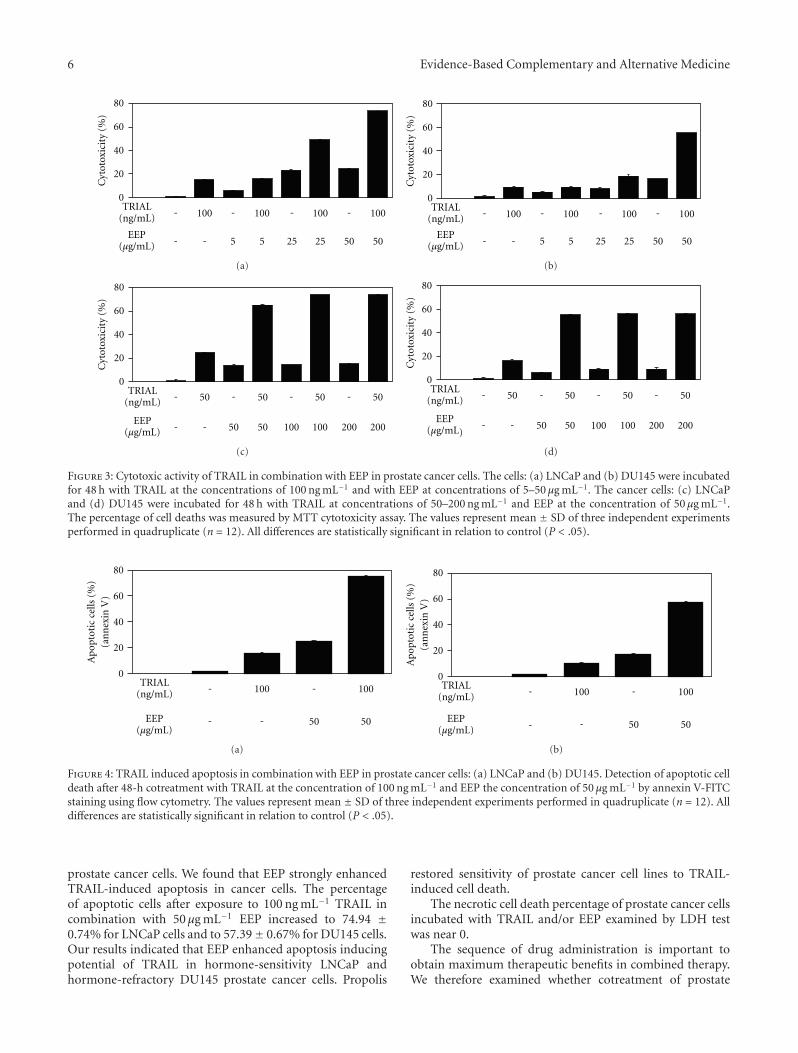
Figure 3: Cytotoxic activity of TRAIL in combination with EEP in prostate cancer cells. The cells: (a) LNCaP and (b) DU145 were incubatedfor 48 h with TRAIL at the concentrations of 100 ng mL−1 and with EEP at concentrations of 5–50 μg mL−1. The cancer cells: (c) LNCaPand (d) DU145 were incubated for 48 h with TRAIL at concentrations of 50–200 ng mL−1 and EEP at the concentration of 50 μg mL−1.The percentage of cell deaths was measured by MTT cytotoxicity assay. The values represent mean ± SD of three independent experimentsperformed in quadruplicate (n = 12). All differences are statistically significant in relation to control (P < .05).
0
20
40
60
80
Ap
opto
tic
cells
(%)
(an
nex
inV
)
100
50
100
50
TRIAL(ng/mL)
EEP(µg/mL)
-
- -
-
(a)
0
20
40
60
80
Ap
opto
tic
cells
(%)
(an
nex
inV
)
100
50
100
50
TRIAL(ng/mL)
EEP(µg/mL)
-
-
-
-
(b)
Figure 4: TRAIL induced apoptosis in combination with EEP in prostate cancer cells: (a) LNCaP and (b) DU145. Detection of apoptotic celldeath after 48-h cotreatment with TRAIL at the concentration of 100 ng mL−1 and EEP the concentration of 50 μg mL−1 by annexin V-FITCstaining using flow cytometry. The values represent mean ± SD of three independent experiments performed in quadruplicate (n = 12). Alldifferences are statistically significant in relation to control (P < .05).
prostate cancer cells. We found that EEP strongly enhancedTRAIL-induced apoptosis in cancer cells. The percentageof apoptotic cells after exposure to 100 ng mL−1 TRAIL incombination with 50 μg mL−1 EEP increased to 74.94 ±0.74% for LNCaP cells and to 57.39± 0.67% for DU145 cells.Our results indicated that EEP enhanced apoptosis inducingpotential of TRAIL in hormone-sensitivity LNCaP andhormone-refractory DU145 prostate cancer cells. Propolis
restored sensitivity of prostate cancer cell lines to TRAIL-induced cell death.
The necrotic cell death percentage of prostate cancer cellsincubated with TRAIL and/or EEP examined by LDH testwas near 0.
The sequence of drug administration is important toobtain maximum therapeutic benefits in combined therapy.We therefore examined whether cotreatment of prostate

Evidence-Based Complementary and Alternative Medicine 7
0–24 h
24–48 h TRIAL
TRIAL EEP
EEP
TRIAL
EEP
EEP
TRIAL
EEP + TRIAL
EEP + TRIAL
0
20
40
60
80
Cyt
otox
icit
y(%
)
-
-
(a)
0
20
40
60
80
Cyt
otox
icit
y(%
)
0–24 h
24–48 h TRIAL
TRIAL EEP
EEP
TRIAL
EEP
EEP
TRIAL
EEP + TRIAL
EEP + TRIAL
-
-
(b)
Figure 5: Cytotoxic activity of TRAIL in combination with EEP, after and before exposure to EEP in prostate cancer cells. (a) LNCaP and(b) DU145 cancer cells were as follws: (1) treated with EEP in combination with TRAIL for 48 h; (2) pretreated with EEP for 24 h, followedby TRAIL for another 24 h; and (3) pretreated with TRAIL for 24 h, followed by EEP for another 24 h. The percentage of cell deaths wasmeasured by MTT cytotoxicity assay. The values represent mean ± SD of three independent experiments performed in quadruplicate (n =12). All differences are statistically significant in relation to control (P < .05).
Quercetin
Kaempferid
Kaempferol
Galangin
Acacetin
Apigenin
Chrysin
CAPE
Caffeic acid
p-coumaric acid
m-coumaric
o-coumaric acid
Cinnamic acid
Control
0 5 10 15 20 25
Cytotoxicity (%)
(a)
TRIAL + p-coumaric acid
TRIAL + m-coumaric acid
TRIAL + o-coumaric acid
TRIAL + 100 ng/mL
Control
0 20 40 60 80
Cytotoxicity (%)
TRIAL + quercetin
TRIAL + kaempferid
TRIAL + kaempferol
TRIAL + galangin
TRIAL + acacetin
TRIAL + apigenin
TRIAL + chrysin
TRIAL + CAPE
TRIAL + caffeic acid
TRIAL + cinnamic acid
(b)
Figure 6: Cytotoxic activity of (a) EEP phenolic components and (b) TRAIL in combination with EEP phenolic components in prostatecancer cells. The LNCaP cells were incubated for 48 h with phenolic compounds at the concentration of 50 μM with or without TRAIL atthe concentration of 100 ng mL−1. The percentage of cell deaths was measured by MTT cytotoxicity assay. The values represent mean ± SDof three independent experiments performed in quadruplicate (n = 12). All differences are statistically significant in relation to control (P <.05).
cancer cells with EEP and TRAIL induced greater apop-tosis than the concurrent pretreatment with EEP followedby TRAIL and vice versa (Figure 5). Interestingly, thecotreatment of both prostate cancer cell lines with EEP incombination with TRAIL induced greater apoptosis thanconcurrent pretreatment or single agent alone. Reversesequence of treatments: pretreatment with EEP followed byTRAIL or pretreatment with TRAIL followed by EEP resulted
in significantly lesser apoptosis than in the cotreatment withEEP and TRAIL.
3.2. Cytotoxicity of Studied Agents in Prostate Cancer Cells
3.2.1. Phenolic Compounds Detected in Propolis. We inves-tigated the cytotoxic effect on LNCaP cells of 13 phe-nolic components of propolis: cinnamic acid, o-coumaric
8 Evidence-Based Complementary and Alternative Medicine
Bcl-2
TRIAL
TRIAL-R1TRIAL-R2
FADD
BID
tBID
Bax Bak
Caspase-9 APAF-1
Caspase-8
Apoptosis
Cytochrome C
Mitochondrium
IAPssurvivin
EEP?LuteolinApigenin
KaempferolQuercetinBaicalein
ApigeninEEP?
ApigeninEEP? Quercetin
EEP?
Caspase-3, -6, -7
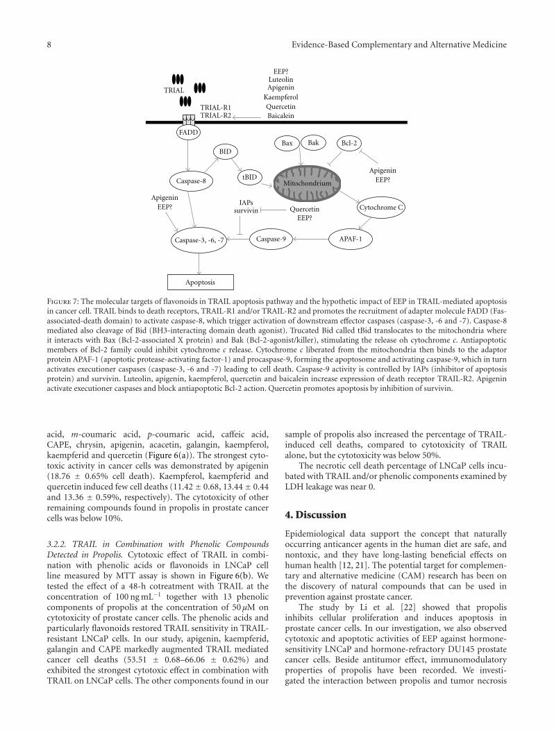
Figure 7: The molecular targets of flavonoids in TRAIL apoptosis pathway and the hypothetic impact of EEP in TRAIL-mediated apoptosisin cancer cell. TRAIL binds to death receptors, TRAIL-R1 and/or TRAIL-R2 and promotes the recruitment of adapter molecule FADD (Fas-associated-death domain) to activate caspase-8, which trigger activation of downstream effector caspases (caspase-3, -6 and -7). Caspase-8mediated also cleavage of Bid (BH3-interacting domain death agonist). Trucated Bid called tBid translocates to the mitochondria whereit interacts with Bax (Bcl-2-associated X protein) and Bak (Bcl-2-agonist/killer), stimulating the release oh cytochrome c. Antiapoptoticmembers of Bcl-2 family could inhibit cytochrome c release. Cytochrome c liberated from the mitochondria then binds to the adaptorprotein APAF-1 (apoptotic protease-activating factor-1) and procaspase-9, forming the apoptosome and activating caspase-9, which in turnactivates executioner caspases (caspase-3, -6 and -7) leading to cell death. Caspase-9 activity is controlled by IAPs (inhibitor of apoptosisprotein) and survivin. Luteolin, apigenin, kaempferol, quercetin and baicalein increase expression of death receptor TRAIL-R2. Apigeninactivate executioner caspases and block antiapoptotic Bcl-2 action. Quercetin promotes apoptosis by inhibition of survivin.
acid, m-coumaric acid, p-coumaric acid, caffeic acid,CAPE, chrysin, apigenin, acacetin, galangin, kaempferol,kaempferid and quercetin (Figure 6(a)). The strongest cyto-toxic activity in cancer cells was demonstrated by apigenin(18.76 ± 0.65% cell death). Kaempferol, kaempferid andquercetin induced few cell deaths (11.42± 0.68, 13.44± 0.44and 13.36 ± 0.59%, respectively). The cytotoxicity of otherremaining compounds found in propolis in prostate cancercells was below 10%.
3.2.2. TRAIL in Combination with Phenolic CompoundsDetected in Propolis. Cytotoxic effect of TRAIL in combi-nation with phenolic acids or flavonoids in LNCaP cellline measured by MTT assay is shown in Figure 6(b). Wetested the effect of a 48-h cotreatment with TRAIL at theconcentration of 100 ng mL−1 together with 13 phenoliccomponents of propolis at the concentration of 50 μM oncytotoxicity of prostate cancer cells. The phenolic acids andparticularly flavonoids restored TRAIL sensitivity in TRAIL-resistant LNCaP cells. In our study, apigenin, kaempferid,galangin and CAPE markedly augmented TRAIL mediatedcancer cell deaths (53.51 ± 0.68–66.06 ± 0.62%) andexhibited the strongest cytotoxic effect in combination withTRAIL on LNCaP cells. The other components found in our
sample of propolis also increased the percentage of TRAIL-induced cell deaths, compared to cytotoxicity of TRAILalone, but the cytotoxicity was below 50%.
The necrotic cell death percentage of LNCaP cells incu-bated with TRAIL and/or phenolic components examined byLDH leakage was near 0.
4. Discussion
Epidemiological data support the concept that naturallyoccurring anticancer agents in the human diet are safe, andnontoxic, and they have long-lasting beneficial effects onhuman health [12, 21]. The potential target for complemen-tary and alternative medicine (CAM) research has been onthe discovery of natural compounds that can be used inprevention against prostate cancer.
The study by Li et al. [22] showed that propolisinhibits cellular proliferation and induces apoptosis inprostate cancer cells. In our investigation, we also observedcytotoxic and apoptotic activities of EEP against hormone-sensitivity LNCaP and hormone-refractory DU145 prostatecancer cells. Beside antitumor effect, immunomodulatoryproperties of propolis have been recorded. We investi-gated the interaction between propolis and tumor necrosis

Evidence-Based Complementary and Alternative Medicine 9
factor-related apoptosis inducing ligand on prostate cancercells. Recombinant human TRAIL used in our study is asoluble protein based on a natural ligand. TRAIL inducesprogrammed death in various cancer cells, in vitro and invivo [16]. However, some tumor cells are resistant to TRAIL-mediated cytotoxicity. We and others demonstrated thatprostate cancer cell lines, LNCaP and DU145, were resistantto TRAIL-induced apoptosis [17–19].
Our study showed the impact of propolis on theanticancer immune defense. Propolis restores sensitivityof tumor cells to immune effectors mechanisms, such asTRAIL-induced apoptosis in prostate cancer cells. For thefirst time, our results demonstrated that EEP markedly aug-mented TRAIL-mediated apoptosis in hormone-sensitivityLNCaP and hormone-refractory DU145 prostate cancercells. The rapid tumor growth and progression of hormonerefractory prostate cancer accounts for most of the morbidityand mortality associated with prostate cancer [1]. The exper-imental data indicated that propolis is a promising anticanceragent also for the prevention of hormone-refractory prostatecancer.
In the field of CAM, immunomodulation through natu-ral or synthetic substances may be considered as an alterna-tive for the prevention of neoplasm disease. EEP enhancesthe apoptosis-inducing potential of TRAIL and sensitizesTRAIL-resistant prostate cancer cells. Further investigationswill be required to recognize and explain the molecularmechanisms and cellular signaling pathways by which EEPsensitizes cancer cells to TRAIL-induced death. Moreover,due to heterogenous complex composition of propolis, itsbiological activity is variable. The presence in propolis of somany compounds makes it difficult to know and understandthe direct and indirect effects of EEP upon transductionpathway of the signal to TRAIL-mediated apoptosis in cancercells.
The flavonoids and phenolic components found inpropolis are known to affect the apoptosis of prostatecancer cells and may play an important role in cancerchemoprevention [2, 3, 23, 24]. We tested in vitro thecytotoxicity of 13 compounds detected in our sample ofpropolis against prostate cancer. The strongest cytotoxicactivity on LNCaP prostate cancer cells was demonstrated byapigenin. Shukla and Gupta [23, 25] reported that apigeninin both in vitro and in vivo studies induced apoptosis inprostate cancer.
It has been suggested that phenolic compounds isolatedfrom propolis induce activities of the immune systemand exert antitumor effects [9–15, 22–25]. To investigatewhich compounds found in propolis may be responsiblefor the enhancement of the apoptosis-inducing potentialof TRAIL, we tested the cytotoxic effect of its phenoliccomponents in combination with TRAIL on prostate cancercells. All detected in our EEP sample compounds usedin combination with TRAIL increased the percentage ofcell deaths compared to cytotoxicity of TRAIL alone. Thephenolic acids and particularly flavonoids restored TRAILsensitivity in TRAIL-resistant LNCaP prostate cancer cells.In our study, apigenin, kaempferid, galangin and CAPEmarkedly augmented TRAIL mediated cancer cells death and
exhibited the strongest cytotoxic effect in combination withTRAIL on LNCaP cells. Apigenin, kaempferid and galangin,the compounds with the most cytotoxic activity with TRAIL,have three hydroxyl groups (positions 5, 7 and 3 or 4′).Every tested flavonoid has hydroxyl groups in fifth andseventh positions. The compounds with only two hydroxylgroups in fifth and seventh positions (chrysin, acacetin),or four (kaempferol) and five hydroxyl groups (quercetin)showed lower cytotoxic activity with TRAIL. The presenceof hydroxyl group in position 3 (galangin versus chrysin)decreased activity of galangin, but addition of TRAILchanged this activity. Probably, this activity is dependent ondifferent mechanisms. The position of hydroxyl groups inflavone structure and their number are very important inreaction with reactive oxygen species as well as can influencecytotoxic and apoptotic activities [26, 27].
In study in vitro on HeLa cell line, we confirmedthat EEP sensitize cancer cells to TRAIL-induced apoptosisand two components identified in propolis, apigenin andCAPE, were the most potent agents inducing cell death incombination with TRAIL in HeLa cells [20]. A similar studywith flavonoids (Figure7) showed that luteolin, apigenin,kaempferol, baicalein and quercetin synergistically inducedapoptosis with TRAIL in human malignant tumor cells [18,28–33]. Horinaka et al. [28] reported that luteolin increasedTRAIL-induced apoptosis in HeLa cells through upregula-tion of death receptor TRAIL-R2. In other investigation,they also showed the enhanced apoptosis-inducing potentialof TRAIL in prostate cancer cell line DU145, leukemic cellline Jurkat, and colon cancer cell line DLD1. The combineduse of apigenin and TRAIL caused Bcl-2-interacting domaincleavage, activation of caspases, and increased expressionof TRAIL-R2 [18]. Yoshida et al. [29] stated that TRAIL-R2 upregulation by kaempferol augments TRAIL actionin colon cancer cells [29]. Chen et al. [30] showed thatsuppression of survivin and induction of TRAIL-R2 byquercetin contribute to sensitization of lung cancer cellsto TRAIL-induced cytotoxicity. Kim et al. [31] examinedthe molecular mechanisms by which quercetin augmentsTRAIL-mediated apoptotic death in prostate cancer cellsand confirmed the ability of quercetin to downregulatesurvivin expression. Taniguchi et al. [32] indicated thatbaicalein increases TRAIL-R2 expression and overcomesTRAIL resistance in prostate cancer cells.
We demonstrated for the first time that kaempferid,galangin and CAPE enhance the cytotoxic potential ofTRAIL in prostate cancer cells. Those polyphenols, besideapigenin, equally firmly sensitize TRAIL-resistant LNCaPcells. The previous study suggested that flavonoids increaseexpression of TRAIL-R2 [18, 28–30, 32]. We hypothesize thatpropolis, as one of the richest sources of flavonoids, such asapigenin, kaempferol, kaempferid, galangin, quercetin andCAPE, can influence the expression of death receptor TRAIL-R2, inhibition of antiapoptotic protein (Bcl-2, survivin), oractivation of caspases (Figure 7).
We showed that EEP and its phenolic components invitro augmented TRAIL mediated cell death in prostatecancer, but further study will be required to examine themolecular mechanisms by which EEP and its compounds act
10 Evidence-Based Complementary and Alternative Medicine
on cellular signaling pathways and sensitize prostate cancercells to TRAIL-induced apoptosis. Our findings suggest thatthe modulation of TRAIL apoptosis pathway may have asignificant potential for prostate cancer chemoprevention,and the overcome of TRAIL-resistance by propolis andits phenolic components may be one of the mechanismsresponsible for their cancer preventive effects. The obtainedresults confirmed the significance of EEP and its componentsin chemoprevention of prostate cancer cells. EEP as a dietarysupplement may be useful in chemoprevention agent againstprostate cancer.
Funding
Research Grant 2-164/08 from Medical University of Silesiain Katowice, Poland.
References
[1] A. Heidenreich, G. Aus, M. Bolla et al., “EAU guidelines onprostate cancer,” European Urology, vol. 53, no. 1, pp. 68–80,2008.
[2] R. P. Singh and R. Agarwal, “Mechanisms of action ofnovel agents for prostate cancer chemoprevention,” Endocrine-Related Cancer, vol. 13, no. 3, pp. 751–778, 2006.
[3] D. N. Syed, Y. Suh, F. Afaq, and H. Mukhtar, “Dietary agentsfor chemoprevention of prostate cancer,” Cancer Letters, vol.265, no. 2, pp. 167–176, 2008.
[4] E. L. Cooper, “The immune system and complementaryand alternative medicine,” Evidence-Based Complementary andAlternative Medicine, vol. 4, no. 1, pp. 5–8, 2007.
[5] S. Salvioli, E. Sikora, E. L. Cooper, and C. Franceschi,“Curcumin in cell death processes: a challenge for CAM ofage-related pathologies,” Evidence-Based Complementary andAlternative Medicine, vol. 4, no. 2, pp. 181–190, 2007.
[6] A. Vojdani and J. Erde, “Regulatory T cells, a potentimmunoregulatory target for CAM researchers: modulatingallergic and infectious disease pathology (II),” Evidence-BasedComplementary and Alternative Medicine, vol. 3, no. 2, pp.209–215, 2006.
[7] C. Gardana, M. Scaglianti, P. Pietta, and P. Simonetti, “Analysisof the polyphenolic fraction of propolis from different sourcesby liquid chromatography-tandem mass spectrometry,” Jour-nal of Pharmaceutical and Biomedical Analysis, vol. 45, no. 3,pp. 390–399, 2007.
[8] V. S. Bankova, S. S. Popov, and N. L. Marekov, “A study onflavonoids of propolis,” Journal of Natural Products, vol. 46, no.4, pp. 471–474, 1983.
[9] W. Krol, S. Scheller, Z. Czuba et al., “Inhibition of neutrophils’chemiluminescence by ethanol extract of propolis (EEP) andits phenolic components,” Journal of Ethnopharmacology, vol.55, no. 1, pp. 19–25, 1996.
[10] N. Orsolic, A. B. Saranovic, and I. Basic, “Direct and indirectmechanism(s) of antitumour activity of propolis and itspolyphenolic compounds,” Planta Medica, vol. 72, no. 1, pp.20–27, 2006.
[11] S. Scheller, W. Krol, J. Swiacik, S. Owczarek, J. Gabrys,and J. Shani, “Antitumoral property of ethanolic extract ofpropolis in mice-bearing Ehrlich carcinoma, as compared tobleomycin,” Zeitschrift fur Naturforschung C, vol. 44, no. 11-12, pp. 1063–1065, 1989.
[12] D. F. Birt, S. Hendrich, and W. Wang, “Dietary agents in cancer
prevention: flavonoids and isoflavonoids,” Pharmacology andTherapeutics, vol. 90, no. 2-3, pp. 157–177, 2001.
[13] C. Chen, M. Weng, C. Wu, and J. Lin, “Comparison ofradical scavenging activity, cytotoxic effects and apoptosisinduction in human melanoma cells by Taiwanese propolisfrom different sources,” Evidence-Based Complementary andAlternative Medicine , vol. 1, pp. 175–185, 2004.
[14] M. C. Bufalo, J. M. Candeias, and J. M. Sforcin, “In vitrocytotoxic effect of Brazilian green propolis on human laryn-geal epidermoid carcinoma (HEP-2) cells,” Evidence-BasedComplementary and Alternative Medicine , vol. 22, pp. 1–5,2007.
[15] F. Missima and J. M. Sforcin, “Green Brazilian propolisaction on macrophages and lymphoid organs of chronicallystressed mice,” Evidence-Based Complementary and AlternativeMedicine, vol. 5, no. 1, pp. 71–75, 2008.
[16] A. Almasan and A. Ashkenazi, “Apo2L/TRAIL: apoptosissignaling, biology, and potential for cancer therapy,” Cytokineand Growth Factor Reviews, vol. 14, no. 3-4, pp. 337–348, 2003.
[17] S. Shankar, T. R. Singh, and R. K. Srivastava, “Ionizingradiation enhances the therapeutic potential of TRAIL inprostate cancer in vitro and in vivo: intracellular mechanisms,”Prostate, vol. 61, no. 1, pp. 35–49, 2004.
[18] M. Horinaka, T. Yoshida, T. Shiraishi, S. Nakata, M. Wakada,and T. Sakai, “The dietary flavonoid apigenin sensitizes malig-nant tumor cells to tumor necrosis factor-related apoptosis-inducing ligand,” Molecular Cancer Therapeutics, vol. 5, no. 4,pp. 945–951, 2006.
[19] E. Szliszka, J. Bronikowska, A. Majcher, J. Miszkiewicz, andW. Krol, “Enhanced sensitivity of hormone-refractory prostatecancer cells to tumor necrosis factor-related apoptosis-inducing ligand (TRAIL) mediated cytotoxicity by taxanes,”Central European Journal of Urology, vol. 62, pp. 29–34, 2009.
[20] E. Szliszka, Z. P. Czuba, M. Domino, B. Mazur, G. Zydowicz,and W. Krol, “Ethanolic extract of propolis (EEP) enhancesthe apoptosis-inducing potential of TRAIL in cancer cells,”Molecules, vol. 14, no. 2, pp. 738–754, 2009.
[21] N. Khan, V. M. Adhami, and H. Mukhtar, “Apoptosis bydietary agents for prevention and treatment of cancer,”Biochemical Pharmacology, vol. 76, no. 11, pp. 1333–1339,2008.
[22] H. Li, A. Kapur, J. X. Yang et al., “Antiproliferation of humanprostate cancer cells by ethanolic extracts of Brazilian propolisand its botanical origin,” International Journal of Oncology, vol.31, no. 3, pp. 601–606, 2007.
[23] S. Shukla and S. Gupta, “Molecular targets for apigenin-induced cell cycle arrest and apoptosis in prostate cancer cellxenograft,” Molecular Cancer Therapeutics, vol. 5, no. 4, pp.843–852, 2006.
[24] K. McEleny, R. Coffey, C. Morrissey, J. M. Fitzpatrick, and R.W. G. Watson, “Caffeic acid phenethyl ester-induced PC-3 cellapoptosis is caspase-dependent and mediated through the lossof inhibitors of apoptosis proteins,” BJU International, vol. 94,no. 3, pp. 402–406, 2004.
[25] S. Shukla and S. Gupta, “Apigenin-induced cell cycle arrestis mediated by modulation of MAPK, PI3K-Akt, and lossof cyclin D1 associated retinoblastoma dephosphorylation inhuman prostate cancer cells,” Cell Cycle, vol. 6, no. 9, pp. 1102–1114, 2007.
[26] Z. P. Czuba and W. Krol, “The importance of hydroxyl sub-stituent in position 4′ in flavonoids for modulation of chemi-luminescence generated by an enzymatic system (horseradishperoxidase-luminol-hydrogen peroxide),” Current Topics inBiophysics, vol. 20, pp. 38–41, 1996.

Evidence-Based Complementary and Alternative Medicine 11
[27] K. Plochmann, G. Korte, E. Koutsilieri et al., “Structure-activity relationships of flavonoid-induced cytotoxicity onhuman leukemia cells,” Archives of Biochemistry and Bio-physics, vol. 460, no. 1, pp. 1–9, 2007.
[28] M. Horinaka, T. Yoshida, T. Shiraishi et al., “The combinationof TRAIL and luteolin enhances apoptosis in human cervicalcancer HeLa cells,” Biochemical and Biophysical ResearchCommunications, vol. 333, no. 3, pp. 833–838, 2005.
[29] T. Yoshida, M. Konishi, M. Horinaka et al., “Kaempferolsensitizes colon cancer cells to TRAIL-induced apoptosis,”Biochemical and Biophysical Research Communications, vol.375, no. 1, pp. 129–133, 2008.
[30] W. Chen, X. Wang, J. Zhuang, L. Zhang, and Y. Lin, “Inductionof death receptor 5 and suppression of survivin contribute tosensitization of TRAIL-induced cytotoxicity by quercetin innon-small cell lung cancer cells,” Carcinogenesis, vol. 28, no.10, pp. 2114–2121, 2007.
[31] Y.-H. Kim, D.-H. Lee, J.-H. Jeong, Z. S. Guo, and Y. J.Lee, “Quercetin augments TRAIL-induced apoptotic death:involvement of the ERK signal transduction pathway,” Bio-chemical Pharmacology, vol. 75, no. 10, pp. 1946–1958, 2008.
[32] H. Taniguchi, T. Yoshida, M. Horinaka et al., “Baicaleinovercomes tumor necrosis factor-related apoptosis-inducingligand resistance via two different cell-specific pathways incancer cells but not in normal cells,” Cancer Research, vol. 68,no. 21, pp. 8918–8927, 2008.
[33] E. Szliszka, Z. P. Czuba, K. Jernas, and W. Krol, “Dietaryflavonoids sensitize HeLa cells to tumor necrosis factor-relatedapoptosis-inducing ligand (TRAIL),” International Journal ofMolecular Sciences, vol. 9, no. 1, pp. 56–64, 2008.

Submit your manuscripts athttp://www.hindawi.com
Stem CellsInternational
Hindawi Publishing Corporationhttp://www.hindawi.com Volume 2014
Hindawi Publishing Corporationhttp://www.hindawi.com Volume 2014
MEDIATORSINFLAMMATION
of
Hindawi Publishing Corporationhttp://www.hindawi.com Volume 2014
Behavioural Neurology
EndocrinologyInternational Journal of
Hindawi Publishing Corporationhttp://www.hindawi.com Volume 2014
Hindawi Publishing Corporationhttp://www.hindawi.com Volume 2014
Disease Markers
Hindawi Publishing Corporationhttp://www.hindawi.com Volume 2014
BioMed Research International
OncologyJournal of
Hindawi Publishing Corporationhttp://www.hindawi.com Volume 2014
Hindawi Publishing Corporationhttp://www.hindawi.com Volume 2014
Oxidative Medicine and Cellular Longevity
Hindawi Publishing Corporationhttp://www.hindawi.com Volume 2014
PPAR Research
The Scientific World JournalHindawi Publishing Corporation http://www.hindawi.com Volume 2014
Immunology ResearchHindawi Publishing Corporationhttp://www.hindawi.com Volume 2014
Journal of
ObesityJournal of
Hindawi Publishing Corporationhttp://www.hindawi.com Volume 2014
Hindawi Publishing Corporationhttp://www.hindawi.com Volume 2014
Computational and Mathematical Methods in Medicine
OphthalmologyJournal of
Hindawi Publishing Corporationhttp://www.hindawi.com Volume 2014
Diabetes ResearchJournal of
Hindawi Publishing Corporationhttp://www.hindawi.com Volume 2014
Hindawi Publishing Corporationhttp://www.hindawi.com Volume 2014
Research and TreatmentAIDS
Hindawi Publishing Corporationhttp://www.hindawi.com Volume 2014
Gastroenterology Research and Practice
Hindawi Publishing Corporationhttp://www.hindawi.com Volume 2014
Parkinson’s Disease
Evidence-Based Complementary and Alternative Medicine
Volume 2014Hindawi Publishing Corporationhttp://www.hindawi.com