equine health program 2008 annual report and research … · evaluating the role of foot...
TRANSCRIPT

North Carolina State University College of Veterinary Medicine
Equine Health Program
2008 Annual Report and
Research Overview

Table of Contents
Page 2 Introduction Letter from Dr. Anthony Blikslager Page 3-7 Development and Scholarship Update, Student Internships Research, Equine Development and Scholarship update from Michael Strader 2008 Veterinary Scholars Program Summer Internships in Equine Research from Sam Jones Page 8-9 Equine Medicine Symposium 2009 –Horse Owners Day PAGES 10-15 OPHTHALMOLOGY SECTION
Bacteria in Eyes with Chronic Equine Recurrent Uveitis from the Southeastern United States -Brian C. Gilger In Vitro and In Vivo Evaluation of an Equine Intraocular Lens -Richard J. McMullen Digital Infrared Photography of the Anterior Segment of the Equine Eye. -Richard J. McMullen Quantitative Differences in mRNA Expression of Toll-Like Receptor (TLR)-2, -4, and -9 in Normal Equine Eyes and Eyes with Equine Recurrent Uveitis -Na Young Yi PAGES 16-19 LAMENESS SECTION
Diagnostic Findings in Horses with Proximal Plantar Metatarsal Pain - A Comparison of MRI and Ultrasonography -Raphael Labens Evaluating the Role of Foot Conformation and Internal Anatomy in the Causes of Equine Foot Lameness Using Photographs, Radiographs and Magnetic Imaging -R.A. Mansmann PAGES 20-22 COLIC SECTION
Analysis of Sodium Carboxymethylcellulose Administration and Related Factors Associated with Postoperative Colic and Survival in Horses with Small Intestinal Disease -Callie Fogle Testing the efficacy of new and old drugs for treatment of colic: COX-2 inhibitors and lidocaine -Anthony T. Blikslager PAGES 23-29 PHARMACOLOGY SECTION
The Effect of Lidocaine on In Vitro Adhesion and Migration of Equine Neutrophils -Sam Jones Pharmacokinetics of intravenous, intramuscular and sublingual buprenorphine in horses -Jennifer L Davis Population Pharmacokinetics of Enrofloxacin in Horses -Jennifer L Davis The Effects of Compounding and Storage Conditions on the Stability of Pergolide Mesylate -Jennifer L Davis PAGES 30-31 IMMUNOLOGY SECTION
Circulating plasma exosomes: nanometer lipid particles bearing IgE and tumor necrosis factor receptor -Bruce Hammerberg
Letter from Director of Equine Health Program As the Director of the Equine Health Program, I am proud to present the Annual Equine Health Program Research Update. Our research programs are critical to the mission of the College of Veterinary Medicine because they allow us to understand those problems that take the lives of horses, and those that keep our horses out of work. In addition, we actively research new methods of increasing and improving breeding programs so that we can become less dependent on breeding programs that are overseas or out of state. We are constantly assessing new ways of funding this research effort as federal and foundation funds become scarce, and private funding becomes more and more competitive. Presently, most of our funding ultimately comes from the horse owner. Anyone interested in assisting in any way should feel free to contact the College of Veterinary Medicine Foundation Office where we now have our own Equine Development Officer (Michael Strader). Ophthalmology is a key area of study, including the study of the vascular supply of the eye (the uvea). Uveitits (moon blindness) has been studied to gain a more in depth appreciation of this complex disease. A novel study on the use of lens implants also demonstrates how advanced equine medicine and surgery has become. Our on-going studies in the area of lameness have focused on imaging. For example, we continue to refine our techniques for assessing the use of magnetic resonance imaging (MRI), particularly for difficult-to-diagnose conditions such as suspensory ligament strain. We have unique abilities to study equine pharmacology, which is a particular strength of this report. We also have a growing team of clinicians that study colic; assessing new drugs that may help us improve survival of horses with this devastating disease. We are also very pleased to have scientists working with us on immunological issues that emphasize our ability to study equine medicine at the cellular and molecular level. I am particularly grateful to Dr. Dick Mansmann and Michael Strader for linking us with the outside world through outreach programs, and our hard working clinicians, students and technicians, who work well beyond the “normal” equine work week to promote our programs. We will have a special session presented by Dr. Sam Jones highlighting the tremendous success we have had recruiting students into our research labs. We have a record number of equine-focused students in the up-coming year, and will strive to provide these students with the clinical and research skills to pursue a long-term career in equine medicine. I am also very pleased to welcome and work with our newly appointed Department Head, Dr. Lizette Hardie, who has already shown a particular interest in the success of the Equine Health Program. I invite you to contact me with any questions about our equine research, clinical, and teaching programs. Best wishes,
Anthony Blikslager, DVM, PhD Director, Equine Health Program

Contributions
Equine Health Program & Gallop of Honor Donors July 1, 2007 – June 30, 2008
Advanced Animal Eye Care Kristine M. Alpi Animal Eye Clinic Animal Eye Consultants Animal Eye Specialty Center, P.A. Anonymous Alison Bailey Sheila C. Bailey Elaine & Robert Baillie Bank of America Foundation Doreen S. Baum Joy E. Beaston Lee R. Bergman Richard & Sharon Berkshire Dr. Anthony T. Blikslager Bluegrass Veterinary Vision, Inc. Mary G. Bowles William S. Bozadjian Sandra Dee Brooks Melanie Buchanan Denise R. Burrell Gretchen P. Burud Burton Foundation, Inc. Nancy Caggia Carole Cameron Jim Cogdell Jennifer B. Collins Tayloe Bond Compton Dr. E. Brian Delp Lucinda Dorsey Barry Dumser & Sandra McShea Alison L. Eastman Robert W. Edwards Eklin Medical Systems, Inc. Eye Clinic for Animals, Inc. Cynthia L. Fawcett Barbara J. Robison Richard P. Rozek Dr. Terry G. Seaks Kimberly H. Shovelin Susan Barnard Marie Burnette Dr. Oscar Fletcher Mt. Misery Farm
Fox Grove Farms, LLC Daniel & Susan Gates Elizabeth C. Gilger Lewis & Olive Greenwald Andrea G. Haney Dr. Elizabeth M. Hardie Lori D. Hattori Ashley Hodges Eva R. Hornak IBM Corporation Cindy Jarman Sharon M. Johnson Charles F. Kane, Jr. Carol L. Knox Kraft Foods Diane Kriesel Dr. Linda J. Kuhn Dwight C. Lamm Barbara A. Lightner Gail Lorzing Rebecca Lula & Scott McLeod Lux Biosciences, Inc. Thelma M. Malone Susan R. Maxwell Donna Falgueso Sandra McDanel Robert & Joanne McLaughlin Lisa E. Morrison Kay M. Murphy
NC Hunter Jumper Association NC Thoroughbred Association New England Equine Cindy Nord Laura Underhill Norment Dr. Adrienne Otto V. Parker Overton Partnar Animal Health, Inc. Glenn & Joan Petty Gloria F. Phillips Shannon G. Preston Caroline C. Quinn John & Gayle Rakosky Raleigh Spring Premier H.S. Donald & Debra Rishel Holly H. Robbins Charles & Christina Robinson Willian B. Thompson, Jr. Kathleen S. Trimeloni University of Florida Janet B. Vicars Wachovia Foundation Elizabeth D. Wall Drs. Lloyd P. & Kathryn Tate Robert H. Tate, Jr. Diana & Ken Swanson Michele D. Swink R.B.Terry Charitable Foundation Stars & Bars Farm, Inc. Kenneth A. Steele Michael Strader Tracey L. McCarthy Elizabeth G. McCrodden Teresa Slack Cameron Slade Christie Boysen Sandra Brooks Debra Garrison Janet Jones Linda Mansmann Dr. Richard Mansmann Christine Nadel Jane Rogers Jones Janet Whetstone

Your Support
Your support can make a difference in the lives of horses and their devoted owners. The North Carolina Veterinary Medical Foundation (NCVMF) graciously accepts donations for the Equine Health Program at the College of Veterinary Medicine and our Equine Health Center at Southern Pines. Tax deductible contributions can be made through:
• The Gallop of Honor. Supports the overall mission and goals of the Equine Health Program. With gifts of $250, $500 or $1,000 you will receive a personalized Bronze, Silver or Gold Horse Shoe, which will be placed on our Gallop of Honor Wall at the location of your choosing (either Southern Pines or Raleigh). The horse shoes are a great way to honor a favorite horse, veterinarian or someone important in the life of your horse. Amounts less than $250 still support the Equine Health Program, but do not qualify for a horseshoe.
• Scholarships for Equine Students. Contributions to this program support fellowships and scholarships for DVM students who are planning to become equine practitioners. These awards will help offset the student debt load that most DVM students experience upon graduation.
• Endowed Chairs, Professorships and Programs. Endowment of equine faculty positions and programs provides both salary and operational support to attract and retain eminent and world class equine faculty. Please contact us if you’d like more information on the requirements for establishing an endowed chair or professorship.
• Capital Improvements. Substantial investment is needed for both new construction and renovation of existing equine facilities at our Southern Pines center and our hospital in Raleigh. Fundraising is currently underway for the addition of a new clinical facility at the Equine Health Center in Southern Pines. Thanks to the support and contributions of NCVMF Board Member Jim Cogdell and other key donors, we’re well on our way to securing the necessary funds to begin construction of this new facility. Your gifts are needed and the future of equine health in North Carolina is dependent upon providing our faculty and staff with the physical resources to ensure exceptional care, research and education.
• Fund for Discovery. Provides financial support for training programs for students involved in the summer research internship program.
For more information about making a contribution to the
Equine Health Program, Gallop of Honor, Equine Health Center or other equine related program, please contact
Michael Strader, Director of Development for Equine Health Programs at the NCVMF Office: (919) 513-6856, or [email protected], or to make your gift online, visit
www.cvm.ncsu.edu/ncvmf/

Scholarship Information
There is a continued need of scholarships and awards for equine oriented veterinary students. On average, veterinary students start their careers with a debt of $70,000 or more. With this large debt load, new veterinarians are tempted to go into a more lucrative small animal practice, where average salaries are higher for the first five years than those in equine practice. Scholarships can offset the cost of this debt, and could be one the deciding factors for students when choosing their practice. Our scholarship program continues to grow, and we are grateful for those who have made contributions that directly impact our students. Due to the generosity of a few very special horse owners, this past year saw the formation of the Fritz & Softy Endowed Equine Scholarship as well as the Abi Normal Endowed Equine Scholarship. Both of these scholarships, in addition to contributions to build towards the endowment, included annual scholarship gifts as well. We were also pleased to receive a gift from Eklin Medical Systems for an Equine Award in Radiology, gift from Vettec Care Products for an Equine Podiatry Award, as well as a gift from Glenn & Joan Petty for an equine student award. These four new scholarships and awards, combined with the Raleigh Spring Premiere Horse Show, the North Carolina Hunter Jumper Association, the North Carolina Thoroughbred Association and the Randall B. Terry Foundation Horse Racing Scholarships formed the basis for our equine oriented scholarships this year. How can you help? Whether you are an individual, a group of individuals, a veterinary practice, a horse organization, a business, etc., your support can make a difference. To set up an endowed scholarship, the minimum contribution is $25,000, which can be a one time gift or can be made over a five year period. Endowed scholarships are those set up in which only the interest accrued is used each year, not the principal. You can also make annual individual contributions which can be given to deserving equine oriented students. Gifts of $1,000 or more could prove very beneficial to students, helping offset yearly tuition. If you have questions, or would like more information concerning our scholarships or if you wish to set up a new scholarship, please contact Michael Strader at (919) 513-6856 or email to [email protected].
Ray Cloninger, manager of the Raleigh Spring Premier Horse Show, presents a gift to Dr. Dick Mansmann, Director of Outreach of the Equine Health Program. This gift supports the Raleigh Spring Premier Horse Show Endowed Equine Scholarship, which provides financial assistance to an equine oriented student or students at the College of Veterinary Medicine. There have been five recipients since its endowment.
2008 Veterinary Scholars Program Summer Internships in
Equine Research
Like other veterinary disciplines, equine veterinary medicine is faced with a shortage of veterinarians trained in research. The importance of research training is not only in producing researchers that work on problems related to equine health and welfare, but also to enrich our students who enter equine practice with knowledge that equips them to better understand diseases and their treatment and to make better use of new information that is being produced every day by equine veterinary research. The equine group at NC State is very involved in all four of the training programs that provide research experience and training to veterinary students. Veterinary Scholars Program Summer Research Internship: The members of the equine faculty are well represented in the program; Dr. Sam Jones, Dr. Anthony Blikslager, Dr. Carlos Pinto, Dr. Sarah Gardner, Dr. Jen Davis, Dr. Mat Gerard, Dr. Brian Gilger, Dr. Dick Mansmann, and Dr. Betta Breuhaus are mentors who have recently trained students. Five students worked on equine research projects during the summer of 2008-Cynthia Ast, Heather Smith, Natalie Morris, Dinah Skorich, and Kate Echeverria. In addition, one alumnus of the internship program, Blaire Holland received a grant from the Morris Animal Foundation to support her work for a second summer of research.
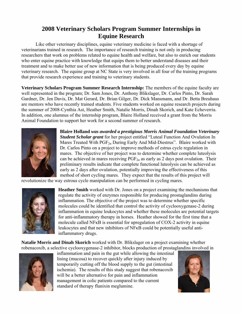
Blaire Holland was awarded a prestigious Morris Animal Foundation Veterinary Student Scholar grant for her project entitled “Luteal Function And Ovulation In Mares Treated With PGF2α During Early And Mid-Diestrus”. Blaire worked with Dr. Carlos Pinto on a project to improve methods of estrus cycle regulation in mares. The objective of her project was to determine whether complete luteolysis can be achieved in mares receiving PGF2α as early as 2 days post ovulation. Their preliminary results indicate that complete functional luteolysis can be achieved as early as 2 days after ovulation, potentially improving the effectiveness of this method of short cycling mares. They expect that the results of this project will
revolutionize the way estrous cycle manipulation can be performed in cycling mares.
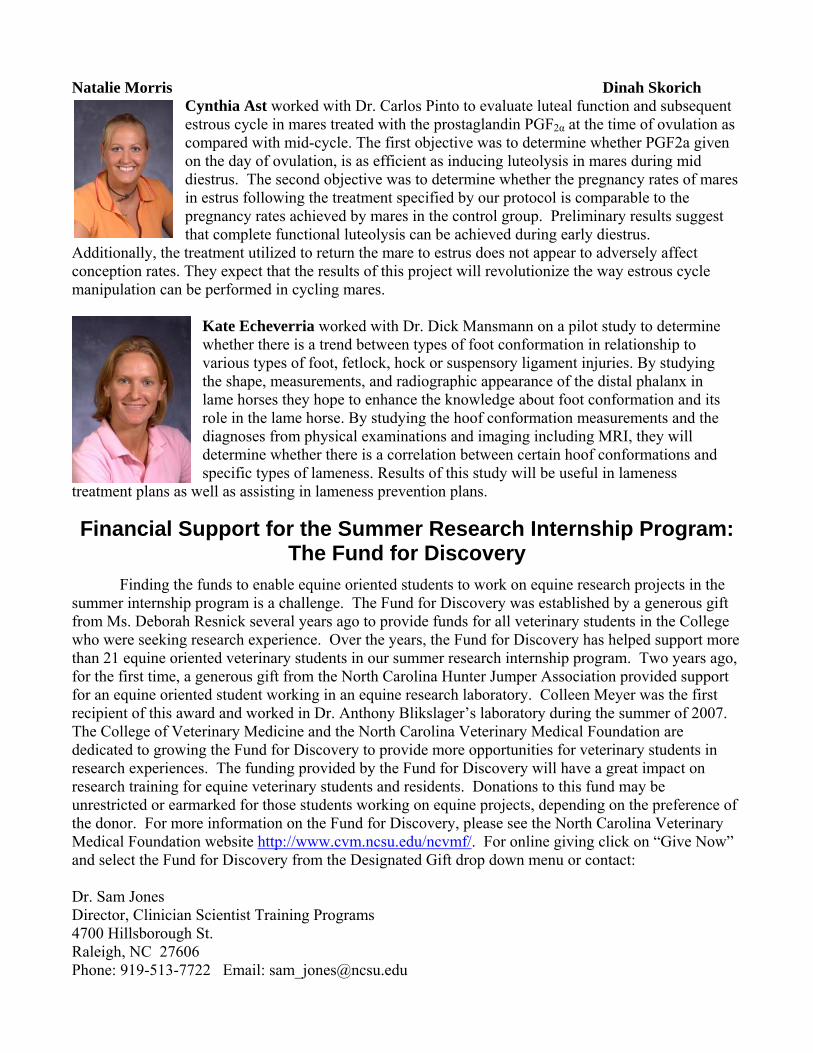
Heather Smith worked with Dr. Jones on a project examining the mechanisms that regulate the activity of enzymes responsible for producing prostaglandins during inflammation. The objective of the project was to determine whether specific molecules could be identified that control the activity of cyclooxygenase-2 during inflammation in equine leukocytes and whether these molecules are potential targets for anti-inflammatory therapy in horses. Heather showed for the first time that a molecule called NFκB is essential for upregulation of COX-2 activity in equine leukocytes and that new inhibitors of NFκB could be potentially useful anti-inflammatory drugs.
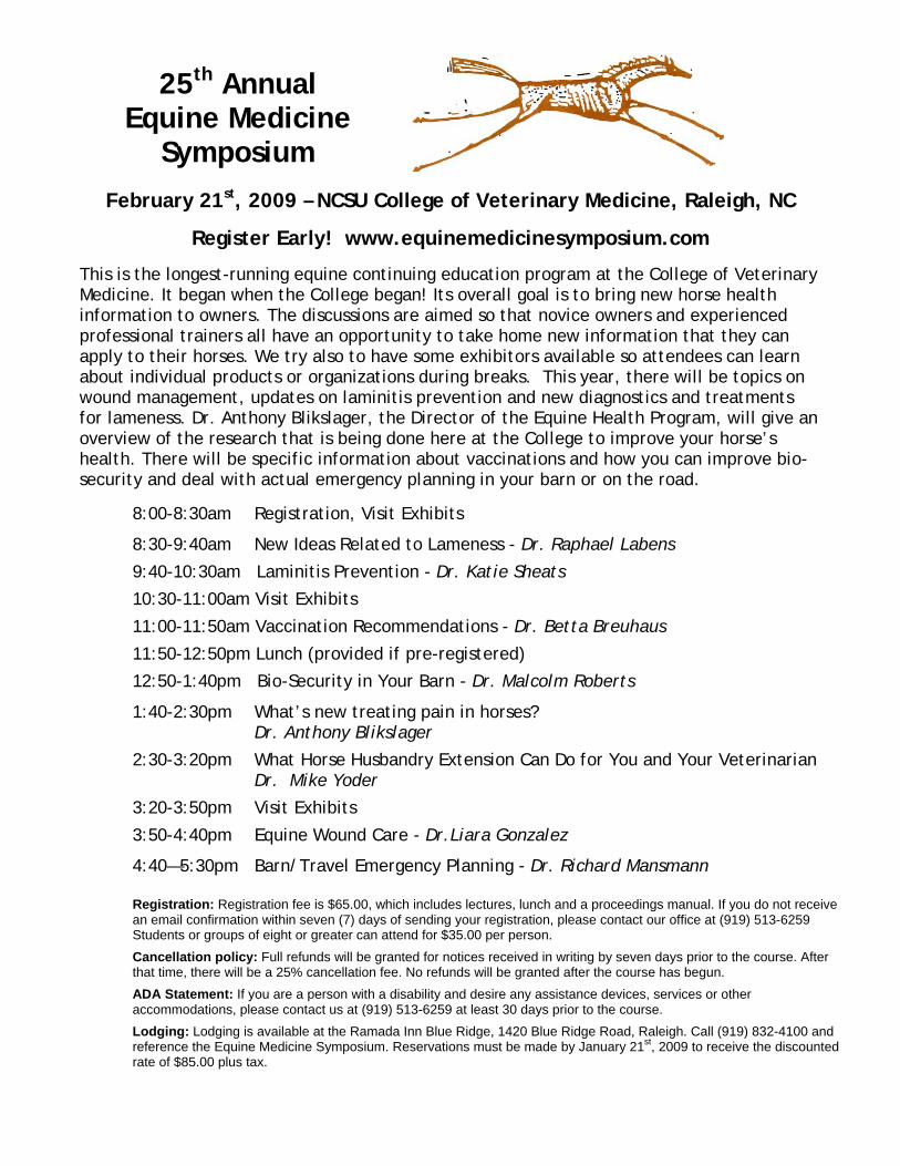
Natalie Morris and Dinah Skorich worked with Dr. Blikslager on a project examining whether robenacoxib, a selective cyclooxygenase-2 inhibitor, blocks production of prostaglandins involved in
inflammation and pain in the gut while allowing the intestinal lining (mucosa) to recover quickly after injury induced by temporarily cutting off the blood supply to the gut (intestinal ischemia). The results of this study suggest that robenacoxib will be a better alternative for pain and inflammation management in colic patients compared to the current standard of therapy flunixin meglumine.
Natalie Morris Dinah Skorich
Cynthia Ast worked with Dr. Carlos Pinto to evaluate luteal function and subsequent estrous cycle in mares treated with the prostaglandin PGF2α at the time of ovulation as compared with mid-cycle. The first objective was to determine whether PGF2a given on the day of ovulation, is as efficient as inducing luteolysis in mares during mid diestrus. The second objective was to determine whether the pregnancy rates of mares in estrus following the treatment specified by our protocol is comparable to the pregnancy rates achieved by mares in the control group. Preliminary results suggest that complete functional luteolysis can be achieved during early diestrus.
Additionally, the treatment utilized to return the mare to estrus does not appear to adversely affect conception rates. They expect that the results of this project will revolutionize the way estrous cycle manipulation can be performed in cycling mares.
Kate Echeverria worked with Dr. Dick Mansmann on a pilot study to determine whether there is a trend between types of foot conformation in relationship to various types of foot, fetlock, hock or suspensory ligament injuries. By studying the shape, measurements, and radiographic appearance of the distal phalanx in lame horses they hope to enhance the knowledge about foot conformation and its role in the lame horse. By studying the hoof conformation measurements and the diagnoses from physical examinations and imaging including MRI, they will determine whether there is a correlation between certain hoof conformations and specific types of lameness. Results of this study will be useful in lameness
treatment plans as well as assisting in lameness prevention plans.
Financial Support for the Summer Research Internship Program: The Fund for Discovery
Finding the funds to enable equine oriented students to work on equine research projects in the summer internship program is a challenge. The Fund for Discovery was established by a generous gift from Ms. Deborah Resnick several years ago to provide funds for all veterinary students in the College who were seeking research experience. Over the years, the Fund for Discovery has helped support more than 21 equine oriented veterinary students in our summer research internship program. Two years ago, for the first time, a generous gift from the North Carolina Hunter Jumper Association provided support for an equine oriented student working in an equine research laboratory. Colleen Meyer was the first recipient of this award and worked in Dr. Anthony Blikslager’s laboratory during the summer of 2007. The College of Veterinary Medicine and the North Carolina Veterinary Medical Foundation are dedicated to growing the Fund for Discovery to provide more opportunities for veterinary students in research experiences. The funding provided by the Fund for Discovery will have a great impact on research training for equine veterinary students and residents. Donations to this fund may be unrestricted or earmarked for those students working on equine projects, depending on the preference of the donor. For more information on the Fund for Discovery, please see the North Carolina Veterinary Medical Foundation website http://www.cvm.ncsu.edu/ncvmf/. For online giving click on “Give Now” and select the Fund for Discovery from the Designated Gift drop down menu or contact: Dr. Sam Jones Director, Clinician Scientist Training Programs 4700 Hillsborough St. Raleigh, NC 27606 Phone: 919-513-7722 Email: [email protected]
25th Annual Equine Medicine
Symposium
February 21st, 2009 – NCSU College of Veterinary Medicine, Raleigh, NC
Register Early! www.equinemedicinesymposium.com
This is the longest-running equine continuing education program at the College of Veterinary Medicine. It began when the College began! Its overall goal is to bring new horse health information to owners. The discussions are aimed so that novice owners and experienced professional trainers all have an opportunity to take home new information that they can apply to their horses. We try also to have some exhibitors available so attendees can learn about individual products or organizations during breaks. This year, there will be topics on wound management, updates on laminitis prevention and new diagnostics and treatments for lameness. Dr. Anthony Blikslager, the Director of the Equine Health Program, will give an overview of the research that is being done here at the College to improve your horse’s health. There will be specific information about vaccinations and how you can improve bio-security and deal with actual emergency planning in your barn or on the road.
8:00-8:30am Registration, Visit Exhibits
8:30-9:40am New Ideas Related to Lameness - Dr. Raphael Labens
9:40-10:30am Laminitis Prevention - Dr. Katie Sheats
10:30-11:00am Visit Exhibits
11:00-11:50am Vaccination Recommendations - Dr. Betta Breuhaus
11:50-12:50pm Lunch (provided if pre-registered)
12:50-1:40pm Bio-Security in Your Barn - Dr. Malcolm Roberts
1:40-2:30pm What’s new treating pain in horses? Dr. Anthony Blikslager
2:30-3:20pm What Horse Husbandry Extension Can Do for You and Your Veterinarian Dr. Mike Yoder
3:20-3:50pm Visit Exhibits
3:50-4:40pm Equine Wound Care - Dr.Liara Gonzalez
4:40—5:30pm Barn/Travel Emergency Planning - Dr. Richard Mansmann Registration: Registration fee is $65.00, which includes lectures, lunch and a proceedings manual. If you do not receive an email confirmation within seven (7) days of sending your registration, please contact our office at (919) 513-6259 Students or groups of eight or greater can attend for $35.00 per person.
Cancellation policy: Full refunds will be granted for notices received in writing by seven days prior to the course. After that time, there will be a 25% cancellation fee. No refunds will be granted after the course has begun.
ADA Statement: If you are a person with a disability and desire any assistance devices, services or other accommodations, please contact us at (919) 513-6259 at least 30 days prior to the course.
Lodging: Lodging is available at the Ramada Inn Blue Ridge, 1420 Blue Ridge Road, Raleigh. Call (919) 832-4100 and reference the Equine Medicine Symposium. Reservations must be made by January 21st, 2009 to receive the discounted rate of $85.00 plus tax.

Please support and thank our Equine Medicine Symposium 2007 Sponsors!
Winners lead the way to optimum nutrition.
www.nutrenaworld.com

OPHTHALMOLOGY SECTION
Bacteria in Eyes with Chronic Equine Recurrent Uveitis from the Southeastern United States
Brian C. Gilger, DVM, MS, Dipl. ACVO Professor of Ophthalmology Jacklyn Salmon, BS Ophthalmology Research Technician
Description of the Problem: Equine recurrent uveitis (ERU) is the leading cause of blindness in horses in the United States. ERU is characterized by recurrent bouts of active ocular inflammation followed by variable-length periods of quiescence. These painful recurrent bouts of inflammation continue until the eye becomes blind.
Study Objectives: The purpose of this study was to help determine the role of bacteria in the pathogenesis of ERU in horses from the Southeastern USA by evaluating known ERU eyes for presence of bacterial DNA using real-time PCR and for antibodies against Leptospira spp (by calculating GWC).
Experimental Approach: Broad-range ribosomal RNA quantitative PCR method (Real time PCR) was used to detect bacterial DNA in aqueous humor (AH) and vitreous humor (VH) from normal horses and horses from Southeastern USA with confirmed, chronic (>3 month), recurrent uveitis (ERU) (n=28). Results were compared to a standard curve of 16s ribosomal DNA ranging from 109 to 101. Aqueous humor and serum was also collected and evaluated for leptospiral titers (Microscopic agglutination testing, Cornell Univ) from normal horses, horses with non-ERU ocular inflammation (OI) (i.e., keratitis)(n=9) and horses with confirmed, chronic (>3 month), ERUAccomplishments/Results:
Bacterial DNA was not detected in AH or VH of ERU or normal samples. Serum leptospiral titers were negative (<1:400) in 91% of normal horses, 89% of horses
with other ocular inflammatory disease, and in 75% of ERU horses. AH leptospiral titers were negative (<1:400) in all non-ERU, 89% of other inflammatory disease, and in 83% ERU of horses. A Goldmann-Witmer coefficient (C-value) was ≤ 1 in all normal, in 89% of other inflammatory, and in 92% of ERU horses.
Benefits to the Equine Industry: Presence of bacterial DNA or leptospiral antigen does not appear to play a direct role in the persistence or recurrence of ERU in horses from the Southeastern USA. C-values ≤1 suggest that leptospiral-specific antibodies detected in aqueous humor were not produced intraocularly. Therefore these results do not support the theory that the production of antibodies against leptospiral antigens is a direct cause for ongoing or recurrent uveitis in the ERU-affected horses in this study. Furthermore, due to the low prevalence of positive results, the use of PCR or titers for bacteria in AH or serum are not useful methods for definitive diagnosis of ERU. This data has been accepted for publication in the American Journal of Veterinary Research

In Vitro and In Vivo Evaluation of an Equine Intraocular Lens Richard J. McMullen, DVM Resident in Ophthalmology Brian C. Gilger, DVM, MS, Dipl. ACVO Professor of Ophthalmology
Description of the Problem: Cataract surgery has become very successful in horses due to advancements in equipment and surgeon skill. An equine intraocular lens (IOL) needs to be developed to return a horse to normal ocular refractive state after surgery. Study Objectives: To determine refractive state and postoperative anterior chamber depth (PACD) of cadaver and live equine eyes implanted with IOLs to determine appropriate IOL power to achieve emmetropia.
Experimental Approach: Extracapsular lens extraction (i.e., cataract surgery) and placement of a +30D IOL was performed on 6 enucleated equine eyes. Four live horses received a +25D IOL implant after lens extraction. Streak retinoscopy and ultrasound (pre- and post-operative globe dimensions: ACD, CLT, and axial length [AxL]) was performed on each eye before and after IOL implantation.
Photograph of a horse with a synthetic intraocular lens in place.

Accomplishments/Results: Pre-operative mean refractive error of -0.46D (SD±1.03) was obtained from cadaver eyes. Pre-operative globe dimensions were: ACD: 7.12 mm (SD±0.82), CLT: 11.32 mm (SD±0.81), and AxL: 40.51 mm (SD±1.26). Post-operative anterior chamber depth (PACD): 10.76 mm (SD±1.16). No IOLs were positioned posterior to the pre-operative ACD + 50% CLT. Implantation of a +30D IOL resulted in mean overcorrection of 2.96D (SD±0.84) in enucleated globes. A +25D IOL in adult horses resulted in a mean overcorrection of 3.94D (SD±1.18) at 30 days after surgery. Benefits to the Equine Industry:
Due to a more anterior PACD than estimated previously, a +30D IOL overcorrected equine cadaver eyes. A +25D IOL overcorrected four live horses.
Further refractive and ultrasonographic data is needed to determine the appropriate refractive power and PACD of the IOL.
Once an equine intraocular lens (IOL) is developed, a horse would be returned to a normal ocular refractive state after surgery.
Research Supported by an American College of Veterinary Ophthalmologist VAF Grant 2006-2007. Study to be presented at the 2008 American College of Veterinary Ophthalmologists Annual Meeting, October 2008, Boston, MA.

Digital Infrared Photography of the Anterior Segment of the Equine Eye Richard J. McMullen, DVM Resident in Ophthalmology Brian C. Gilger, DVM, MS, Dipl. ACVO Professor of Ophthalmology
Description of the Problem: With cloudy ocular media (i.e., cornea, aqueous humor, lens, and vitreous humor) it is nearly impossible to evaluate the anterior segment of the eyes, especially the equine iris and lens. Digital ultraviolet (UV) cameras have recently become available. These cameras have been used extensively in the forensic fields, but only recently described in ophthalmology. The value of IR imaging in ophthalmology is that it allows better imaging of the relatively warmer structures in the eye, such as the uveal tract, and allows these structures to be examined. Study Objectives: To demonstrate benefits of digital infrared photography of the anterior segment of the equine eye.
Experimental Approach: Normal and diseased anterior segments were photographed with a digital infrared Nikon D70 camera converted by LifePixel (www.lifepixel.com) for infrared imaging (wavelength > 760 nm) and a Nikor 18-55 mm AF or 105 mm lens. Color images were taken with a Nikon D200 digital SLR camera with a Tamron 90 mm macro lens and a Nikon SB-800 flash.
Accomplishments/Results:
The normal, brown equine iris appeared grayish-blue with variable amounts of dark blue foci throughout the iridal surface.
The darker the original tissue, the deeper the blue color. Infrared images of heterochromic eyes appeared darker than color images.
Infrared images were rich in contrast, and the pupil was easily identifiable as a black ellipse surrounded by pupillary ruff.
Pigmented masses within the anterior chamber were readily visible even with corneal opacification.
Corneal edema was visually eliminated and some opacifications become transparent allowing for better evaluation of their position within the cornea and corneal vessels and other isolated opacifications appeared to be suspended above the iris and easily visualized.

Benefits to the Equine Industry:
Increased contrast using infrared photography and ability to visualize anterior iris through an opaque cornea was a valuable method to evaluate the pupil and assess effects of inflammation in eyes with uveitis and/or glaucoma.
This method may make diagnosis of equine ocular disease more accurate. Study to be presented at the 2008 American College of Veterinary Ophthalmologists Annual Meeting, October 2008, Boston, MA.
Figure 1. A. Digital photograph of a horse with keratitis and uveitis. The internal structures of the eye are not clearly visible. B. IR photograph of the same eye demonstrating excellent uveal detail. A. B.
Figure 2. A. Horse with a corneal stromal abscess and uveitis. The uveal track is poorly visible. B. IR photograph of the same eye with details of the iris visible. A. B

Quantitative Differences in mRNA Expression of Toll-Like Receptor (TLR)-2, -4, and -9 in Normal Equine Eyes and Eyes with Equine Recurrent Uveitis Na Young Yi, DVM, PhD, Post-doctorate fellow Ophthalmology Brian C. Gilger, DVM, MS, Dipl. ACVO Professor of Ophthalmology
Description of the Problem: Equine recurrent uveitis (ERU) is the most common cause of blindness in horses. The pathogenesis of the disease, its initation, and causes of recurrence is not known. Toll-like receptors (TLRs) detect various microbial components and play an important role in the host innate immunity. Study Objectives: This study was performed to evaluate the quantitative differences in mRNA expression of TLR-2, -4, and -9 in normal equine eyes and eyes with equine recurrent uveitis (ERU). Experimental Approach: Ocular tissues, including the ciliary body (normal: n=6; ERU: n=6), iris (normal: n=6; ERU: n=4), choroid/retina and corneal epithelium (normal: n=6; ERU: n=3) of normal eyes and eyes with naturally-occurring ERU were collected. Real time PCR assay was performed to compare mRNA expression of TLR-2, -4, and -9 between normal and ERU eyes. Western blot analysis and immunohistochemical localization are being performed currently.
Accomplishments/Results: A 3 to 4-fold elevation of TLR-2 and -9 mRNA in the iris and ciliary body and a 2
to 6-fold elevation of TLR-2, -4, and -9 mRNA in the choroid and retina from eyes with ERU were found compared to the mRNA levels in these same tissues of normal equine eyes. In the cornea there were no remarkable differences in the expression of the TLR-2, -4, and -9 between normal and ERU eyes.
This preliminary study demonstrated up-regulation of TLR-2 and-9 mRNA in the iris, ciliary body, and choroid/retina and up-regulation of TLR-4 mRNA in the choroid/retina of ERU eyes. The current data suggest the potential involvement of TLR-2, -4, and -9 in the pathogenesis of ERU.
Benefits to the Equine Industry: If TLRs are found to play a role in the pathogenesis or ERU, new, specific treatment can be developed to manage this blinding disease.
Study to be presented at the 2008 American College of Veterinary Ophthalmologists Annual Meeting, October 2008, Boston, MA.
LAMENESS SECTION
Diagnostic Findings in Horses with Proximal Plantar Metatarsal Pain - A Comparison of MRI and Ultrasonography Raphael Labens, DVM, Dec-EQ, CertES(Orth), MVM Rich Redding
DVM, DiplACVS Michael Schramme DVM,CertES(Orth), PhD, DiplECVS
Description of the Problem: Lameness in horses which improves significantly following local anaesthesia of the deep branch of the lateral plantar nerve (DBLPN) while anaesthesia of the plantar, plantar and dorsal metatarsal nerves (low six point) has little effect, is thought to originate from the proximal plantar metatarsus, more precisely from the area of the proximal suspensory ligament (PSL). Pain is frequently caused by desmitis of the proximal suspensory ligament (PSD) but recently neuritis of the DBLPN has also been proposed as a cause of proximal suspensory pain (PSP) and lameness (Toth et al 2008). Diagnosis of PSD or PSP is based on results with local anaesthesia and diagnostic imaging which may involve radiographic, scintigraphic, ultrasonographic or magnetic resonance imaging (MRI) techniques. Ultrasonography (US), being the most available soft tissue imaging modality enables accurate diagnosis of PSD (Dyson 1994). However, accurate ultrasonographic definition of structures in the proximal plantar metatarsal region can be extremely difficult. Therefore MRI has been considered the gold standard for detection of soft tissue injury due to its high soft tissue detail and contrast. An objective comparison of US, low field MRI and post mortem histology has been reported for healthy Warmblood horses (Bischofberger et al. 2005). For horses with PSD comparison of the two diagnostic imaging modalities has not yet been performed.
Study Objectives: This retrospective study focused on the diagnostic findings in horses with proximal plantar metatarsal pain and, using qualitative and quantitative methods, compares US to high field MRI for its accuracy in diagnosing PSD.
Experimental Approach: Retrospective study design, using the hospital population and records of clinical and diagnostic findings for analysis.

Accomplishments/Results: Results of this retrospective study show that ultrasonographic examination of the proximal metatarsus for detection of PSD is of limited accuracy when compared to MRI findings. MRI and ultrasonographic measurements of cross sectional surface area were significantly different at 2 and 4 but not at 6cm distal to the head of the fourth metatarsal bone. The privileged anatomic location of the PSL embedded between the 4th and 2nd metatarsal bone complicate ultrasonographic examination of the entire structure. At the distal level (6cm) the PSL becomes more accessible which allows better ultrasonographic visualisation and explains the degree of correlation. This retrospective study has shown that ultrasonographic examination of the PSL for diagnosis of PSD carries a fair sensitivity (0.75-0.77) but unacceptable specificity (0.36-0.4). The sensitivity for accurate lesion localisation is poor (0.63-0.66). Clinical and diagnostic findings have also shown that anaesthesia of the DBLPN has the ability to desensitize a larger anatomical area than presumed. Benefits to the Equine Industry: Accurate diagnostic treatment modalities are paramount when selecting appropriate treatments. Ultrasonographic findings are of limited accuracy to determine the appropriate management of horses with PSP or PSD. MRI examinations should be preferred.
Evaluating the Role of Foot Conformation and Internal Anatomy in the causes of Equine Foot Lameness Using Photographs, Radiographs and Magnetic Imaging
R. A. Mansmann, VMD, PhD K. Echeverria, DVM Student, 2010
Description of the Problem Equine lameness can be very a frustrating and time-consuming problem. Lameness can result in financial loss for the owners as well as time away from the competition venue. According to a 1998 USDA study, lameness was by far the most significant cost to owners evaluated at $678 million due to death, loss of use and veterinary services. The most common areas of lameness is in the foot, fetlock, hock and suspensory ligaments. The hypothesis of our project is that poor hoof conformation can be related to lameness etiologies within the foot. Study Objectives • To determine a standardized examination form and
imaging techniques that can be used for a data bank for lameness studies related to the equine foot.
• To determine if there is a trend between types of foot conformation in relationship to various types of foot injuries and lameness. By studying the shape, measurements and radiographic appearance of the hoof and distal phalanx in lame horses we hope to enhance the knowledge about foot conformation and its role in the lame horse.
Experimental Approach: Horses referred to NCSU CVM for an MRI are asked to participate in the study. MRI horses where chosen as study participants because they potentially have the most detailed lameness workups since it is imperative to have the 12 sq. cm anatomical area of interest isolated for the MRI. At least 2 distal posterior digital nerve blocks need to have been done to verify the source of the lameness within the foot. The sample group is from the eastern United States mainly North Carolina and are mostly competition horses. The day prior to the MRI hoof measurements, photographs and radiographs of all four shod feet are taken. Using the software program ‘E-Films’ measurements are taken from the lateral radiograph to determine the anatomy of the distal phalanx in relation to the hoof capsule. The measurements of interest from the lateral radiograph are; sole depth, H/L zone, sole plane angle, breakover to tip of distal phalanx and percent support of the shoe or trim of the DIP joint. The MRI is performed at NCSU CVM in the 1/5 Tesla IAMS Siemens Symphony MRI unit. After the MRI, a full written report of the MRI findings including final diagnoses is added to the study record.

Currently we are working with three potential sets of controls for this project. The first set of controls is to compare our data in to spare literature on the front feet lateral radiographic measurements of sound horses and an unpublished study evaluating front feet conformation on sound, low level competition and pleasure horses in work residing in central North Carolina. The measurements of the front feet taken in this MRI/foot conformation study are the same measurements in the previous unpublished study. The second set of controls is to determine some normal measurements of hind feet of sound horses. Currently there is no significant literature describing measurements of the radiographic anatomy of the hind feet in horses especially as they relate to front feet conformation and imaging. The authors have a small number of low level exercising sound horses with all four feet radiographed for measuring. Also in another pilot study, horses were examined with or without palpable gluteal pain solely in relationship to breakover, so this particular measurement could be helpful in determining a trend toward a normal hind foot breakover measurement. Lastly, another area of controls in this study is to determine the effect of various positioning of the camera and radiographic machine in relation to the equine foot and its effect on specific values of the above measurements. We are using the software program ‘Epona Tech Metron’ to assist in verifying the accuracy of the photographic measurements. Accomplishments/Results: We have funding for 50 cases in this pilot study. We have twenty cases finished. They have established the standardization of our forms, photographs and radiographs. Benefits to the Equine Industry: Potentially studies determining what is the normal horse’s feet conformational anatomy and relating conformation to lameness can be useful in lameness treatment plans as well as assisting in lameness prevention plans. Also this type of information may help in pre-purchase and breeding situations.
Fig. 1 Metron program overlay of lateral radiograph over photograph of a foot. This horse has a positive sole plane angle of its coffin bone (normal), a normal dorsal wall thickness, but a minimal sole depth of 10mm and a very extended breakover in front of the coffin bone of 34mm. Funded by: The North Carolina Horse Council
COLIC SECTION
Analysis of Sodium Carboxymethylcellulose Administration and Related Factors Associated with Postoperative Colic and Survival in Horses with Small Intestinal Disease
Callie Fogle, DVM, DACVS Clinical Assistant Professor of Equine Surgery Mathew Gerard, BVSc, PhD, DACVS Yvonne Elce, DVM, DACVS Dianne Little, BVSc, PhD, DACVS Alison Morton, VMD, MSpVM, DACVS Maria Correa, MS, PhD Anthony Blikslager, DVM, PhD, DACVS Published in Veterinary Surgery, 37:501-506, 2008
Description of the Problem: In the horse, surgery for disease of the small intestine is associated with a higher rate of postoperative complications and consequently a lower rate of survival when compared to surgery for disease of the large intestine. Postoperative colic occurred in 28.1% of horses recovering from colic surgery in one recent study, and was the most frequent postoperative complication prior to discharge. Adhesions cause a clinical problem in 15-22% of horses recovering from surgery of the small intestine. Serosal trauma, ischemia, distension, and resulting inflammation, are common in surgery of the small intestine, and have crucial roles in the development of adhesions. Experimental use of carboxymethylcellulose suggests that this product may decrease adhesion formation in the horse.
Study Objectives To retrospectively analyze the effect of the intraoperative use of sodium carboxymethylcellulose (CBMC) and related perioperative factors on postoperative colic and survival in horses undergoing colic surgery. Experimental Approach: Animals-203 horses that had surgery for disease of the small intestine, 33 of which had intraoperative use of CBMC. Materials and Methods--Information from medical records was obtained for 33 cases with

intraoperative use of CBMC and 170 cases prior to the use of CBMC. Kaplan-Meier survival curves were used to estimate median survival time and the Cox Proportional Hazards Model to estimate the hazard ratio for the effect of CBMC and other perioperative variables on survival.
Accomplishments/Results: Seventy-five percent of horses in which CBMC was used survived to 180 days, compared to 75% of untreated horses surviving 8 days. (The median survival time for this group was 18 days). Horses that did not receive CBMC were twice as likely to die compared to horses in which CBMC was used. Horses that suffered from postoperative ileus were 1.4 times more likely to die than horses without ileus. Similarly, horses with colic after surgery were 1.3 times more likely to die than horses without colic postoperatively. Benefits to the Equine Industry: Conclusions--The results suggest that CBMC is protective against death and prolongs survival when used intraoperatively in horses with disease of the small intestine, particularly in horses with postoperative colic or postoperative ileus. Both ileus and colic placed horses at an increased risk of death after surgery. Clinical Relevance--The intraoperative use of CBMC in surgery of the small intestine may improve survival postoperatively, possibly by reducing early adhesion formation. Testing the efficacy of new and old drugs for treatment of colic: COX-2 inhibitors and lidocaine
Anthony T. Blikslager, DVM, PhD, DACVS Professor of Equine Surgery Vanessa Cook, MA, VetMB, PhD, DACVS Research Associate, Equine Gastroenterology
Description of the Problem: Colic is the leading cause of death in horses behind old age. This is principally because of strangulating obstruction, during which lipopolysaccharide (LPS) is absorbed because of degeneration of the gut mucosa. Current treatments prior to and after surgery include non-steroidal anti-inflammatory drugs such as flunixin meglumine (Banamine®) which ameliorates
LPS-induced shock, but also slows down repair of injured intestinal mucosa. Possible solutions to this problem include use of cyclooxygenase (COX)-2 inhibitors, which more specifically target the enzyme responsible for shock or the use of
lidocaine. The latter has been used as a local anesthetic for years, but has recently been suggested to have anti-inflammatory properties.
Study Objectives: To determine if COX-2 inhibitors or lidocaine promote repair of the intestine while providing optimal pain control in horses with colic Experimental Approach: Horses donated specifically for this research were taken to surgery. Intestine was subjected to 2-hours of ischemia, after which it was reperfused and horses were recovered. All horses were treated with the analgesic butorphanol. In addition, horses were treated with either intravenous flunixin, firocoxib (Equioxx®), or lidocaine. Horses were carefully assessed for any behavioral evidence of colic. At 18-hours following the surgery, normal and recovering intestinal mucosa was placed in Ussing chambers for evaluation of COX expression, prostaglandin production, and permeability to LPS. Accomplishments/Results:
COX-1 and COX-2 are expressed in normal intestine, and are upregulated following injury. This expression pattern is unusual in that COX-1 is typically the only isoform expressed in normal intestine, whereas COX-2 is the only isoforms that is upregulated.
Flunixin completely inhibited COX-produced prostaglandins, whereas firocoxib only inhibited prostaglandins produced by COX-2. Lidocaine had no effect on prostaglandin production.
Horses treated with either flunixin or firocoxib had significantly reduced behavioral scores as compared to horses on butorphanol alone. Lidocaine partially reduced behavioral scores, but not to the same extent as the non-steroidal anti-inflammatory drugs
Horses treated with flunixin had increased mucosal permeability to LPS. This was prevented when horses were treated with both lidocaine and flunixin. Alternatively, firocoxib allowed full recovery of the intestinal mucosal.
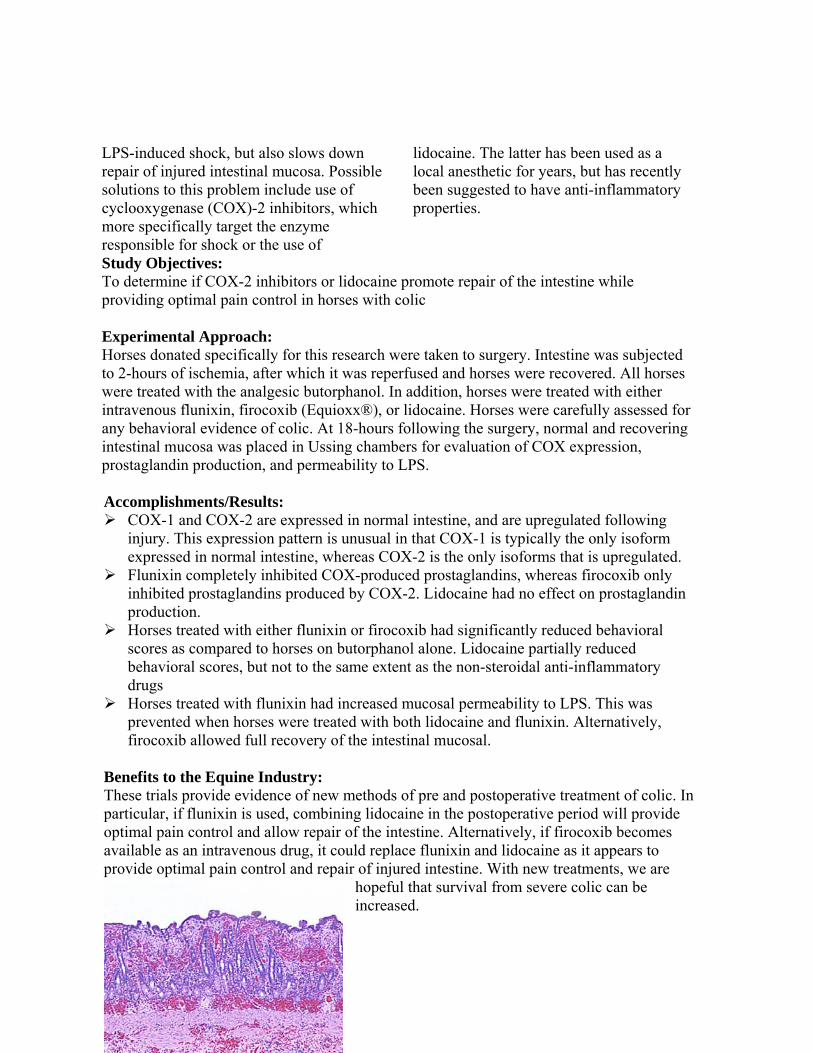
Benefits to the Equine Industry: These trials provide evidence of new methods of pre and postoperative treatment of colic. In particular, if flunixin is used, combining lidocaine in the postoperative period will provide optimal pain control and allow repair of the intestine. Alternatively, if firocoxib becomes available as an intravenous drug, it could replace flunixin and lidocaine as it appears to provide optimal pain control and repair of injured intestine. With new treatments, we are
hopeful that survival from severe colic can be increased.
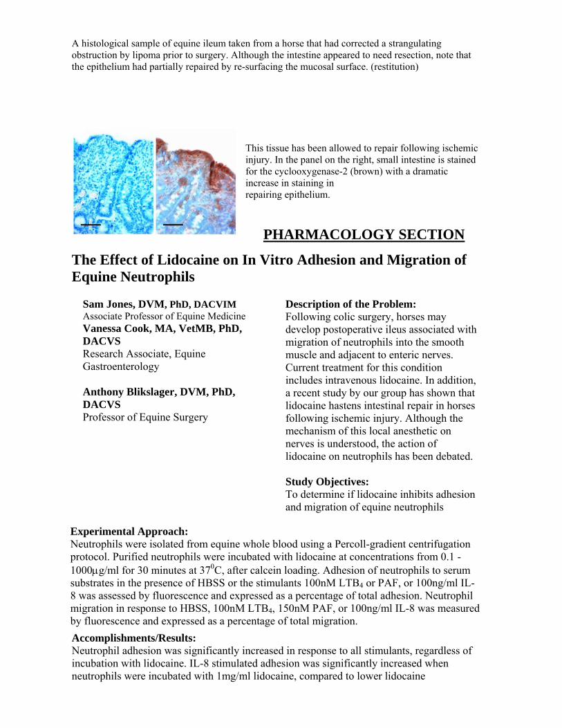
A histological sample of equine ileum taken from a horse that had corrected a strangulating obstruction by lipoma prior to surgery. Although the intestine appeared to need resection, note that the epithelium had partially repaired by re-surfacing the mucosal surface. (restitution)
This tissue has been allowed to repair following ischemic injury. In the panel on the right, small intestine is stained for the cyclooxygenase-2 (brown) with a dramatic increase in staining in repairing epithelium.
PHARMACOLOGY SECTION
The Effect of Lidocaine on In Vitro Adhesion and Migration of Equine Neutrophils
Sam Jones, DVM, PhD, DACVIM Associate Professor of Equine Medicine Vanessa Cook, MA, VetMB, PhD, DACVS Research Associate, Equine Gastroenterology Anthony Blikslager, DVM, PhD, DACVS Professor of Equine Surgery
Description of the Problem: Following colic surgery, horses may develop postoperative ileus associated with migration of neutrophils into the smooth muscle and adjacent to enteric nerves. Current treatment for this condition includes intravenous lidocaine. In addition, a recent study by our group has shown that lidocaine hastens intestinal repair in horses following ischemic injury. Although the mechanism of this local anesthetic on nerves is understood, the action of lidocaine on neutrophils has been debated. Study Objectives: To determine if lidocaine inhibits adhesion and migration of equine neutrophils
Experimental Approach: Neutrophils were isolated from equine whole blood using a Percoll-gradient centrifugation protocol. Purified neutrophils were incubated with lidocaine at concentrations from 0.1 -1000μg/ml for 30 minutes at 370C, after calcein loading. Adhesion of neutrophils to serum substrates in the presence of HBSS or the stimulants 100nM LTB4 or PAF, or 100ng/ml IL-8 was assessed by fluorescence and expressed as a percentage of total adhesion. Neutrophil migration in response to HBSS, 100nM LTB4, 150nM PAF, or 100ng/ml IL-8 was measured by fluorescence and expressed as a percentage of total migration.
Accomplishments/Results: Neutrophil adhesion was significantly increased in response to all stimulants, regardless of incubation with lidocaine. IL-8 stimulated adhesion was significantly increased when neutrophils were incubated with 1mg/ml lidocaine, compared to lower lidocaine

concentrations. LTB4 stimulated adhesion was significantly increased when neutrophils were incubated with 1mg/ml lidocaine compared to that at 5μg/ml lidocaine. Migration was significantly increased in response to IL-8 at all lidocaine concentrations, and in response to LTB4 at lidocaine concentrations >1μg/ml. In conclusion, lidocaine did not inhibit neutrophil migration or adhesion in vitro at therapeutic concentrations, and increased migration and adhesion at higher concentrations.
Benefits to the Equine Industry: Use of lidocaine for treatment of horses following colic surgery has dramatically increased in recent years. Lidocaine was initially used because of a perceived benefit in horses with postoperative ileus, but has also been used because of evidence that it enhances intestinal repair. Although these benefits of lidocaine have been shown, this study indicates that lidocaine does not work by inhibiting neutrophil adhesion or migration. Therefore, further work is needed to determine the mechanisms of lidocaine activity. Pharmacokinetics of intravenous, intramuscular and sublingual buprenorphine in horses
Jennifer L Davis, DVM, PhD, DACVIM, DACVCP Assistant Professor of Equine Medicine Kristen Messesnger, DVM Anesthesiology Resident D. Heath LaFevers, BS Equine Medicine Research Technician
Description of the Problem: Pain management and the recognition of pain are essential components of veterinary medicine, and are of extreme importance in animal welfare to reduce patient morbidity and mortality. Equine analgesia has historically been limited primarily to nonsteroidal anti-inflammatory drugs (NSAIDs) and alpha-2 agonists. While NSAIDs are effective for the management of mild to moderate pain, they are also associated with side effects such as gastrointestinal ulceration and kidney damage. Alpha-2 agonists are associated with heavy sedation and cardiovascular depression. In some instances, veterinarians must rely on other classes of drugs to alleviate pain, particularly the opioid analgesics. However, the opioid drugs commonly used in horses have a short duration of action and require frequent injections. Overall, there is a lack of analgesic options available for owners to administer to their horses for the treatment of severe pain, which necessitates the investigation of longer acting, more potent drugs and alternative drug delivery methods. Buprenorphine has the potential to be an effective, easily administered alternative analgesic in horses with acute or chronic pain that cannot tolerate other forms of pain medication. Study Objectives:
To describe the absorption (bioavailability), distribution, metabolism and excretion of intravenous (IV), Intramuscular (IM) and sublingual administration of buprenorphine in healthy horses. To evaluate the side effects of buprenorphine administered by the IV, IM and sublingual routes in the horse.
Experimental Approach:
For the IV and sublingual study, 5 healthy horses were used in a crossover design and administered 0.006 mg/kg buprenorphine IV and sublingually. For the IV and IM study, 6 healthy adult horses were used in a crossover design and administered 0.005 mg/kg buprenorphine IV and IM. Heparinized blood samples were drawn via an indwelling jugular catheter immediately prior to drug administration, and then at 10, 20, 30, 45, and 60 minutes, and 1.5, 2, 4, 6, 8, 12, and 24 hours after drug administration. Plasma was harvested and the samples were stored at -80ºC until analysis by ultra-high pressure liquid chromatography with mass spectrometry (UPLC-MS). Horses were monitored continuously for the first 4 hours for signs of abnormal behavior, and then at every sampling point after that for the duration of the study. Temperature, pulse, respiratory rates, and gut sounds were monitored prior to drug administration, one hour after drug administration and at each sampling point thereafter. Fecal and urine output were also monitored throughout the study. Accomplishments/Results:
Several horses developed signs of CNS excitement after administration of IV or IM buprenorphine. These signs included muscle tremors and continuous stall walking, pawing and sweating. The severity of the signs varied with each individual horse. No horses in the sublingual group exhibited any signs of CNS excitement.
A decrease in gastrointestinal sounds was noted after IV and, to a lesser extent IM administration of buprenorphine in all horses. The time to first passage of feces after IV and IM administration was increased compared to sublingual. However, the total number of fecal piles passed was similar among all treatment groups.
A highly sensitive and specific UPLC-MS method has been developed for the analysis of the plasma samples, with a limit of detection of 0.1 ng/mL.
Sample analysis is ongoing. Benefits to the Equine Industry: Based on the preliminary results of this study, sublingual buprenorphine has the potential to be used as an alternative analgesic drug in horses with minimal side effects and would be easy and practical for owners and veterinarians to administer.


Population Pharmacokinetics of Enrofloxacin in Horses Jennifer L Davis, DVM, PhD, DACVIM, DACVCP Assistant Professor of Equine Medicine Katie Sheats, DVM Equine Medicine Resident Mark G. Papich, DVM, MS, DACVCP
Description of the Problem: Reports on the pharmacokinetics (PK) of enrofloxacin in horses have been published; however these reports studied the pharmacokinetics of the drug in a small population of healthy, homogenous animals following the collection of a large number of samples from each animal. There are currently no reports of the PK of enrofloxacin in diseased or hospitalized patients. Population pharmacokinetics is a tool used to assess intra- and inter-individual variability in drug disposition among a population of patients in a clinical environment. Pharmacodynamic (PD) principles can also be incorporated into the analysis. Population PK modeling is ideal for horses in a hospital setting, as it uses fewer samples from a larger number of patients. By studying the population PK of enrofloxacin in horses, we will be better able to determine a dosing regimen that will be effective in our target population, based on commonly used pharmacodynamic indices. Study Objectives:
To determine the population pharmacokinetics of enrofloxacin and its active metabolite, ciprofloxacin, in horses following intravenous administration.
To determine the dosing regimen of enrofloxacin that would most likely be effective in a hospitalized patient population, based on Monte Carlo simulations and the known pharmacokinetic-pharmacodynamic interactions of fluoroquinolone antibiotics.
Experimental Approach: Horses admitted to the large animal teaching hospital that were subsequently placed on enrofloxacin therapy for treatment of any disease process were included. A minimum of 21 horses will be included in the study. Each horse had a maximum of 5 blood samples drawn, including a pre-treatment sample. Plasma was harvested and frozen at -70ºC until analysis. All drug concentrations were analyzed using high-pressure liquid chromatography (HPLC) with fluorescence detection based on a method that has been previously validated and published. Additional information was collected regarding signalment, disease status, concurrent medications, culture and sensitivity testing. Pharmacokinetic parameters were generated using a specialized computer software program (WinNonlin and WinNonMix, Pharsight Corporation, Cary, NC). Monte Carlo simulations (17) will be performed using a software program (Crystal Ball, Decisioneering Inc. Denver, CO). This allows us to define pharmacokinetics variables that vary within the population as well as decision variable cells that contain the values that are within our control to change (dose and interval). Finally, predictions in the population can be generated, for values such as plasma concentrations or PK-PD parameters.
Accomplishments/Results:
Initial results indicate that hospitalized horses have a much higher variability in the distribution, metabolism and excretion of enrofloxacin, compared to data previously reported in healthy horses.
The half-life of the drug appears prolonged in horses with liver disease and those exhibiting signs of dehydration and/or kidney dysfunction.
Additional horses need to be monitored to determine the significance of these effects. Data is available from only 19 horses at this point.
Benefits to the Equine Industry: Population pharmacokinetic modeling of enrofloxacin (and its metabolite ciprofloxacin) will produce predictive models of the disposition of these drugs in equine clinical patients. Other factors that affect pharmacokinetics (covariates in the model) can be identified in order to modify therapy in sub-populations of treated animals. These differences will result in modifications to current dosing regimens for hospitalized patients.
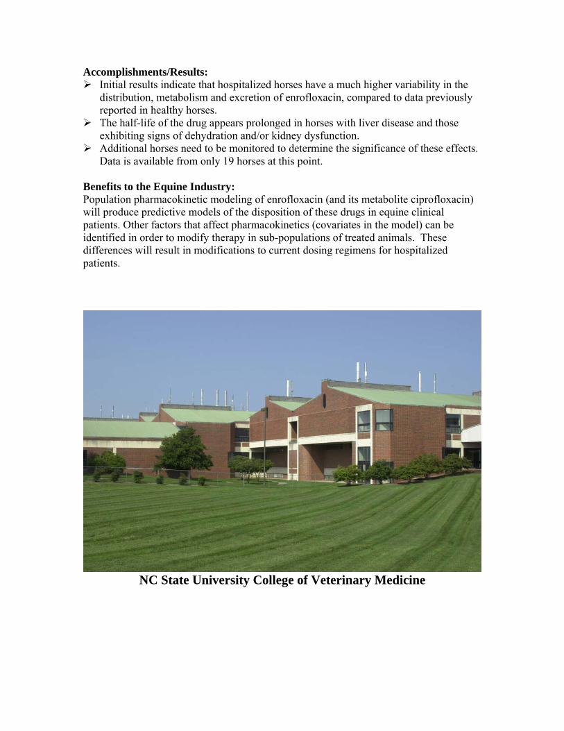
NC State University College of Veterinary Medicine

The Effects of Compounding and Storage Conditions on the Stability of Pergolide Mesylate Jennifer L Davis, DVM, PhD, DACVIM, DACVCP Assistant Professor of Equine Medicine Loren Madden Kirk Gigi Davidson, RPh, DICVP Mark G. Papich, DVM, MS, DACVCP
Description of the Problem: Pergolide mesylate is an orally administered drug used to treat equine Cushing’s syndrome. In March 2007, the FDA announced a voluntary withdrawal of human products containing pergolide due to concerns over a link between its use and the development of cardiac side effects, including valvular dysfunction, in people. Because there are no products containing pergolide registered for administration to horses, this creates a dilemma for veterinarians treating horses with this drug. In order to address the concerns over the availability of pergolide to treat horses with Cushing’s, the FDA Center for Veterinary Medicine is working with the drug’s manufacturers to make the product available for use in the horse. This includes making a special exception in the case of pergolide so that products compounded from bulk powders will be considered to be under discretionary enforcement by the FDA. Such products will be allowed, but will still be closely monitored for the development of any adverse or unexpected side effects. The stability of liquid preparations of pergolide when stored under different storage conditions after being admixed with vehicles commonly used by veterinary compounding pharmacies, has not been reported, to the authors’ knowledge.
Study Objectives:
The purpose of this experiment was to prepare a compounded aqueous formulation of pergolide mesylate, and subject the formulation to storage conditions that would be consistent with caregiver administration of this drug to horses.
Following the preparation and storage, we determined the stability of pergolide, including the effects of temperature and light, over a 35 day period.
Experimental Approach: The compounded suspension of pergolide was prepared by a licensed pharmacist using standard compounding pharmacy protocols at a concentration of 1 mg/mL. 500μL of the suspension was dispensed into 30 separate 5mL freezer storage tubes. Each tube was appropriately labeled and stored in their respective storage environments, which included heat (37°C), room temperature (25-27°C), room temperature with light exposure, refrigeration (8°C), and freezer (-20°C). The samples exposed to light were stored in a clear, zip lock bag. The heated, room temperature, refrigeration, and freezer samples

0 7 14 21 28 35 42
Days after compounding
0
0.2
0.4
0.6
0.8
1.0
1.2
Conc
entra
tion
(mg/
mL)
- - -20°C- - 8°C- - 25°C- - 37°C- - Light
were stored in a brown, light protective zip lock bag. For each day of analysis, one sample from each storage environment was brought to room temperature before being analyzed. Analysis was performed by high pressure liquid chromatography immediately after compounding (Day 0) and 1, 7, 14, 21, and 35 days after compounding. At each time point, a visual inspection of the samples was made to detect any changes in color. Accomplishments/Results
The initial concentrations of pergolide mesylate in the compounded formulation tested were 1.05 ± 0.08 mg/mL.
The concentrations had undergone excessive degradation by day 14 when exposed to light, day 21 when stored at 37ºC, and day 35 at room temperature (Figure 1).
The decrease in expected concentrations corresponded with the appearance of degradation peaks in the chromatograph, as well as a color change in the formulation (Figure 2).
Benefits to the Equine Industry: Commercial products of pergolide mesylate are not currently available; therefore the FDA has issued a limited exemption for compounding pergolide from bulk substances, so that horses can continue receiving appropriate therapy. The results of this study indicate that pergolide mesylate is not stable in an aqueous suspension. Additionally, degradation of the drug occurs more rapidly when exposed to light or high temperatures. It is recommended that formulations be stored under refrigerated conditions, and protected from light. Formulations that have undergone a color change should be considered unstable and be discarded.
Figure 1. Concentration versus time curves of pergolide mesylate in an aqueous vehicle under different storage conditions.
Figure 2. Color change reaction occurring in pergolide mesylate formulations 35 days after compounding under different storage conditions.

IMMUNOLOGY SECTION
Circulating plasma exosomes: nanometer lipid particles bearing IgE and tumor necrosis factor receptor Bruce Hammerberg, DVM, PhD Professor of Immunoparasitology
Description of the Problem: Diagnosis of allergic disease in horses is facilitated by measurement of circulating blood levels of antibodies of the immunoglobulin subclass IgE. IgE antibodies activate mast cells and basophils to release potent inflammatory mediators at various tissue sites in response to allergens and this results in the clinical manifestations of allergies; such as hives, chronic obstructive pulmonary disease or dermatitis. The problem is that, as in humans, allergen-specific IgE measurements often do not correlate well with allergic disease manifestation and the reason for this is not known. Study Objectives: To determine the role of nanometer size lipid particles bearing IgE and other cell membrane proteins in the accurate measurement of circulating IgE. Experimental Approach: Affinity chromatography with immobilized antibodies against the receptor for tumor necrosis factor alpha (TNFRI) was performed on sera from horses with high levels of circulating IgE. The material bound by these antibodies was isolated and analysed by enzyme linked immunoadsorption assay (ELISA) for IgE and TNFRI content. Transmission electron microscopy was conducted to demonstrate the presence of lipid particles between 30 and 100 nanometers in size (see figure)
Accomplishments/Results: Exosomes have been isolated from sera of horses with allergic disease and high levels of serum IgE. These exosomes bear receptors for the inflammatory mediator TNF-alpha, IgG as well as IgE. The aggregation of these immunoglobulins and possible inflammatory mediators on nano-particles, and their presence in circulation may affect the accurate measurement of serum IgE and may also play a role in the pathogenesis of allergic diseases. Benefits to the Equine Industry: The discovery of a previously unrecognized circulating aggregation of immunoglobulins on exosomes may facilitate improved detection and diagnosistic value of serum IgE. Analysis of exosomes could reveal how co-aggregated allegen-specific IgE and inflammatory mediators are distributed to tissue sites where allergic diseases are manifested.
