endoscopic approach of juvenile nasopharyngeal...the sphenoidal extension of the angiofibroma can be...
TRANSCRIPT
Remedy Publications LLC.
World Journal of Vascular Surgery
2018 | Volume 1 | Issue 1 | Article 10051
Endoscopic Approach of Juvenile Nasopharyngeal Angiofibroma - A Sustained Surgical Challenge
OPEN ACCESS
*Correspondence:Vlad Andrei Budu, Department of ENT Microsurgery, "Prof Dr Dorin Hociota"
Institute for Phono-Audiology and Functional ENT Surgery, Bucharest,
Romania,E-mail: [email protected] Date: 11 Feb 2018Accepted Date: 19 Feb 2018Published Date: 26 Feb 2018
Citation: Budu VA. Endoscopic Approach of
Juvenile Nasopharyngeal Angiofibroma - A Sustained Surgical Challenge. World
J Vasc Surg. 2018; 1(1): 1005.
Copyright © 2018 Vlad Andrei Budu. This is an open access article
distributed under the Creative Commons Attribution License, which permits unrestricted use, distribution,
and reproduction in any medium, provided the original work is properly
cited.
Surgical TechniquePublished: 26 Feb, 2018
IntroductionJuvenile Angiofibroma (JNA) is a benign vascular tumor arising usually from the posterior part
of the nasal cavity (pterygoid region), which appears at young male teenagers. The tumor has a destructive growth in the surrounding areas; therefore the management of JNA needs to be initiated in early stages of tumor development.
The ethiopathology of JNA is controversial and not yet completely explained. There are several theories to be considered. Monoclonal factors like exogenous testosterone can induce tumor hypertrophy [1]. There is also a genetic theory of JNA appearance which is still to be proved by studies on large series of angiofibomas [2]. There are authors who try to demonstrate the role of viral infections (Herpes simplex virus, Epstein Barr virus, HPV) in the appearance and growing of JNA [3]. The vascular theory, which reveals the over expression of receptors for vascular endothelial growth factor in presence of JNA, can explain the development of this tumor [4].
Preoperative ProtocolFirst step in this pathology is to have an accurate diagnosis. In every patient of our series we
counted on clinical history, endoscopic exam and CT imaging. Unilateral nasal obstruction with moderate growth, associated with repetitive unilateral expistaxis, facial pressure, headache and conductive hearing loss on the same side, which appeared at an adolescent man in the last 6 months, determined us to take into account a JNA.
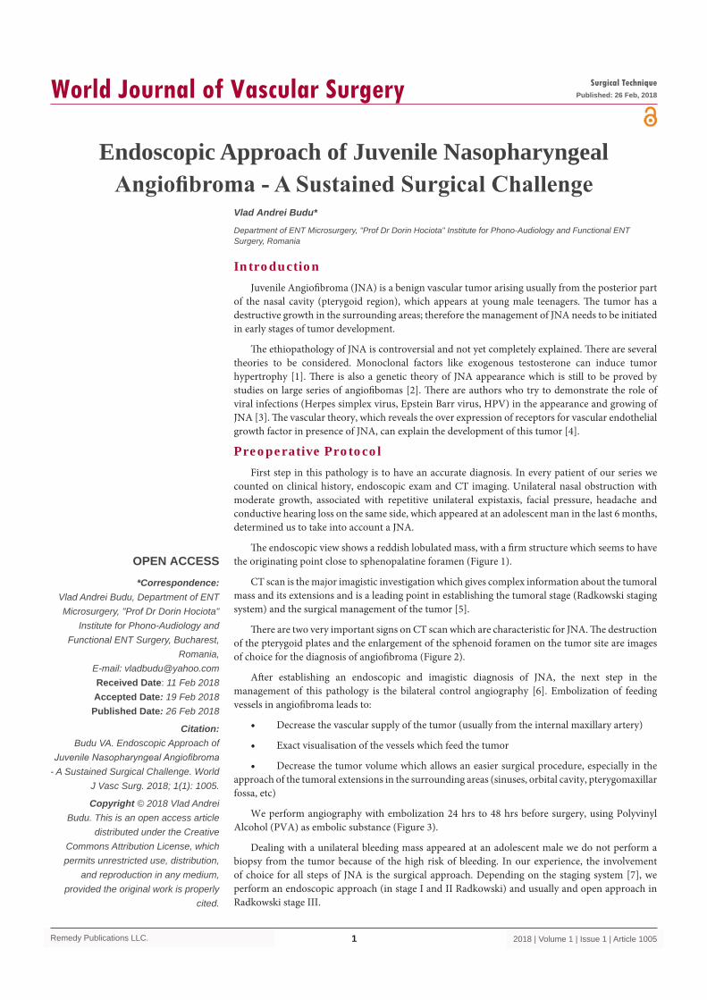
The endoscopic view shows a reddish lobulated mass, with a firm structure which seems to have the originating point close to sphenopalatine foramen (Figure 1).
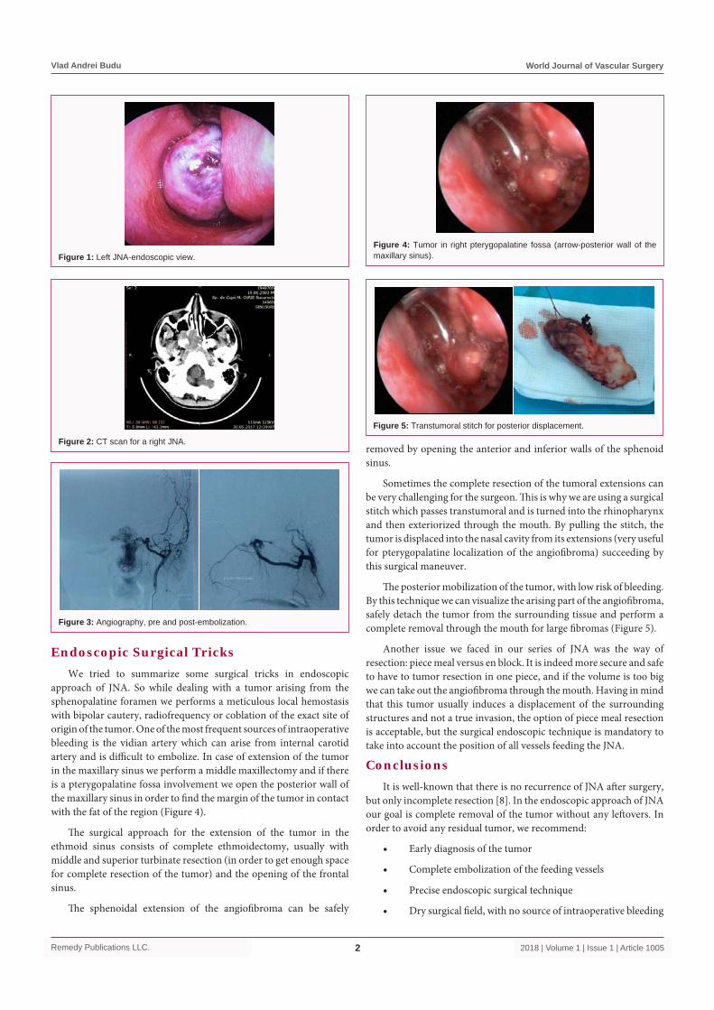
CT scan is the major imagistic investigation which gives complex information about the tumoral mass and its extensions and is a leading point in establishing the tumoral stage (Radkowski staging system) and the surgical management of the tumor [5].
There are two very important signs on CT scan which are characteristic for JNA. The destruction of the pterygoid plates and the enlargement of the sphenoid foramen on the tumor site are images of choice for the diagnosis of angiofibroma (Figure 2).
After establishing an endoscopic and imagistic diagnosis of JNA, the next step in the management of this pathology is the bilateral control angiography [6]. Embolization of feeding vessels in angiofibroma leads to:
• Decrease the vascular supply of the tumor (usually from the internal maxillary artery)
• Exact visualisation of the vessels which feed the tumor
• Decrease the tumor volume which allows an easier surgical procedure, especially in the approach of the tumoral extensions in the surrounding areas (sinuses, orbital cavity, pterygomaxillar fossa, etc)
We perform angiography with embolization 24 hrs to 48 hrs before surgery, using Polyvinyl Alcohol (PVA) as embolic substance (Figure 3).
Dealing with a unilateral bleeding mass appeared at an adolescent male we do not perform a biopsy from the tumor because of the high risk of bleeding. In our experience, the involvement of choice for all steps of JNA is the surgical approach. Depending on the staging system [7], we perform an endoscopic approach (in stage I and II Radkowski) and usually and open approach in Radkowski stage III.
Vlad Andrei Budu*
Department of ENT Microsurgery, "Prof Dr Dorin Hociota" Institute for Phono-Audiology and Functional ENT Surgery, Romania
Vlad Andrei Budu World Journal of Vascular Surgery
Remedy Publications LLC. 2018 | Volume 1 | Issue 1 | Article 10052
Endoscopic Surgical TricksWe tried to summarize some surgical tricks in endoscopic
approach of JNA. So while dealing with a tumor arising from the sphenopalatine foramen we performs a meticulous local hemostasis with bipolar cautery, radiofrequency or coblation of the exact site of origin of the tumor. One of the most frequent sources of intraoperative bleeding is the vidian artery which can arise from internal carotid artery and is difficult to embolize. In case of extension of the tumor in the maxillary sinus we perform a middle maxillectomy and if there is a pterygopalatine fossa involvement we open the posterior wall of the maxillary sinus in order to find the margin of the tumor in contact with the fat of the region (Figure 4).
The surgical approach for the extension of the tumor in the ethmoid sinus consists of complete ethmoidectomy, usually with middle and superior turbinate resection (in order to get enough space for complete resection of the tumor) and the opening of the frontal sinus.
The sphenoidal extension of the angiofibroma can be safely
removed by opening the anterior and inferior walls of the sphenoid sinus.
Sometimes the complete resection of the tumoral extensions can be very challenging for the surgeon. This is why we are using a surgical stitch which passes transtumoral and is turned into the rhinopharynx and then exteriorized through the mouth. By pulling the stitch, the tumor is displaced into the nasal cavity from its extensions (very useful for pterygopalatine localization of the angiofibroma) succeeding by this surgical maneuver.
The posterior mobilization of the tumor, with low risk of bleeding. By this technique we can visualize the arising part of the angiofibroma, safely detach the tumor from the surrounding tissue and perform a complete removal through the mouth for large fibromas (Figure 5).
Another issue we faced in our series of JNA was the way of resection: piece meal versus en block. It is indeed more secure and safe to have to tumor resection in one piece, and if the volume is too big we can take out the angiofibroma through the mouth. Having in mind that this tumor usually induces a displacement of the surrounding structures and not a true invasion, the option of piece meal resection is acceptable, but the surgical endoscopic technique is mandatory to take into account the position of all vessels feeding the JNA.
ConclusionsIt is well-known that there is no recurrence of JNA after surgery,
but only incomplete resection [8]. In the endoscopic approach of JNA our goal is complete removal of the tumor without any leftovers. In order to avoid any residual tumor, we recommend:
• Early diagnosis of the tumor
• Complete embolization of the feeding vessels
• Precise endoscopic surgical technique
• Dry surgical field, with no source of intraoperative bleeding
Figure 1: Left JNA-endoscopic view.
Figure 2: CT scan for a right JNA.
Figure 3: Angiography, pre and post-embolization.
Figure 4: Tumor in right pterygopalatine fossa (arrow-posterior wall of the maxillary sinus).
Figure 5: Transtumoral stitch for posterior displacement.
Vlad Andrei Budu World Journal of Vascular Surgery
Remedy Publications LLC. 2018 | Volume 1 | Issue 1 | Article 10053
• Meticulous endoscopic control of the tumoral extensions site of the end of the surgery
• Rigurous follow-up (endoscopic exam, CT imaging)
In practice, our experience has shown that even if all the steps above are followed, JNA is still a challenging pathology and the surgeon has to be able at any time to assure complete hemostasis during surgery (endoscopic or if needed open approach).
Endoscopic endonasal surgery of JNA is only limited by the extensions of the tumor in surrounding anatomical regions and the experience of the surgical team.
References1. Riggs S, Orlandi RR. Juvenile nasopharyngeal angiofibroma recurrence
associated with exogenous testosterone therapy. Head Neck. 2010;32(6):812-5.
2. Coutinho-Camillo CM, Brentani MM, Nagai MA. Genetic alterations injuvenile nasopharyngeal angiofibromas. Head Neck. 2008;30:390-400.
3. Carlos R, Thompson LD, Netto AC, Pimenta LG, Correia-Silva Jde F, Gomes CC, et al. Epstein-Barr virus and human herpes virus-8 are not associated with juvenile nasopharyngeal angiofibroma. Head Neck Pathol. 2008;2(3):145-9.
4. Ponti G, Losi L, Pellacani G, Rossi GB, Presutti L, Mattioli F, et al. Wnt pathway, angiogenetic and hormonal markers in sporadic and familial adenomatous polyposis-associated juvenile nasopharyngeal angiofibromas (JNA). Appl Immunohistochem Mol Morphol. 2008;16(2):173-8.
5. Lloyd G, Howard D, Lund VJ, Savy L. Imaging for juvenile angiofibroma. J Laryngol Otol. 2000;114(9):727-30.
6. Wu AW, Mowry SE, Vinuela F, Abemayor E, Wang MB. Bilateral vascular supply in juvenile nasopharyngeal angiofibromas. Laryngoscope. 2011;121(3):639-43.
7. Radkowski D, McGill T, Healy GB, Ohlms L, Jones DT. Angiofibroma. changes in staging and treatment. Arch Otolaryngol Head Neck Surg. 1996;122(2):122-9.
8. Tyagi I, Syal R, Goyal A. Recurrent and residual juvenile angiofibromas. J Laryngol Otol. 2007;121(5):460-7.